aerobic microbial skin flora in jeddah city, saudi arabiaegyptianjournal.xyz/5_5.pdfthe egyptian...
TRANSCRIPT

The Egyptian Journal of Hospital Medicine Vol., 5 : 49–77 Dce 2001
I.S.S.N: 12084
Aerobic Microbial Skin Flora in Jeddah City, Saudi Arabia
Rajaa M. Milyani
Department of Biological Sciences, Faculty of Science,
King Abdulaziz University.
Abstract
The aerobic microbial skin flora of 40 healthy subjects living in Jeddah city
(Saudi Arabia) was determined. Two age groups: children and adults; including males
and females were investigated. Seven sites were studied: forehead, axilla, chest, groin,
leg, toe web and anterior nares. The skin was sampled by rubbing the chosen site with a
surfactant substance (Tween 80) moistened cotton swab which was dipped back in the
surfactant container and the resulted suspension was agitated for one minute.
Thirty three microbial species were isolated from the seven sites of the study
group, in which Acinetobacter baumannii, Acinetobacter lwoffii, corynebacterium
species and Staphylococcus (Staph.) aureus dominated among children (30% each). The
most other prevalent isolates recovered were Alkaligenes species, Bacillus species,
Chryseomonas luteola, Staph. epidermidis, Enterococcus faecalis and Staph. hominis
(27.5% each). Organisms including Candida albicans, Enterobacter agglomerans,
Escherichia coli, Flavobacterium meningosepticum, Klebsiella oxytoca, Micrococcus
luteus, Micrococcus roseus, Micrococcus varians, Micrococcus species, Burkholderia
cepacia, Stenotrophomonas maltophilia, Pseudomonas paucimobilis, Pseudomonas
fluorescence, Pseudomonas species, Staph. capitis, Staph. cohnii, Staph. saprophyticus,
Staph. simulans, Staph. warneri, Staph. xylosus, viridans-type streptococcus and yeasts
were also found in different percentage. Higher isolation rates of Acinetobacter lwoffii,
Staph. aureus, Alkaligenes species, Corynebacterium species, Chryseomonas luteola,
Enterobacter agglomerans, Staph. epidermidis and other coagulase negative
Staphylococci were noted in children from the seven sites. However, Chryseomonas
luteola, and Pseudomonas species, were found only in the groin area among males.
Otherwise, no significant differences were recorded in the isolation rates from each site
separately in relation to age and sex. The role of the isolated microorganisms in
endogenous, exogenous and nosocomial infections was emphasized.
Introduction “Surprisingly little is known about
the microflora of the skin and its role in
common skin infections such as
psoriasis and eczema and even less
known of the flora in rare diseases”. A
statement that had been written in 1974
by Noble and Somerville and
regrettably, the same can be applied to
our knowledge in 1990s, not only to the
skin flora but also to the whole
microflora of man, in a country that
receives millions of people for religious
purposes, from all over the world at
almost all the year around namely:
Saudi Arabia. The importance of such
knowledge is certainly obvious,
particularly in preventing exogenous
and or endogenous infections. Milyani
et. al. (1987) stressed upon the medical
significance of such topic and the lack
of information amongst Saudi subjects,
and took a step towards that by studying
49

Aerobic Microbial Skin Flora……..
50
the throat microbial flora in Jeddah city
as a start. Since then our knowledge
have accumulated, from other parts of
the world, and the role played by the
normal microbial flora in different
infections became clearly apparent
(Maibach and Aly 1981; Noble 1983;
Boyce et. al. (1990). Though, this
phenomenon has gained attention as
early as 1843 by Oliver Wendell
Holmes who was known to teach at
Boston that puerperal fever was caused
by germs on the hands of physicians
and midwives which were transmitted
to the vaginas of women during internal
examination. Few took these ideas
seriously until Joseph Lister in 1865
used carbolic acid to disinfect the skin
of the operation site and the operator‟s
hands (Sethna, 1978). Furthermore, and
above all Prophet Mohammad peace
be upon him, before more than 14
century, taught the world to wash their
bodies, faces, rinse the mouth, clear
with water the inside of the nose
(sniffing), wash hands, forearms to the
elbows and feet to the ankles (around
five times a day); the wisdom behind
that is obviously to rid the body of any
transient pathogens, minimize the
number of microbial flora to a balanced
state and thus preventing the mischief of
these organisms that may act as
opportunistic pathogens causing
different diseases and infections
(Milyani, 1998).
With the advent of antibiotics,
cytotoxic and immunosuppressive
drugs, in parallel to the increase in
compromised patients, allowed the so-
called innocent normal flora to establish
itself and emerge as opportunistic
pathogen if not as a true pathogen
causing high rates of morbidity and
mortality (Mandell et. al., 2000).
The aim of the present study was
to determine the microbial flora of the
skin among children and adults, in
Jeddah city, highlighting its medical
significance and to discuss the role of
the isolated organisms in endogenous,
exogenous and nosocomial infections.
Materials And Methods Subjects: 40 apparently healthy
22 females and 18 males consisted of
two age groups. 1 -12 years (24 children
before menarche and puberty); and 13 -
17 years (16 early adults after puberty).
None had been receiving antibiotics for
nine weeks or using deodorants.
Axillary hair was not removed.
Media Used: Five growth media
were used: nutrient agar + 0.5% glucose
+ 0.5% Tween80, for the isolation of
skin lipophilic organisms; blood agar +
crystal violet 1:666.666; was used to
select Streptococcus pyogenes.
(Milyani, 1976); Tinsdale agar base;
MacConkey agar and Cystine-Lactose-
Deficient agar (Oxoid Ltd, London).
Sampling method and culture:
A modified standardized swabbing
method by Selwyn and Ellis (1972) was
used. It involved rubbing thoroughly an
area of 2 cm
2 of the chosen site with
moderate pressure, for one minute with
a sterile cotton-wool swab which had
been moistened with a sterile solution of
0.5 % Tween 80 (Fluka) in 0.075M
phosphate buffer at pH 7.9. The swab
was dipped back into a sterile 10ml test
tube containing 2ml of the sampling
solution (Tween 80, the surfactant
substance) and the suspension was
mechanically agitated for one minute on
a Rotamixer. Whereas, anterior nares
were sampled by, rotating the Tween 80
moistened cotton swab inside the nares
for one minute. Culturing the samples
was within one to two hours of
collection . The following procedure
was carried out for culturing: the cotton
swab was removed from the tube and
streaked evenly all over the surface of
each growth medium (dipping it in the

Rajaa M. Milyani
51
suspension each time prior to
inoculation of the new medium). The
inoculated plates were then incubated
aerobically at 37°C for 48 – 72 hours.
Samples were collected within five
months when ambient temperature
varied between 37°C - 45°C.
Identification: Commercial identification kits were
used: API Staph., API 20E (Analytab
Product, Plainview, N.Y) and Spectrum
10 System (ABL, Austin, Texas); In
general, identification was carried out
according to Koneman et.al. ( 1997).
Statistical analysis: The data were
analyzed using Fisher‟s exact test and
Pearson chi-square (each when
applicable) to determine the significant
differences between the microbial
isolation rates in relation to age, sex,
seven sites and each site separately.
Results: The isolation rates of different
organisms recovered from the total
seven sites of the skin and from each
site separately, in relation to age and
sex are demonstrated in Tables 1- 16
and Figure 1. Figures 2-7 show the
isolation rates of some of the isolates in
each site.
Thirty three microbial species
were isolated from the seven sites of the
study group including Gram positive
and Gram negative microorganisms; the
Gram positive comprised 20 species
while the Gram negative comprised 13
species. Acinetobacter (A.) baumannii,
Acinetobacter (A.) lwoffii, Corynebac-
terium species and Staphylococcus
(Staph.) aureus dominated among
children comprising 30% of each
isolate. The other microbial isolates
followed in prevalence among the same
group, were Alkaligenes species,
Bacillus species, Chryseomonas (C.)
luteola, Staph. epidermidis,
Enterococcus (E.) faecalis and Staph.
hominis giving an incidence of 27.5%
each (Table 1 and Figure 1). Organisms
including Candida (C.) albicans,
Enterobacter (E.) agglomerans,
Escherichia (E.) coli, Flavobacterium
(F.) meningosepticum, Klebsiella (K.)
oxytoca, Micrococcus luteus,
Micrococcus roseus, Micrococcus
varians, Micrococcus species,
Burkholderia (B.) cepacia,
Stenotrophomonas (S.) maltophilia,
Pseudomonas (P.) paucimobilis, P.
fluorescence, Pseudomonas species,
Staph. capitis, Staph. cohnii, Staph.
haemolyticus, Staph. saprophyticus,
Staph. simulans, Staph. warneri, Staph.
xylosus, viridas-type streptococcus and
yeasts were also found in different
percentage among both age groups,
males and females (Table 1 and 2). On
the other hand, significant differences
between children and adults were found
in the isolation rates of A. lwoffii (P=
0.004), Alkaligenes species (P= 0.027),
Corynebacterium species (P= 0.015),
C. luteola (P= 0.027), E. agglomerans
(P= 0.032), Staph. aureus (P= 0.015),
Staph. capitis (P= 0.032), Staph. cohnii
(P= 0.027), Staph. epidermidis (P=
0.012), Staph. haemolyticus (P= 0.032),
Staph. hominis (P= 0.012), Staph.
simulans (P= 0.027), Staph. warneri
(P= 0.027) and Staph. xylosus (P=
0.027) from the seven sites (Table 1).
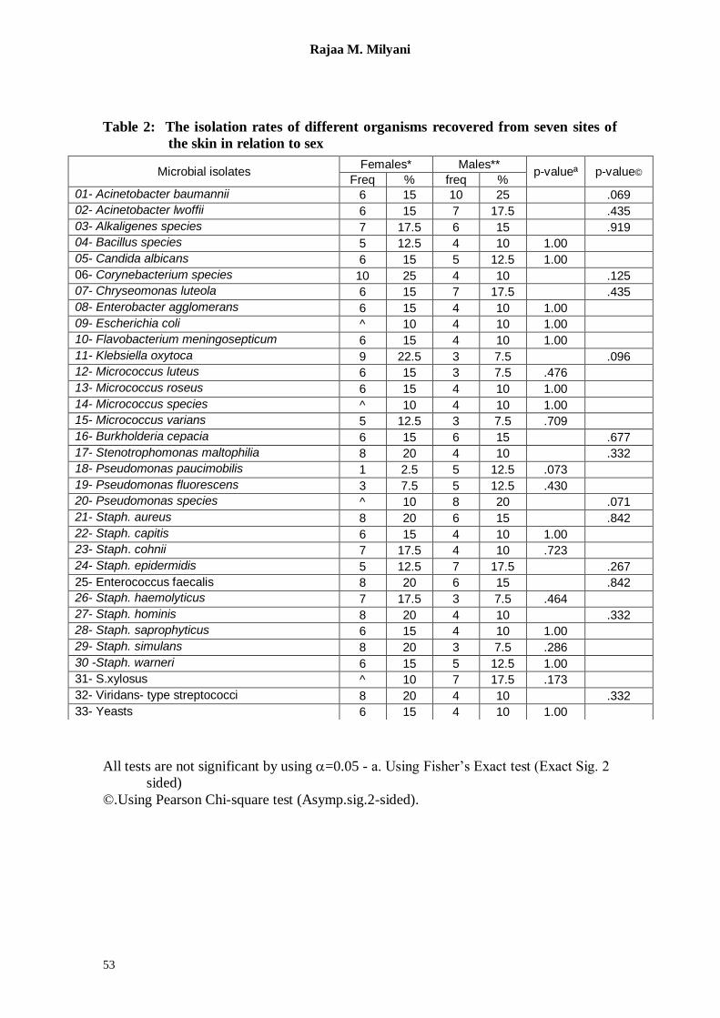
However, in relation to sex, significant
differences were evident only in the
isolation rates of C. luteola (P= 0.013),
and P. species (P= 0.013) in the groin
area among males (Table 12).
Otherwise, no significant differences
were recorded in the isolation rates from
each site separately in relation to age
and sex (Tables 3-16 and Figures 2-7).
Nonetheless, as shown in Figure 2
and 3 within the age range 1-12 and 13 -
17 years the carriage rate of Staph.
aureus among children and adults,

Aerobic Microbial Skin Flora……..
52
males and females, was found to be 5%
and 0% in the forehead, 10% and 0% in
the anterior nares, 2.5% and 5% in the
axilla (both age groups, females and
males respectively), whereas, in the
chest area it was 7.5% and 0% in
children and adults and 2.5% and 7.5%
in females and males respectively. On
the other hand, the carriage rate in the
groin was 10% in children, 2.5% in
adults, 7.5% in females and 5% in
males; in the leg 10% in children and
0% in adults, 2.5% in females and 7.5%
in males; finally in the toe webs, 12.5%
in children and 2.5% in adults, whereas,
the same carriage rate of 7.5% was
noted in both females and males.
Turning to other isolates, the incidence
of A. baumannii and A. lwoffii in the
seven sites of the skin is shown in
Figures 4 and 5 in relation to age.
Whereas, Figures 6-7 recorded the
isolation rate of C. albicans among both
age groups and sex.
Table 1: The isolation rates of different organisms recovered from the seven sites
of the skin in relation to age
a. Using Fisher‟s Exact test (Exact Sig. 2 sided), using =0.05
b. The test is significant (otherwise the test is not significant).
©.Using Pearson Chi-square test (Asymp.sig.2-sided).
* Age 1- 12 years were 24 subjects
** Age 13 -17 years were 16 subjects
Microbial isolates age 1-12* age 13-17**
p-valueª p-value© Freq % freq %
01- Acinetobacter baumannii 12 30 4 10 .114
02- Acinetobacter lwoffii 12 30 1 2.5 .004 b
03- Alkaligenes species 11 27.5 2 5 .027 b
04- Bacillus species 6 15 3 7.5 .717
05- Candida albicans 7 17.5 4 10 1.00
06- Corynebacterium species 12 30 2 5 .015 b
07- Chryseomonas luteola 11 27.5 2 5 .027 b
08- Enterobacter agglomerans 9 22.5 1 2.5 .032 b
09- Escherichia coli 6 15 2 5 .439
10- Flavobacterium meningosepticum 6 15 4 10 1.00
11- Klebsiella oxytoca 8 20 4 10 .729
12- Micrococcus luteus 8 20 1 2.5 .061
13- Micrococcus roseus 8 20 2 5 .263
14- Micrococcus species 6 15 2 5 .439
15- Micrococcus varians 7 17.5 1 2.5 .114
16- Burkholderia cepacia 9 22.5 3 7.5 .297
17- Stenotrophomonas maltophilia 8 20 4 10 .729
18- Pseudomonas paucimobilis 3 7.5 3 7.5 .668
19- Pseudomonas fluorescens 7 17.5 1 2.5 .114
20- Pseudomonas species 7 17.5 5 12.5 1.00
21- Staph. aureus 12 30 2 5 .015 b
22- Staph. capitis 9 22.5 1 2.5 .032 b
23- Staph. cohnii 10 25 1 2.5 .027 b
24- Staph. epidermidis 11 27.5 1 2.5 .012 b
25- Enterococcus faecalis 11 27.5 3 7.5 .079
26- Staph. haemolyticus 9 22.5 1 2.5 .032 b
27- Staph. hominis 11 27.5 1 2.5 .012 b
28- Staph. saprophyticus 8 20 2 5 .263
29- Staph. simulans 10 25 1 2.5 .027 b
30 -Staph. warneri 10 25 1 2.5 .027 b
31- S.xylosus 10 25 1 2.5 .027 b
32- Viridans- type streptococci 10 25 2 5 .079
33- Yeasts 7 17.5 3 7.5 .711
Rajaa M. Milyani
53
Table 2: The isolation rates of different organisms recovered from seven sites of
the skin in relation to sex
All tests are not significant by using =0.05 - a. Using Fisher‟s Exact test (Exact Sig. 2
sided)
©.Using Pearson Chi-square test (Asymp.sig.2-sided).
Microbial isolates Females* Males**
p-valueª p-value© Freq % freq %
01- Acinetobacter baumannii 6 15 10 25 .069
02- Acinetobacter lwoffii 6 15 7 17.5 .435
03- Alkaligenes species 7 17.5 6 15 .919
04- Bacillus species 5 12.5 4 10 1.00
05- Candida albicans 6 15 5 12.5 1.00
06- Corynebacterium species 10 25 4 10 .125
07- Chryseomonas luteola 6 15 7 17.5 .435
08- Enterobacter agglomerans 6 15 4 10 1.00
09- Escherichia coli ^ 10 4 10 1.00
10- Flavobacterium meningosepticum 6 15 4 10 1.00
11- Klebsiella oxytoca 9 22.5 3 7.5 .096
12- Micrococcus luteus 6 15 3 7.5 .476
13- Micrococcus roseus 6 15 4 10 1.00
14- Micrococcus species ^ 10 4 10 1.00
15- Micrococcus varians 5 12.5 3 7.5 .709
16- Burkholderia cepacia 6 15 6 15 .677
17- Stenotrophomonas maltophilia 8 20 4 10 .332
18- Pseudomonas paucimobilis 1 2.5 5 12.5 .073
19- Pseudomonas fluorescens 3 7.5 5 12.5 .430
20- Pseudomonas species ^ 10 8 20 .071
21- Staph. aureus 8 20 6 15 .842
22- Staph. capitis 6 15 4 10 1.00
23- Staph. cohnii 7 17.5 4 10 .723
24- Staph. epidermidis 5 12.5 7 17.5 .267
25- Enterococcus faecalis 8 20 6 15 .842
26- Staph. haemolyticus 7 17.5 3 7.5 .464
27- Staph. hominis 8 20 4 10 .332
28- Staph. saprophyticus 6 15 4 10 1.00
29- Staph. simulans 8 20 3 7.5 .286
30 -Staph. warneri 6 15 5 12.5 1.00
31- S.xylosus ^ 10 7 17.5 .173
32- Viridans- type streptococci 8 20 4 10 .332
33- Yeasts 6 15 4 10 1.00
Aerobic Microbial Skin Flora……..
54
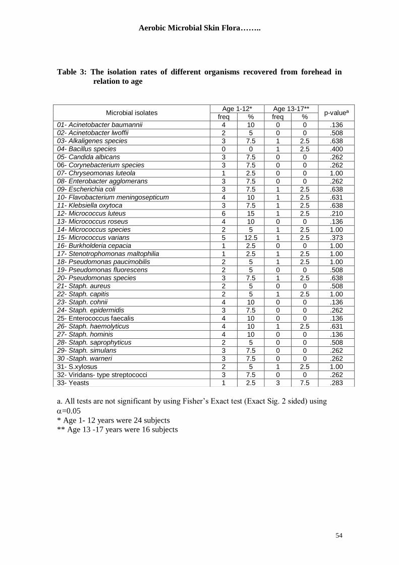
Table 3: The isolation rates of different organisms recovered from forehead in
relation to age
a. All tests are not significant by using Fisher‟s Exact test (Exact Sig. 2 sided) using
=0.05
* Age 1- 12 years were 24 subjects
** Age 13 -17 years were 16 subjects
Microbial isolates Age 1-12* Age 13-17**
p-valueª freq % freq %
01- Acinetobacter baumannii 4 10 0 0 .136 02- Acinetobacter lwoffii 2 5 0 0 .508 03- Alkaligenes species 3 7.5 1 2.5 .638 04- Bacillus species 0 0 1 2.5 .400 05- Candida albicans 3 7.5 0 0 .262 06- Corynebacterium species 3 7.5 0 0 .262 07- Chryseomonas luteola 1 2.5 0 0 1.00 08- Enterobacter agglomerans 3 7.5 0 0 .262 09- Escherichia coli 3 7.5 1 2.5 .638 10- Flavobacterium meningosepticum 4 10 1 2.5 .631 11- Klebsiella oxytoca 3 7.5 1 2.5 .638 12- Micrococcus luteus 6 15 1 2.5 .210 13- Micrococcus roseus 4 10 0 0 .136 14- Micrococcus species 2 5 1 2.5 1.00 15- Micrococcus varians 5 12.5 1 2.5 .373 16- Burkholderia cepacia 1 2.5 0 0 1.00 17- Stenotrophomonas maltophilia 1 2.5 1 2.5 1.00 18- Pseudomonas paucimobilis 2 5 1 2.5 1.00 19- Pseudomonas fluorescens 2 5 0 0 .508 20- Pseudomonas species 3 7.5 1 2.5 .638 21- Staph. aureus 2 5 0 0 .508 22- Staph. capitis 2 5 1 2.5 1.00 23- Staph. cohnii 4 10 0 0 .136 24- Staph. epidermidis 3 7.5 0 0 .262
25- Enterococcus faecalis 4 10 0 0 .136 26- Staph. haemolyticus 4 10 1 2.5 .631 27- Staph. hominis 4 10 0 0 .136 28- Staph. saprophyticus 2 5 0 0 .508 29- Staph. simulans 3 7.5 0 0 .262 30 -Staph. warneri 3 7.5 0 0 .262
31- S.xylosus 2 5 1 2.5 1.00
32- Viridans- type streptococci 3 7.5 0 0 .262
33- Yeasts 1 2.5 3 7.5 .283

Rajaa M. Milyani
55
Table 4: The isolation rates of different organisms recovered from forehead in
relation to sex
a. All tests are not significant by using Fisher‟s Exact test (Exact Sig. 2 sided) using
=0.05
Microbial isolates Females Males
p-valueª freq % freq %
01- Acinetobacter baumannii 2 5 2 5 1.00 02- Acinetobacter lwoffii 1 2.5 1 2.5 1.00 03- Alkaligenes species 2 5 2 5 1.00 04- Bacillus species 0 0 1 2.5 .450 05- Candida albicans 2 5 1 2.5 1.00 06- Corynebacterium species 2 5 1 2.5 1.00 07- Chryseomonas luteola 0 0 1 2.5 .450 08- Enterobacter agglomerans 2 5 1 2.5 1.00 09- Escherichia coli 1 2.5 3 7.5 .310 10- Flavobacterium meningosepticum 4 10 1 2.5 .355 11- Klebsiella oxytoca 3 7.5 1 2.5 .613 12- Micrococcus luteus 4 10 3 7.5 1.00 13- Micrococcus roseus 2 5 2 5 1.00 14- Micrococcus species 2 5 1 2.5 1.00 15- Micrococcus varians 3 7.5 3 7.5 1.00 16- Burkholderia cepacia 1 2.5 0 0 1.00 17- Stenotrophomonas maltophilia 1 2.5 1 2.5 1.00 18- Pseudomonas paucimobilis 1 2.5 2 5 .579 19- Pseudomonas fluorescens 0 0 2 5 .196 20- Pseudomonas species 2 5 2 5 1.00 21- Staph. aureus 2 5 0 0 .492 22- Staph. capitis 2 5 1 2.5 1.00 23- Staph. cohnii 2 5 2 5 1.00 24- Staph. epidermidis 2 5 1 2.5 1.00
25- Enterococcus faecalis 4 10 0 0 .114 26- Staph. haemolyticus 2 5 3 7.5 .642 27- Staph. hominis 3 7.5 1 2.5 .613 28- Staph. saprophyticus 1 2.5 1 2.5 1.00 29- Staph. simulans 1 2.5 2 5 .579 30 -Staph. warneri 2 5 1 2.5 1.00
31- S.xylosus 1 2.5 2 5 .579
32- Viridans- type streptococci 2 5 1 2.5 1.00
33- Yeasts 2 5 2 5 1.00

Aerobic Microbial Skin Flora……..
56
Table 5: The isolation rates of different organisms recovered from the anterior
nares in relation to age
a. All tests are not significant by using Fisher‟s Exact test (Exact Sig. 2 sided) using
=0.05
h. No statistics are computed because ANTERIOR NARES are a constant.
* Age 1- 12 years were 24 subjects
** Age 13 -17 years were 16 subjects
Microbial isolates age 1-12* age 13-17**
p-valueª freq % freq %
01- Acinetobacter baumannii 5 12.5 1 2.5 .373 02- Acinetobacter lwoffii 3 7.5 0 0 .262 03- Alkaligenes species 4 10 1 2.5 .631 04- Bacillus species 3 7.5 0 0 .262 05- Candida albicans 4 10 0 0 .136 06- Corynebacterium species 1 2.5 0 0 1.00 07- Chryseomonas luteola 2 5 0 0 .508 08- Enterobacter agglomerans 2 5 0 0 .508 09- Escherichia coli 1 2.5 0 0 1.00 10- Flavobacterium meningosepticum 2 5 0 0 .508 11- Klebsiella oxytoca 1 2.5 0 0 1.00 12- Micrococcus luteus 3 7.5 0 0 .262 13- Micrococcus roseus 2 5 1 2.5 1.00 14- Micrococcus species 3 7.5 1 2.5 .638 15- Micrococcus varians 0 0 0 0 h 16- Burkholderia cepacia 4 10 0 0 .136 17- Stenotrophomonas maltophilia 3 7.5 1 2.5 .638 18- Pseudomonas paucimobilis 1 2.5 0 0 1.00 19- Pseudomonas fluorescens 1 2.5 0 0 1.00 20- Pseudomonas species 1 2.5 2 5 .553 21- Staph. aureus 4 10 0 0 .136 22- Staph. capitis 1 2.5 0 0 1.00 23- Staph. cohnii 3 7.5 0 0 .262 24- Staph. epidermidis 3 7.5 0 0 .262
25- Enterococcus faecalis 2 5 0 0 .508 26- Staph. haemolyticus 2 5 0 0 .508 27- Staph. hominis 4 10 1 2.5 .631 28- Staph. saprophyticus 3 7.5 0 0 .262 29- Staph. simulans 5 12.5 0 0 .071 30 -Staph. warneri 2 5 1 2.5 1.00
31- S.xylosus 3 7.5 0 0 .262
32- Viridans- type streptococci 3 7.5 0 0 .262
33- Yeasts 2 5 0 0 .508

Rajaa M. Milyani
57
Table 6: The isolation rates of different organisms recovered from the anterior
nares in relation to sex
a. All tests are not significant by using Fisher‟s Exact test (Exact Sig. 2 sided) using
=0.05
h. No statistics are computed because ANTERIOR NARES are a constant.
Microbial isolates Females Males
p-valueª freq % freq %
01- Acinetobacter baumannii 4 10 2 5 .673 02- Acinetobacter lwoffii 2 5 1 2.5 1.00 03- Alkaligenes species 3 7.5 2 5 1.00 04- Bacillus species 2 5 1 2.5 1.00 05- Candida albicans 3 7.5 1 2.5 .613 06- Corynebacterium species 1 2.5 0 0 1.00 07- Chryseomonas luteola 2 5 0 0 .492 08- Enterobacter agglomerans 2 5 0 0 .492 09- Escherichia coli 1 2.5 0 0 1.00 10- Flavobacterium meningosepticum 2 5 0 0 .492 11- Klebsiella oxytoca 1 2.5 0 0 1.00 12- Micrococcus luteus 2 5 1 2.5 1.00 13- Micrococcus roseus 1 2.5 2 5 .579 14- Micrococcus species 2 5 2 5 1.00 15- Micrococcus varians 0 0 0 0 h 16- Burkholderia cepacia 4 10 0 0 .114 17- Stenotrophomonas maltophilia 3 7.5 1 2.5 .613 18- Pseudomonas paucimobilis 1 2.5 0 0 1.00 19- Pseudomonas fluorescens 1 2.5 0 0 1.00 20- Pseudomonas species 2 5 1 2.5 1.00 21- Staph. aureus 4 10 0 0 .114 22- Staph. capitis 1 2.5 0 0 1.00 23- Staph. cohnii 2 5 1 2.5 1.00 24- Staph. epidermidis 1 2.5 2 5 .579
25- Enterococcus faecalis 2 5 0 0 .492 26- Staph. haemolyticus 2 5 0 0 .492 27- Staph. hominis 4 10 1 2.5 .355 28- Staph. saprophyticus 2 5 1 2.5 1.00 29- Staph. simulans 3 7.5 2 5 1.00 30 -Staph. warneri 2 5 1 2.5 1.00
31- S.xylosus 1 2.5 2 5 .579
32- Viridans- type streptococci 3 7.5 0 0 .238
33- Yeasts 2 5 0 0 .492

Aerobic Microbial Skin Flora……..
58
Table 7: The isolation rates of different organisms recovered from the axilla in
relation to age
a. All tests are not significant by using Fisher‟s Exact test (Exact Sig. 2 sided) using
=0.05
h. No statistics are computed because AXILLA is a constant.
* Age 1- 12 years were 24 subjects
** Age 13 -17 years were 16 subjects
Microbial isolates age 1-12* age 13-17**
p-valueª freq % freq %
01- Acinetobacter baumannii 1 2.5 2 5 .553 02- Acinetobacter lwoffii 8 20 1 2.5 .061 03- Alkaligenes species 2 5 1 2.5 1.00 04- Bacillus species 2 5 0 0 .508 05- Candida albicans 1 2.5 0 0 1.00 06- Corynebacterium species 4 10 0 0 .136 07- Chryseomonas luteola 1 2.5 1 2.5 1.00 08- Enterobacter agglomerans 5 12.5 0 0 .071 09- Escherichia coli 4 10 0 0 .136 10- Flavobacterium meningosepticum 4 10 1 2.5 .631 11- Klebsiella oxytoca 1 2.5 4 10 .138 12- Micrococcus luteus 3 7.5 0 0 .262 13- Micrococcus roseus 3 7.5 1 2.5 .638 14- Micrococcus species 1 2.5 0 0 1.00 15- Micrococcus varians 5 12.5 0 0 .071 16- Burkholderia cepacia 4 10 0 0 .136 17- Stenotrophomonas maltophilia 3 7.5 1 2.5 .638 18- Pseudomonas paucimobilis 3 7.5 2 5 1.00 19- Pseudomonas fluorescens 2 5 0 0 .508 20- Pseudomonas species 2 5 1 2.5 1.00 21- Staph. aureus 1 2.5 2 5 .553 22- Staph. capitis 4 10 1 2.5 .631 23- Staph. cohnii 3 7.5 0 0 .262 24- Staph. epidermidis 0 0 0 0 h
25- Enterococcus faecalis 2 5 1 2.5 1.00 26- Staph. haemolyticus 3 7.5 0 0 .262 27- Staph. hominis 4 10 1 2.5 .631 28- Staph. saprophyticus 3 7.5 0 0 .262 29- Staph. simulans 3 7.5 0 0 .262 30 -Staph. warneri 4 10 1 2.5 .631
31- S.xylosus 1 2.5 0 0 1.00
32- Viridans- type streptococci 5 12.5 0 0 .071
33- Yeasts 3 7.5 1 2.5 .638

Rajaa M. Milyani
59
Table 8: The isolation rates of different organisms recovered from the axilla in
relation to sex
a. All tests are not significant by using Fisher‟s Exact test (Exact Sig. 2 sided) using
=0.05
h. No statistics are computed because AXILLA is a constant.
Microbial isolates Female Males
p-valueª freq % freq %
01- Acinetobacter baumannii 1 2.5 2 5 .579 02- Acinetobacter lwoffii ^ 10 5 12.5 .705 03- Alkaligenes species 2 5 1 2.5 1.00 04- Bacillus species 1 2.5 1 2.5 1.00 05- Candida albicans 0 0 1 2.5 .450 06- Corynebacterium species 2 5 2 5 1.00 07- Chryseomonas luteola 2 5 0 0 .492 08- Enterobacter agglomerans 2 5 3 7.5 .642 09- Escherichia coli 3 7.5 1 2.5 .613 10- Flavobacterium meningosepticum 3 7.5 2 5 1.00 11- Klebsiella oxytoca 3 7.5 2 5 1.00 12- Micrococcus luteus 2 5 1 2.5 1.00 13- Micrococcus roseus 3 7.5 1 2.5 .613 14- Micrococcus species 0 0 1 2.5 .450 15- Micrococcus varians 3 7.5 2 5 1.00 16- Burkholderia cepacia 2 5 2 5 1.00 17- Stenotrophomonas maltophilia 2 5 2 5 1.00 18- Pseudomonas paucimobilis 1 2.5 4 10 .155 19- Pseudomonas fluorescens 0 0 2 5 .196 20- Pseudomonas species 0 0 3 7.5 .083 21- Staph. aureus 1 2.5 2 5 .579 22- Staph. capitis 3 7.5 2 5 1.00 23- Staph. cohnii 2 5 1 2.5 1.00 24- Staph. epidermidis 0 0 0 0 h
25- Enterococcus faecalis 3 7.5 0 0 .238 26- Staph. haemolyticus 1 2.5 2 5 .579 27- Staph. hominis 2 5 3 7.5 .642 28- Staph. saprophyticus 1 2.5 2 5 .579 29- Staph. simulans 1 2.5 2 5 .579 30 -Staph. warneri 2 5 3 7.5 .642
31- S.xylosus 1 2.5 0 0 1.00
32- Viridans- type streptococci 4 10 1 2.5 .355
33- Yeasts 4 10 0 0 .114

Aerobic Microbial Skin Flora……..
60
Table 9: The isolation rates of different organisms recovered from the chest skin
in relation to age
a. All tests are not significant by using Fisher‟s Exact test (Exact Sig. 2 sided) using
=0.05
h. No statistics are computed because CHEST is a constant.
* Age 1- 12 years were 24 subjects
** Age 13 -17 years were 16 subjects
Microbial isolates age 1-12* age 13-17**
p-valueª freq % freq %
01- Acinetobacter baumannii 2 5 2 5 1.00 02- Acinetobacter lwoffii 2 5 0 0 .508 03- Alkaligenes species 4 10 1 2.5 .631 04- Bacillus species 2 5 2 5 1.00 05- Candida albicans 2 5 1 2.5 1.00 06- Corynebacterium species 1 2.5 0 0 1.00 07- Chryseomonas luteola 1 2.5 0 0 1.00 08- Enterobacter agglomerans 4 10 0 0 .136 09- Escherichia coli 3 7.5 0 0 .262 10- Flavobacterium meningosepticum 1 2.5 3 7.5 .283 11- Klebsiella oxytoca 3 7.5 0 0 .262 12- Micrococcus luteus 2 5 1 2.5 1.00 13- Micrococcus roseus 3 7.5 1 2.5 .638 14- Micrococcus species 1 2.5 2 5 .553 15- Micrococcus varians 3 7.5 0 0 .262 16- Burkholderia cepacia 4 10 0 0 .136 17- Stenotrophomonas maltophilia 3 7.5 3 7.5 .668 18- Pseudomonas paucimobilis 0 0 0 0 h 19- Pseudomonas fluorescens 1 2.5 1 2.5 1.00 20- Pseudomonas species 2 5 1 2.5 1.00 21- Staph. aureus 4 10 0 0 .136 22- Staph. capitis 3 7.5 0 0 .262 23- Staph. cohnii 3 7.5 1 2.5 .638 24- Staph. epidermidis 3 7.5 0 0 .262
25- Enterococcus faecalis 4 10 0 0 .136 26- Staph. haemolyticus 1 2.5 0 0 1.00 27- Staph. hominis 4 10 0 0 .136 28- Staph. saprophyticus 4 10 0 0 .136 29- Staph. simulans 1 2.5 0 0 1.00 30 -Staph. warneri 2 5 0 0 .508
31- S.xylosus 5 12.5 0 0 .071
32- Viridans- type streptococci 3 7.5 0 0 .262
33- Yeasts 3 7.5 2 5 1.00

Rajaa M. Milyani
61
Table 10: The isolation rates of different organisms recovered from the chest
skin in relation to sex
a. All tests are not significant by using Fisher‟s Exact test (Exact Sig. 2 sided) using
=0.05
h. No statistics are computed because CHEST is a constant.
Microbial isolates Females Males
p-valueª freq % freq %
01- Acinetobacter baumannii 2 5 2 5 1.00 02- Acinetobacter lwoffii 2 5 0 0 .492 03- Alkaligenes species 2 5 3 7.5 .642 04- Bacillus species 3 7.5 1 2.5 .613 05- Candida albicans 2 5 1 2.5 1.00 06- Corynebacterium species 1 2.5 0 0 1.00 07- Chryseomonas luteola 1 2.5 0 0 1.00 08- Enterobacter agglomerans 3 7.5 1 2.5 .613 09- Escherichia coli 2 5 1 2.5 1.00 10- Flavobacterium meningosepticum 2 5 2 5 1.00 11- Klebsiella oxytoca 3 7.5 0 0 .238 12- Micrococcus luteus 2 5 1 2.5 1.00 13- Micrococcus roseus 1 2.5 3 7.5 .310 14- Micrococcus species 3 7.5 0 0 .238 15- Micrococcus varians 2 5 1 2.5 1.00 16- Burkholderia cepacia 1 2.5 3 7.5 .310 17- Stenotrophomonas maltophilia 4 10 2 5 .673 18- Pseudomonas paucimobilis 0 0 0 0 h 19- Pseudomonas fluorescens 2 5 0 0 .492 20- Pseudomonas species 2 5 1 2.5 1.00 21- Staph. aureus 1 2.5 3 7.5 .310 22- Staph. capitis 1 2.5 2 5 .579 23- Staph. cohnii 3 7.5 1 2.5 .613 24- Staph. epidermidis 1 2.5 2 5 .579
25- Enterococcus faecalis 3 7.5 1 2.5 .613 26- Staph. haemolyticus 1 2.5 0 0 1.00 27- Staph. hominis 3 7.5 1 2.5 .613 28- Staph. saprophyticus 3 7.5 1 2.5 .613 29- Staph. simulans 1 2.5 0 0 1.00 30 -Staph. warneri 2 5 0 0 .492
31- S.xylosus 1 2.5 4 10 .155
32- Viridans- type streptococci 2 5 1 2.5 1.00
33- Yeasts 2 5 3 7.5 .642

Aerobic Microbial Skin Flora……..
62
Table 11: The isolation rates of different organisms recovered from the groin in
relation to age
a. All tests are not significant by using Fisher‟s Exact test (Exact Sig. 2 sided) using
=0.05
h. No statistics are computed because GROIN is a constant.
* Age 1- 12 years were 24 subjects
** Age 13 -17 years were 16 subjects
Microbial isolates age 1-12* age 13-17**
p-valueª freq % freq %
01- Acinetobacter baumannii 4 10 2 5 1.00 02- Acinetobacter lwoffii 1 2.5 0 0 1.00 03- Alkaligenes species 2 5 0 0 .508 04- Bacillus species 1 2.5 0 0 1.00 05- Candida albicans 4 10 3 7.5 1.00 06- Corynebacterium species 5 12.5 2 5 .681 07- Chryseomonas luteola 4 10 1 2.5 .631 08- Enterobacter agglomerans 6 15 1 2.5 .210 09- Escherichia coli 0 0 0 0 h 10- Flavobacterium meningosepticum 2 5 1 2.5 1.00 11- Klebsiella oxytoca 5 12.5 0 0 .071 12- Micrococcus luteus 4 10 0 0 .136 13- Micrococcus roseus 2 5 0 0 .508 14- Micrococcus species 1 2.5 0 0 1.00 15- Micrococcus varians 3 7.5 0 0 .262 16- Burkholderia cepacia 2 5 1 2.5 1.00 17- Stenotrophomonas maltophilia 3 7.5 0 0 .262 18- Pseudomonas paucimobilis 0 0 1 2.5 .400 19- Pseudomonas fluorescens 1 2.5 0 0 1.00 20- Pseudomonas species 3 7.5 2 5 1.00 21- Staph. aureus 4 10 1 2.5 .631 22- Staph. capitis 2 5 1 2.5 1.00 23- Staph. cohnii 2 5 0 0 .508 24- Staph. epidermidis 3 7.5 0 0 .262
25- Enterococcus faecalis 4 10 2 5 1.00 26- Staph. haemolyticus 3 7.5 0 0 .262 27- Staph. hominis 2 5 0 0 .508 28- Staph. saprophyticus 3 7.5 2 5 1.00 29- Staph. simulans 6 15 1 2.5 .210 30 -Staph. warneri 4 10 0 0 .136
31- S.xylosus 1 2.5 0 0 1.00
32- Viridans- type streptococci 4 10 2 5 1.00
33- Yeasts 0 0 0 0 h

Rajaa M. Milyani
63
Table 12: The isolation rates of different organisms recovered from the groin in
relation to sex
a. Using Fisher‟s Exact test (Exact Sig. 2 sided) using =0.05
b. The test is significant (otherwise the test is not significant).
h. No statistics are computed because GROIN is a constant.
Microbial isolates Females Males
p-valueª freq % freq %
01- Acinetobacter baumannii 2 5 4 10 .381 02- Acinetobacter lwoffii 0 0 1 2.5 .450 03- Alkaligenes species 2 5 0 0 .492 04- Bacillus species 1 2.5 0 0 1.00 05- Candida albicans 3 7.5 4 10 .680 06- Corynebacterium species 4 10 3 7.5 1.00 07- Chryseomonas luteola 0 0 5 12.5 .013 b 08- Enterobacter agglomerans 3 7.5 4 10 .680 09- Escherichia coli 0 0 0 0 h 10- Flavobacterium meningosepticum 1 2.5 2 5 .579 11- Klebsiella oxytoca 3 7.5 2 5 1.00 12- Micrococcus luteus 2 5 2 5 1.00 13- Micrococcus roseus 1 2.5 1 2.5 1.00 14- Micrococcus species 0 0 1 2.5 .450 15- Micrococcus varians 1 2.5 2 5 .579 16- Burkholderia cepacia 2 5 1 2.5 1.00 17- Stenotrophomonas maltophilia 2 5 1 2.5 1.00 18- Pseudomonas paucimobilis 0 0 1 2.5 .450 19- Pseudomonas fluorescens 0 0 1 2.5 .450 20- Pseudomonas species 0 0 5 12.5 .013 b 21- Staph. aureus 3 7.5 2 5 1.00 22- Staph. capitis 3 7.5 0 0 .238 23- Staph. cohnii 1 2.5 1 2.5 1.00 24- Staph. epidermidis 1 2.5 2 5 .579
25- Enterococcus faecalis 1 2.5 5 12.5 .073 26- Staph. haemolyticus 2 5 1 2.5 1.00 27- Staph. hominis 2 5 0 0 .492 28- Staph. saprophyticus 3 7.5 2 5 1.00 29- Staph. simulans 6 15 1 2.5 .105 30 -Staph. warneri 2 5 2 5 1.00
31- S.xylosus 1 2.5 0 0 1.00
32- Viridans- type streptococci 4 10 2 5 .673
33- Yeasts 0 0 0 0 h

Aerobic Microbial Skin Flora……..
64
Table 13: The isolation rates of different organisms recovered from the leg in
relation to age
a. All tests are not significant by using Fisher‟s Exact test (Exact Sig. 2 sided) using
=0.05
h. No statistics are computed because LEG is a constant.
* Age 1- 12 years were 24 subjects
** Age 13 -17 years were 16 subjects* Age 1- 12 years were 24 subjects
Microbial isolates age 1-12* age 13-17**
p-valueª freq % freq %
01- Acinetobacter baumannii 3 7.5 1 2.5 .638 02- Acinetobacter lwoffii 3 7.5 0 0 .262 03- Alkaligenes species 4 10 0 0 .136 04- Bacillus species 0 0 0 0 h 05- Candida albicans 0 0 1 2.5 .400 06- Corynebacterium species 2 5 0 0 .508 07- Chryseomonas luteola 2 5 0 0 .508 08- Enterobacter agglomerans 2 5 0 0 .508 09- Escherichia coli 0 0 0 0 h 10- Flavobacterium meningosepticum 3 7.5 1 2.5 .638 11- Klebsiella oxytoca 1 2.5 1 2.5 1.00 12- Micrococcus luteus 3 7.5 0 0 .262 13- Micrococcus roseus 4 10 0 0 .136 14- Micrococcus species 3 7.5 0 0 .262 15- Micrococcus varians 2 5 0 0 .508 16- Burkholderia cepacia 1 2.5 2 5 .553 17- Stenotrophomonas maltophilia 1 2.5 1 2.5 1.00 18- Pseudomonas paucimobilis 0 0 0 0 h 19- Pseudomonas fluorescens 2 5 1 2.5 1.00 20- Pseudomonas species 2 5 1 2.5 1.00 21- Staph. aureus 4 10 0 0 .136 22- Staph. capitis 1 2.5 0 0 1.00 23- Staph. cohnii 3 7.5 1 2.5 .638 24- Staph. epidermidis 3 7.5 0 0 .262
25- Enterococcus faecalis 3 7.5 1 2.5 .638 26- Staph. haemolyticus 4 10 0 0 .136 27- Staph. hominis 3 7.5 0 0 .262 28- Staph. saprophyticus 2 5 0 0 .508 29- Staph. simulans 3 7.5 0 0 .262 30 -Staph. warneri 5 12.5 0 0 .071
31- S.xylosus 3 7.5 0 0 .262
32- Viridans- type streptococci 1 2.5 1 2.5 1.00
33- Yeasts 2 5 0 0 .508

Rajaa M. Milyani
65
Table 14: The isolation rates of different organisms recovered from the leg in
relation to sex
a. All tests are not significant by using Fisher‟s Exact test (Exact Sig. 2 sided) using
=0.05
h. No statistics are computed because LEG is a constant.
Microbial isolates Females Males
p-valueª freq % freq %
01- Acinetobacter baumannii 2 5 2 5 1.00 02- Acinetobacter lwoffii 3 7.5 0 0 .238 03- Alkaligenes species 3 7.5 1 2.5 .613 04- Bacillus species 0 0 0 0 h 05- Candida albicans 1 2.5 0 0 1.00 06- Corynebacterium species 2 5 0 0 .492 07- Chryseomonas luteola 2 5 0 0 .492 08- Enterobacter agglomerans 2 5 0 0 .492 09- Escherichia coli 0 0 0 0 h 10- Flavobacterium meningosepticum 3 7.5 1 2.5 .613 11- Klebsiella oxytoca 2 5 0 0 .492 12- Micrococcus luteus 2 5 1 2.5 1.00 13- Micrococcus roseus 3 7.5 1 2.5 .613 14- Micrococcus species 1 2.5 2 5 .579 15- Micrococcus varians 1 2.5 1 2.5 1.00 16- Burkholderia cepacia 3 7.5 0 0 .238 17- Stenotrophomonas maltophilia 1 2.5 1 2.5 1.00 18- Pseudomonas paucimobilis 0 0 0 0 h 19- Pseudomonas fluorescens 2 5 1 2.5 1.00 20- Pseudomonas species 1 2.5 2 5 .579 21- Staph. aureus 1 2.5 3 7.5 .310 22- Staph. capitis 1 2.5 0 0 1.00 23- Staph. cohnii 2 5 2 5 1.00 24- Staph. epidermidis 2 5 1 2.5 1.00
25- Enterococcus faecalis 3 7.5 1 2.5 .613 26- Staph. haemolyticus 3 7.5 1 2.5 .613 27- Staph. hominis 2 5 1 2.5 1.00 28- Staph. saprophyticus 2 5 0 0 .492 29- Staph. simulans 2 5 1 2.5 1.00 30 -Staph. warneri 4 10 1 2.5 .355
31- S.xylosus 3 7.5 0 0 .238
32- Viridans- type streptococci 2 5 0 0 .492
33- Yeasts 2 5 0 0 .492

Aerobic Microbial Skin Flora……..
66
Table 15: The isolation rates of different organisms recovered from the toe webs in
relation to age
a. All tests are not significant by using Fisher‟s Exact test (Exact Sig. 2 sided) using
=0.05
* Age 1- 12 years were 24 subjects
** Age 13 -17 years were 16 subjects
Microbial isolates age 1-12* age 13-17**
p-valueª freq % freq %
01- Acinetobacter baumannii 1 2.5 0 0 1.00 02- Acinetobacter lwoffii 3 7.5 0 0 .262 03- Alkaligenes species 1 2.5 0 0 1.00 04- Bacillus species 1 2.5 2 5 .553 05- Candida albicans 3 7.5 3 7.5 .668 06- Corynebacterium species 3 7.5 0 0 .262 07- Chryseomonas luteola 5 12.5 0 0 .071 08- Enterobacter agglomerans 1 2.5 0 0 1.00 09- Escherichia coli 3 7.5 2 5 1.00 10- Flavobacterium meningosepticum 3 7.5 1 2.5 .638 11- Klebsiella oxytoca 4 10 0 0 .136 12- Micrococcus luteus 4 10 0 0 .136 13- Micrococcus roseus 3 7.5 0 0 .262 14- Micrococcus species 1 2.5 1 2.5 1.00 15- Micrococcus varians 4 10 1 2.5 .631 16- Burkholderia cepacia 6 15 0 0 .064 17- Stenotrophomonas maltophilia 3 7.5 1 2.5 .638 18- Pseudomonas paucimobilis 2 5 0 0 .508 19- Pseudomonas fluorescens 3 7.5 1 2.5 .638 20- Pseudomonas species 2 5 1 2.5 1.00 21- Staph. aureus 5 12.5 1 2.5 .373 22- Staph. capitis 5 12.5 0 0 .071 23- Staph. cohnii 3 7.5 0 0 .262 24- Staph. epidermidis 3 7.5 1 2.5 .638
25- Enterococcus faecalis 4 10 0 0 .136 26- Staph. haemolyticus 4 10 1 2.5 .631 27- Staph. hominis 1 2.5 0 0 1.00 28- Staph. saprophyticus 2 5 0 0 .508 29- Staph. simulans 2 5 0 0 .508 30 -Staph. warneri 2 5 1 2.5 1.00
31- S.xylosus 2 5 0 0 .508
32- Viridans- type streptococci 2 5 0 0 .508
33- Yeasts 2 5 0 0 .508

Rajaa M. Milyani
67
Table 16: The isolation rates of different organisms recovered from the toe webs in
relation to sex
a. All tests are not significant by using Fisher‟s Exact test (Exact Sig. 2 sided) using
=0.05
Microbial isolates Females Males
p-valueª freq % freq %
01- Acinetobacter baumannii 1 2.5 0 0 1.00 02- Acinetobacter lwoffii 1 2.5 2 5 .579 03- Alkaligenes species 0 0 1 2.5 .450 04- Bacillus species 1 2.5 2 5 .579 05- Candida albicans 3 7.5 3 7.5 1.00 06- Corynebacterium species 2 5 1 2.5 1.00 07- Chryseomonas luteola 1 2.5 4 10 .155 08- Enterobacter agglomerans 0 0 1 2.5 .450 09- Escherichia coli 2 5 3 7.5 .642 10- Flavobacterium meningosepticum 2 5 2 5 1.00 11- Klebsiella oxytoca 2 5 2 5 1.00 12- Micrococcus luteus 1 2.5 3 7.5 .310 13- Micrococcus roseus 2 5 1 2.5 1.00 14- Micrococcus species 1 2.5 1 2.5 1.00 15- Micrococcus varians 3 7.5 2 5 1.00 16- Burkholderia cepacia 2 5 4 10 .381 17- Stenotrophomonas maltophilia 2 5 2 5 1.00 18- Pseudomonas paucimobilis 1 2.5 1 2.5 1.00 19- Pseudomonas fluorescens 1 2.5 3 7.5 .310 20- Pseudomonas species 0 0 3 7.5 .083 21- Staph. aureus 3 7.5 3 7.5 1.00 22- Staph. capitis 2 5 3 7.5 .642 23- Staph. cohnii 2 5 1 2.5 1.00 24- Staph. epidermidis 2 5 2 5 1.00
25- Enterococcus faecalis 2 5 2 5 1.00 26- Staph. haemolyticus 2 5 3 7.5 .642 27- Staph. hominis 1 2.5 0 0 1.00 28- Staph. saprophyticus 1 2.5 1 2.5 1.00 29- Staph. simulans 0 0 2 5 .196 30 -Staph. warneri 1 2.5 2 5 .579
31- S.xylosus 0 0 2 5 .196
32- Viridans- type streptococci 1 2.5 1 2.5 1.00
33- Yeasts 1 2.5 1 2.5 1.00

Aerobic Microbial Skin Flora……..
68
* The microbial isolates are numbered as shown in Tables 1-16
1 3 5 7 9 11 13 15 17 19 21 23 25 27 29 31 33 0
3
6
9
12
15
18
21
24
27
30
%
Microbial isolates *
Figure 1: The isolation rates of different organisms recovered from
the seven sites of the skin in relation to age
age 1-12
age 13 - 17
0
2
4
6
8
10
12
14
%
Age 1-12 Age 13-17
Age groups
Figure 2 : Isolation rates of Staph . aureus among children and adults from seven skin sites
Forhead
Anterior nares
Axilla
Chest
Groin
Leg
Toe web

Rajaa M. Milyani
69
0
1
2
3
4
5
6
7
8
9
10
%
Females males
Sex
Figure 3 : Isolation rates of Staph . aureus among
females and males from seven skin sites
Forhead
Anterior nares
Axilla
Chest
Groin
Leg
Toe web
0
2
4
6
8
10
12
14
%
Age 1-12 Age 13-17
Age groups
Figure 4 : Isolation rates of Acinetobacter baumannii among children and adults from seven skin sites
Forhead
Anterior nares
Axilla
Chest
Groin
Leg
Toe web

Aerobic Microbial Skin Flora……..
70
0
2
4
6
8
10
12
14
16
18
20
%
Age 1-12 Age 13-17
Age groups
Figure 5 : Isolation rates of Acinetobacter lwoffii among children and adults from seven skin sites
Forhead
Anterior nares
Axilla
Chest
Groin
Leg
Toe web
0
1
2
3
4
5
6
7
8
9
10
%
Age 1-12 Age 13-17
Age groups
Figure 6 : Isolation rates of Candida albicans among children and adults from seven skin sites
Forhead
Anterior nares
Axilla
Chest
Groin
Leg
Toe web

Rajaa M. Milyani
71
Discussion
Reproducibility of the sampling
method:
Although there are different
methods for the investigation of skin
microflora, rubbing technique was used
for sampling the skin in the present
study as a qualitative method rather than
quantitative. Moistening the cotton
swab with 0.5% Tween 80 and scrub -
bing the skin with moderate pressure for
one minute, gave reproducible results.
Agitation of the samples in addition to
using a surfactant substance (Tween 80)
allowed the removal of bacteria from
the skin and the dispersal of the
majority of microorganisms held on the
cotton swab; moreover this procedure
had lead to the breakdown of bacterial
aggregates, thus achieving an optimal
recovery of organisms (DeCoursey
et.al., 1956; Milyani and Selwyn, 1976).
In the standardized swabbing method by
Selwyn and Ellis (1972), Triton X-100
was used as a surfactant substance.
However, Tween 80 was replaced in
this study since Triton X-100 was found
to have to some extent bactericidal
activity if there was a delay in
processing the specimen for more than
three hours. The lipophilic
Corynebacterium species are destroyed
(Noble and Somerville, 1974).
Furthermore, the variety of media and
identification techniques used in the
present work allowed adequate isolation
and identification of various species and
a higher yield of organisms.
Incidence of the isolated microorg-
anisms:
Thirty three species including
Gram positive and Gram negative
microorganisms have been isolated
from the seven sites of the skin; the
Gram positive microorganisms
comprised 20 species while the Gram
negative ones comprised 13 species. It
is well known that Gram negative rods
other than Acinetobacter species are
rare on normal skin, and only occasio-
nally are found in quantity in moist
0
1
2
3
4
5
6
7
8
9
10
%
Females males
Sex
Figure 7 : Isolation rates of Candida albicans among females and males from seven skin sites
Forhead
Anterior nares
Axilla
Chest
Groin
Leg
Toe web

Aerobic Microbial Skin Flora……..
72
areas (Noble and Somerville, 1974). In
the present study, the incidence of A.
baumannii and A. lwoffii dominated
among children within the seven sites,
comprising 30% of each isolate.
However, the isolation rates of these
species varied in each separate site
(Figures 4-5), and this is in agreement
with researchers who isolated this
species from different skin sites
(Marples, 1965; Somerville, 1966;
Noble and Somerville, 1974). Though,
the role of Acinetobacter species as a
ubiquitous opportunistic pathogen is
now appreciated (Mandell, et.al., 2000).
Surprisingly, other Gram negative rods
isolates followed in prevalence among
the same group within all sites with
significant difference namely:
Alkaligenes species, C. luteola (an
incidence of 27.5% each); E. agglo-
merans (22.5%) and E. coli (15%). On
the other hand, F. meningosepticum
(15%); K. oxytoca (20%), B. cepacia
(22.5%) and different species of
Pseudomonas (7.5%-17.5%) were
isolated from children and adults and
females and males but no significant
difference was evident. Also, similar
incidence was noted between different
sites among the age groups. However,
in relation to sex, significant differences
were recorded only in the isolation rates
of C. luteola and P. species (P= 0.013
each) in the groin area among males;
and this could be attributed to the
micro-climate and humidity at this
occluded area which favoured the
establishment of these species. Differ-
ences were expected between children
and adults especially in the microbial
flora of the axilla, since axillary hair
was not removed in addition to the
presence of excessive sebum in adults,
thus providing a green house
atmosphere. Yet the isolation rates of
microbial isolates were not significantly
different at this site in spite of variations
in hygienic habits, dietary habits,
different activities and hormonal factors
between children before puberty and
adults after puberty in both sexes which
are known to play a role in the
qualitative and quantitative composition
of skin microbial flora (Marples, 1965;
Noble and Somerville, 1974; Skinner
and Carr, 1974). In this respect,
Prophet Mohammad peace be upon
him 14 century ago instructed us to
remove axillary and pubic hair
periodically.
Although, desiccation is known to be
the prime reason for the failure of many
organisms to survive on skin, such as
the Gram negative rods yet, a
considerable species numbers of these
organisms have been isolated. However,
it has been reported that increasing
humidity lead to an immense increase in
the bacterial population (Marples, 1965;
Noble and Somerville, 1974); and these
paradoxical results may be attributed to
the humid and hot climate of Jeddah
city which may have encouraged these
organisms to survive on the skin of the
study group. Factors such as occlusion
(clothing), hormonal changes, nutrients,
pH, body temperature and hygiene
should not be ignored since they are
indeed important in the diversity of
microbial flora not only from time to
time but also from site to site and within
the same site (McBride et.al, 1975;
McBride et.al, 1977). Moreover, most
of the Gram negative rods that have
been isolated in this study have not been
isolated elsewhere as normal skin flora,
but have been reported only as common
isolates from clinical specimens
especially from compromised patients
(Noble, 1981; Koneman et.al., 1997;
Mandell, et.al., 2000).
The Gram positive isolates
consisted of rods and cocci, where
Corynebacterium species (coryneforms)
and Bacillus species represented the

Rajaa M. Milyani
73
rods, while members of the Family
Micrococcaceae, viridans-type streptoc-
occi and yeasts represented the coccal
forms in the present work. The isolation
of coryneforms from the skin is
expected since it is well established that
they are dominant residents of the skin
flora and mucous surfaces. Coryne-
forms have been isolated from the seven
skin sites in different percentage among
the study group as well as by others
(Marples, 1969b; Noble and Somerville,
1974; Maibach and Aly, 1981).
Unfortunately, because of their varied
characteristics and unavailable (local)
simple identification methods, further
identification up to the species level was
hindered in this work. On the other hand
the recovery of Bacillus species is
considered as transient flora, nonet-
heless, it has been implicated in serious
nosocomial infections and in intrave-
nous drug abusers injection (Sliman, et.
al., 1987; Richard, et.al., 1988;
Mandell, et.al 2000)
The Family Micrococcaceae
includes the genus Staphylococcus and
the genus Micrococcus. The genus
Staphylococcus comprises the coagulase
negative staphylococci and the coagu-
lase positive staphylococci, the former
staphylococci are the second normal
resident flora of the human skin, in
addition to members of the genus
Micrococcus (Leyden et.al., 1981;
Leyden et.al., 1987). Different species
of coagulase negative staphylococci,
micrococcus and coagulase positive
staphylococci namely: Staph. aureus
have been isolated in the present work
from the seven sites at different
percentage as shown in all Tables.
Other researchers have also reported
similar findings (Kloos and
Musselwhite, 1975; Maibach and Aly,
1981; Romero, et. al., 1990). However,
Staph. aureus was not recovered from
chest and legs of adults and surprisingly
also from forehead and anterior nares of
both adults and males (Figures 2-3),
though Milyani and Memish (1987)
reported a carriage rate of 12% and 9%
in the throat of children and adults in
Jeddah city and from the anterior nares
of female adults (Milyani, unpublished
data). However, significant difference
was not found when each site was
studied separately at the present study,
and this could be attributed to the rather
small number of subjects investigated.
Among the streptococci, viridans –
type streptococcus was isolated at
variable rates from different sites.
Though, in other studies streptococci
were not recovered frequently from skin
(unpublished data). However, Noble
(1981) reported that Alpha and Gamma
–haemolytic streptococci are found well
over the body especially in infants and
children. On the other hand,
Streptococcus pyogenes was not
recovered in this work as expected.
Apart from bacterial species, C.
albicans (Figure 6-7) and yeasts were
noted among the aerobic isolated
organisms in different sites of the skin
and at different isolation rates, this is
also in accordance with Marples,
(1965); Noble, (1981) and O‟Connell et.
al., (1995).
The role of the isolated organisms in
endogenous, exogenous and
nosocomial infections:
The diversity of microorganisms
isolated from the skin in the present
study highlights the importance of
pursuing similar investigation on a wide
scale; since it is known that every
movement an individual makes
dislodges epidermal fragments which
act as „rafts‟, carrying microorganisms
to the surrounding habitat (Smith and
Bruch, 1969). Thus, most of these
isolates or the carriage of a true
pathogen might be a potential risk for
the individual himself or others,

Aerobic Microbial Skin Flora……..
74
especially, in the hospital environment.
It has been documented that Coagulase
negative staphylococci especially Staph.
epidermidis are significant nosocomial
pathogens complicating central venous
catheters, indwelling prosthetic medical
devices, intensive care units, catheter
related septisaemia and a cause of
infection of peritoneal dialysis catheters
and postoperative wound infections
Lowy, et. al., 1983; Andremont, et. al.
1988; Boyce, et. al.,1990; Kamath et.
al., 1992; Mandell, et. el. 2000). It is
obvious that the source of these
organisms could be mainly from the
skin or the body normal flora in
addition to the hospital environment
(Maibach and Aly, 1981; Benson,
2000). Staph. aureus, Micrococcus,
Corynebacterium, Acinetobacter,
Pseudomonas, a wide variety of Gram
negative rods and C. albicans are also
incriminated in a wide range of
endogenous, exogenous and nosocomial
infections particularly, in compromised
patients (Flynn, et. el. 1987; Govan, et.
al.1996; Rangel-Frausto, et. al. 1999).
In view of the most common
organisms encountered in three main
hospitals in Jeddah city during two
years (1995- 1997), the predominant
organisms reported were Staph. aureus,
Staph. epidermidis, Staph. Haemoly-
ticus, Staph. saprophyticus, Staph. war-
neri, Staph. capitis, E. faecalis, E.
agglomerans, E. coli, Proteus, Serratia,
C. luteola, S. maltophilia, F. mening-
osepticum, Klebsiella oxytoca,
Klebsiella pneumoniae, B. cepacia,
Pseudomonas species, viridans strepto-
cocci and yeasts. These organisms were
incriminated in upper respiratory tract,
urinary tract, postoperative wound and
burn infections, bacteraemia, and
infections related to indwelling medical
devices and in intensive–care units
(personal communication). Preventive
measures should be undertaken and
strictly followed to reduce the morbidity
and mortality associated with microbial
infections. Basic hygiene, hand hygiene
and care with catheter insertion and
maintenance using aseptic techniques,
are of all importance in prevention.
Also, rapid diagnosis and aggressive
effective treatment may reduce
morbidity and mortality, bearing in
mind the growing microbial resistance
in hospitals, at different parts of the
world (Bergogne-Berezin, et. al., 1993;
Roberts, et. al., 2001; Wang, et. al.,
2001).
Acknowlegment I am indebted to King Abdulaziz
University for the financial support of
this project. I am also grateful to
Professor W. C. Noble(St John‟s
Hospital for Diseases of the Skin,
London) for his valuable consultations
at the beginning of this work. I also
acknowledge the help of Miss Fatin
Alharatani (B.Sc.) who carried out the
technical work with patience and
accuracy.
References 1. Andremont, A.; Paulet, R.;
Nittenberg, G.; Hill, C. (1988) Value Of Semiquantitative Cultures of Blood
Drawn through Catheter Hubs for
Estimating the Risk of Catheter Tip Colonization in Cancer Patients. J.
Clin. Microbio 26:2297-2299.
2. Benson, K. (2000) Bacterial
Contamination of Blood Components. Infect Med 17(4): 248-250.
3. Bergogne-Berezin, E.; Decre, D.;
Joly-Guillou, ML. (1993) Opportunistic nosocomial multiply
resistant bacterial infections- their
treatment and prevention. J Antimicrob-Chemother. 32 suppl A: 39-47.
4. Boyce, M. J.; Bynoe, P. G.; Opal, M.
S. (1990) A common-Source Outbreak
of Staphylococcus epidermidis Infections among Patients Undergoing

Rajaa M. Milyani
75
Cardiac Surgery. J Infect Diseases
161: 493-499.
5. De Coursey, J.D., McGuire, C.D.,
Otto, J. S. and Durant, R. C., (1956). T he role of flies in the transmission of
non-enteric diseases in the Middle East,
Naval Medical Research Unit No.3, Cairo, Egypt, Report pp. 1-4.
6. Flynn, D.M.; Weinstein, R.A.;
Nathan, C. et. el. (1987) Patients‟ endogenous flora as the source of
“nosocomial” Enterobacter in cardiac
surgery. J Infect Dis 156: 363- 368.
7. Govan, J.R.; Hughes, J.E.; Vandamme, P. (1996) Burkholderia
cepacia: medical, taxonomic and
ecological issues. J. Med Microbiol. 45(6): 395- 407.
8. Kamath, U.; Singer, C.; Isenberg, D.
H. (1992) Clinical Significance of Staphylococcus warneri Bacteremia. J.
Clin Microbio 30: 261-264.
9. Kloos, W. E.; and Musselwhite, M. S.
(1975) Distribution and persistence of Staphylococcus and Micrococcus
species and other aerobic bacteria on
human skin. Appl. Microbiol. 30: 81-395.
10. Koneman, W. E.; Allen, D. S.;
Janda, M. W. et. al. (1997) Color Atlas and Text Book of Diagnostic
Microbiology. Lippincott- raven
publishers.
11. Leyden,J,K. McGinley, E. Hoelzle, J. Labows, and kligman, A. (1981) The
microbiology of the human axilla and
its relationship to axillary odor. J. Invest. Dermatol. 77: 413- 416.
12. Leyden,J,K. McGinley, K.
Nordstrom, and G. Webster. (1987)
Skin Microflora. J. Invest. Dermatol. 88(Suppl.): 65-72.
13. Lowy, D. F. and Hammer, M. S.
(1983) Staphylococcus epidermidis Infections. Annals of Internal Medicine
99: 834-839.
14. Maibach, H. I. and Aly Raza (1981) Skin Microbiology, Relevance to
Clinical Infection. Springer-Verlag,
New York
15. Maibach, H. T. and Hildick-Smith , G . (1965) Skin Bacteria and their Role
in Infection. McGraw-Hill , New York.
16. Mandell, L.G.; Bennett, E.J.; Dolin,
R. (2000) Mandell, Douglas, and
Bennett‟s Principles and Practice of Infectious Diseases. Churchill
Livingstone. New York, London.
17. Marples, M. I. (1965a) The Ecology of
the Human Skin. Springfield , Illinois, Thomas.
18. Marples, R. R. (1969b) Diphtheroids
of normal human skin. Br. J. Derm., 81, supplement 1, 47-54.
19. McBri de, M. E., Duncan, W. C. and
Knox, J. M. (1975) Physiological and
environmental control of Gram-negative bacteria on skin. Br. J. Derm.,
93, 191-99.
20. McBride, M., Duncan, W. and Knox J. (1975) The Environment and the
microbial ecology of human skin. Appl
Environ. Microbiol. 33: 603-608. 21. Milyani, M. R. (1976) Studies on
interactions of human skin micro-
organisms on solid surfaces. Ph.D.
Thesis, University of London. 22. Milyani, M. R. (1998) Principles of
Medical Bacteriology (Arabic
Language). Bookshops, Publishing and distribution Co., Ltd. Jeddah, Saudi
Arabia: PP 13-15; 219.
23. Milyani, M. R. and Selwyn, S. S. (1976) Errors in viable counts due to
bacterial aggregations. J. Clin. Path.
Abstract, 9.
24. Milyani, M. R. and Memish A. T. (1987) Studies on Throat Microbial
Flora in Jeddah, Saudi Arabia, II.
Occurrence of Pathogenic Bacteria in the Throat of Female School Children
and University Students. Researches
Sci , K.A.U. pp 123-128.
25. Milyani, R. M.; Memish A. T. and A. H. Salama (1987). Studies on Throat
Microbial Flora in Jeddah, Saudi
Arabia, I. In Relation to Age and Environmental Factors. Researches
Sci. , K.A.U. pp 109-122.
26. Noble, W. C. (1983) Microbial Skin Disease: its epidemiology. Edward
Arnold (publishers) Ltd.
27. Noble , W. C. and Somerville, D. A.
(1974) Microbiology of Human Skin. W. B. Saunders Co. Ltd.., London.

Aerobic Microbial Skin Flora……..
76
28. O’Connell, B.; Coleman, D.C.;
Bennett, D. et. al. (1995) An
epidemiological study of Candida
species infection in cancer patients
using genetic fingerprinting and
morphotyping. J Hosp Infect 31(3): 2117-7.
29. Rangel-Frausto, M.S.; Wiblin, T.;
Blumberg, H.M. et. al. (1999) National epidemiology of mycosis
survey (NEMIS): variations in rates of
blood stream infections due to Candida
species in seven surgical intensive care units and six neonatal intensive care
units. Clin Infect Dis 29: 253-58.
30. Richard, V. ; Van der Auwera P, Smoek R, et. Nosocomail bacteremia
caused by Bacillus species. Eur J Clin
Microbiol Infect Dis. 1988; 7:783 –
785.
31. Roberts, S.A.; Findlat, R.; Lang S.D.
(2001) Investigation of an outbreak of
multi-drug resistant Acinetobacter baumannii in an intensive care
burns unit. J Hosp Infect. 48: 228-
32.
32. Romero, S. ; Witek, T.; Balish, E. (1990) Adherence of Skin Bacteria
to Human Epithelial Cells. J. Clin.
Microbio. 28: (1) 27-31.
33. Selwyn, S. and Ellis, H. (1972)
Skin bacteria and skin disinfection
reconsidered. Br. Med. J., I, 136-
40. 34. Sethna, N.T. (1978) In Vitro and
In Vivo Studies of An Antibiotic-
Producing Staphylococcus From Human skin. Ph.D. Thesis,
University of London.
35. Skinner, A. F. and Carr, G. J. (1974) The Normal Microbial Flora
of Man. Academic Press. London.
New York.
36. Sliman, R; Rehm, S; Shlaes, D. M. Serious infections caused by Bacillus
species. Medicine. (1987); 66:218 –
223
37. Smith, F. W. and Bruch, M. (1969)
Reduction of Microbiological Shedding
in Clean Rooms. Devs. Ind. Microbiol. 10, 290.
38. Wang, JT; Chang, Sc; KoW, J. et.
al.( 2001) A hospital-acquired outbreak of methicillin-resistant Staphylococcus
aureus infection initiated by a surgeon
carrier. J Hosp Infect 47: 104-09.
Microbial isolates�age 1-12*�age 13-17**�p-valueª�
age 1-12*�age 13-17**�p-valueª�
age 13-17**�p-valueª�
p-valueª�

Rajaa M. Milyani
77

Aerobic Microbial Skin Flora……..
78
المملكة العربية -الفلىرا الميكروبيه الهىائية للجلد في مدينة جدة
السعىدية
رجاء محمىد مليانيالوولكح العستح -جدج –جاهعح الولك عثد العصص –كلح العلىم –قعن علىم األحاء
ىدحالعع
لقد ذن الرعسف عل الفلىزا الوكسوتح الهىائح لجلد أزتعيي خصاياي عنيىى مي هديح
أطفيا وتيالني : وذضود الدزاظح هجوىعري هي األعواز(. الوولكح العستح الععىدح)جدج
الجثي، االتظ، االزب، السجل، الجلدج تي أصاتع: هي الركىز واإلاز، خولد ظثعح هاطق
وقد أخرخ عاخ الجلد تدعك الوطقح تىاظطح هاظيحح قطيح . القدم شن مرحر األف األهاهح
ذىضع تعدها م الىعاء الوحريى علي هير الويادج شين ( 08ذىي ) هنثعح توادج هظفح للجلد .صض العلق الاذج لودج دققح واحدج
عح قد الدزاظح حس ظادخ تي عصلد شالز وشالشىى ىعاي هي العثع هاطق هي الوجوى
األطفييا كييل هييي أظييارىتكرس تىهييا، أظييارىتكرس لىمييا، كييىزاثكرسن، والوكييىزاخ
يى هيي : وهيي أكصيس العيصالخ األخيسي خيىعاي (. لكيل ههيا% 08تعيثح ) العقىدح الرهثح
الجلديييح، الكييياالجص، يييى هيييي تاظيييلالض، كساصوهىييياض الذيييىال، الوكيييىزاخ العقىديييح
كويا (. لكيل ههيا% 72تعيثح )الوكىزاخ الوعىح الثساشيح والوكيىزاخ العقىديح هىهياض
كاديدا الثكياص، نرسوتكريس أجليىهساط، اإلنسنيا القىلىيح، : ظهسخ هكسوتاخ هصيل
مالمييىتكرسن هجىظييثركن، كلثعييلال أوكصرىكييا، الوكييىزاخ الدققييح لييىذط، الوكييىزاخ الدققييح الىزدييح، الوكييىزاخ الدققييح مييسط، ييى هييي الوكييىزاخ الدققييح، تسكهىلييدسا
ظثنيييييا، ظيييييرىذسومىهىاض هالرىمليييييا، ظيييييىدوهىاض تىظيييييوىتالض، ظيييييىدوهىاض
ملىزعيياط، ييى هييي العييىدوهىاض، الوكييىزاخ العقىدييح كيياترط، الوكييىزاخ العقىدييح
ورسهوح، الوكىزاخ العقىدح ظوىالط، الوكىزاخ العقىدح كىا، الوكىزاخ العقىدح ال
وزسا، الوكيىزاخ العقىديح شاليىشط، الوكيىزاخ العقديح هيي األيىا الوصضيسج ز و
أظيارىتكرس : كوا لىحع لدي األطفا م العثع هاطق عية أعلي هيي. خوائس تعة هصرلفح
الكيياالجص، كييىزاثكرسن، كساصوهىيياض لىمييا، الوكييىزاخ العقىدييح الرهثييح، ييى هيييالذىال، نرسوتكريس أجليىهساط، الوكيىزاخ العقىديح الجلديح وهكيىزاخ عقىديح اخيسي
تويا وجيد كيل هيي كساصوهىياض الذيىال ويى هيي العيىدوهىاض مي . ظالثح للكىأجيىلص
ىيح مي عية العيص لكيل عدا ذلك لن ذعجل أ مسوقاخ هع. هطقح اإلزب لدي الركىز مقظ
. هطقح عل حدج تالعثح للعوس والجط
وقد ذن الرأكد عل دوز الوكسوتاخ الوعصولح م العيدوي الداخليح والصازجيح والعيدوي
.الوكرعثح داخل الوعرنفاخ