aensi journals australian journal of basic and...
TRANSCRIPT

Australian Journal of Basic and Applied Sciences, 7(13) November 2013, Pages: 269-286
AENSI Journals
Australian Journal of Basic and Applied Sciences
Journal home page: www.ajbasweb.com
Corresponding Author: Amr Elkarargy, Department of Periodontology College of Dentistry, Qassim University, KSA.
Biomimetic Implant Coatings: Do They Improve The Biological Performance Of Dental Implants? An Experimental Comparative Study Evaluating the Effect of Fibronectin versus Hyaluronate as Biomimetic Organic Coatings 1Amr EL-Karargy and 2Ghada Bassiouny
1Department of Periodontology College of Dentistry, Qassim University, KSA. 2Department of Periodontology and Oral Medicine, Faculty of Dentistry, Alexandria University, Egypt and College of Dentistry, Qassim University, KSA.
A R T I C L E I N F O A B S T R A C T Article history: Received 10 October 2013 Received in revised form 21 November 2013 Accepted 22 November 2013 Available online 20 December 2013 Key words: Dental implants, Osseointegration, Biomimetic implant coatings, Fibronectin, Hyaluronate
Background: Biomaterial surface properties regulate cellular and host responses to implanted biomedical prostheses. Surface biomodifications of titanium implants using various modalities aim to improve the initial healing and promote biological performance of dental implants. Objectives: This study was conducted to evaluate hyaluronate versus fibronectin as bioactive implant coatings and to investigate their role in promoting the biological performance of dental implant. Material and methods: Twenty New Zealand white mature male rabbits weighing 2.5-4kg were divided equally into two groups and received implants in each tibia. In group I the implants in experimental side were coated with fibronectin whilst on the contralateral side was uncoated (control). In group II, the implants in the experimental side were coated with hyaluronate whilst on the contralateral side were uncoated control .Five rabbits for each group were sacrificed at 4 and 8 weeks. Following fixation and decalcification, the bone was embedded in paraffin, serial 6-µm sections were obtained and stained with Hematoxylin and Eosin and Masson's Trichrome stain for histoiogical examination. Histomorphometric estimation of the mean area percent filled by bone trabeculae was estimated using image analyzer computer system.Results: Histological examination revealed the superiority of hyaluronate over fibronectin and control at both observation periods revealing thick trabeculae of newly formed bone containing viable osteocytes and enclosing variable-sized marrow spaces with reversal lines denoting the rhythmic pattern of bone apposition. Histomorphometric estimation of the mean area percent filled by bone trabeculae 4 weeks post-operatively was greater in the hyaluronate group (63.68± 1.68) compared to the fibronectin group (60.75±5.37). The control group revealed the lowest area % of bone trabeculae (54.83±4.94). The same pattern was observed eight weeks post-operatively, where the area percent of bone trabeculae was greater in the group hyaluronate (87.93±3.18) compared to the fibronectin group (82.04±4.12), whereas the control group revealed the lowest area % of bone trabeculae (77.64±3.45).Conclusion:Titanium surfaces passively coated with the hyaluronate-mimetic approach significantly enhanced osseointegration of titanium implants in rabbit cortical bone. This study established a biologically active and clinically relevant implant coating strategy that enhances bone repair and orthopaedic implant integration.
© 2013 AENSI Publisher All rights reserved. To Cite This Article: Amr EL-Karargy and Ghada Bassiouny., Biomimetic Implant Coatings: Do They Improve The Biological Performance Of Dental Implants? An Experimental Comparative Study Evaluating the Effect of Fibronectin versus Hyaluronate as Biomimetic Organic Coatings. Aust. J. Basic & Appl. Sci., 7(13): 269-286, 2013
INTRODUCTION
The number of dental implant procedures has increased steadily worldwide per year. (Guehennec, Soueidan
et al. 2007)After implantation, titanium implants interact with biological fluids and tissues. There are two types of response. The first type involves the formation of a fibrous soft tissue capsule around the implant. This fibrous tissue capsule does not ensure proper biomechanical fixation and leads to clinical failure of the dental implant. The second type of bone response is related to direct bone–implant contact without an intervening connective tissue layer. This is what is known as osseointegration. This biological fixation is considered a prerequisite for implant-supported prostheses and their long-term success. (Albrektsson, Branemark et al. 1981). The clinical success of oral implants is related to their early osseointegration. The osseointegration rate of titanium dental implants is related to their composition and surface roughness. Rough-surfaced implants favor

270 Amr ELkarargy and Ghada.Bassiouny, 2013 Australian Journal of Basic and Applied Sciences, 7(13) November 2013, Pages: 269-286
both bone anchoring and biomechanical stability. (Steinemann 1998; Buser, Broggini et al. 2004). Various methods have been developed in order to create a rough surface and improve the osseointegration of titanium dental implants. These methods use titanium plasma-spraying(Roccuzzo, Bunino et al. 2001; Martini, Fini et al. 2003; Taba Junior, Novaes Junior et al. 2003; Ong, Carnes et al. 2004), blasting with ceramic particles(Abron, Hopfensperger et al. 2001; Novaes, Souza et al. 2002; Piatelli, Scarano et al. 2002; Aparicio, Gil et al. 2003; Mueller, Gross et al. 2003)acid-etching (Cho and Park 2003;. Papalexiou, Novaes et al. 2004; Novaes, Papalexiou et al. 2004; Zinger, Anselme et al. 2004) and anodization (Sul, Byon et al. 2004; Huang, Xiropaidis et al. 2005; Jungner, Lundqvist et al. 2005; Sul, Johansson et al. 2005; Xiropaidis, Qahash et al. 2005) and modifications of surface roughness at the nanoscale level for promoting protein adsorption and cell adhesion. (Bigerelle, Anselme et al. 2002; Zhu, Chen et al. 2004; Zinger, Anselme et al. 2004;) However, the Cochrane collaboration has not found any clinical evidence demonstrating the superiority of any particular implant surface. (Esposito, Coulthard et al. 2005).
The concept of functionalizing the implant surfaces with native or synthetic molecules based on proteins and growth factors emerged from the hypothesis that the ability of imitating the environment of bone, which is composed of an organic matrix (mainly collagenous proteins) and inorganic calcium phosphat, could enhance the implant surface performance and encouraging the initial biologic response. (Novaes, de Souza et al. 2010).
The osseointegration of titanium implants coated with biomimetic inorganic calcium phosphate has been investigated in pre-clinical comparative models. These studies have demonstrated a higher bone-to-implant contact for biomimetic calcium phosphate coatings than for uncoated titanium implants. However, the osseointegration of titanium dental implants coated biomimetically has not yet been compared with other surface treatments in pre-clinical models. (Barrere, van der Valk et al. 2003; Habibovic, Valk. et al. 2005).
Biomimetic organic coatings by proteins found in the extracellular matrix (ECM) such as collagen, fibronectin, osteopontin and bone sialoprotein, a part from cell attachment, they also act on cellular migration and proliferation events. Growth factors, mainly members of the transforming growth factor, bone morphogenetic proteins, platelet-derived growth factor and insulin-like growth factors are some of the most promising candidates for this purpose. (Tatakis, Koh et al. 2002; Boyne and Jones 2004; Stenport, Roos-Jansake et al. 2004).
Fibronectin is a major ECM component acting as a substrate for cell attachment. It is one of the growth-factors that induce cell proliferation, differentiation, and migration. (Hynes 1986) Moreover fibronectin belongs to a group of high molecular weight glycoproteins that exist on cell surfaces. It is found in connective tissues, basement membranes,as well as extracellular fluids, and is known to play a role in cell-to-cell and cell-to-substrate adhesion. (Tatakis, Koh et al. 2002) The first biological reaction at the interface between the biomaterial and tissue after implantation is the adsorption of body fluid proteins onto its surface, and subsequent biological responses at the interface are controlled by these adsorbed proteins. (Horbett 1993; Puleo 1999) The selective adsorption of beneficial molecules has been attempted by modifying the implant surfaces.It has been demonstrated that coating the surface with cell-adhesive proteins, such as fibronectin, collagen, and/or laminin, improved initial cell attachment, cell spreading, and cell activity. (Novaes, de Souza et al. 1995; Roehlecke, Witt et al. 2001).
In addition to the better-known biomaterials used as biomimetics &bioactive agents for implant coating, there are number of substances which are less used at present, and at the same time, have the potential to augment results of implant therapy. One such molecule is 'Hyaluronate'. (Moseley, Waddington et al. 2002). Hyaluronate is a polysaccharide present in the connective tissue of vertebrates, a polymer of glucoronic acid and N-acetyglucosylamine, and is a member of glycosamine family with a high molecular weight. It has many properties that make it a potentially ideal molecule for assisting wound healing by inducing early granulation tissue formation, inhibiting inflammation, promoting epithelial turnover and also connective tissue angiogenesis. (Moseley, Waddington et al. 2002).
Hyaluronate accelerates bone regeneration by means of chemotaxis, proliferation and successive differentiation of mesenchymal cells. It shares bone induction characteristics with osteogenic substances such as bone morphogenetic protein-2 and osteopontin (Embery, Waddington et al. 2000) It has been proposed that an accelerated wound healing in the bone matrix will occur due to stimulation of angiogenesis by hyaluronate, moreover High molecular weight of hyaluronate has shown to stimulate osteoinduction during wound healing (Sakasi and Watanabe 1995).
The aim of the present study was focused on comparing the impact of fibronectin and hyaluronate in promoting the biological performance of dental implants.
MATERIALS AND METHODS
The study was performed according to the experimental research ethics committee of the Animal house,
Faculty of Medicine, Cairo University.

271 Amr ELkarargy and Ghada.Bassiouny, 2013 Australian Journal of Basic and Applied Sciences, 7(13) November 2013, Pages: 269-286
Twenty New-Zealand white mature male rabbits weighing 2.5-4kg were used in this study. During the
experimental period each animal was kept in separate cage and they were fed on standard food, temperature and had free access to tap water.
Study groups:
The rabbit population was divided into two groups. Group I (n=10) one implant was inserted into each tibia; the implant in the right limb was coated with fibronectin (experimental side), whilst on the contralateral side was uncoated (control).
Group II (n=10) one implant was inserted into each tibia; the implant in the right limb was coated with hyaluronate (experimental side), whilst on the contralateral side was uncoated (control).
1-Coating of implants:
Forty titanium implants of 4.2 mm in diameter and 8.0 mm in length (Implantium, Dentium, Seoul, Korea) were used in this study. Twenty implants were implanted without coating in left limb of all rabbits, while the experimental condition was implanted by other twenty implants in right limb after coating with fibronectin and hyaluronate as follows: 10μg of fibronectin (Biochrom, Germany)and hyaluronate (Hyadent, BioScience Gmbh ,Germany)were dissolved in 1 ml of 0.9M phosphate buffered saline, pH 7.2. Then10 implants were incubated for two hours in 300 μl of fibronectin solution and the other10 implants were incubated for two hours in 300 ul of hyaluronate solution. The treated implants were removed from the coating solutions and allowed to dry under sterile conditions for 12 hours at room temperature. Thereafter, the coated implants were ready for implantation. (Wuttke, Müller et al. 2001; Petrie, Catherine et al. 2009)
2-Surgical protocol:
Under aseptic conditions the surgical procedure was carried out under general anaesthesia produced by an intramuscular injection of Xylazine (Chanazine, Chanelle Pharmacuetical, Ireland) 5mg/kg body weight and ketamine hydrochloride (Ketamine, Pharmazeutische Pröparate, Germany). 30mg/kg bodyweight. Local anesthesia with 1ml of 5% Xylocaine (Astra, Sweden) was administrated to the tibial metaphysis where the implants were to be inserted. Once general anaesthesia was established, the medial aspects in the region of the proximal tibia were shaved; the skin was carefully swabbed with mixture of iodine and 70% ethanol. A 30 mm incision was made along the medial aspect of the proximal tibia and the wound advanced down to and through the periosteum. A subperiosteal dissection was then advanced up to the inferior attachment of the knee joint capsule and laterally to the full extent of the flat medial bone surface. Under continuous irrigation with sterile saline, the forty implants installation procedure in tibiae bone was carried out according to the manufacturer’s instructions. The prophylactic administration of procaine penicillin (Wyeth Pharmaceuticals, Parramatta, New South Wales.) 60 000 units/kg intramuscularly was commenced during the surgery and continued for three post-operative days to reduce the potential for wound infection. 3-Animal sacrifice:
Five rabbits for each group were sacrificed at 4 and 8 weeks for histological and histomorphometrical studies using an intramuscular injection of sodium phenobarbitone (Phenobarbitone, Fawns &McAllan Pty Ltd, Melbourne, Victoria). 4-Histological Examination:
The tibia were dissected and fixed in 10 percentage normal saline for 48 hours. After that time, the specimens were decalcified with a 50 percentage formic acid and 20 percentage sodium citrate solution for 6 weeks. Eighteen days after the beginning of the decalcification process, the implant was removed from the bone tissue by an incision following the long axis of implant. Following fixation and decalcification, the bone was embedded in paraffin, serial 6-µm sections were obtained and stained with Hematoxylin and Eosin (H & E) and Masson's Trichrome stain. Measuring Of The Area Percent Of Bone Trabeculae:
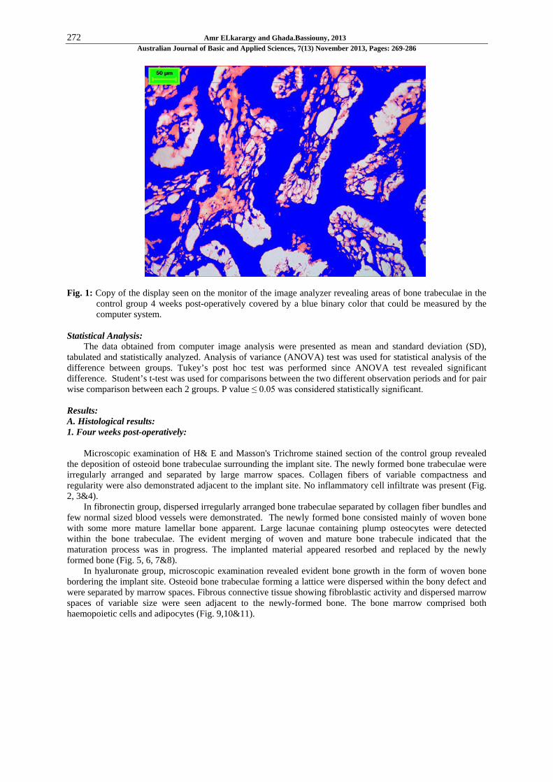
The area percent of bone trabeculae was estimated using Leica Quin 500 analyzer computer system, (Leica Microsystems, Swizerland). The cursor was used to outline the areas of bone trabeculae, which were then masked by a blue binary color that could be measured by the computer. The image analyzer is calibrated automatically to convert the measurement units (pixels) produced by the image analyzer program into actual 1micrometer units. The area percent of bone trabeculae was estimated in 5 different fields in each slide using magnification (x100), (Fig.1). Mean values and standard deviation were calculated for each group.
272 Amr ELkarargy and Ghada.Bassiouny, 2013 Australian Journal of Basic and Applied Sciences, 7(13) November 2013, Pages: 269-286
Fig. 1: Copy of the display seen on the monitor of the image analyzer revealing areas of bone trabeculae in the
control group 4 weeks post-operatively covered by a blue binary color that could be measured by the computer system.
Statistical Analysis:
The data obtained from computer image analysis were presented as mean and standard deviation (SD), tabulated and statistically analyzed. Analysis of variance (ANOVA) test was used for statistical analysis of the difference between groups. Tukey’s post hoc test was performed since ANOVA test revealed significant difference. Student’s t-test was used for comparisons between the two different observation periods and for pair wise comparison between each 2 groups. P value ≤ 0.05 was considered statistically significant.
Results: A. Histological results: 1. Four weeks post-operatively:
Microscopic examination of H& E and Masson's Trichrome stained section of the control group revealed
the deposition of osteoid bone trabeculae surrounding the implant site. The newly formed bone trabeculae were irregularly arranged and separated by large marrow spaces. Collagen fibers of variable compactness and regularity were also demonstrated adjacent to the implant site. No inflammatory cell infiltrate was present (Fig. 2, 3&4).
In fibronectin group, dispersed irregularly arranged bone trabeculae separated by collagen fiber bundles and few normal sized blood vessels were demonstrated. The newly formed bone consisted mainly of woven bone with some more mature lamellar bone apparent. Large lacunae containing plump osteocytes were detected within the bone trabeculae. The evident merging of woven and mature bone trabecule indicated that the maturation process was in progress. The implanted material appeared resorbed and replaced by the newly formed bone (Fig. 5, 6, 7&8).
In hyaluronate group, microscopic examination revealed evident bone growth in the form of woven bone bordering the implant site. Osteoid bone trabeculae forming a lattice were dispersed within the bony defect and were separated by marrow spaces. Fibrous connective tissue showing fibroblastic activity and dispersed marrow spaces of variable size were seen adjacent to the newly-formed bone. The bone marrow comprised both haemopoietic cells and adipocytes (Fig. 9,10&11).

273 Amr ELkarargy and Ghada.Bassiouny, 2013 Australian Journal of Basic and Applied Sciences, 7(13) November 2013, Pages: 269-286
Fig. 2: Photomicrograph of control group 4 weeks post-operatively revealing, thin irregular newly formed bone
trabeculae (NB) containing viable osteocytes (arrows) located adjacent to the implant site. Collagen fibers of variable compactness and regularity are also demonstrated (F), (H&Ex200).
Fig. 3: Photomicrograph of control group at 4 weeks post-operatively, revealing an implant site (I) bordered by
newly formed unclacified bone trabeculae (N) stained blue-green with Masson’s Trichrome. Focal areas of calcification exhibiting red histochemial staining are seen within the newly formed bone. Original bone (O) is seen at the periphery, (Masson’s Trichrome x100).
Fig. 4: Photomicrograph of control group at 4 weeks post-operatively, revealing newly formed unclacified bone
trabeculae (N) stained blue-green with Masson’s Trichrome. Areas of calcified bone exhibiting red histochemial staining are demonstrated (arrow). Large marrow spaces (M) are also demonstrated, (Masson’s Trichrome x200).

274 Amr ELkarargy and Ghada.Bassiouny, 2013 Australian Journal of Basic and Applied Sciences, 7(13) November 2013, Pages: 269-286
Fig. 5: Photomicrograph of fibronectin group 4 weeks post-operatively. Indentations created by the implant
(green arrows) are seen on the surface of the bone adjacent to the implant site (I). Vitality of bone is confirmed by the entrapped osteocytes (black arrows). Parallel dense collagen fibers are also demonstrated (F), (H&Ex200).
Fig. 6: Photomicrograph of fibronectin group 4 weeks post-operatively illustrating trabeculae of newly formed
bone (NB) with entrapped osteocytes (arrows) enclosing marrow spaces (M) of variable sizes. Parallel dense collagen fibers are also demonstrated (F), (H&Ex200).
Fig. 7: Photomicrograph of fibronectin group at 4 weeks post-operatively revealing, filling of the implant site
with newly-formed uncalcified woven bone (arrows) separated by wide marrow spaces (M), (Masson’s Trichrome x100).

275 Amr ELkarargy and Ghada.Bassiouny, 2013 Australian Journal of Basic and Applied Sciences, 7(13) November 2013, Pages: 269-286
Fig. 8: Photomicrograph of fibronectin group at 4 weeks post-operatively revealing, filling of the implant site
with newly-formed uncalcified woven bone separated by wide marrow spaces. Osteocytes are enclosed with lacuna denoting bone vitality (arrows), (Masson’s Trichrome x200).
Fig. 9: Photomicrograph of hyaluronate group 4 weeks post-operatively showing irregular trabeculae of newly
formed bone (NB) infiltrating dense collagen fibers (F). Osteocytes (arrows) and marrow spaces (M) are demonstrated within bone trabeculae,(H&Ex200).

276 Amr ELkarargy and Ghada.Bassiouny, 2013 Australian Journal of Basic and Applied Sciences, 7(13) November 2013, Pages: 269-286
Fig. 10: Photomicrograph of hyaluronate group 4 weeks post-operatively showing thick trabeculae of newly
formed bone containing viable osteocytes (arrows) and enclosing variable-sized marrow spaces (M). Parallel dense collagen fibers (F) are seen adjacent to the implant site, (H&Ex200).
Fig. 11: Photmicrograph of hyaluronate group at 4 weeks post-operatively revealing Thin parallel trabeculae are
dispersed by localized areas of calcified bone recognized by the red histochemical stain are dispersed within the new bone trabeculae, (Masson’s trichromex100).
2.Eight weeks post operatively:
Microscopic examination of H&E and Masson’s Trichrome stained section of the control group revealed that the implant site was completely surrounded by thick bone trabecular, separated by a fibro-vascular stroma. Vitality of bone was confirmed by osteocytes enclosed in lacunae within bone. Reversal lines denoting the rhythmic pattern of bone formation were also demonstrated (Fig. 12, 13, 14 &15).
In fibronectin group, there was circumferential bone regeneration in the form of thick regular bone trabeculae, separated by marrow spaces. Randomly arranged osteocytes were embedded within the trabeculae denoting vitality and enhanced bone apposition. Variable-sized fibro-vascular marrow separated the newly formed bone trabeculae. Reversal lines denoting the rhythmic pattern of bone apposition were observed (Fig. 16, 17, 18 &19).
Microscopic examination of hyaluronate group revealed newly formed bone trabeculae that were mainly directed toward the implant site. Thick bone trabeculae were seen coalescing together in certain areas, but separated by marrow spaces in other sites. Osteocytes were embedded within the newly formed trabeculae confirming the maintained bone vitality. Reversal lines denoting the rhythmic pattern of bone apposition were observed. No inflammatory cells could be detected (Fig. 20, 21, 22, 23&24).
Fig. 12: Photomicrograph of control group 8 weeks post-operatively revealing thick trabeculae of newly formed
bone trabeculae containing viable osteocytes (black arrows) surrounding the implant site (I). Reversal lines (yellow arrows) denoting the rhythmic pattern of bone formation are demonstrated, (H&Ex200).

277 Amr ELkarargy and Ghada.Bassiouny, 2013 Australian Journal of Basic and Applied Sciences, 7(13) November 2013, Pages: 269-286
Fig. 13: Photomicrograph of control group 8 weeks post-operatively revealing ,elongated trabeculae of newly
formed bone (NB) of varying thickness and containing viable osteocytes (arrow) surrounding the implant site (I). Dense fibro-vascular tissue (F) separates the newly formed trabeculae, (H&Ex200).
Fig. 14: Photomicrograph of control group at 8 weeks post-operatively, revealing an implant site (I) bordered by
newly formed unclacified bone trabeculae (N). Original bone (O) is demonstrated at the periphery, (Masson’s Trichrome x100).
Fig. 15: A higher magnification of the previous photomicrograph of control group at 8 weeks post-operatively,
revealing large marrow space (M) within the newly formed unclacified bone trabeculae (N). Original bone (O) is demonstrated at the periphery, (Masson’s Trichrome x200).

278 Amr ELkarargy and Ghada.Bassiouny, 2013 Australian Journal of Basic and Applied Sciences, 7(13) November 2013, Pages: 269-286
Fig. 16: Photomicrograph of fibronectin group 8 weeks post-operatively revealing thick trabeculae of newly
formed bone containing viable osteocytes (black arrows) and enclosing variable-sized marrow spaces (M). Reversal lines (yellow arrows) denoting the rhythmic pattern of bone apposition are demonstrated, (H&Ex200).
Fig. 17: Photomicrograph of fibronectin group 8 weeks post-operatively revealing the implant site (I) separated
by thick trabeculae of newly formed bone containing viable osteocytes (black arrows) and enclosing marrow spaces (M). Reversal lines (yellow arrows) denoting the rhythmic pattern of bone apposition are demonstrated, (H&Ex200).
Fig. 18: Photmicrograph of fibronectin group at 8 weeks post-operatively revealing newly formed unclacified
bone trabeculae filling the implant site. Focal areas of calcified bone recognized by the red histochemical stain are dispersed within the new bone trabeculae. Wide marrow spaces (M) are demonstrated, (Masson’s Trichrome x200).

279 Amr ELkarargy and Ghada.Bassiouny, 2013 Australian Journal of Basic and Applied Sciences, 7(13) November 2013, Pages: 269-286
Fig. 19: Higher magnification of the previous photmicrograph of fibronectin group at 8 weeks post-operatively
revealing newly formed unclacified bone trabeculae filling the implant site.Focal areas of calcified bone recognized by the red histochemical stain (arrows). Marrow spaces (M) are demonstrated, (Masson’s Trichrome x200).
Fig. 20: Photomicrograph of hyaluronate group 8 weeks post-operatively revealing thick trabeculae of newly
formed bone containing viable osteocytes (black arrows) and enclosing variable-sized marrow spaces (M). Reversal lines (yellow arrows) denoting the rhythmic pattern of bone apposition are demonstrated, (H&Ex100).
Fig. 21: Higher magnification of the previous photomicrograph of hyaluronate group 8 weeks post-operatively
revealing thick trabeculae of newly formed bone containing viable osteocytes (black arrows) and enclosing fibro-vascular marrow (M). Reversal lines (yellow arrows) are observed, (H&Ex200).

280 Amr ELkarargy and Ghada.Bassiouny, 2013 Australian Journal of Basic and Applied Sciences, 7(13) November 2013, Pages: 269-286
Fig. 22: Photmicrograph of hyaluronate group at 8 weeks post-operatively revealing newly formed unclacified
bone trabeculae bordering the implant site (I). Focal areas of calcified bone recognized by the red histochemical stain are dispersed within the new bone trabeculae. Wide marrow spaces (M) are demonstrated, (Masson’s Trichrome x100).
Fig. 23: Higher magnification of the previous photomicrograph of hyaluronate group at 8 weeks post-
operatively revealing newly formed unclacified bone trabeculae bordering the implant site (I). Focal areas of calcified bone recognized by the red histochemical stain are dispersed within the new bone trabeculae (arrows). Wide marrow spaces (M) are demonstrated, (Masson’s Trichrome x200).
Fig. 24: Photomicrograph of hyaluronate group at 8 weeks post-operatively revealing calcification of the newly
formed bone stained red, with localized areas of un-calcified bone stained blue- green with Masson's trichrome. Wide marrow spaces (arrows) are demonstrated, (Masson's Trichrome x200).

281 Amr ELkarargy and Ghada.Bassiouny, 2013 Australian Journal of Basic and Applied Sciences, 7(13) November 2013, Pages: 269-286
B. Histomorphometrical results: I-Comparison between control and experimental groups:
Histomorphometric estimation of the mean area percent filled by bone trabeculae 4 weeks postoperatively was greater in the hyaluronate group (63.68± 1.68) compared to the fibronectin group (60.75±5.37). The control group revealed the lowest area % of bone trabeculae (54.83±4.94). Analysis of variance (ANOVA) test revealed that the difference between the 3 groups was extremely statistically significant (p<0.0001). Tukey’s post hoc test revealed that the hyaluronate and fibronectin groups were not statistically different, as confirmed by Student’s t test (Table 1, 2; Fig.25).
The same pattern was observed eight weeks post-operatively, where the area percent of bone trabeculae was greater in the group hyaluronate (87.93±3.18) compared to the fibronectin group (82.04±4.12), whereas the control group revealed the lowest area % of bone trabeculae (77.64±3.45). Analysis of variance (ANOVA) test revealed that the difference between the 3 groups was highly statistically significant (p<0.0001). Tukey’s post hoc test revealed that each two groups were statistically different, as confirmed by Student’s t test (Table 1, 2; Fig.25).
Table 1: Values of area % filled by bone trabeculae and statistical significance of the different between all groups at each observation
period (ANOVA test)
Values 4 weeks 8 weeks C F H C F H
Mean 54.83 a 60.75 b 63.68 b 77.64 a 82.04 b 87.93c StdDev 4.94 5.37 1.68 3.45 4.12 3.18 Std Error 6.682 6.871 0.751 6.013 1.843 1.424 Max 74.26 71.681 65.497 92.569 87.005 93.295 Min 34.363 30.815 61.007 57.878 74.769 83.706 2-s Range 59.769 61.46 6.714 53.786 16.486 12.735 F value 10.877 20.510 P value <0.0001*** <0.0001***
***Highly statistically significant Table 2: Results of Student’s t test used for paiwise comparison between groups
Observation time Groups t value p value 4 weeks C versus F
C versus H F versus H
2.5657 5.3635 1.6467
0.0195* <0.0001*** 0.1170
8 weeks C versus F Cversus H F versus H
2.5893 6.9352 3.5788
0.0185* <0.0001*** 0.0021**
*Statistically significant **Very statistically significant *** Highly statistically significant
Fig. 25: Mean area % of bone trabeculae in all groups throughout the experiment
II- Change By Time In Bone Trabeculae In Control And Experimental Groups:
All groups exhibited an increase in the area percent occupied by bone trabeculae throughout time. Student’s t test revealed that the difference in the mean area % throughout the experiment (four and eight weeks post-operatively) was extremely statistically significant (p<0.0001) in the control and experimental groups. The

282 Amr ELkarargy and Ghada.Bassiouny, 2013 Australian Journal of Basic and Applied Sciences, 7(13) November 2013, Pages: 269-286
percent increase was greater in the control group (41.60%), compared to the fibronection group (35.05%), and hyaluronate (38.08%) (Table 3, Fig.26&27).
Table 3: Change by time in mean area % of bone trabeculae of each group and statistical significance of the difference (Paired Student’s t
test)
Groups Values
Control Fibronection Hyaluronate
4 weeks 8 weeks 4 weeks 8 weeks 4 weeks 8 weeks Mean 54.83 77.64 60.75 82.04 63.68 87.93 StdDev 4.94 3.45 5.37 4.12 1.68 3.18 % increase 41.60% 35.05% 38.08% t value 11.9711 9.9469 21.3222 P value <0.0001*** <0.0001*** <0.0001***
*** Highly statistically significant
Fig. 26: Change by time in area % of bone trabeculae in all groups
Fig. 27: Percent increase in area filled by bone trabeculae throughout the experiment Discussion:
Surface modifications of titanium implants using various modalities aim to improve the initial healing and promote faster healing times.(Dohan, Coelho et al. 2010) With this surface modification, immediate or early loading has become a predictable treatment protocol.(Atieh, Payne et al. 2010) The future of dental implantology should aim to develop surfaces with controlled and standardized topography or chemistry. This approach will be the only way to understand the interactions between proteins, cells and tissues, and implant surfaces. The local release of bone stimulating or antiresorptive subestances in the peri-implant region may also respond to difficult clinical situations with poor bone quality and quantity. These therapeutic strategies should

283 Amr ELkarargy and Ghada.Bassiouny, 2013 Australian Journal of Basic and Applied Sciences, 7(13) November 2013, Pages: 269-286
ultimately enhance the osseointegration process of dental implants for their immediate loading and long-term success. (Zhao, Schwartz et al. 2005)
The present study was designed to entail the tibial bone that constituted mainly compact cortical bone, with a structure comparable to that of human. Hence, the tibia was considered an ideal model for mandibular bone, this model was documented to provide ideal conditions for the investigation of bone regeneration and implant osseointegration. (Chong-Hyun and Dong-Hoo 1994)
The present research was focused on the development of surface modifications that mimic the existing biological situation by promoting specific cell attachment, and thereby enhancing osseointegration of the implant surface. To achieve this goal this study was conducted to characterize the osseointegration of fibronectin vs hyaluronate-coated titanium dental implants as a biologically active and clinically relevant implant coating strategy that enhances bone repair and implant integration.
This modification on the topographic pattern of implant surface increases not only the bone-implant contact, but also the biomechanical interaction of that interface at early implantation periods. (Roccuzzo, Bunino et al. 2001)
Roccuzzo, Bunino et al. observed in an in vivo study that the topography is not the only surface characteristic capable of enhancing new bone apposition. (Roccuzzo, Bunino et al. 2001) Besides,Germanier, Tosatti et al. showed in an in vitro study that surface chemistry also influenced the attachment and morphology of cells. In general, an increase in cell number and more spread cells were observed on bioactive substrate compared to bio-inactive surfaces according to their findings. (Germanier, Tosatti et al. 2006)
However, Bioactive surface modification approaches, including calcium-phosphate ceramic coatings and macro/microporosity, have had limited success in promoting integration because of a chipping effect of apatite crystals from the surface, causing a phagocytic response by macrophages or even a foreign body reaction and peri-implantitis in the long run. (Schuler, Owen et al. 2006)
In attempts to avoid such effects and to improve osseointegration, titanium surfaces were biofunctionalized with ECM-mimetic peptides, this biomimetic implant coating is generated using a simple, single-step procedure that readily translates to a clinical environment with minimal processing and cytotoxicity concerns. (Iezzi, Pecora et al. 2009)
These findings were in consistence with the strategy of our study that combines the benefits of both topographic surface characteristics of sand blasted acid etched dental implant together with surface biomodification using two of the most important bioactive ECM proteins hyaluronate and fibronectin in an attempt to mimic the existing biological situation by promoting specific cell attachment, and thereby enhancing osseointegration of the implant surface.
Importantly, In this study, biomodification of implant surface utilized a simple, dip-coating method relied on physical adsorption of fibronectin and Hyaluronate to pre-sterilized titanium implants as a simple, clinically-translatable strategy to functionalize dental implants. This is in accordance with Petrie et al whom described this simple, one-step coating procedure that relies on the passive adsorption of an ECM peptide onto titanium implants to enhance osseointegration. (Petrie, Catherine et al. 2009)
On the other hand, the other approaches for implant coating and biofunctionalization, although are successful in many cases, they can be restricted by slow rates of osseointegration and poor mechanical anchorage, especially in challenging clinical cases, such as those associated with large bone loss and poor bone quality. (Catherine, Timothy et al. 2007)In addition, these surface modification approaches rely on costly and manufacturing-intensive processes. As an alternative to these surface technologies, emerging biomimetic strategies that focused on the presentation of extracellular matrix sequences and growth factors.(Dillow, Ochsenhirt et al. 2001) The general paradigm of these approaches is the covalent immobilization of the biological entities onto the underlying implant surface, which often involves multi-step procedures to render the surface suitable for biofunctionalization. (Chung, Gilbert et al. 2006) In contrast, the method used in our study for implant biofunctionalization ,relies on Petrie, Catherine et al.technique which offers a quick and versatile surface application conducted under physiological conditions that the surgeon can employ seconds before implantation. This single-step procedure, in turn, minimizes the chance of infection, reduces implant surface treatment variability, and minimizes cytotoxicity concerns inherent with covalent immobilization schemes, while maintaining the surgeon’s dexterity. (Petrie, Catherine et al. 2009)
Clinical and histologic results revealed that all implants were clinically stable and non-mobile. The os-seointegration of each implant was confirmed. Osteoblasts had actively deposited osteoid matrix on the implant surface. The newly formed bone in the interthread space was in close contact with the implant surface, without any gaps or dense fibrous connective tissue. No apical epithelial migration was found. No inflammatory cell infiltrate was present around the implants. Areas of bone remodeling with osteoblasts and osteoid matrix with reversal lines were observed specially in hyaluronate group more than in fibronectin while the least of these histological findings were observed in the uncoated control implants especially at the early healing period (4 weeks post-operative).

284 Amr ELkarargy and Ghada.Bassiouny, 2013 Australian Journal of Basic and Applied Sciences, 7(13) November 2013, Pages: 269-286
Histomorphometric estimation of the mean area percent filled by bone trabeculae was greater in the
hyaluronate group (63.68± 1.68) (87.93±3.18) at 4&8weeks post-operatively respectively compared to the fibronectin group (60.75±5.37) (82.04±4.12),while the control group revealed the lowest area % of bone trabeculae (54.83±4.94) (77.64±3.45) at the same observation periods. Analysis of variance (ANOVA) test revealed that the difference between the 3 groups was highly statistically significant (p<0.0001) at both estimation periods. While Tukey’s post hoc test revealed that the difference between hyaluronate and fibronectin was not statistically significant at 4 weeks, however each two groups were statistically different at 8 weeks post-operatively, as confirmed by Student’s t test.
Regarding Change by time in bone trabeculae in control and experimental groups, all groups exhibited an increase in the area percent occupied by bone trabeculae throughout time. Student’s t test revealed that the difference in the mean area % throughout the experiment (four and eight weeks post-operatively) was highly statistically significant (p<0.0001) in the control and experimental groups. The percent increase was greater in the control group (41.60%), compared to the fibronection group (35.05%), and hyaluronate (38.08%).This can be explained by the observation that the area % of bone fill was greater in experimental groups at an earlier time (in the first observation period), consequently, the percent increase was slightly lower in experimental compared to the control, which needed longer time for new bone formation.
Different mechanisms have been proposed to explain the effect of hyaluronate and fibronectin in accelerating wound healing and promoting osteogenesis. The biomimetic coating with fibronectin has been tested in previous studies. (Urban, Jacobs et al. 2000; Brett, Harle et al. 2004; Lutolf and Hubbell 2005)It is believed that application of fibronectin to the implant surface can enhance the osseointegration of dental implants to the bone. (Martini, Fini et al. 2003) This can be explained by the researches that demonstrated that coating the surface with cell-adhesive proteins, such as fibronectin, improved initial cell attachment, cell spreading, and cell activity. (Urban, Jacobs et al. 2000; Brett, Harle et al. 2004; Lutolf and Hubbell 2005) Others have documented that fibronectin is actively involved in cytoskeletal reorganization and bone tissue formation, in part by regulating the survival of osteoblasts. (Roccuzzo, Bunino et al. 2001) Moreover fibronectin is known to play a role in cell-to-cell and cell-to-substrate adhesion as well as an important role in osseointegration due to its capacity to make osteoblasts attach to ECM components. (Browne and Gregson 2000)
However, the present study supported the superiority of hyaluronate over fibronectin both qualitatively by the microscopic evaluation of the quality of the newly formed bone around hyaluronate coated implants vs fibronectin and quantitatively by histomorphometric analysis of the area percent occupied by bone trabeculae .
There are two mechanisms to explain these findings , firstly, Hyaluronate accelerates wound healing and promoting osteogenesis by means of chemotaxis, proliferation and successive differentiation of mesenchymal cells as well as by a certain role on hydroxyapatite crystal formation. (Pilloni, Rimondini et al. 2003) These important roles mediated by hyaluronate have confirmed by other investigators who documented that hyaluronate shares bone induction characteristics with osteogenic substances such as bone morphogenetic protein-2 and osteopontin. (Mendes, Silva et al. 2008)
Secondly, hyaluronate has anti-inflammatory properties through modulation of inflammatory cells, interaction with the proteoglycans of the extracellular matrix and scavenging of free radicals. Thus inhibiting tissue destruction and facilitates healing. (Moseley, Waddington et al. 2002), (Boskey and Dick 1991)
Based on our findings and on the previous mentioned studies it can be stated that hyaluronate-mimetic approach significantly enhanced osseointegration of titanium implants in rabbit cortical bone. This study establishes a biologically active and clinically relevant implant coating strategy that enhances bone repair and dental implant integration.
Conclusion:
Biofunctionalization of implant surfaces is still a poorly investigated area. The enhancement of early osseointegration around the hyaluronate-coated surface is promising. Further in vitro and in vivo studies are necessary to elucidate how and in which extent biofunctionalization of the implant surfaces could represent an advantageous modification.
REFERENCES
Abron, A., M. Hopfensperger, et al. 2001. "Evaluation of a predictive model for implant surface topography
effects on early osseointegration in the rat tibia model." J Prosth Dent, 85: 40-60. Albrektsson, T., P. Branemark, et al. 1981. "Osseointegrated titanium implants. Requirements for ensuring
a long-lasting, direct bone-to-implant anchorage in man." Acta Orthop Scand, 52: 155-170. Aparicio, C., F. Gil, et al. 2003. "Corrosion behavior of commercially pure titanium shot blasted with
different materials and size of shot particles for dental implant applications." Biomaterials, 24: 263-273.

285 Amr ELkarargy and Ghada.Bassiouny, 2013 Australian Journal of Basic and Applied Sciences, 7(13) November 2013, Pages: 269-286
Atieh, M., A. Payne, et al. 2010. "Immediate placement or immediate restoration/loading of single implants
for molar tooth replacement: a system¬atic review and meta-analysis." Int J Oral Maxillofac Imlplants., 25: 401-415.
Barrere, F., C. van der Valk, et al. 2003. "Osteointegration of biomimetic apatite coating applied onto dense and porous metal implants in femurs of goats." J Biomed Mater Res., 67: 655-665.
Bigerelle, M., K. Anselme, et al. 2002. " Improvement in the morphology of Ti-based surfaces: a new process to increase in vitro human osteoblast response." Biomaterials., 23: 1563-1577.
Boskey, A. and B. Dick 1991. "Hyaluronan interactions with hydroxyapatite do not alter in vitro hydroxyapatite crystal proliferation and growth." Matrix. 11: 442-446.
Boyne, P. and S. Jones 2004. "Demonstration of the osseoinductive effect of bone morphogenetic protein within endosseous dental implants." Implant Dent., 13: 180-184.
Brett, P., J. Harle, et al. 2004. "Roughness response genes in osteoblasts." Bone. 35: 124-133. Browne, M. and P. Gregson 2000. "Effect of mechanical surface pretreatment on metal ions release."
Biomaterials., 21: 385-392. Buser, D., N. Broggini, et al. 2004. "Enhanced bone apposition to a chemically modified SLA titanium
surface." J Dent Res., 83(7): 529-533. Catherine, D., A. Timothy, et al. 2007. "Biomolecular surface coating to enhance orthopaedic tissue healing
and integration " Biomaterials., 28: 3228-3235. Cho, S. and K. Park 2003. "The removal torque of titanium screw inserted in rabbit tibia treated by dual
acid etching " Biomaterials., 24: 3611-3617. Chong-Hyun, M. and H. Dong-Hoo 1994. "A study on shear-bond strength of the interface between bone
and titanium plasma-sprayed IMZ implants in rabbits." Int J Oromaxillofac Imp. 9: 698-707. Chung, E., M. Gilbert, et al. 2006. " Biomimetic artificial ECMs stimulate bone regeneration." J Biomed
Mater Res., A. 79: 815-826. Dillow, A., S. Ochsenhirt, et al. 2001. "Adhesion of alpha 5 beta1 receptors to biomimetic substrates
constructed from peptide amphiphiles. ." Biomaterials., 22: 1493-1505. Dohan, D., P. Coelho, et al. 2010. "Classification of osseointegrated implant sur¬faces: materials, chemistry
and topography." Trends Biotechnol., 28: 198-206. Embery, G., R. Waddington, et al. 2000. "Connective tissue elements as diagnostic aids in periodontology."
Periodontol., 24: 193-214. Esposito, M., P. Coulthard, et al. 2005. "Interventions for replacing missing teeth: different types of dental
implants." Cochrane Database Syst Rev., 25(CD003815.). Germanier, Y., S. Tosatti, et al. 2006. "Enhanced bone apposition around biofunctionalized sand blasted
and acid etched titanium implant surfaces. A histomorphometric study in miniature pigs." Clin Oral Impl Res., 17: 251-257.
Guehennec, LE., LA. Soueidan,et al. 2007. "Surface treatments of titanium dental implants for rapid osseointegration." Dent Mater, 23(7): 844-854.
Habibovic, P., V. Valk, et al. 2005. "Biological performance of uncoated and octacalcium phosphate-coated Ti6Al4V." Biomaterials., 26: 23-36.
Horbett, T. (1993). "Principles underlying the role of adsorbed plasma proteins in blood interactions with for¬eign materials." Cardiovasc Pathol., 2: 137-148.
Huang, Y., A. Xiropaidis, et al. 2005. "Bone formation at titanium porous oxide (TiUnite) oral implants in type IV bone." Clin Oral Implants Res., 16: 105-111.
Hynes, R. (1986). "Fibronectins." Sci Am. 254: 42-51. Iezzi, G. O., S. G. Pecora, et al. 2009. "Histologic and histomorphometric evaluation of the bone response
around a hydroxyapatite- coated implant retrieved after 15 years." International Journal of Periodontics Restorative Dentistry, 29: 99-105.
Jungner, M., P. Lundqvist, et al. 2005. "Oxidized titanium implants (Nobel Biocare TiUnite) compared with turned titanium implants (Nobel Biocare mark III) with respect to implant failure in a group of consecutive patients treated with early functional loading and two-stage protocol." Clin Oral Implants Res., 16: 308-312.
Lutolf, M. and J. Hubbell 2005. "Synthetic biomaterials as instructive extracellular microenvironments for morphogenesis in tissue engineering." Nat Biotechnol., 23: 47-55.
Martini, D., M. Fini, et al. 2003. "Detachment of titanium and fluorohydroxyapatite particles in unloaded endosseous implants." Biomaterials, 24: 1309-1316.
Mendes, R., G. Silva, et al. 2008. "Sodium hyaluronate accelerates the healing process in tooth sockets of rats." Arch Oral Biol., 53: 1155-1162.
Moseley, R., R. Waddington, et al. 2002. " Hyaluronan and its potential role in periodontal healing." Dent Update, 29: 144-148.
Mueller, W. D., U. Gross, et al. 2003. "Evaluation of the interface between bone and titanium surfaces being blasted by aluminium oxide or bioceramic particles." Clin Oral Implants Res., 3: 349-356.

286 Amr ELkarargy and Ghada.Bassiouny, 2013 Australian Journal of Basic and Applied Sciences, 7(13) November 2013, Pages: 269-286
Novaes, A., S. de Souza, et al. 1995. "Fibronectin and laminin enhance gingival cell attachment to dental
implant surfaces in vitro." Int J Oral Maxillofac Implants, 10: 721-728. Novaes, A., S. d. B. de Souza, RR., et al. 2010. " Influence of Implant Surfaces on Osseointegration."
Invited Review Article Braz Dent, J. 21: 471-481. Novaes, A., V. Papalexiou, et al. 2004. "Influence of implant microstructure on the osseointegration of
immediate implants placed in periodontally infected sites. A histomorphometric study in dogs." Clin Oral Implants Res., 15: 34-43.
Novaes, A., S. Souza, et al. 2002. "Histomorphometric analysis of the bone-implant contact obtained with 4 different implant surface treatments placed side by side in the dog mandible." Int J Oral Maxillofac Implants, 17: 377-388.
Ong, J.L., D.L. Carnes, et al. 2004. "Evaluation of titanium- plasma sprayed and plasma -sprayed hydroxyapatite implants in vivo." Biomaterials, 25: 4601-4606.
Petrie, T., D. Catherine, et al. 2009. "Simple application of fibronectin-mimetic coating enhances osseointegration of titanium implants." J Cell Mol Med., 13: 2602-2612.
Papalexiou, V., J. Novaes, et al. 2004. "Influence of implant microstructure on the dynamics of bone healing around immediate implants placed into periodontally infected sites." Clin Oral Implants Res., 15: 44-53.
Petrie, T., D. Catherine, et al. 2009. "Simple application of fibronectin-mimetic coating enhances osseointegration of titanium implants." J Cell Mol Med., 13: 2602-2612.
Piatelli, M., A. Scarano, et al. 2002. "Bone response to machined and resorbable blast material titanium implants: an experimental study in rabbits." J Oral Implantol, 28: 2-8.
Pilloni, A., L. Rimondini, et al. 2003. "Effect of hyaluronan on calcification-nodule formation from human periodontal ligament cell culture." Journal of Applied Biomaterials & Biomechanics, 1: 1-7.
Puleo, D. N., A. 1999. "Understanding and controlling the bone-implant interface." Biomaterials 1999; 20:2311-21. 20: 2311-2321.
Roccuzzo, M., M. Bunino, et al. 2001. "Early loading of sand blasted and acid-etched (SLA) implants: A prospective split-mouth comparative study." Clin Oral Implants Res., 12: 572-578.
Roehlecke, C., M. Witt, et al. 2001. "Synergistic effect of titanium alloy and collagen type I on cell adhesion, proliferation and differentiation of osteoblast-like cells." Cells Tissues Organs., 168: 178-187.
Sakasi, T. and C. Watanabe 1995. "Stimulation of osteoinduction in bone wound healing by high-molecular hyaluronic acid." Bone., 16: 9-15.
Schuler, M., G. Owen, et al. 2006. "Biomimetic modification of titanium dental implant model surfaces using the RGDSP-peptide sequence: a cell morphology study." Biomaterials., 27: 4003-4015.
Steinemann, S. 1998. "Titanium -the material of choice ?" Periodontology 2000 17: 7-21. Stenport, V., A. M. Roos-Jansake , et al. 2004. "Failure to induce supracrestal bone growth between and
around partially inserted titanium implants using bone morphogenetic protein (BMP): an experimental study in dogs." Clin Oral Implants Res., 14: 219-225.
Sul, Y., E. Byon, et al. 2004. "Biomechanical measurements of calcium-incorporated oxidized implants in rabbit bone: effect of calcium surface chemistry of a novel implant. ." Clin Implant Dent Relat Res., 6: 101-110.
Sul, Y., C. Johansson, et al. 2005. "Optimum surface properties of oxidized implants for reinforcement of osseointegration: surface chemistry, oxide thickness, porosity, roughness, and crystal structure." Int J Oral Maxillofac Implants., 20: 349-359.
Taba Junior, M., A. B. Novaes Junior, et al. 2003. "Radiographic evaluation of dental implants with different surface treatments: an experimental study in dogs." Implant Dent., 12: 252-258.
Tatakis, D., A. Koh, et al. 2002. "Peri-implant bone regeneration using recombinant human bone morphogenetic protein-2 in a canine model: a dose-response study." J Periodontal Res., 37: 93-100.
Urban, R. M., J. Jacobs, et al. 2000. "Dissemination of wear particles to the liver, spleen and abdominal lymph nodes of eepatients with hip or knee replacement." Bone Jt Surg Am., 82: 457-477.
Wuttke, M., S. Müller, et al. 2001. "Structural characterization of human recombinant and bone-derivede bone sialoprotein." J Biol Chem 276: 36839-36848.
Xiropaidis, A., M. Qahash, et al. 2005. "Bone-implant contact at calcium phosphate-coated and porous titanium oxide (TiUnite)-modified oral implants." Clin Oral Implants Res., 16: 532-539.
Zhao, G., Z. Schwartz, et al. 2005. "High surface energy enhances cell response to titanium substrate microstructure." Biomed Mater Res., A. 74: 49-58.
Zhu, X., J. Chen, et al. 2004. " Cellular reactions of osteoblasts to micron and submicron-scale porous structures of titanium surfaces." Cells Tissues Organs., 178: 13-22.
Zinger, O., K. Anselme, et al. 2004. "Time-dependent morphology and adhesion of osteoblastic cells on titanium model surfaces featuring scale-resolved topography." Biomaterials, 25: 2695.