advertiment. lʼaccés als continguts dʼaquesta tesi queda ... · el riesgo de progresión precoz...
TRANSCRIPT
ADVERTIMENT. Lʼaccés als continguts dʼaquesta tesi queda condicionat a lʼacceptació de les condicions dʼúsestablertes per la següent llicència Creative Commons: http://cat.creativecommons.org/?page_id=184
ADVERTENCIA. El acceso a los contenidos de esta tesis queda condicionado a la aceptación de las condiciones de usoestablecidas por la siguiente licencia Creative Commons: http://es.creativecommons.org/blog/licencias/
WARNING. The access to the contents of this doctoral thesis it is limited to the acceptance of the use conditions setby the following Creative Commons license: https://creativecommons.org/licenses/?lang=en
TESIS DOCTORAL
Clinical, radiological and pathological prognostic factors for local
relapse, distant metastases and long-term survival in patients with
locally advanced rectal cancer treated with neoadjuvant long-course
oral fluoropyrimidine- and oxaliplatin-based chemoradiotherapy and
total mesorectal excision:
Can we move towards a more personalised approach?
ROBERTO PEDRO DÍAZ BEVERIDGE
DEPARTAMENT DE MEDICINA
UNIVERSIDAD AUTÒNOMA DE BARCELONA
CURSO 2015-2016
DIRECTORES:
JORGE APARICIO URTASUN
JORDI GIRALT LÓPEZ DE SAGREDO
RAFAEL ROSELL COSTA
TUTOR:
CARLOS GUARNER AGUILAR
2
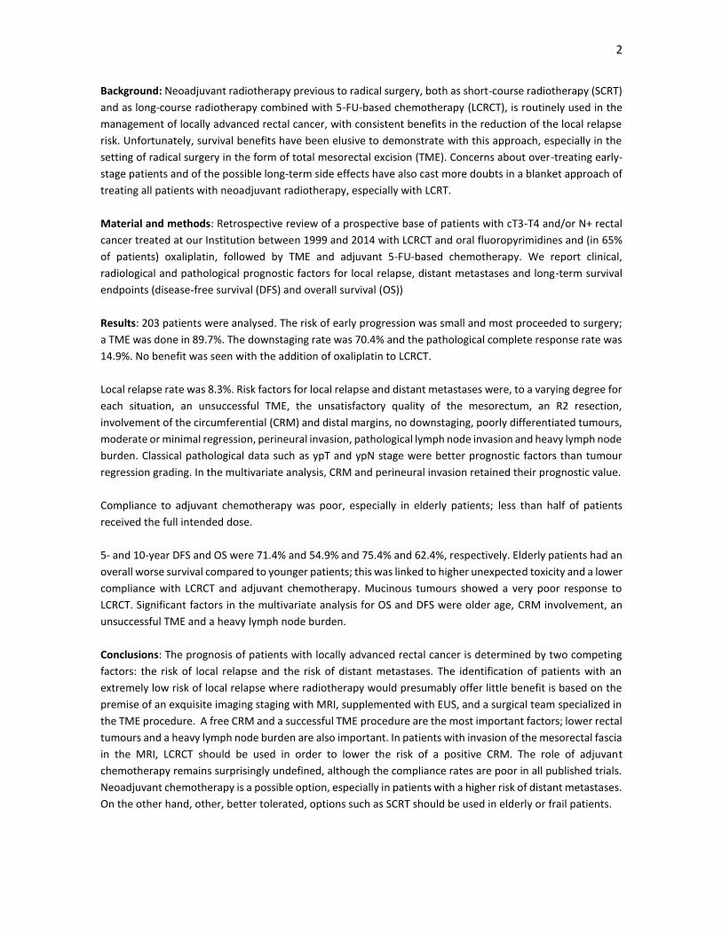
Background: Neoadjuvant radiotherapy previous to radical surgery, both as short-course radiotherapy (SCRT)
and as long-course radiotherapy combined with 5-FU-based chemotherapy (LCRCT), is routinely used in the
management of locally advanced rectal cancer, with consistent benefits in the reduction of the local relapse
risk. Unfortunately, survival benefits have been elusive to demonstrate with this approach, especially in the
setting of radical surgery in the form of total mesorectal excision (TME). Concerns about over-treating early-
stage patients and of the possible long-term side effects have also cast more doubts in a blanket approach of
treating all patients with neoadjuvant radiotherapy, especially with LCRT.
Material and methods: Retrospective review of a prospective base of patients with cT3-T4 and/or N+ rectal
cancer treated at our Institution between 1999 and 2014 with LCRCT and oral fluoropyrimidines and (in 65%
of patients) oxaliplatin, followed by TME and adjuvant 5-FU-based chemotherapy. We report clinical,
radiological and pathological prognostic factors for local relapse, distant metastases and long-term survival
endpoints (disease-free survival (DFS) and overall survival (OS))
Results: 203 patients were analysed. The risk of early progression was small and most proceeded to surgery;
a TME was done in 89.7%. The downstaging rate was 70.4% and the pathological complete response rate was
14.9%. No benefit was seen with the addition of oxaliplatin to LCRCT.
Local relapse rate was 8.3%. Risk factors for local relapse and distant metastases were, to a varying degree for
each situation, an unsuccessful TME, the unsatisfactory quality of the mesorectum, an R2 resection,
involvement of the circumferential (CRM) and distal margins, no downstaging, poorly differentiated tumours,
moderate or minimal regression, perineural invasion, pathological lymph node invasion and heavy lymph node
burden. Classical pathological data such as ypT and ypN stage were better prognostic factors than tumour
regression grading. In the multivariate analysis, CRM and perineural invasion retained their prognostic value.
Compliance to adjuvant chemotherapy was poor, especially in elderly patients; less than half of patients
received the full intended dose.
5- and 10-year DFS and OS were 71.4% and 54.9% and 75.4% and 62.4%, respectively. Elderly patients had an
overall worse survival compared to younger patients; this was linked to higher unexpected toxicity and a lower
compliance with LCRCT and adjuvant chemotherapy. Mucinous tumours showed a very poor response to
LCRCT. Significant factors in the multivariate analysis for OS and DFS were older age, CRM involvement, an
unsuccessful TME and a heavy lymph node burden.
Conclusions: The prognosis of patients with locally advanced rectal cancer is determined by two competing
factors: the risk of local relapse and the risk of distant metastases. The identification of patients with an
extremely low risk of local relapse where radiotherapy would presumably offer little benefit is based on the
premise of an exquisite imaging staging with MRI, supplemented with EUS, and a surgical team specialized in
the TME procedure. A free CRM and a successful TME procedure are the most important factors; lower rectal
tumours and a heavy lymph node burden are also important. In patients with invasion of the mesorectal fascia
in the MRI, LCRCT should be used in order to lower the risk of a positive CRM. The role of adjuvant
chemotherapy remains surprisingly undefined, although the compliance rates are poor in all published trials.
Neoadjuvant chemotherapy is a possible option, especially in patients with a higher risk of distant metastases.
On the other hand, other, better tolerated, options such as SCRT should be used in elderly or frail patients.
3
Fundamentos: La radioterapia (RT) neoadyuvante previa a la cirugía, ya sea la radioterapia de duración corta
(RTDC) como la radioterapia de duración larga combinada con quimioterapia (QT) basada en 5-FU (QRTLD), es
usada de forma rutinaria en el manejo del cáncer de recto localmente avanzado, con beneficios consistentes
en el riesgo de recidiva local. Desafortunadamente, no se han podido demostrar mejorías en la supervivencia,
especialmente en los casos tratados con cirugía radical en forma de una escisión mesorectal total (EMT). El
riesgo de sobretratar a algunos pacientes y los posibles efectos secundarios a largo plazo han provocado a su
vez dudas sobre el manejo con RT neoadyuvante, especialmente con QRTLD, en todos los pacientes con cáncer
de recto localmente avanzado independientemente de su riesgo basal de recidiva local.
Material y métodos: Revisión retrospectiva de una base prospectiva de pacientes con cáncer de recto cT3-T4
y/o cN+, tratados entre 1999 y 2014 con QRTLD basada en fluoropirimidinas orales y (en un 65%) oxaliplatino,
seguido de EMT y QT adyuvante basada en 5-FU. Evaluamos factores pronóstico clínicos, radiológicos y
patológicos para un mayor riesgo de recidiva local y de metástasis a distancia y una menor supervivencia libre
de progresión (SLP) y supervivencia global (SG).
Resultados: 203 pacientes fueron analizados. El riesgo de progresión precoz fue bajo y la mayor parte de
pacientes procedieron a cirugía; hubo una EMT satisfactoria en el 89.7%. La tasa de infraestadiaje fue del
70.4% y el porcentaje de respuestas completas patológicas fue del 14.9%. No hubo ningún beneficio con la
adición de oxaliplatino a la QRTLD. La tasa de recidivas locales fue del 8.3%. Los factores de riesgo para la
recidiva local y para las metástasis a distancia fueron, con un valor variable para las dos situaciones, una EMT
no exitosa, la calidad insuficiente del mesorecto, una resección R2, afectación del margen circunferencial
radial (MCR) y del margen distal, no infraestadiaje, tumores pobremente diferenciados, regresión tumoral
moderada o mínima, invasión perineural, afectación patológica linfática y una gran carga tumoral linfática.
Los factores pronóstico clásicos como el estadio ypT ó ypN tuvieron mayor importancia que la regresión
tumoral patológica. En el análisis multivariante, la afectación del MCR y la invasión perineural mantuvieron la
significación. La cumplimentación de la QT adyuvante fue pobre, especialmente en los pacientes ancianos;
menos de la mitad recibieron la dosis completa prevista.
La SLP y SG a 5 y 10 años fue del 71.4% y 54.9% y del 75.4% y 62.4%, respectivamente. Los pacientes ancianos
tuvieron una peor SLP y SG; ello estaba ligado a un aumento de las toxicidades graves no previsibles y una
menor cumplimentación de la QRTLD y de la QT adyuvante. Los tumores mucinosos mostraron una respuesta
muy pobre a la QRTLD. Factores significativos en el análisis multivariante para SLP y SG fueron una mayor
edad, afectación del MRC, una EMT no exitosa y una gran carga tumoral linfática.
Conclusiones: El pronóstico de los pacientes con un cancer de recto está determinado por dos factores
competitivos: el riesgo de recidiva local y el de las metástasis a distancia. La identificación de los pacientes
con un riesgo muy bajo de recidiva local, en donde el beneficio de la RT sea escaso depende de una exquisita
estadificación con RMN y de un equipo quirúrgico especializado en la EMT. Un MRC libre y una EMT exitosa
son los factores más importantes; los tumores rectales bajos y la carga linfática son también importantes. La
QRTLD debería ser usada en los pacientes con una fascia mesorectal afecta clínica. El papel de la QT adyuvante
es controvertido, aunque la cumplimentación es pobre. La QT neoadyuvante es una opción atractiva,
especialmente en los pacientes con un mayor riesgo de metástasis a distancia. Por el contrario, otras opciones
menos agresivas y mejor toleradas, como la RTCD, deberían ser usadas en pacientes ancianos o frágiles.

4
5
INDEX 1. Background 7-8 2. Material and methods 9-17 .
2.1. Primary tumour staging 9 2.2. Chemotherapy administration 9-10 2.3 Radiotherapy administration 11 2.4. Surgery 11 2.5. Pathological analysis 11-14 2.6. Adjuvant chemotherapy 15 2.7. Follow-up 15 2.8. Aims of the study 15-16 2.9. Statistical analysis 17
3. Results 18-91 3.1. Baseline characteristics 18-22 3.2. Neoadjuvant chemoradiotherapy 23-26 3.3. Surgery 27-29 3.4. Pathology 30-40 3.5. Adjuvant chemotherapy 41-42 3.6. Other secondary endpoints 43-44 3.7. Local relapse and distant metastases 45-58
3.7.1. Risk factors for local relapse and stage III disease 50-52 3.7.2. Relationship between neoadjuvant therapy with local relapse and pathological endpoints of response 53-55 3.7.3. Pathological features and risk of local relapse and metastases 56-58
3.8. Special groups of patients: Elderly patients and patients with mucinous tumours 59-61 3.9. Survival analysis 62-91
3.9.1. Univariate analysis for prognostic factors for DFS and OS 68-78 3.9.2. Multivariate analysis for prognostic factors for DFS and OS 79-81 3.9.3. Adjuvant chemotherapy and risk of local relapse and metastases 82-83 3.9.4. Validation of the study group with the Valentini nomogram 84-91 and the Dhadda score
4. Discussion 92-178 4.1. Introduction 92-93
4.2. Treatment of locally advanced rectal cancer. Where are we at the moment? 93-106
4.2.1. What is the basis for the use of LCRCT? 93-96
4.2.2. Short-course radiotherapy 97-99
4.2.3. Can we select which patients will benefit either from SCRT or LCRCT? 100-102
4.2.4. Applicability of these results in the TME-era 103-106
4.3. How can we improve the results of long-course chemoradiotherapy? 106-116
4.3.1. Can we maximize the efficacy of radiotherapy? 106-110
4.3.2. What is the role of intensification of neoadjuvant
chemotherapy alongside long-course radiotherapy? 111-113
4.3.3. Is there a role of induction or consolidation chemotherapy
alongside neoadjuvant chemoradiotherapy? 113-116
4.4. Is there any role of adjuvant chemotherapy after
long-course chemoradiotherapy and surgery? 117-124 4.4.1. Can we select which patients would benefit most
from adjuvant chemotherapy? 120-123
6
4.5. How can we improve clinical staging in order to define which patients have a higher risk of local relapse and distant metastases? 125-142
4.5.1. Primary tumour staging 125-137 4.5.2. Lymph node staging 138-140
4.5.3. Why do we not restage patients after neoadjuvant therapy? 141
4.6. Why did our older patients not benefit from our multimodality approach? 143-146
4.7. Can TME-surgery be improved? The problem of low rectal tumours
and of lateral pelvic lymph nodes 147-152
4.8. Pathological prognostic factors after LCRCT. Can we move forward
from the TNM classification? 153-172
4.8.1. The circumferential resection margin remains
the most important prognostic factor 153-154
4.8.2. Pathological residual lymph node disease implies
a highly aggressive phenotype 154-157
4.8.3. Perineural invasion is an under-recognised
but major pathological prognostic factor 158-160
4.8.4. Downstaging and tumour regression grade were
weaker prognostic factors than we expected 161-171
4.8.5. We need new ways to grade tumours, especially
after neoadjuvant therapy 171
4.9. What is the prognostic role of mucinous tumours? 173-178
5. Recapitulation of conclusions 179-184
6. What is the future in the management of locally advanced rectal cancer? 185-187
7. References 188-217
8. Acknowledgments 218
7
1. Background
Surgery remains the mainstay of curative treatment in the management of localised rectal cancer. However,
chemotherapy and radiotherapy are frequently used alongside surgery in the management of resectable
rectal cancer patients, due to the high local and systemic relapse seen in patients treated with only surgery.
Unfortunately, the best way to integrate all of these treatment approaches is highly controversial. Short-
course preoperative radiotherapy (5 Gy in 5 fractions, SCRT) is an easy and economic option and has improved
local control rates in several phase III trials [1–5]. In one trial it improved overall survival (OS) [3]. Despite this,
it has not gained widespread acceptance in our medium.
Most efforts have focused on the use of preoperative combined therapy with chemotherapy and
conventionally fractionated long-course radiotherapy (LCRCT), usually with bolus or infusional 5-fluorouracil
(5-FU) (LCRCT). The possible advantages of long-course preoperative treatment include lower toxicity,
increased resectability and an increased rate of conservative surgery of the anal sphincter [6, 7]. A German
trial that compared preoperative combined 5-FU-based therapy with standard 5-FU-based postoperative
chemoradiotherapy in 823 patients with T3-4 or N-positive disease showed a higher local control rate and less
toxicity with the neoadjuvant approach, although there was no disease-free survival (DFS) or overall survival
(OS) benefit [6]. A smaller American trial of 267 patients randomised to one of those approaches showed a
DFS but no OS benefit with the preoperative treatment. Strikingly, there was no local control benefit and
grade 4 or higher toxicity was increased with neoadjuvant treatment [7].
Novel regimens with new agents or targeted therapies have been tested in several phase I–II trials, with
promising results in improving pathological complete response (pCR) rates compared to historical
comparisons, although recent phase III trials have been more disappointing. In this sense, oral
fluoropyrimidines, because of their ease of administration, constitute an attractive alternative to 5-FU. UFT is
one of the oral formulations of the fluoropyrimidines that combines uracil and tegafur in a fixed molar ration
of 4:1. Tegafur is a prodrug that is converted to FU by the mitochondrial system of the liver. Uracil
competitively inhibits dihydropyrimidine dehydrogenase, the principal enzyme responsible for the catabolism
of 5-FU. Pharmacokinetic studies have demonstrated that UFT administered orally reaches plasma
concentrations of 5-FU similar to when 5-FU is administered in continuous infusion [8]. Encouraging results
were found with the use of UFT alongside preoperative RT in a Spanish trial of 94 patients; toxicity was mild
and the pCR was 9% [9].
Capecitabine is a fluoropyrimidine carbamate that is absorbed intact through the intestinal wall and then
converted to 5-FU in three sequential enzymatic reactions. The third enzyme, thymidine phosphorylase, is
present at consistently higher levels in tumour compared to normal tissue, thereby providing the basis for
enhanced selectivity for tumour cells and better tolerability. A phase II trial of concomitant capecitabine and
radiotherapy demonstrated a pCR rate of 12%, a rate similar to that expected with infusional 5-FU [10]. Results
of a phase III trial that compared capecitabine with infusional 5-FU-based chemoradiotherapy in 161 patients
with resectable rectal cancer demonstrated a higher downstaging rate, although there was no survival benefit
[11].
Oxaliplatin, a platinum analogue, has become an important component of treatment for advanced colorectal
cancer; in addition, oxaliplatin plus 5-FU and leucovorin (LV) outperforms 5-FU/LV in the adjuvant treatment
of stage III colon cancer and has been adopted as a standard regimen [12]. Oxaliplatin is usually given in
8
combination with 5-FU. The use of oral fluoropyrimidines and oxaliplatin is especially attractive due to the
activity of the combination and the ease of administration for patients. A number of uncontrolled studies with
oxaliplatin-based combined therapy (with 5-FU or oral fluoropyrimidines) in the last decade suggested at least
a short-term benefit with higher rates of pCR and downstaging [13-16]. However, a possible benefit in the
long-term survival rates with the use of oxaliplatin in the context of randomised trials has not been reported
until recently.
In this context, we report our long-term experience with the combined use of oral fluoropyrimidines alongside
oxaliplatin in a prospective fashion in the management of resectable rectal cancer treated at our institution.
Our aim is to report clinical, radiological and pathological prognostic factors for local relapse, distant
metastases and long-term survival endpoints (disease-free survival (DFS) and overall survival (OS)) in a fairly
homogeneous population of patients treated with fluoropyrimide-based long-course neoadjuvant
chemoradiotherapy.
Secondary endpoints include oncological (compliance with treatment, toxicity rates, differences with the use
or not of oxaliplatin), surgical (rate of successful total mesorectal excision (TME), sphincter-preserving surgery,
frequency of R1-R2 resections, early and late surgical complications) and pathological (quality of mesorectum,
lymph node yield, tumour regression grade, rates of pathological complete response, downstaging rates)
variables.
9
2. Material and methods
A retrospective study on a prospectively maintained database was performed on patients undergoing
neoadjuvant radiochemotherapy for locally advanced rectal cancer at the University Hospital La Fe of Valencia,
Spain between March 1999 and March 2014.
Patients needed to have been diagnosed with an adenocarcinoma of the rectum that was histologically proven
and localised by rigid rectoscopy evaluation to the proximal, mid or distal third (between 1 and 15 cm from
the pectineal line). Endorectal ultrasound (EUS) and/or magnetic resonance imaging (MRI) had to show cT3
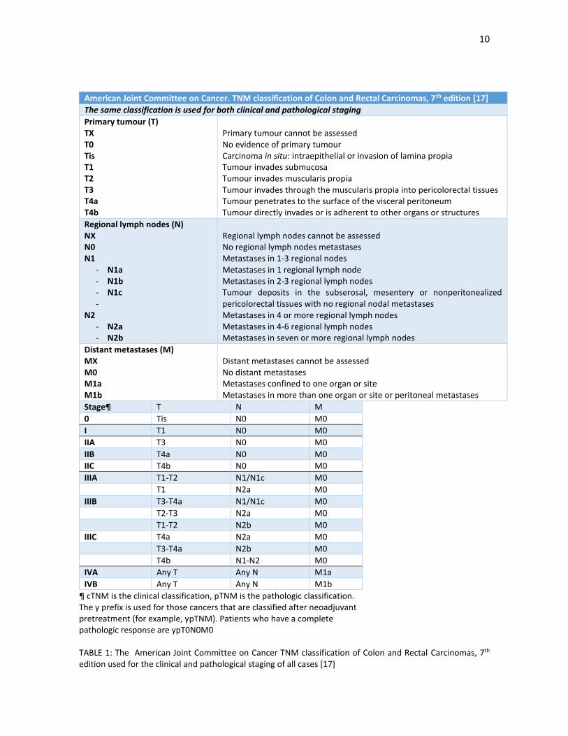
or cT4 or any cT with positive N. Clinical and pathological staging was done according to the American Joint
Committee on Cancer, TNM classification of Colon and Rectal Carcinomas, 7th edition (TABLE 1) [17]. All
patients had an Eastern Cooperative Oncology Group performance status (PS) less than 2, adequate renal and
hepatic function, and adequate bone marrow reserve (white blood cell count > 4000/mm3, haemoglobin >10
g/dl and platelet count >150,000/mm3). For each patient, we calculated the neutrophil-lymphocyte ratio
(RNL) on the 7 days previous to the start of neoadjuvant therapy and on the 7 days previous to the surgical
procedure. Patients with distant metastases were excluded. Staging evaluation included a complete
colonoscopy, a thoracic-abdomen-pelvis computed tomography (CT) scan, a serum carcinoembryonic antigen
(CEA) level and a serum carbohydrate antigen 19-9 (CA 19.9) level. All patients were discussed at our weekly
multidisciplinary colon cancer committee.
2.1. Primary tumour staging
Primary tumour staging could be performed with an EUS and/or an MRI. cT and CN stage was collected in all
patients; where it was performed, invasion of the levator muscle and the internal sphincter was also analyzed.
EUS was the preferred staging method until 2004, when MRI was introduced in our Hospital and it became
our standard imaging method of choice, although EUS is still routinely used in low rectal cancers. Specific MRI
features such as mesorectal fascia invasion, extramural venous invasion, depth of invasion in T3 tumours as
described by the MERCURY group (“a” (< 1 mm outside the wall), “b” (1–5 mm), “c” (5–15 mm), and “d” (> 15
mm)) and lateral pelvic lymph nodes were also analysed.
2.2. Chemotherapy administration
Preoperative treatment was administered on an outpatient basis and scheduled for 5 weeks. The initial 107
patients received UFT at a dose of 400 mg/m2 in three fractions per day between meals during the days of RT
administration (Monday to Friday, with the weekend as a rest period). Because the packets of UFT contained
100 mg of tegafur, the administered dose was rounded up when the fraction was >50 mg. From November
2007, due to the removal of UFT from the Spanish market, capecitabine was given alongside RT at a dose of
825 mg/m2 every 12 h during the days of RT administration (Monday to Friday, with the weekend as a rest
period).
Oxaliplatin was given as a 2-h intravenous infusion at a dose of 85 mg/m2 every two weeks during RT in all
but the first 32 patients from October 2001 and until August 2011 (131 patients in total), moment when we
decided to remove oxaliplatin from the regimen, when the initial results of several phase III trials showed that
oxaliplatin was not effective in this setting [18-21]. Thus, from that moment onwards and till the present time,
patients were treated with capecitabine monotherapy alongside radiotherapy (56 patients have been treated
in this manner)
10
American Joint Committee on Cancer. TNM classification of Colon and Rectal Carcinomas, 7th edition [17]
The same classification is used for both clinical and pathological staging
Primary tumour (T) TX T0 Tis T1 T2 T3 T4a T4b
Primary tumour cannot be assessed No evidence of primary tumour Carcinoma in situ: intraepithelial or invasion of lamina propia Tumour invades submucosa Tumour invades muscularis propia Tumour invades through the muscularis propia into pericolorectal tissues Tumour penetrates to the surface of the visceral peritoneum Tumour directly invades or is adherent to other organs or structures
Regional lymph nodes (N) NX N0 N1
- N1a - N1b - N1c -
N2 - N2a - N2b
Regional lymph nodes cannot be assessed No regional lymph nodes metastases Metastases in 1-3 regional nodes Metastases in 1 regional lymph node Metastases in 2-3 regional lymph nodes Tumour deposits in the subserosal, mesentery or nonperitonealized pericolorectal tissues with no regional nodal metastases Metastases in 4 or more regional lymph nodes Metastases in 4-6 regional lymph nodes Metastases in seven or more regional lymph nodes
Distant metastases (M) MX M0 M1a M1b
Distant metastases cannot be assessed No distant metastases Metastases confined to one organ or site Metastases in more than one organ or site or peritoneal metastases
Stage¶ T N M
0 Tis N0 M0
I T1 N0 M0 IIA T3 N0 M0 IIB T4a N0 M0 IIC T4b N0 M0 IIIA T1-T2 N1/N1c M0 T1 N2a M0 IIIB T3-T4a N1/N1c M0 T2-T3 N2a M0 T1-T2 N2b M0 IIIC T4a N2a M0 T3-T4a N2b M0 T4b N1-N2 M0 IVA Any T Any N M1a
IVB Any T Any N M1b
¶ cTNM is the clinical classification, pTNM is the pathologic classification. The y prefix is used for those cancers that are classified after neoadjuvant pretreatment (for example, ypTNM). Patients who have a complete pathologic response are ypT0N0M0 TABLE 1: The American Joint Committee on Cancer TNM classification of Colon and Rectal Carcinomas, 7th edition used for the clinical and pathological staging of all cases [17]
11
2.3 Radiotherapy administration
A pelvic plane CT in the treatment position for virtual simulation was performed on all patients. CT images
were obtained using a 5 mm slice thickness at 1 cm separation (5 mm slices through the tumour), from L5-S1
to 1.5 cm distal to the anus. The planning target volume (PTV) was defined as clinical target volume (CTV)1+
1 cm margin: (1) diagnostic imaging (CT) was used to define gross target volume (GTV); (2) CTV1, including the
GTV+2 cm in all directions, perirectal, internal iliac and presacral nodes up to the promontory; for T4 (vesicle
involvement, prostate, vagina or uterus), external iliac nodes were also included. The inguinal areas were
irradiated in those patients who had invasion of the anal canal. Treatment was delivered through three to four
fields via the axial beam technique, shaped with multi-leaf collimator and high-energy photons of 10 MV (this
being possible using photons of 6 MV in the PA).
The total dose administered was 45 Gy with conventional fractions of 1.8 Gy/day five times per week in the
first 94 patients. In the following 109 patients, the total dose was increased to 50.4 Gy with a prespecified
tumour area boost of 5.4 Gy, after the initial results of the phase III ACCORD-12/0405-Prodige-2 and other
trials [21]. which showed better local control with the higher dose. The prescribed dose was specified at the
International Commission on Radiation Units and Measurements point (intersection of the central axes of the
3–4 beams) and isodose distribution to the PTV (95–105%).
Toxicity of the combination treatment was evaluated weekly in each patient. A complete blood count was
obtained; toxicity scoring was performed using the Common Terminology Criteria for Adverse Events, Version
4.0. No restaging procedure was done routinely after the end of chemo-radiotherapy, except if there was a
suspicion of progressive disease or other unexpected findings.
2.4. Surgery
Patients were scheduled for surgery between the sixth and eighth week following the conclusion of the
combination therapy and were treated with TME. Relevant surgical end-points were collected. Of these, the
definition of the resection status took into account both the clinical intraoperative judgment and the
pathologic results [22-24]. With regards to the first factor, the surgeon was to report at the end of operation
if this was considered curative and if the TME was deemed successful, based on the absence of gross residual
distant metastases or loco-regional tumour, otherwise the resection was considered non-curative (R2). With
regards to the second factor, R1 was defined in potentially curative cases as microscopic tumour at or less
than 1mm from the cut surgical margin, and R0 where microscopic tumour was greater than 1mm away from
the cut surgical margin.
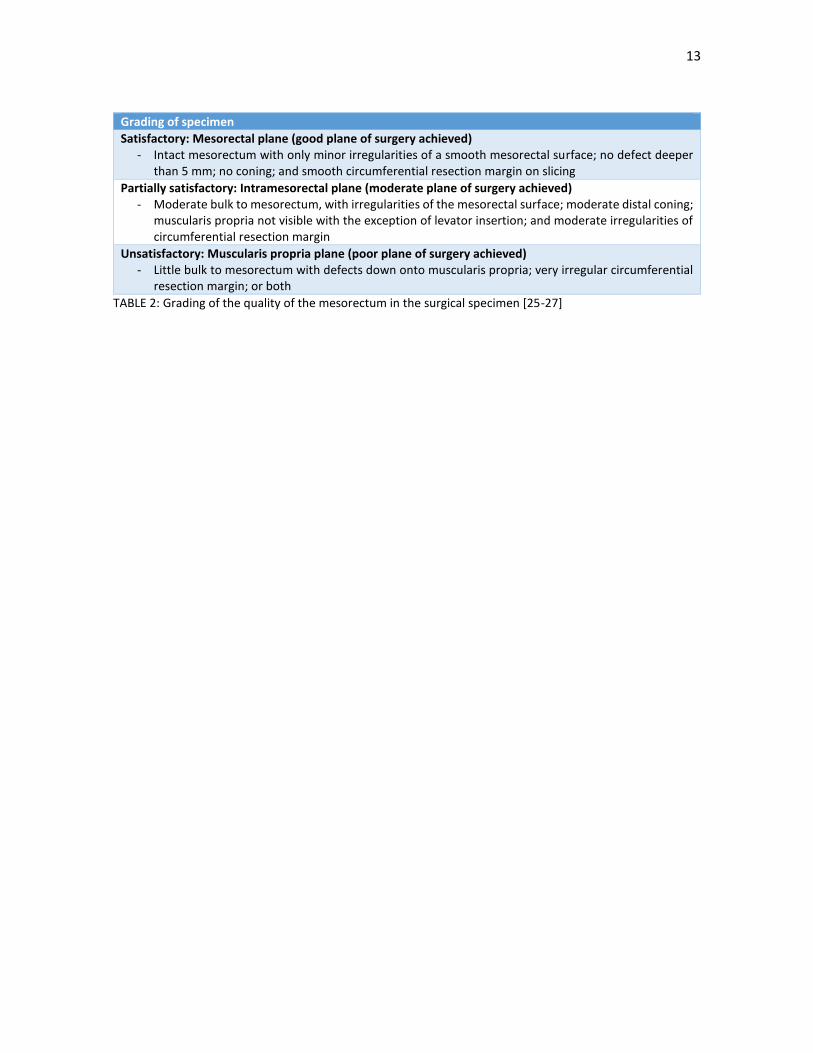
3.5. Pathological analysis
The analysis of the quality of the mesorectum was performed according to previously defined criteria as
satisfactory, partially satisfactory and unsatisfactory [25-27]. The criteria are shown in TABLE 2. Pathological
staging was done according to the American Joint Committee on Cancer, TNM classification of Colon and
Rectal Carcinomas, 7th edition [17]. Analysis of the response to preoperative treatment was defined clinically
as well as pathologically. Downstaging was considered when pathologic T (pT) or N (pN) was less than
ultrasound or MRI-defined T (cT) or N (cN). No response was considered when pT and cT were similar. Disease
12
progression was when pT was more than cT, when pN was more than cN or when metastases were observed
during surgery. We also analysed the different rates of downstaging according to the imaging criteria used
(MRI or EUS) in case both were performed in the same case.
As stated previously, R1 was defined in potentially curative cases as microscopic tumor at or less than 1mm
from the cut surgical margin; we differentiated between the circumferential margin invasion (CRM) and the
distal margin invasion accordingly [23-24].
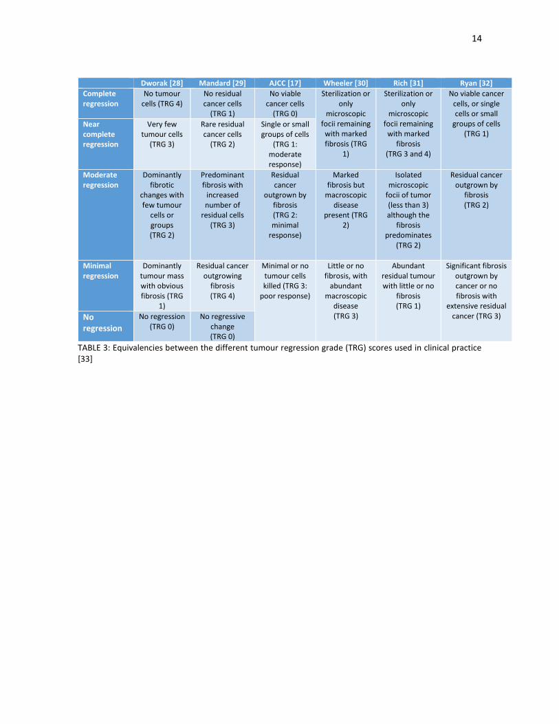
Due to the heterogeneity of the tumour regression grades scores used in these 15 years of follow-up [17, 28-
32] (at our institution, the Dworak, Mandard, Rich and Wheeler scores were most frequently used), we
decided to unify these scores in common groups: tumour with complete response, tumours with nearly-
complete response, tumours with moderate regression and tumour with minimal or no regression. The
equivalencies are shown in TABLE 3 [33]. If a tumour had differing scores, we chose the score with a worse
prognosis and allocated the patient accordingly. A pCR was considered when no malignant cells were
observed, when the sample contained nonviable cells or only large acellular pools of mucin. Surprisingly no
mention is made of the lymph node status in these classifications; however, we also collected the data of
patients with ypT0 N-positive disease. Nonviable cells were those that showed pyknosis, karyorrhexis,
karyolysis, cytolysis, or extreme distortion and hyperchromasia of the nucleus. Other pathological signs of
interest collected include the presence of perineural invasion, lymphatic or vascular embolization and
infrequent types of rectal adenocarcinoma (mucinous and signe-cell carcinomas). RAS analysis was not done
routinely except in cases where it was necessary for the beginning of advanced-disease treatment.
13
Grading of specimen
Satisfactory: Mesorectal plane (good plane of surgery achieved) - Intact mesorectum with only minor irregularities of a smooth mesorectal surface; no defect deeper
than 5 mm; no coning; and smooth circumferential resection margin on slicing
Partially satisfactory: Intramesorectal plane (moderate plane of surgery achieved) - Moderate bulk to mesorectum, with irregularities of the mesorectal surface; moderate distal coning;
muscularis propria not visible with the exception of levator insertion; and moderate irregularities of circumferential resection margin
Unsatisfactory: Muscularis propria plane (poor plane of surgery achieved) - Little bulk to mesorectum with defects down onto muscularis propria; very irregular circumferential
resection margin; or both
TABLE 2: Grading of the quality of the mesorectum in the surgical specimen [25-27]
14
Dworak [28] Mandard [29] AJCC [17] Wheeler [30] Rich [31] Ryan [32]
Complete regression
No tumour cells (TRG 4)
No residual cancer cells
(TRG 1)
No viable cancer cells
(TRG 0)
Sterilization or only
microscopic focii remaining
with marked fibrosis (TRG
1)
Sterilization or only
microscopic focii remaining
with marked fibrosis
(TRG 3 and 4)
No viable cancer cells, or single cells or small
groups of cells (TRG 1)
Near complete regression
Very few tumour cells
(TRG 3)
Rare residual cancer cells
(TRG 2)
Single or small groups of cells
(TRG 1: moderate response)
Moderate regression
Dominantly fibrotic
changes with few tumour
cells or groups (TRG 2)
Predominant fibrosis with
increased number of
residual cells (TRG 3)
Residual cancer
outgrown by fibrosis (TRG 2: minimal
response)
Marked fibrosis but
macroscopic disease
present (TRG 2)
Isolated microscopic
focii of tumor (less than 3) although the
fibrosis predominates
(TRG 2)
Residual cancer outgrown by
fibrosis (TRG 2)
Minimal regression
Dominantly tumour mass with obvious fibrosis (TRG
1)
Residual cancer outgrowing
fibrosis (TRG 4)
Minimal or no tumour cells killed (TRG 3:
poor response)
Little or no fibrosis, with
abundant macroscopic
disease (TRG 3)
Abundant residual tumour with little or no
fibrosis (TRG 1)
Significant fibrosis outgrown by cancer or no fibrosis with
extensive residual cancer (TRG 3) No
regression
No regression (TRG 0)
No regressive change (TRG 0)
TABLE 3: Equivalencies between the different tumour regression grade (TRG) scores used in clinical practice [33]
15
2.6. Adjuvant chemotherapy
Following surgery, patients treated with UFT and UFT-oxaliplatin received four cycles of FU (425 mg/m2) and
LV (20 mg/m2) on days 1–5. This scheme was repeated every 28 days. Patients treated with capecitabine-
oxaliplatin or capecitabine monotherapy received four cycles of adjuvant capecitabine (1000 mg/m2 every 12
h for 14 days) and oxaliplatin (130 mg/m2) every 21 days. There was not fixed date for the beginning of
treatment, although treatment was usually begun on the fourth to sixth week after surgery. No adjuvant
chemotherapy was started twelve weeks after the surgical procedure.
2.7. Follow-up
Following the conclusion of treatment, patients had outpatient clinic appointments every 3 months for the
first 2 years, at which time chest X-ray, abdominal ultrasound, CT scans of the thorax, abdomen and pelvis,
and a blood analysis, including CEA, were performed. Between the third and fifth years, the appointments
were every 6 months. A complete physical examination was conducted at each clinical visit, as was CEA
measurement. CT scans were done in an alternating fashion with chest X-rays and abdominal ultrasound.
Follow-up colonoscopies were done at the end of adjuvant chemotherapy (if the colonoscopy at diagnosis was
not complete), one year after diagnosis, five years after diagnosis and every five years from then on. These
intervals were shorter if any abnormalities appeared, such as advanced polyps.
2.8. Aims of the study
Our aim is to report clinical, radiological and pathological prognostic factors for local relapse, distant
metastases and long-term survival endpoints (DFS and OS) in patients treated with neoadjuvant
chemoradiotherapy for locally advanced rectal cancer. We also analysed if our study population would fit the
Valentini nomogram [34] and the Dhadda score [35], two prognostic scores in patients with locally advanced
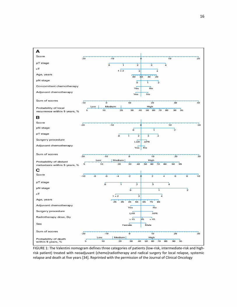
rectal cancer, both with differing elements included. The Valentini nomogram (FIGURE 1) defines three
categories of patients (low-risk, intermediate-risk and high-risk patient) treated with neoadjuvant
(chemo)radiotherapy and radical surgery for local relapse, systemic relapse and death at five years and
includes items such as pT, pN, cT, type of surgery (abdominoperineal resection versus low anterior resection),
age, sex, radiotherapy dose and use of concomitant and adjuvant chemotherapy.
On the other hand, the Dhadda score defines four groups of patients (excellent, good, moderate and poor
prognosis) treated also with neoadjuvant (chemo)radiotherapy and radical surgery for local relapse, DFS and
OS and includes four items: pN, perineural invasion, invasion of the circumferential margin and the tumour
regression grade. A score for each factor was calculated: TRG 1 = 0, TRG 2 =1, TRG 3-5 = 2; 0 nodes positive =
0, 1-3 nodes positive = 2, 4 or more nodes positive = 4; perineural invasion absent = 0, perineural invasion
present = 4; CRM clear = 0, CRM involved = 4). A final score was thereafter calculated for each individual
patient with a higher value indicative of a worse prognosis. This allowed a value between 0 and 14, with four
groups defined: excellent prognosis group (score 0), good prognosis group (score 1-3), moderate prognosis
group (score 4-8), poor prognosis group (score 9-14).
16
FIGURE 1: The Valentini nomogram defines three categories of patients (low-risk, intermediate-risk and high-risk patient) treated with neoadjuvant (chemo)radiotherapy and radical surgery for local relapse, systemic relapse and death at five years [34]. Reprinted with the permission of the Journal of Clinical Oncology
17
2.9. Statistical analysis
Local relapse was defined as the presence of any anastomotic, pelvic, or perineal tumor documented by
proctoscopic, radiologic, or histopathologic examination. Distant relapse was defined as evidence of recurrent
disease in any other location. Calculation of local relapse rates included patients who developed local relapse
only and patients who developed both local and distant recurrence. DFS was defined as the time from
diagnosis to the date of cancer relapse or death by any cause. OS was defined as the time from diagnosis to
the date of death by any cause. Cancer-specific survival (CSS) was defined as the time from diagnosis to the
date of relapse or death by cancer-related deaths; patients with other causes of death were not included in
the analysis.
Univariate analysis was performed using 2-tailed chi-square test or Fisher’s exact test for categorical variables
and Mann–Whitney test for numerical variables. Multivariate analyses were performed using a Cox
proportional hazards regression model. Time to local and overall relapses (cumulative risk at certain time)
were estimated using the Kaplan–Meier method and comparison was done using log rank test. Univariate
analysis of DFS and OS were calculated using the Kaplan-Meier procedure-limit method and reported as
median survival and estimated 5-year survival. Multivariate analysis was performed using the Cox proportional
hazards regression method. For each variant, hazard ratio was calculated including 95% confidence intervals.
All tests were 2-tailed and statistical significance was set at p < 0.05. The Statistical Package for the Social
Sciences (SPSS version 23; Chicago, Ill, USA) was used for data management and statistical analyses. The
analysis was performed on July 2016.
18
3. Results
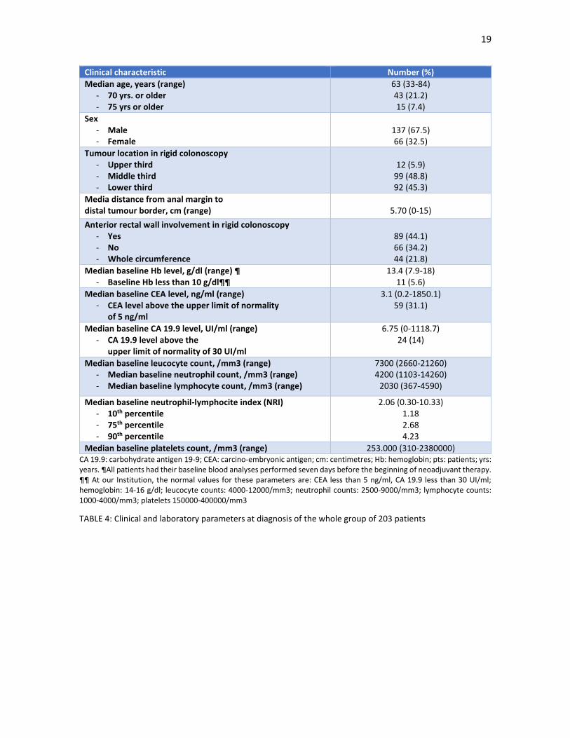
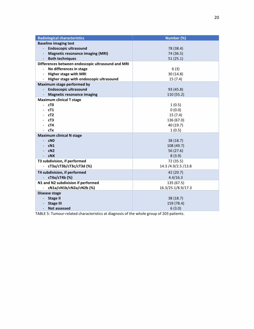
3.1. Baseline characteristics
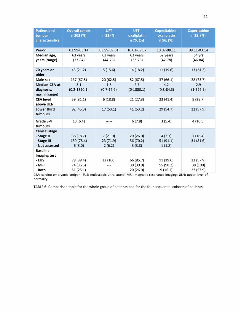
A total of 203 patients were included in a prospective fashion in this treatment regimen between March 1999
and March 2014. Four sequential cohorts of patients according to the oral fluoropyrimidine given and to the
use of oxaliplatin or not were identified. Clinical and tumour-related characteristics of all patients and for each
of these cohorts are shown in TABLES 4, 5 and 6, respectively. Baseline characteristics were similar for the
four subgroups, except for a non-statistically increase of stage III patients and patients with a higher CEA level
(p 0.056 and p 0.088, respectively) in the third cohort, compared to the earlier groups.
The most frequent clinical stage was cT3N1 (69 patients, 34.0%), followed by cT3N2 (35 patients, 17.2%),
cT4N1 (20 patients, 9.8%) and cT4N2 (14 patients, 6.9%). Clinical lymph node involvement was seen in 159
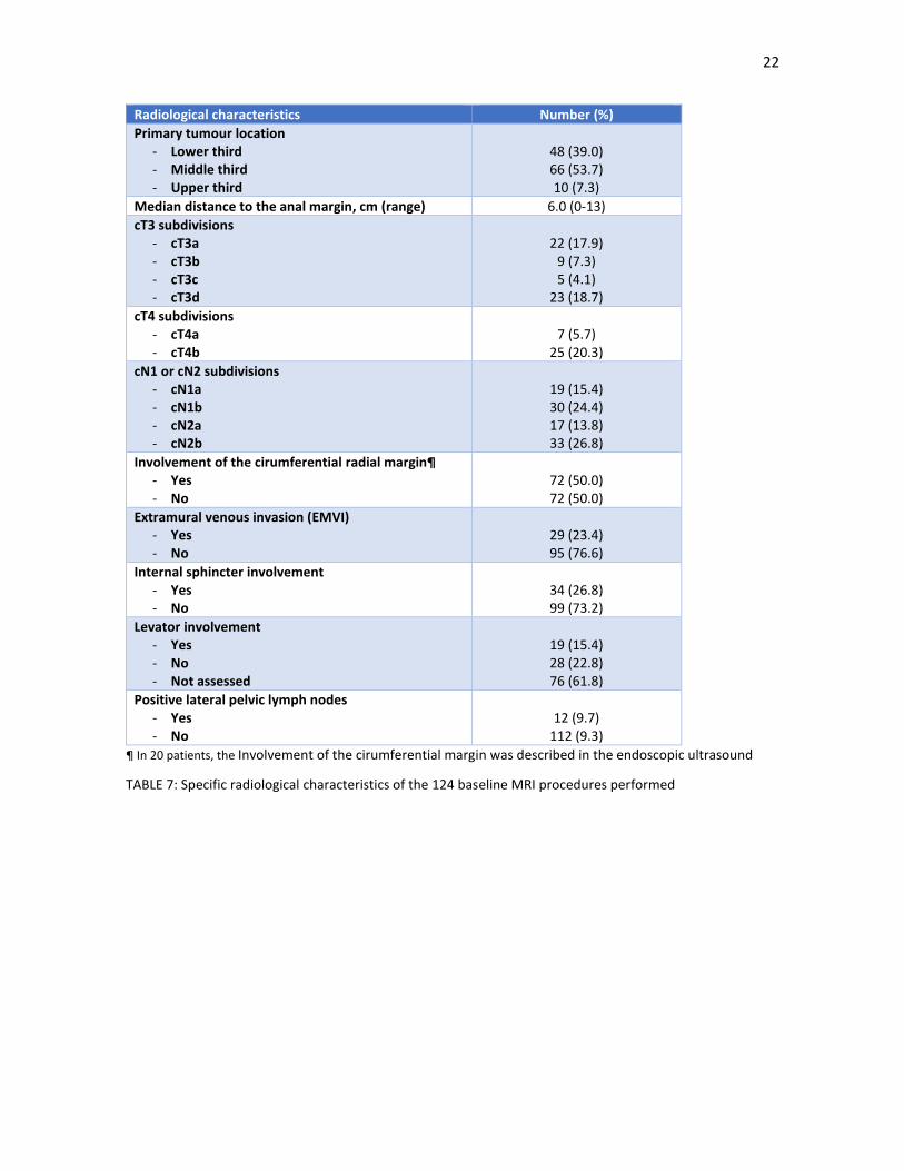
patients (78.4%). More than a half of patients (55.2%) were staged with MRI; specific radiological
characteristics of the 124 MRI procedures done are shown in TABLE 7. Of note, in a half of patients staged
with MRI there were findings suggestive of involvement of the circumferential radial margin and in 12 patients
(9.2%) there were positive lateral pelvic lymph nodes (9.7%). In 20 patients, the Involvement of the
cirumferential margin was described in the endoscopic ultrasound
19
Clinical characteristic Number (%)
Median age, years (range) - 70 yrs. or older - 75 yrs or older
63 (33-84) 43 (21.2) 15 (7.4)
Sex - Male - Female
137 (67.5) 66 (32.5)
Tumour location in rigid colonoscopy - Upper third - Middle third - Lower third
12 (5.9)
99 (48.8) 92 (45.3)
Media distance from anal margin to distal tumour border, cm (range)
5.70 (0-15)
Anterior rectal wall involvement in rigid colonoscopy - Yes - No - Whole circumference
89 (44.1) 66 (34.2) 44 (21.8)
Median baseline Hb level, g/dl (range) ¶ - Baseline Hb less than 10 g/dl¶¶
13.4 (7.9-18) 11 (5.6)
Median baseline CEA level, ng/ml (range) - CEA level above the upper limit of normality
of 5 ng/ml
3.1 (0.2-1850.1) 59 (31.1)
Median baseline CA 19.9 level, UI/ml (range) - CA 19.9 level above the
upper limit of normality of 30 UI/ml
6.75 (0-1118.7) 24 (14)
Median baseline leucocyte count, /mm3 (range) - Median baseline neutrophil count, /mm3 (range) - Median baseline lymphocyte count, /mm3 (range)
7300 (2660-21260) 4200 (1103-14260)
2030 (367-4590)
Median baseline neutrophil-lymphocite index (NRI) - 10th percentile - 75th percentile - 90th percentile
2.06 (0.30-10.33) 1.18 2.68 4.23
Median baseline platelets count, /mm3 (range) 253.000 (310-2380000) CA 19.9: carbohydrate antigen 19-9; CEA: carcino-embryonic antigen; cm: centimetres; Hb: hemoglobin; pts: patients; yrs: years. ¶All patients had their baseline blood analyses performed seven days before the beginning of neoadjuvant therapy. ¶¶ At our Institution, the normal values for these parameters are: CEA less than 5 ng/ml, CA 19.9 less than 30 UI/ml; hemoglobin: 14-16 g/dl; leucocyte counts: 4000-12000/mm3; neutrophil counts: 2500-9000/mm3; lymphocyte counts: 1000-4000/mm3; platelets 150000-400000/mm3
TABLE 4: Clinical and laboratory parameters at diagnosis of the whole group of 203 patients
20
Radiological characteristics Number (%)
Baseline imaging test - Endoscopic ultrasound - Magnetic resonance imaging (MRI) - Both techniques
78 (38.4) 74 (36.5) 51 (25.1)
Differences between endoscopic ultrasound and MRI - No differences in stage - Higher stage with MRI - Higher stage with endoscopic ultrasound
6 (3)
30 (14.8) 15 (7.4)
Maximum stage performed by - Endoscopic ultrasound - Magnetic resonance imaging
93 (45.8)
110 (55.2)
Maximum clinical T stage - cT0 - cT1 - cT2 - cT3 - cT4 - cTx
1 (0.5) 0 (0.0)
15 (7.4) 136 (67.0) 40 (19.7)
1 (0.5)
Maximum clinical N stage - cN0 - cN1 - cN2 - cNX
38 (18.7)
108 (49.7) 56 (27.6)
8 (3.9)
T3 subdivision, if performed - cT3a/cT3b/cT3c/cT3d (%)
72 (35.5) 14.3 /4.9/2.5 /13.8
T4 subdivision, if performed - cT4a/cT4b (%)
42 (20.7) 4.4/16.3
N1 and N2 subdivision if performed - cN1a/cN1b/cN2a/cN2b (%)
135 (67.5) 16.3/25.1/8.9/17.3
Disease stage - Stage II - Stage III - Not assessed
38 (18.7)
159 (78.4) 6 (3.0)
TABLE 5: Tumour-related characteristics at diagnosis of the whole group of 203 patients.
21
Patient and tumour characteristics
Overall cohort n 203 (%)
UFT n 32 (%)
UFT-oxaliplatin n 75, (%)
Capecitabine-oxaliplatin n 56, (%)
Capecitabine n 38, (%)
Period 03.99-03.14 03.99-09.01 10.01-09.07 10.07-08.11 09.11-03.14
Median age, years (range)
63 years (33-84)
63 years (44-76)
63 years (33-76)
62 years (42-78)
64 yrs (46-84)
70 years or older
43 (21.2)
5 (15.6) 14 (18.2) 11 (19.6) 13 (34.2)
Male sex 137 (67.5) 20 (62.5) 52 (67.5) 37 (66.1) 28 (73.7)
Median CEA at diagnosis, ng/ml (range)
3.1 (0.2-1850.1)
1.8 (0.7-17.6)
2.7 (0-1850.1)
4.2 (0.8-84.3)
2.9 (1-326.9)
CEA level above ULN
59 (31.1) 6 (18.8) 21 (27.3) 23 (41.4) 9 (25.7)
Lower third tumours
92 (45.3) 17 (53.1) 41 (53.2) 29 (54.7) 22 (57.9)
Grade 3-4 tumours
13 (6.4) ----- 6 (7.8) 3 (5.4) 4 (10.5)
Clinical stage - Stage II - Stage III - Not assessed
38 (18.7)
159 (78.4) 6 (3.0)
7 (21.9)
23 (71.9) 2 (6.2)
20 (26.0) 56 (70.2)
3 (3.8)
4 (7.1)
51 (91.1) 1 (1.8)
7 (18.4)
31 (81.6) ------
Baseline imaging test - EUS - MRI - Both
78 (38.4) 74 (36.5) 51 (25.1)
32 (100) --- ---
66 (85.7) 30 (39.0) 20 (26.0)
11 (19.6) 55 (98.2) 9 (16.1)
22 (57.9) 38 (100) 22 (57.9)
CEA: carcino-embryonic antigen; EUS: endoscopic ultra-sound; MRI: magnetic resonance imaging; ULN: upper level of normality
TABLE 6: Comparison table for the whole group of patients and for the four sequential cohorts of patients
22
Radiological characteristics Number (%)
Primary tumour location - Lower third - Middle third - Upper third
48 (39.0) 66 (53.7) 10 (7.3)
Median distance to the anal margin, cm (range) 6.0 (0-13)
cT3 subdivisions - cT3a - cT3b - cT3c - cT3d
22 (17.9)
9 (7.3) 5 (4.1)
23 (18.7)
cT4 subdivisions - cT4a - cT4b
7 (5.7)
25 (20.3)
cN1 or cN2 subdivisions - cN1a - cN1b - cN2a - cN2b
19 (15.4) 30 (24.4) 17 (13.8) 33 (26.8)
Involvement of the cirumferential radial margin¶ - Yes - No
72 (50.0) 72 (50.0)
Extramural venous invasion (EMVI) - Yes - No
29 (23.4) 95 (76.6)
Internal sphincter involvement - Yes - No
34 (26.8) 99 (73.2)
Levator involvement - Yes - No - Not assessed
19 (15.4) 28 (22.8) 76 (61.8)
Positive lateral pelvic lymph nodes - Yes - No
12 (9.7)
112 (9.3)
¶ In 20 patients, the Involvement of the cirumferential margin was described in the endoscopic ultrasound
TABLE 7: Specific radiological characteristics of the 124 baseline MRI procedures performed
23
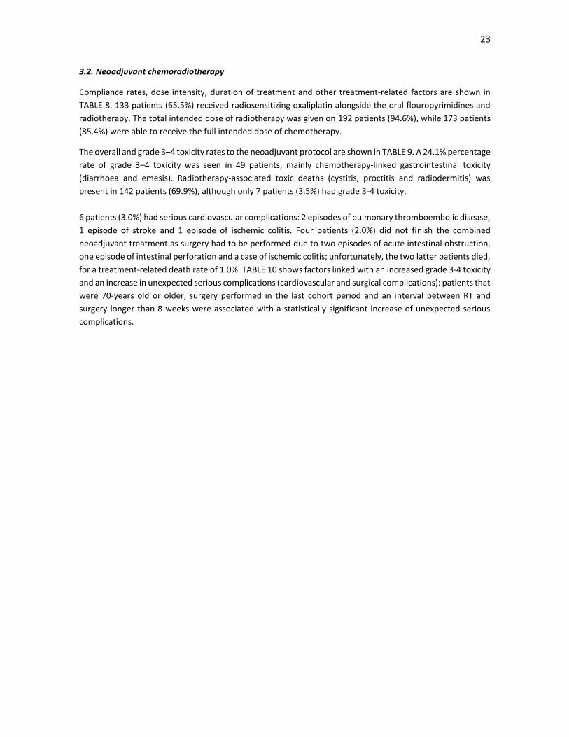
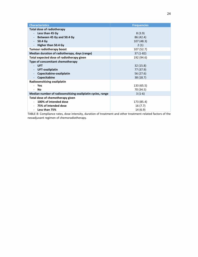
3.2. Neoadjuvant chemoradiotherapy
Compliance rates, dose intensity, duration of treatment and other treatment-related factors are shown in
TABLE 8. 133 patients (65.5%) received radiosensitizing oxaliplatin alongside the oral flouropyrimidines and
radiotherapy. The total intended dose of radiotherapy was given on 192 patients (94.6%), while 173 patients
(85.4%) were able to receive the full intended dose of chemotherapy.
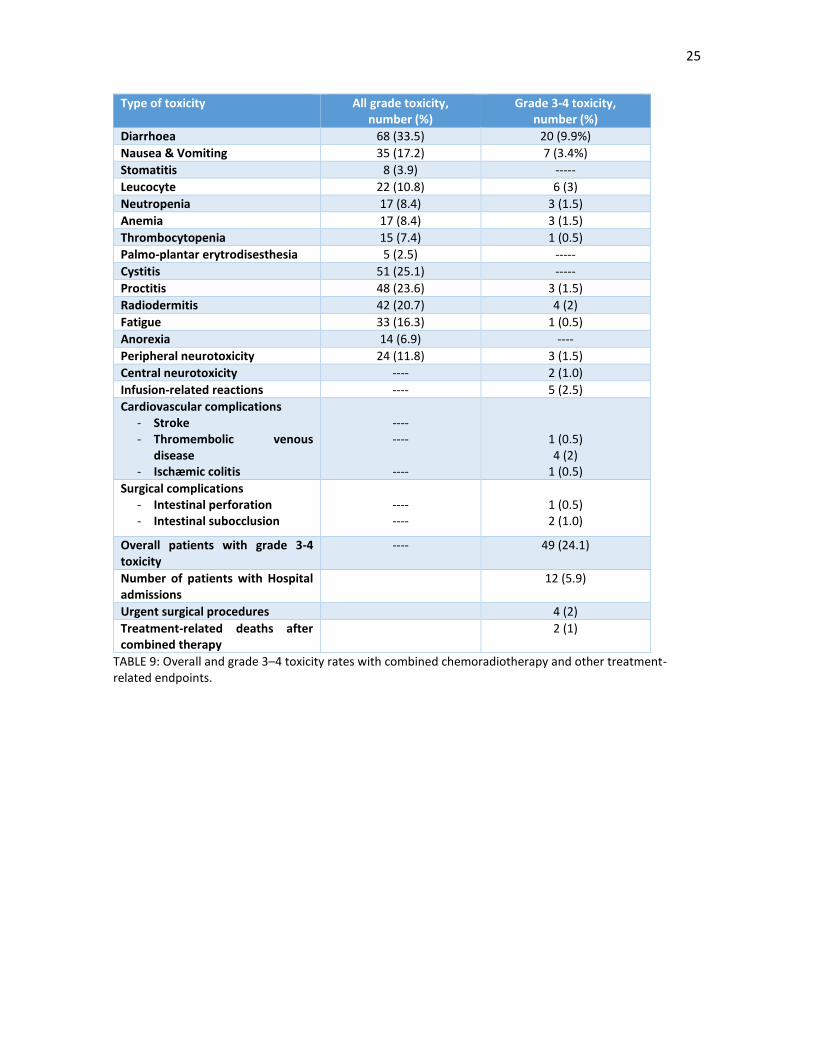
The overall and grade 3–4 toxicity rates to the neoadjuvant protocol are shown in TABLE 9. A 24.1% percentage
rate of grade 3–4 toxicity was seen in 49 patients, mainly chemotherapy-linked gastrointestinal toxicity
(diarrhoea and emesis). Radiotherapy-associated toxic deaths (cystitis, proctitis and radiodermitis) was
present in 142 patients (69.9%), although only 7 patients (3.5%) had grade 3-4 toxicity.
6 patients (3.0%) had serious cardiovascular complications: 2 episodes of pulmonary thromboembolic disease,
1 episode of stroke and 1 episode of ischemic colitis. Four patients (2.0%) did not finish the combined
neoadjuvant treatment as surgery had to be performed due to two episodes of acute intestinal obstruction,
one episode of intestinal perforation and a case of ischemic colitis; unfortunately, the two latter patients died,
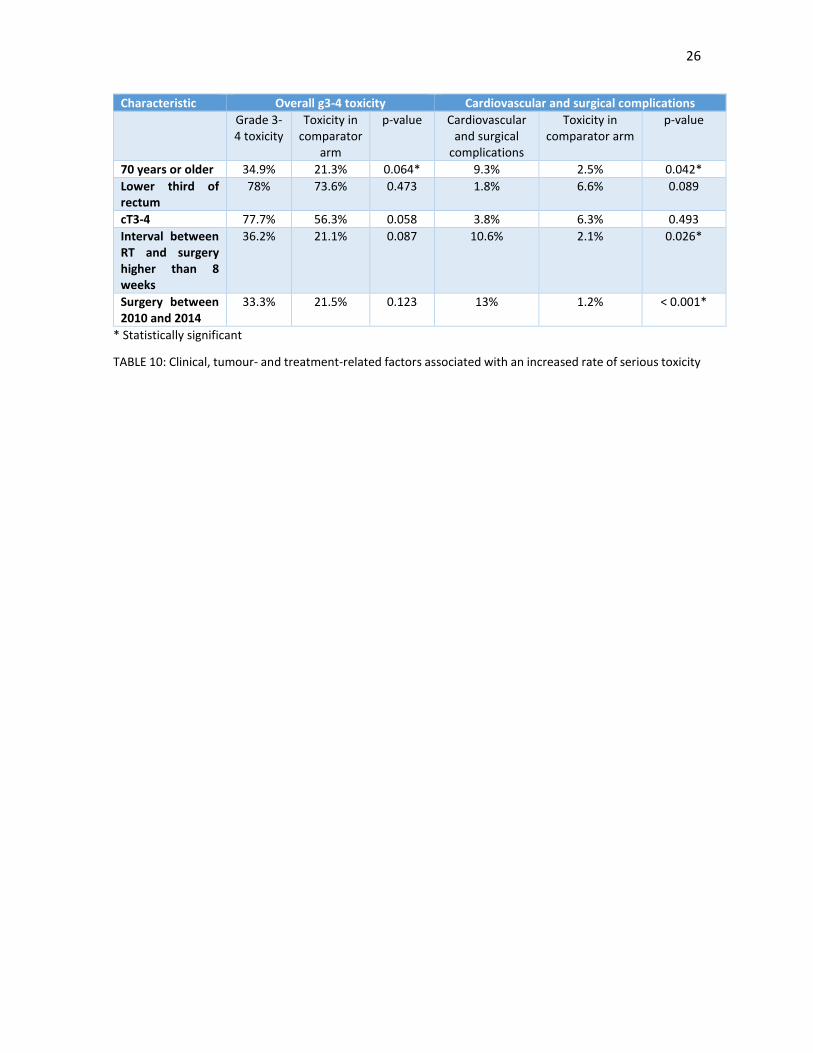
for a treatment-related death rate of 1.0%. TABLE 10 shows factors linked with an increased grade 3-4 toxicity
and an increase in unexpected serious complications (cardiovascular and surgical complications): patients that
were 70-years old or older, surgery performed in the last cohort period and an interval between RT and
surgery longer than 8 weeks were associated with a statistically significant increase of unexpected serious
complications.
24
Characteristics Frequencies
Total dose of radiotherapy - Less than 45 Gy - Between 45 Gy and 50.4 Gy - 50.4 Gy - Higher than 50.4 Gy
8 (3.9)
86 (42.4) 107 (48.3)
2 (1)
Tumour radiotherapy boost 107 (52.7)
Median duration of radiotherapy, days (range) 37 (1-82)
Total expected dose of radiotherapy given 192 (94.6)
Type of concomitant chemotherapy - UFT - UFT-oxaliplatin - Capecitabine-oxaliplatin - Capecitabine
32 (15.8) 77 (37.9) 56 (27.6) 38 (18.7)
Radiosensitizing oxaliplatin - Yes - No
133 (65.5) 70 (34.5)
Median number of radiosensitizing oxaliplatin cycles, range 3 (1-6)
Total dose of chemotherapy given - 100% of intended dose - 75% of intended dose - Less than 75%
173 (85.4)
16 (7.7) 14 (6.9)
TABLE 8: Compliance rates, dose intensity, duration of treatment and other treatment-related factors of the neoadjuvant regimen of chemoradiotherapy.
25
Type of toxicity All grade toxicity, number (%)
Grade 3-4 toxicity, number (%)
Diarrhoea 68 (33.5) 20 (9.9%)
Nausea & Vomiting 35 (17.2) 7 (3.4%)
Stomatitis 8 (3.9) -----
Leucocyte 22 (10.8) 6 (3)
Neutropenia 17 (8.4) 3 (1.5)
Anemia 17 (8.4) 3 (1.5)
Thrombocytopenia 15 (7.4) 1 (0.5)
Palmo-plantar erytrodisesthesia 5 (2.5) -----
Cystitis 51 (25.1) -----
Proctitis 48 (23.6) 3 (1.5)
Radiodermitis 42 (20.7) 4 (2)
Fatigue 33 (16.3) 1 (0.5)
Anorexia 14 (6.9) ----
Peripheral neurotoxicity 24 (11.8) 3 (1.5)
Central neurotoxicity ---- 2 (1.0)
Infusion-related reactions ---- 5 (2.5)
Cardiovascular complications - Stroke - Thromembolic venous
disease - Ischæmic colitis
---- ----
----
1 (0.5) 4 (2)
1 (0.5)
Surgical complications - Intestinal perforation - Intestinal subocclusion
---- ----
1 (0.5) 2 (1.0)
Overall patients with grade 3-4 toxicity
---- 49 (24.1)
Number of patients with Hospital admissions
12 (5.9)
Urgent surgical procedures 4 (2)
Treatment-related deaths after combined therapy
2 (1)
TABLE 9: Overall and grade 3–4 toxicity rates with combined chemoradiotherapy and other treatment-related endpoints.
26
Characteristic Overall g3-4 toxicity Cardiovascular and surgical complications
Grade 3-4 toxicity
Toxicity in comparator
arm
p-value Cardiovascular and surgical
complications
Toxicity in comparator arm
p-value
70 years or older 34.9% 21.3% 0.064* 9.3% 2.5% 0.042*
Lower third of rectum
78% 73.6% 0.473 1.8% 6.6% 0.089
cT3-4 77.7% 56.3% 0.058 3.8% 6.3% 0.493
Interval between RT and surgery higher than 8 weeks
36.2% 21.1% 0.087 10.6% 2.1% 0.026*
Surgery between 2010 and 2014
33.3% 21.5% 0.123 13% 1.2% < 0.001*
* Statistically significant
TABLE 10: Clinical, tumour- and treatment-related factors associated with an increased rate of serious toxicity
27
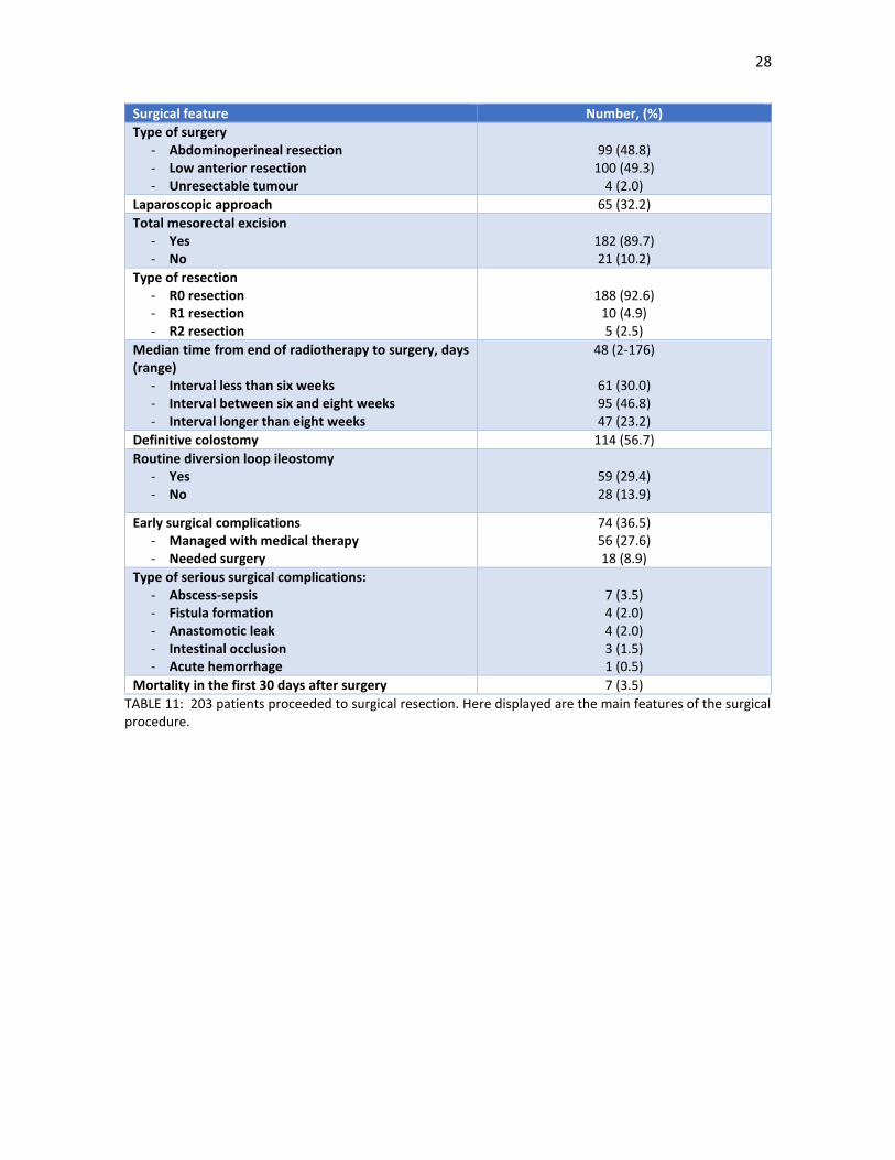
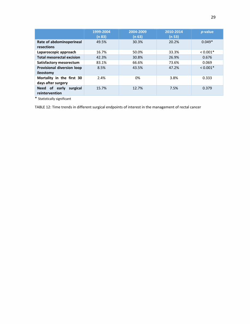
3.3. Surgery
202 patients (99.5%) proceeded to surgery. The main surgical endpoints are shown in TABLE 11. 4 patients
(2.0%) were deemed unresectable at the moment of surgery. Almost half of the remaining patients (99
patients, 48.8%) required an abdominoperineal resection. A total mesorectal excision was technically feasible
in 182 patients (89.7%) and an R0 resection was described by the surgeon in 188 patients (92.6%); in the
remaining patients, there were doubts about the completeness of the surgery in 10 patients (4.9%) and there
was macroscopical residual disease in five patients (2.5%). A provisional diversion loop ileostomy was
performed in 59 patients of the 100 patients with a lower anterior resection (59.0%). Early surgical
complications were seen in 74 patients (36.5%), although only in 18 patients (8.9%) a surgical reintervention
was needed. 7 patients (3.5%) died in the first 30 days after surgery.
In order to evaluate the surgical trends in the 15 years of the study, we performed an analysis of these surgical
endpoints in three approximate time periods of five years; the results are shown in TABLE 12. Compared to
earlier periods, the rate of abdominoperineal resections has decreased, while the laparoscopic approach and
the routine use of diversion loop ileostomies has increased.
28
Surgical feature Number, (%)
Type of surgery - Abdominoperineal resection - Low anterior resection - Unresectable tumour
99 (48.8)
100 (49.3) 4 (2.0)
Laparoscopic approach 65 (32.2)
Total mesorectal excision - Yes - No
182 (89.7) 21 (10.2)
Type of resection - R0 resection - R1 resection - R2 resection
188 (92.6)
10 (4.9) 5 (2.5)
Median time from end of radiotherapy to surgery, days (range)
- Interval less than six weeks - Interval between six and eight weeks - Interval longer than eight weeks
48 (2-176)
61 (30.0) 95 (46.8) 47 (23.2)
Definitive colostomy 114 (56.7)
Routine diversion loop ileostomy - Yes - No
59 (29.4) 28 (13.9)
Early surgical complications - Managed with medical therapy - Needed surgery
74 (36.5) 56 (27.6) 18 (8.9)
Type of serious surgical complications: - Abscess-sepsis - Fistula formation - Anastomotic leak - Intestinal occlusion - Acute hemorrhage
7 (3.5) 4 (2.0) 4 (2.0) 3 (1.5) 1 (0.5)
Mortality in the first 30 days after surgery 7 (3.5)
TABLE 11: 203 patients proceeded to surgical resection. Here displayed are the main features of the surgical procedure.
29
1999-2004 (n 83)
2004-2009 (n 63)
2010-2014 (n 53)
p-value
Rate of abdominoperineal resections
49.5% 30.3% 20.2% 0.049*
Laparoscopic approach 16.7% 50.0% 33.3% < 0.001*
Total mesorectal excision 42.3% 30.8% 26.9% 0.676
Satisfactory mesorectum 83.1% 66.6% 73.6% 0.069
Provisional diversion loop ileostomy
8.5% 43.5% 47.2% < 0.001*
Mortality in the first 30 days after surgery
2.4% 0% 3.8% 0.333
Need of early surgical reintervention
15.7% 12.7% 7.5% 0.379
* Statistically significant
TABLE 12: Time trends in different surgical endpoints of interest in the management of rectal cancer
30
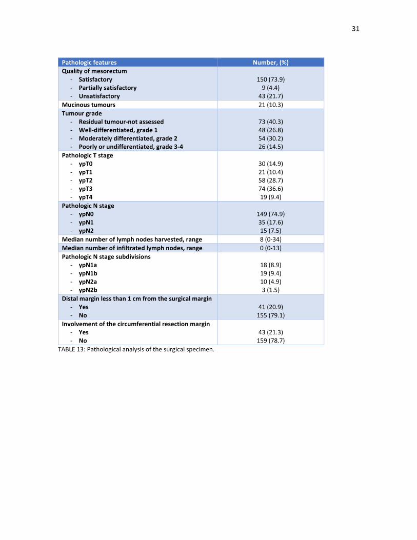
3.4. Pathology
The main endpoints of the pathological analysis of the surgical specimen are shown in TABLE 13. The quality
of the mesorectum was deemed satisfactory in 150 patients (73.9%), although involvement of the
circumferential radial margin was only seen in 43 patients (21.6%). 26 patients (14.5%) had poorly or
undifferentiated tumours. The most frequent ypT stage was ypT3 in 74 patients (36.6%), followed by ypT2 in
57 patients (28.7%); there were 30 cases (14.9%) with ypT0 (pathological complete response). There was no
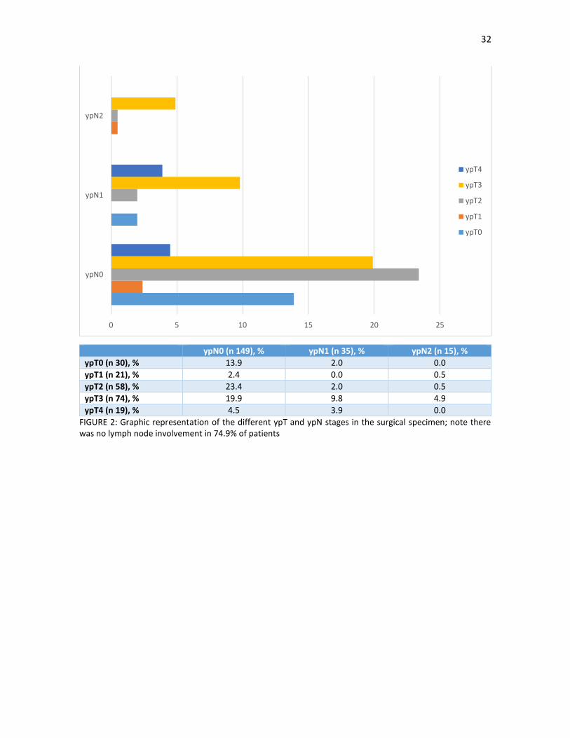
lymph node involvement in 149 patients (74.9%). FIGURE 2 is a graphic representation of these ypT and ypN
substages.
31
Pathologic features Number, (%)
Quality of mesorectum - Satisfactory - Partially satisfactory - Unsatisfactory
150 (73.9)
9 (4.4) 43 (21.7)
Mucinous tumours 21 (10.3)
Tumour grade - Residual tumour-not assessed - Well-differentiated, grade 1 - Moderately differentiated, grade 2 - Poorly or undifferentiated, grade 3-4
73 (40.3) 48 (26.8) 54 (30.2) 26 (14.5)
Pathologic T stage - ypT0 - ypT1 - ypT2 - ypT3 - ypT4
30 (14.9) 21 (10.4) 58 (28.7) 74 (36.6) 19 (9.4)
Pathologic N stage - ypN0 - ypN1 - ypN2
149 (74.9) 35 (17.6) 15 (7.5)
Median number of lymph nodes harvested, range 8 (0-34)
Median number of infiltrated lymph nodes, range 0 (0-13)
Pathologic N stage subdivisions - ypN1a - ypN1b - ypN2a - ypN2b
18 (8.9) 19 (9.4) 10 (4.9) 3 (1.5)
Distal margin less than 1 cm from the surgical margin - Yes - No
41 (20.9)
155 (79.1)
Involvement of the circumferential resection margin - Yes - No
43 (21.3)
159 (78.7)
TABLE 13: Pathological analysis of the surgical specimen.
32
ypN0 (n 149), % ypN1 (n 35), % ypN2 (n 15), %
ypT0 (n 30), % 13.9 2.0 0.0
ypT1 (n 21), % 2.4 0.0 0.5
ypT2 (n 58), % 23.4 2.0 0.5
ypT3 (n 74), % 19.9 9.8 4.9
ypT4 (n 19), % 4.5 3.9 0.0
FIGURE 2: Graphic representation of the different ypT and ypN stages in the surgical specimen; note there was no lymph node involvement in 74.9% of patients
0 5 10 15 20 25
ypN0
ypN1
ypN2
ypT4
ypT3
ypT2
ypT1
ypT0

33
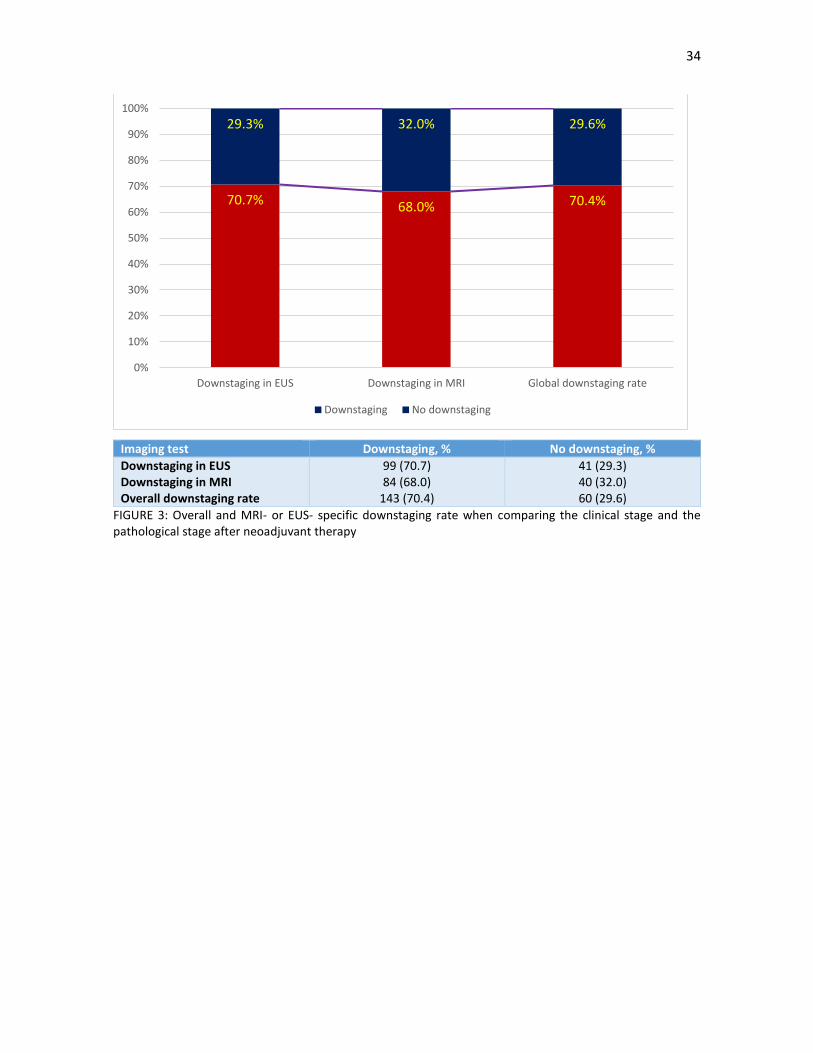
There was a 70.4% rate (143 patients) of downstaging compared to the clinical stage; the rate was broadly
similar for patients staged either with EUS and MRI (70.7% vs 68.0; FIGURE 3). This downstaging was driven
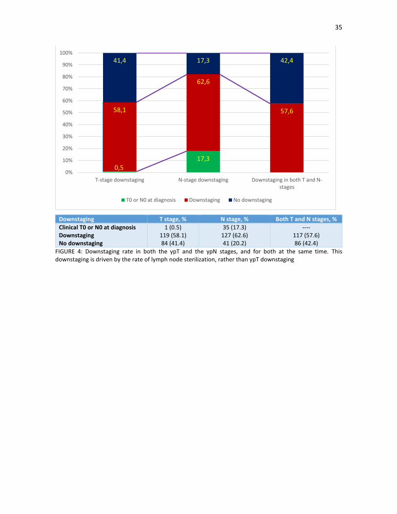
by the rate of lymph node sterilization, rather than T downstaging, as seen in FIGURE 4. The absolute numbers
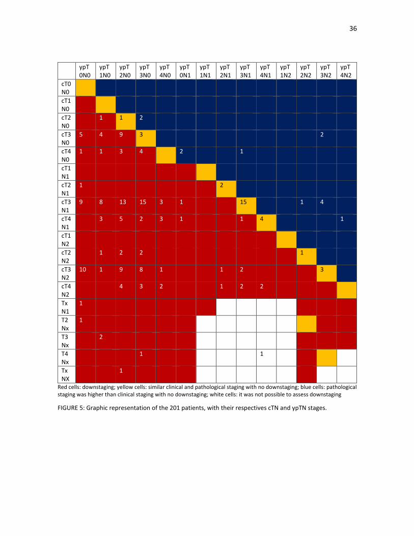
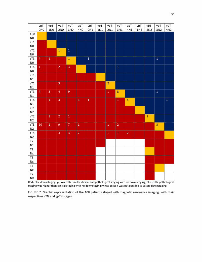
of patients, with their associated clinical and pathological staging, both in the overall group of patients and in
those patients staged either by EUS or MRI, are shown in FIGURE 5, 6 AND 7, respectively.
34
Imaging test Downstaging, % No downstaging, %
Downstaging in EUS Downstaging in MRI Overall downstaging rate
99 (70.7) 84 (68.0)
143 (70.4)
41 (29.3) 40 (32.0) 60 (29.6)
FIGURE 3: Overall and MRI- or EUS- specific downstaging rate when comparing the clinical stage and the pathological stage after neoadjuvant therapy
70.7% 68.0% 70.4%
29.3% 32.0% 29.6%
0%
10%
20%
30%
40%
50%
60%
70%
80%
90%
100%
Downstaging in EUS Downstaging in MRI Global downstaging rate
Downstaging No downstaging
35
Downstaging T stage, % N stage, % Both T and N stages, %
Clinical T0 or N0 at diagnosis Downstaging No downstaging
1 (0.5) 119 (58.1) 84 (41.4)
35 (17.3) 127 (62.6) 41 (20.2)
---- 117 (57.6) 86 (42.4)
FIGURE 4: Downstaging rate in both the ypT and the ypN stages, and for both at the same time. This downstaging is driven by the rate of lymph node sterilization, rather than ypT downstaging
0,5
17,3
58,1
62,6
57,6
41,4 17,3 42,4
0%
10%
20%
30%
40%
50%
60%
70%
80%
90%
100%
T-stage downstaging N-stage downstaging Downstaging in both T and N-stages
T0 or N0 at diagnosis Downstaging No downstaging
36
ypT0N0
ypT1N0
ypT2N0
ypT3N0
ypT4N0
ypT0N1
ypT1N1
ypT2N1
ypT3N1
ypT4N1
ypT1N2
ypT2N2
ypT3N2
ypT4N2
cT0N0
cT1N0
cT2N0
1 1 2
cT3N0
5 4 9 3 2
cT4N0
1 1 3 4 2 1
cT1N1
cT2N1
1 2
cT3N1
9 8 13 15 3 1 15 1 4
cT4N1
3 5 2 3 1 1 4 1
cT1N2
cT2N2
1 2 2 1
cT3N2
10 1 9 8 1 1 2 3
cT4N2
4 3 2 1 2 2
TxN1
1
T2Nx
1
T3Nx
2
T4Nx
1 1
TxNX
1
Red cells: downstaging; yellow cells: similar clinical and pathological staging with no downstaging; blue cells: pathological staging was higher than clinical staging with no downstaging; white cells: it was not possible to assess downstaging
FIGURE 5: Graphic representation of the 201 patients, with their respectives cTN and ypTN stages.

37
ypT0N0
ypT1N0
ypT2N0
ypT3N0
ypT4N0
ypT0N1
ypT1N1
ypT2N1
ypT3N1
ypT4N1
ypT1N2
ypT2N2
ypT3N2
ypT4N2
cT0N0
cT1N0
cT2N0
1 1
cT3N0
1 3 9 2 1 1
cT4N0
1 1 1
cT1N1
cT2N1
1 1
cT3N1
5 5 9 6 3 1 7 1 3
cT4N1
2 2 2
cT1N2
cT2N2
1
cT3N2
1
cT4N2
1
TxN1
1
T2Nx
1
T3Nx
2
T4Nx
1 1 1
TxNX
1
Red cells: downstaging; yellow cells: similar clinical and pathological staging with no downstaging; blue cells: pathological staging was higher than clinical staging with no downstaging; white cells: it was not possible to assess downstaging
FIGURE 6: Graphic representation of the 93 patients staged with endoscopic ultrasound, with their respectives cTN and ypTN stages.
38
ypT0N0
ypT1N0
ypT2N0
ypT3N0
ypT4N0
ypT0N1
ypT1N1
ypT2N1
ypT3N1
ypT4N1
ypT1N2
ypT2N2
ypT3N2
ypT4N2
cT0N0
cT1N0
cT2N0
1 1
cT3N0
4 1 1 1 1
cT4N0
1 2 3 1
cT1N1
cT2N1
3 2
cT3N1
4 3 4 9 1 8 1
cT4N1
1 3 3 1 1 4 1
cT1N2
cT2N2
1 2 1 1
cT3N2
10 1 9 7 1 1 2 3
cT4N2
4 3 2 1 1 2
TxN1
T2Nx
T3Nx
T4Nx
TxNX
Red cells: downstaging; yellow cells: similar clinical and pathological staging with no downstaging; blue cells: pathological staging was higher than clinical staging with no downstaging; white cells: it was not possible to assess downstaging
FIGURE 7: Graphic representation of the 108 patients staged with magnetic resonance imaging, with their respectives cTN and ypTN stages.

39
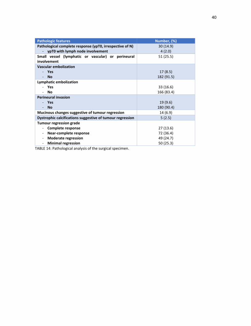
Other relevant pathological endpoints are shown in TABLE 14. There were four patients (2%) with ypT0 disease
but with lymph node involvement. Vascular, lymphatic and perineural invasion was found in 8.5%, 16.6% and
9.6% of patients, respectively. A complete or nearly-complete TRG was seen in 99 patients (50.0%), while
there was minimal or no regression in 50 patients (25.3%). Other signs of regression, such as acellular
mucinous lakes and dystrophic calcifications were found in 6.9% and 2.5% of patients, respectively.
40
Pathologic features Number, (%)
Pathological complete response (ypT0, irrespective of N) - ypT0 with lymph node involvement
30 (14.9) 4 (2.0)
Small vessel (lymphatic or vascular) or perineural involvement
51 (25.5)
Vascular embolization - Yes - No
17 (8.5)
182 (91.5)
Lymphatic embolization - Yes - No
33 (16.6)
166 (83.4)
Perineural invasion - Yes - No
19 (9.6)
180 (90.4)
Mucinous changes suggestive of tumour regression 14 (6.9)
Dystrophic calcifications suggestive of tumour regression 5 (2.5)
Tumour regression grade - Complete response - Near-complete response - Moderate regression - Minimal regression
27 (13.6) 72 (36.4) 49 (24.7) 50 (25.3)
TABLE 14: Pathological analysis of the surgical specimen.
41
3.5. Adjuvant chemotherapy
Adjuvant chemotherapy administration was possible in 165 patients (81.3%); the details of the adjuvant
chemotherapy regimen are shown in TABLE 15. The most frequent reasons for no administration were patient-
related factors, such as advanced age, comorbidities and poor or long recovery from the surgical procedure
(25 patients, 12.8%). The median time from surgery to adjuvant chemotherapy was 5 weeks, although a
quarter of patients did not begin adjuvant treatment until 7 weeks after surgery. Only 93 patients (45.9%)
received the full intended adjuvant dose of chemotherapy; most patients needed a dose reduction or an early
stop to treatment due to toxicity or poor tolerance. There were 25 (15.6%) treatment-related hospital
admissions compared to 12 admissions (5.9%) in the neoadjuvant part of the regimen. However, no
treatment-related deaths were observed.
42
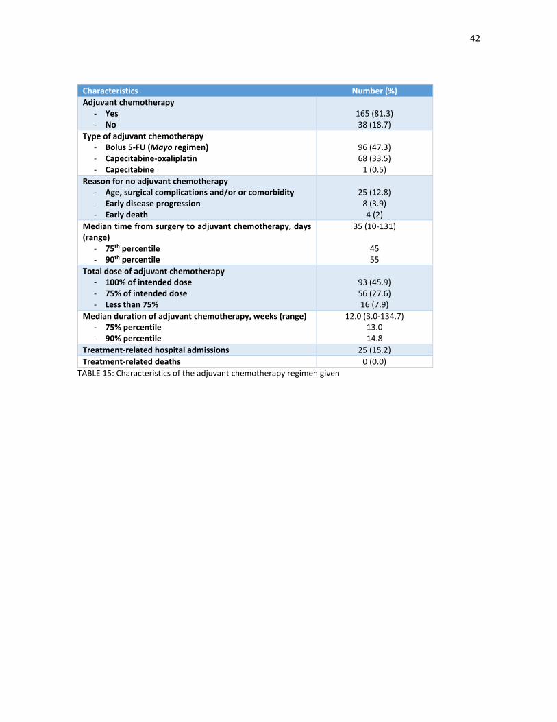
Characteristics Number (%)
Adjuvant chemotherapy - Yes - No
165 (81.3) 38 (18.7)
Type of adjuvant chemotherapy - Bolus 5-FU (Mayo regimen) - Capecitabine-oxaliplatin - Capecitabine
96 (47.3) 68 (33.5)
1 (0.5)
Reason for no adjuvant chemotherapy - Age, surgical complications and/or or comorbidity - Early disease progression - Early death
25 (12.8)
8 (3.9) 4 (2)
Median time from surgery to adjuvant chemotherapy, days (range)
- 75th percentile - 90th percentile
35 (10-131)
45 55
Total dose of adjuvant chemotherapy - 100% of intended dose - 75% of intended dose - Less than 75%
93 (45.9) 56 (27.6) 16 (7.9)
Median duration of adjuvant chemotherapy, weeks (range) - 75% percentile - 90% percentile
12.0 (3.0-134.7) 13.0 14.8
Treatment-related hospital admissions 25 (15.2)
Treatment-related deaths 0 (0.0)
TABLE 15: Characteristics of the adjuvant chemotherapy regimen given
43
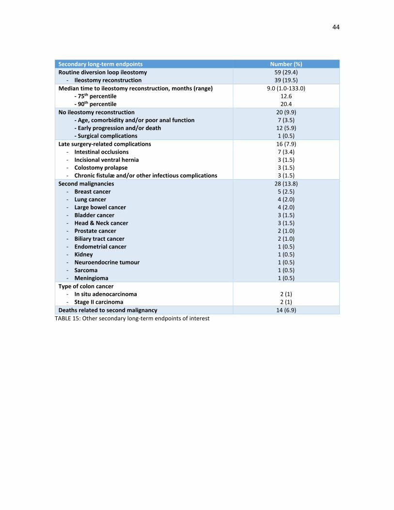
3.6. Other secondary endpoints
Other secondary long-term endpoints are shown in TABLE 16. Late surgery-related complications were
observed in 16 patients (7.9%). The median time to ileostomy reconstruction was nine months, although in a
quarter of patients it was more than one year after the initial surgery. In a third of patients with a routine
provisional loop ileostomy, it was not corrected, due to age, comorbidity and/or poor anal function, early
progression and/or death or surgical complications. There were 28 cases (13.8%) of metachronous malignant
tumours in the follow-up of these patients; of these, 4 cases (2%) were early stage colon cancers treated with
local therapies; no adjuvant treatment was necessary. 3 (1.5%) genetic syndromes were diagnosed in the
follow-up of these patients: one patient with a mutation of the BRCA1 gene, one patient with a mutation of
the DOG1 gene and one patient with an attenuated familiar colonic polyposis and a mutation of the APC gene.
44
Secondary long-term endpoints Number (%)
Routine diversion loop ileostomy - Ileostomy reconstruction
59 (29.4) 39 (19.5)
Median time to ileostomy reconstruction, months (range) - 75th percentile - 90th percentile
9.0 (1.0-133.0) 12.6 20.4
No ileostomy reconstruction - Age, comorbidity and/or poor anal function - Early progression and/or death - Surgical complications
20 (9.9) 7 (3.5)
12 (5.9) 1 (0.5)
Late surgery-related complications - Intestinal occlusions - Incisional ventral hernia - Colostomy prolapse - Chronic fistulæ and/or other infectious complications
16 (7.9) 7 (3.4) 3 (1.5) 3 (1.5) 3 (1.5)
Second malignancies - Breast cancer - Lung cancer - Large bowel cancer - Bladder cancer - Head & Neck cancer - Prostate cancer - Biliary tract cancer - Endometrial cancer - Kidney - Neuroendocrine tumour - Sarcoma - Meningioma
28 (13.8) 5 (2.5) 4 (2.0) 4 (2.0) 3 (1.5) 3 (1.5) 2 (1.0) 2 (1.0) 1 (0.5) 1 (0.5) 1 (0.5) 1 (0.5) 1 (0.5)
Type of colon cancer - In situ adenocarcinoma - Stage II carcinoma
2 (1) 2 (1)
Deaths related to second malignancy 14 (6.9)
TABLE 15: Other secondary long-term endpoints of interest
45
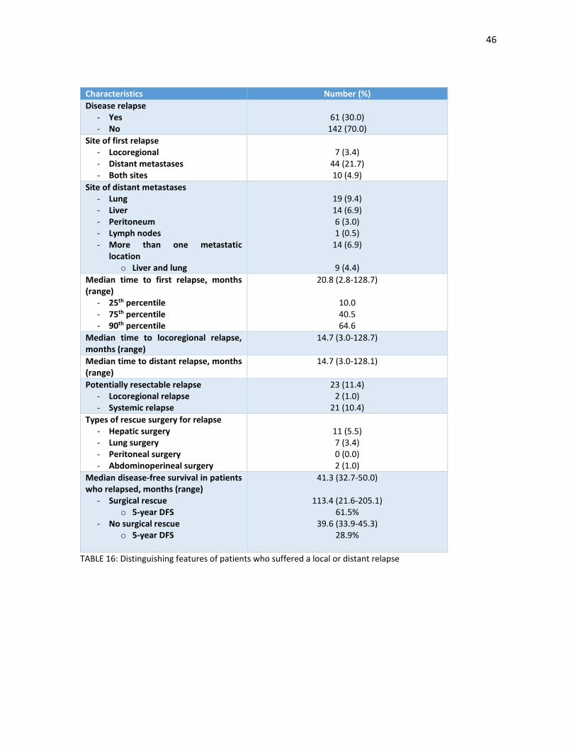
3.7. Local relapse and distant metastases
61 patients (30%) relapsed (TABLE 16). As the first site a recurrence, a local relapse was seen in 17 patients
(8.3%), although in 10 patients (4.9%), there was a local and systemic relapse at the same time. Distant
metastases were seen in 54 patients (26.6%). The most common site of distant relapse was in the lungs (19
patients, 9.4%) followed by the liver (14 patients, 6.9%) and both in the liver and the lungs (9 patients, 4.4%).
K-RAS analysis was performed in 28 patients (13.8%); of these patients, 19 (9.4%) had wild-type disease while
9 patients (4.4%) had K-RAS mutations.
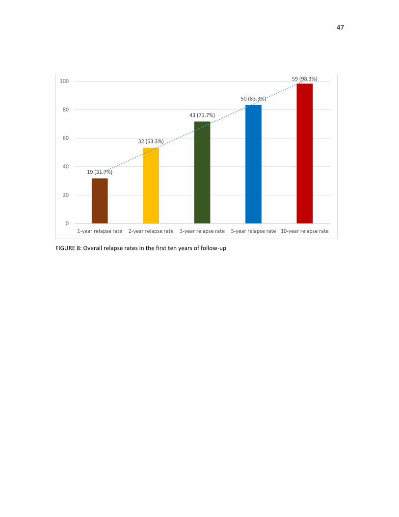
The median time to relapse was 20 months and in 50 patients (88.3%), the relapses took place in the first five
years of follow-up; FIGURE 8 and 9 are graphic representations of these findings. There were no differences
in the median time to relapse between patients with a local recurrence and those with distant metastases
(FIGURE 10). In 23 patients (11.4%) there was a potentially resectable relapse, of which, only 2 patients (1%)
had a local relapse.
46
Characteristics Number (%)
Disease relapse - Yes - No
61 (30.0)
142 (70.0)
Site of first relapse - Locoregional - Distant metastases - Both sites
7 (3.4)
44 (21.7) 10 (4.9)
Site of distant metastases - Lung - Liver - Peritoneum - Lymph nodes - More than one metastatic
location o Liver and lung
19 (9.4) 14 (6.9) 6 (3.0) 1 (0.5)
14 (6.9)
9 (4.4)
Median time to first relapse, months (range)
- 25th percentile - 75th percentile - 90th percentile
20.8 (2.8-128.7)
10.0 40.5 64.6
Median time to locoregional relapse, months (range)
14.7 (3.0-128.7)
Median time to distant relapse, months (range)
14.7 (3.0-128.1)
Potentially resectable relapse - Locoregional relapse - Systemic relapse
23 (11.4) 2 (1.0)
21 (10.4)
Types of rescue surgery for relapse - Hepatic surgery - Lung surgery - Peritoneal surgery - Abdominoperineal surgery
11 (5.5) 7 (3.4) 0 (0.0) 2 (1.0)
Median disease-free survival in patients who relapsed, months (range)
- Surgical rescue o 5-year DFS
- No surgical rescue o 5-year DFS
41.3 (32.7-50.0)
113.4 (21.6-205.1) 61.5%
39.6 (33.9-45.3) 28.9%
TABLE 16: Distinguishing features of patients who suffered a local or distant relapse
47
FIGURE 8: Overall relapse rates in the first ten years of follow-up
19 (31.7%)
32 (53.3%)
43 (71.7%)
50 (83.3%)
59 (98.3%)
0
20
40
60
80
100
1-year relapse rate 2-year relapse rate 3-year relapse rate 5-year relapse rate 10-year relapse rate
48
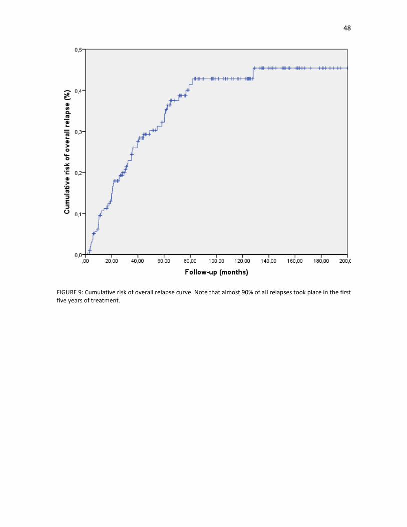
FIGURE 9: Cumulative risk of overall relapse curve. Note that almost 90% of all relapses took place in the first five years of treatment.
49
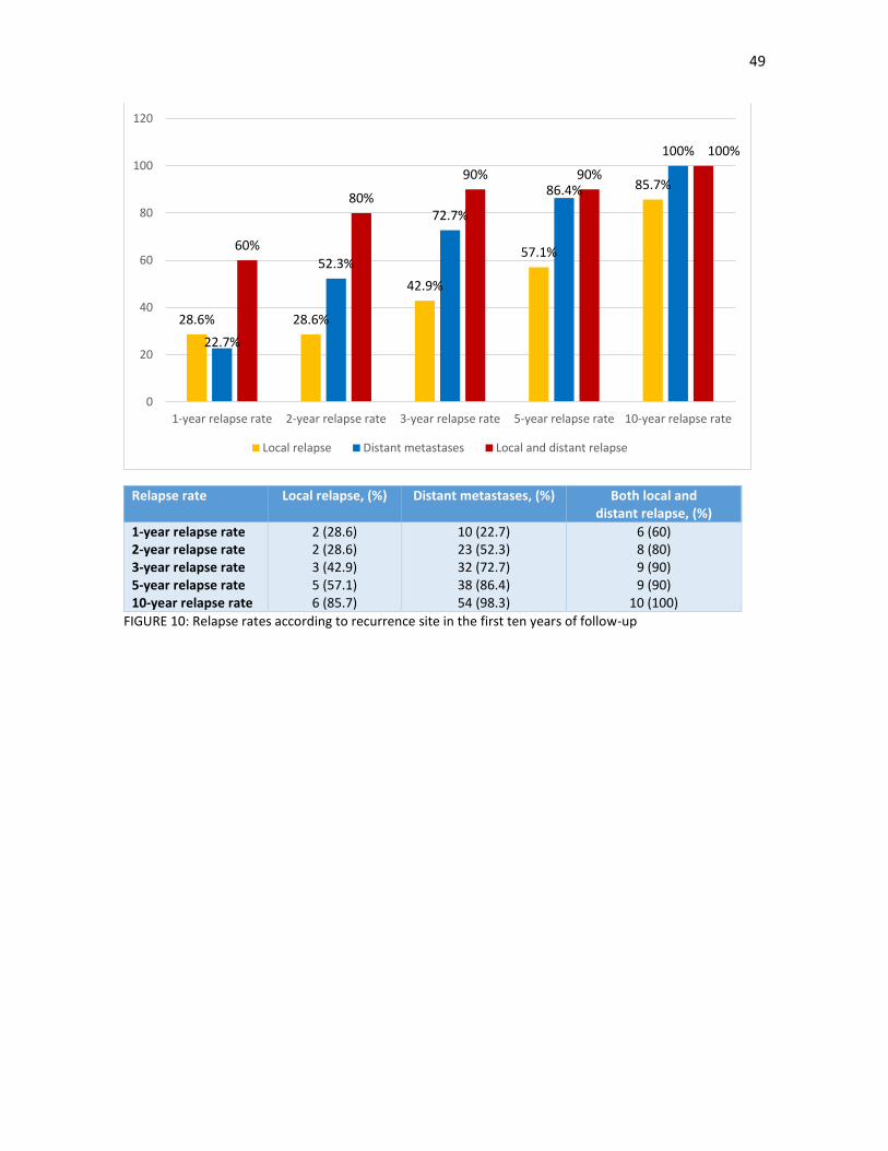
Relapse rate Local relapse, (%) Distant metastases, (%) Both local and distant relapse, (%)
1-year relapse rate 2-year relapse rate 3-year relapse rate 5-year relapse rate 10-year relapse rate
2 (28.6) 2 (28.6) 3 (42.9) 5 (57.1) 6 (85.7)
10 (22.7) 23 (52.3) 32 (72.7) 38 (86.4) 54 (98.3)
6 (60) 8 (80) 9 (90) 9 (90)
10 (100)
FIGURE 10: Relapse rates according to recurrence site in the first ten years of follow-up
28.6% 28.6%
42.9%
57.1%
85.7%
22.7%
52.3%
72.7%
86.4%
100%
60%
80%
90% 90%
100%
0
20
40
60
80
100
120
1-year relapse rate 2-year relapse rate 3-year relapse rate 5-year relapse rate 10-year relapse rate
Local relapse Distant metastases Local and distant relapse

50
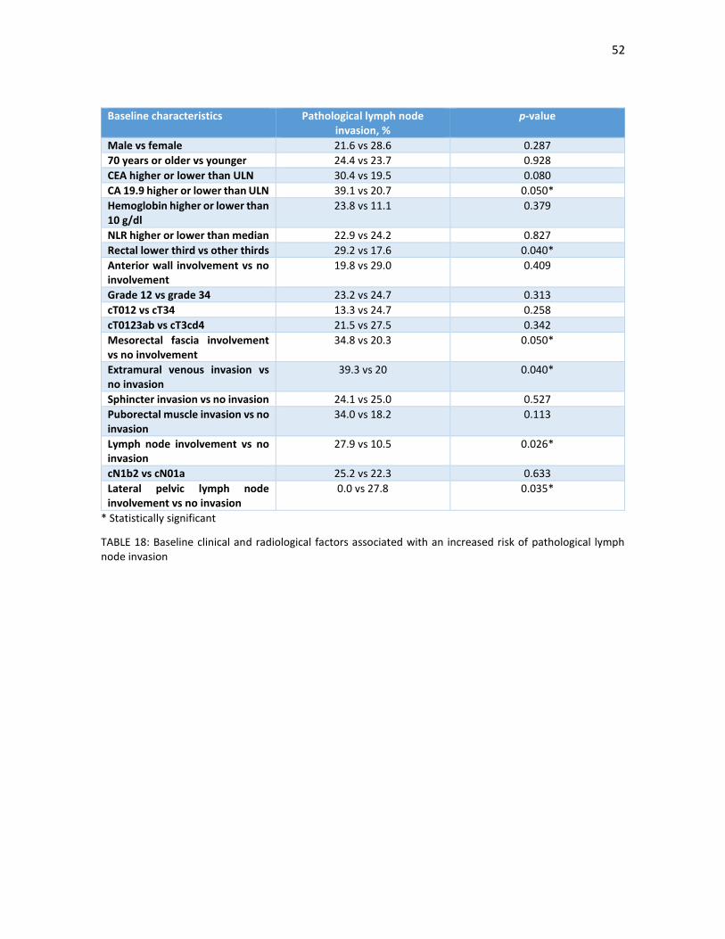
3.7.1. Risk factors for local relapse and pathological stage III disease
We performed an analysis of baseline clinical and radiological factors that could increase the local relapse and
the presence of pathological lymph nodes in the surgical specimen, as they are the factors most closely linked
to the prescription of adjuvant chemotherapy. The results are shown in TABLE 17 and 18.
A higher cT stage, poorly and undifferentiated tumours, a hemoglobin level less than 10 g/dl and patients 70
years or older had an increased risk of a local relapse. We did not see an association however with lymph node
positive disease (cN), a clinical higher lymph node burden, involvement of the mesorectal fascia, lower rectal
tumours and an elevated NRL ratio.
On the other hand, there was an increase in the risk pathological lymph nodes with clinical mesorectal fascia
involvement, extramural venous invasion, elevated values of CEA and CA 19.9, positive lymph node disease
and lower rectal tumours. There was no significant link with age, an elevated NRL ratio and age. Curiously,
those patients with lateral pelvic lymph node disease had a lower risk of pathological lymph nodes.
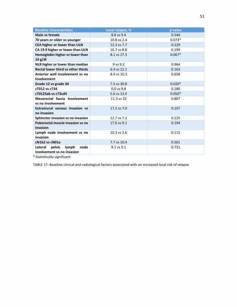
51
Baseline characteristics Local relapse, % p-value
Male vs female 8.8 vs 9.4 0.544
70 years or older vs younger 10.8 vs 2.4 0.073*
CEA higher or lower than ULN 12.3 vs 7.7 0.229
CA 19.9 higher or lower than ULN 16.7 vs 8.8 0.199
Hemoglobin higher or lower than 10 g/dl
8.1 vs 27.3 0.067*
NLR higher or lower than median 9 vs 9.2 0.964
Rectal lower third vs other thirds 6.4 vs 12.1 0.163
Anterior wall involvement vs no involvement
8.4 vs 10.3 0.658
Grade 12 vs grade 34 7.5 vs 30.8 0.020*
cT012 vs cT34 0.0 vs 9.8 0.280
cT0123ab vs cT3cd4 5.6 vs 13.4 0.050*
Mesorectal fascia involvement vs no involvement
11.3 vs 10 0.807
Extramural venous invasion vs no invasion
17.2 vs 7.0 0.107
Sphincter invasion vs no invasion 12.7 vs 7.2 0.225
Puborectal muscle invasion vs no invasion
17.6 vs 9.1 0.194
Lymph node involvement vs no invasion
10.3 vs 2.6 0.115
cN1b2 vs cN01a 7.7 vs 10.4 0.501
Lateral pelvic lymph node involvement vs no invasion
9.1 vs 9.1 0.731
* Statistically significant
TABLE 17: Baseline clinical and radiological factors associated with an increased local risk of relapse
52
Baseline characteristics Pathological lymph node invasion, %
p-value
Male vs female 21.6 vs 28.6 0.287
70 years or older vs younger 24.4 vs 23.7 0.928
CEA higher or lower than ULN 30.4 vs 19.5 0.080
CA 19.9 higher or lower than ULN 39.1 vs 20.7 0.050*
Hemoglobin higher or lower than 10 g/dl
23.8 vs 11.1 0.379
NLR higher or lower than median 22.9 vs 24.2 0.827
Rectal lower third vs other thirds 29.2 vs 17.6 0.040*
Anterior wall involvement vs no involvement
19.8 vs 29.0 0.409
Grade 12 vs grade 34 23.2 vs 24.7 0.313
cT012 vs cT34 13.3 vs 24.7 0.258
cT0123ab vs cT3cd4 21.5 vs 27.5 0.342
Mesorectal fascia involvement vs no involvement
34.8 vs 20.3 0.050*
Extramural venous invasion vs no invasion
39.3 vs 20 0.040*
Sphincter invasion vs no invasion 24.1 vs 25.0 0.527
Puborectal muscle invasion vs no invasion
34.0 vs 18.2 0.113
Lymph node involvement vs no invasion
27.9 vs 10.5 0.026*
cN1b2 vs cN01a 25.2 vs 22.3 0.633
Lateral pelvic lymph node involvement vs no invasion
0.0 vs 27.8 0.035*
* Statistically significant
TABLE 18: Baseline clinical and radiological factors associated with an increased risk of pathological lymph node invasion
53
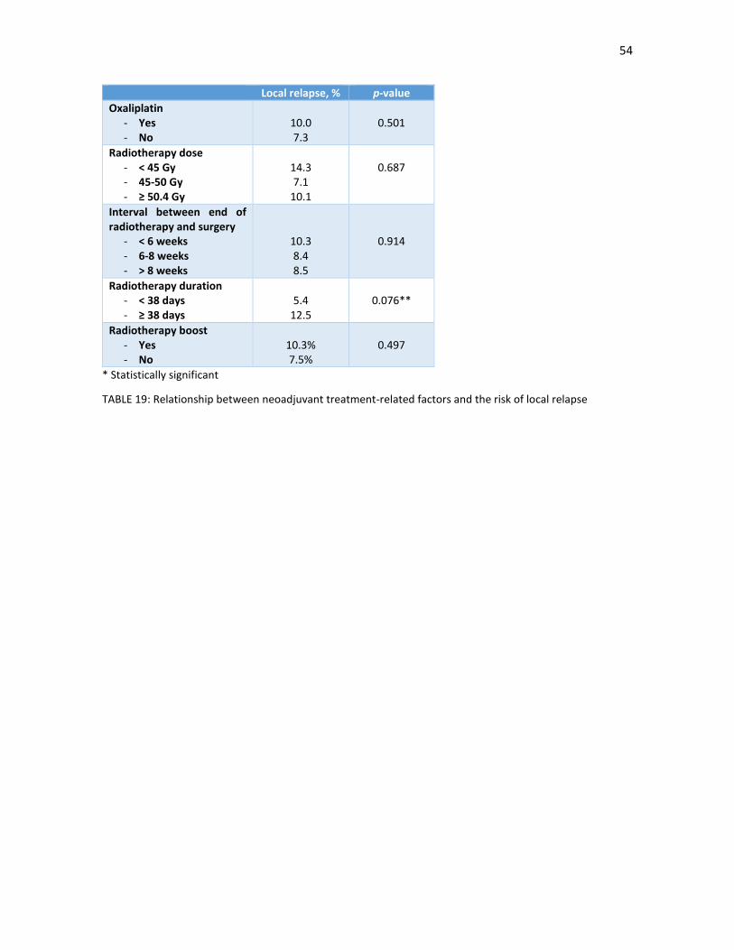
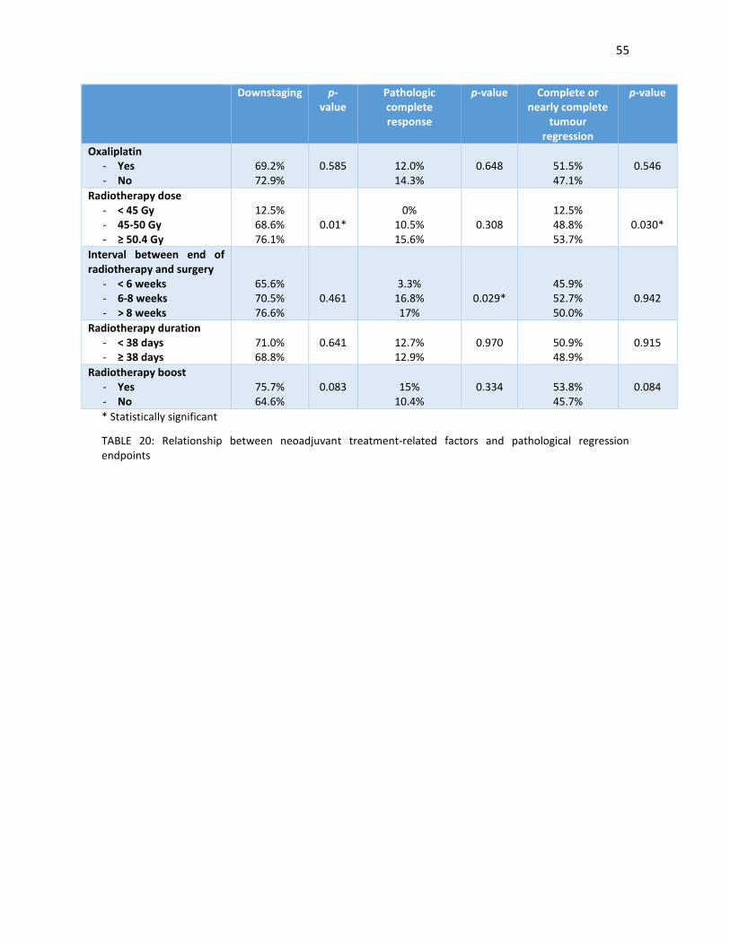
3.7.2. Relationship between neoadjuvant therapy and local relapse and pathological endpoints of response
Due to the relationship between the use of neoadjuvant (chemo)radiotherapy and the decreased risk of local
relapse and an increased downstaging rate, we performed an analysis of these factors. The results are shown
in TABLES 19 and 20.
There was a numerically but not statistically significant increase of local relapse with increasing durations of
the radiotherapy regimen. However, we did not see any difference with the use of radiosensitizing oxaliplatin,
with the total dose of radiotherapy or with the use of a radiotherapy boost. There were also no differences
according to the interval between the end of radiotherapy and the surgical procedure.
With regards to pathological regression endpoints, there was no improvement with the use of oxaliplatin, the
use of a radiotherapy boost and the duration of the radiotherapy regimen. However, we did see an increased
rate of downstaging and complete or nearly complete tumour regression with higher doses of radiotherapy
and an increased rate of pathological complete response with longer intervals between the end of
radiotherapy and the surgical procedure.
54
Local relapse, % p-value
Oxaliplatin - Yes - No
10.0 7.3
0.501
Radiotherapy dose - < 45 Gy - 45-50 Gy - ≥ 50.4 Gy
14.3 7.1
10.1
0.687
Interval between end of radiotherapy and surgery
- < 6 weeks - 6-8 weeks - > 8 weeks
10.3 8.4 8.5
0.914
Radiotherapy duration - < 38 days - ≥ 38 days
5.4
12.5
0.076**
Radiotherapy boost - Yes - No
10.3% 7.5%
0.497
* Statistically significant
TABLE 19: Relationship between neoadjuvant treatment-related factors and the risk of local relapse
55
Downstaging p-value
Pathologic complete response
p-value Complete or nearly complete
tumour regression
p-value
Oxaliplatin - Yes - No
69.2% 72.9%
0.585
12.0% 14.3%
0.648
51.5% 47.1%
0.546
Radiotherapy dose - < 45 Gy - 45-50 Gy - ≥ 50.4 Gy
12.5% 68.6% 76.1%
0.01*
0%
10.5% 15.6%
0.308
12.5% 48.8% 53.7%
0.030*
Interval between end of radiotherapy and surgery
- < 6 weeks - 6-8 weeks - > 8 weeks
65.6% 70.5% 76.6%
0.461
3.3% 16.8% 17%
0.029*
45.9% 52.7% 50.0%
0.942
Radiotherapy duration - < 38 days - ≥ 38 days
71.0% 68.8%
0.641
12.7% 12.9%
0.970
50.9% 48.9%
0.915
Radiotherapy boost - Yes - No
75.7% 64.6%
0.083
15%
10.4%
0.334
53.8% 45.7%
0.084
* Statistically significant
TABLE 20: Relationship between neoadjuvant treatment-related factors and pathological regression endpoints
56
3.7.3. Pathological features and risk of local relapse and distant metastases.
Pathological features in the surgical specimen and an increased risk of local relapse and distant metastases
are shown in TABLES 21 and 22, respectively. Factors linked to both endpoints were an unsuccessful TME, the
unsatisfactory quality of the mesorectum, an R2 resection, involvement of the circumferential and distal
margins, no downstaging, poorly differentiated tumours, moderate or minimal regression, perineural
invasion, pathological lymph node invasion and heavy lymph node burden. In the multivariate analysis, CRM
invasion and perineural invasion retained their prognostic significance for both endpoints.
A pathological complete response was associated with a decreased risk of local relapse, but with no difference
in distant metastases. Vascular invasion was linked with an increased risk of distant metastases, but not with
local relapses. There was a borderline statistically significant link of abdominoperineal resection with a higher
local relapse rate and of mucinous tumours with increased distant metastases.
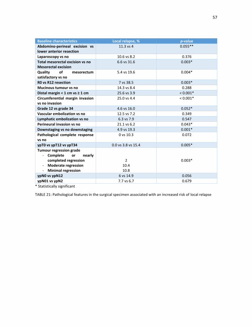
57
Baseline characteristics Local relapse, % p-value
Abdomino-perineal excision vs lower anterior resection
11.3 vs 4 0.055**
Laparoscopy vs no 10.6 vs 8.2 0.376
Total mesorectal excision vs no Mesorectal excision
6.6 vs 31.6 0.003*
Quality of mesorectum satisfactory vs no
5.4 vs 19.6 0.004*
R0 vs R12 resection 7 vs 38.5 0.003*
Mucinous tumour vs no 14.3 vs 8.4 0.288
Distal margin < 1 cm vs ≥ 1 cm 25.6 vs 3.9 < 0.001*
Circumferential margin invasion vs no invasion
25.0 vs 4.4 < 0.001*
Grade 12 vs grade 34 4.6 vs 16.0 0.052*
Vascular embolization vs no 12.5 vs 7.2 0.349
Lymphatic embolization vs no 6.3 vs 7.9 0.547
Perineural invasion vs no 21.1 vs 6.2 0.043*
Downstaging vs no downstaging 4.9 vs 19.3 0.001*
Pathological complete response vs no
0 vs 10.3 0.072
ypT0 vs ypT12 vs ypT34 0.0 vs 3.8 vs 15.4 0.005*
Tumour regression grade - Complete or nearly
completed regression - Moderate regression - Minimal regression
2 10.4 10.8
0.003*
ypN0 vs ypN12 6 vs 14.9 0.056
ypN01 vs ypN2 7.7 vs 6.7 0.679
* Statistically significant
TABLE 21: Pathological features in the surgical specimen associated with an increased risk of local relapse
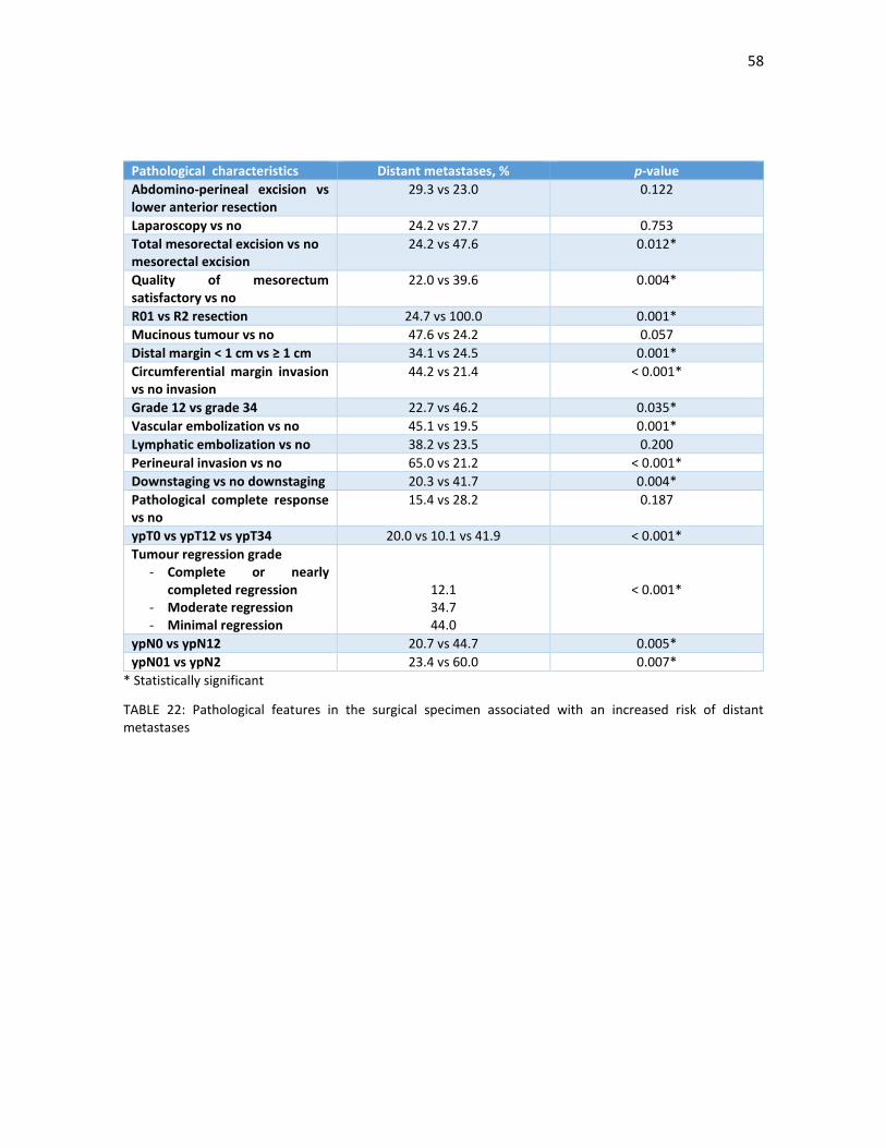
58
Pathological characteristics Distant metastases, % p-value
Abdomino-perineal excision vs lower anterior resection
29.3 vs 23.0 0.122
Laparoscopy vs no 24.2 vs 27.7 0.753
Total mesorectal excision vs no mesorectal excision
24.2 vs 47.6 0.012*
Quality of mesorectum satisfactory vs no
22.0 vs 39.6 0.004*
R01 vs R2 resection 24.7 vs 100.0 0.001*
Mucinous tumour vs no 47.6 vs 24.2 0.057
Distal margin < 1 cm vs ≥ 1 cm 34.1 vs 24.5 0.001*
Circumferential margin invasion vs no invasion
44.2 vs 21.4 < 0.001*
Grade 12 vs grade 34 22.7 vs 46.2 0.035*
Vascular embolization vs no 45.1 vs 19.5 0.001*
Lymphatic embolization vs no 38.2 vs 23.5 0.200
Perineural invasion vs no 65.0 vs 21.2 < 0.001*
Downstaging vs no downstaging 20.3 vs 41.7 0.004*
Pathological complete response vs no
15.4 vs 28.2 0.187
ypT0 vs ypT12 vs ypT34 20.0 vs 10.1 vs 41.9 < 0.001*
Tumour regression grade - Complete or nearly
completed regression - Moderate regression - Minimal regression
12.1 34.7 44.0
< 0.001*
ypN0 vs ypN12 20.7 vs 44.7 0.005*
ypN01 vs ypN2 23.4 vs 60.0 0.007*
* Statistically significant
TABLE 22: Pathological features in the surgical specimen associated with an increased risk of distant metastases
59
3.8. Special groups of patients: Elderly patients and patients with mucinous tumours.
Long-term outcomes for elderly patients (defined as 70-year patients or older) were poorer than for younger
patients. We performed an analysis of possible factors that could explain these findings; the most significant
results are shown in TABLE 23. Although no differences where seen with respects to classical pathological
findings, we found that elderly patients had a poorer compliance to overall treatment, with more than half of
patients receiving a radiotherapy dose less than 45 Gy, with a borderline statistically significant increase of
grade 3-4 toxicity and of unexpected cardiovascular and surgical events, compared to their younger
counterparts. The rate of abdominoperineal resections was higher in elderly patients while the poor
compliance to the neoadjuvant treatment was linked to a lower rate of complete or near complete tumour
regressions. The compliance of adjuvant treatment was particularly poor, with less than a quarter of elderly
patients receiving the full intended dose, compared to more than half of younger patients.
Mucinous tumours, defined as tumours with more than 50% of a mucoid component were found in 21 patients
(10.3%). These tumours were linked to higher overall rates of poor response to neoadjuvant chemo-
radiotherapy and other poor-prognosis pathological endpoints compared to non-mucinous tumours, as
shown in TABLE 24.
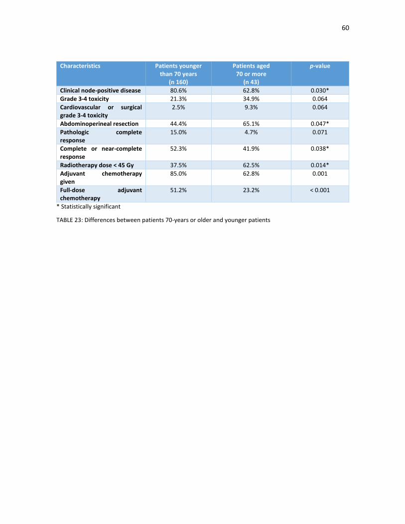
60
Characteristics Patients younger than 70 years
(n 160)
Patients aged 70 or more
(n 43)
p-value
Clinical node-positive disease 80.6% 62.8% 0.030*
Grade 3-4 toxicity 21.3% 34.9% 0.064
Cardiovascular or surgical grade 3-4 toxicity
2.5% 9.3% 0.064
Abdominoperineal resection 44.4% 65.1% 0.047*
Pathologic complete response
15.0% 4.7% 0.071
Complete or near-complete response
52.3% 41.9% 0.038*
Radiotherapy dose < 45 Gy 37.5% 62.5% 0.014*
Adjuvant chemotherapy given
85.0% 62.8% 0.001
Full-dose adjuvant chemotherapy
51.2% 23.2% < 0.001
* Statistically significant
TABLE 23: Differences between patients 70-years or older and younger patients
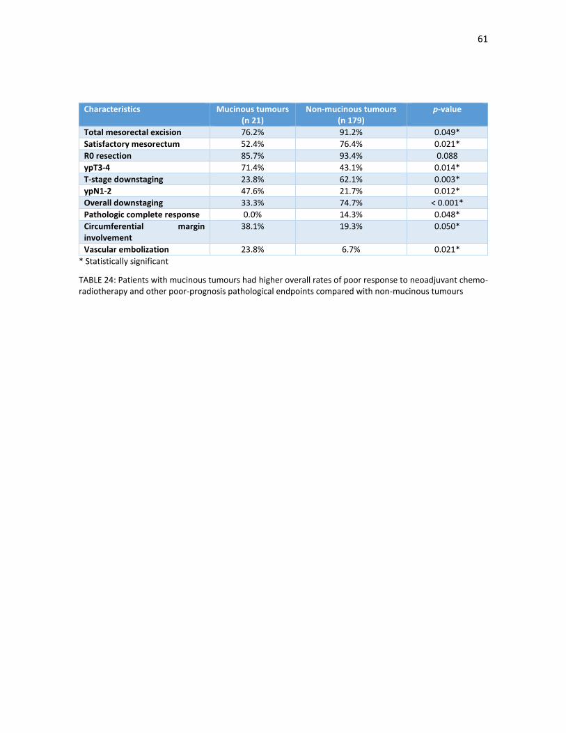
61
Characteristics Mucinous tumours (n 21)
Non-mucinous tumours (n 179)
p-value
Total mesorectal excision 76.2% 91.2% 0.049*
Satisfactory mesorectum 52.4% 76.4% 0.021*
R0 resection 85.7% 93.4% 0.088
ypT3-4 71.4% 43.1% 0.014*
T-stage downstaging 23.8% 62.1% 0.003*
ypN1-2 47.6% 21.7% 0.012*
Overall downstaging 33.3% 74.7% < 0.001*
Pathologic complete response 0.0% 14.3% 0.048*
Circumferential margin involvement
38.1% 19.3% 0.050*
Vascular embolization 23.8% 6.7% 0.021*
* Statistically significant
TABLE 24: Patients with mucinous tumours had higher overall rates of poor response to neoadjuvant chemo-radiotherapy and other poor-prognosis pathological endpoints compared with non-mucinous tumours
62
3.9. Survival analysis
With a median follow-up of 124.9 months (range 14.4-212.3), median DFS and median CSS were 139.8 months
(106.0-173.6) and 198.1 months (133.8-262.4), respectively; median OS has not been reached yet. Estimated
5-year DFS, CSS and OS are 71.4%, 75.8% and 75.6%, respectively (FIGURES 11, 12 and 13 and TABLE 25). At
the moment of the present analysis, 114 (55.7%) of patients are alive and free of disease, 11 (5.4%) are alive
but with active disease, 8 (3.9%) are alive and free of disease after resection of the disease relapse, 44 (21.7%)
have died secondary to cancer-related causes, 7 (3.4%) have had treatment-related deaths and 20 (9.9%) have
died for non-related causes.
The median DFS and OS times of the 61 patients that relapsed according to the location of the relapse are
shown in TABLE 26. Of note, 5-year DFS was significantly improved in those patients who were candidates for
surgical rescue of the relapse versus those where it was not (61.5% vs 28.9%); These differences were
statistically significant (p < 0.001).
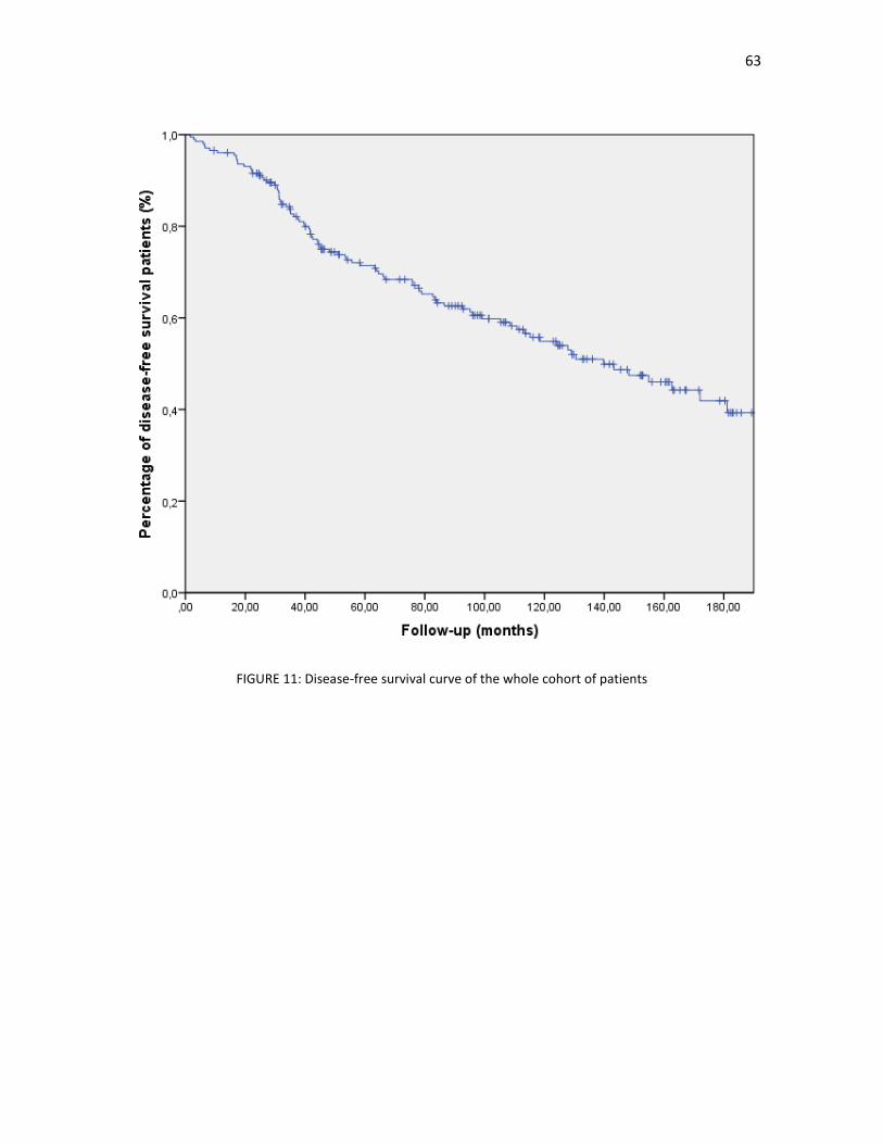
63
FIGURE 11: Disease-free survival curve of the whole cohort of patients
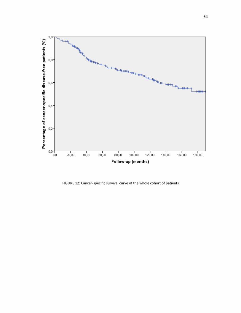
64
FIGURE 12: Cancer-specific survival curve of the whole cohort of patients
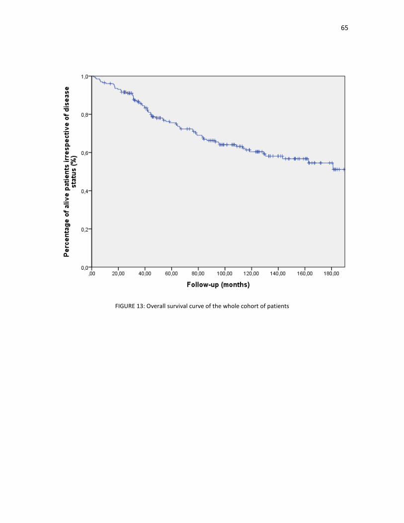
65
FIGURE 13: Overall survival curve of the whole cohort of patients
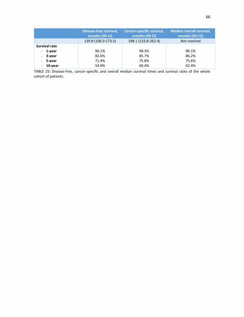
66
Disease-free survival, months (95 CI)
Cancer-specific survival, months (95 CI)
Median overall survival, months (95 CI))
139.8 (106.0-173.5) 198.1 (133.8-262.4) Not reached
Survival rate - 1-year - 3-year - 5-year - 10-year
96.1% 82.6% 71.4% 54.9%
98.3% 85.7% 75.8% 60.4%
96.1% 86.2% 75.6% 62.4%
TABLE 25: Disease-free, cancer-specific and overall median survival times and survival rates of the whole cohort of patients.
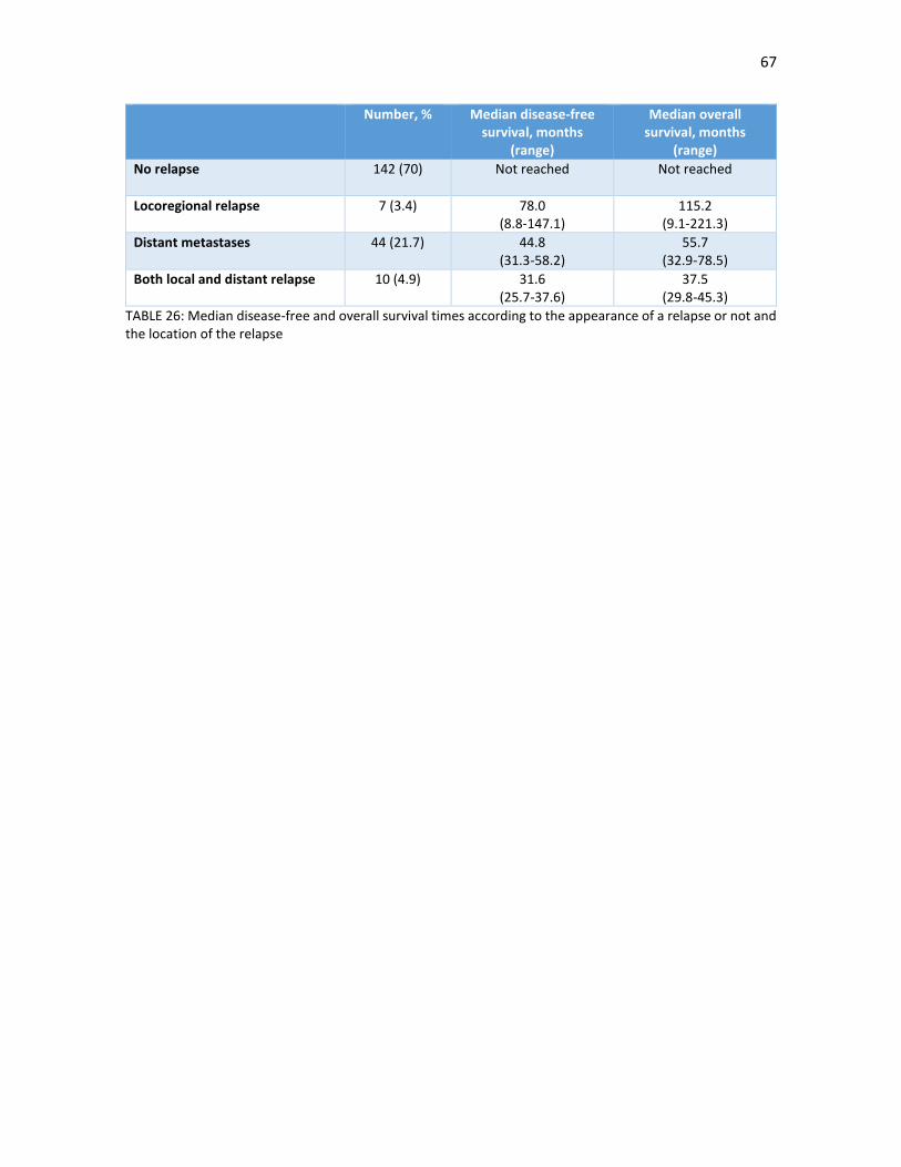
67
Number, % Median disease-free survival, months
(range)
Median overall survival, months
(range)
No relapse
142 (70) Not reached Not reached
Locoregional relapse 7 (3.4)
78.0 (8.8-147.1)
115.2 (9.1-221.3)
Distant metastases 44 (21.7)
44.8 (31.3-58.2)
55.7 (32.9-78.5)
Both local and distant relapse 10 (4.9) 31.6 (25.7-37.6)
37.5 (29.8-45.3)
TABLE 26: Median disease-free and overall survival times according to the appearance of a relapse or not and the location of the relapse
68
3.9.1. Univariate analysis for prognostic factors for DFS and OS
Possible clinical, neoadjuvant treatment-related, surgical and pathological factors were included in the
univariate analysis, both for DFS and OS; the results are summarized in TABLES 27, 28, 29 and 30.
Significant baseline clinical factors included older age (FIGURE 14), elevated values of CEA and CA 19.9, poorly
differentiated tumours in the diagnostic biopsy, radiological sphincter involvement and a clinical heavy lymph
node burden (defined as more than one lymph node involvement, cN1b-2ab). There was no relationship with
tumour location, anterior wall involvement, clinical cT3-4 stage or cN stage, independently of the number of
nodes. No link was seen between the baseline NLR and improved survival at any level (quartiles, median, 10th
or 90th percentiles or > 5).
With regards to treatment-related factors, both neoadjuvant and surgical, only a successful TME improved
DSF and OS (FIGURE 15), while a shorter interval between the end of radiotherapy and surgery improved DFS;
results were poorer if the interval was longer than eight weeks. Curiously, a low presurgical RNL was
associated with poor survival outcomes, both in DFS and OS.
Pathological endpoints which improved DFS and OS included a satisfactory mesorectum, no lymph node
invasion, a lower lymph node burden (FIGURE 16), overall, T-stage and N-stage downstaging, circumferential
margin involvement (FIGURE 17), no vascular and perineural invasion, no complete or nearly-complete
tumour regression (FIGURE 18) and a non-mucinous histology. On the other hand, prognosis was especially
poor in patients with ypN2 disease (TABLE 31 and FIGURE 19). Improvement only in DFS were seen with lower
T stages, well differentiated tumours and a pathologic complete or or nearly-complete response (ypT01).
Lymphatic embolization and an involved distal margin were not significant factors.
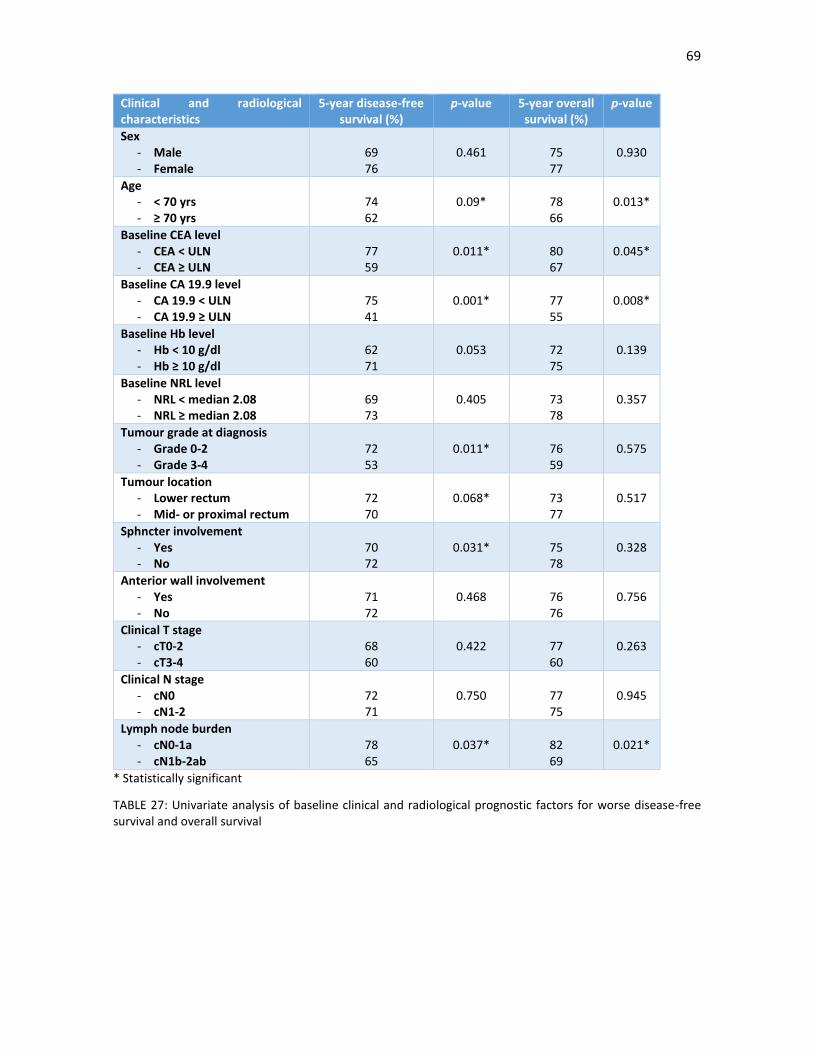
69
Clinical and radiological characteristics
5-year disease-free survival (%)
p-value 5-year overall survival (%)
p-value
Sex - Male - Female
69 76
0.461
75 77
0.930
Age - < 70 yrs - ≥ 70 yrs
74 62
0.09*
78 66
0.013*
Baseline CEA level - CEA < ULN - CEA ≥ ULN
77 59
0.011*
80 67
0.045*
Baseline CA 19.9 level - CA 19.9 < ULN - CA 19.9 ≥ ULN
75 41
0.001*
77 55
0.008*
Baseline Hb level - Hb < 10 g/dl - Hb ≥ 10 g/dl
62 71
0.053
72 75
0.139
Baseline NRL level - NRL < median 2.08 - NRL ≥ median 2.08
69 73
0.405
73 78
0.357
Tumour grade at diagnosis - Grade 0-2 - Grade 3-4
72 53
0.011*
76 59
0.575
Tumour location - Lower rectum - Mid- or proximal rectum
72 70
0.068*
73 77
0.517
Sphncter involvement - Yes - No
70 72
0.031*
75 78
0.328
Anterior wall involvement - Yes - No
71 72
0.468
76 76
0.756
Clinical T stage - cT0-2 - cT3-4
68 60
0.422
77 60
0.263
Clinical N stage - cN0 - cN1-2
72 71
0.750
77 75
0.945
Lymph node burden - cN0-1a - cN1b-2ab
78 65
0.037*
82 69
0.021*
* Statistically significant
TABLE 27: Univariate analysis of baseline clinical and radiological prognostic factors for worse disease-free survival and overall survival
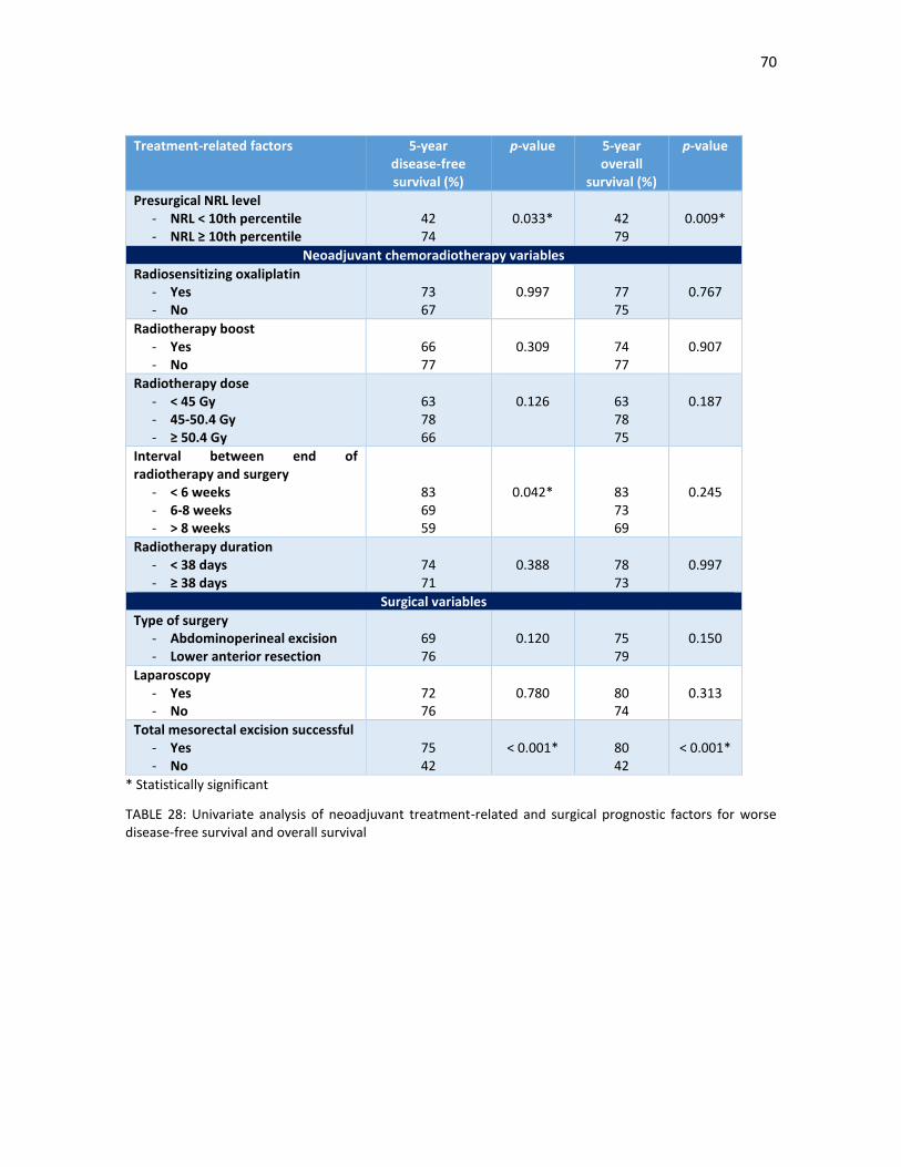
70
Treatment-related factors 5-year disease-free survival (%)
p-value 5-year overall
survival (%)
p-value
Presurgical NRL level - NRL < 10th percentile - NRL ≥ 10th percentile
42 74
0.033*
42 79
0.009*
Neoadjuvant chemoradiotherapy variables
Radiosensitizing oxaliplatin - Yes - No
73 67
0.997
77 75
0.767
Radiotherapy boost - Yes - No
66 77
0.309
74 77
0.907
Radiotherapy dose - < 45 Gy - 45-50.4 Gy - ≥ 50.4 Gy
63 78 66
0.126
63 78 75
0.187
Interval between end of radiotherapy and surgery
- < 6 weeks - 6-8 weeks - > 8 weeks
83 69 59
0.042*
83 73 69
0.245
Radiotherapy duration - < 38 days - ≥ 38 days
74 71
0.388
78 73
0.997
Surgical variables
Type of surgery - Abdominoperineal excision - Lower anterior resection
69 76
0.120
75 79
0.150
Laparoscopy - Yes - No
72 76
0.780
80 74
0.313
Total mesorectal excision successful - Yes - No
75 42
< 0.001*
80 42
< 0.001*
* Statistically significant
TABLE 28: Univariate analysis of neoadjuvant treatment-related and surgical prognostic factors for worse disease-free survival and overall survival
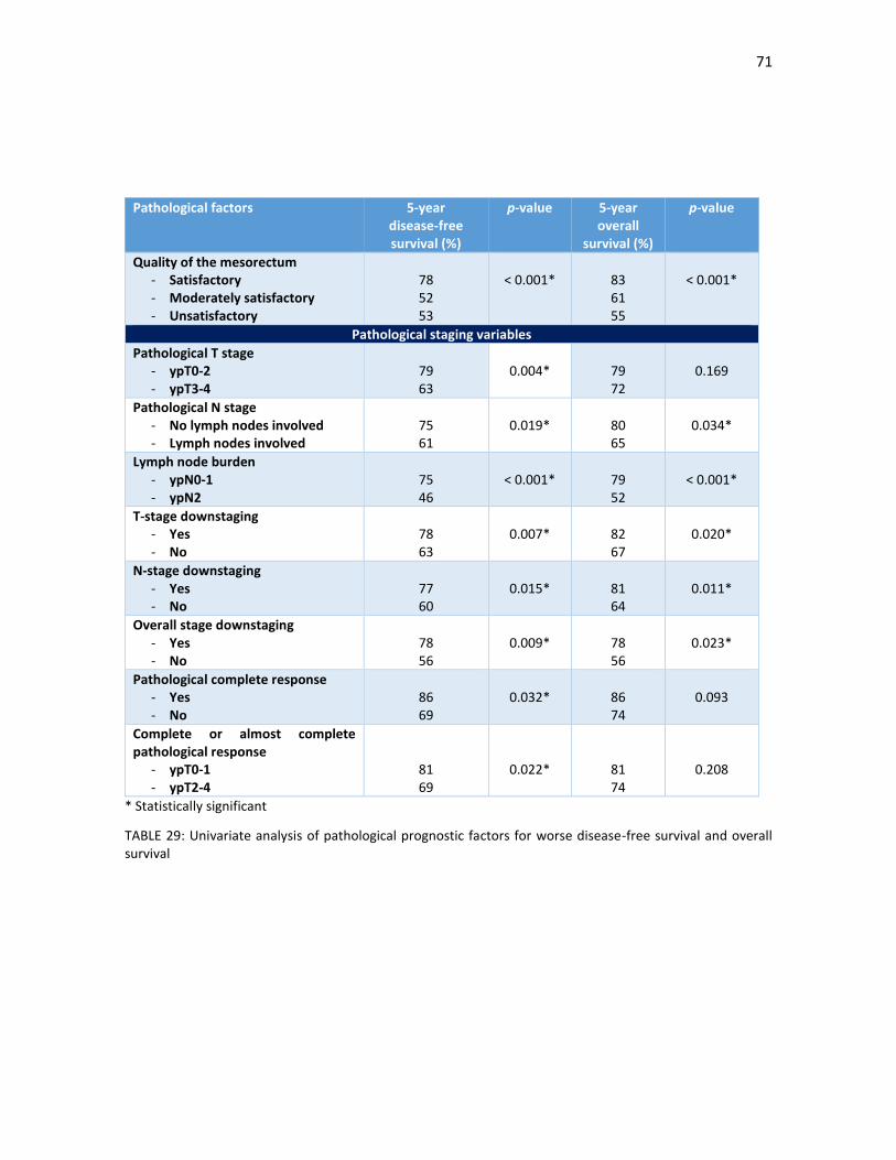
71
Pathological factors 5-year disease-free survival (%)
p-value 5-year overall
survival (%)
p-value
Quality of the mesorectum - Satisfactory - Moderately satisfactory - Unsatisfactory
78 52 53
< 0.001*
83 61 55
< 0.001*
Pathological staging variables
Pathological T stage - ypT0-2 - ypT3-4
79 63
0.004*
79 72
0.169
Pathological N stage - No lymph nodes involved - Lymph nodes involved
75 61
0.019*
80 65
0.034*
Lymph node burden - ypN0-1 - ypN2
75 46
< 0.001*
79 52
< 0.001*
T-stage downstaging - Yes - No
78 63
0.007*
82 67
0.020*
N-stage downstaging - Yes - No
77 60
0.015*
81 64
0.011*
Overall stage downstaging - Yes - No
78 56
0.009*
78 56
0.023*
Pathological complete response - Yes - No
86 69
0.032*
86 74
0.093
Complete or almost complete pathological response
- ypT0-1 - ypT2-4
81 69
0.022*
81 74
0.208
* Statistically significant
TABLE 29: Univariate analysis of pathological prognostic factors for worse disease-free survival and overall survival
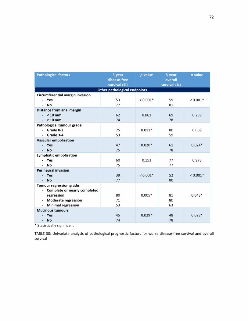
72
Pathological factors 5-year disease-free survival (%)
p-value 5-year overall
survival (%)
p-value
Other pathological endpoints
Circumferential margin invasion - Yes - No
53 77
< 0.001*
59 81
< 0.001*
Distance from anal margin - < 10 mm - ≥ 10 mm
62 74
0.061
69 78
0.239
Pathological tumour grade - Grade 0-2 - Grade 3-4
75 53
0.011*
80 59
0.069
Vascular embolization - Yes - No
47 75
0.020*
61 78
0.024*
Lymphatic embolization - Yes - No
60 75
0.153
77 77
0.978
Perineural invasion - Yes - No
39 77
< 0.001*
52 80
< 0.001*
Tumour regression grade - Complete or nearly completed
regression - Moderate regression - Minimal regression
80 71 53
0.005*
81 80 63
0.043*
Mucinous tumours - Yes - No
45 74
0.029*
48 78
0.023*
* Statistically significant
TABLE 30: Univariate analysis of pathological prognostic factors for worse disease-free survival and overall survival
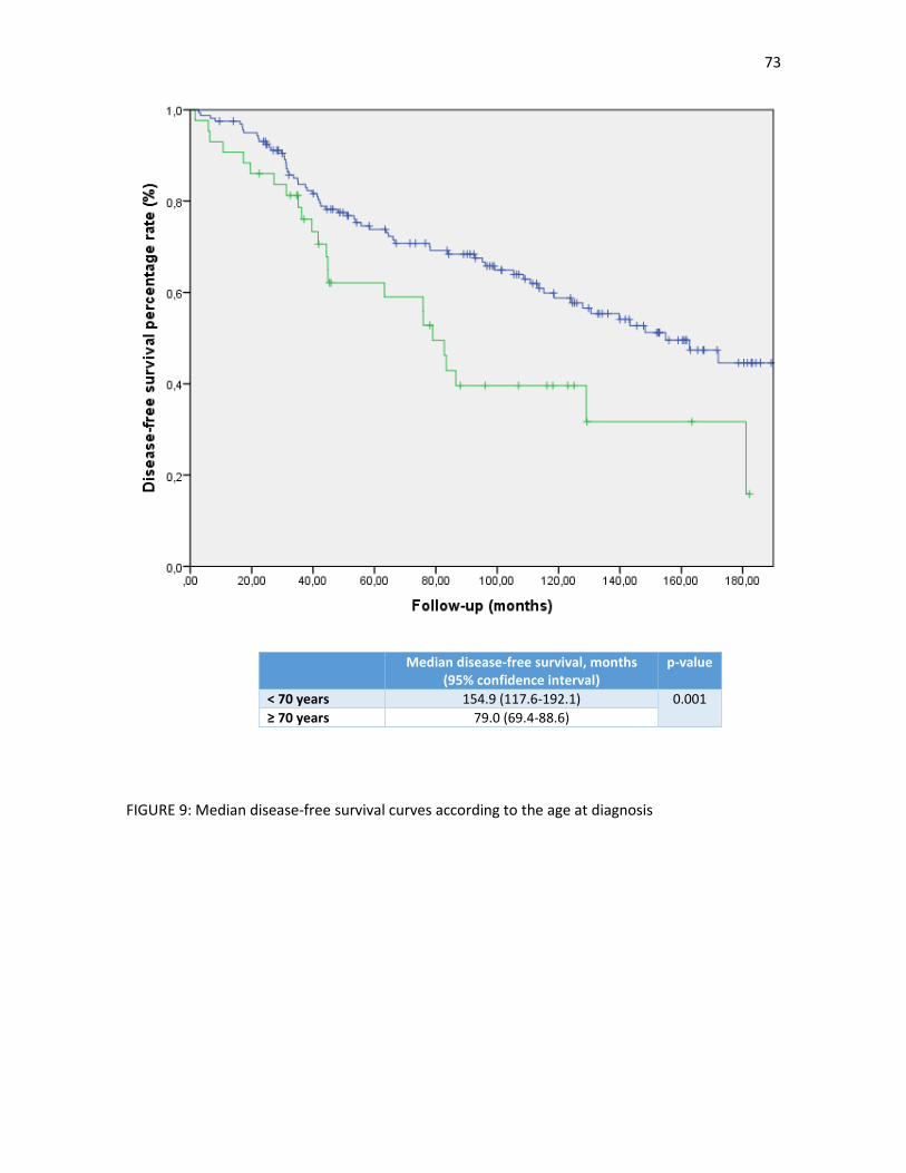
73
FIGURE 9: Median disease-free survival curves according to the age at diagnosis
Median disease-free survival, months (95% confidence interval)
p-value
< 70 years 154.9 (117.6-192.1) 0.001
≥ 70 years 79.0 (69.4-88.6)
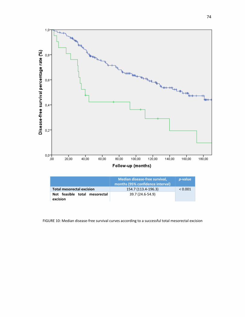
74
FIGURE 10: Median disease-free survival curves according to a successful total mesorectal excision
Median disease-free survival, months (95% confidence interval)
p-value
Total mesorectal excision 154.7 (113.4-196.3) < 0.001
Not feasible total mesorectal excision
39.7 (24.6-54.9)
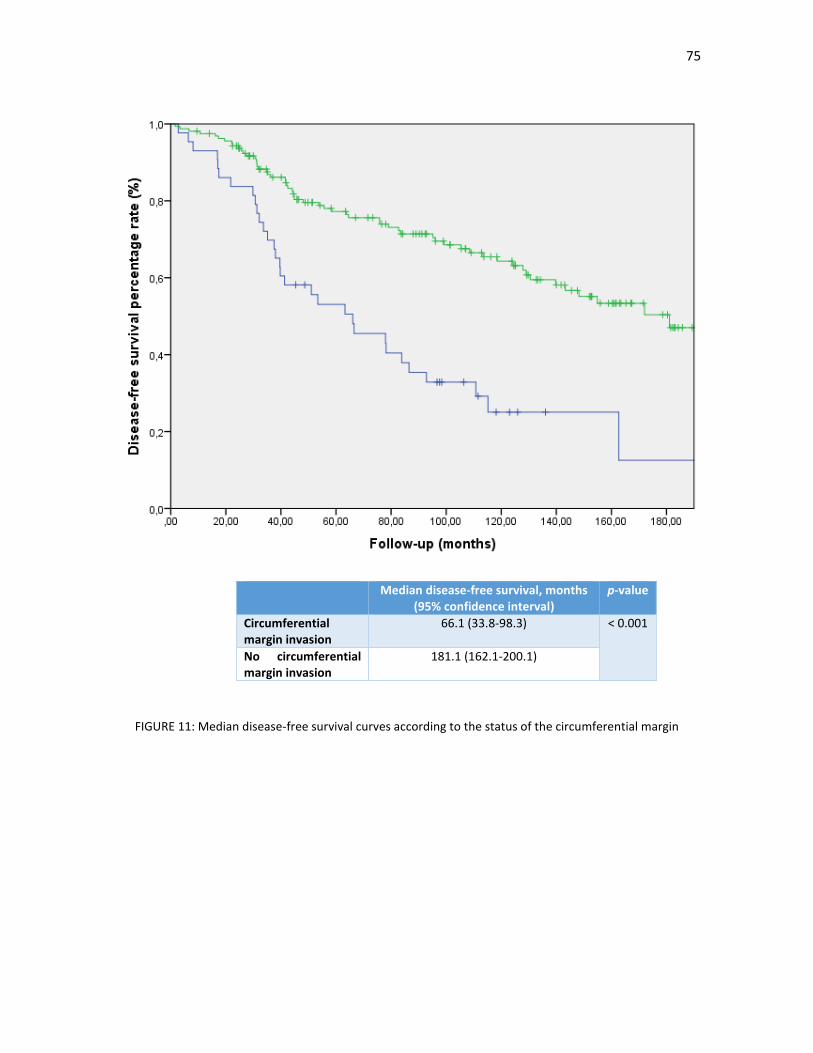
75
FIGURE 11: Median disease-free survival curves according to the status of the circumferential margin
Median disease-free survival, months (95% confidence interval)
p-value
Circumferential margin invasion
66.1 (33.8-98.3) < 0.001
No circumferential margin invasion
181.1 (162.1-200.1)
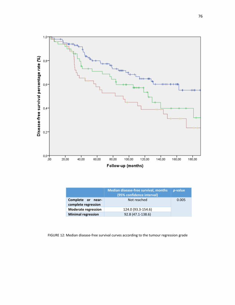
76
FIGURE 12: Median disease-free survival curves according to the tumour regression grade
Median disease-free survival, months (95% confidence interval)
p-value
Complete or near-complete regression
Not reached 0.005
Moderate regression 124.0 (93.3-154.6)
Minimal regression 92.8 (47.1-138.6)
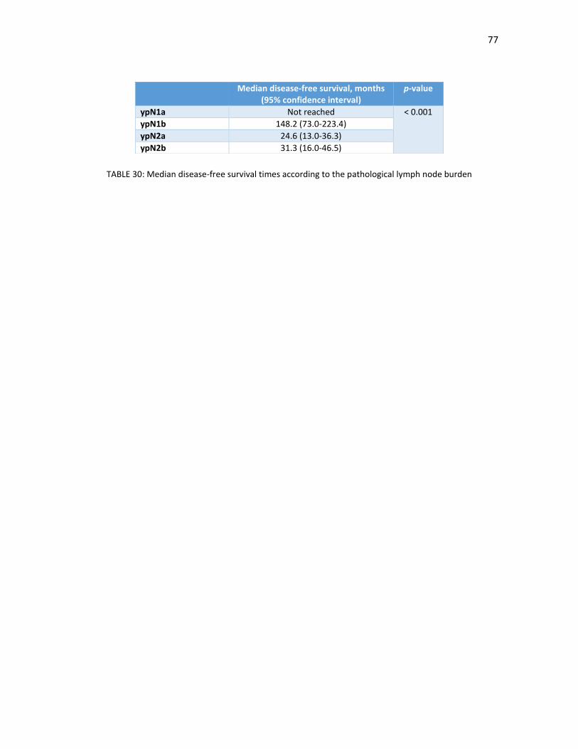
77
TABLE 30: Median disease-free survival times according to the pathological lymph node burden
Median disease-free survival, months (95% confidence interval)
p-value
ypN1a Not reached < 0.001
ypN1b 148.2 (73.0-223.4)
ypN2a 24.6 (13.0-36.3)
ypN2b 31.3 (16.0-46.5)
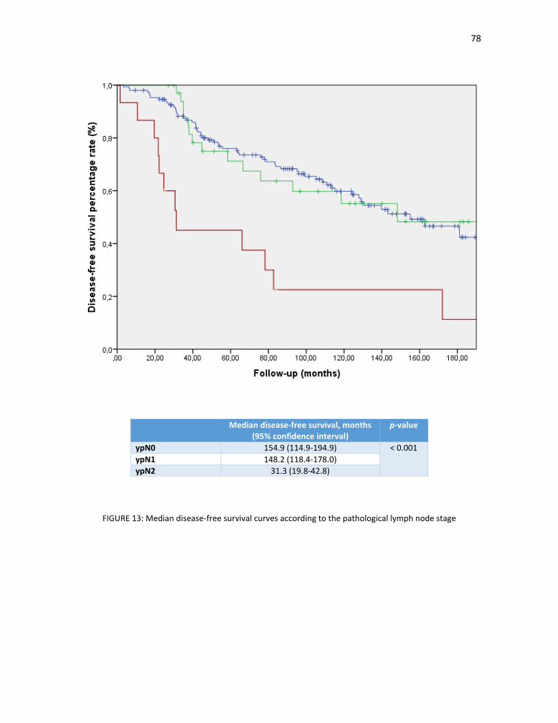
78
FIGURE 13: Median disease-free survival curves according to the pathological lymph node stage
Median disease-free survival, months (95% confidence interval)
p-value
ypN0 154.9 (114.9-194.9) < 0.001
ypN1 148.2 (118.4-178.0)
ypN2 31.3 (19.8-42.8)

79
3.9.2. Multivariate analysis for prognostic factors for DFS and OS
Significant factors in the univariate analysis were included in the multivariate analysis, both for DFS and OS.
Results are shown in TABLES 32 and 33. In our model, age 70 years or older, circumferential margin invasion,
an unsuccessful TME procedure and a pathological heavy lymph node burden remained statistically significant
factors for worse survival outcomes.
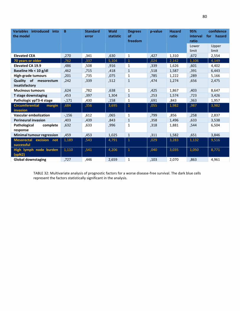
80
TABLE 32: Multivariate analysis of prognostic factors for a worse disease-free survival. The dark blue cells represent the factors statistically significant in the analysis.
Variables introduced into the model
B Standard error
Wald statistic
Degrees of freedom
p-value Hazard ratio
95% confidence interval for hazard ratio
Lower limit
Upper limit
Elevated CEA ,270 ,341 ,630 1 ,427 1,310 ,672 2,554
70 years or older ,762 ,337 5,104 1 ,024 2,142 1,106 4,149
Elevated CA 19.9 ,486 ,508 ,916 1 ,339 1,626 ,601 4,402
Baseline Hb < 10 g/dl ,462 ,715 ,418 1 ,518 1,587 ,391 6,443
High-grade tumours ,201 ,735 ,075 1 ,785 1,222 ,289 5,166
Quality of mesorectum insatisfactory
,242 ,339 ,512 1 ,474 1,274 ,656 2,475
Mucinous tumours ,624 ,782 ,638 1 ,425 1,867 ,403 8,647
T stage downstaging ,453 ,397 1,304 1 ,253 1,574 ,723 3,426
Pathologic ypT3-4 stage -,171 ,430 ,158 1 ,691 ,843 ,363 1,957
Circumferential margin invasion
,684 ,356 3,695 1 ,055 1,982 ,987 3,982
Vascular embolization -,156 ,612 ,065 1 ,799 ,856 ,258 2,837
Perineural invasion ,403 ,439 ,843 1 ,358 1,496 ,633 3,538
Pathological complete response
,632 ,633 ,996 1 ,318 1,881 ,544 6,504
Minimal tumour regression ,459 ,453 1,025 1 ,311 1,582 ,651 3,846
Mesorectal excision not successful
1,189 ,543 4,791 1 ,029 3,283 1,132 9,516
High lymph node burden (ypN2)
1,110 ,541 4,206 1 ,040 3,035 1,050 8,771
Global downstaging ,727 ,446 2,659 1 ,103 2,070 ,863 4,961
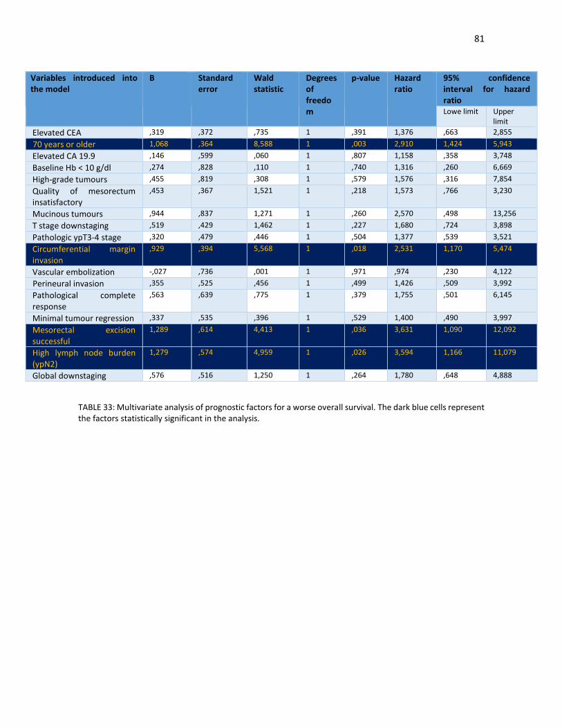
81
TABLE 33: Multivariate analysis of prognostic factors for a worse overall survival. The dark blue cells represent the factors statistically significant in the analysis.
Variables introduced into the model
B Standard error
Wald statistic
Degrees of freedom
p-value Hazard ratio
95% confidence interval for hazard ratio Lowe limit Upper
limit
Elevated CEA ,319 ,372 ,735 1 ,391 1,376 ,663 2,855
70 years or older 1,068 ,364 8,588 1 ,003 2,910 1,424 5,943
Elevated CA 19.9 ,146 ,599 ,060 1 ,807 1,158 ,358 3,748
Baseline Hb < 10 g/dl ,274 ,828 ,110 1 ,740 1,316 ,260 6,669
High-grade tumours ,455 ,819 ,308 1 ,579 1,576 ,316 7,854
Quality of mesorectum insatisfactory
,453 ,367 1,521 1 ,218 1,573 ,766 3,230
Mucinous tumours ,944 ,837 1,271 1 ,260 2,570 ,498 13,256
T stage downstaging ,519 ,429 1,462 1 ,227 1,680 ,724 3,898
Pathologic ypT3-4 stage ,320 ,479 ,446 1 ,504 1,377 ,539 3,521
Circumferential margin invasion
,929 ,394 5,568 1 ,018 2,531 1,170 5,474
Vascular embolization -,027 ,736 ,001 1 ,971 ,974 ,230 4,122
Perineural invasion ,355 ,525 ,456 1 ,499 1,426 ,509 3,992
Pathological complete response
,563 ,639 ,775 1 ,379 1,755 ,501 6,145
Minimal tumour regression ,337 ,535 ,396 1 ,529 1,400 ,490 3,997
Mesorectal excision successful
1,289 ,614 4,413 1 ,036 3,631 1,090 12,092
High lymph node burden (ypN2)
1,279 ,574 4,959 1 ,026 3,594 1,166 11,079
Global downstaging ,576 ,516 1,250 1 ,264 1,780 ,648 4,888
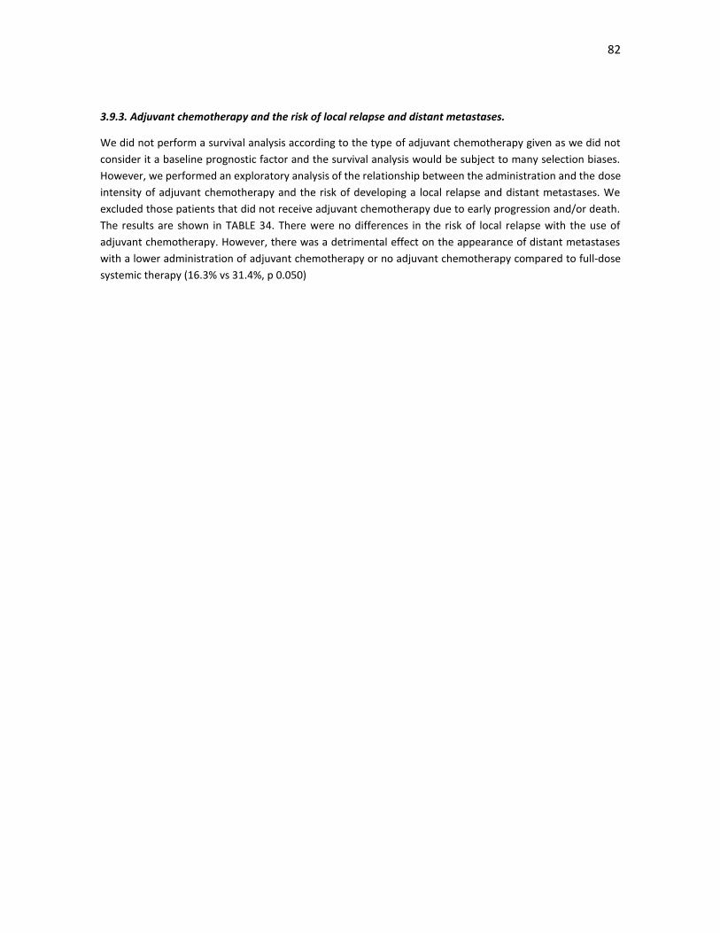
82
3.9.3. Adjuvant chemotherapy and the risk of local relapse and distant metastases.
We did not perform a survival analysis according to the type of adjuvant chemotherapy given as we did not
consider it a baseline prognostic factor and the survival analysis would be subject to many selection biases.
However, we performed an exploratory analysis of the relationship between the administration and the dose
intensity of adjuvant chemotherapy and the risk of developing a local relapse and distant metastases. We
excluded those patients that did not receive adjuvant chemotherapy due to early progression and/or death.
The results are shown in TABLE 34. There were no differences in the risk of local relapse with the use of
adjuvant chemotherapy. However, there was a detrimental effect on the appearance of distant metastases
with a lower administration of adjuvant chemotherapy or no adjuvant chemotherapy compared to full-dose
systemic therapy (16.3% vs 31.4%, p 0.050)
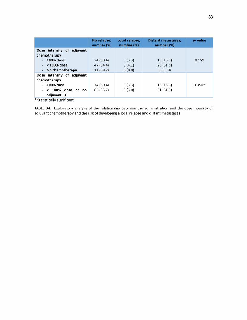
83
No relapse, number (%)
Local relapse, number (%)
Distant metastases, number (%)
p- value
Dose intensity of adjuvant chemotherapy
- 100% dose - < 100% dose - No chemotherapy
74 (80.4) 47 (64.4) 11 (69.2)
3 (3.3) 3 (4.1) 0 (0.0)
15 (16.3) 23 (31.5) 8 (30.8)
0.159
Dose intensity of adjuvant chemotherapy
- 100% dose - < 100% dose or no
adjuvant CT
74 (80.4) 65 (65.7)
3 (3.3) 3 (3.0)
15 (16.3) 31 (31.3)
0.050*
* Statistically significant
TABLE 34: Exploratory analysis of the relationship between the administration and the dose intensity of adjuvant chemotherapy and the risk of developing a local relapse and distant metastases

84
4.9.4. Validation of the study group with the Valentini nomogram and the Dhadda score
Finally, we analysed if our study population would fit the Valentini nomogram [34] and the Dhadda score [35].
We categorized our patients in one of the three Valentini prognostic subgroups; the results are shown in
TABLE 35 and in FIGURES 14 and 15. The use of nomogram predicted fairly accurately a fairly large group of
patients with a high risk of distant relapse (61.8%) and worse OS (52.5%): in contrast, only 16% of our patients
had a high risk of local relapse with the use of the nomogram.
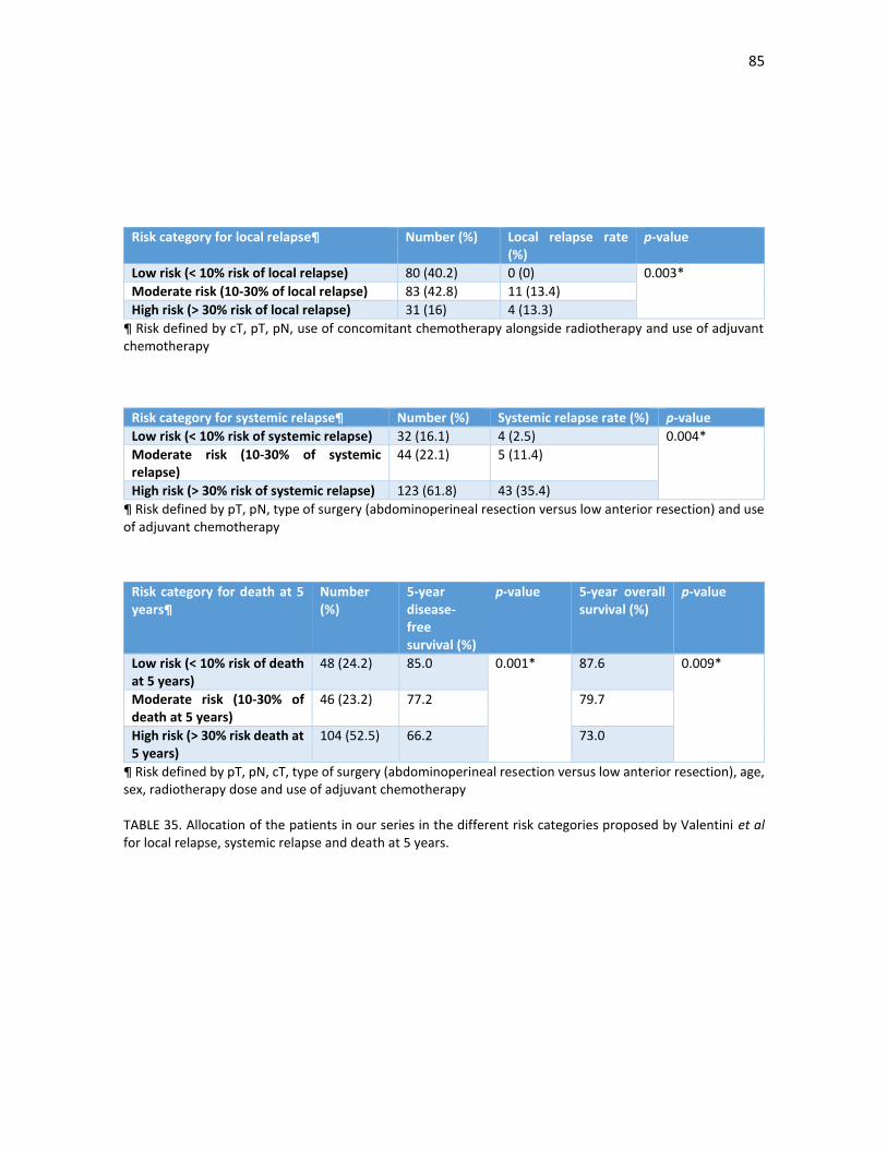
85
Risk category for local relapse¶ Number (%) Local relapse rate (%)
p-value
Low risk (< 10% risk of local relapse) 80 (40.2) 0 (0) 0.003*
Moderate risk (10-30% of local relapse) 83 (42.8) 11 (13.4)
High risk (> 30% risk of local relapse) 31 (16) 4 (13.3)
¶ Risk defined by cT, pT, pN, use of concomitant chemotherapy alongside radiotherapy and use of adjuvant chemotherapy
Risk category for systemic relapse¶ Number (%) Systemic relapse rate (%) p-value
Low risk (< 10% risk of systemic relapse) 32 (16.1) 4 (2.5) 0.004*
Moderate risk (10-30% of systemic relapse)
44 (22.1) 5 (11.4)
High risk (> 30% risk of systemic relapse) 123 (61.8) 43 (35.4)
¶ Risk defined by pT, pN, type of surgery (abdominoperineal resection versus low anterior resection) and use of adjuvant chemotherapy
¶ Risk defined by pT, pN, cT, type of surgery (abdominoperineal resection versus low anterior resection), age, sex, radiotherapy dose and use of adjuvant chemotherapy TABLE 35. Allocation of the patients in our series in the different risk categories proposed by Valentini et al for local relapse, systemic relapse and death at 5 years.
Risk category for death at 5 years
Number (%)
5-year disease-free survival (%)
p-value 5-year overall survival (%)
p-value
Low risk (< 10% risk of death at 5 years)
48 (24.2) 85.0 0.001* 87.6 0.009*
Moderate risk (10-30% of death at 5 years)
46 (23.2) 77.2 79.7
High risk (> 30% risk death at 5 years)
104 (52.5) 66.2 73.0
86
FIGURE 16: Disease-free survival curves for patients allocated to the three risk categories proposed by Valentini et al [34].
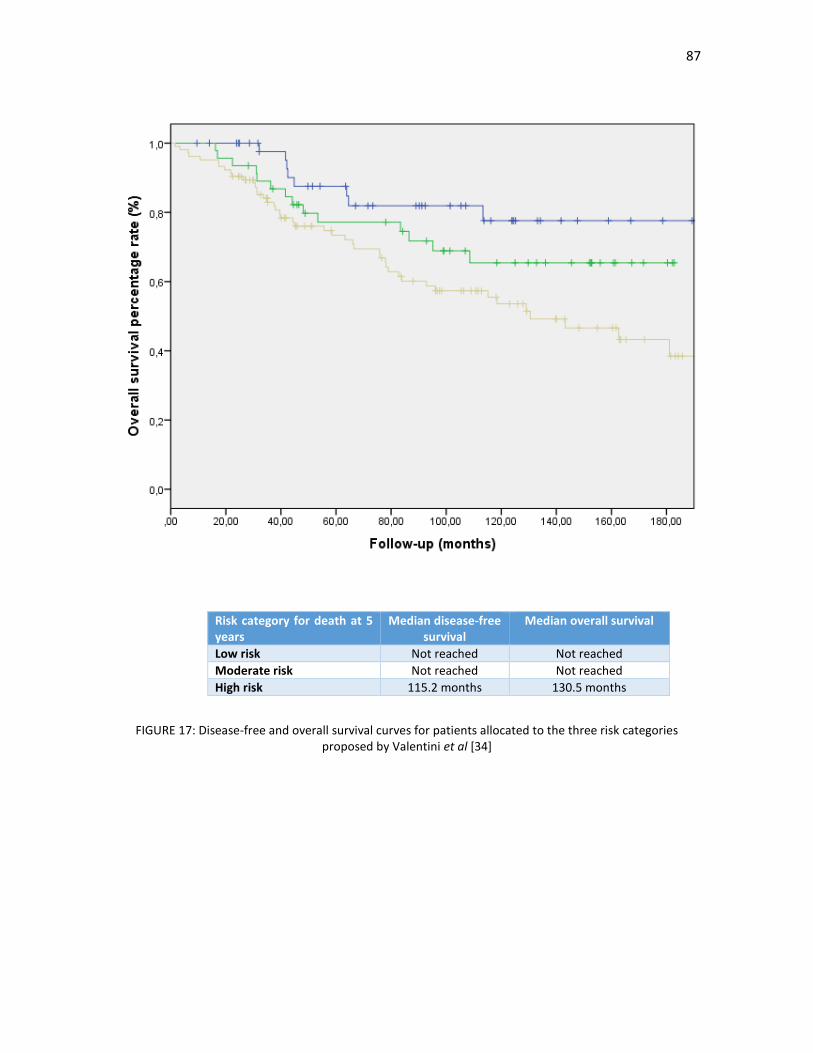
87
FIGURE 17: Disease-free and overall survival curves for patients allocated to the three risk categories proposed by Valentini et al [34]
Risk category for death at 5 years
Median disease-free survival
Median overall survival
Low risk Not reached Not reached
Moderate risk Not reached Not reached
High risk 115.2 months 130.5 months

88
We also categorized our patients in one of the four Dhadda prognostic subgroups; the results are shown in
TABLE 38 and in FIGURES 16 and 17. With this classification, most patients belonged to the excellent (42.6%)
and good (26.2%) prognosis groups; there were no differences in DFS and OS between these two groups in
our population, although there was a slightly higher risk of local relapse (5.9% vs 0%) in the good prognosis
group. 23.6% and 7.7% of patients belonged to the moderate and poor prognosis groups and there were clear
differences in the DFS and OS between these two stages and with the other two stages; however, the risk of
local relapse was curiously higher in the moderate risk group than in the poor risk group.
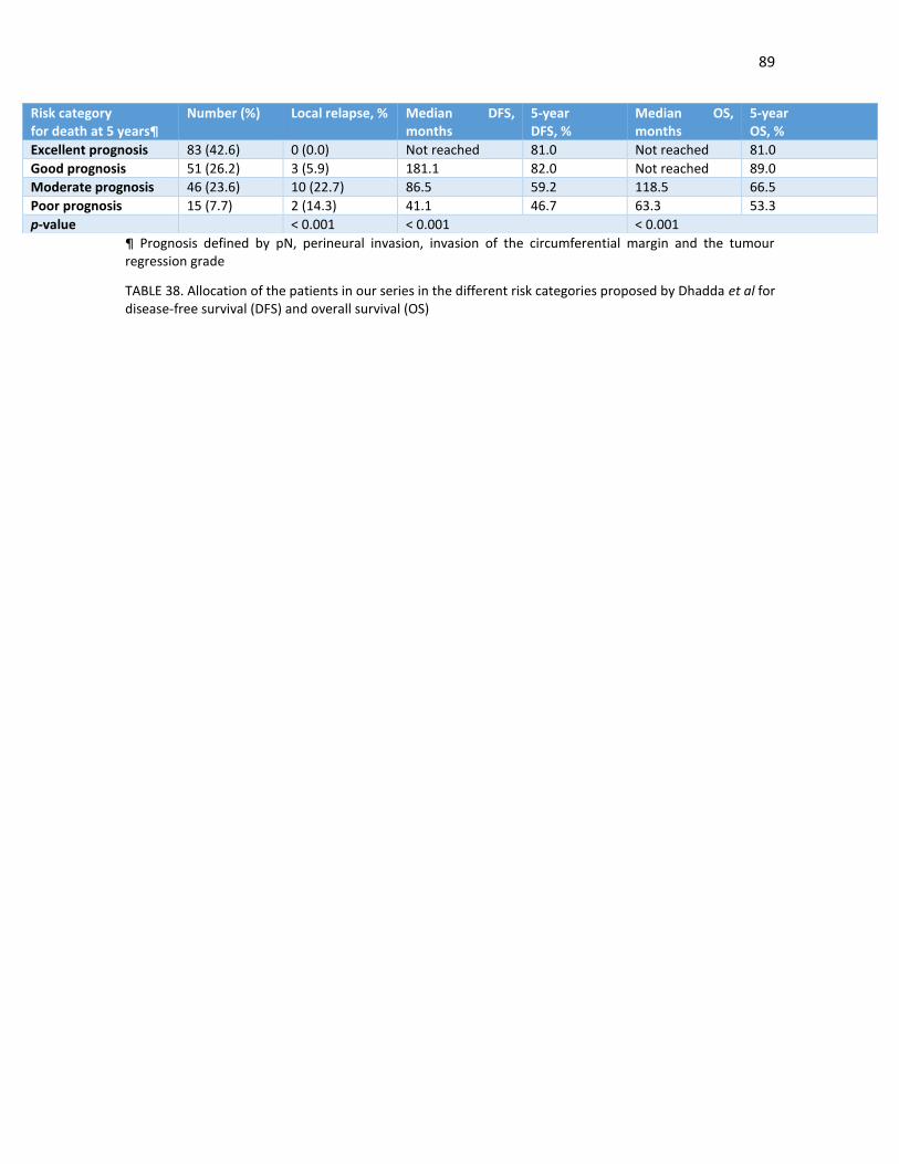
89
¶ Prognosis defined by pN, perineural invasion, invasion of the circumferential margin and the tumour regression grade
TABLE 38. Allocation of the patients in our series in the different risk categories proposed by Dhadda et al for disease-free survival (DFS) and overall survival (OS)
Risk category for death at 5 years
Number (%) Local relapse, % Median DFS, months
5-year DFS, %
Median OS, months
5-year OS, %
Excellent prognosis 83 (42.6) 0 (0.0) Not reached 81.0 Not reached 81.0
Good prognosis 51 (26.2) 3 (5.9) 181.1 82.0 Not reached 89.0
Moderate prognosis 46 (23.6) 10 (22.7) 86.5 59.2 118.5 66.5
Poor prognosis 15 (7.7) 2 (14.3) 41.1 46.7 63.3 53.3
p-value < 0.001 < 0.001 < 0.001
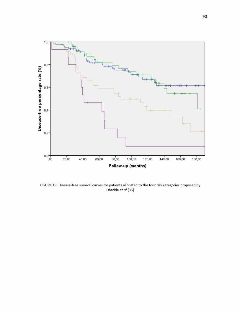
90
FIGURE 18: Disease-free survival curves for patients allocated to the four risk categories proposed by Dhadda et al [35]
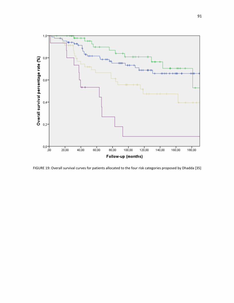
91
FIGURE 19: Overall survival curves for patients allocated to the four risk categories proposed by Dhadda [35]
92
4. Discussion
4.1. Introduction
The treatment of locally advanced rectal cancer has been one of the success stories in Oncology of the last
decades. From where we were in the 1980s, when up to 50% of the patients suffering from a locally advanced
tumour developed a pelvic recurrence, the decrease in this event to less than 10% with the incorporation of
radiotherapy and without any contribution from new drugs, except of 5-FU, is a landmark observation in the
management of this malignancy. Several factors are behind these improvements: the widespread introduction
of TME as the standard surgical approach, the improvement in the clinical staging of these patients with EUS
and, especially, with high-quality MRI, the integration of (neo)adjuvant radiotherapy and chemotherapy in
localized disease, a better pathological diagnosis with the recognition of the fundamental prognostic role of
the CRM and, lastly but not less important, the management of these patients in the setting of
multidisciplinary units, where the most appropriate approach can be decided between the different
disciplines [36].
However, despite these benefits, several shortcomings are now evident in the management of our patients.
With the marked improvement in the risk of pelvic recurrences, distant metastases are now the main cause
of death in around 30% of patients; unfortunately, in the vast majority of randomized trials of chemotherapy
and/or radiotherapy, no overall survival benefits have been observed, despite the marked improvement in
local control rate. The integration of adjuvant chemotherapy, in the same manner as in colon cancer, has been
difficult and compliance rates have been poor; moreover, the benefit is much less clear than in colon cancer,
especially after LCRCT and surgery. Although clinical staging has improved enormously, there is still an
important group of patients that are over- or understaged, especially in the identification of pathological
lymph nodes, probably the most important prognostic factor alongside the invasion of the CRM. Finally, and
perhaps more under-recognised, are the risks of late toxicity. Recognised long-term side-effects of pelvic
radiotherapy for rectal cancer include faecal incontinence, sexual and urinary dysfunction, and sterility [37-
39]. There are also suggestions of an increase in the risk of second malignancies [40-41]. These risks can be
accumulative; quality-of-life data from some trials have demonstrated a significant impairment in sexual
function attributable to surgery and a further detriment due to radiotherapy and a similar pattern for faecal
incontinence [37].
In this sense, the long-term results of our study of patients with locally advanced rectal cancer are broadly
similar to other studies of LCRCT followed by surgery and we think that they can be considered moderately
successful: there was a local relapse rate of less than 10%, with a downstaging rate of 70% and a pCR rate of
14.8%. 30% of patients relapsed, the vast majority in the form of distant metastases and the 10-year OS rate
was 62.4%, with most relapses taking place in the first five years of follow-up. However, there were several
concerning factors: older patients fared poorly compared to their younger counterparts; despite the use of
LCRC and specialized surgery, the quality of the mesorectum was unsatisfactory in around a quarter of patients
and 21% had a positive CRM; finally, the compliance in the use of adjuvant chemotherapy was rather poor,
with less than half of patients receiving the full intended dose and around a quarter of patients not receiving
any type of adjuvant treatment. We followed a blanket approach of treating all patients with cT3-4 and/or
lymph node positive disease with LCRCT and adjuvant chemotherapy, irrespective of response, as our
standard treatment, as is usually done in the United States and in many countries of Europe. However, as we
will see, the risk factors of local relapse and distant metastases are different although overlapping. In this
sense, how can we improve our results? Can we offer a more personalized approach taking into account these
differing risks of local and metastatic relapse, as is recommended by ESMO and NICE? Can we better select
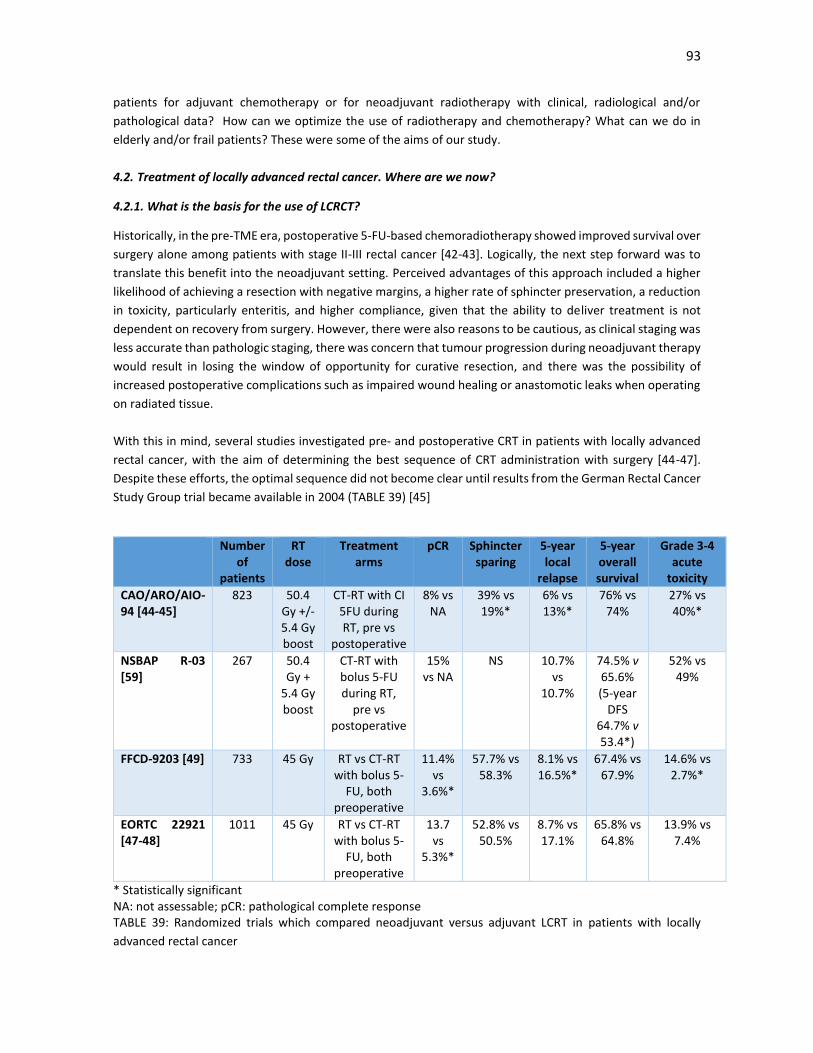
93
patients for adjuvant chemotherapy or for neoadjuvant radiotherapy with clinical, radiological and/or
pathological data? How can we optimize the use of radiotherapy and chemotherapy? What can we do in
elderly and/or frail patients? These were some of the aims of our study.
4.2. Treatment of locally advanced rectal cancer. Where are we now?
4.2.1. What is the basis for the use of LCRCT?
Historically, in the pre-TME era, postoperative 5-FU-based chemoradiotherapy showed improved survival over
surgery alone among patients with stage II-III rectal cancer [42-43]. Logically, the next step forward was to
translate this benefit into the neoadjuvant setting. Perceived advantages of this approach included a higher
likelihood of achieving a resection with negative margins, a higher rate of sphincter preservation, a reduction
in toxicity, particularly enteritis, and higher compliance, given that the ability to deliver treatment is not
dependent on recovery from surgery. However, there were also reasons to be cautious, as clinical staging was
less accurate than pathologic staging, there was concern that tumour progression during neoadjuvant therapy
would result in losing the window of opportunity for curative resection, and there was the possibility of
increased postoperative complications such as impaired wound healing or anastomotic leaks when operating
on radiated tissue.
With this in mind, several studies investigated pre- and postoperative CRT in patients with locally advanced
rectal cancer, with the aim of determining the best sequence of CRT administration with surgery [44-47].
Despite these efforts, the optimal sequence did not become clear until results from the German Rectal Cancer
Study Group trial became available in 2004 (TABLE 39) [45]
Number of
patients
RT dose
Treatment arms
pCR Sphincter sparing
5-year local
relapse
5-year overall survival
Grade 3-4 acute
toxicity
CAO/ARO/AIO-94 [44-45]
823 50.4 Gy +/- 5.4 Gy boost
CT-RT with CI 5FU during RT, pre vs
postoperative
8% vs NA
39% vs 19%*
6% vs 13%*
76% vs 74%
27% vs 40%*
NSBAP R-03 [59]
267 50.4 Gy +
5.4 Gy boost
CT-RT with bolus 5-FU during RT,
pre vs postoperative
15% vs NA
NS 10.7% vs
10.7%
74.5% v 65.6% (5-year
DFS 64.7% v 53.4*)
52% vs 49%
FFCD-9203 [49] 733 45 Gy RT vs CT-RT with bolus 5-
FU, both preoperative
11.4% vs
3.6%*
57.7% vs 58.3%
8.1% vs 16.5%*
67.4% vs 67.9%
14.6% vs 2.7%*
EORTC 22921 [47-48]
1011 45 Gy RT vs CT-RT with bolus 5-
FU, both preoperative
13.7 vs
5.3%*
52.8% vs 50.5%
8.7% vs 17.1%
65.8% vs 64.8%
13.9% vs 7.4%
* Statistically significant NA: not assessable; pCR: pathological complete response TABLE 39: Randomized trials which compared neoadjuvant versus adjuvant LCRT in patients with locally
advanced rectal cancer
94
The German Rectal Cancer Study Group trial was initiated in February 1995, and enrollment was extended
through September 2002 [44]. The study enrolled 823 patients with clinical stage T3 or T4 or node-positive
disease. Patients were then randomly assigned to receive either preoperative or postoperative LCRCT. The
preoperative treatment consisted of 50.40 Gy concurrently with infusional 5-FU. The primary endpoint was
OS. All patients underwent surgery 6 weeks after the completion of LCRCT. One month after this surgery, 4
additional cycles of adjuvant 5-FU were administered. LCRCT was identical in the postoperative treatment
group, except for the delivery of a boost of 54 Gy in the latter group.
Results from a 46-month median follow-up analysis showed that preoperative treatment was associated with
a significantly lower cumulative incidence of local relapse than postoperative CRT. However, there were no
significant differences in 5-year DFS and OS. Following preoperative CRT, there was a significant shift toward
earlier TNM staging, which was suggestive of a significant downstaging effect. 8% of the patients in the
preoperative CRT group had a pCR, and only 25% had positive lymph nodes (compared with 40% in the
postoperative group). Of note, in patients with tumours that had been predicted preoperatively by a surgeon
to require an APR, the rate of sphincter-preserving surgery was more than twice as high after preoperative
CRT compared with postoperative CRT (39% vs 19%). However, postoperative treatment was associated with
a significant increase in the rate of short- (27% vs 40%) and long-term toxic effects (14% vs 24%). A recent
update of the trial with an 11-year follow-up confirmed these conclusions, with a significant, albeit smaller,
benefit in the risk of local relapse but no difference in DFS and OS [45]. This trial established the superiority
of preoperative over postoperative LCRCT.
However, two concerning facts were also evident: 18% of patients in the postoperative treatment group with
no CRT prior to surgery who were determined preoperatively to have T3 or T4 disease or lymph node
metastasis were found to actually have T1 or T2 or node-negative tumors on pathologic examination of the
resected specimen. This important observation clearly highlights the limitations of preoperative staging,
especially at the time the study was performed. Also, compliance with the postoperative arm was worse than
in the preoperative arm, with 92% of patients in the preoperative arm receiving a full dose of RT, compared
with only 54% in the postoperative arm and 89% compared with 50% receiving a full dose of chemotherapy;
both of these findings were statistically significant and may have justified the local control benefit observed.
Another important earlier randomized trial tried to answer two relevant questions: the benefit of
preoperative versus postoperative radiotherapy and the benefits of concomitant chemotherapy alongside
radiotherapy. With this in mind, The European Organisation for Research and Treatment of Cancer (EORTC)
Radiotherapy Group initiated the EORTC 22921 trial in 1993, which randomized more than 1,000 patients with
T3-T4 resectable rectal cancer, using a 2 × 2 factorial design, to one of the following four treatment regimens
(along with surgery): 1) preoperative radiotherapy (the control arm), 2) preoperative LCRCT, 3) preoperative
radiotherapy and postoperative chemotherapy, or 4) preoperative CRT and postoperative chemotherapy.
Preoperative and postoperative chemotherapy regimens involved bolus 5-FU plus leucovorin. In addition to
effects on local tumor control, the ability of the four treatment approaches to improve OS and progression-
free survival (PFS) was analyzed. [47]
Combined chemoradiotherapy resulted in downstaging of tumors and increased local control rates. Local
failure rates were significantly lower in all three groups that received chemotherapy, compared with the
control arm (preoperative RT alone), regardless of whether chemotherapy was given prior to or following
surgery. Patients undergoing preoperative CRT had a significantly higher rate of pathologic complete response
(pCR; 14% vs 5%). However, the addition of chemotherapy, either concurrently with preoperative RT or
95
postoperatively again was not associated with any improvement in PFS or OS. Updated long-term results of
this study were published in 2014. The reported cumulative incidence of local relapse at 10 years was 22.4%
in patients who received RT alone, compared with 11% to 15% in the three groups that received chemotherapy
[49-50] Ten-year DFS and OS was similar in patients who received preoperative CRT vs RT alone. One
important finding was that adjuvant 5-FU–based chemotherapy after preoperative RT (with or without
chemotherapy) still did not affect DFS or OS [48].
Other studies, such as the Fédération Francophone de Cancérologie Digestive (FFCD) trial 9203 [53] yielded
similar results and showed that the addition of 5-FU to RT significantly increased the pCR and local disease
control rate, with no benefit in DFS or OS.
In a Cochrane analysis that included the EORTC, the FFCD and four other trials which compared SCRT and
LCRCT [52-56] the addition of chemotherapy to preoperative RT for the treatment of locally advanced rectal
cancer was associated with a lower risk of local relapse that was statistically significant [57]. With all these
results, the addition of chemotherapy to conventional fractionation RT became a standard approach in the
treatment of locally advanced rectal cancer, likely because of the reduction in local recurrence and
improvement in pCR rates observed with CRT.
On the other hand, in addition to these trials, two other prospective randomized trials with the same aim were
initiated in the United States by the Radiation Therapy Oncology Group (RTOG; trial 94-01) and the National
Surgical Adjuvant Breast and Bowel Project (NSABP; protocol R-03). Unfortunately, both studies were closed
prematurely due to poor accrual. However, the NSABP Group published their results with the 257 patients
accrued [58]. Surprisingly, there was a significant benefit in DFS with the preoperative approach, with no
benefit in the local relapse rate; there was surprisingly a higher acute grade 3-4 toxicity with the preoperative
arm. These findings are difficult to harmonize with the European trials. The unusual chemotherapy regimen,
alongside the high RT dose and the non- standard use of TME, alongside the early closure of the trial, may
justify some of these findings, although the explanation remains elusive [59].
With the establishment of 5-FU based LCRT, following the development of orally active fluoropyrimidines (ie,
capecitabine or UFT), the natural question was whether these drugs could replace infusional 5-FU in this
setting. Two clinical trials studied the effect of using capecitabine in place of infusional 5-FU, combined with
RT, on pCR, sphincter-sparing surgery, and surgical downstaging. Unfortunately, there are no phase III trials
with UFT and it was removed from the Spanish market on 2007.
The first trial [61] was a neoadjuvant, open-label, multicenter, noninferiority, randomized phase III study that
explored the substitution of capecitabine for infusional 5-FU [60]. A total of 401 patients with locally advanced
rectal cancer were enrolled and randomly assigned to either the oral or intravenous fluoropyrimidine group.
There were no differences in 5-year OS and and 3-year DFS was higher in the capecitabine group in both the
adjuvant and neoadjuvant cohorts. The local recurrence rate was similarly low for capecitabine and
intravenous 5-FU (6% vs 7%), but fewer patients developed distant metastases in the capecitabine group.
The second phase III trial, which was conducted by the NSABP [61] employed a 2 × 2 factorial noninferiority
design to compare four regimens administered concomitantly with RT (45 Gy in 25 fractions over 5 weeks,
followed by a boost). The aim of this study was to determine the optimal neoadjuvant chemotherapy regimen
for stage II/III rectal cancer, including the best sequence of administration of preoperative CRT. The trial
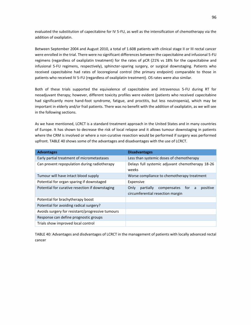
96
evaluated the substitution of capecitabine for IV 5-FU, as well as the intensification of chemotherapy via the
addition of oxaliplatin.
Between September 2004 and August 2010, a total of 1.608 patients with clinical stage II or III rectal cancer
were enrolled in the trial. There were no significant differences between the capecitabine and infusional 5-FU
regimens (regardless of oxaliplatin treatment) for the rates of pCR (21% vs 18% for the capecitabine and
infusional 5-FU regimens, respectively), sphincter-sparing surgery, or surgical downstaging. Patients who
received capecitabine had rates of locoregional control (the primary endpoint) comparable to those in
patients who received IV 5-FU (regardless of oxaliplatin treatment). OS rates were also similar.
Both of these trials supported the equivalence of capecitabine and intravenous 5-FU during RT for
neoadjuvant therapy; however, different toxicity profiles were evident (patients who received capecitabine
had significantly more hand-foot syndrome, fatigue, and proctitis, but less neutropenia), which may be
important in elderly and/or frail patients. There was no benefit with the addition of oxaliplatin, as we will see
in the following sections.
As we have mentioned, LCRCT is a standard treatment approach in the United States and in many countries
of Europe. It has shown to decrease the risk of local relapse and it allows tumour downstaging in patients
where the CRM is involved or where a non-curative resection would be performed if surgery was performed
upfront. TABLE 40 shows some of the advantages and disadvantages with the use of LCRCT.
Advantages Disadvantages
Early partial treatment of micrometastases Less than systemic doses of chemotherapy
Can prevent repopulation during radiotherapy Delays full systemic adjuvant chemotherapy 18-26
weeks
Tumour will have intact blood supply Worse compliance to chemotherapy treatment
Potential for organ sparing if downstaged Expensive
Potential for curative resection if downstaging Only partially compensates for a positive
circumferential resection margin
Potential for brachytherapy boost
Potential for avoiding radical surgery?
Avoids surgery for resistant/progressive tumours
Response can define prognostic groups
Trials show improved local control
TABLE 40: Advantages and disdvantages of LCRCT in the management of patients with locally advanced rectal
cancer
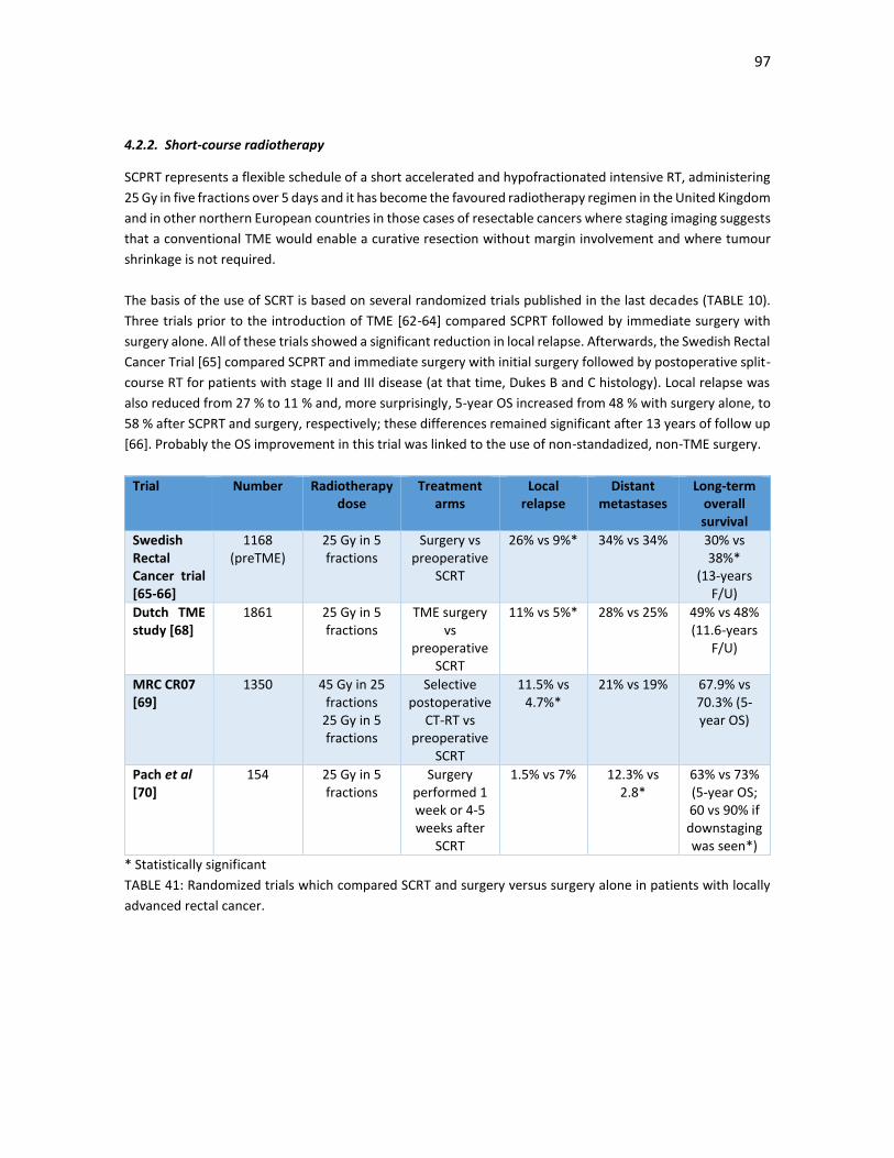
97
4.2.2. Short-course radiotherapy
SCPRT represents a flexible schedule of a short accelerated and hypofractionated intensive RT, administering
25 Gy in five fractions over 5 days and it has become the favoured radiotherapy regimen in the United Kingdom
and in other northern European countries in those cases of resectable cancers where staging imaging suggests
that a conventional TME would enable a curative resection without margin involvement and where tumour
shrinkage is not required.
The basis of the use of SCRT is based on several randomized trials published in the last decades (TABLE 10).
Three trials prior to the introduction of TME [62-64] compared SCPRT followed by immediate surgery with
surgery alone. All of these trials showed a significant reduction in local relapse. Afterwards, the Swedish Rectal
Cancer Trial [65] compared SCPRT and immediate surgery with initial surgery followed by postoperative split-
course RT for patients with stage II and III disease (at that time, Dukes B and C histology). Local relapse was
also reduced from 27 % to 11 % and, more surprisingly, 5-year OS increased from 48 % with surgery alone, to
58 % after SCPRT and surgery, respectively; these differences remained significant after 13 years of follow up
[66]. Probably the OS improvement in this trial was linked to the use of non-standadized, non-TME surgery.
Trial Number Radiotherapy dose
Treatment arms
Local relapse
Distant metastases
Long-term overall survival
Swedish Rectal Cancer trial [65-66]
1168 (preTME)
25 Gy in 5 fractions
Surgery vs preoperative
SCRT
26% vs 9%* 34% vs 34% 30% vs 38%*
(13-years F/U)
Dutch TME study [68]
1861 25 Gy in 5 fractions
TME surgery vs
preoperative SCRT
11% vs 5%* 28% vs 25% 49% vs 48% (11.6-years
F/U)
MRC CR07 [69]
1350 45 Gy in 25 fractions
25 Gy in 5 fractions
Selective postoperative
CT-RT vs preoperative
SCRT
11.5% vs 4.7%*
21% vs 19% 67.9% vs 70.3% (5-year OS)
Pach et al [70]
154 25 Gy in 5 fractions
Surgery performed 1 week or 4-5 weeks after
SCRT
1.5% vs 7% 12.3% vs 2.8*
63% vs 73% (5-year OS; 60 vs 90% if downstaging was seen*)
* Statistically significant
TABLE 41: Randomized trials which compared SCRT and surgery versus surgery alone in patients with locally
advanced rectal cancer.
98
In this sense, subsequent trials were designed to test whether SCPRT still reduced local relapse if TME was
performed. By then, it was recognised that the risk of pelvic recurrence, after a potentially curative resection,
was mainly explained by microscopic tumour cells within 1 mm of the CRM [67].
The Dutch trial [68] and the Medical Research Council CR07 trial [69] compared routine SCPRT and immediate
surgery against initial surgery with a policy of selective postoperative treatment restricted to patients with
involvement of the CRM (the Dutch trial used radiotherapy alone and the MRC CR07 trial used concurrent
5FU-based LCRCT). Both trials showed a sustained reduction in LR in the SCPRT group, although there was no
survival benefit and the risk of distant metastases prevailed. In the MRC 07 trial, all patients benefited from
the use of SCRT, even those deemed of low risk. However, in both trials, independently of radiotherapy
administration, a positive CRM was associated with local recurrence and decreased survival. As such, while
the results of this trial support the use of radiotherapy in locally advanced patients, the data underscore the
importance of obtaining a negative CRM with high quality surgery. Unfortunately, radiotherapy is not able to
compensate fully for a positive CRM; however, the same can be said of LCRCT.
Unfortunately, there were several shortcomings in the trial that have made it somewhat different to interpret
these results. For example, the number of patients who were intended to, and actually received postoperative
radiotherapy in the Dutch trial, is not known. In the CR07 trial there were no details on compliance in the 53
patients who received selective postoperative LCRCT, while only 19% received radiotherapy alone and 9 % did
not receive any type of radiotherapy.
At the same time, compared to the CAO/AOC/AIO 94 German trial [49], it was not clear whether the majority
of recurrences occurred in the treated or non-treated patients. Adjuvant chemotherapy was not used in the
Dutch trial, whereas most patients with stage III received 5FU chemotherapy in the CR07 trial. In the CR07
trial, doses of RT mandated were low (45 Gy) compared with standard postoperative CRT trials (such as 55.4
Gy in the German Trial).
Finally, accrual for the MRC 07 trial began in 1998, and surgical quality control was not emphasized at the start
of the trial. As such, it is difficult to know the extent to which variation in the quality of surgical resection may
have played a role in the higher local recurrence rate in the selective therapy arm. As we have said previously,
the overall survival improvement in the Uppsala trial was probably linked to the use of non-TME surgery.
However, the CR07 trial did show that the plane of surgical resection, which may be considered a surrogate
of surgical quality, was highly correlated with outcome [70]. For patients treated within the proper plane
whose radial margins were clear, there was a very low rate of local treatment failure of 6%, even among
patients with node positive disease found on pathologic evaluation, an important finding, as we will see;
patients with low burden lymph node disease may not need necessarily neoadjuvant radiotherapy, as the risk
of local relapse will probably be low in case high quality surgery is performed.
There are several advantages to the use of SCRT (TABLE 42). Compliance is high because toxicity, mainly
digestive and genitourinary, is usually only experienced after treatment is completed. The short overall
treatment time, with immediate surgery (ideally within 7 days), leaves an insufficient interval for radiation-
induced fibrosis to appear and avoids the accelerated repopulation that may occur in the latter part of LCRCT.
It is also a cheap alternative. Decisions on the requirement for postoperative adjuvant chemotherapy can be
made without modification of the pathological stage or effacement of the nodes. Adjuvant chemotherapy
with systemically active regimens, such as FOLFOX, can be started with minimal delay, within a few weeks of
diagnosis. However, an important criticism is the difficult in integrating systemic chemotherapy in the
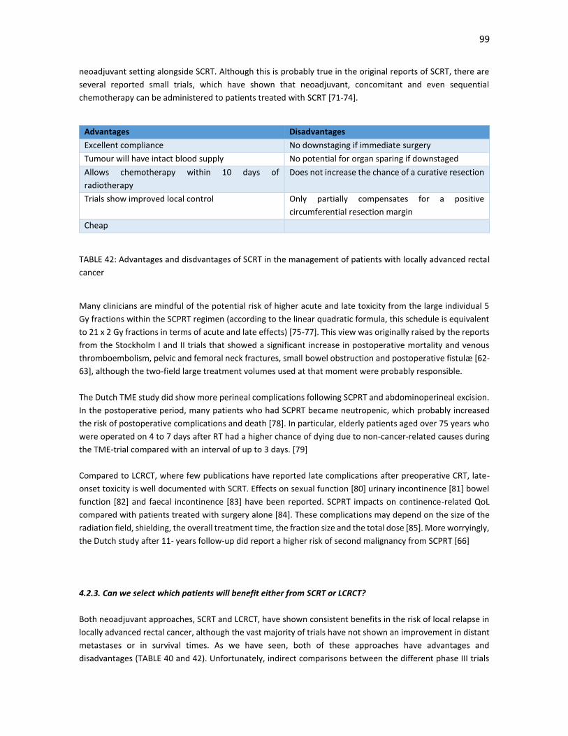
99
neoadjuvant setting alongside SCRT. Although this is probably true in the original reports of SCRT, there are
several reported small trials, which have shown that neoadjuvant, concomitant and even sequential
chemotherapy can be administered to patients treated with SCRT [71-74].
Advantages Disadvantages
Excellent compliance No downstaging if immediate surgery
Tumour will have intact blood supply No potential for organ sparing if downstaged
Allows chemotherapy within 10 days of
radiotherapy
Does not increase the chance of a curative resection
Trials show improved local control Only partially compensates for a positive
circumferential resection margin
Cheap
TABLE 42: Advantages and disdvantages of SCRT in the management of patients with locally advanced rectal
cancer
Many clinicians are mindful of the potential risk of higher acute and late toxicity from the large individual 5
Gy fractions within the SCPRT regimen (according to the linear quadratic formula, this schedule is equivalent
to 21 x 2 Gy fractions in terms of acute and late effects) [75-77]. This view was originally raised by the reports
from the Stockholm I and II trials that showed a significant increase in postoperative mortality and venous
thromboembolism, pelvic and femoral neck fractures, small bowel obstruction and postoperative fistulæ [62-
63], although the two-field large treatment volumes used at that moment were probably responsible.
The Dutch TME study did show more perineal complications following SCPRT and abdominoperineal excision.
In the postoperative period, many patients who had SCPRT became neutropenic, which probably increased
the risk of postoperative complications and death [78]. In particular, elderly patients aged over 75 years who
were operated on 4 to 7 days after RT had a higher chance of dying due to non-cancer-related causes during
the TME-trial compared with an interval of up to 3 days. [79]
Compared to LCRCT, where few publications have reported late complications after preoperative CRT, late-
onset toxicity is well documented with SCRT. Effects on sexual function [80] urinary incontinence [81] bowel
function [82] and faecal incontinence [83] have been reported. SCPRT impacts on continence-related QoL
compared with patients treated with surgery alone [84]. These complications may depend on the size of the
radiation field, shielding, the overall treatment time, the fraction size and the total dose [85]. More worryingly,
the Dutch study after 11- years follow-up did report a higher risk of second malignancy from SCPRT [66]
4.2.3. Can we select which patients will benefit either from SCRT or LCRCT?
Both neoadjuvant approaches, SCRT and LCRCT, have shown consistent benefits in the risk of local relapse in
locally advanced rectal cancer, although the vast majority of trials have not shown an improvement in distant
metastases or in survival times. As we have seen, both of these approaches have advantages and
disadvantages (TABLE 40 and 42). Unfortunately, indirect comparisons between the different phase III trials
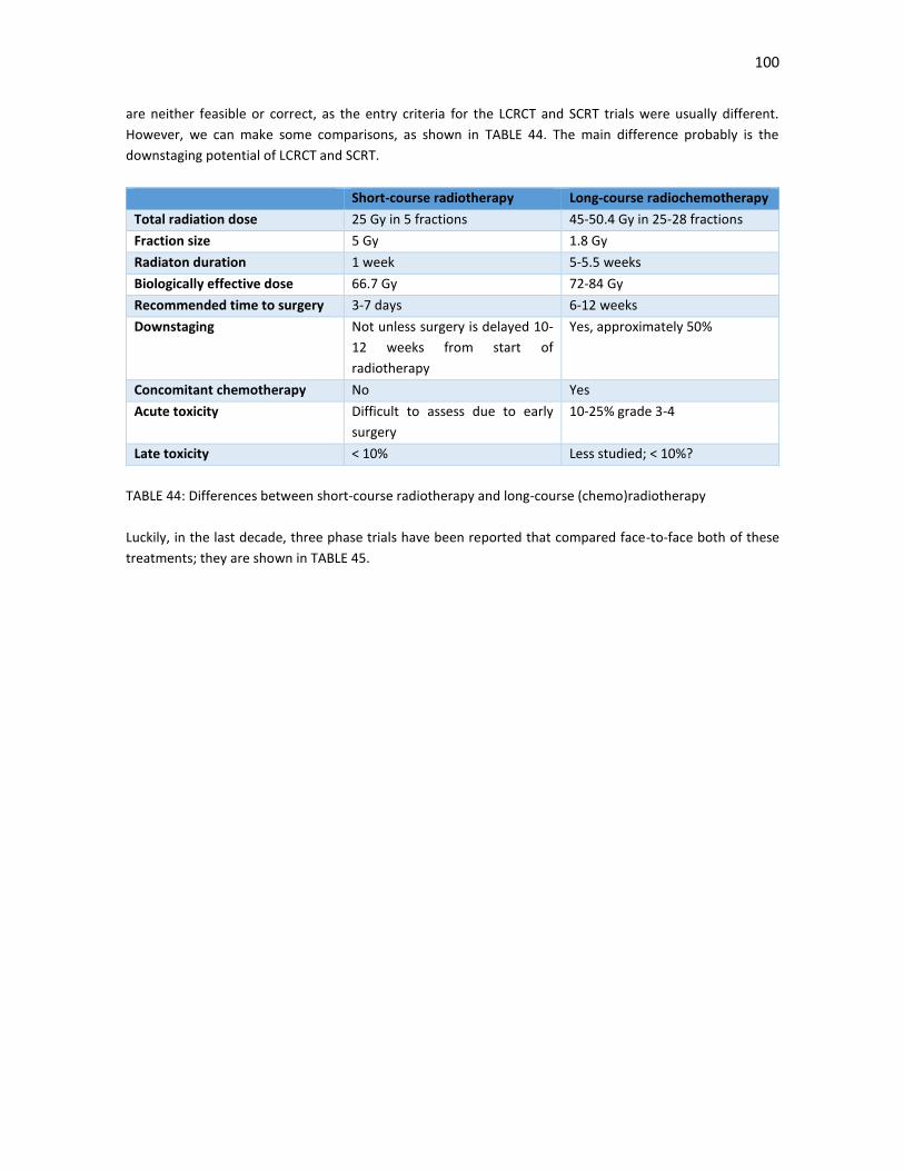
100
are neither feasible or correct, as the entry criteria for the LCRCT and SCRT trials were usually different.
However, we can make some comparisons, as shown in TABLE 44. The main difference probably is the
downstaging potential of LCRCT and SCRT.
Short-course radiotherapy Long-course radiochemotherapy
Total radiation dose 25 Gy in 5 fractions 45-50.4 Gy in 25-28 fractions
Fraction size 5 Gy 1.8 Gy
Radiaton duration 1 week 5-5.5 weeks
Biologically effective dose 66.7 Gy 72-84 Gy
Recommended time to surgery 3-7 days 6-12 weeks
Downstaging Not unless surgery is delayed 10-
12 weeks from start of
radiotherapy
Yes, approximately 50%
Concomitant chemotherapy No Yes
Acute toxicity Difficult to assess due to early
surgery
10-25% grade 3-4
Late toxicity < 10% Less studied; < 10%?
TABLE 44: Differences between short-course radiotherapy and long-course (chemo)radiotherapy
Luckily, in the last decade, three phase trials have been reported that compared face-to-face both of these
treatments; they are shown in TABLE 45.
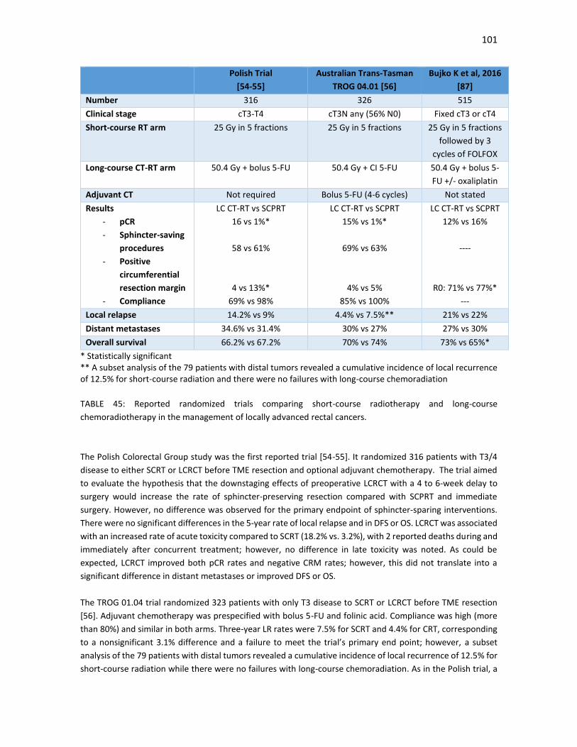
101
Polish Trial
[54-55]
Australian Trans-Tasman
TROG 04.01 [56]
Bujko K et al, 2016
[87]
Number 316 326 515
Clinical stage cT3-T4 cT3N any (56% N0) Fixed cT3 or cT4
Short-course RT arm 25 Gy in 5 fractions
25 Gy in 5 fractions
25 Gy in 5 fractions
followed by 3
cycles of FOLFOX
Long-course CT-RT arm 50.4 Gy + bolus 5-FU 50.4 Gy + CI 5-FU
50.4 Gy + bolus 5-
FU +/- oxaliplatin
Adjuvant CT Not required Bolus 5-FU (4-6 cycles) Not stated
Results
- pCR
- Sphincter-saving
procedures
- Positive
circumferential
resection margin
- Compliance
LC CT-RT vs SCPRT
16 vs 1%*
58 vs 61%
4 vs 13%*
69% vs 98%
LC CT-RT vs SCPRT
15% vs 1%*
69% vs 63%
4% vs 5%
85% vs 100%
LC CT-RT vs SCPRT
12% vs 16%
----
R0: 71% vs 77%*
---
Local relapse 14.2% vs 9% 4.4% vs 7.5%** 21% vs 22%
Distant metastases 34.6% vs 31.4% 30% vs 27% 27% vs 30%
Overall survival 66.2% vs 67.2% 70% vs 74% 73% vs 65%*
* Statistically significant ** A subset analysis of the 79 patients with distal tumors revealed a cumulative incidence of local recurrence of 12.5% for short-course radiation and there were no failures with long-course chemoradiation
TABLE 45: Reported randomized trials comparing short-course radiotherapy and long-course
chemoradiotherapy in the management of locally advanced rectal cancers.
The Polish Colorectal Group study was the first reported trial [54-55]. It randomized 316 patients with T3/4
disease to either SCRT or LCRCT before TME resection and optional adjuvant chemotherapy. The trial aimed
to evaluate the hypothesis that the downstaging effects of preoperative LCRCT with a 4 to 6-week delay to
surgery would increase the rate of sphincter-preserving resection compared with SCPRT and immediate
surgery. However, no difference was observed for the primary endpoint of sphincter-sparing interventions.
There were no significant differences in the 5-year rate of local relapse and in DFS or OS. LCRCT was associated
with an increased rate of acute toxicity compared to SCRT (18.2% vs. 3.2%), with 2 reported deaths during and
immediately after concurrent treatment; however, no difference in late toxicity was noted. As could be
expected, LCRCT improved both pCR rates and negative CRM rates; however, this did not translate into a
significant difference in distant metastases or improved DFS or OS.
The TROG 01.04 trial randomized 323 patients with only T3 disease to SCRT or LCRCT before TME resection
[56]. Adjuvant chemotherapy was prespecified with bolus 5-FU and folinic acid. Compliance was high (more
than 80%) and similar in both arms. Three-year LR rates were 7.5% for SCRT and 4.4% for CRT, corresponding
to a nonsignificant 3.1% difference and a failure to meet the trial’s primary end point; however, a subset
analysis of the 79 patients with distal tumors revealed a cumulative incidence of local recurrence of 12.5% for
short-course radiation while there were no failures with long-course chemoradiation. As in the Polish trial, a
102
higher rate of pCR was achieved with LCRCT, but no difference in DFS or OS was noted and the rate of late
toxicity was similar.
What can we make of these two trials? Both of them were underpowered to detect modest but clinically
significant differences in long-term outcomes and neither trial provides sufficient evidence to definitively
compare efficacy between SCPRT and LCRCT. Both approaches seem broadly similar. However, LCRCT does
have some advantages in those patients where a downstaging is needed, especially if the mesorectal fascia is
threatened, and in lower tumours, where the risk of local relapse is higher. On the other hand, SCRT seems to
be more effective in mid-rectal tumours. Acute toxicity is higher with LCRCT (although it is usually
manageable), with the exception of acute neurogenic pain, which is observed in <1–2 % of patients receiving
SCPRT, is usually reversible and dependent on the radiotherapy fields used [88-89]. With a more limited field
size, this side effect has not been observed in more recent trials, such as in the CR07 trial, and have not been
reported after LCRCT.
The third trial recently reported is another study of The Polish Colorectal Group study [87]. The trial included
515 patients with fixed cT3 or cT4 rectal cancer. Patients were randomly assigned to SCRCT and three cycles
of FOLFOX4 after one-week rest, or to standard LCRCT, with bolus 5-FU, leucovorin and oxaliplatin; of note,
after data showed there was no advantage of adding oxaliplatin to LCRCT in 2012, the study protocol was
amended and oxaliplatin could be omitted at the discretion of the participating study site.
No significant difference was found between patients assigned both arms in respect to the primary endpoint,
the rate of radical resection. There were also no differences in the pCR rates, nor in DFS and incidence of local
and/or distant relapse. However, surprisingly, although the follow-up is short, the 3-year OS rate was
significantly higher for patients assigned to the SCRCT and combined chemotherapy arm. Similar downstaging
results were recently reported with the interim results of the Stockholm III trial comparing SCRT and SCRT
with surgery delayed 4–8 weeks; the trial demonstrated higher rates of primary tumor downstaging and
pathologic complete response (11.8% vs 1.7%) in the delayed surgery arm [90]. A caveat of the Polish trial
however, is the use of oxaliplatin alongside LCRCT, which we know now is ineffective and more toxic and can
reduce the dose-intensity of LCRCT and worsen outcomes.
All these preliminary results seem to show that the differences between LCRCT and SCRT seem to be blurring.
Perhaps the combination of SCRT and induction chemotherapy can be a better tolerated option, especially in
more frail and/or elderly patients that is similarly effective than standard LCRCT. The use of SCRCT with a delay
to surgery in these locally advanced tumours when the patient is not fit for LCRCT has been reported
previously with encouraging results [91-93].
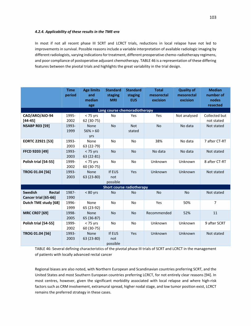
103
4.2.4. Applicability of these results in the TME-era
In most if not all recent phase III SCRT and LCRCT trials, reductions in local relapse have not led to
improvements in survival. Possible reasons include a variable interpretation of available radiologic imaging by
different radiologists, varying indications for treatment, different preoperative chemo-radiotherapy regimens,
and poor compliance of postoperative adjuvant chemotherapy. TABLE 46 is a representation of these differing
features between the pivotal trials and highlights the great variability in the trial design.
Time period
Age limits and
median age
Standard staging
MRI
Standard staging
EUS
Total mesorectal
excision
Quality of mesorectal
excision
Median number of
nodes resected
Long course chemoradiotherapy
CAO/ARO/AIO-94 [44-45]
1995-2002
< 75 yrs 62 (30-75)
No Yes Yes
Not analysed
Collected but not stated
NSABP R03 [59] 1993-1999
None 56% > 60
yrs
No Not stated
No No data Not stated
EORTC 22921 [53] 1993-2003
None 63 (22-79)
No No 38% No data 7 after CT-RT
FFCD 9203 [49] 1993-2003
< 75 yrs 63 (22-81)
No No No data No data Not stated
Polish trial [54-55] 1999-2002
< 75 yrs 60 (30-75)
No No Unknown Unknown 8 after CT-RT
TROG 01.04 [56] 1993-2003
None 63 (23-80)
If EUS not
possible
Yes Unknown Unknown Not stated
Short course radiotherapy
Swedish Rectal Cancer trial [65-66]
1987-1990
< 80 yrs No No No No Not stated
Dutch TME study [68] 1996-1999
None 65 (23-92)
No No Yes 50% 7
MRC CR07 [69] 1998-2005
None 65 (36-87)
No No Recommended 52% 11
Polish trial [54-55] 1999-2002
< 75 yrs 60 (30-75)
No No Unknown Unknown 9 after SCRT
TROG 01.04 [56] 1993-2003
None 63 (23-80)
If EUS not
possible
Yes Unknown Unknown Not stated
TABLE 46: Several defining characteristics of the pivotal phase III trials of SCRT and LCRCT in the management
of patients with locally advanced rectal cancer
Regional biases are also noted, with Northern European and Scandinavian countries preferring SCRT, and the
United States and most Southern European countries preferring LCRCT, for not entirely clear reasons [94]. In
most centres, however, given the significant morbidity associated with local relapse and where high-risk
factors such as CRM involvement, extramural spread, higher nodal stage, and low tumor position exist, LCRCT
remains the preferred strategy in these cases.
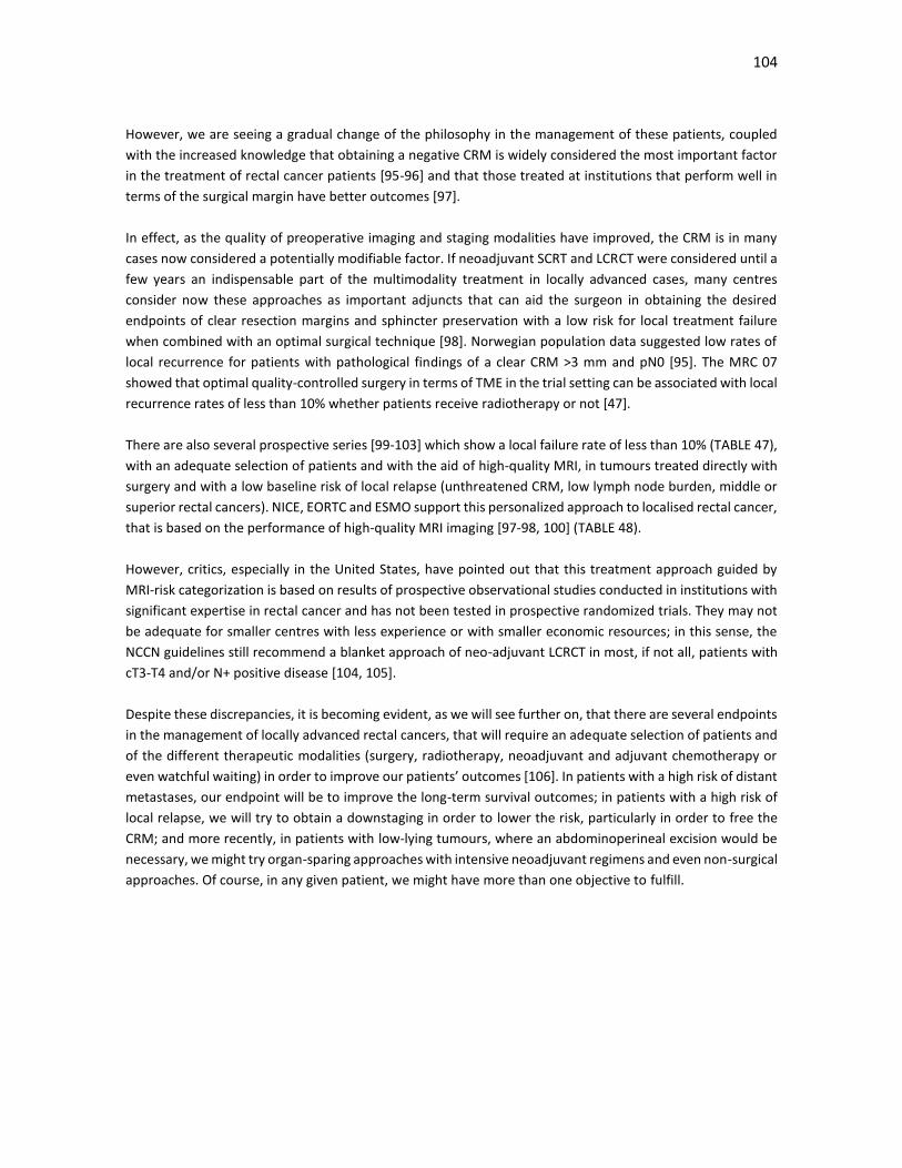
104
However, we are seeing a gradual change of the philosophy in the management of these patients, coupled
with the increased knowledge that obtaining a negative CRM is widely considered the most important factor
in the treatment of rectal cancer patients [95-96] and that those treated at institutions that perform well in
terms of the surgical margin have better outcomes [97].
In effect, as the quality of preoperative imaging and staging modalities have improved, the CRM is in many
cases now considered a potentially modifiable factor. If neoadjuvant SCRT and LCRCT were considered until a
few years an indispensable part of the multimodality treatment in locally advanced cases, many centres
consider now these approaches as important adjuncts that can aid the surgeon in obtaining the desired
endpoints of clear resection margins and sphincter preservation with a low risk for local treatment failure
when combined with an optimal surgical technique [98]. Norwegian population data suggested low rates of
local recurrence for patients with pathological findings of a clear CRM >3 mm and pN0 [95]. The MRC 07
showed that optimal quality-controlled surgery in terms of TME in the trial setting can be associated with local
recurrence rates of less than 10% whether patients receive radiotherapy or not [47].
There are also several prospective series [99-103] which show a local failure rate of less than 10% (TABLE 47),
with an adequate selection of patients and with the aid of high-quality MRI, in tumours treated directly with
surgery and with a low baseline risk of local relapse (unthreatened CRM, low lymph node burden, middle or
superior rectal cancers). NICE, EORTC and ESMO support this personalized approach to localised rectal cancer,
that is based on the performance of high-quality MRI imaging [97-98, 100] (TABLE 48).
However, critics, especially in the United States, have pointed out that this treatment approach guided by
MRI-risk categorization is based on results of prospective observational studies conducted in institutions with
significant expertise in rectal cancer and has not been tested in prospective randomized trials. They may not
be adequate for smaller centres with less experience or with smaller economic resources; in this sense, the
NCCN guidelines still recommend a blanket approach of neo-adjuvant LCRCT in most, if not all, patients with
cT3-T4 and/or N+ positive disease [104, 105].
Despite these discrepancies, it is becoming evident, as we will see further on, that there are several endpoints
in the management of locally advanced rectal cancers, that will require an adequate selection of patients and
of the different therapeutic modalities (surgery, radiotherapy, neoadjuvant and adjuvant chemotherapy or
even watchful waiting) in order to improve our patients’ outcomes [106]. In patients with a high risk of distant
metastases, our endpoint will be to improve the long-term survival outcomes; in patients with a high risk of
local relapse, we will try to obtain a downstaging in order to lower the risk, particularly in order to free the
CRM; and more recently, in patients with low-lying tumours, where an abdominoperineal excision would be
necessary, we might try organ-sparing approaches with intensive neoadjuvant regimens and even non-surgical
approaches. Of course, in any given patient, we might have more than one objective to fulfill.
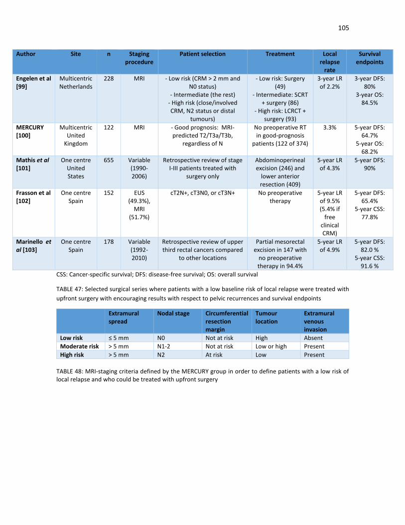
105
CSS: Cancer-specific survival; DFS: disease-free survival; OS: overall survival
TABLE 47: Selected surgical series where patients with a low baseline risk of local relapse were treated with
upfront surgery with encouraging results with respect to pelvic recurrences and survival endpoints
Extramural spread
Nodal stage Circumferential resection margin
Tumour location
Extramural venous invasion
Low risk ≤ 5 mm N0 Not at risk High Absent
Moderate risk > 5 mm N1-2 Not at risk Low or high Present
High risk > 5 mm N2 At risk Low Present
TABLE 48: MRI-staging criteria defined by the MERCURY group in order to define patients with a low risk of local relapse and who could be treated with upfront surgery
Author Site n Staging procedure
Patient selection Treatment Local relapse
rate
Survival endpoints
Engelen et al [99]
Multicentric Netherlands
228 MRI - Low risk (CRM > 2 mm and N0 status)
- Intermediate (the rest) - High risk (close/involved CRM, N2 status or distal
tumours)
- Low risk: Surgery (49)
- Intermediate: SCRT + surgery (86)
- High risk: LCRCT + surgery (93)
3-year LR of 2.2%
3-year DFS: 80%
3-year OS: 84.5%
MERCURY [100]
Multicentric United
Kingdom
122 MRI - Good prognosis: MRI-predicted T2/T3a/T3b,
regardless of N
No preoperative RT in good-prognosis
patients (122 of 374)
3.3% 5-year DFS: 64.7%
5-year OS: 68.2%
Mathis et al [101]
One centre United States
655 Variable (1990-2006)
Retrospective review of stage I-III patients treated with
surgery only
Abdominoperineal excision (246) and
lower anterior resection (409)
5-year LR of 4.3%
5-year DFS: 90%
Frasson et al [102]
One centre Spain
152 EUS (49.3%),
MRI (51.7%)
cT2N+, cT3N0, or cT3N+ No preoperative therapy
5-year LR of 9.5% (5.4% if
free clinical CRM)
5-year DFS: 65.4%
5-year CSS: 77.8%
Marinello et al [103]
One centre Spain
178 Variable (1992-2010)
Retrospective review of upper third rectal cancers compared
to other locations
Partial mesorectal excision in 147 with
no preoperative therapy in 94.4%
5-year LR of 4.9%
5-year DFS: 82.0 %
5-year CSS: 91.6 %
106
We used a blanket-approach of treating all our locally advanced rectal cancer patients, defined as cT3-T4
and/or N+ disease with LCRCT followed by TME-surgery and adjuvant chemotherapy, regardless of the
pathological tumour response. However, all the previous data have shown us that the future lies in a more
personalised approach, especially in the setting of better clinical imaging and improvement in surgical
techniques. Taking all these factors into account, from a practical point of view, we will centre on several
questions in the management of our patients and we will try to determine which were the prognostic and
predictive factors that may help us in that goal of a more personalised approach in the future and a more
rational sequencing of the different therapeutic modalities in locally advanced rectal cancer
4.3. How can we improve the results of long-course chemoradiotherapy?
4.3.1. Can we maximize the efficacy of radiotherapy?
Our protocol required the use of LCRCT followed by surgery in the 5-8 weeks after the end of the radiotherapy,
as is standard with most of these regimens reported. Compliance to the radiotherapy regimen was excellent,
with almost 95% of patients receiving the full intended dose and almost a half of patients proceeding to
surgery in the sixth to eight-week after the end of radiotherapy. Serious acute radiotherapy-related toxicity
was low, with only 3.4% of patients suffering from grade 3-4 toxicity. In this sense, modern and better-
tolerated radiotherapy techniques, with more precise radiation fields, and better supportive care have
improved the results with this technique (FIGURE 20 shows the radiotherapy planification imaging of one of
our patients)
Unfortunately, as in most other centres, most of the radiotherapy treatment parameters were determined in
an empiric manner. For example, why 6 to 8 weeks? Do we really know what is the ideal interval between
radiotherapy and surgery in order to obtain the maximal downstaging benefit? Is there a radiotherapy dose-
response lineal relationship, like in other tumours, such as in head and neck cancer? Can we maximize
radiotherapy in order to obtain more pCR and subsequently to increase the possibility of organ-sparing
surgical approaches or even watch- and- wait approaches? Most of these questions have not been answered
in a satisfactory manner.
Regarding the first question, the optimal interval between (chemo)radiotherapy and surgery has long been a
subject of investigation. The Lyon R90-01 trial established the current reference standard of 6 to 8 weeks
between neoadjuvant LCRCT and surgical resection nearly 2 decades ago after demonstrating superior
outcomes compared to a 2-week interlude [108]. The primary endpoint of the study was the rate of sphincter-
preserving resection. Although increased rates of objective response (71.7% versus 53.1) and major
pathological tumour regression (26% versus 13%) were observed in the delayed-surgery group, these did not
translate into a statistically significantly higher rate of sphincter-preserving surgery in the intention-to-treat
population. Similarly, a numerically higher but not statistically significant difference in favour of the delayed-
surgery group (41% versus 23%) was found in the low rectal cancer patient population.
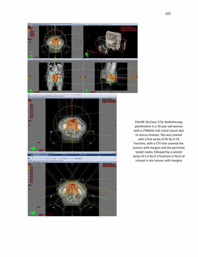
107
FIGURE 30 (Case 175): Radiotherapy
planification in a 70-year old woman
with a cT4bN1b mid-rectal cancer due
to uterus invasion. She was treated
with a first series of 45 Gy in 25
fractions, with a CTV that covered the
tumour with margins and the perirectal
lymph nodes, followed by a second
series of 5.4 Gy in 3 fractions in form of
a boost in the tumour with margins
108
However, other more recent publications suggest an even longer time frame may lead to still better tumour
regression and downstaging with no increase in surgical morbidity and long-term oncologic and functional
outcomes [109]. A meta-analysis of 13 mostly retrospective studies also evaluated the association between
LCRCT and time to surgery in 3584 patients and found that pCR rates improved where the interval was 6 to 8
weeks or more [110]; however, this did not translate into a DFS or OS advantage. Other smaller retrospective
reviews have advocated a longer time frame of 10 to 14 weeks as a result of higher pCR rates and tumor
volume reduction; although follow-up was too short to report long-term survival data [111-112]. In effect, in
one of these studies [111], the rate of pCR gradually increased with time and reached the peak after 15-16
weeks from the start of standard LCRCT. In the multivariate analysis, the time interval between
chemoradiotherapy and surgery was an independent predictive factor for pCR.
Finally, outcomes were reported from a retrospective review of data on 6805 patients captured by the
American National Cancer Data Base [113] A significant association between an increasing LCRCT-to-surgery
interval and improved pCR existed up to 75 days although it disappeared thereafter. Surprisingly, beyond a
60-day interval, there was an increment in the rate of positive surgical margins and, more importantly, a
significant decrease of 25% in OS. However, these worrying data must be taken with caution, as, probably,
those patients where surgery was delayed where poor-prognosis patients, where there was a longer recovery
time from LCRCT due to higher toxicity or other causes. The retrospective nature of the analysis is also a
problem for definitive conclusions to be obtained.
As we have seen previously, there is also data with SCRT that a longer interval between SCRT and surgery may
allow downstaging and a higher pCR. Pre-planned interim analyses of the Stockholm III trial have
demonstrated that postponing surgery until 4-8 weeks after SCRT does not increase the risk of post-operative
complications and is associated with significantly higher rates of pCR (11.8% versus 1.7%) and tumour
regression (grade 2-4 according to Dworak, 43% versus 20.1%) and a lower tumour stage compared to
immediate surgery [90]. The recent phase III Polish trial of LCRCT vs SCRT with a delayed interval to surgery
showed similar results [87].
Despite these encouraging results, possible benefits deriving from a delayed surgery are to be carefully
weighed against potential disadvantages including an increased risk of tumour regrowth/metastases, of peri-
operative complications and of a delayed start of post-operative systemic chemotherapy. In this sense,
Increasing the interval between neoadjuvant CRT and surgery warrants further investigation in a randomized
controlled trial. The NCT01037049 trial is aiming to recruit 218 patients to neoadjuvant CRT and TME surgery
after either 6 or 12 weeks, with a planned primary end point of T-stage downstaging on MRI.
Regarding the question of dose intensity, there is little data for a dose-response relationship in locally
advanced rectal cancer, particularly for larger T4 or fixed tumours. Traditional preoperative doses in the
randomised trials are, conventionally, 45 to 50.4 Gy using 1.8 Gy per fraction. However, the most effective
total dose and schedule are not known. Meta-analyses of preoperative radiotherapy for rectal cancer suggest
that there is an advantage to treating to a biological equivalent dose (BED) of at least 30 Gy [114]. Three
different doses were investigated in a Canadian sequential phase II trial [55]. Sequential schedules of radiation
combined with 5-FU dose-escalated from 40 Gy in 20 fractions to 46 Gy in 23 fractions, and finally to 50 Gy in
25 fractions [115]. Local control was significantly better for doses of 46 Gy and above, but there was no
difference between 46 Gy and 50 Gy. There was a trend to higher pathological complete response rates with
increasing the radiation dose of 13%, 21% and 31% for 40 Gy, 46 Gy and 50 Gy, respectively. The trial
concluded that there was no advantage in routinely increasing total dose to or above 46 Gy. In contrast, the
109
conclusion of another randomised trial comparing both oxaliplatin and 50 Gy with capecitabine and 45 Gy, is
that 50 Gy is a new standard [116]; in fact, we adopted that dose from that moment.
A pooled analysis did suggest that higher rates of pCR were associated with higher total doses of radiation
[117]. In practice, however, few prospective phase I-II studies have used total doses > 50 Gy, the exception
being the EXPERT trial, which boosted to 54 Gy [118]. Results of this study appeared impressive with respects
to pCR, with a 29% rate but we do not know the relative contributions of the induction chemotherapy and the
concurrent chemoradiotherapy schedule or the high dose of pelvic radiotherapy. For instance, in the same
group’s subsequent study, in the arm with an identical chemotherapy schedule but with a lower radiotherapy
dose of 50.4 Gy, the pathological complete response fell to 15% [119].
In this sense, higher doses are likely to result in better tumour responses but this will necessarily entail greater
acute and late toxicity. Waiting for the highest degree of radiotherapy-induced tumour regression may
optimise the selection of patients who are candidates for a local excision or a “watch and wait approach”
following the achievement of a complete or near clinical complete tumour [120]. This assumption is supported
by the data reported by several groups (especially in Brazil) which suggest that a delayed but strict assessment
of clinical response, could identify the true responders (while excluding those with suboptimal response or
early tumour re-growth/progression) and lead to an improved outcome for patients who are managed
nonoperatively [121].
Our data are consistent with all these findings (TABLE 19 and 20); there was an increased rate of downstaging
and complete or nearly complete tumour regression with higher doses of radiotherapy and an increased rate
of pathological complete response with longer intervals between the end of radiotherapy and the surgical
procedure. Increased duration of radiotherapy (due to delays or other causes) was detrimental, with a
numerically (although statistically not significant) increase in the risk of local relapse. This is most likely due to
the increased risk of malignant repopulation at the end of long-course radiotherapy regimens [75-77].
Although there was an increased downstaging rate with longer intervals, this did not translate into a survival
benefit. On the contrary, longer intervals were associated worse DFS. This apparent discrepancy can be
explained in the same manner as the American registry study. In our protocol, surgery was specified to be 6
to 8 weeks after the end of radiotherapy; those patients in where the interval was longer, there was a cause
for the delay, usually toxicity or other serious complications, that imply a poorer prognosis and which would
obscure any benefit with an increased downstaging rate.
110
Practical conclusions of radiotherapy:
1. International guidelines recommend that patients who are candidate for neoadjuvant treatment
should undergo surgery from 6 to 8 weeks (ESMO) or 5 to 12 weeks (NCCN) after completion of long-
course chemoradiotherapy or within 7-10 days of completion of short-course radiotherapy.
2. However, there is compelling data that an even longer time frame may lead to still better tumor
regression and downstaging, both in LCRCT and in SCRT. Our data are consistent with these data.
With this in mind, we can tailor the treatment in individual cases. For example, SCRT followed by a
longer interval in order to increase downstaging can be useful in elderly or frail patients, where a
LCRCT would be necessary but not feasible. However, we must wait the results of prospective
randomized trials in order to adopt a widespread approach of increasing the interval between RT and
surgery.
3. There is an imperfect relationship between higher radiotherapy dose and an increased pCR. These
differences are probably not significant with doses higher than 45 Gy, before radical surgery, but will
probably be useful in patients where a non-operative management is planned. Again, prospective
randomized trials are needed in this sense.
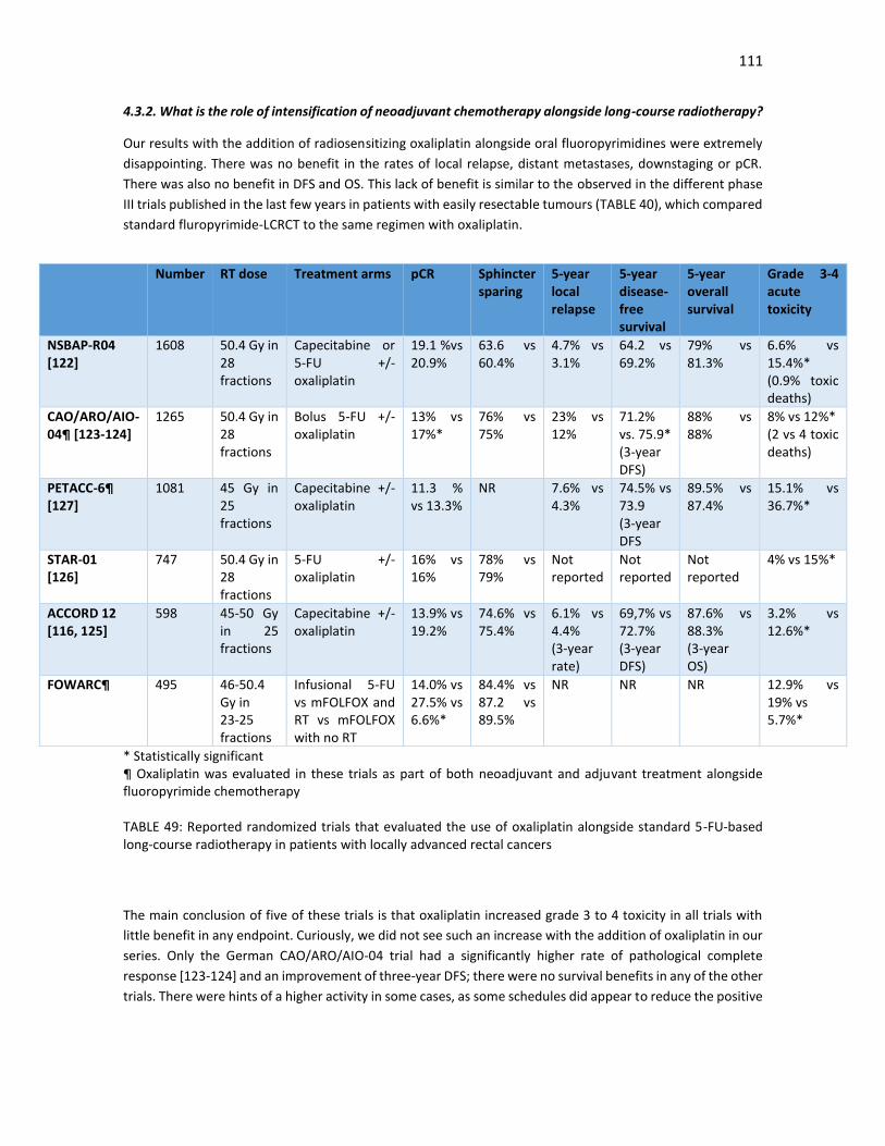
111
4.3.2. What is the role of intensification of neoadjuvant chemotherapy alongside long-course radiotherapy?
Our results with the addition of radiosensitizing oxaliplatin alongside oral fluoropyrimidines were extremely
disappointing. There was no benefit in the rates of local relapse, distant metastases, downstaging or pCR.
There was also no benefit in DFS and OS. This lack of benefit is similar to the observed in the different phase
III trials published in the last few years in patients with easily resectable tumours (TABLE 40), which compared
standard fluropyrimide-LCRCT to the same regimen with oxaliplatin.
* Statistically significant ¶ Oxaliplatin was evaluated in these trials as part of both neoadjuvant and adjuvant treatment alongside fluoropyrimide chemotherapy TABLE 49: Reported randomized trials that evaluated the use of oxaliplatin alongside standard 5-FU-based long-course radiotherapy in patients with locally advanced rectal cancers The main conclusion of five of these trials is that oxaliplatin increased grade 3 to 4 toxicity in all trials with
little benefit in any endpoint. Curiously, we did not see such an increase with the addition of oxaliplatin in our
series. Only the German CAO/ARO/AIO-04 trial had a significantly higher rate of pathological complete
response [123-124] and an improvement of three-year DFS; there were no survival benefits in any of the other
trials. There were hints of a higher activity in some cases, as some schedules did appear to reduce the positive
Number RT dose Treatment arms pCR Sphincter sparing
5-year local relapse
5-year disease-free survival
5-year overall survival
Grade 3-4 acute toxicity
NSBAP-R04 [122]
1608 50.4 Gy in 28 fractions
Capecitabine or 5-FU +/- oxaliplatin
19.1 %vs 20.9%
63.6 vs 60.4%
4.7% vs 3.1%
64.2 vs 69.2%
79% vs 81.3%
6.6% vs 15.4%* (0.9% toxic deaths)
CAO/ARO/AIO-04¶ [123-124]
1265 50.4 Gy in 28 fractions
Bolus 5-FU +/- oxaliplatin
13% vs 17%*
76% vs 75%
23% vs 12%
71.2% vs. 75.9* (3-year DFS)
88% vs 88%
8% vs 12%* (2 vs 4 toxic deaths)
PETACC-6¶ [127]
1081 45 Gy in 25 fractions
Capecitabine +/- oxaliplatin
11.3 % vs 13.3%
NR 7.6% vs 4.3%
74.5% vs 73.9 (3-year DFS
89.5% vs 87.4%
15.1% vs 36.7%*
STAR-01 [126]
747 50.4 Gy in 28 fractions
5-FU +/- oxaliplatin
16% vs 16%
78% vs 79%
Not reported
Not reported
Not reported
4% vs 15%*
ACCORD 12 [116, 125]
598 45-50 Gy in 25 fractions
Capecitabine +/- oxaliplatin
13.9% vs 19.2%
74.6% vs 75.4%
6.1% vs 4.4% (3-year rate)
69,7% vs 72.7% (3-year DFS)
87.6% vs 88.3% (3-year OS)
3.2% vs 12.6%*
FOWARC¶ 495 46-50.4 Gy in 23-25 fractions
Infusional 5-FU vs mFOLFOX and RT vs mFOLFOX with no RT
14.0% vs 27.5% vs 6.6%*
84.4% vs 87.2 vs 89.5%
NR NR NR 12.9% vs 19% vs 5.7%*
112
CRM rate [123-124, 127]. Due to the short follow-up period, there are no data yet on late toxicity, especially
persistent neurotoxicity.
The benefit in DFS in the German Trial, although statistically significant, is difficult to quantify, as the standard
LCRCT regimen included substandard doses of infusional bolus 5-FU. Adjuvant oxaliplatin-based
chemotherapy was also used in the oxaliplatin arm and we do not know which part of the treatment is
responsible of the benefit. As in all the other trials, serious toxicity was more frequent with the use of
oxaliplatin and there were 4 treatment-related deaths compared to 2 in the standard arm.
The short-term outcomes of 4 of these trials (NSABP R-04, CAO/ARO/AIO-04, STAR-01, and ACCORD 12/0405)
have been collated and examined in a recent meta-analysis [129]. An overall improvement in pCR rates and
decreased perioperative metastases were reported with the addition of oxaliplatin. However, this was
associated with increased grade 3-4 toxicity and a lower rate of planned radiotherapy completion, although
there was no difference in chemotherapy completion rates.
Why did oxaliplatin fail? The theoretical basis for its use is evident. Oxaliplatin in combination with
fluoropyrimidine therapy improves outcomes for colon cancer in the adjuvant setting and in metastatic
colorectal cancer [12], and it has known radiosensitizing properties. There were also promising phase II data
which were the initial basis of the use of oxaliplatin in a majority of our patients [13-16].
The most logical explanation is the increased toxicity rate, which had a deleterious effect on the compliance
of the proven beneficial treatment, namely 5-FU- based LCRCT. Systemically active doses of chemotherapy
are difficult to deliver concurrently with chemoradiotherapy schedules because of overlapping toxicities. The
(small) benefits in increased pCR rates and free CRM margin in some of the trials were not able to compensate
this poor radiotherapy compliance.
A second important factor is that all these randomised trials in rectal cancer selected patients with easily
resectable tumours. The majority of these patients were probably node negative. Based on the results of the
QUASAR and MOSAIC studies [130-131], stage II patients are unlikely to show a large benefit in terms of DFS
even from 5-FU chemotherapy – and minimal benefit from the addition of oxaliplatin. This lack of benefit of
oxaliplatin is probably more pronounced in elderly patients [132]. The poorer tolerance to capecitabine to
infusional 5-FU in elderly patients is also well known and may justify this higher toxicity, when combined with
oxaliplatin.
However, there are also some positive trials. In more advanced unresectable tumours, oxaliplatin has shown
to improve DFS and OS alongside standard LCRCT in a randomized phase III trial and is used frequently [133].
Also, initial results from the FOWARC study have been recently reported [128] that show that compared with
single-agent 5-FU, mFOLFOX6 concurrent with radiotherapy preoperatively results in a higher rate of pCR
(14.0% v 27.5%), a higher good response rate, good compliance, and acceptable toxicity for Asian patients
with stage II-III rectal cancer. The results also show that a comparable rate of downstaging can be achieved
with perioperative mFOLFOX6 alone compared with traditional 5-FU-based LCRCT. These preliminary results
suggest that a strategy of combining full-dose chemotherapy with radiation over chemosensitizing radiation
may be a new option for neoadjuvant treatment. Finally, a recent meta-analysis that included seven
randomized trials showed an improvement in the distant metastasis rate, in the rate of pCR and a marginal
improvement in DFS, albeit at the expense of a higher toxicity rate [134].
113
Despite these positive studies, there is little evidence to support the use of concomitant oxaliplatin in
resectable rectal cancer patients. Oxaliplatin has activity in locally advanced rectal cance, but its
administration concomitant to LCRT is probably not the best way to administer the drug. In this sense, the
drive to potentiate LCRCT with a second cytotoxic drug, which caused so much interest in the last two decades
appears to have waned as we have realized that this may cause additional toxicities and possibly enhance
surgical morbidity [135].
4.3.3. Is there a role of induction or consolidation chemotherapy alongside neoadjuvant chemoradiotherapy? As we will analyze posteriorly, we observed poor compliance rates of adjuvant chemotherapy in our group of
patients, especially in the elderly. Induction or consolidation chemotherapy before surgery has several
theoretical advantages. It treats occult micrometastases several months earlier and increases treatment
compliance, potentially enhancing the efficacy of chemotherapy in preventing distant metastases and
ultimately improving survival. Other benefits include increased response of the primary tumor, early
identification of nonresponders, and earlier removal of the loop ileostomy.
Unfortunately, several phase 2 trials have administered 2 to 4 cycles of induction capecitabine-oxaliplatin or
FOLFOX before LCRCT (TABLE 50), with mixed results in most cases [118, 136-141]. The addition of induction
chemotherapy showed a modest increase in the downstaging rate, but with no benefit in pCR or long-term
outcomes in most cases. Grade 3-4 toxicity was increased with the addition of induction chemotherapy.
Worryingly, in some trials there was an increase in serious cardiovascular events, especially in the elderly.
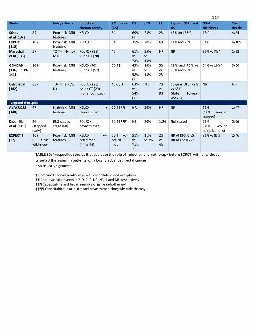
114
TABLE 50: Prospective studies that evaluate the role of induction chemotherapy before LCRCT, with or without
targeted therapies, in patients with locally advanced rectal cancer
* Statistically significant ¶ Combined chemoradiotherapy with capecitabine and oxaliplatin ¶¶ Cardiovascular events in 2, 9, 0, 2, NR, NR, 1 and NR, respectively ¶¶¶ Capecitabine and bevacizumab alongside radiotherapy ¶¶¶¶ Capecitabine, oxaliplatin and bevacizumab alongside radiotherapy
Study n Entry criteria Induction chemotherapy
RT dose (Gy)
DS pCR LR 5-year DSF and OS
G3-4 toxicity¶¶
Toxic deaths
Schou et al [137]
84 Poor-risk MRI features
XELOX 54 69% (T)
23% 2% 63% and 67% 18% 4/84
EXPERT [118]
105 Poor-risk MRI features
XELOX 54
35% 20% 6% 64% and 75% 34% 4/105
Marechal et al [138]
57 T2-T4 N+ by MRI
FOLFOX (28) vs no CT (20)
45 61% vs 72%
25% vs 28%
NR NR 36% vs 7%* 1/28
GEMCAD [136, 139-141]
108 Poor-risk MRI features
XELOX (56) vs no CT (52)
50.4¶ 43% vs 58% (T)
14% vs 13%
5% vs 2%
62% and 75% vs 75% and 78%
54% vs 19%* 3/56
Calvo et al [142]
335 T3-T4 and/or N+
FOLFOX (28) vs no CT (20) (no randomized)
45-50.4 63% vs 54% (T)*
NR 7% vs 9%
10-year DFS: 72% vs 68% Global 10-year OS: 75%
NR NR
Targeted therapies
AVACROSS [148]
47 High-risk MRI features
XELOX + bevacizumab
50.4¶¶¶ NR 36% NR NR 33% (18% needed surgery)
1/47
Dipetrillo et al [149]
26 (stopped early)
EUS-staged stage II-III
FOLFOX-bevacizumab
50.4¶¶¶¶ N$ 20% 1/26 Not stated 76% (36% wound complications)
0/26
EXPERT 2 [57]
165 (90 KRAS wild-type)
Poor-risk MRI features
XELOX +/- cetuximab (44 vs 46)
50.4 +/- cetuxi-mab
51% vs 71%*
11% vs 7%
2% vs 4%
HR of DFS: 0.65 HR of OS: 0.27*
81% vs 83% 2/46
115
Perhaps of these trials, the most interesting is the Spanish Grupo Cancer de Recto [136, 139-141]. This large
prospective trial recruited 108 patients with MRI-defined poor risk disease 47 and compared four cycles of
neoadjuvant or adjuvant capecitabine-oxaliplatin with TME-surgery. Serious toxicity was significantly
increased in the adjuvant arm, as could be expected, and a higher relative dose intensity for both capecitabine
and oxaliplatin was achieved with preoperative treatment (91% vs. 61% and 94% vs. 73% respectively).
However, 3 deaths were reported in each cohort, including 2 related to cardiovascular or thromboembolic
events. There was no benefit in pCR, local relapse, distant metastases or 5-year DFS and OS. Despite this,
although the study was underpowered to detect long-term benefits, the neoadjuvant approach showed better
tolerability and compliance and equivalent efficacy parameters.
The chemotherapy backbone can also be of interest. We know in advanced colon cancer that is similar in efficacy
to FOLFOX, although in most trials, the tolerance of FOLFOX, with its infusional 5-FU, is better [143]. A recent study
investigated the safety and efficacy of FOLFOX before LCRCT, demonstrating excellent treatment compliance and
no evidence of serious adverse effects requiring treatment delay. All patients undergoing TME had an R0 resection,
and nearly half had a tumor response greater than 90%, including 30% who had a pCR [144].
Another option is to give chemotherapy after LCRCT. A possible advantage of this approach is that, as we
know, extending neoadjuvant chemotherapy requires a delay to surgery after chemoradiation, and we know
this delay is significantly associated with a better response and a higher proportion of patients achieving a
pathological complete response [145]. There is less evidence of this strategy, however. The TIMING trial,
which completed accrual in 2012, did show that delivering two, four, or six cycles of FOLFOX after CRT in
patients with locally advanced tumours increased the pCR rates up to 25%, 30%, and 38%, respectively,
compared with CRT alone (18%), without any associated increase in adverse events or surgical complications
[146]. Eighty percent of patients received consolidation CT without interruption. [147]. Despite these
interesting results, we have concerns with this approach, as, in our anecdiotal evidence, tolerance to
chemotherapy after LCRCT, even before surgery, tends to be poorer, not only in rectal cancer but in othe
tumours such as pancreatic or esophageal cancer.
The addition of targeted antiangiogenic therapies, such as bevacizumab, have yielded disappointing results,
with an increase in surgical complications in the two largest studies reported [148-149]. These results are not
altogether surprising, as there is no activity of bevacizumab in the adjuvant treatment of colon cancer [150]
and there are no great increases in the response rate with the addition of bevacizumab to standard
chemotherapy in advanced colon cancer [151]. Curiously, in both trials, bevacizumab was used with
capecitabine as induction chemotherapy and also alongside LCRCT, which probably explains the high rate of
surgical complications.
More promising are the results of anti-EGFR agents, such as cetuximab, alongside induction chemotherapy in
patients with KRAS wild-type tumours [119, 152-153], reported in trials such as the EXPERT-C trial [119], which
showed an increase of the downstaging rate in the experimental arm, with an improvement in overall survival,
although the follow-up was short. Unfortunately, the grade 3-4 toxicity rate was high in both arms of induction
CT. These benefits have been proven to be higher in patients with all-RAS native tumours [154-155]
116
Practical aspects of chemotherapy intensification:
1. Although theoretically promising, the addition of concomitant oxaliplatin alongside standard LCRCT
is not beneficial. The integration of systemic CT alongside 5-FU-based CRT is difficult, due to the
existence of synergistic toxicities which worsen the compliance rate and the chance of completing
successfully the treatment plan
2. Results with induction chemotherapy (before LCRCT) or consolidation chemotherapy (after LCRCT)
are somewhat more promising and are included in some guidelines. Compliance is better compared
to adjuvant chemotherapy. However, patients should be selected accordingly, as toxicity is higher,
especially in elderly and/or frail patients, and there is an increased risk of serious cardiovascular
events.
3. Despite the lack of data from large prospective studies, some guidelines recommend neoadjuvant
chemotherapy alongside standard LCRCT is included as a treatment option.
4. We have concerns with the consolidation approach, as, in our anecdotal evidence, tolerance to
chemotherapy after LCRCT, even before surgery, tends to be poorer, not only in rectal cancer but in
othe tumours such as pancreatic or esophageal cancer.
5. There is no benefit with the addition of antiangiogenic targeted therapies, with an increased risk of
surgical complications. There are hints of better activity of the combination of induction CT with
antiEGFR targeted therapies, although, again, serious toxicities are worrying.
117
4.4. Is there any role of adjuvant chemotherapy after long-course chemoradiotherapy and surgery?
In our series, adjuvant chemotherapy was given to all patients, if feasible, regardless of the pathological
response observed; this was possible in more than 80% of patients. However, as in most other published trials,
the tolerance to the adjuvant regimen was poor with less than half of these patients receiving the full intended
dose and there were almost three-times more hospital admissions related to toxicity than with neoadjuvant
treatment. This was especially evident in elderly patients, where almost a quarter of patients did not receive
any kind of treatment after surgery. The median time from the surgery to the beginning of adjuvant treatment
was 5 weeks, but a quarter of patients did not begin treatment until seven weeks after. There did not seem
to be a benefit in local relapse rates with the use of adjuvant therapy, but there was an improvement in the
rate of distant metastasis with the administration of full-dose adjuvant therapy. On the other hand, patients
with ypN-positive disease that were not treated with adjuvant treatment, fared very poorly.
We know that with neoadjuvant (chemo)radiotherapy and modern surgical techniques, such as TME, the risk
of local relapse is low and the risk of distant metastases predominates. In the CAO/ARO/AIO-94 study nearly
30% of the patients treated with neoadjuvant therapy developed distant metastatic disease by 10 years,
findings similar to our series [6]. In this sense, locally advanced rectal cancer would seem to be a prime
candidate for true multimodality therapy, in where patients would benefit from systemic chemotherapy at
full doses, beyond what is delivered as a radiosensitizer concurrently with radiation therapy. There is also the
proven role of adjuvant chemotherapy in colon cancer, which showed first the superiority of adjuvant 5-FU
and leucovorin relative to surgery alone, and subsequently the value of adding oxaliplatin to 5-FU [12, 156].
In reality, for patients treated without preoperative (chemo)radiotherapy or TME-surgery (which results in
high numbers of locoregional recurrences) adjuvant chemotherapy does seem to be effective. A systematic
review and meta-analysis of of 21 randomized controlled trials showed that adjuvant chemotherapy did
improve DFS and OS in this group of patients [157]; however, this review included only two studies in which
patients had had preoperative (chemo)radiotherapy [47, 131]. The QUASAR [131, 158] study showed a
borderline-significant improvement in OS for patients with rectal cancer; however, only 21% of patients with
rectal cancer or both colon and rectal cancer received preoperative radiotherapy. Furthermore, results of a
Japanese trial also showed improved overall survival and disease-free survival in patients with stage III rectal
cancer who were randomly assigned to adjuvant chemotherapy after standardised mesorectal excision [158]
However, none of the patients in this trial received preoperative (chemo)radiotherapy and standardised
mesorectal excision included selective lateral lymphadenectomy.
The current recommendations for use of adjuvant chemotherapy in patients treated with LCRCT and TME is
based in part on these data and extrapolation from colon cancer data, and these patients are usually treated
with 5-FU or capecitabine +/- oxaliplatin-based adjuvant chemotherapyCT [12, 156] However, as was seen in
our study, despite these recommendations, up to 27% of eligible patients with locally advanced rectal cancer
never start adjuvant chemotherapy and less than 50% [48] receive the full prescribed treatment without
interruptions or delays [48, 123], resulting from postoperative complications, slow recovery, interference with
closure of their temporary ileostomy [160] or simply refusal of treatment.
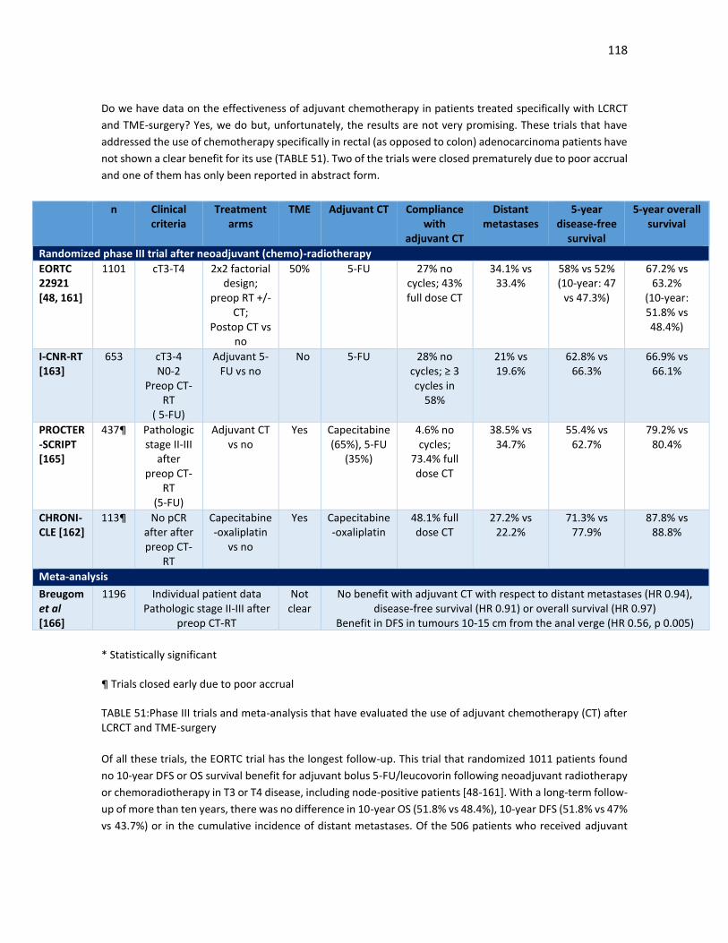
118
Do we have data on the effectiveness of adjuvant chemotherapy in patients treated specifically with LCRCT
and TME-surgery? Yes, we do but, unfortunately, the results are not very promising. These trials that have
addressed the use of chemotherapy specifically in rectal (as opposed to colon) adenocarcinoma patients have
not shown a clear benefit for its use (TABLE 51). Two of the trials were closed prematurely due to poor accrual
and one of them has only been reported in abstract form.
n Clinical criteria
Treatment arms
TME Adjuvant CT Compliance with
adjuvant CT
Distant metastases
5-year disease-free
survival
5-year overall survival
Randomized phase III trial after neoadjuvant (chemo)-radiotherapy
EORTC 22921 [48, 161]
1101 cT3-T4 2x2 factorial design;
preop RT +/- CT;
Postop CT vs no
50% 5-FU 27% no cycles; 43% full dose CT
34.1% vs 33.4%
58% vs 52% (10-year: 47
vs 47.3%)
67.2% vs 63.2%
(10-year: 51.8% vs 48.4%)
I-CNR-RT [163]
653 cT3-4 N0-2
Preop CT-RT
( 5-FU)
Adjuvant 5-FU vs no
No 5-FU 28% no cycles; ≥ 3 cycles in
58%
21% vs 19.6%
62.8% vs 66.3%
66.9% vs 66.1%
PROCTER-SCRIPT [165]
437¶ Pathologic stage II-III
after preop CT-
RT (5-FU)
Adjuvant CT vs no
Yes Capecitabine (65%), 5-FU
(35%)
4.6% no cycles;
73.4% full dose CT
38.5% vs 34.7%
55.4% vs 62.7%
79.2% vs 80.4%
CHRONI-CLE [162]
113¶ No pCR after after preop CT-
RT
Capecitabine-oxaliplatin
vs no
Yes Capecitabine-oxaliplatin
48.1% full dose CT
27.2% vs 22.2%
71.3% vs 77.9%
87.8% vs 88.8%
Meta-analysis
Breugom et al [166]
1196 Individual patient data Pathologic stage II-III after
preop CT-RT
Not clear
No benefit with adjuvant CT with respect to distant metastases (HR 0.94), disease-free survival (HR 0.91) or overall survival (HR 0.97)
Benefit in DFS in tumours 10-15 cm from the anal verge (HR 0.56, p 0.005)
* Statistically significant ¶ Trials closed early due to poor accrual TABLE 51:Phase III trials and meta-analysis that have evaluated the use of adjuvant chemotherapy (CT) after LCRCT and TME-surgery
Of all these trials, the EORTC trial has the longest follow-up. This trial that randomized 1011 patients found
no 10-year DFS or OS survival benefit for adjuvant bolus 5-FU/leucovorin following neoadjuvant radiotherapy
or chemoradiotherapy in T3 or T4 disease, including node-positive patients [48-161]. With a long-term follow-
up of more than ten years, there was no difference in 10-year OS (51.8% vs 48.4%), 10-year DFS (51.8% vs 47%
vs 43.7%) or in the cumulative incidence of distant metastases. Of the 506 patients who received adjuvant
119
chemotherapy, 57% did not receive the intended 4 cycles as scheduled, and 27% could not start adjuvant
treatment at all [48, 161].
The CHRONICLE study randomly assigned patients receiving neoadjuvant chemoradiotherapy to 6 months of
postoperative chemotherapy (six cycles of capecitabine-oxaliplatin) versus observation [162]. Patients were
required to have received at least 45 Gy with concurrent fluoropyrimidine-based chemotherapy. Only 113
patients were accrued. Surprisingly, despite the observation group having more node-positive patients, there
was no trend toward improved outcomes with adjuvant chemotherapy, even though we know patients with
pathological stage III disease fare very poorly. However, these results are difficult to interpret because of the
small numbers of patients randomized.
In the larger study I-CNR-RT trial [163], 655 patients with cT3 or T4 disease were randomly assigned to six
cycles of bolus 5-FU versus observation; the clinical design was very similar to the EORTC study and the
chemotherapy was given in a similarly reduced-dose bolus fashion postoperatively. Despite this, compliance
was poor and there were no differences in survival. The cumulative incidence of local relapse was 7.4% versus
8.7% and there there were no differences in distant metastases (24.3% for postoperative chemotherapy
versus 23.9% for no postoperative chemotherapy).
Finally, investigators from the Dutch Colorectal Cancer Group performed a randomized in 470 patients treated
initially with radiation or chemoradiotherapy followed by TME [164]. Again, despite a high incidence of
pathological stage III disease (81.9% of patients in the postoperative chemotherapy arm and 85.5% in the
observation) there was no difference in DFS and OS at a median of 5 years of follow-up. Again, this study was
similarly small and underpowered to detect a small survival benefit.
A meta-analysis of these trials [165], showed, that with a median follow-up of 7 years, the cumulative
incidence of distant recurrences at 5 years was 36·5% in patients in the observation group and 35·5% for
patients who received adjuvant chemotherapy (hazard ratio 0·94, 95% CI 0·78–1·14; p=0·523) . OS did not
differ significantly between groups. Adjuvant chemotherapy did not provide a benefit for any endpoint in any
of the ypN subgroups. With regards to oxaliplatin, the investigators limited their analysis to trials that had an
observation group without adjuvant chemotherapy, and no randomised trials testing oxaliplatin were eligible.
All these trials concluded that the benefit of adjuvant chemotherapy noted for patients with colon cancer
seems to be lost for patients with rectal cancer neoadjuvant (chemo)radiotherapy. The reasons for this
discrepancy are difficult to explain. Lack of activity might be due to the fact that after neoadjuvant treatment
and the surgically complex TME, which often has prolonged recovery period, there is a chemotherapy-free
period of approximately 20 weeks until adjuvant systemic treatment can be administered, many times at a
reduced dose [48, 123]. In all trials, compliance rates were suboptimal.
Secondly, the absolute benefit of postoperative chemotherapy on OS may be less in the modern era after
preoperative radiation as a result of stage migration and other mitigating factors. It is also true that in colon
cancer, the benefit of adjuvant chemotherapy is largely restricted to patients with node-positive disease, and
we still do not know how to identify those stage II patients that will benefit from adjuvant CT; the same can
probably apply to rectal cancer, where, of course, the weak point in the clinical staging is the identification of
N+ positive disease; most clinicians in these cases, even despite an excellent response after LCRCT, as we will
see, are afraid to omit postoperative chemotherapy “in case the nodes were originally positive.”. On the other
hand, there is the concern that some of the patients with pathologic node negative disease actually had nodal
120
disease that responded to therapy. These patients are at increased risk for systemic disease recurrence given
the nodal status. In addition, these patients have treatment sensitive disease and might be more likely to
benefit from additional chemotherapy. [166]
Other authors suggest that this difference in response to adjuvant chemotherapy between rectal and colon
cancer could be the result of biological and genetic differences between the two diseases [165]. The finding
in the recent meta-analysis that adjuvant chemotherapy improved DFSl and distant recurrences (although still
not OS) in those tumours 10–15 cm from the anal verge lends some support to the idea of a possible difference
between cancer of rectum and of the colon. An analysis of individual patient data pooled from five randomised
trials showed that adjuvant chemotherapy significantly contributed to local control, had no effect on distant
recurrences, and had a small effect on overall survival [34]. The assumption that (chemo)radiotherapy could
mask the effect of chemotherapy on local spread of the primary tumour is intriguing, but no clinical trial has
directly addressed this issue. Some ongoing trials with a chemotherapy-only preoperative group could offer
some insight on the effect of chemotherapy on local control, but they will not definitively answer this
question. Of note, we did not show any benefit in local relapse rates with the use of adjuvant chemotherapy.
However, although this is an intriguing hypothesis, it is not entirely convincing, as there is little data in different
molecular studies to suggest that rectal cancers behave differently to colon cancers, especially to distal colon
cancers [167]
4.4.1. Can we select which patients would benefit most from adjuvant chemotherapy?
To add more controversy to the role of adjuvant chemotherapy, the need for adjuvant treatment in patients
with a complete or near-complete response after CRT has been questioned [168-169]. Recent work from a
multi-institutional retrospective analysis of 3133 patients shows that the benefit of adjuvant therapy differs
between LARC subgroups. For example, patients with ypT1-2 or ypT3-4 tumors benefitted the most from
adjuvant therapy compared with patients who had ypT0N0 tumours [170].
There are centres that now adapt adjuvant chemotherapy according to the pathological response after LCRCT.
A recent phase 2 randomized study in 321 patients with postoperative pathologic stage II or stage III rectal
cancer following preoperative fluoropyrimidine-based LCRCT found that adjuvant 5-FU and oxaliplatin
improved 3-year DFS (71.6%) compared to 5-FU (62.9%) [171]. This trial restricted the use of adjuvant
chemotherapy to patients with LARC who had ypT3-4N0 or ypTanyN1-2 tumors afterneoadjuvant treatment.
Patients were randomly assigned to adjuvant chemotherapy with either four cycles of 5-FU or eight cycles of
FOLFOX. The administration of FOLFOX after surgery was associated with prolonged DFS in stage III patients
but not in stage II patients. A main strength of this study is that 96% of the patients completed the intended
4 cycles of adjuvant chemotherapy, which may justify this benefit, alongside the selection of patients with
stage II or III residual disease. A risk-adapted approach proposed by a Spanish Group showed interesting DFS
benefits [172], in the intention-to-treat analysis, when patients were selected according to their pathological
response. In effect, 5-year DFS for patients in the good-prognosis group (downstaging to ypT0-2 N0, who
received only 5-FU adjuvant chemotherapy) and for patients in the poor- prognosis group (pT3-4 or N positive
disease, who received oxaliplatin-based combination chemotherapy) were 79.4% and 66.3%, respectively.
However, other studies suggest that these patients who have a higher downstaging with LCRCT are those that
benefit most from adjuvant chemotherapy. These were the original findings of the EORTC trial, where patients
downstaged to ypT02 benefitted from adjuvant CT, whereas none was seen in ypT34 patients [48].
121
Interestingly, the benefit of adjuvant chemotherapy in patients with ypT0-2 seen in the first analysis has
disappeared with a longer follow-up [161].
What can we conclude of all these trials? Patients with poor downstaging should probably be offered some
type of adjuvant chemotherapy, for only the lack of better options, especially in patients with node-positive
disease. Involved regional lymph nodes at histopathology after radical surgery for T3-4 cancers confer a high
risk of locoregional recurrence and distant metastases; however, compliance can be a problem. In patients
not treated with neoadjuvant radiotherapy, we do not support the use of postoperative (chemo)radiotherapy
if the surgical specimen shows involved margins or other factors of local relapse. In the Trans-Tasman
Radiation Oncology Group (TROG) trial, positive resection margins, histopathogically involved lymph nodes
following radiotherapy in the resected specimen, and baseline carcinoembryonic antigen level were
independently associated with LR [56]. In the MRC 07 trial, significant risk factors included anterior quadrant
tumor involvement, extramural vascular invasion, and N stage (N2 > N1) [173]. However, not only we know
that postoperative RCT is poorly tolerated but also that these factors also increase the risk of distant
metastases and worse survival; in our study, CRM invasion was one of the significant factors in the multivariate
analysis for a worse DFS and OS.
More difficult is to decide what to do with patients with a good downstaging and especially those with a pCR.
Selecting patients with fluoropyrimidine-sensitive disease is an attractive option, but the data suggest that
the absolute benefit is relatively small. It is certainly likely that there is a subset of patients with good
prognostic markers (such as cT3N0 disease, tumors high in the rectum, tumors with pCR) that do not gain
much additional benefit from adjuvant chemotherapy. However, currently we do not have enough
information on how to identify these patients. Patients with a pCR from neoadjuvant chemotherapy have
better outcomes in terms of DFS and OS than patients who do not [6, 170, 174]. Is that enough basis for not
giving adjuvant chemotherapy.?. Probably not, but we can take solace in that, in this group of patients, the
benefit of adjuvant chemotherapy is probably relatively small [175].
In terms of specific molecular biomarkers, microsatellite instability testing might be of value if considering
single-agent 5-FU therapy, although MSI-high tumours are rate in the rectum [167-168]. Unfortunately, in
most circumstances, more advanced gene expression assays are not likely to be beneficial in determining the
need for adjuvant chemotherapy in this setting.
The potential, unproven, benefits of adjuvant chemotherapy should be balanced with its potential short-term
and long-term negative effects. Such effects include deterioration in quality of life; increased mortality
associated with postoperative chemotherapy, probably higher among elderly patients; increased grade 3 and
higher toxic effects caused by the combination of fluorouracil and oxaliplatin; the need to postpone diverting
stoma; and direct and indirect health-care costs. Other, long-term adverse events include permanent
deterioration in quality of life and neuropathy in patients who take oxaliplatin.
Unfortunately, we are not going to have high-quality randomized trials evaluating adjuvant CT after
(chemo)radiotherapy in the future. Instead, we should turn to exploration of large research archives, cancer
centre networks, and population registries. This approach will be more affordable and should be used to
address the ongoing uncertainties of prescribing adjuvant chemotherapy after preoperative
(chemo)radiotherapy for rectal cancer.
122
In a case-by-case basis, when deciding which patients to consider for adjuvant chemotherapy with ypT3-T4N0
rectal cancer the decision needs to be made on an individual basis discussing the risks and benefits for each
patient. Patient comorbidities and performance status following neoadjuvant chemoradiation and their
operation are also important. In general, the likely benefit for adjuvant chemotherapy remains quite small in
this specific setting, although there is a minority of patients that might derive significant benefit. In our study,
both the Valentini nomograms and the Dhadda score defined poor-prognosis patients, with a high risk of local
relapse and distant metastases. The use of these nomograms may aid us in the selection of patients’
candidates for adjuvant chemotherapy, although, again, as they are scores based in pathological criteria, they
can only be used after surgery.
Another possible approach to resolve this conundrum is the use of neoadjuvant chemotherapy; the objective
in this case is not to increase downstaging, as the induction trials we mentioned previously, but to decrease
the risk of distant metastases [175]. We know from all clinical trials that adjuvant chemotherapy is poorly
tolerated after neoadjuvant radiotherapy and surgery for rectal cancer, with 25% of patients not receiving
anything. With this in mind, several authors have proposed the use of systemic treatment before, rather than
after, surgery. Randomised trials with neoadjuvant systemic therapy are in progress; they usually do not
include radiotherapy or, if they do, only SCRT (RAPIDO, PROSPECT, COPERNICUS). In the meantime, four small
phase 2 studies have addressed this novel concept of neoadjuvant chemotherapy without LCRCT with
encouraging results (TABLE 52).
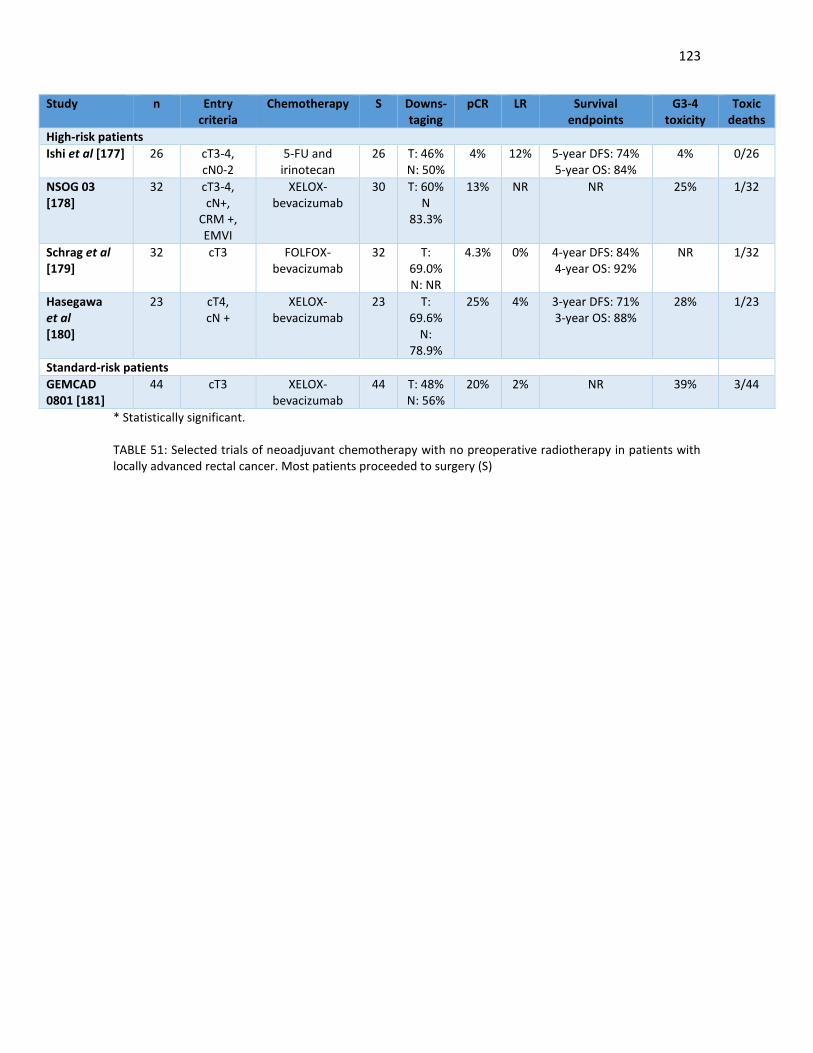
123
* Statistically significant. TABLE 51: Selected trials of neoadjuvant chemotherapy with no preoperative radiotherapy in patients with locally advanced rectal cancer. Most patients proceeded to surgery (S)
Study n Entry criteria
Chemotherapy S Downs-taging
pCR LR Survival endpoints
G3-4 toxicity
Toxic deaths
High-risk patients
Ishi et al [177] 26 cT3-4, cN0-2
5-FU and irinotecan
26 T: 46% N: 50%
4% 12% 5-year DFS: 74% 5-year OS: 84%
4% 0/26
NSOG 03 [178]
32 cT3-4, cN+,
CRM +, EMVI
XELOX- bevacizumab
30 T: 60% N
83.3%
13% NR NR 25% 1/32
Schrag et al [179]
32 cT3 FOLFOX-bevacizumab
32 T: 69.0% N: NR
4.3% 0% 4-year DFS: 84% 4-year OS: 92%
NR 1/32
Hasegawa et al [180]
23 cT4, cN +
XELOX- bevacizumab
23 T: 69.6%
N: 78.9%
25% 4% 3-year DFS: 71% 3-year OS: 88%
28% 1/23
Standard-risk patients
GEMCAD 0801 [181]
44 cT3 XELOX- bevacizumab
44 T: 48% N: 56%
20% 2% NR 39% 3/44
124
Practical aspects of adjuvant chemotherapy.
1. We offered adjuvant chemotherapy to all patients after surgery, regardless the response rate.
However, as in most other published trials, the tolerance to the adjuvant regimen was poor with less
than half of these patients receiving the full intended dose; this was especially evident in elderly
patients.
2. We observed a small but significant decrease in the rate of distant metastases with adjuvant CT; no
benefit in local control was obtained.
3. There is controversial evidence on the use of adjuvant CT after LCRCT and TME-surgery; most trials
have not shown any survival benefit. However, compliance to treatment was uniformly poor, which
may explain this lack of benefit. Unfortunately, we are not going to have randomized trials which will
answer this question
4. Patients with poor downstaging, high lymph node burden or other poor prognostic factors (high
grade, minimal regression perineural or vascular invasion, involved CRM, mucinous tumours) should
probably be offered adjuvant chemotherapy if feasible, if only for a lack of better options. The use of
nomograms and scores, such as the Valentini momogram or the Dhadda score, were helpful in our
case in selecting patients with poor prognosis.
5. If there is a benefit, it is probable small in patients with downstaging and especially in those who
have pCR; the doubts over the effectiveness of adjuvant chemotherapy should be transmitted to
patients and a shared-decision should be taken over its use.
6. We did not use neoadjuvant chemotherapy regimens, alone or alongside LCRCT or SCRT, as there are
only phase II evidence for its activity, but results are promising.
7. Improvements in clinical staging, with a better identification of pathological lymph nodes, are needed
to identify those patients with a higher risk of systemic relapse, who will benefit from more intensive
oxaliplatin-containing (neo)adjuvant regimens, probably in the neoadjuvant setting.
125
4.5. How can we improve clinical staging in order to define which patients have a higher risk of local relapse and distant metastases? 4.5.1. Primary tumour staging The importance of local staging in rectal cancer is dependent on the multimodality approach policy that is
used. In patients with locally advanced tumours treated with a blanket approach of neoadjuvant LCRCT, as
was our case, staging procedures are less important, although we know that there is an inherent higher risk
of over- and under-treating patients. In centres where a more progressive and selective policy is adopted,
relation of the tumour margin to the CRM is imperative for decision making, as involvement of the CRM
supercedes all other prognostic indicators for the use of neoadjuvant chemoradiation. In both cases, however,
the challenge for an imaging study is not just to be able to use it to determine resectability, but also to be able
to use it to predict the likelihood of local relapse and distant metastases—and thereby define the optimal
neoadjuvant strategy, and determine whether the tumor will respond to standard fluoropyrimidine-based
CRT.
As we used in all patients neoadjuvant LCRCT, the utility of clinical factors that increase the risk of local relapse
are probably not very useful. We observed a higher risk of local relapse in older patients, anaemic patients
(where we know that radiotherapy is less effective), poorly differentiated tumours and bulky tumours.
Curiously, involvement of the mesorectal fascia was not a significant factor, probably due to the fact that
almost 40% of patients were staged with EUS, which does not identify well CRM invasion in most cases, and
that LCRCT compensates somehow for an involved CRM. What is perhaps most interesting is that there was
no relationship between local relapse and lymph node involvement. The poor sensitivity of cN staging, as we
will see later, is probably a factor. However, it is also true that with better surgical rechniques, a successful
TME procedure would probably remove all pathological lymph nodes and that would not necessarily indicate
a higher risk of local relapse.
Both EUS and MRI are commonly recommended for rectal cancer staging, although most guidelines now agree
that MRI has become a required standard for evaluating locally advanced disease, particularly in those
patients with potential CRM involvement. This is despite the fact that in the the published meta-analysis, EUS
does not perform any differently to either CT or MRI. However, these analyses limited assessment to T and N
staging only and did not take into account the important prognostic variables that should also be assessed by
imaging, such as depth of extramural spread in millimetres, relationship of tumour to the mesorectal fascia
and extramural vascular invasion [182]. EUS has other disadvantages, such as its inability to stage high, bulky
and obstructing tumours [183]. The limited views of the whole mesorectum and pelvis with EUS does not
allow us to exclude tumour deposits, pathological lymph nodes, discontinuous vascular invasion and
mesorectal fascia involvement by tumour, which will inevitably lead to understaging [184]. In effect, that is
what we saw in our study, as in most patients staged by both imaging modalities, EUS understaged in
comparison to MRI, especially in cN disease, upper rectal cancers and stenotic tumours (FIGURES 32 and 33)
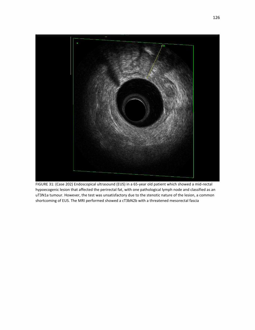
126
FIGURE 31: (Case 202) Endoscopical ultrasound (EUS) in a 65-year old patient which showed a mid-rectal
hypoecogenic lesion that affected the perirectal fat, with one pathological lymph node and classified as an
uT3N1a tumour. However, the test was unsatisfactory due to the stenotic nature of the lesion, a common
shortcoming of EUS. The MRI performed showed a cT3bN2b with a threatened mesorectal fascia
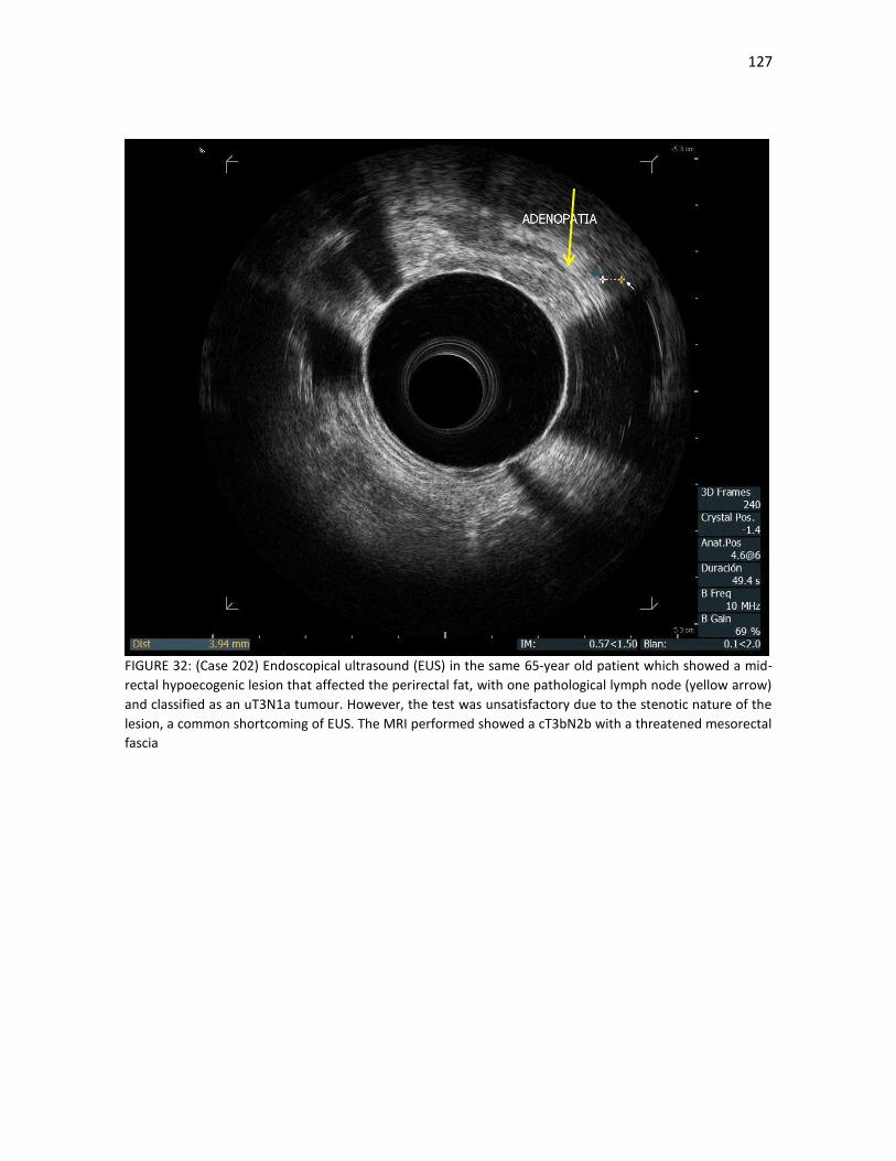
127
FIGURE 32: (Case 202) Endoscopical ultrasound (EUS) in the same 65-year old patient which showed a mid-
rectal hypoecogenic lesion that affected the perirectal fat, with one pathological lymph node (yellow arrow)
and classified as an uT3N1a tumour. However, the test was unsatisfactory due to the stenotic nature of the
lesion, a common shortcoming of EUS. The MRI performed showed a cT3bN2b with a threatened mesorectal
fascia
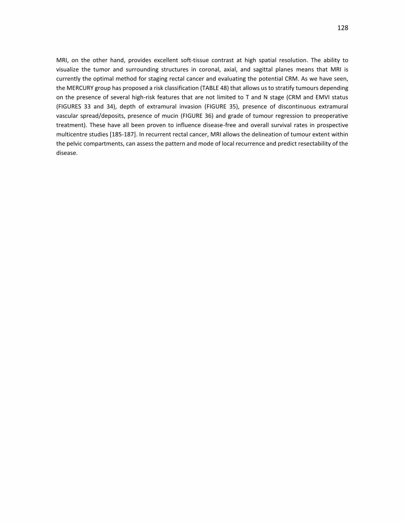
128
MRI, on the other hand, provides excellent soft-tissue contrast at high spatial resolution. The ability to
visualize the tumor and surrounding structures in coronal, axial, and sagittal planes means that MRI is
currently the optimal method for staging rectal cancer and evaluating the potential CRM. As we have seen,
the MERCURY group has proposed a risk classification (TABLE 48) that allows us to stratify tumours depending
on the presence of several high-risk features that are not limited to T and N stage (CRM and EMVI status
(FIGURES 33 and 34), depth of extramural invasion (FIGURE 35), presence of discontinuous extramural
vascular spread/deposits, presence of mucin (FIGURE 36) and grade of tumour regression to preoperative
treatment). These have all been proven to influence disease-free and overall survival rates in prospective
multicentre studies [185-187]. In recurrent rectal cancer, MRI allows the delineation of tumour extent within
the pelvic compartments, can assess the pattern and mode of local recurrence and predict resectability of the
disease.
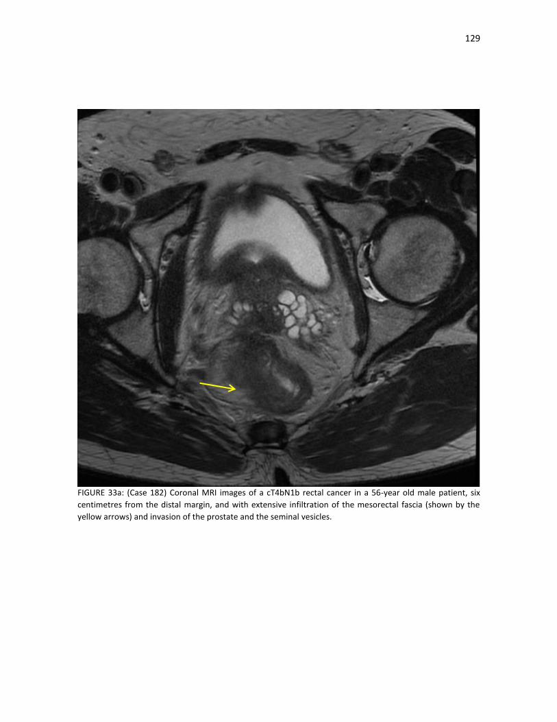
129
FIGURE 33a: (Case 182) Coronal MRI images of a cT4bN1b rectal cancer in a 56-year old male patient, six
centimetres from the distal margin, and with extensive infiltration of the mesorectal fascia (shown by the
yellow arrows) and invasion of the prostate and the seminal vesicles.
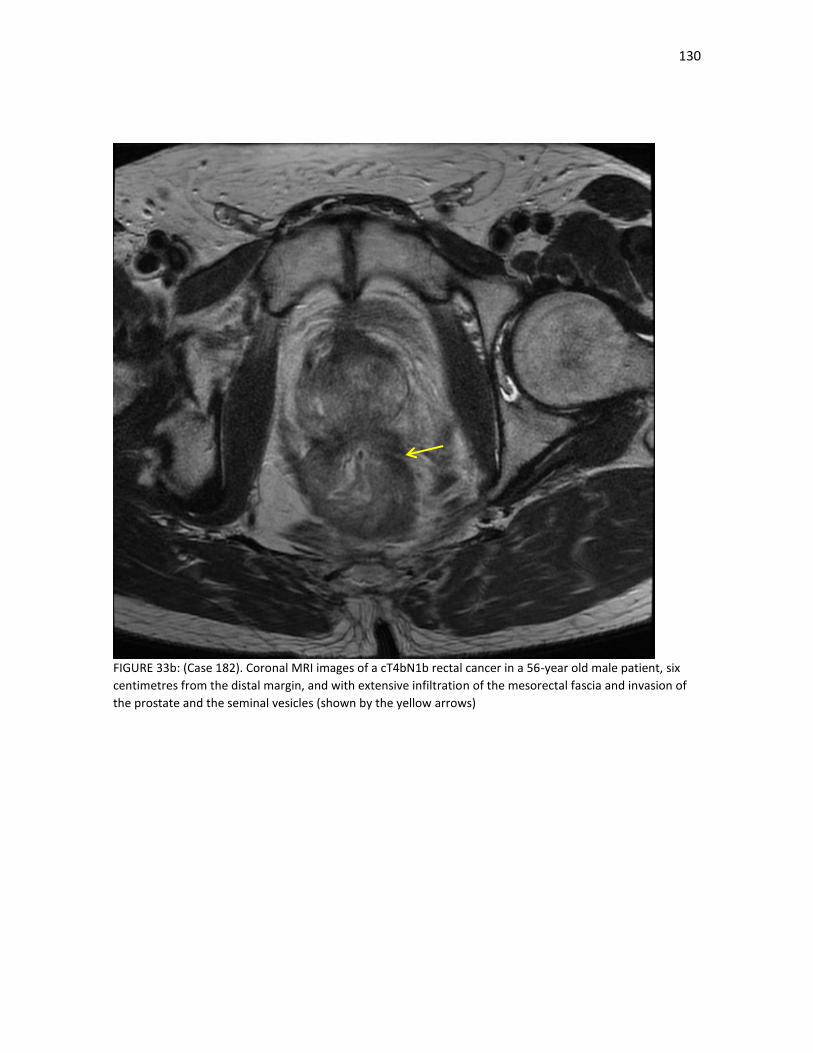
130
FIGURE 33b: (Case 182). Coronal MRI images of a cT4bN1b rectal cancer in a 56-year old male patient, six
centimetres from the distal margin, and with extensive infiltration of the mesorectal fascia and invasion of
the prostate and the seminal vesicles (shown by the yellow arrows)
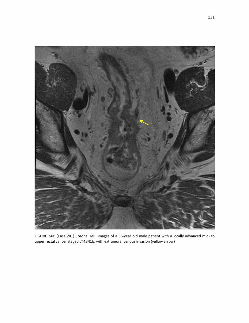
131
FIGURE 34a: (Case 201) Coronal MRI images of a 56-year old male patient with a locally advanced mid- to
upper rectal cancer staged cT4aN1b, with extramural venous invasion (yellow arrow)
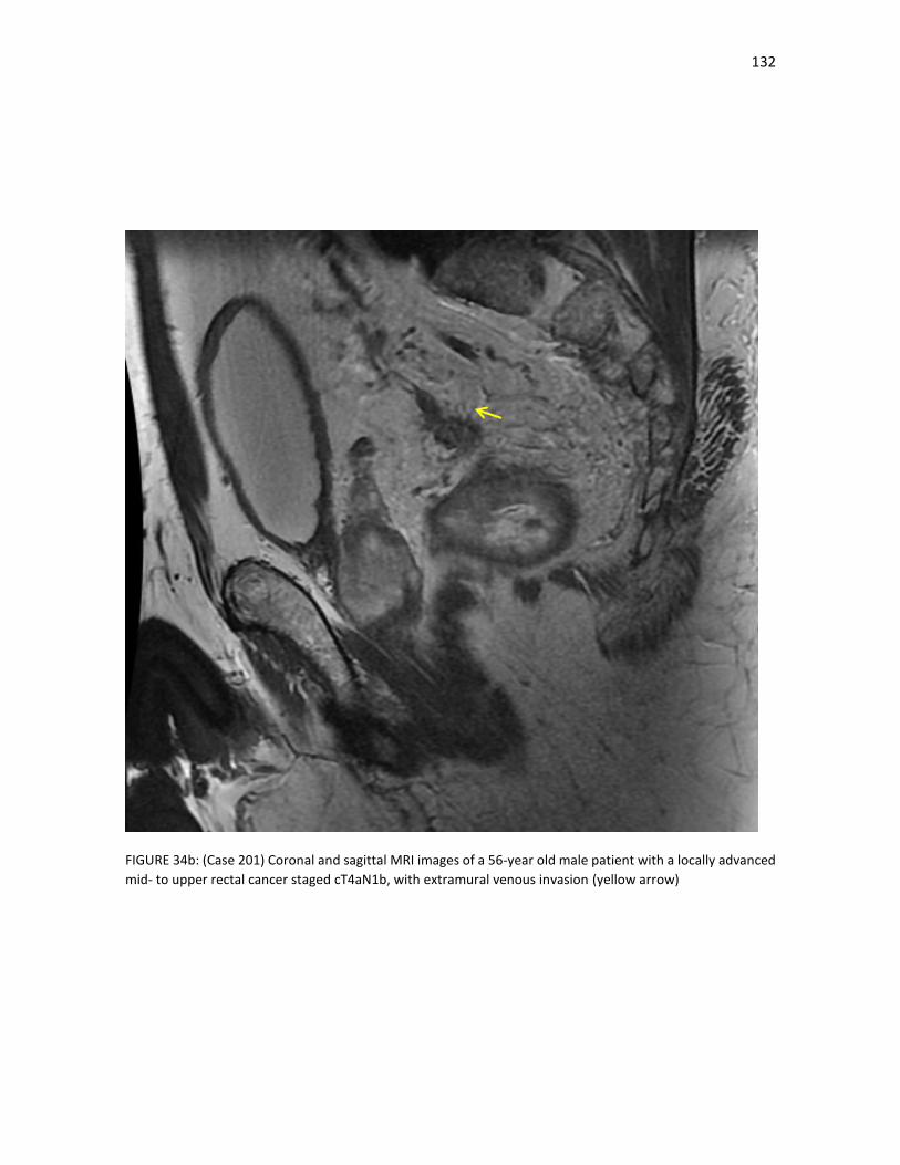
132
FIGURE 34b: (Case 201) Coronal and sagittal MRI images of a 56-year old male patient with a locally advanced
mid- to upper rectal cancer staged cT4aN1b, with extramural venous invasion (yellow arrow)
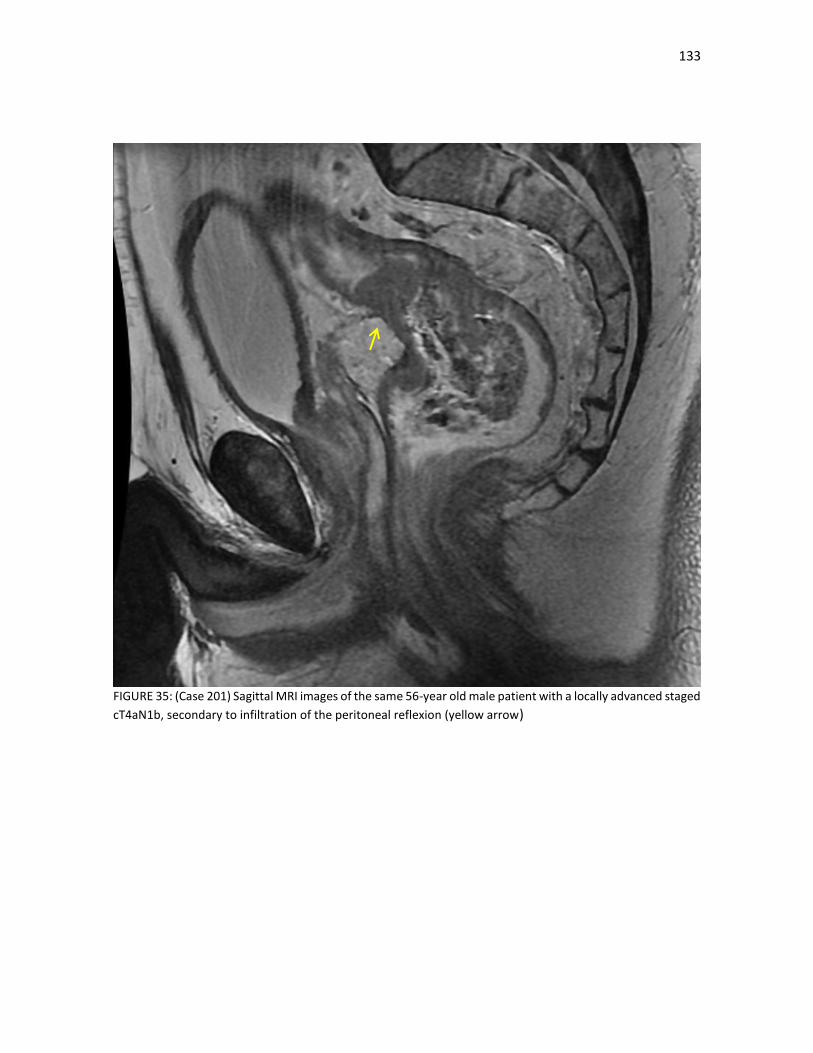
133
FIGURE 35: (Case 201) Sagittal MRI images of the same 56-year old male patient with a locally advanced staged
cT4aN1b, secondary to infiltration of the peritoneal reflexion (yellow arrow)
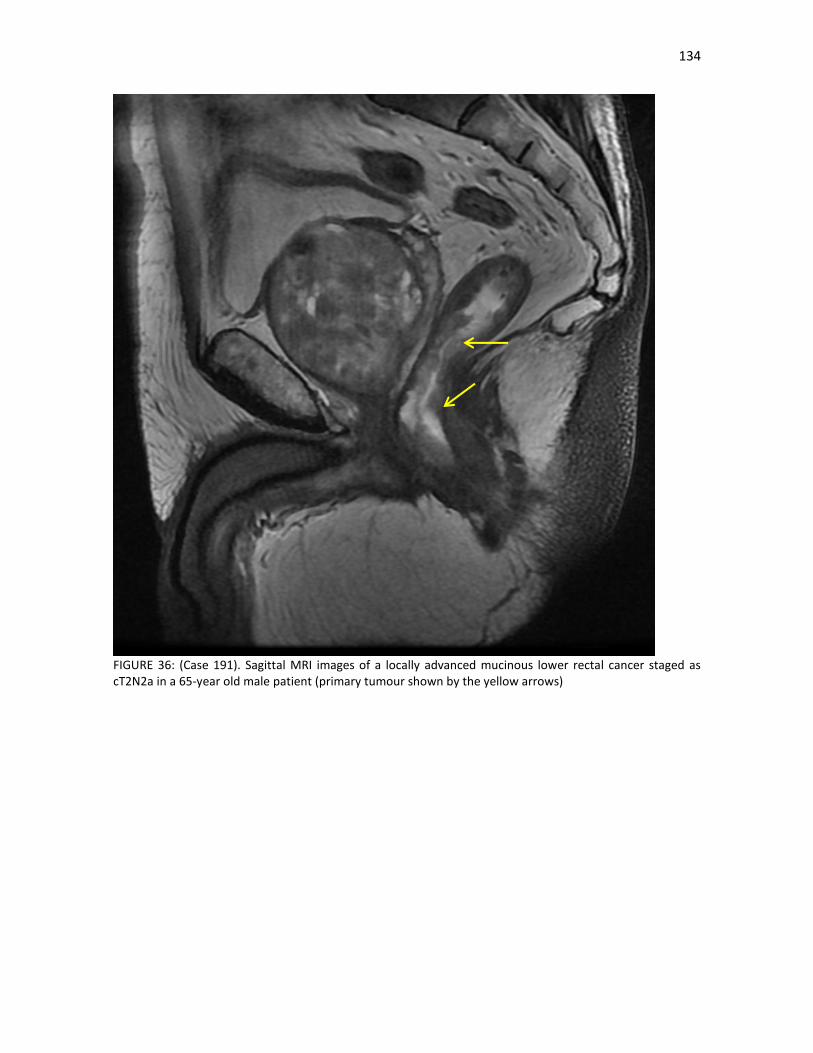
134
FIGURE 36: (Case 191). Sagittal MRI images of a locally advanced mucinous lower rectal cancer staged as cT2N2a in a 65-year old male patient (primary tumour shown by the yellow arrows)
135
MRI can also give us more important information. The risks of local failure are much higher for cancers below
the peritoneal reflection, and for those in the lower rectum below or involving the levators (FIGURE 37). Low
rectal cancers that are anteriorly located and fixed are more difficult to resect and have a higher risk of local
relapse [188-189]. The depth of tumour penetration in those patients staged as cT3 with MRI has also
prognostic importance. As we have mentioned, the MERCURY Study Group used preoperative MRI to extend
the clinical subclassification of T3 into four groups: “a” (< 1 mm outside the wall), “b” (1–5 mm), “c” (5–15
mm), and “d” (> 15 mm). An exploratory subset analysis of T3 tumors demonstrated local relapse rates for
SCPRT vs selective postoperative LCCRT of 2.7% vs 5.8% in T3a (≤ 1 mm) tumors; 2.9% vs 9.6% in T3b (> 1–5
mm) tumors; and 9.6% vs 21.9% in T3c (> 5–15 mm) tumors. [173]. Other groups have also shown that T3c
and T3d rectal cancers have markedly worse progression-free and cancer-specific survival compared with T3a
and T3b cancers [190]
As we have seen, in those centres where a risk-adapted approach is used, relation of the tumour margin to
the CRM is imperative for decision making. In a landmark study, rigorous histopathologic analysis revealed
27% of all rectal cancers demonstrated an occult positive CRM (< 1 mm) after potentially curative surgery
[191]. MRI can accurately predict CRM status and direct risk-stratified management strategies so as to select
patients appropriate for preoperative CRT. MRI prediction of CRM as compared with histopathology suggested
the use of a wider threshold with MRI (2 mm) than with pathology [56]; however, the MERCURY group based
their analysis on an MRI cutoff of < 1 mm [100] and a prospective study also showed that a 1-mm cutoff on
MRI predicts clear margins in 96.7% of cases [192]. Finally, In the MRC CR07 trial, the rate of finding a positive
CRM fell from 21% in 1998 to a low of 10% in 2005 [70] secondary to an improvement in the quality of the
mesorectal plane of excision (predominantly in patients undergoing anterior resection). The preoperative
results of MRI were compared with the histopathologic findings in the resected specimens, and showed an
accuracy for preoperative MRI of 90.9% [193]
These are impressive results and have been adopted by NICE, EORTC and ESMO for a more personalized
approach to localised rectal cancer, that is based on the performance of high-quality MRI imaging [97-98, 100].
However, we must also point out that this treatment approach guided by MRI-risk categorization is based on
results of prospective observational studies conducted in institutions with significant expertise in rectal cancer
and has not been tested in prospective randomized trials. They may not be adequate for smaller centres with
less experience or with smaller economic resources.
There are also other problems with MRI imaging. A current limitation is the significant learning curve
associated with image evaluation; there is also the uncertainty regarding nodal involvement. The widespread
use of preoperative LCRCT (leading in most cases to nodal downstaging) offers limited opportunities to the
radiologist to audit performances in pretreatment MRI assessments. Another caveat is the evaluation of
levator involvement in low rectal cancer (FIGURE 37) and the diagnosis of nodal involvement, which remain
difficult even with newer MRI sequences, such as diffusion-weighted imaging. Finally, there is no clear
evidence currently that MRI can discriminate between tumor grades; there are no specific texture features on
standard images, and no clear relationship with diffusion-weighted MRI in the small studies published to date.
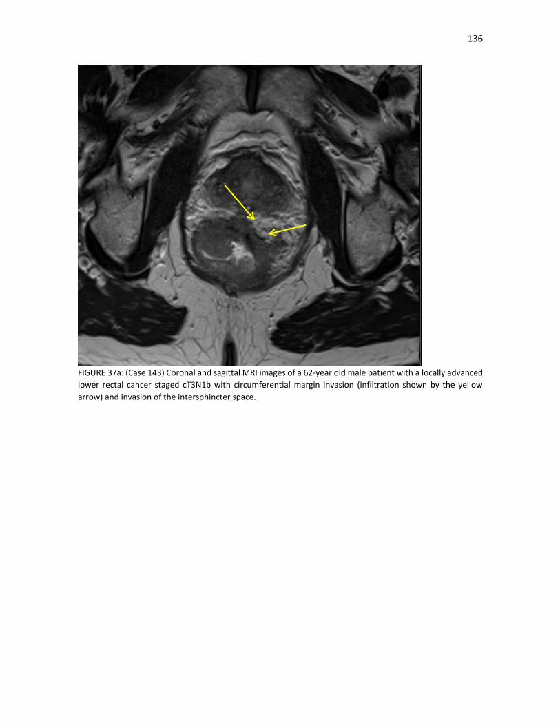
136
FIGURE 37a: (Case 143) Coronal and sagittal MRI images of a 62-year old male patient with a locally advanced
lower rectal cancer staged cT3N1b with circumferential margin invasion (infiltration shown by the yellow
arrow) and invasion of the intersphincter space.
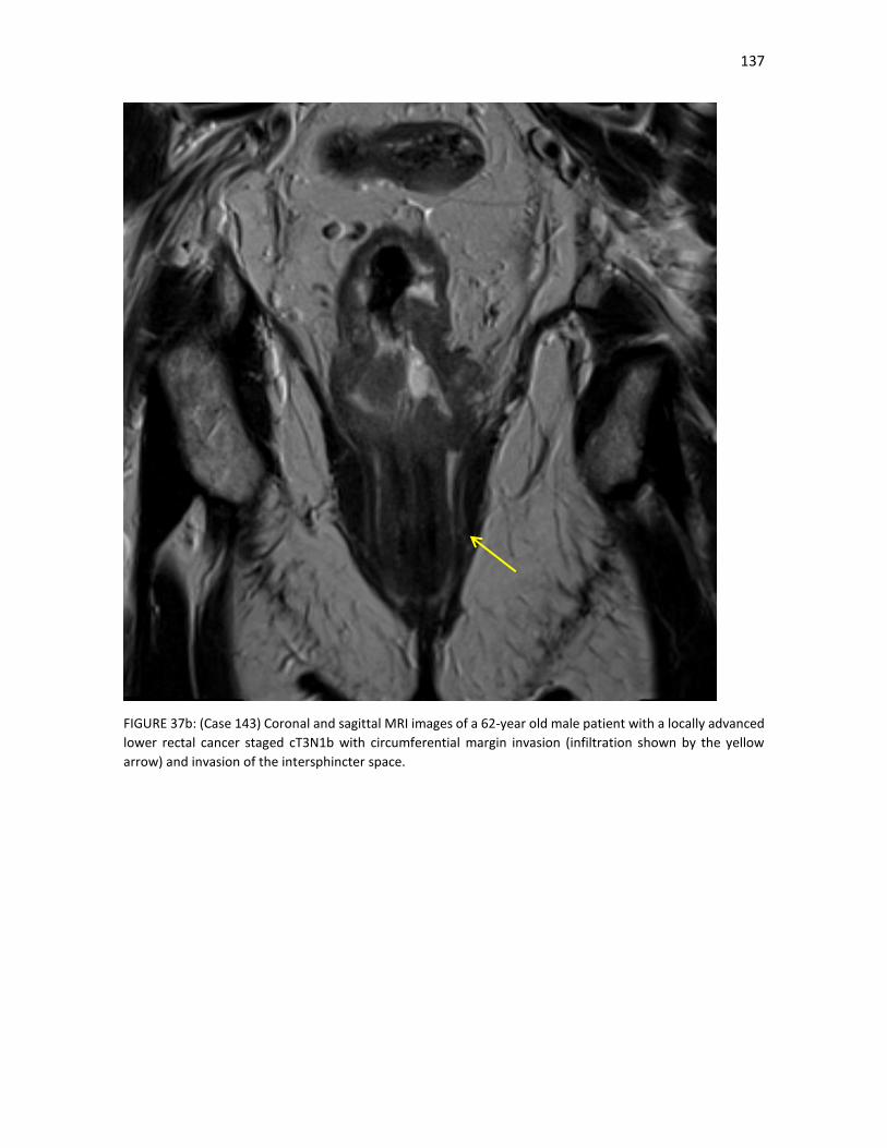
137
FIGURE 37b: (Case 143) Coronal and sagittal MRI images of a 62-year old male patient with a locally advanced
lower rectal cancer staged cT3N1b with circumferential margin invasion (infiltration shown by the yellow
arrow) and invasion of the intersphincter space.
138
4.5.2. Lymph node staging
Pathological stage III disease is the most important prognostic factor in localized rectal cancer and patients
with a heavy lymph node burden fare very poorly. This is especially important now that surgical improvements
and better identification of risk factors for local relapse, such as the circumferential margin invasion, have
decreased the risk of pelvic recurrence and the risk of distant metastases predominates. We also know that
neoadjuvant (chemo)radiotherapy does not improve survival and that adjuvant chemotherapy compliance is
poor and difficult to administer after LCRCT.
The logical step, as we have discussed previously, is to identify those patients with a high risk of distant
metastases, which would include most patients with lymph node disease, and offer them neoadjuvant
chemotherapy alone or alongside LCRCT and SCRT. In our series, where the majority of patients were staged
by MRI, baseline factors that predicted an increased risk of pathological lymph nodes included positive clinical
lymph node disease (as would be expected) and other poor prognostic factors, such as mesorectal fascia
invasion, extramural venous invasion, lower rectal tumours and elevated CEA and CA 19.9 levels. On the other
hand, nodal staging is of questionable importance as a predictor for local recurrence in patients receiving
radical surgery for primary tumours; however, it still should be assessed if local excision is considered.
Unfortunately, the clinical lymph node staging remains the Achilles heel in the baseline staging of patients
with locally advanced rectal cancer, even with the use of high-quality MRI. In effect, although the preoperative
MRI has proven to predict both T category and circumferential resection margin with good accuracy, the
accuracy for predicting lymph node metastasis has been poor [182, 194-195], with moderate sensitivity (60-
84%) and specificity (59–81%). One limitation in detecting positive lymph nodes by MRI is that a great number
of detected nodes turn out to be normal or benign reactive nodes. In a node-to-node study of lymph nodes in
rectal cancer, only 76 out of 521 nodes (14.6%) were confirmed malignant nodes by histology [196]. Currently,
identifying malignant nodes mainly relies on the size of nodes (FIGURES 38 and 39). That is a useful, but
imperfect strategy, as studies have confirmed that the proportion of positive nodes increases with larger nodal
size [197-199]. However, there is no widely accepted consensus on the size criterion of enlarged lymph nodes,
and substantial overlap exists between benign and malignant nodes. Although very small lymph nodes can be
observed by high resolution MRI, the diagnosis of metastasis from detected lymph nodes is challenging, and
the interobserver agreement in diagnosis of lymph nodes metastases varies among radiologists, especially for
sub-centimeter lymph nodes [200-201].
Others have found that besides nodal size, lymph nodes with spiculated or indistinct border and mottled
heterogeneous appearance were useful to predict positive nodes [198-199]. Despite this, in one study, even
when the criteria of positive lymph nodes were strict (heterogeneous texture, irregular margin, and nodal size
> 10 mm), the total accuracy was only 63%, with a sensitivity of 85% and specificity of 41% [202]. Most of
these studies only had a small number of patients and multivariate analyses were rarely used to assess the
relationship between these parameters and the nodal size. In other cases, the border, shape or intrinsic signal
is difficult and inaccurate for very small nodes (less than 3–5 mm) in MRI. To make things more difficult, some
low-signal-intensity rim in small lymph nodes is difficult to distinguish between normal lymph node capsule
and tumor with necrosis, and tumor without necrosis in lymph node will present similar intensity with normal
lymph nodes.
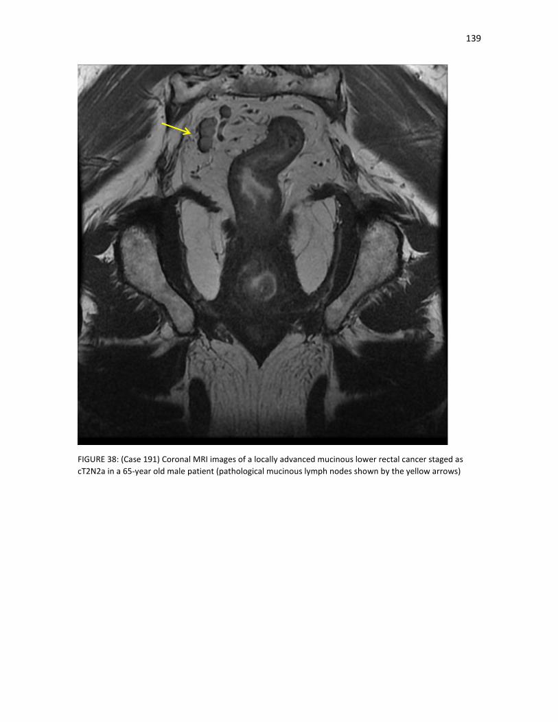
139
FIGURE 38: (Case 191) Coronal MRI images of a locally advanced mucinous lower rectal cancer staged as
cT2N2a in a 65-year old male patient (pathological mucinous lymph nodes shown by the yellow arrows)
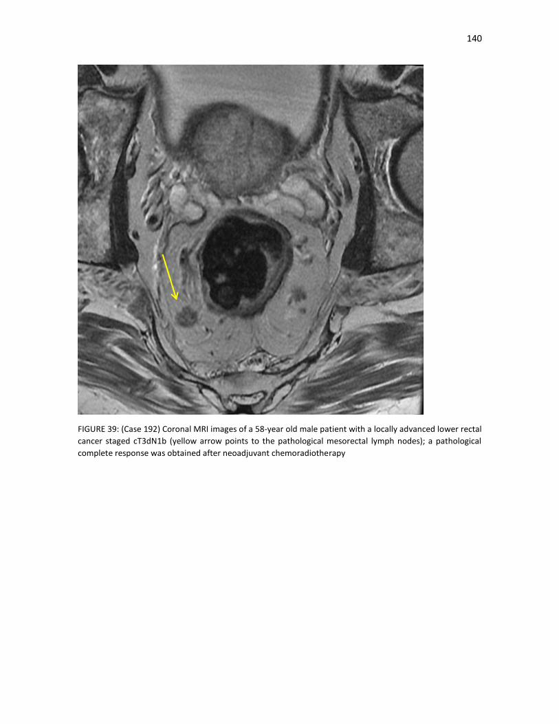
140
FIGURE 39: (Case 192) Coronal MRI images of a 58-year old male patient with a locally advanced lower rectal
cancer staged cT3dN1b (yellow arrow points to the pathological mesorectal lymph nodes); a pathological
complete response was obtained after neoadjuvant chemoradiotherapy
141
4.5.3. Why do we not restage patients after neoadjuvant therapy?
We found CRM invasion, a heavy lymph node burden and an unsuccessful TME as pathological risk factors of
a worse survival. In this sense, accurate assessment of tumour response could be useful in order to stratify
patients into low- and high-risk groups after neoadjuvant treatment, but before surgical resection and
pathologic evaluation, intensified strategies could be planned.
However, as recommended by most organizations, we did not perform post-neoadjuvant therapy imaging
routinely, except in patients where progression is suspected. There are several reasons for this. Unfortunately,
tumour downstaging is usually overestimated by conventional MRI evaluation. EUS does not fare much better.
Around 50% of patients have tumours that are relatively resistant to CRT and that change little. Larger T3/T4
tumors tend to respond by fragmentation and can be difficult to assess after CRT, whereas smaller T2/T3a
tumors tend to shrink back to the endoluminal point of origin. Individual lymph node size also is a poor
predictor of nodal metastases following CRT [199] although there is a high negative predictive value if
previously imaged involved nodes return to normal.
Lastly, theoretically, a good clinical response could offer the opportunity to change the original plan for
surgery, and to perform a less mutilating resection if an adjacent organ was originally involved or facilitate
sphincter-sparing surgery in distal tumours. On the real world, however, there is very little evidence to support
the view that preoperative CRT increases the chance of a sphincter-preserving resection [203]. In the majority
of patients with a mid and upper rectal cancer, an anterior resection is feasible without tumour shrinkage.
Very low tumours less than 4 cm from the anal verge usually require an abdomino-perineal excision.
Therefore, it is only in a very small group of patients whose distal tumour extent is 4–6 cm from the anal verge
where preoperative CRT may play a role in achieving a sphincter-preserving procedure
There are some studies in this regard. The MERCURY group assessed a modified Mandard grading system of
tumour response and proved it to be a reliable tool for assessing tumour response in rectal cancer with MRI
[204]. This grading scale is based on qualitative assessment of intermediate signal vs low signal (the latter
considered as fibrosis and the former as residual disease) within the treated tumour. MR TRG 1–2 can be
described as low signal intensity scar or low density fibrosis with no evident macroscopic intermediate signal
intensity within it, mrTRG3 dominant fibrosis outgrowing the tumour mass and mrTRG 4–5 predominantly
intermediate signal intensity with minimal or no signs of fibrosis. MERCURY experience showed that patients
with complete and near complete response (mrTRG 1–3) have better prognosis, disease-free and overall
survival compared to poor responders [205]. Although interesting findings, what should we do these findings?
As we will see later, fibrosis after neoadjuvant treatment is not a uniform phenomenon and is not always
synonymous of response. Pathological stage is probably a more important prognostic factor than TRG. Lymph
node invasion remains difficult to identify, even more so after neoadjuvant radiotherapy.
In this sense, prospective trials should be done in order to assess of tumour response in order in order to
stratify patients into low- and high-risk groups after neoadjuvant treatment, but before surgical resection and
pathologic evaluation, intensified strategies could be planned
.

142
Practical aspects of radiological staging:
1. The use of a blanket-approach of treating all locally advanced rectal cancers with LCRCT, as we
used, lowers somewhat the importance of a precise primary tumour staging, although we know
there is an inherent higher risk of over- and under-treating patients. The importance of baseline
prognostic factors for local relapse and distant metastases for selecting patients for risk-adapted
approaches also diminishes with this approach.
2. Broadly speaking. MRI and EUS show a similar sensitivity and specificity with respect to cT
staging; EUS is particulary useful for the staging of early tumours, such as T1-T2. However, a
limited view field, poor assessment of the CRM and the difficulty in staging stenotic tumours are
disadvantages with EUS, with a tendency to understage tumours. In our series, when both tests
were performed, EUS understaged in comparison to MRI, especially in cN disease, upper rectal
cancers and stenotic tumours.
3. MRI is especially preferred in locally advanced rectal cancer by its assessment of cT and invasion
of the mesorectal fascia and other prognostic factors, such as EMVI, lateral lymph pelvic nodes
or tumour deposits. Any risk-adapted approach should include a high-quality MRI, as the
mesorectal rectal status is the most important factor in determining the need of downstaging
and of neoadjuvant (chemo)radiotherapy.
4. Clinical N staging remains a problem, as MRI, and even more so EUS, has poor sensitivity and
specificity for this indication. Lymph node size is a poor distinguishing factor. Other features such
as irregular borders and a heterogeneous signal can help, although results are still far from
satisfactory.
5. Despite the difficulties in cN staging, the presence of cN positive disease, mesorectal fascia
invasion, extramural venous invasion, lower rectal tumours and elevated CEA and CA 19.9 levels
predicted a higher risk of distant metastases. These are poor-prognosis patients and they would
be prime candidates to any neoadjuvant full-dose chemotherapy approach
6. We found that there was an increased risk of local relapse in poorly differentiated tumours and
bulky tumours; perhaps more importantly, as in other studies, we did not find a higher risk with
cN positive disease, especially in N1 tumours. This may reflect the difficulties in staging lymph
node disease with all imaging modalities but also that a successful TME will probably remove
satisfactorily all pathological nodes.
7. We did not use restaging procedures after neoadjuvant therapy. Their use, although attractive,
is misleading in many cases, as MRI tends to over-estimate downstaging and there is no evidence
that surgeons will change their intended original approach with these results. EUS fares even
worse. Radiological TRG, such as those proposed by the MERCURY group, suffer from the same
problems as histological TRG; they could probably be useful in patients complete or near-
complete regression, but not in patients with lesser degrees of regression.
143
4.6. Why did our older patients not benefit from our multimodality approach?
On our series, age was the most important clinical factor in predicting worse OS and DFS after neoadjuvant
LCRCT (TABLE 13). Patients older than 70 years fared poorly in almost all aspects of the multimodality
treatment. We found that elderly patients had a poorer compliance to overall treatment, with more than half
of patients receiving a radiotherapy dose less than 45 Gy, with a borderline statistically significant increase of
grade 3-4 toxicity and of unexpected cardiovascular and surgical events, compared to their younger
counterparts. The rate of abdominoperineal resections was higher in elderly patients while the poor
compliance to the neoadjuvant treatment was linked to a lower rate of complete or near complete tumour
regressions and a higher risk of local relapse. The compliance of adjuvant treatment was particularly poor,
with less than a quarter of elderly patients receiving the full intended dose, compared to more than half of
younger patients. Despite these results, there was no evidence of differences in classical pathological risk
factors. More strikingly, there were even hints of less aggressiveness in these older patients’ tumours, with
less cN-positive staging. How can we explain these differing conclusions? The most plausible explanation is
that LCRCT is probably not the best approach in elderly and/or frail patients, and less aggressive multimodality
treatments should be considered.
Other studies support this view. Recent analyses of the Dutch total mesorectal excision (TME) study and two
large population-based registries in the Netherlands showed that, unlike younger patients, patients more than
75 years of age did not experience gains in overall survival after the introduction of preoperative radiotherapy
and TME surgery [207]. In addition, analyses from the ACCENT database and MOSAIC trial failed to show a
benefit to adjuvant combination chemotherapy in elderly patients [130, 208]. This lack of benefit seen in
elderly patients compared with younger patients may partly be due to high rates of treatment deviation. This
poorer tolerance is essentially due to non-hematological side effects of various origins reflecting the
heterogeneity among older patients in terms of physiology and frailty. Our study, like others, provides useful
insight into how older patients tolerate therapy, especially considering that 30% of 40% of rectal cancer cases
occur in patients 75 years and older.
There are also population-based studies that indicate a high rate of deviation from a full course of combined
modality therapy [209-210]. A Surveillance, Epidemiology, and End Results (SEER)–Medicare study of patients
more than 65 years of age who were receiving postoperative therapy for rectal cancer showed that whereas
97% of Stage III rectal cancer patients completed radiation therapy, only 68.2% completed chemotherapy
[209]. Among stage II cancer patients, 91.5% completed radiation therapy but only 49.8% completed
chemotherapy. Unfortunately, population-based studies are not able to identify why doctors choose to
discontinue adjuvant treatment for elderly patients, or why elderly patients decline further treatment. In this
study, the most commonly cited reason for early discontinuation of adjuvant chemotherapy was the patient’s
performance status. In effect, that was the main reason for giving no adjuvant chemotherapy in our patients.
Another poorly recognized factor is that even low-grade toxicities can lead to treatment discontinuation in
elderly patients, with a lower threshold than in younger patients [211].
The two previously mentioned SEER–Medicare studies [209-210] showed that patients who completed
adjuvant chemoradiotherapy had improved cancer-specific survival compared with those who do not,
supporting the efforts of oncologists to enable patients to complete a full course of therapy. Yet such studies
are limited because of selection bias and other potential confounders of the relationship between age and
treatment completion, such as comorbidity. A Canadian study also showed that elderly patients with stage III
colon cancer frequently received either no adjuvant chemotherapy or only capecitabine monotherapy
144
because of advanced age and comorbidities., although the effect of adjuvant chemotherapy on survival was
similar across age groups, with comparable side effects and rates of treatment modifications [212]. However,
in another Dutch registry study, the combination of capecitabine and oxaliplatin was associated with
significantly more grade 3-4 toxicities than capecitabine monotherapy, which had a pronounced impact on
the cumulative dosage received and completion of all planned cycles [213]
Does that mean that elderly patients should not be offered multimodality treatment?. Probably not. Most
experts agree that chronological age itself is not sufficient grounds for withholding cancer treatment [214], as
it is not possible to judge whether a patient is fit for radical rectal cancer treatment merely by looking at their
date of birth. Unfortunately, however, multiple studies have demonstrated that treatment decisions in elderly
cancer with colorectal cancer patients are still largely based on age [215-218]. Tailoring of care is needed, and
for this reason, some form of geriatric evaluation is increasingly being incorporated in oncologic care [219-
220]. A geriatric evaluation can help identify previously unrecognized health issues that have been
demonstrated to be associated with prognosis and treatment-related complications and can provide guidance
in balancing the risks and benefits of treatment. [219]. Such studies should not only incorporate a geriatric
evaluation but also those outcome measures most relevant to older patients in their treatment decisions,
such as care dependence and quality of life.
TABLE 49 shows the few trials that have evaluated specifically the role of LCRCT or SCRT in elderly patients
with locally advanced rectal cancer; most used some kind of geriatric or comorbidity grading of patients.
Although there is wide variability between all these studies and the numbers are relatively small, all the trials
seem to agree with our findings. Compliance with the multimodality treatment was poor. Patients with
comorbidities or poor performance status fared worse, as could be expected, secondary to higher acute
toxicities. There were no clear evidences that, if the treatment was given accordingly to plan, the efficacy
results were worse than those in younger patients. Surgical complications were not higher, but were more
serious and took longer to resolve than in younger patients. Of all these studies, the study of the ACCORD12
phase III trial is probable the most interesting, as it showed, that in the context of a well-defined phase III trial,
elderly patients (defined as patients older than 70 years), although there were no differences in efficacy
endpoints, there was a higher grade 3-4 toxicity and a statistically significant decrease in the number of
surgical resections.
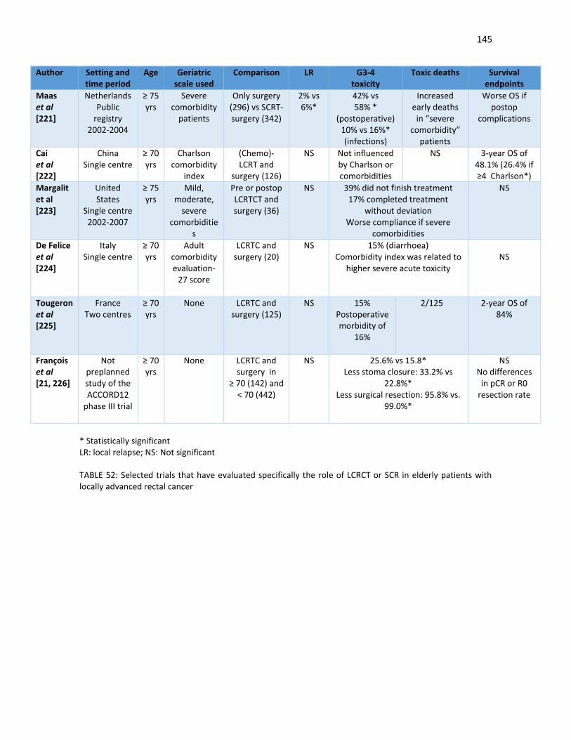
145
Author Setting and time period
Age Geriatric scale used
Comparison LR G3-4 toxicity
Toxic deaths Survival endpoints
Maas et al [221]
Netherlands Public
registry 2002-2004
≥ 75 yrs
Severe comorbidity
patients
Only surgery (296) vs SCRT-surgery (342)
2% vs 6%*
42% vs 58% *
(postoperative) 10% vs 16%* (infections)
Increased early deaths
in “severe comorbidity”
patients
Worse OS if postop
complications
Cai et al [222]
China Single centre
≥ 70 yrs
Charlson comorbidity
index
(Chemo)- LCRT and
surgery (126)
NS Not influenced by Charlson or comorbidities
NS 3-year OS of 48.1% (26.4% if ≥4 Charlson*)
Margalit et al [223]
United States
Single centre 2002-2007
≥ 75 yrs
Mild, moderate,
severe comorbiditie
s
Pre or postop LCRTCT and surgery (36)
NS 39% did not finish treatment 17% completed treatment
without deviation Worse compliance if severe
comorbidities
NS
De Felice et al [224]
Italy Single centre
≥ 70 yrs
Adult comorbidity evaluation-
27 score
LCRTC and surgery (20)
NS 15% (diarrhoea) Comorbidity index was related to
higher severe acute toxicity
NS
Tougeron et al [225]
France Two centres
≥ 70 yrs
None LCRTC and surgery (125)
NS 15% Postoperative morbidity of
16%
2/125
2-year OS of 84%
François et al [21, 226]
Not preplanned study of the ACCORD12
phase III trial
≥ 70 yrs
None LCRTC and surgery in
≥ 70 (142) and < 70 (442)
NS 25.6% vs 15.8* Less stoma closure: 33.2% vs
22.8%* Less surgical resection: 95.8% vs.
99.0%*
NS No differences
in pCR or R0 resection rate
* Statistically significant LR: local relapse; NS: Not significant TABLE 52: Selected trials that have evaluated specifically the role of LCRCT or SCR in elderly patients with locally advanced rectal cancer
146
With all these results in mind, although older patients should benefit from some kind of multimodality
therapy, perhaps standard LCRCT may not be the most appropriate approach and SCRT would be preferable.
In the Dutch TME-study, patients aged 75 years and older showed a better response in the study arm when
compared to younger patients with SCRT. Younger patients have a significantly lower local recurrence rate of
5.2% after preoperative radiotherapy versus 11% for patients without preoperative radiotherapy. However,
overall survival at 5 years, distant metastases free survival and cancer-free survival were not improved. In the
elderly, however, apart from the local recurrence rate, DFS and cancer-specific survival were improved. All
this suggests that the biological behavior of rectal cancer in the elderly in response to radiotherapy is better
than in younger patients [227]. No significant differences with respect to postoperative morbidity and
mortality were found between neoadjuvant RT and surgery only. Therefore, neoadjuvant RT and surgery may
be the optimal treatment strategy in elderly patients, especially in patients medically fit for the operation. In
effect, this was the main conclusion of the ACCORD12 trial in elderly patients. [226]. In very frail patients, new
radiotherapy approaches, such as image-guided radiotherapy (IMRT), may be used to lower acute toxicity and
to improve their tolerance to radiation and allow them to have curative resection despite the associated
comorbidity. Also, in cases where downstaging is needed, we know that there are promising results with SCRT
and a longer interval to allow downstaging in patients not fit for LCRCT [71-74]. The recent results of the
Stockholm III and the Polish Trial show that this can be a feasible approach [87, 90]
Practical aspects of age as a prognostic factor:
1. Our elderly patients fared poorly in almost all aspects of the multimodality treatment. This was
especially evident in an increased risk of serious unexpected (usually cardiovascular or surgical)
complications, and a lower compliance to neoadjuvant radiotherapy and, especially, to adjuvant
chemotherapy.
2. Disease-free and overall survival were worse in the elderly, even though we did not find any signs
of higher aggressiveness in these tumours, compared to their younger counterparts. There is
also no evidence to suggest that older patients have poor responses to (chemo)radiotherapy. In
particular, there seems to be a greater radiosensitivity in these patients.
3. Other secondary endponts, such as stoma closure or the rate of sphincter-conserving surgery,
were also worse for the older patients.
4. Chronological age, however, should not be the only determining factor un the decision of the
type of multimodality treatment offered. Evaluation of comobirbidites, especially
cardiovascular, and more formal geriatric assessments would be helpful in this regard.
5. LCRCT perhaps is not the best option for older patients, especially in frail or with other
comorbidities. All these results warrant the development of specific therapeutic strategies more
optimal for patients above 70–75 years, including fit patients over 80 years. SCRT followed by
delayed surgery to decrease toxicities and permit tumor downsizing is a promising approach.
Developing less toxic treatments should allow a higher percentage of patients to undergo
surgery.
147
4.7. Can TME-surgery be improved? The problem of low rectal tumous and of lateral pelvic lymph nodes
The analysis of our surgical series shows, in a long period of 15 years, an improvement in the surgical expertise
and evidence of a learning curve; the rate of abdominoperineal resections decreased from almost half of
patients in the first period between 1999 and 2004 to just 20% in the period of 2010-2014; the laparoscopic
approach increased significantly and the rates of unsuccessful TME also decreased. 30-days mortality after
surgery was consistently low. A total mesorectal excision was technically feasible in almost 90% of patients,
while an R0 resection was described by the surgeon in more than 90% of patients. These results were
obtained, we must remember, in a poor prognosis-group of patients, with invasion of the mesorectal fascia in
50% of the 144 patients where it was evaluable (unfortunately, that was not possible in the initial group of
patients staged only by EUS). In the pathological analysis, there was a 70% downstaging rate, although there
was still involvement of the circumferential resection margin in 21% of patients.
We also observed a progressive increase in the use of provisional diversion loop ileostomies after LCRCT as it
became clearer the higher risk of anastomotic leakages, especially in ultra-low anastomosis, where there are
reports of an incidence ranging from 10% to 28%. A recent trial and meta-analysis and systematic review of
the literature in 2008 both found that a diverting stoma is associated with fewer clinical leaks and rec-
ommended a diverting stoma in all low rectal anastomoses [228-229] Similar findings were reported from
pooled data from major multicentre European trials. These results, in general, seem satisfactory and in line
with other series [230].
However, as we have mentioned, this blanket-approach of treating all patients with neoadjuvant LCRCT
probably under- and overtreats a group of patients. We also know that neoadjuvant (chemo)radiotherapy
lowers the local relapse rate but does not improve survival and, more importantly, limits the possibility of
giving the full-intended dose of adjuvant chemotherapy and can have long-term side effects. On the other
hand, we also know from the MRC07 trial of SCRT, that all patients, even those with low risk tumours, benefit
from radiotherapy. We also know that these possible long-term side effects will probably diminish with the
continuous improvements in radiotherapy techniques (smaller fields, use of IMRT, better delimitation of the
tumour with MRI etc). Finally, as we have seen, despite the difficulties in staging, the risk factors of local
relapse and distant metastases are overlapping but not entirely the same.
How can we balance the proven benefits of radiotherapy in the risk of local relapse with the most certain
overtreatment if we treat all patients with neoadjuvant (chemo)radiotherapy?
In centres with surgical expertise in rectal cancer surgery and where high-quality MRI can be performed, in a
multidisciplinary setting, we can probably select patients with a very low risk of local relapse, who could be
treated with surgery, with or without SCRT. Mid rectal tumours with little penetration into the meso-rectal
fat (the MERCURY group defined a 5 mm cut-off point, while others have defined a 4 mm cut-off point) and,
perhaps, more importantly, with no or minimal clinical lymph node disease (N1a or N1b) and with a clear MRF,
are probably the best candidates for this approach, which we have adopted in the last few years.
However, the main problem, in our opinion, are the low rectal tumours. Even when TME principles are applied,
for low tumours, the local relapse rates are higher for tumors treated with an abdominoperineal excision
compared with a sphincter-saving procedure, such a lower anterior resection [4]. Originally, this different risk
148
was attributed to the difficulties in staging and in possible differences in tumour biology and patterns of
spread: for example, low rectal cancers 3-4 centimetres from the margin are at a higher risk of positive CRM
involvement, especially those located anteriorly [231]. It is also more difficult to predict levator involvement
even with high-quality MRI and there is a 15% risk of lateral pelvic lymph nodes. However, apart from these
well known factors, there is evidence from pathology audits that inadequate surgery may also play a role [232-
233].
A locally advanced rectal cancer located in the vicinity of the anorectal ring likely involves the levator muscle
or external anal sphincter. Extending the dissection along the mesorectal fascia to the level of the anorectal
ring separates the rectal wall from the levators and risks exposing the tumour closer to the CRM [233].
Pathologic studies of abdomino-perineal resection specimens have found a smaller volume of tissue around
the muscularis propria of the rectum and a higher rate of positive CRMs compared with specimens from low
anterior resections [234]. The Dutch TME-trial found higher positive CRMs and more perforations in
specimens from patients with low rectal cancer [232]. In their study, patients undergoing an
abdominoperineal excision had a higher risk of a positive CRM, independent of tumor height as well as higher
local relapse rates.
In order to improve these results, some authors have proposed a more radical abdominoperineal excision, in
which the dissection along the mesorectal fascia ends at the upper level of the levators, and the levator
muscles are left in their natural position, attached to the distal rectum. This procedure has been called
cylindrical or extralevator abdominoperineal excision (ELAPE), distinguishing it from the standard
abdominoperineal excision (SAPE) in which the levators are not removed with the specimen [233, 235]. The
results of a single institution randomized trial [236] and a recent meta-analysis of several case series indicate
that ELAPE is associated with lower rates of intraoperative perforation, lower rates of positive CRM and local
relapse, and similar complication rates compared with SAPE [237]. On the other hand, other population-based
tumor registry analyses have not demonstrated the benefit of ELAPE compared with SAPE [237-238].
Introduction of ELAPE principles has raised awareness about the importance of adequate preoperative
imaging for surgical planning in patients with locally advanced rectal tumours who would require an
abdominoperineal excision. In the future, we might see a more selective approach in patients with low rectal
tumours, especially if an abdominoperineal excision is not required, but at the moment, due to the surgical
difficulties and the higher risk of local relapse, these patients are probably best served with some type of
neoadjuvant therapy at the moment; in our centre and in most others, the majority of these patients are still
treated with standard LCRCT and TME-surgery.
We observed in our series a 9.7% rate of lateral pelvic lymph nodes (in those patients staged with MRI, FIGURE
40). There is controversy as to the management and relevance of these lateral pelvic side-wall nodes. In Japan
the presence of these lymph nodes mandates their surgical resection via lateral pelvic lymphadenectomy, a
more aggressive surgical approach that carries an increase in morbidity and long-term quality of life
complications [239]. In a retrospective review of 237 patients with T3-4 low rectal cancer who underwent R0
resection, including lateral pelvic lymphadenectomy, the incidence of lateral nodal involvement increased
with decreasing tumour height, with an incidence of 45% in those tumours less than two centimetres away
from the margin. Other factors predicting an increased risk of involved lateral nodes were involved mesorectal
nodes, female patients, advanced T stage, poor tumour differentiation, lymphovascular invasion and low
rectal cancer [240]. However, in Europe, these patients are still usually treated with LCRCT and TME-surgery.
There is evidence that this approach may be as effective as nodal removal, with less morbidity. Of note, in our

149
series, none of the patients with involved lateral pelvic lymph nodes at diagnosis had ypN disease in the
surgical specimen, casting more doubts in the significance of these clinically staged lymph nodes.
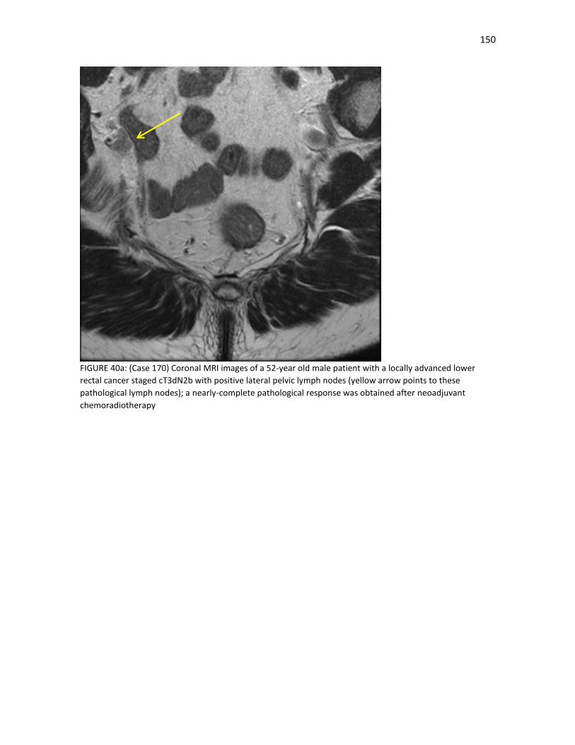
150
FIGURE 40a: (Case 170) Coronal MRI images of a 52-year old male patient with a locally advanced lower
rectal cancer staged cT3dN2b with positive lateral pelvic lymph nodes (yellow arrow points to these
pathological lymph nodes); a nearly-complete pathological response was obtained after neoadjuvant
chemoradiotherapy
151
FIGURE 40b: (Case 170) Coronal MRI images of a 52-year old male patient with a locally advanced lower
rectal cancer staged cT3dN2b with positive lateral pelvic lymph nodes (yellow arrow points to these
pathological lymph nodes); a nearly-complete pathological response was obtained after neoadjuvant
chemoradiotherapy
152
Practical aspects of surgery as part of the multimodality approach
1. A blanket-approach of neoadjuvant (chemo)radiotherapy to all patients with locally advanced rectal
lowers the local relapse rate but does not improve survival and, more importantly, limits the
possibility of giving the full-intended dose of adjuvant chemotherapy and can have long-term side
effects. On the other hand, all patients, even those with low risk tumours, benefit from radiotherapy
and we can expect all long-term side effects will probably diminish with the continuous
improvements in radiotherapy techniques.
2. Mid rectal tumours with little penetration into the meso-rectal fat and with no or minimal clinical
lymph node disease (N1a or N1b) and with a clear MRF, are probably the best candidates for a
surgery-first approach.
3. Low rectal tumours, especially if treated with abdominoperineal resection, have a higher risk of local
relapse. Risk factors include anterior rectal location, levator involvement and the difficulty in
obtaining a successful TME. These patients are probably best served with some type of neoadjuvant
therapy for the moment. Improvements in surgical techniques such as extra-levator
abdominoperineal resection should also help us move forward.
4. The significance of lateral pelvic lymph nodes in MRI is unclear. Although a lateral pelvic
lymphadenectomy could be performed, as is done in the East, these patients do as well with LCRCT
and TME-surgery, with less morbidity and long-term complications.
153
4.8. Pathological prognostic factors after LCRCT. Can we move forward from the TNM clasification?
The 1999 consensus statement of the College of American Pathologists indicated that pathologic TNM stage,
extramural venous invasion, and preoperative CEA level were the most important prognostic factors in
colorectal cancer [242]. These remain the most widely used factors in making treatment decisions. Important
recent additions that pertain to rectal carcinomas are the quality of the mesorectal excision procedure and
the status of the CRM. Other important factors include tumour grade, perineural invasion, lymphatic
embolization, other types of carcinoma histologies (such as mucinous tumours), Finally, we must also take
into account other factors secondary to the use of neoadjuvant treatment, namely downstaging, tumour
regression grade (TRG) and the rate of pCR.
In line with this statement, in our series, pathological factors that predicted a worse DFS and OS included most
of these factors, such as an unsatisfactory mesorectum, lymph node invasion, a higher lymph node burden,
no overall, T-stage or N-stage downstaging, circumferential margin involvement, vascular or perineural
invasion, minimal or moderate regression and a non-mucinous histology. Improvement in only DFS were seen
with lower T stages, well differentiated tumours and a pathologic complete or or nearly-complete response
(ypT01). Lymphatic embolization and an involved distal margin were not significant factors. On the
multivariate analysis, however, of these factors, only a positive CRM, a heavy pathological lymph burden and
perineural invasion retained their significance (alongside older age as a clinical factors).
4.8.1 The circumferential resection margin remains the most important prognostic factor
Even after neoadjuvant treatment, the CRM status trumps all other prognostic factors (FIGURE 41 and 42). As
we have seen repeatedly, awareness of the importance of this factor has altered both the management of
this disease and the pathologic assessment of rectal cancer resection specimens. In 1986, Quirke first
described the importance of lateral spread and the role of involvement of the radial margin in predicting
recurrence of rectal cancer [27]. After slicing 52 unopened rectal resection specimens serially and analyzing
whole-mount sections, positive lateral resection margins were found in 14 (27%), and recurrent tumors
developed in 12 of these 14 patients [27]. As we know, these results were confirmed in larger studies in
which positive radial margins predicted not only local recurrence but survival [26]. These studies were the
basis of the introduction of TME. Not only that, we have also been able to recognize that the effectiveness of
TME is not simply the result of prevention of positive margins but of the removal of unrecognized tumor
deposits that are often located deep within the perirectal adipose tissue [243]. Results from the Dutch TME
trial showed that even when the radial margin is negative, the distance (in millimeters) from the deepest point
of tumor penetration to the margin is predictive of local and distant recurrence [244] Based on these data, it
is recommended that pathologists provide an exact measurement of the distance of the tumor to the deep
margin in pathology reports.
We also know from the Dutch-TME study, where approximately 55% of TMEs were complete, 20% were nearly
complete, and 25% were considered incomplete, that, in cases in which the CRM was negative, the TME status
was predictive of tumor recurrence [4]. The MRC-07 trial did also show that the quality of the mesorectum
was a prognostic factor independent of the CRM status [5,70]. However, others have not replicated these
findings, and a poor-quality mesorectum or an unsatisfactory TME is probably not as important in the long-
term if the CRM is free [105, 245].
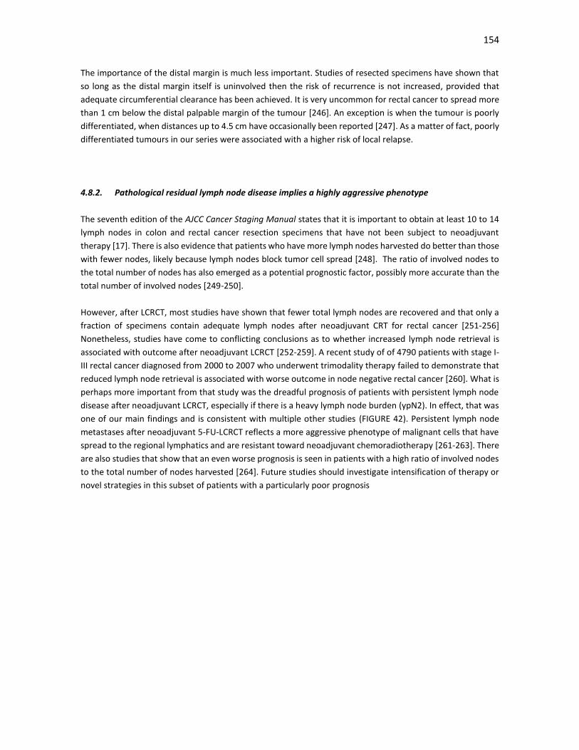
154
The importance of the distal margin is much less important. Studies of resected specimens have shown that
so long as the distal margin itself is uninvolved then the risk of recurrence is not increased, provided that
adequate circumferential clearance has been achieved. It is very uncommon for rectal cancer to spread more
than 1 cm below the distal palpable margin of the tumour [246]. An exception is when the tumour is poorly
differentiated, when distances up to 4.5 cm have occasionally been reported [247]. As a matter of fact, poorly
differentiated tumours in our series were associated with a higher risk of local relapse.
4.8.2. Pathological residual lymph node disease implies a highly aggressive phenotype
The seventh edition of the AJCC Cancer Staging Manual states that it is important to obtain at least 10 to 14
lymph nodes in colon and rectal cancer resection specimens that have not been subject to neoadjuvant
therapy [17]. There is also evidence that patients who have more lymph nodes harvested do better than those
with fewer nodes, likely because lymph nodes block tumor cell spread [248]. The ratio of involved nodes to
the total number of nodes has also emerged as a potential prognostic factor, possibly more accurate than the
total number of involved nodes [249-250].
However, after LCRCT, most studies have shown that fewer total lymph nodes are recovered and that only a
fraction of specimens contain adequate lymph nodes after neoadjuvant CRT for rectal cancer [251-256]
Nonetheless, studies have come to conflicting conclusions as to whether increased lymph node retrieval is
associated with outcome after neoadjuvant LCRCT [252-259]. A recent study of of 4790 patients with stage I-
III rectal cancer diagnosed from 2000 to 2007 who underwent trimodality therapy failed to demonstrate that
reduced lymph node retrieval is associated with worse outcome in node negative rectal cancer [260]. What is
perhaps more important from that study was the dreadful prognosis of patients with persistent lymph node
disease after neoadjuvant LCRCT, especially if there is a heavy lymph node burden (ypN2). In effect, that was
one of our main findings and is consistent with multiple other studies (FIGURE 42). Persistent lymph node
metastases after neoadjuvant 5-FU-LCRCT reflects a more aggressive phenotype of malignant cells that have
spread to the regional lymphatics and are resistant toward neoadjuvant chemoradiotherapy [261-263]. There
are also studies that show that an even worse prognosis is seen in patients with a high ratio of involved nodes
to the total number of nodes harvested [264]. Future studies should investigate intensification of therapy or
novel strategies in this subset of patients with a particularly poor prognosis
155
FIGURE 41: The invasion of the circumferential margin is an important predictor of local relapse, distant
metastases and worse disease-free and overall survival. Several surgical specimens with this feature can be
seen in this figure
156
FIGURE 42 (Case 182): Minimal regression and no downstaging was seen in this 62-year old male patient
with a locally advanced lower rectal cancer with invasion of the mesorectal fascia. Unfortunately, there was
circumferential margin invasion in the surgical specimen (infiltration shown by the yellow arrow) and
invasion of the intersphincter space. The final pathological diagnosis was ypT3bN1G2, Mandard grade 3,
Wheeler grade 2.
157
FIGURE 43 (Case 168): Three pathological lymph nodes, all with capsular rupture (yellow arrow), were found
in this 51-year old male patient with a ypT3N1b after neoadjuvant treatment for a cT3Nx upper rectal
tumour; the lymph nodes were not seen in the baseline MRI
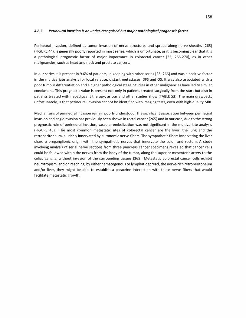
158
4.8.3. Perineural invasion is an under-recognised but major pathological prognostic factor Perineural invasion, defined as tumor invasion of nerve structures and spread along nerve sheaths [265]
(FIGURE 44), is generally poorly reported in most series, which is unfortunate, as it is becoming clear that it is
a pathological prognostic factor of major importance in colorectal cancer [35, 266-270], as in other
malignancies, such as head and neck and prostate cancers.
In our series it is present in 9.6% of patients, in keeping with other series [35, 266] and was a positive factor
in the multivariate analysis for local relapse, distant metastases, DFS and OS. It was also associated with a
poor tumour differentiation and a higher pathological stage. Studies in other malignancies have led to similar
conclusions. This prognostic value is present not only in patients treated surgically from the start but also in
patients treated with neoadjuvant therapy, as our and other studies show (TABLE 53). The main drawback,
unfortunately, is that perineural invasion cannot be identified with imaging tests, even with high-quality MRI.
Mechanisms of perineural invasion remain poorly understood. The significant association between perineural
invasion and angioinvasion has previously been shown in rectal cancer [265] and in our case, due to the strong
prognostic role of perineural invasion, vascular embolization was not significant in the multivariate analysis
(FIGURE 45). The most common metastatic sites of colorectal cancer are the liver, the lung and the
retroperitoneum, all richly innervated by autonomic nerve fibers. The sympathetic fibers innervating the liver
share a preganglionic origin with the sympathetic nerves that innervate the colon and rectum. A study
involving analysis of serial nerve sections from three pancreas cancer specimens revealed that cancer cells
could be followed within the nerves from the body of the tumor, along the superior mesenteric artery to the
celiac ganglia, without invasion of the surrounding tissues [265]. Metastatic colorectal cancer cells exhibit
neurotropism, and on reaching, by either hematogenous or lymphatic spread, the nerve-rich retroperitoneum
and/or liver, they might be able to establish a paracrine interaction with these nerve fibers that would
facilitate metastatic growth.
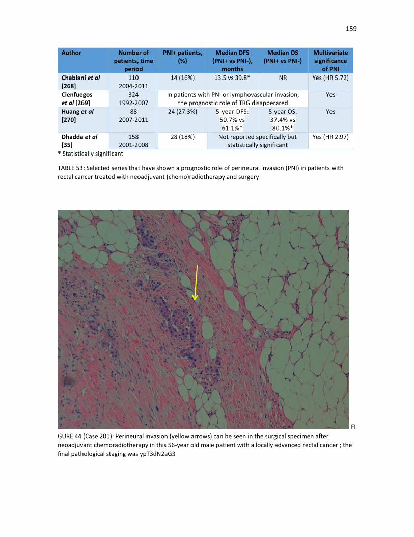
159
Author Number of patients, time
period
PNI+ patients, (%)
Median DFS (PNI+ vs PNI-),
months
Median OS (PNI+ vs PNI-)
Multivariate significance
of PNI
Chablani et al [268]
110 2004-2011
14 (16%) 13.5 vs 39.8* NR Yes (HR 5.72)
Cienfuegos et al [269]
324 1992-2007
In patients with PNI or lymphovascular invasion, the prognostic role of TRG disapperared
Yes
Huang et al [270]
88 2007-2011
24 (27.3%) 5-year DFS: 50.7% vs 61.1%*
5-year OS: 37.4% vs 80.1%*
Yes
Dhadda et al [35]
158 2001-2008
28 (18%) Not reported specifically but statistically significant
Yes (HR 2.97)
* Statistically significant
TABLE 53: Selected series that have shown a prognostic role of perineural invasion (PNI) in patients with
rectal cancer treated with neoadjuvant (chemo)radiotherapy and surgery
FI
GURE 44 (Case 201): Perineural invasion (yellow arrows) can be seen in the surgical specimen after
neoadjuvant chemoradiotherapy in this 56-year old male patient with a locally advanced rectal cancer ; the
final pathological staging was ypT3dN2aG3
160
FIGURE 45 (Case 201): Vascular embolization (yellow arrows) can be seen in the surgical specimen after
neoadjuvant chemoradiotherapy in the same patient of FIGURE 44; perineural invasion is frequently
associated with vascular embolization
161
4.8.4. Downstaging and tumour regression grade were weaker prognostic factors than we expected.
We did not find TRG a strong prognostic factor. Although there was an improvement in local relapse, distant
metastases and survival endpoints, these were modest and basically centered in those patients with complete
or nearly-complete pathological responses. However, the prognostic role in cases with moderate and minimal
regression were more difficult to ascertain. As we explained, due to the heterogeneity of the TRG scores used
in our centre [26-31] (at our institution, the Dworak, Mandard, Rich and Wheeler scores were most frequently
used), we decided to unify these scores in common groups; this was probably easier in patients with very good
regressions, but subject to more errors in patients with lesser grades of regression.
Why is there such controversy with the role of TRG? TRG as a measurement of response to neoadjuvant
(chemo)radiotherapy was first proposed by Mandard in 1994 for use in the assessment of pathological
specimens of squamous cell carcinoma of the oesophagus following neoadjuvant chemoradiotherapy [29].
From then onwards, several TRG scales have been proposed, with different pathological criteria, although
most categorize three- or four-tiered groups: patients with complete or nearly complete regression, moderate
regression and minimal or no regression. The most used TRG used in rectal cancer have been shown in TABLE
3. This is unfortunate, as it has made difficult to perform cross-trial comparisons. There is also an imperfect
relationship between TRG and downstaging: a tumour may have regressed little but may have been
downstaged from T3 to T2, whereas a tumour showing a good response with only microscopic foci of tumour
cells in the subserosa may still be staged as T3 (FIGURE 46).
Additional histological features such as mucin pools (FIGURE 47), necrosis, foamy macrophages, haemosiderin
and dystrophic calcifications (FIGURE 48) may also indicate regression (or not), but they are not included in
most TRG. Most TRG scores (such as the frequently used Mandard and Dworak scores) tend to assess the
predominance of fibrosis over tumour cells as a major criteria of a good response (FIGURES 49, 50 and 51);
however, fibrosis can be seen without previous chemoradiation and is not specific for regression, as it can be
also secondary to usual tumour desmoplasia or to a response of normal tissue to chemoradiation. The true
origin of fibrosis can be difficult to ascertain; some authors have suggested that the amount of tumour present
is the most important factor, irrespective of the amount of fibrosis [271]. Other problems with currently used
TRG scores is that they do not provide a clear indication of whether a grade is reached on the ‘worst’ section
(containing the most tumour) or an average of the grades of regression observed in all slides.
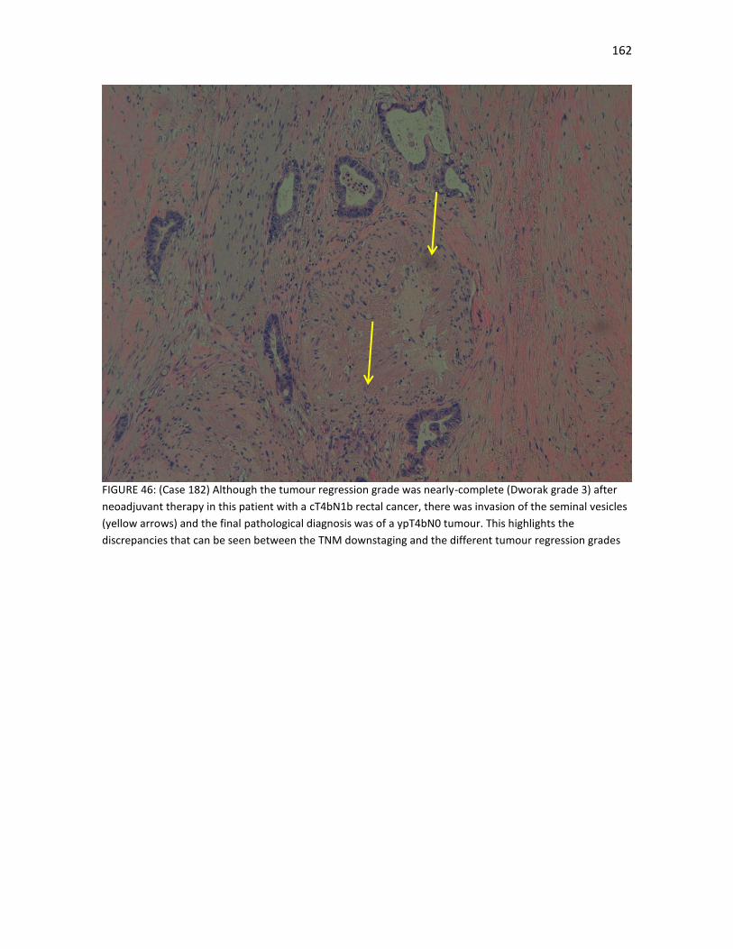
162
FIGURE 46: (Case 182) Although the tumour regression grade was nearly-complete (Dworak grade 3) after
neoadjuvant therapy in this patient with a cT4bN1b rectal cancer, there was invasion of the seminal vesicles
(yellow arrows) and the final pathological diagnosis was of a ypT4bN0 tumour. This highlights the
discrepancies that can be seen between the TNM downstaging and the different tumour regression grades
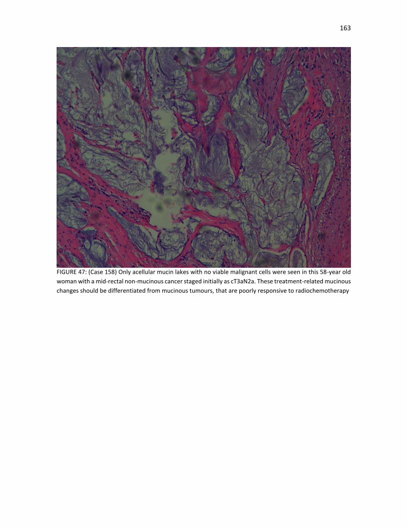
163
FIGURE 47: (Case 158) Only acellular mucin lakes with no viable malignant cells were seen in this 58-year old
woman with a mid-rectal non-mucinous cancer staged initially as cT3aN2a. These treatment-related mucinous
changes should be differentiated from mucinous tumours, that are poorly responsive to radiochemotherapy
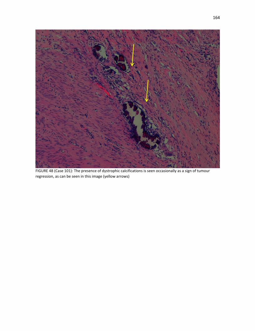
164
FIGURE 48 (Case 101): The presence of dystrophic calcifications is seen occasionally as a sign of tumour
regression, as can be seen in this image (yellow arrows)
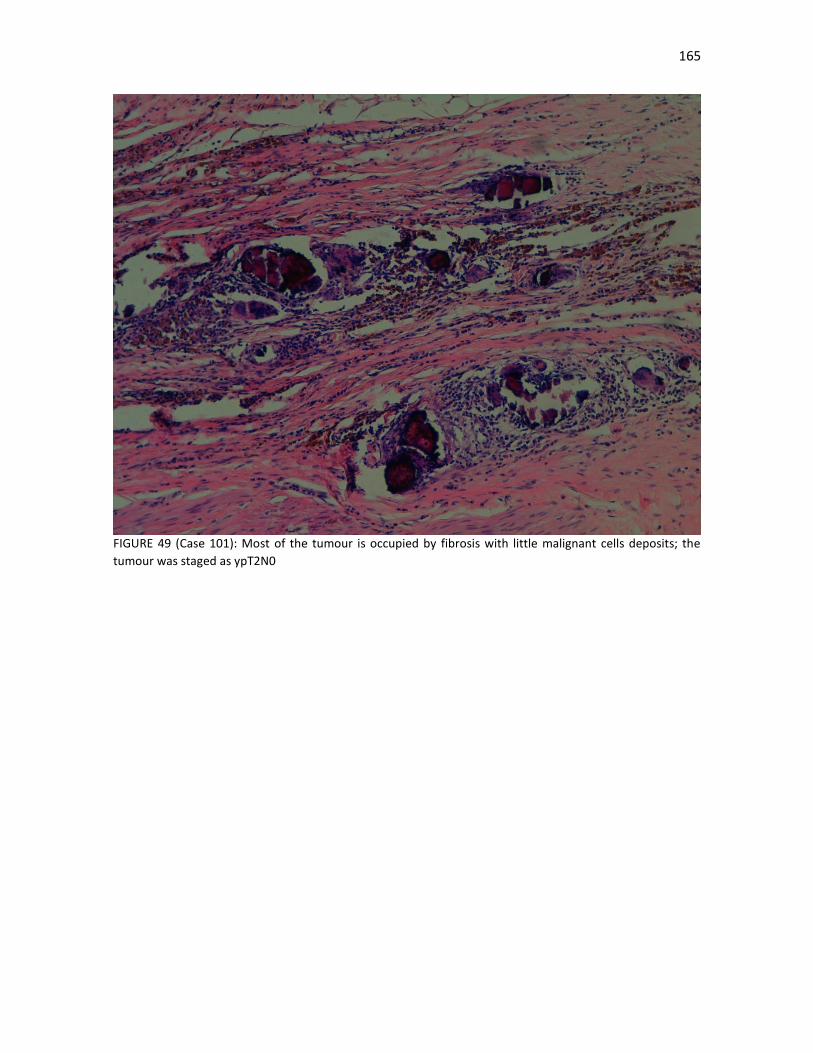
165
FIGURE 49 (Case 101): Most of the tumour is occupied by fibrosis with little malignant cells deposits; the
tumour was staged as ypT2N0
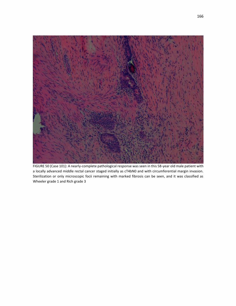
166
FIGURE 50 (Case 101): A nearly-complete pathological response was seen in this 58-year old male patient with
a locally advanced middle rectal cancer staged initially as cT4bN0 and with circumferential margin invasion.
Sterilization or only microscopic focii remaining with marked fibrosis can be seen, and it was classified as
Wheeler grade 1 and Rich grade 3
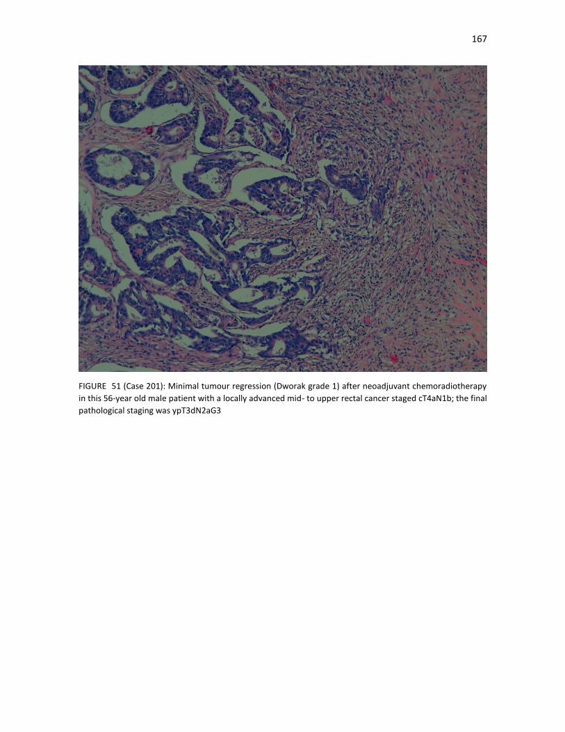
167
FIGURE 51 (Case 201): Minimal tumour regression (Dworak grade 1) after neoadjuvant chemoradiotherapy
in this 56-year old male patient with a locally advanced mid- to upper rectal cancer staged cT4aN1b; the final
pathological staging was ypT3dN2aG3
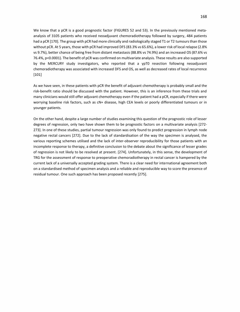
168
We know that a pCR is a good prognostic factor (FIGURES 52 and 53). In the previously mentioned meta-
analysis of 3105 patients who received neoadjuvant chemoradiotherapy followed by surgery, 484 patients
had a pCR [170]. The group with pCR had more clinically and radiologically staged T1 or T2 tumours than those
without pCR. At 5 years, those with pCR had improved DFS (83.3% vs 65.6%), a lower risk of local relapse (2.8%
vs 9.7%), better chance of being free from distant metastasis (88.8% vs 74.9%) and an increased OS (87.6% vs
76.4%, p<0.0001). The benefit of pCR was confirmed on multivariate analysis. These results are also supported
by the MERCURY study investigators, who reported that a ypT0 resection following neoadjuvant
chemoradiotherapy was associated with increased DFS and OS, as well as decreased rates of local recurrence
[101]
As we have seen, in these patients with pCR the benefit of adjuvant chemotherapy is probably small and the
risk-benefit ratio should be discussed with the patient. However, this is an inference from these trials and
many clinicians would still offer adjuvant chemotherapy even if the patient had a pCR, especially if there were
worrying baseline risk factors, such as cN+ disease, high CEA levels or poorly differentiated tumours or in
younger patients.
On the other hand, despite a large number of studies examining this question of the prognostic role of lesser
degrees of regression, only two have shown them to be prognostic factors on a multivariate analysis [272-
273]. In one of these studies, partial tumour regression was only found to predict progression in lymph node
negative rectal cancers [272]. Due to the lack of standardisation of the way the specimen is analysed, the
various reporting schemes utilised and the lack of inter-observer reproducibility for those patients with an
incomplete response to therapy, a definitive conclusion to the debate about the significance of lesser grades
of regression is not likely to be resolved at present. [274]. Unfortunately, in this sense, the development of
TRG for the assessment of response to preoperative chemoradiotherapy in rectal cancer is hampered by the
current lack of a universally accepted grading system. There is a clear need for international agreement both
on a standardised method of specimen analysis and a reliable and reproducible way to score the presence of
residual tumour. One such approach has been proposed recently [275].
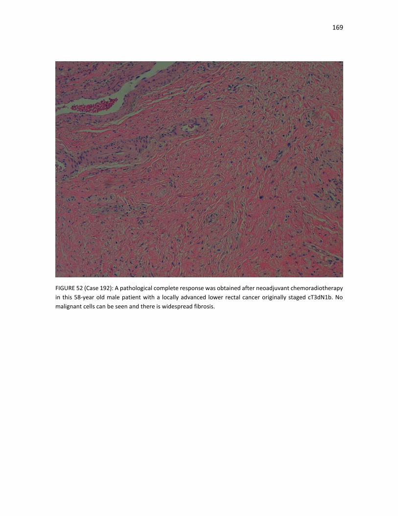
169
FIGURE 52 (Case 192): A pathological complete response was obtained after neoadjuvant chemoradiotherapy
in this 58-year old male patient with a locally advanced lower rectal cancer originally staged cT3dN1b. No
malignant cells can be seen and there is widespread fibrosis.
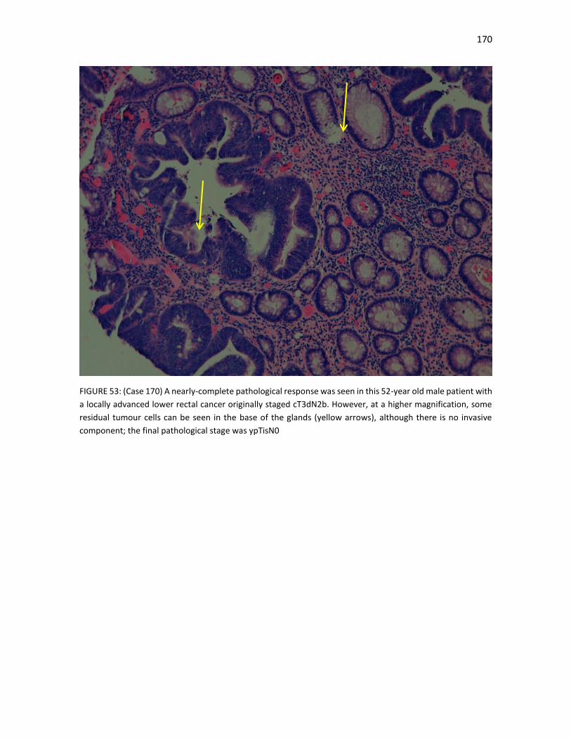
170
FIGURE 53: (Case 170) A nearly-complete pathological response was seen in this 52-year old male patient with
a locally advanced lower rectal cancer originally staged cT3dN2b. However, at a higher magnification, some
residual tumour cells can be seen in the base of the glands (yellow arrows), although there is no invasive
component; the final pathological stage was ypTisN0
171
The use of downstaging as a prognostic factor, although more straightforward than TRG, also has its
shortcomings. There are not uniformly standardized criteria to define downstaging and its effectiveness
depends basically in the sensitivity and specificity of the clinical staging. In our study, the downstaging rate
was basically driven by cN downstaging, more than cT staging. In effect, in our series, 81.3% of patients were
stages as cN+ disease, while in the pathological analysis 74.9% of patients were ypN negative. Although there
was undoubtedly a lymph node sterilization in an important number of patients, due to the poor sensitivity of
MRI, and especially EUS, in N-staging, there will probably have been false-positive cases. Differences between
ypT1 and ypT2 are probably not very significantly. However, as we have mentioned before, the persistence of
ypT34, and especially, ypN12 disease, are very poor prognostic factors, as they imply an aggressive phenotype
with a high risk of distant metastases. These were the findings of our study, where patients with a heavy lymph
node burden after LCRCT had worse DFS and OS in the multivariate analysis, independently from the CRM
status or perineural invasion.
4.8.5. We need new ways to grade tumours, especially after neoadjuvant therapy.
The importance of histologic grading as a prognostic factor is supported by the consistent finding of
independent statistical significance in studies with multivariate analyses [240]. Unfortunately, well-
differentiated tumors make up only a small fraction of tumors in most series, and the difference in outcome
between well-differentiated versus moderately differentiated tumors is not as clear as the difference between
either well- or moderately differentiated tumors and poorly differentiated tumors. Most authorities now
recommend that the traditional three-grade system be collapsed into a two-tiered grading system where low-
grade tumors encompass both well- and moderately differentiated tumors, and high-grade tumors represent
poorly differentiated tumors. Although not evaluated in our trial, the presence of poorly differentiated
clusters of malignant cells is a very promising approach that may surpass the classical histological grading and
can be performed easily in the initial diagnostic biopsy and in the surgical specimen [276].
172
Practical conclusions of the pathological analysis of the surgical specimen
1. Most of the classical factors described as poor prognostic features in rectal cancer were significant
in our analysis. These included an unsatisfactory mesorectum, lymph node invasion, a higher lymph
node burden, no overall, T-stage or N-stage downstaging, circumferential margin involvement,
vascular or perineural invasion, minimal or moderate regression and a non-mucinous histology.
2. Circumferential margin involvement, perineural invasion and ypN2 disease (alongside old age) were
the only remaining independent prognostic factors for worse survival in the multivariate analysis.
3. CRM invasion portends a poor prognosis, even despite the use of LCRCT. Distal margin involvement,
on the other hand, is a much less important factor if the CRM is uninvolved, except in poorly
differentiated tumours, where the risk of local relapse remains high due to the infiltrative
dissemination of these undifferentiated tumours.
4. Perineural invasion is a consistent prognostic factor in most studies for worse local relapse, distant
metastases, DFS and OS. Unfortunately, it is not evaluated routinely in many centres. It is also not
visible in imaging tests, included high-quality MRI, which limits its usefulness in the clinical staging.
5. Patients with complete or nearly-complete pathological response fared very well and many
physicians opt not to offer adjuvant chemotherapy, due to the small expected benefit. However,
many other clinicians would still offer adjuvant chemotherapy, especially if there were worrying
baseline risk factors, such as cN+ disease, high CEA levels or poorly differentiated tumours or in
younger patients.
6. The prognostic role of lesser grades of regression remains unconvincing, especially compared to
other pathological risk factors. Variability in the different TRG scores is problematic and the current
lack of a universally accepted grading system remains a problem.
7. There are not uniformly standardized criteria to define downstaging and its effectiveness depends
basically in the sensitivity and specificity of the clinical imaging performed. Clinical lymph node
staging remains unsatisfactory and N-downstaging is probably not very reliable. However, the
persistence of ypT34, and especially, ypN12 disease, portend a poor prognosis and high risk of distant
metastases.
173
4.9. What is the prognostic role of mucinous tumours?
10.3% of our patients had mucinous tumours, defined as tumours with more than 50% of a mucoid
component. These tumours were linked to higher overall rates of poor response to LCRCT and other poor-
prognosis pathological endpoints compared to non-mucinous tumours (TABLE 24). They were also linked in
the univariate analysis to worse DFS and OS; however, this was lost in the multivariate analysis. These data
are similar to other studies published in the literature [277-279]
They should be differentiated from adenocarcinoma with mucinous features or adenocarcinoma with
mucinous differentiation, terms which are often used to describe tumors that have a significant mucinous
component (>10% but <50%). Most mucinous adenocarcinomas have free-floating strips of neoplastic
epithelium or individual tumor cells in the mucin (FIGURES 54 and 55). A variable number of signet ring cells
also may be seen. If more than 50% of the tumor is composed of signet ring cells, the tumor is best classified
as signet ring cell carcinoma, even if more than 50% of the tumour is composed of extracellular mucin; these
tumours represent 1% of all colon carcinomas, are aggressive and usually present in advanced stages.
Curiously we did not find any cases of signet ring cell carcinoma in our series. This is an important distinction,
as most studies that have evaluated the prognostic role of mucinous tumours have included patients both
with mucinous and signet ring cell tumours, making it difficult to evaluate the real prognostic role of these
mucinous tumours. Another problem, as we have mentioned previously, is that, as the Dutch TME study
results showed, there are two distinct classes of mucinous carcinoma: those that are pre-existing and
mucinous carcinomas that are induced by preoperative radiotherapy (FIGURE 47), with a better prognosis for
those that are induced. Acellular mucin pools, these are not considered to represent residual tumor, although
some studies have suggested that the presence of acellular mucin pools is associated with earlier local
recurrence [280-281]
Another problem is the difficulty in identifying the mucinous or signet ring cell subtype before starting
treatment. As the diagnosis is based on the limited material available from colonoscopy, a satisfactory
diagnosis of both of these carcinoma histologies is usually difficult to establish. One method to establish this
would be to identify the mucinous component using MRI and grouping these tumors as mucinous neoplasms,
which may be superior in identifying mucinous tumors to biopsy (FIGURE 56) [282]
The margins of the tumor are often dissecting and infiltrative, which probably contributes to the overall poor
prognosis in some patients with these tumors, which we observed in our study, where there were statistically
worse rates of a successful TME and of a satisfactory quality of the meso-rectum.
Mucinous tumors represent a greater proportion of right colon tumors, are more common in females, and are
less frequent in the distal colon and rectum. A large national database analysis from the MD Anderson Cancer
Center reported that among rectal adenocarcinoma cases, the incidence of mucinous tumours was 7.08% and
that of signet ring cell carcinomas was 0.7% [283] Another single-institution series from China reported an
incidence of 5% and 1.6% of cases, respectively, among rectal cancer cases [284]
They are more common in young individuals and in patients with Lynch syndrome, and they are more likely
to be at an advanced stage at presentation. The type of genetic alterations in these tumors suggests that the
molecular pathogenesis is different from that of usual adenocarcinomas (TABLE 54). Defects in DNA MMR and
MSI-H are more common in mucinous adenocarcinomas although a mucinous phenotype shows a low degree
of sensitivity as a marker for this genotype [285] MSI-H mucinous tumors are found more often in younger
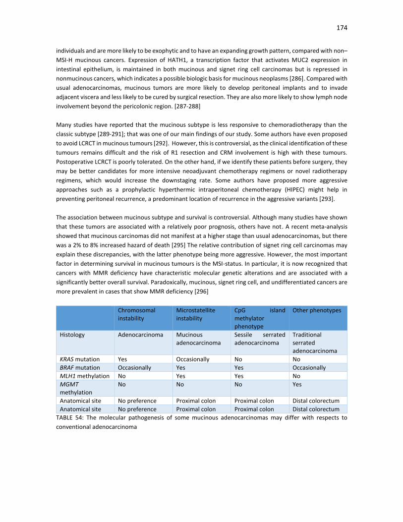
174
individuals and are more likely to be exophytic and to have an expanding growth pattern, compared with non–
MSI-H mucinous cancers. Expression of HATH1, a transcription factor that activates MUC2 expression in
intestinal epithelium, is maintained in both mucinous and signet ring cell carcinomas but is repressed in
nonmucinous cancers, which indicates a possible biologic basis for mucinous neoplasms [286]. Compared with
usual adenocarcinomas, mucinous tumors are more likely to develop peritoneal implants and to invade
adjacent viscera and less likely to be cured by surgical resection. They are also more likely to show lymph node
involvement beyond the pericolonic region. [287-288]
Many studies have reported that the mucinous subtype is less responsive to chemoradiotherapy than the
classic subtype [289-291]; that was one of our main findings of our study. Some authors have even proposed
to avoid LCRCT in mucinous tumours [292]. However, this is controversial, as the clinical identification of these
tumours remains difficult and the risk of R1 resection and CRM involvement is high with these tumours.
Postoperative LCRCT is poorly tolerated. On the other hand, if we identify these patients before surgery, they
may be better candidates for more intensive neoadjuvant chemotherapy regimens or novel radiotherapy
regimens, which would increase the downstaging rate. Some authors have proposed more aggressive
approaches such as a prophylactic hyperthermic intraperitoneal chemotherapy (HIPEC) might help in
preventing peritoneal recurrence, a predominant location of recurrence in the aggressive variants [293].
The association between mucinous subtype and survival is controversial. Although many studies have shown
that these tumors are associated with a relatively poor prognosis, others have not. A recent meta-analysis
showed that mucinous carcinomas did not manifest at a higher stage than usual adenocarcinomas, but there
was a 2% to 8% increased hazard of death [295] The relative contribution of signet ring cell carcinomas may
explain these discrepancies, with the latter phenotype being more aggressive. However, the most important
factor in determining survival in mucinous tumours is the MSI-status. In particular, it is now recognized that
cancers with MMR deficiency have characteristic molecular genetic alterations and are associated with a
significantly better overall survival. Paradoxically, mucinous, signet ring cell, and undifferentiated cancers are
more prevalent in cases that show MMR deficiency [296]
Chromosomal instability
Microstatellite instability
CpG island methylator phenotype
Other phenotypes
Histology Adenocarcinoma Mucinous adenocarcinoma
Sessile serrated adenocarcinoma
Traditional serrated adenocarcinoma
KRAS mutation Yes Occasionally No No
BRAF mutation Occasionally Yes Yes Occasionally
MLH1 methylation No Yes Yes No
MGMT methylation
No No No Yes
Anatomical site No preference Proximal colon Proximal colon Distal colorectum
Anatomical site No preference Proximal colon Proximal colon Distal colorectum
TABLE 54: The molecular pathogenesis of some mucinous adenocarcinomas may differ with respects to
conventional adenocarcinoma
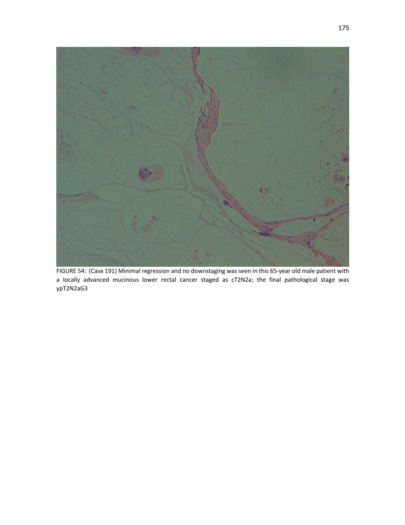
175
FIGURE 54: (Case 191) Minimal regression and no downstaging was seen in this 65-year old male patient with
a locally advanced mucinous lower rectal cancer staged as cT2N2a; the final pathological stage was
ypT2N2aG3
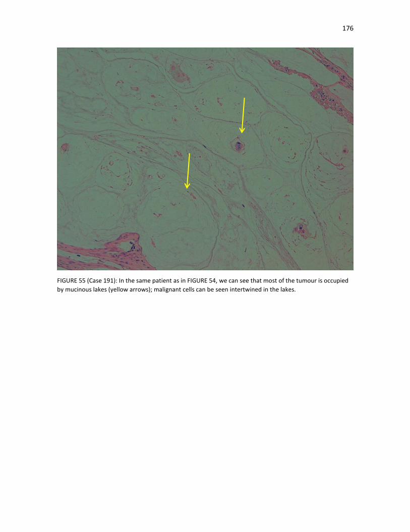
176
FIGURE 55 (Case 191): In the same patient as in FIGURE 54, we can see that most of the tumour is occupied
by mucinous lakes (yellow arrows); malignant cells can be seen intertwined in the lakes.
177
FIGURE 56 (Case 139) Coronal MRI images of a 59-year old female patient with a locally advanced voluminous
mucinous upper rectal cancer (shown by red arrows) staged cT4N1b with invasion of the uterus and collapse
of the endometrial lumen (infiltration shown by the yellow arrows)
178
Practical aspects in the management of mucinous tumours:
1. We observed a 10% rate of mucinous tumours in our study, a rate similar to other studies in the
Western World. These tumours were linked to higher overall rates of poor response to LCRCT and
other poor-prognosis pathological endpoints compared to non-mucinous tumours.
2. The correct characterization of the prognostic role of mucinous tumours is hampered by the difficulty
in obtaining a definitive diagnosis in the initial biopsy, its association with signet cell cancer cells
(which have a poorer prognosis) and the presence of mucin-induced changes after radiotherapy, that
have a much better prognosis than non-induced mucinous tumours.
3. MRI may help in the clinical identification of mucinous tumours.
4. Most studies do show that there is a usually poor response to neoadjuvant therapy in mucinous
tumours. However, worse survival outcomes are difficult to prove and results are contradictory; the
relative contribution of signet cell cancer cells in some studies may explain these discrepancies.
5. The MSI status is probably more important; MSI-low or MSS mucinous tumours are aggresive
compared with MSI-high tumours. Unfortunately, we do not perform it routinely.
6. Mucinous tumours, especially those MSS or MSI-low if identified, would be optimal candidates for
more intensive neoadjuvant chemotherapy regimens or novel radiotherapy regimens, which would
increase the downstaging rate and the possibility of a radical resection
179
5. Recapitulation of conclusions
The long-term results of our study of patients with locally advanced rectal cancer are broadly similar to other
studies of LCRCT followed by surgery and we think that they can be considered moderately successful: there
was a local relapse rate of less than 10%, with a downstaging rate of 70% and a pCR rate of 14.8%. 30% of
patients relapsed, the vast majority in the form of distant metastases and the 10-year OS rate was 62.4%, with
most relapses taking place in the first five years of follow-up. However, there were several concerning factors:
older patients fared poorly compared to their younger counterparts; despite the use of LCRC and specialized
surgery, the quality of the mesorectum was unsatisfactory in around a quarter of patients and 21% had a
positive CRM; finally, the compliance in the use of adjuvant chemotherapy was rather poor, with less than
half of patients receiving the full intended dose and around a quarter of patients not receiving any type of
adjuvant treatment. We followed a blanket approach of treating all patients with cT3-4 and/or lymph node
positive disease with LCRCT and adjuvant chemotherapy, irrespective of response, as our standard treatment,
as is usually done in the United States and in many countries of Europe.
We analyzed the different aspects in the diagnosis and management of our series in a stepwise manner, which
we summarize in the following sections:
5.1. Radiotherapy:
1. International guidelines recommend that patients who are candidate for neoadjuvant treatment
should undergo surgery from 6 to 8 weeks (ESMO) or 5 to 12 weeks (NCCN) after completion of long-
course chemoradiotherapy or within 7-10 days of completion of short-course radiotherapy.
2. However, there is compelling data that an even longer time frame may lead to still better tumor
regression and downstaging, both in LCRCT and in SCRT. Our data are consistent with these data.
With this in mind, we can tailor the treatment in individual cases. For example, SCRT followed by a
longer interval in order to increase downstaging can be useful in elderly or frail patients, where a
LCRCT would be necessary but not feasible. However, we must wait the results of prospective
randomized trials in order to adopt a widespread approach of increasing the interval between RT and
surgery.
3. There is an imperfect relationship between higher radiotherapy dose and an increased pCR. These
differences are probably not significant with doses higher than 45 Gy, before radical surgery, but will
probably be useful in patients where a non-operative management is planned. Again, prospective
randomized trials are needed in this sense.
180
5.2. Neoadjuvant chemotherapy intensification
1. Although theoretically promising, the addition of concomitant oxaliplatin alongside standard LCRCT
is not beneficial. The integration of systemic CT alongside 5-FU-based CRT is difficult, due to the
existence of synergistic toxicities which worsen the compliance rate and the chance of completing
successfully the treatment plan
2. Results with induction chemotherapy (before LCRCT) or consolidation chemotherapy (after LCRCT)
are somewhat more promising and are included in some guidelines. Compliance is better compared
to adjuvant chemotherapy. However, patients should be selected accordingly, as toxicity is higher,
especially in elderly and/or frail patients, and there is an increased risk of serious cardiovascular
events.
3. Despite the lack of data from large prospective studies, some guidelines recommend neoadjuvant
chemotherapy alongside standard LCRCT is included as a treatment option.
4. We have concerns with the consolidation approach, as, in our anecdotal evidence, tolerance to
chemotherapy after LCRCT, even before surgery, tends to be poorer, not only in rectal cancer but in
othe tumours such as pancreatic or esophageal cancer.
5. There is no benefit with the addition of antiangiogenic targeted therapies, with an increased risk of
surgical complications. There are hints of better activity of the combination of induction CT with
antiEGFR targeted therapies, although, again, serious toxicities are worrying.
5.3. Role of adjuvant chemotherapy
1. We offered adjuvant chemotherapy to all patients after surgery, regardless the response rate.
However, as in most other published trials, the tolerance to the adjuvant regimen was poor with less
than half of these patients receiving the full intended dose; this was especially evident in elderly
patients.
2. We observed a small but significant decrease in the rate of distant metastases with adjuvant CT; no
benefit in local control was obtained.
3. There is little high-quality evidence on the use of adjuvant CT after LCRCT and TME-surgery; most
trials have not shown any survival benefit. However, compliance to treatment was uniformly poor,
which may explain this lack of benefit. Unfortunately, we are not going to have randomized trials
which will answer this question
4. Patients with poor downstaging, high lymph node burden or other poor prognostic factors (high
grade, minimal regression perineural or vascular invasion, involved CRM, mucinous tumours) should
probably be offered adjuvant chemotherapy if feasible, if only for a lack of better options. The use of
nomograms and scores, such as the Valentini momogram or the Dhadda score, were helpful in our
case in selecting patients with poor prognosis.
5. If there is a benefit, it is probable small in patients with downstaging and especially in those who
have pCR; the doubts over the effectiveness of adjuvant chemotherapy should be transmitted to
patients and a shared-decision should be taken over its use.
6. We did not use neoadjuvant chemotherapy regimens, alone or alongside LCRCT or SCRT, as there are
only phase II evidence for its activity, but results are promising.
7. Improvements in clinical staging, with a better identification of pathological lymph nodes, are needed
to identify those patients with a higher risk of systemic relapse, who will benefit from more intensive
oxaliplatin-containing (neo)adjuvant regimens, probably in the neoadjuvant setting.
181
5.4. Radiological staging
1. The use of a blanket-approach of treating all locally advanced rectal cancers with LCRCT, as we
used, lowers somewhat the importance of a precise primary tumour staging, although we know
there is an inherent higher risk of over- and under-treating patients. The importance of baseline
prognostic factors for local relapse and distant metastases for selecting patients for risk-adapted
approaches also diminishes with this approach.
2. Broadly speaking. MRI and EUS show a similar sensitivity and specificity with respect to cT
staging; EUS is particulary useful for the staging of early tumours, such as T1-T2. However, a
limited view field, poor assessment of the CRM and the difficulty in staging stenotic tumours are
disadvantages with EUS, with a tendency to understage tumours. In our series, when both tests
were performed, EUS understaged in comparison to MRI, especially in cN disease, upper rectal
cancers and stenotic tumours.
3. MRI is especially preferred in locally advanced rectal cancer by its assessment of cT and invasion
of the mesorectal fascia and other prognostic factors, such as EMVI, lateral lymph pelvic nodes
or tumour deposits. Any risk-adapted approach should include a high-quality MRI, as the
mesorectal rectal status is the most important factor in determining the need of downstaging
and of neoadjuvant (chemo)radiotherapy.
4. Clinical N staging remains a problem, as MRI, and even more so EUS, has poor sensitivity and
specificity for this indication. Lymph node size is a poor distinguishing factor. Other features such
as irregular borders and a heterogeneous signal can help, although results are still far from
satisfactory.
5. Despite the difficulties in cN staging, the presence of cN positive disease, mesorectal fascia
invasion, extramural venous invasion, lower rectal tumours and elevated CEA and CA 19.9 levels
predicted a higher risk of distant metastases. These are poor-prognosis patients and they would
be prime candidates to any neoadjuvant full-dose chemotherapy approach
6. We found that there was an increased risk of local relapse in poorly differentiated tumours and
bulky tumours; perhaps more importantly, as in other studies, we did not find a higher risk with
cN positive disease, especially in N1 tumours. This may reflect the difficulties in staging lymph
node disease with all imaging modalities but also that a successful TME will probably remove
satisfactorily all pathological nodes.
7. We did not use restaging procedures after neoadjuvant therapy. Their use, although attractive,
is misleading in many cases, as MRI tends to over-estimate downstaging and there is no evidence
that surgeons will change their intended original approach with these results. EUS fares even
worse. Radiological TRG, such as those proposed by the MERCURY group, suffer from the same
problems as histological TRG; they could probably be useful in patients complete or near-
complete regression, but not in patients with lesser degrees of regression.
182
5.5. Role of surgery in the multidisciplinary approach
1. A blanket-approach of neoadjuvant (chemo)radiotherapy to all patients with locally advanced rectal
lowers the local relapse rate but does not improve survival and, more importantly, limits the
possibility of giving the full-intended dose of adjuvant chemotherapy and can have long-term side
effects. On the other hand, all patients, even those with low risk tumours, benefit from radiotherapy
and we can expect all long-term side effects will probably diminish with the continuous
improvements in radiotherapy techniques.
2. Mid rectal tumours with little penetration into the meso-rectal fat and with no or minimal clinical
lymph node disease (N1a or N1b) and with a clear MRF, are probably the best candidates for a
surgery-first approach.
3. Low rectal tumours, especially if treated with abdominoperineal resection, have a higher risk of local
relapse. Risk factors include anterior rectal location, levator involvement and the difficulty in
obtaining a successful TME. These patients are probably best served with some type of neoadjuvant
therapy for the moment. Improvements in surgical techniques such as extra-levator
abdominoperineal resection should also help us move forward.
4. The significance of lateral pelvic lymph nodes in MRI is unclear. Although a lateral pelvic
lymphadenectomy could be performed, as is done in the East, these patients do as well with LCRCT
and TME-surgery, with less morbidity and long-term complications.
5.6. Management of older patients
1. Our elderly patients fared poorly in almost all aspects of the multimodality treatment. This was
especially evident in an increased risk of serious unexpected (usually cardiovascular or surgical)
complications, and a lower compliance to neoadjuvant radiotherapy and, especially, to adjuvant
chemotherapy.
2. Disease-free and overall survival were worse in the elderly, even though we did not find any signs
of higher aggressiveness in these tumours, compared to their younger counterparts. There is
also no evidence to suggest that older patients have poor responses to (chemo)radiotherapy. In
particular, there seems to be a greater radiosensitivity in these patients.
3. Other secondary endponts, such as stoma closure or the rate of sphincter-conserving surgery,
were also worse for the older patients.
4. Chronological age, however, should not be the only determining factor un the decision of the
type of multimodality treatment offered. Evaluation of comobirbidites, especially
cardiovascular, and more formal geriatric assessments would be helpful in this regard.
5. LCRCT perhaps is not the best option for older patients, especially in frail or with other
comorbidities. All these results warrant the development of specific therapeutic strategies more
optimal for patients above 70–75 years, including fit patients over 80 years. SCRT followed by
delayed surgery to decrease toxicities and permit tumor downsizing is a promising approach.
Developing less toxic treatments should allow a higher percentage of patients to undergo
surgery.
183
5.7. Pathological prognostic factors
1. Most of the classical factors described as poor prognostic features in rectal cancer were significant
in our analysis. These included an unsatisfactory mesorectum, lymph node invasion, a higher lymph
node burden, no overall, T-stage or N-stage downstaging, circumferential margin involvement,
vascular or perineural invasion, minimal or moderate regression and a non-mucinous histology.
2. Circumferential margin involvement, perineural invasion and ypN2 disease (alongside old age) were
the only remaining independent prognostic factors for worse survival in the multivariate analysis.
3. CRM invasion portends a poor prognosis, even despite the use of LCRCT. Distal margin involvement,
on the other hand, is a much less important factor if the CRM is uninvolved, except in poorly
differentiated tumours, where the risk of local relapse remains high due to the infiltrative
dissemination of these undifferentiated tumours.
4. Perineural invasion is a consistent prognostic factor in most studies for worse local relapse, distant
metastases, DFS and OS. Unfortunately, it is not evaluated routinely in many centres. It is also not
visible in imaging tests, included high-quality MRI, which limits its usefulness in the clinical staging.
5. Patients with complete or nearly-complete pathological response fared very well and many
physicians opt not to offer adjuvant chemotherapy, due to the small expected benefit. However,
many other clinicians would still offer adjuvant chemotherapy, especially if there were worrying
baseline risk factors, such as cN+ disease, high CEA levels or poorly differentiated tumours or in
younger patients.
6. The prognostic role of lesser grades of regression remains unconvincing, especially compared to
other pathological risk factors. Variability in the different TRG scores is problematic and the current
lack of a universally accepted grading system remains a problem.
7. There are not uniformly standardized criteria to define downstaging and its effectiveness depends
basically in the sensitivity and specificity of the clinical imaging performed. Clinical lymph node
staging remains unsatisfactory and N-downstaging is probably not very reliable. However, the
persistence of ypT34, and especially, ypN12 disease, portend a poor prognosis and high risk of distant
metastases.
5.8. Management of mucinous tumours
1. We observed a 10% rate of mucinous tumours in our study, a rate similar to other studies in the
Western World. These tumours were linked to higher overall rates of poor response to LCRCT and
other poor-prognosis pathological endpoints compared to non-mucinous tumours.
2. The correct characterization of the prognostic role of mucinous tumours is hampered by the difficulty
in obtaining a definitive diagnosis in the initial biopsy, its association with signet cell cancer cells
(which have a poorer prognosis) and the presence of mucin-induced changes after radiotherapy, that
have a much better prognosis than non-induced mucinous tumours.
3. MRI may help in the clinical identification of mucinous tumours.
4. Most studies do show that there is a usually poor response to neoadjuvant therapy in mucinous
tumours. However, worse survival outcomes are difficult to prove and results are contradictory; the
relative contribution of signet cell cancer cells in some studies may explain these discrepancies.
5. The MSI status is probably more important; MSI-low or MSS mucinous tumours are aggresive
compared with MSI-high tumours. Unfortunately, we do not perform it routinely.

184
6. Mucinous tumours, especially those MSS or MSI-low if identified, would be optimal candidates for
more intensive neoadjuvant chemotherapy regimens or novel radiotherapy regimens, which would
increase the downstaging rate and the possibility of a radical resection
185
6. What is the future in the management of locally advanced rectal cancer?
The prognosis of patients with locally advanced rectal cancer is determined by two competing factors: the risk
of local relapse and the risk of distant metastases. The identification of patients with an extremely low risk of
local relapse where radiotherapy would presumably offer little benefit is based on the premise of an exquisite
imaging staging with MRI, supplemented with EUS, and a surgical team specialized in the TME procedure.
With the current imaging tests, we can define fairly reasonably those patients with a high risk of local relapse,
who will benefit from neoadjuvant radiotherapy and we can probably avoid radiotherapy in those patients
with a low risk of relapse. There are also promising data with the combination of SCRT and chemotherapy,
with promising results with regards to downstaging and pCR rates; in this sense, the differences between
LCRCT and SCRT are blurring and we will probably be able to tailor these multimodality treatments according
to the characteristics of the patient (age, comorbidities) and of the tumour.
In this sense, the future management of rectal cancer patients will depend on what is the objective we are
trying to achieve: in patients with a high risk of distant metastases, we know that induction chemotherapy is
a suboptimal approach and these patients will benefit from a better integration of systemic chemotherapy in
the neoadjuvant setting. In those patients with a high risk of local relapse, the role of radiotherapy will remain
essential and we will try to maximize its use, be it with improved radiotherapy technique (IMRT, better-
defined fields), higher doses, longer intervals between radiotherapy and surgery, better integration of
concomitant and neoadjuvant chemotherapy and novel radiotherapy approaches, like the use of
brachytherapy boosts. This essential role of neoadjuvant (chemo)radiotherapy will play also a role in organ-
sparing approaches or even non-surgical management of patients with complete clinical responses. Although
in its infancy, compared to other tumours, such as lymphomas, breast or lung cancer, newer insights into the
molecular subtypes of colorectal cancer may help us tailor the systemic approaches in each individual patient
(TABLE 55) [297].
All these improvements need the input of the multidisciplinary team, where improvements in radiological
staging, in the pathological diagnosis and in the introduction of better surgical techniques will benefit our
patients and allow us a more personalised approach to the management of our patients. We offer finally a
graphical representation of what we think the future management of locally advanced rectal cancer will be.
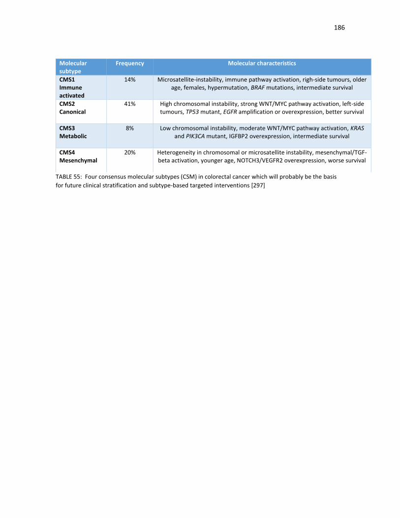
186
TABLE 55: Four consensus molecular subtypes (CSM) in colorectal cancer which will probably be the basis
for future clinical stratification and subtype-based targeted interventions [297]
Molecular subtype
Frequency Molecular characteristics
CMS1 Immune activated
14% Microsatellite-instability, immune pathway activation, righ-side tumours, older age, females, hypermutation, BRAF mutations, intermediate survival
CMS2 Canonical
41% High chromosomal instability, strong WNT/MYC pathway activation, left-side tumours, TP53 mutant, EGFR amplification or overexpression, better survival
CMS3 Metabolic
8% Low chromosomal instability, moderate WNT/MYC pathway activation, KRAS and PIK3CA mutant, IGFBP2 overexpression, intermediate survival
CMS4 Mesenchymal
20% Heterogeneity in chromosomal or microsatellite instability, mesenchymal/TGF-beta activation, younger age, NOTCH3/VEGFR2 overexpression, worse survival
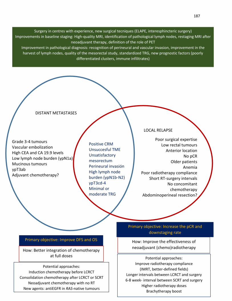
187
DISTANT METASTASES
LOCAL RELAPSE
Positive CRM Unsuccesful TME Unsatisfactory mesorectum Perineural invasión High lymph node burden (ypN1b-N2) ypT3cd-4 Minimal or moderate TRG
Poor surgical expertise Low rectal tumours
Anterior location No pCR
Older patients Anemia
Poor radiotherapy compliance Short RT-surgery intervals
No concomitant chemotherapy
Abdominoperineal resection?
Grade 3-4 tumours Vascular embolization High CEA and CA 19.9 levels Low lymph node burden (ypN1a) Mucinous tumours ypT3ab Adjuvant chemotherapy?
Primary objective: Improve DFS and OS
Primary objective: Increase the pCR and
downstaging rate
How: Better integration of chemotherapy at full doses
How: Improve the effectiveness of
neoadjuvant (chemo)radiotherapy
Potential approaches:
Induction chemotherapy before LCRCT
Consolidation chemotherapy after LCRCT or SCRT
Neoadjuvant chemotherapy with no RT
New agents: antiEGFR in RAS-native tumours
Potential approaches:
Improve radiotherapy compliance
(IMRT, better-defined fields)
Longer intervals between LCRCT and surgery
6-8 week- interval between SCRT and surgery
Higher radiotherapy doses
Brachytherapy boost
….
Surgery in centres with experience, new surgical tecniques (ELAPE, interesphincteric surgery)
Improvements in baseline staging: High-quality MRI, identification of pathological lymph nodes, restaging MRI after
neoadjuvant therapy, definition of the role of PET
Improvement in pathological diagnosis: recognition of perineural and vascular invasion, improvement in the
harvest of lymph nodes, quality of the mesorectal study, standardized TRG, new prognostic factors (poorly
differentiated clusters, immune infilitrates)
188
7. References
[1] O'Connell MJ, Martenson JA, Wieand HS, Krook JE, Macdonald JS, Haller DG, Mayer RJ, Gunderson LL, Rich
TA. Improving adjuvant therapy for rectal cancer by combining protracted-infusion fluorouracil with radiation
therapy after curative surgery. N Engl J Med. 1994 Aug 25;331(8):502-7. PMID: 8041415
[2] Smalley SR, Benedetti JK, Williamson SK, Robertson JM, Estes NC, Maher T, Fisher B, Rich TA, Martenson
JA, Kugler JW, Benson AB 3rd, Haller DG, Mayer RJ, Atkins JN, Cripps C, Pedersen J, Periman PO, Tanaka MS Jr,
Leichman CG, Macdonald JS. Phase III trial of fluorouracil-based chemotherapy regimens plus radiotherapy in
postoperative adjuvant rectal cancer: GI INT 0144. J Clin Oncol. 2006 Aug 1;24(22):3542-7. PMID: 16877719
[3] (No authors listed) Improved survival with preoperative radiotherapy in resectable rectal cancer. N Engl J
Med. 1997 Apr 3;336(14):980-7. Erratum in: N Engl J Med 1997 May 22;336(21):1539. PMID: 9091798.
[4] Kapiteijn E, Marijnen CA, Nagtegaal ID, Putter H, Steup WH, Wiggers T, Rutten HJ, Pahlman L, Glimelius B,
van Krieken JH, Leer JW, van de Velde CJ; Dutch Colorectal Cancer Group. Improved survival with preoperative
radiotherapy in resectable rectal cancer. Swedish Rectal Cancer Trial. N Engl J Med. 2001 Aug 30;345(9):638-
46. PMID: 11547717
[5] Sebag-Montefiore D, Stephens RJ, Steele R, Monson J, Grieve R, Khanna S, Quirke P, Couture J, de Metz C,
Myint AS, Bessell E, Griffiths G, Thompson LC, Parmar M. Preoperative radiotherapy versus selective
postoperative chemoradiotherapy in patients with rectal cancer (MRC CR07 and NCIC-CTG C016): a
multicentre, randomised trial. Lancet. 2009 Mar 7;373(9666):811-20. doi: 10.1016/S0140-6736(09)60484-0.
PMID: 19269519.
[6] Dewdney A, Cunningham D, Chau I. Selecting patients with locally advanced rectal cancer for neoadjuvant
treatment strategies. Oncologist. 2013;18(7):833-42. doi: 10.1634/theoncologist.2013-0022. PMID:
23821325.
[7] Gollins S, Sebag-Montefiore D. Neoadjuvant Treatment Strategies for Locally Advanced Rectal Cancer. Clin
Oncol (R Coll Radiol). 2016 Feb;28(2):146-51. doi: 10.1016/j.clon.2015.11.003. PMID: 26645661.
[8] Ho DH, Pazdur R, Covington W, Brown N, Huo YY, Lassere Y, Kuritani J. Comparison of 5-fluorouracil
pharmacokinetics in patients receiving continuous 5-fluorouracil infusion and oral uracil plus N1-(2'-
tetrahydrofuryl)-5-fluorouracil Clin Cancer Res. 1998 Sep;4(9):2085-8. PMID: 9748123.
[9] Fernández-Martos C, Aparicio J, Bosch C, Torregrosa M, Campos JM, Garcera S, Vicent JM, Maestu I,
Climent MA, Mengual JL, Tormo A, Hernandez A, Estevan R, Richart JM, Viciano V, Uribe N, Campos J, Puchades
R, Arlandis F, Almenar D. Preoperative uracil, tegafur, and concomitant radiotherapy in operable rectal cancer:
a phase II multicenter study with 3 years' follow-up. J Clin Oncol. 2004 Aug 1;22(15):3016-22. PMID: 15210740.
[10] Kim JC, Kim TW, Kim JH, Yu CS, Kim HC, Chang HM, Ryu MH, Park JH, Ahn SD, Lee SW, Shin SS, Kim JS, Choi
EK. Preoperative concurrent radiotherapy with capecitabine before total mesorectal excision in locally
advanced rectal cancer. Int J Radiat Oncol Biol Phys. 2005 Oct 1;63(2):346-53. PMID: 15913913.
189
[11] Fernández-Martos C, Nogué M, Cejas P, Moreno-García V, Machancoses AH, Feliu J. The role of
capecitabine in locally advanced rectal cancer treatment: an update. Drugs. 2012 May 28;72(8):1057-73. doi:
10.2165/11633870-000000000-00000. PMID: 22621694.
[12] André T, Boni C, Mounedji-Boudiaf L, Navarro M, Tabernero J, Hickish T, Topham C, Zaninelli M, Clingan
P, Bridgewater J, Tabah-Fisch I, de Gramont A; Multicenter International Study of Oxaliplatin/5-
Fluorouracil/Leucovorin in the Adjuvant Treatment of Colon Cancer (MOSAIC) Investigators. Oxaliplatin, fl
uorouracil, and leucovorin as adjuvant treatment for colon cancer. N Engl J Med. 2004 Jun 3;350(23):2343-51.
PMID: 15175436.
[13] Fakih MG, Bullarddunn K, Yang GY, Pendyala L, Toth K, Andrews C, Rustum YM, Ross ME, Levea C,
Puthillath A, Park YM, Rajput A. Phase II study of weekly intravenous oxaliplatin combined with oral daily
capecitabine and radiotherapy with biologic correlates in neoadjuvant treatment of rectal adenocarcinoma.
Int J Radiat Oncol Biol Phys. 2008 Nov 1;72(3):650-7. doi: 10.1016/j.ijrobp.2008.01.020. PMID: 18565686.
[14] Koeberle D, Burkhard R, von Moos R, Winterhalder R, Hess V, Heitzmann F, Ruhstaller T, Terraciano L,
Neuweiler J, Bieri G, Rust C, Toepfer M. Phase II study of capecitabine and oxaliplatin given prior to and
concurrently with preoperative pelvic radiotherapy in patients with locally advanced rectal cancer. Br J Cancer.
2008 Apr 8;98(7):1204-9. doi: 10.1038/sj.bjc.6604297. PMID: 18349837.
[15] Gérard JP, Chapet O, Nemoz C, Romestaing P, Mornex F, Coquard R, Barbet N, Atlan D, Adeleine P, Freyer
G. Preoperative concurrent chemoradiotherapy in locally advanced rectal cancer with high-dose radiation and
oxaliplatin-containing regimen: The Lyon R0-04 phase II trial. J Clin Oncol. 2003 Mar 15;21(6):1119-24. PMID:
12637479.
[16] Hospers GA, Punt CJ, Tesselaar ME, Cats A, Havenga K, Leer JW, Marijnen CA, Jansen EP, Van Krieken HH,
Wiggers T, Van de Velde CJ, Mulder NH. Preoperative chemoradiotherapy with capecitabine and oxaliplatin in
locally advanced rectal cancer. A phase I–II multicenter study of the Dutch Colorectal Cancer Group. Ann Surg
Oncol. 2007 Oct;14(10):2773-9. Epub 2007 Jul 26. PMID: 17653805.
[17] Edge SB, Byrd SR, Compton CC, Fritz, AG, Greene FL, Trotti, A, editors. AJCC Cancer Staging Manual. 7th
edition Springer-Verlag; New York (NY): 2010.
[18] Allegra CJ, Yothers G, O'Connell MJ, Beart RW, Wozniak TF, Pitot HC, Shields AF, Landry JC, Ryan DP, Arora
A, Evans LS, Bahary N, Soori G, Eakle JF, Robertson JM, Moore DF Jr, Mullane MR, Marchello BT, Ward PJ,
Sharif S, Roh MS, Wolmark N. Neoadjuvant 5-FU or Capecitabine Plus Radiation With or Without Oxaliplatin
in Rectal Cancer Patients: A Phase III Randomized Clinical Trial. J Natl Cancer Inst. 2015 Sep 14;107(11). pii:
djv248. doi: 10.1093/jnci/djv248. Print 2015 Nov. Erratum in: J Natl Cancer Inst. 2016 Apr;108(4). pii: djw057.
doi: 10.1093/jnci/djw057. PMID: 26374429.
[19] Rödel C, Liersch T, Becker H, Fietkau R, Hohenberger W, Hothorn T, Graeven U, Arnold D, Lang-
Welzenbach M, Raab HR, Sülberg H, Wittekind C, Potapov S, Staib L, Hess C, Weigang-Köhler K, Grabenbauer
GG, Hoffmanns H, Lindemann F, Schlenska-Lange A, Folprecht G, Sauer R; German Rectal Cancer Study Group.
Preoperative chemoradiotherapy and postoperative chemotherapy with fluorouracil and oxaliplatin versus
fluorouracil alone in locally advanced rectal cancer: initial results of the German CAO/ARO/AIO-04 randomised
phase 3 trial. Lancet Oncol. 2012 Jul;13(7):679-87. doi: 10.1016/S1470-2045(12)70187-0. PMID: 22627104.
190
[20] Gerard JP, Rostom Y, Gal J, Benchimol D, Ortholan C, Aschele C, Levi JM. Can we increase the chance of
sphincter saving surgery in rectal cancer with neoadjuvant treatments: lessons from a systematic review of
recent randomized trials.Crit Rev Oncol Hematol. 2012 Jan;81(1):21-8. doi: 0.1016/j.critrevonc.2011.02.001.
PMID: 21377377.
[21] Valentini V, Glimelius B, Haustermans K, Marijnen CA, Rödel C, Gambacorta MA, Boelens PG, Aristei C,
van de Velde CJ. EURECCA consensus conference highlights about rectal cancer clinical management: the
radiation oncologist's expert review. Radiother Oncol. 2014 Jan;110(1):195-8. doi:
10.1016/j.radonc.2013.10.024. PMID: 24286634.
[22] Wittekind C, Compton CC, Greene FL, Sobin LH. TNM residual tumor classification revisited. Cancer.
2002 May 1;94(9):2511-6. PMID: 12015777.
[23] Nagtegaal ID, van de Velde CJ, van der Worp E, Kapiteijn E, Quirke P, van Krieken JH; Cooperative Clinical
Investigators of the Dutch Colorectal Cancer Group. Macroscopic evaluation of rectal cancer resection
specimen: clinical significance of the pathologist in quality control. J Clin Oncol. 2002 Apr 1;20(7):1729-34.
PMID: 11919228.
[24] Gravante G, Hemingway D, Stephenson JA, Sharpe D, Osman A, Haines M, Pirjamali V, Sorge R, Yeung JM,
Norwood M, Miller A, Boyle K. Rectal cancers with microscopic circumferential resection margin involvement
(R1 resections): Survivals, patterns of recurrence, and prognostic factors. J Surg Oncol. 2016 Jul 7. doi:
10.1002/jso.24360. [Epub ahead of print]. PMID: 27383398.
[25] Maughan NJ, Quirke P. Modern management of colorectal cancer - a pathologist's view. Scand J Surg.
2003;92(1):11-9. PMID: 12705546.
[26] Adam IJ, Mohamdee MO, Martin IG, Scott N, Finan PJ, Johnston D, Dixon MF, Quirke P. Role of
circumferential margin involvement in the local recurrence of rectal cancer. Lancet. 1994 Sep
10;344(8924):707-11. PMID: 7915774.
[27] Quirke P, Durdey P, Dixon MF, Williams NS. Local recurrence of rectal adenocarcinoma due to inadequate
surgical resection: histopathological study of lateral tumour spread and surgical excision. Lancet. 1986 Nov
1;2(8514):996-9. PMID: 2430152.
[28] Dworak O, Keilholz L, Hoffmann A. Pathological features of rectal cancer after preoperative
radiochemotherapy. Int J Colorectal Dis. 1997;12(1):19-23. PMID: 9112145.
[29] Mandard AM, Dalibard F, Mandard JC, Marnay J, Henry-Amar M, Petiot JF, Roussel A, Jacob JH, Segol P,
Samama G. Pathologic assessment of tumor regression after preoperative chemoradiotherapy of esophageal
carcinoma. Clinicopathologic correlations. Cancer. 1994 Jun 1;73(11):2680-6. PMID: 8194005.
[30] Wheeler JM, Warren BF, Mortensen NJ, Ekanyaka N, Kulacoglu H, Jones AC, George BD, Kettlewell MG.
Quantification of histologic regression of rectal cancer after irradiation: a proposal for a modified staging
system. Dis Colon Rectum. 2002 Aug;45(8):1051-6. PMID: 12195189.
191
[31] Rich TA, Skibber JM, Ajani JA, Buchholz DJ, Cleary KR, Dubrow RA, Levin B, Lynch PM, Meterissian SH,
Roubein LD. Preoperative infusional chemoradiation therapy for stage T3 rectal cancer. Int J Radiat Oncol Biol
Phys. 1995 Jul 15;32(4):1025-9. PMID: 7607922.
[32] Ryan R, Gibbons D, Hyland JM, Treanor D, White A, Mulcahy HE, O'Donoghue DP, Moriarty M, Fennelly
D, Sheahan K. Pathological response following long-course neoadjuvant chemoradiotherapy for locally
advanced rectal cancer. Histopathology. 2005 Aug;47(2):141-6. PMID: 16045774.
[33] Kim SH, Chang HJ, Kim DY, Park JW, Baek JY, Kim SY, Park SC, Oh JH, Yu A, Nam BH. What Is the Ideal
Tumor Regression Grading System in Rectal Cancer Patients after Preoperative Chemoradiotherapy?. Cancer
Res Treat. 2016 Jul;48(3):998-1009. doi: 10.4143/crt.2015.254. Epub 2015 Oct 22. PMID: 26511803.
[34] Valentini V, van Stiphout RG, Lammering G, Gambacorta MA, Barba MC, Bebenek M, Bonnetain F, Bosset
JF, Bujko K, Cionini L, Gerard JP, Rödel C, Sainato A, Sauer R, Minsky BD, Collette L, Lambin P. Nomograms for
predicting local recurrence, distant metastases, and overall survival for patients with locally advanced rectal
cancer on the basis of European randomized clinical trials. J Clin Oncol. 2011 Aug 10;29(23):3163-72. doi:
10.1200/JCO.2010.33.1595. PMID: 21747092.
[35] Dhadda AS, Bessell EM, Scholefield J, Dickinson P, Zaitoun AM. Mandard tumour regression grade,
perineural invasion, circumferential resection margin and post-chemoradiation nodal status strongly predict
outcome in locally advanced rectal cancer treated with preoperative chemoradiotherapy. Clin Oncol (R Coll
Radiol). 2014 Apr;26(4):197-202. doi: 10.1016/j.clon.2014.01.001. PMID: 24485884.
[36] Lutz MP, Zalcberg JR, Glynne-Jones R, Ruers T, Ducreux M, Arnold D, Aust D, Brown G, Bujko K,
Cunningham C, Evrard S, Folprecht G, Gerard JP, Habr-Gama A, Haustermans K, Holm T, Kuhlmann KF, Lordick
F, Mentha G, Moehler M, Nagtegaal ID, Pigazzi A, Puciarelli S, Roth A, Rutten H, Schmoll HJ, Sorbye H, Van
Cutsem E, Weitz J, Otto F.Second St. Gallen European Organisation for Research and Treatment of Cancer
Gastrointestinal Cancer Conference: consensus recommendations on controversial issues in the primary
treatment of rectal cancer. Eur J Cancer. 2016 Aug; 63:11-24. doi: 10.1016/j.ejca.2016.04.010. PMID:
27254838.
[37] Stephens RJ, Thompson LC, Quirke P, Steele R, Grieve R, Couture J, Griffiths GO, Sebag-Montefiore D.
Impact of short-course preoperative radiotherapy for rectal cancer on patients' quality of life: data from the
Medical Research Council CR07/National Cancer Institute of Canada Clinical Trials Group C016 randomized
clinical trial. J Clin Oncol. 2010 Sep 20;28(27):4233-9. doi: 10.1200/JCO.2009.26.5264. PMID: 20585099.
[38] Peeters KC, van de Velde CJ, Leer JW, Martijn H, Junggeburt JM, Kranenbarg EK, Steup WH, Wiggers T,
Rutten HJ, Marijnen CA. Late side effects of short-course preoperative radiotherapy combined with total
mesorectal excision for rectal cancer: increased bowel dysfunction in irradiated patients – a Dutch Colorectal
Cancer Group study. J Clin Oncol. 2005 Sep 1;23(25):6199-206. PMID: 16135487.
[39]. Marijnen CA, van de Velde CJ, Putter H, van den Brink M, Maas CP, Martijn H, Rutten HJ, Wiggers T,
Kranenbarg EK, Leer JW, Stiggelbout AM. Impact of short-term preoperative radiotherapy on health-related
quality of life and sexual functioning in primary rectal cancer: report of a multicenter randomized trial. J Clin
Oncol. 2005 Mar 20;23(9):1847-58. PMID: 15774778.
192
[40] van Gijn W, Marijnen CA, Nagtegaal ID, Kranenbarg EM, Putter H, Wiggers T, Rutten HJ, Påhlman L,
Glimelius B, van de Velde CJ; Dutch Colorectal Cancer Group. Preoperative radiotherapy combined with total
mesorectal excision for resectable rectal cancer: 12-year follow-up of the multicentre, randomised controlled
TME trial. Lancet Oncol. 2011 Jun;12(6):575-82. doi: 10.1016/S1470-2045(11)70097-3. PMID: 21596621.
[41]. Birgisson H, Påhlman L, Gunnarsson U, Glimelius B. Occurrence of second cancers in patients treated
with radiotherapy for rectal cancer. J Clin Oncol. 2005 Sep 1;23(25):6126-31. PMID: 16135478.
[42] (No authors listed) Radiation therapy and fluorouracil with or without semustine for the treatment of
patients with surgical adjuvant adenocarcinoma of the rectum. Gastrointestinal Tumor Study Group. J Clin
Oncol. 1992; 10 (4):549-557. PMID: 1548520.
[43] Krook JE, Moertel CG, Gunderson LL, Wieand HS, Collins RT, Beart RW, Kubista TP, Poon MA, Meyers WC,
Mailliard JA. Effective surgical adjuvant therapy for high-risk rectal carcinoma. N Engl J Med. 1991 Mar
14;324(11):709-15. PMID: 1997835.
[44] Sauer R, Becker H, Hohenberger W, Rödel C, Wittekind C, Fietkau R, Martus P, Tschmelitsch J, Hager E,
Hess CF, Karstens JH, Liersch T, Schmidberger H, Raab R; German Rectal Cancer Study Group. Preoperative
versus postoperative chemoradiotherapy for rectal cancer. N Engl J Med. 2004 Oct 21;351(17):1731-40. PMID:
15496622.
[45]. Sauer R, Liersch T, Merkel S, Fietkau R, Hohenberger W, Hess C, Becker H, Raab HR, Villanueva MT,
Witzigmann H, Wittekind C, Beissbarth T, Rödel C. Preoperative versus postoperative chemoradiotherapy for
locally advanced rectal cancer: results of the German CAO/ARO/AIO-94 randomized phase III trial after a
median follow-up of 11 years. J Clin Oncol. 2012 Jun 1;30(16):1926-33. doi: 10.1200/JCO.2011.40.1836. PMID:
22529255.
[46]. Mohiuddin M, Regine WF, John WJ, Hagihara PF, McGrath PC, Kenady DE, Marks G. Preoperative
chemoradiation in fixed distal rectal cancer: dose time factors for pathological complete response. Int J Radiat
Oncol Biol Phys. 2000 Mar 1;46(4):883-8. PMID: 10705009.
[47]. Bosset JF, Calais G, Mineur L, Maingon P, Radosevic-Jelic L, Daban A, Bardet E, Beny A, Briffaux A, Collette
L. Enhanced tumorocidal effect of chemotherapy with preoperative radiotherapy for rectal cancer:
preliminary results—EORTC 22921J Clin Oncol. 2005 Aug 20;23(24):5620-7. PMID: 16009958.
[48]. Bosset JF, Calais G, Mineur L, Maingon P, Stojanovic-Rundic S, Bensadoun RJ, Bardet E, Beny A, Ollier JC,
Bolla M, Marchal D, Van Laethem JL, Klein V, Giralt J, Clavère P, Glanzmann C, Cellier P, Collette L; EORTC
Radiation Oncology Group. Fluorouracil-based adjuvant chemotherapy after preoperative
chemoradiotherapy in rectal cancer: long-term results of the EORTC 22921 randomised study. Lancet Oncol.
2014 Feb;15(2):184-90. doi: 10.1016/S1470-2045(13)70599-0. PMID: 24440473.
[49] Gérard JP, Conroy T, Bonnetain F, Bouché O, Chapet O, Closon-Dejardin MT, Untereiner M, Leduc B,
Francois E, Maurel J, Seitz JF, Buecher B, Mackiewicz R, Ducreux M, Bedenne L. Preoperative radiotherapy
with or without concurrent fluorouracil and leucovorin in T3-4 rectal cancers: results of FFCD 9203. J Clin
Oncol. 2006 Oct 1;24(28):4620-5. PMID: 17008704.
193
[50] O'Connell MJ, Martenson JA, Wieand HS, Krook JE, Macdonald JS, Haller DG, Mayer RJ, Gunderson LL,
Rich TA. Improving adjuvant therapy for rectal cancer by combining protracted-infusion fluorouracil with
radiation therapy after curative surgery. N Engl J Med. 1994 Aug 25;331(8):502-7. PMID: 8041415.
[51] Kapiteijn E, Marijnen CA, Nagtegaal ID, Putter H, Steup WH, Wiggers T, Rutten HJ, Pahlman L, Glimelius
B, van Krieken JH, Leer JW, van de Velde CJ; Dutch Colorectal Cancer Group. Preoperative radiotherapy
combined with total mesorectal excision for resectable rectal cancer. N Engl J Med. 2001 Aug 30;345(9):638-
46. PMID: 11547717.
[52]. van Gijn W, Marijnen CA, Nagtegaal ID, Kranenbarg EM, Putter H, Wiggers T, Rutten HJ, Påhlman L,
Glimelius B, van de Velde CJ; Dutch Colorectal Cancer Group. Preoperative radiotherapy combined with total
mesorectal excision for resectable rectal cancer: 12-year follow-up of the multicentre, randomised controlled
TME trial. Lancet Oncol. 2011 Jun;12(6):575-82. doi: 10.1016/S1470-2045(11)70097-3. PMID: 21596621.
[53]. Boulis-Wassif S, Gerard A, Loygue J, Camelot D, Buyse M, Duez N. Final results of a randomized trial on
the treatment of rectal cancer with preoperative radiotherapy alone or in combination with 5-fluorouracil,
followed by radical surgery. Trial of the European Organization on Research and Treatment of Cancer
Gastrointestinal Tract Cancer Cooperative Group. Cancer. 1984 May 1;53(9):1811-8. PMID: 6423263.
[54] Bujko K, Nowacki MP, Nasierowska-Guttmejer A, Michalski W, Bebenek M, Kryj M. Long-term results of a
randomized trial comparing preoperative short-course radiotherapy with preoperative conventionally
fractionated chemoradiation for rectal cancer. Br J Surg. 2006 Oct;93(10):1215-23. PMID: 16983741.
[55] Latkauskas T, Pauzas H, Gineikiene I, Janciauskiene R, Juozaityte E, Saladzinskas Z, Tamelis A, Pavalkis D.
Initial results of a randomized controlled trial comparing clinical and pathological downstaging of rectal cancer
after preoperative short-course radiotherapy or long-term chemoradiotherapy, both with delayed surgery.
Colorectal Dis. 2012 Mar;14(3):294-8. doi: 10.1111/j.1463-1318.2011.02815.x. PMID: 21899712.
[56]. Ngan SY, Burmeister B, Fisher RJ, Solomon M, Goldstein D, Joseph D, Ackland SP, Schache D, McClure B,
McLachlan SA, McKendrick J, Leong T, Hartopeanu C, Zalcberg J, Mackay J. Randomized trial of short-course
radiotherapy versus long-course chemoradiation comparing rates of local recurrence in patients with T3 rectal
cancer: Trans-Tasman Radiation Oncology Group trial 01.04. J Clin Oncol. 2012 Nov 1;30(31):3827-33. doi:
10.1200/JCO.2012.42.9597. PMID: 23008301.
[57] McCarthy K, Pearson K, Fulton R, Hewitt J. Pre-operative chemoradiation for non-metastatic locally
advanced rectal cancer. Cochrane Database Syst Rev. 2012 Dec 12;12:CD008368. doi:
10.1002/14651858.CD008368.pub2. PMID: 23235660.
[58] Preoperative multimodality therapy improves disease-free survival in patients with carcinoma of the
rectum: NSABP R-03. Roh MS, Colangelo LH, O'Connell MJ, Yothers G, Deutsch M, Allegra CJ, Kahlenberg MS,
Baez-Diaz L, Ursiny CS, Petrelli NJ, Wolmark N. J Clin Oncol. 2009 Nov 1;27(31):5124-30. doi:
10.1200/JCO.2009.22.0467. PMID: 19770376.
[59] Is the NSABP R-03 study in line with other chemoradiation studies? Glynne-Jones R, Harrison M, Hughes
R. J Clin Oncol. 2010 Jun 20;28(18):e305-6; author reply e307. doi: 10.1200/JCO.2010.28.4356. PMID:
20479393.
194
[60]. Hofheinz RD, Wenz F, Post S, Matzdorff A, Laechelt S, Hartmann JT, Müller L, Link H, Moehler M, Kettner
E, Fritz E, Hieber U, Lindemann HW, Grunewald M, Kremers S, Constantin C, Hipp M, Hartung G, Gencer D,
Kienle P, Burkholder I, Hochhaus A. Chemoradiotherapy with capecitabine versus fluorouracil for locally
advanced rectal cancer: a randomised, multicentre, non-inferiority, phase 3 trial. Lancet Oncol. 2012
Jun;13(6):579-88. doi: 10.1016/S1470-2045(12)70116-X. PMID: 2503032.
[61]. O'Connell MJ, Colangelo LH, Beart RW, Petrelli NJ, Allegra CJ, Sharif S, Pitot HC, Shields AF, Landry JC,
Ryan DP, Parda DS, Mohiuddin M, Arora A, Evans LS, Bahary N, Soori GS, Eakle J, Robertson JM, Moore DF Jr,
Mullane MR, Marchello BT, Ward PJ, Wozniak TF, Roh MS, Yothers G, Wolmark N. Capecitabine and oxaliplatin
in the preoperative multimodality treatment of rectal cancer: surgical end points from National Surgical
Adjuvant Breast and Bowel Project trial R-04. J Clin Oncol. 2014 Jun 20;32(18):1927-34. doi:
10.1200/JCO.2013.53.7753. Epub 2014 May 5. PMID: 24799484.
[62]. Cedermark B, Johansson H, Rutqvist LE, Wilking N. The Stockholm I trial of preoperative short term
radiotherapy in operable rectal carcinoma. A prospective randomized trial, Stockholm Colorectal Cancer Study
Group. Cancer. 1995 May 1;75(9):2269-75. PMID: 7712435.
[63]. Martling A, Holm T, Johansson H, Rutqvist LE, Cedermark B; Stockholm Colorectal Cancer Study Group.
The Stockholm II trial on preoperative radiotherapy in rectal carcinoma: long-term follow-up of a population-
based study. Cancer. 2001 Aug 15;92(4):896-902. PMID: 11550163.
[64]. [No authors listed]. Improved survival with preoperative radiotherapy in resectable rectal cancer,
Swedish Rectal Cancer Trial. N Engl J Med. 1997 Apr 3;336(14):980-7. Erratum in: N Engl J Med 1997 May
22;336(21):1539. PMID: 9091798.
[65]. Frykholm GJ, Påhlman L, Glimelius B. Int J Radiat Oncol Biol Phys. Combined chemo and radiotherapy vs
radiotherapy alone in the treatment of primary non-resectable adenocarcinoma of the rectum. 2001 Jun
1;50(2):427-34. PMID: 11380230.
[66]. Folkesson J, Birgisson H, Pahlman L, Cedermark B, Glimelius B, Gunnarsson U. Swedish Rectal Cancer
Trial: long lasting benefits from radiotherapy on survival and local recurrence rate. J Clin Oncol. 2005 Aug
20;23(24):5644-50. PMID: 16110023.
[67] Quirke P, Durdey P, Dixon MF, Williams NS. Local recurrence of rectal adenocarcinoma due to inadequate
surgical resection, Histopathological study of lateral tumour spread and surgical excision. Lancet. 1986 Nov
1;2(8514):996-9. PMID: 2430152.
[68] Kapiteijn E, Marijnen CA, Nagtegaal ID, Putter H, Steup WH, Wiggers T, Rutten HJ, Pahlman L, Glimelius
B, van Krieken JH, Leer JW, van de Velde CJ; Dutch Colorectal Cancer Group. Preoperative radiotherapy
combined with total mesorectal excision for resectable rectal cancer. N Engl J Med. 2001 Aug 30;345(9):638-
46. PMID: 11547717.
[69] Sebag-Montefiore D, Stephens RJ, Steele R, Monson J, Grieve R, Khanna S, Quirke P, Couture J, de Metz
C, Myint AS, Bessell E, Griffiths G, Thompson LC, Parmar M. Preoperative radiotherapy versus selective
postoperative chemoradiotherapy in patients with rectal cancer (MRC CR07 and NCIC-CTG C016): a
195
multicentre, randomised trial. Lancet. 2009 Mar 7;373(9666):811-20. doi: 10.1016/S0140-6736(09)60484-
0. PMID: 19269519.
[70] Quirke P, Steele R, Monson J, Grieve R, Khanna S, Couture J, O'Callaghan C, Myint AS, Bessell E, Thompson
LC, Parmar M, Stephens RJ, Sebag-Montefiore D; MRC CR07/NCIC-CTG CO16 Trial Investigators; NCRI
Colorectal Cancer Study Group. Effect of the plane of surgery achieved on local recurrence in patients with
operable rectal cancer: a prospective study using data from the MRC CR07 and NCIC-CTG CO16 randomised
clinical trial. Lancet. 2009 Mar 7;373(9666):821-8. doi: 10.1016/S0140-6736(09)60485-2. PMID: 19269520.
[71] Shin SJ, Yoon HI, Kim NK, Lee KY, Min BS, Ahn JB, Keum KC, Koom WS. Upfront systemic chemotherapy
and preoperative short course radiotherapy with delayed surgery for locally advanced rectal cancer with
distant metastases. Radiat Oncol. 2011 Aug 24;6:99. doi: 10.1186/1748-717X-6-99. PMID: 21864368.
[72]. Yeo SG, Oh JH, Kim DY, Baek JY, Kim SY, Park JW, Kim MJ, Chang HJ, Kim TH, Lee JH, Jang HS, Kim JG, Lee
MA, Nam TK. Preoperative short-course concurrent chemoradiation therapy followed by delayed surgery for
locally advanced rectal cancer: a phase II multicenter study (KROG 10-01). Int J Radiat Oncol Biol Phys. 2013
May 1;86(1):34-9. doi: 10.1016/j.ijrobp.2012.11.018. PMID: 23265569.
[73] Myerson RJ, Parikh PJ, Tan B, et al. A single institution phase II trial of five fractions of radiotherapy
followed by four courses of FOLFOX chemotherapy as preoperative therapy for rectal adenocarcinoma J Clin
Oncol, 2012;30(Suppl 4); Abstr 553.
[74] Nilsson PJ, van Etten B, Hospers GA, Påhlman L, van de Velde CJ, Beets-Tan RG, Blomqvist L, Beukema JC,
Kapiteijn E, Marijnen CA, Nagtegaal ID, Wiggers T, Glimelius B. Short-course radiotherapy followed by
neoadjuvant chemotherapy in locally advanced rectal cancer – the RAPIDO trial. BMC Cancer. 2013 Jun 7;13:
279. doi: 10.1186/1471-2407-13-279. PMID: 23742033.
[75] Minsky BD. Short-course radiation versus long-course chemoradiation for rectal cancer: making progress.
J Clin Oncol. 2012 Nov 1;30(31):3777-8. doi: 10.1200/JCO.2012.45.0551. PMID: 23008292.
[76] Fletcher GH, Hypofractionation: lessons from complications. Radiother Oncol. 1991 Jan;20(1):10-5.
Review. PMID: 2020750.
[77] Hall EH, Time, dose, and fractionation in radiotherapy, In: Hall EJ, editor, Radiobiology for the Radiologist,
ed 5, Philadelphia, PA, Lippincott Williams and Wilkins, 2000;397–418.
[78] Fokstuen T, Holm T, Glimelius B. Postoperative morbidity and mortality in relation to leukocyte counts
and time to surgery after short-course preoperative radiotherapy for rectal cancer. Radiother Oncol. 2009
Nov;93(2):293-7. doi: 10.1016/j.radonc.2009.08.034. PMID: 19748694.
[79] van den Broek CB, Vermeer TA, Bastiaannet E, Rutten HJ, van de Velde CJ, Marijnen CA. Impact of the
interval between short-course radiotherapy and surgery on outcomes of rectal cancer patients. Eur J Cancer.
2013 Oct;49(15):3131-9. doi: 10.1016/j.ejca.2013.05.025. PMID: 23800669.
[80] Marijnen CA, van de Velde CJ, Putter H, van den Brink M, Maas CP, Martijn H, Rutten HJ, Wiggers T,
Kranenbarg EK, Leer JW, Stiggelbout AM. Impact of short-term preoperative radiotherapy on health-related
196
quality of life and sexual functioning in primary rectal cancer: report of a multicenter randomized trial. J Clin
Oncol. 2005 Mar 20;23(9):1847-58. PMID: 15774778.
[81] Peeters KC, Marijnen CA, Nagtegaal ID, Kranenbarg EK, Putter H, Wiggers T, Rutten H, Pahlman L,
Glimelius B, Leer JW, van de Velde CJ; Dutch Colorectal Cancer Group. The TME Trial after a median follow-up
of 6 Years: Increased local control but no survival benefit in irradiated patients with resectable rectal
carcinoma. Ann Surg. 2007 Nov;246(5):693-701. PMID: 17968156.
[82]. Pollack J, Holm T, Cedermark B, Holmström B, Mellgren A. Long-term effect of preoperative radiation
therapy on anorectal function. Dis Colon Rectum. 2006 Mar;49(3):345-52. PMID: 16532369.
[83] Lange MM, den Dulk M, Bossema ER, Maas CP, Peeters KC, Rutten HJ, Klein Kranenbarg E, Marijnen CA,
van de Velde CJ; Cooperative Clinical Investigators of the Dutch Total Mesorectal Excision Trial. Risk factors
for faecal incontinence after rectal cancer treatment. Br J Surg. 2007 Oct;94(10):1278-84. PMID: 1757934.
[84] Stephens RJ, Thompson LC, Quirke P, Steele R, Grieve R, Couture J, Griffiths GO, Sebag-Montefiore D.
Impact of short-course preoperative radiotherapy for rectal cancer on patients’ quality of life: data from the
Medical Research Council CR07/ National Cancer Institute of Canada Clinical Trials Group C016 randomized
clinical trial. J Clin Oncol. 2010 Sep 20;28(27):4233-9. doi: 10.1200/JCO.2009.26.5264. PMID: 20585099.
[85] Murata A, Brown CJ, Raval M, Phang PT. Impact of short-course radiotherapy and low anterior resection
on quality of life and bowel function in primary rectal cancer. Am J Surg. 2008 May;195(5):611-5; discussion
615. doi: 10.1016/j.amjsurg.2007.12.034. PMID: 18367144.
[86] Tiv M, Puyraveau M, Mineur L, Calais G, Maingon P, Bardet E, Mercier M, Bosset JF. Long-term quality of
life in patients with rectal cancer treated with preoperative (chemo)-radiotherapy within a randomized trial.
Cancer Radiother. 2010 Oct;14(6-7):530-4. doi: 10.1016/j.canrad.2010.06.017. PMID: 20797891.
[87] Bujko K, Wyrwicz L, Rutkowski A, Malinowska M, Pietrzak L, Kryński J, Michalski W, Olędzki J, Kuśnierz J,
Zając L, Bednarczyk M, Szczepkowski M, Tarnowski W, Kosakowska E, Zwoliński J, Winiarek M, Wiśniowska K,
Partycki M, Bęczkowska K, Polkowski W, Styliński R, Wierzbicki R, Bury P, Jankiewicz M, Paprota K, Lewicka M,
Ciseł B, Skórzewska M, Mielko J, Bębenek M, Maciejczyk A, Kapturkiewicz B, Dybko A, Hajac Ł, Wojnar A,
Leśniak T, Zygulska J, Jantner D, Chudyba E, Zegarski W, Las-Jankowska M, Jankowski M, Kołodziejski L,
Radkowski A, Żelazowska-Omiotek U, Czeremszyńska B, Kępka L, Kolb-Sielecki J, Toczko Z, Fedorowicz Z, Dziki
A, Danek A, Nawrocki G, Sopyło R, Markiewicz W, Kędzierawski P, Wydmański J; Polish Colorectal Study Group.
Long-course oxaliplatin-based preoperative chemoradiation versus 5 × 5 Gy and consolidation chemotherapy
for cT4 or fixed cT3 rectal cancer: results of a randomized phase III study. Ann Oncol. 2016 May;27(5):834-42.
doi: 10.1093/annonc/mdw062. PMID: 26884592
[88] Frykholm GJ, Sintorn K, Montelius A, Jung B, Påhlman L, Glimelius B. Acute lumbosacral plexopathy during
and after preoperative radiotherapy of rectal adenocarcinoma. Radiother Oncol. 1996 Feb;38(2):121-30.
PMID: 8966224.
197
[89]. Folkesson J, Birgisson H, Pahlman L, Cedermark B, Glimelius B, Gunnarsson U. Swedish Rectal Cancer
Trial: long lasting benefits from radiotherapy on survival and local recurrence rate. J Clin Oncol. 2005 Aug
20;23(24):5644-50. PMID: 16110023.
[90] Pettersson D, Lörinc E, Holm T, Iversen H, Cedermark B, Glimelius B, Martling A. Tumour regression in the
randomized Stockholm III Trial of radiotherapy regimens for rectal cancer. Br J Surg. 2015 Jul;102(8):972-8;
discussion 978. doi: 10.1002/bjs.9811. PMID: 26095256.
[91]. Hatfield P, Hingorani M, Radhakrishna G, Cooper R, Melcher A, Crellin A, Kwok-Williams M, Sebag-
Montefiore D. Short‐course radiotherapy, with elective delay prior to surgery, in patients with unresectable
rectal cancer who have poor performance status or significant co‐morbidity. Radiother Oncol. 2009
Aug;92(2):210-4. doi: 10.1016/j.radonc.2009.04.007. PMID: 19409638.
[92]. Radu C, Berglund A, Påhlman L, Glimelius B. Short‐course preoperative radiotherapy with delayed surgery
in rectal cancer – a retrospective study. Radiother Oncol. 2008 Jun;87(3):343-9. PMID: 18093674.
[93]. Pettersson D, Holm T, Iversen H, Blomqvist L, Glimelius B, Martling A. Preoperative short‐course
radiotherapy with delayed surgery in primary rectal cancer. Br J Surg. 2012 Apr;99(4):577-83. doi:
10.1002/bjs.7796. PMID: 22241246.
[94] Minsky BD, Rödel C, Valentini V. Short-course radiation versus long-course chemoradiation for rectal
cancer. J Natl Compr Canc Netw. 2012 Oct 1;10(10):1223-31. PMID: 23054876.
{95] Wibe A, Eriksen MT, Syse A, Myrvold HE, Søreide O; Norwegian Rectal Cancer Group. Total mesorectal
excision for rectal cancer—What can be achieved by a national audit? Colorectal Dis. 2003 Sep;5(5):471-7.
PMID: 12925083.
[96]. Wibe A, Carlsen E, Dahl O, Tveit KM, Weedon-Fekjaer H, Hestvik UE, Wiig JN; Norwegian Rectal Cancer
Group. Nationwide quality assurance of rectal cancer treatment. Colorectal Dis. 2006 Mar;8(3):224-9. PMID:
16466564.
[97] Glimelius B, Tiret E, Cervantes A, Arnold D; ESMO Guidelines Working Group. Rectal cancer: ESMO Clinical
Practice Guidelines for diagnosis, treatment and follow‐up. Ann Oncol. 2013 Oct;24 Suppl 6:vi81-8. doi:
10.1093/annonc/mdt240. PMID: 24078665.
[98]. Lutz MP, Zalcberg JR, Glynne-Jones R, Ruers T, Ducreux M, Arnold D, Aust D, Brown G, Bujko K,
Cunningham C, Evrard S, Folprecht G, Gerard JP, Habr-Gama A, Haustermans K, Holm T, Kuhlmann KF, Lordick
F, Mentha G, Moehler M, Nagtegaal ID, Pigazzi A, Puciarelli S, Roth A, Rutten H, Schmoll HJ, Sorbye H, Van
Cutsem E, Weitz J, Otto F. Second St. Gallen European Organisation for Research and Treatment of Cancer
Gastrointestinal Cancer Conference: consensus recommendations on controversial issues in the primary
treatment of rectal cancerEur J Cancer. 2016 Aug; 63:11-24. doi: 10.1016/j.ejca.2016.04.010. PMID:
27254838.
[100] Engelen SM, Maas M, Lahaye MJ, Leijtens JW, van Berlo CL, Jansen RL, Breukink SO, Dejong CH, van de
Velde CJ, Beets-Tan RG, Beets GL. Modern multidisciplinary treatment of rectal cancer based on staging with
198
magnetic resonance imaging leads to excellent local control, but distant control remains a challenge. Eur J
Cancer. 2013 Jul;49(10):2311-20. doi: 10.1016/j.ejca.2013.03.006. PMID: 23571146
[101] Taylor FG, Quirke P, Heald RJ, Moran B, Blomqvist L, Swift I, Sebag-Montefiore DJ, Tekkis P, Brown G;
MERCURY study group. Preoperative high-resolution magnetic resonance imaging can identify good prognosis
stage I, II, and III rectal cancer best managed by surgery alone: a prospective, multicenter, European study.
Ann Surg. 2011 Apr;253(4):711-9. doi: 10.1097/SLA.0b013e31820b8d52. PMID: 21475011
[102] Mathis KL, Larson DW, Dozois EJ, Cima RR, Huebner M, Haddock MG, Wolff BG, Nelson H, Pemberton
JH. Outcomes following surgery without radiotherapy for rectal cáncer. Br J Surg. 2012 Jan;99(1):137-43. doi:
10.1002/bjs.7739. PMID: 22052336.
[103] Frasson M, Garcia-Granero E, Roda D, Flor-Lorente B, Roselló S, Esclapez P, Faus C, Navarro S, Campos
S, Cervantes A. Preoperative chemoradiation may not always be needed for patients with T3 and T2Nþ rectal
cancer. Cancer. 2011 Jul 15;117(14):3118-25. doi: 10.1002/cncr.25866. PMID: 21264832.
[104] Marinello FG, Frasson M, Baguena G, Flor-Lorente B, Cervantes A, Roselló S, Espí A, García-Granero
E. Selective approach for upper rectal cancer treatment: total mesorectal excision and preoperative
chemoradiation are seldom necessary. Dis Colon Rectum. 2015 Jun;58(6):556-65. doi:
10.1097/DCR.0000000000000349. PMID: 25944427.
[105] Smith JJ, Garcia-Aguilar J. Advances and challenges in treatment of locally advanced rectal cancer. J Clin
Oncol. 2015 Jun 1;33(16):1797-808. doi: 10.1200/JCO.2014.60.1054. PMID: 25918296
[106] Lee M, Gibbs P, Wong R. Multidisciplinary Management of Locally Advanced Rectal Cancer--An Evolving
Landscape? . Clin Colorectal Cancer. 2015 Dec;14(4):251-61. doi: 10.1016/j.clcc.2015.06.002.PMID: 26210575.
[107] Massarweh NN, Artinyan A, Chang GJ. Neoadjuvant treatment for rectal cancer. A value-based
proposition J Surg Oncol. 2016 Sep;114(3):304-10. doi: 10.1002/jso.24267. PMID: 27111410.
[108] Francois Y, Nemoz CJ, Baulieux J, Vignal J, Grandjean JP, Partensky C, Souquet JC, Adeleine P, Gerard JP.
Influence of the interval between preoperative radiation therapy and surgery on downstaging and on the rate
of sphincter-sparing surgery for rectal cancer: The Lyon R90-01 randomized trial. J Clin Oncol. 1999
Aug;17(8):2396. PMID: 10561302.
[109] Wasserberg N. Interval to surgery after neoadjuvant treatment for colorectal cancer World J
Gastroenterol. 2014 Apr 21;20(15):4256-62. doi: 10.3748/wjg.v20.i15.4256. PMID: 24764663.
[110] Petrelli F, Sgroi G, Sarti E, Barni S. Increasing the interval between neoadjuvant chemoradiotherapy and
surgery in rectal cancer: a meta-analysis of published studies. Ann Surg. 2016 Mar;263(3):458-64. doi:
10.1097/SLA.0000000000000368. PMID: 24263329.
[111] Dhadda AS, Zaitoun AM, Bessell EM. Regression of rectal cancer with radiotherapy with or without
concurrent capecitabine—optimising the timing of surgical resection. Clin Oncol (R Coll Radiol). 2009
Feb;21(1):23-31. doi: 10.1016/j.clon.2008.10.011. PMID: 19027272.
199
[112] Sloothaak DA, Geijsen DE, van Leersum NJ, Punt CJ, Buskens CJ, Bemelman WA, Tanis PJ; Dutch Surgical
Colorectal Audit. Optimal time interval between neoadjuvant chemoradiotherapy and surgery for rectal
cancer. Br J Surg. 2013 Jun;100(7):933-9. doi: 10.1002/bjs.9112. PMID: 23536485.
[113] Huntington CR, Boselli D, Symanowski J, Hill JS, Crimaldi A, Salo JC. Optimal timing of surgical resection
after radiation therapy in locally advanced rectal adenocarcinoma: an analysis of the National Cancer
Database (NCDB). Ann Surg Oncol. 2016 Mar;23(3):877-87. doi: 10.1245/s10434-015-4927-z. PMID:
26514119.
[114] Sanghera P, Wong DW, McConkey CC, Geh JI, Hartley A. Chemoradiotherapy for rectal cancer: an
updated analysis of factors affecting pathological response. Clin Oncol (R Coll Radiol). 2008 Mar;20(2):176-83.
doi: 10.1016/j.clon.2007.11.013. PMID: 18248971.
[115] Wiltshire KL, Ward IG, Swallow C, Oza AM, Cummings B, Pond GR, Catton P, Kim J, Ringash J, Wong
CS, Wong R, Siu LL, Moore M, Brierley J. Preoperative radiation with concurrent chemotherapy for
resectable rectal cancer: effect of dose escalation on pathologic complete response, local recurrence-free
survival, disease-free survival, and overall survival. Int J Radiat Oncol Biol Phys. 2006 Mar 1;64(3):709-16.
PMID: 16242252.
[116] Gérard JP, Azria D, Gourgou-Bourgade S, Martel-Laffay I, Hennequin C, Etienne PL, Vendrely V,
François E, de La Roche G, Bouché O, Mirabel X, Denis B, Mineur L, Berdah JF, Mahé MA, Bécouarn Y, Dupuis
O, Lledo G, Montoto-Grillot C, Conroy T. Comparison of two neoadjuvant chemoradiotherapy regimens
forlocally advanced rectal cancer: results of the Phase III trial ACCORD 12/0405-Prodige 2. J Clin Oncol. 2010
Apr 1;28(10):1638-44. doi: 10.1200/JCO.2009.25.8376. PMID: 20194850.
[117] Sanghera P, Wong DW, McConkey CC, Geh JI, Hartley A. Chemoradiotherapy for rectal cancer: an
updated analysis of factors affecting pathological response Clin Oncol (R Coll Radiol). 2008 Mar;20(2):176-83.
doi: 10.1016/j.clon.2007.11.013. PMID: 18248971.
[118] Chua YJ, Barbachano Y, Cunningham D, Oates JR, Brown G, Wotherspoon A, Tait D, Massey A, Tebbutt
NC, Chau I. Neoadjuvant capecitabine and oxaliplatin before chemoradiation andtotal mesorectal excision in
MRI-defined poor-risk rectal cancer: a phase 2trial.Lancet Oncol. 2010 Mar;11(3):241-8. doi: 10.1016/S1470-
2045(09)70381-X. PMID: 20106720.
[119] Dewdney A, Cunningham D, Tabernero J, Capdevila J, Glimelius B, Cervantes A, Tait D, Brown G,
Wotherspoon A, Gonzalez de Castro D, Chua YJ, Wong R, Barbachano Y, Oates J, Chau I. Multicenter
randomized phase II clinical trial comparing neoadju-vant oxaliplatin, capecitabine, and preoperative
radiotherapy with or without cetuximab followed by total mesorectal excision in patients with high-risk rectal
cancer (EXPERT-C). J Clin Oncol. 2012 May 10;30(14):1620-7. doi: 10.1200/JCO.2011.39.6036. PMID:
22473163.
[120] Habr-Gama A, Perez RO, Proscurshim I, Nunes Dos Santos RM, Kiss D, Gama-Rodrigues J, Cecconello I.
Interval between surgery and neoadjuvant chemoradiation therapy for distal rectal cancer: Does delayed
surgery have an impact on outcome? Int J Radiat Oncol Biol Phys. 2008 Jul 15;71(4):1181-8. doi:
10.1016/j.ijrobp.2007.11.035. PMID: 18234443.
200
[121] Habr-Gama A, Perez RO, Nadalin W, Sabbaga J, Ribeiro U Jr, Silva e Sousa AH Jr, Campos FG, Kiss DR,
Gama-Rodrigues J. Operative versus nonoperative treatment for stage 0 distal rectal cancer following
chemoradiation therapy: long-term results. Ann Surg. 2004 Oct;240(4):711-7. PMID: 5383798.
[122] O'Connell MJ, Colangelo LH, Beart RW, Petrelli NJ, Allegra CJ, Sharif S, Pitot HC, Shields AF, Landry JC,
Ryan DP, Parda DS, Mohiuddin M, Arora A, Evans LS, Bahary N, Soori GS, Eakle J, Robertson JM, Moore DF Jr,
Mullane MR, Marchello BT, Ward PJ, Wozniak TF, Roh MS, Yothers G, Wolmark N. Capecitabine and oxaliplatin
in the preoperative multimodality treatment of rectal cancer: surgical end points from National Surgical
Adjuvant Breast and Bowel Project trial R-04. J Clin Oncol. 2014 Jun 20;32(18):1927-34. doi:
10.1200/JCO.2013.53.7753. PMID: 24799484.
[123] Rödel C, Graeven U, Fietkau R, Hohenberger W, Hothorn T, Arnold D, Hofheinz RD, Ghadimi M, Wolff
HA, Lang-Welzenbach M, Raab HR, Wittekind C, Ströbel P, Staib L, Wilhelm M, Grabenbauer GG, Hoffmanns
H, Lindemann F, Schlenska-Lange A, Folprecht G, Sauer R, Liersch T; German Rectal Cancer Study Group.
Oxaliplatin added to fluorouracil-based preoperative chemoradiotherapy and postoperative chemotherapy
of locally advanced rectal cancer (the German CAO/ARO/AIO-04 study): final results of the multicentre,
open-label, randomised, phase 3 trial. Lancet Oncol. 2015 Aug;16(8):979-89. doi: 10.1016/S1470-
2045(15)00159-X. PMID: 26189067.
[124] Rodel C, Liersch T, Fietkau R, et al. Preoperative chemoradiotherapy and postoperative chemotherapy
with 5-fluorouracil and oxaliplatin versus 5-fluorouracil alone in locally advanced rectal cancer: results of the
German CAO/ARO/AIO-04 randomized phase III trial. J Clin Oncol 2014; 32:3500.
[125] Gérard JP, Azria D, Gourgou-Bourgade S, Martel-Lafay I, Hennequin C, Etienne PL, Vendrely V, François
E, de La Roche G, Bouché O, Mirabel X, Denis B, Mineur L, Berdah JF, Mahé MA, Bécouarn Y, Dupuis O, Lledo
G, Seitz JF, Bedenne L, Juzyna B, Conroy T. Clinical outcome of the ACCORD 12/0405 PRODIGE 2 randomized
trial in rectal cancer. J Clin Oncol. 2012 Dec 20;30(36):4558-65. doi: 10.1200/JCO.2012.42.8771. PMID:
23109696.
[126]. Aschele C, Cionini L, Lonardi S, Pinto C, Cordio S, Rosati G, Artale S, Tagliagambe A, Ambrosini G, Rosetti
P, Bonetti A, Negru ME, Tronconi MC, Luppi G, Silvano G, Corsi DC, Bochicchio AM, Chiaulon G, Gallo M, Boni
L. Primary tumor response to preoperative chemoradiation with or without oxaliplatin in locally advanced
rectal cancer: pathologic results of the STAR-01 randomized phase III trial. J Clin Oncol. 2011 Jul
10;29(20):2773-80. doi: 10.1200/JCO.2010.34.4911. PMID: 21606427.
[127] Schmoll H-J, Haustermans K, Price TJ, et al. Preoperative chemoradiotherapy and postoperative
chemotherapy with capecitabine and oxaliplatin versus capecitabine alone in locally advanced rectal cancer:
disease-free survival results at interim analysis. J Clin Oncol 2014; 32: abstract 3501.
[128] Deng Y, Chi P, Lan P, Wang L, Chen W, Cui L, Chen D, Cao J, Wei H, Peng X, Huang Z, Cai G, Zhao R, Huang
Z, Xu L, Zhou H, Wei Y, Zhang H, Zheng J, Huang Y, Zhou Z, Cai Y, Kang L, Huang M, Peng J, Ren D, Wang J.
Modified FOLFOX6 With or Without Radiation Versus Fluorouracil and Leucovorin With Radiation in
201
Neoadjuvant Treatment of Locally Advanced Rectal Cancer: Initial Results of the Chinese FOWARC
Multicenter, Open-Label, Randomized Three-Arm Phase III Trial. J Clin Oncol. 2016 Aug 1. pii: JCO666198.
[Epub ahead of print]. PMID: 27480145.
[129] An X, Lin X, Wang FH, Goodman K, Cai PQ, Kong LH, Fang YJ, Gao YH, Lin JZ, Wan DS, Pan ZZ, Ding PR.
Short term results of neoadjuvant chemoradiotherapy with fluoropyrimidine alone or in combination with
oxaliplatin in locally advanced rectal cancer: a meta analysis. Eur J Cancer. 2013 Mar;49(4):843-51. doi:
10.1016/j.ejca.2012.09.026. PMID: 23063351.
[130] . André T, Boni C, Navarro M, Tabernero J, Hickish T, Topham C, Bonetti A, Clingan P, Bridgewater J,
Rivera F, de Gramont A. Improved overall survival with oxaliplatin, fluorouracil, and leucovorin as adjuvant
treatment in stage II or III colon cancer in the MOSAIC trial. J Clin Oncol. 2009 Jul 1;27(19):3109-16. doi:
10.1200/JCO.2008.20.6771. PMID: 19451431.
[131] [No authors listed]. Comparison of fluorouracil with additional levamisole, higher-dose folinic acid, or
both, as adjuvant chemotherapy for colorectal cancer: a randomised trial. QUASAR Collaborative Group.
Lancet. 2000 May 6;355(9215):1588-96. PMID: 10821362
[132] McCleary NJ, Meyerhardt JA, Green E, Yothers G, de Gramont A, Van Cutsem E, O'Connell M, Twelves
CJ, Saltz LB, Haller DG, Sargent DJ. Impact of age on the efficacy of newer adjuvant therapies in patients with
stage II/III colon cancer: findings from the ACCENT database J Clin Oncol. 2013 Jul 10;31(20):2600-6. doi:
10.1200/JCO.2013.49.6638. PMID: 23733765.
[133] Braendengen M, Tveit KM, Berglund A, Birkemeyer E, Frykholm G, Påhlman L, Wiig JN, Byström P, Bujko
K, Glimelius B. Randomized phase III study comparing preoperative radiotherapy with chemoradiotherapy in
nonresectable rectal cancer J Clin Oncol. 2008 Aug 1;26(22):3687-94. doi: 10.1200/JCO.2007.15.3858. PMID:
18669453.
[134] Fluorouracil-based neoadjuvant chemoradiotherapy with or without oxaliplatin for treatment of locally
advanced rectal cancer: An updated systematic review and meta-analysis. Yang YJ, Cao L, Li ZW, Zhao L, Wu
HF, Yue D, Yang JL, Zhou ZR, Liu SX. Oncotarget. 2016 Jun 14. doi: 10.18632/oncotarget.9995. [Epub ahead of
print]. PMID: 27322422.
[135] De Felice F, Musio D, Izzo L, Tombolini V. Neoadjuvant chemoradiotherapy for locally advanced rectal
cancer: the debate continues. World J Gastrointest Oncol. 2014 Dec 15;6(12):438-40. doi:
10.4251/wjgo.v6.i12.438. PMID: 25516776.
[136] Fernández-Martos C, Pericay C, Aparicio J, Salud A, Safont M, Massuti B, Vera R, Escudero P, Maurel J,
Marcuello E, Mengual JL, Saigi E, Estevan R, Mira M, Polo S, Hernandez A, Gallen M, Arias F, Serra J, Alonso V.
Phase II, randomized study of concomitant chemoradiotherapy followed by surgery and adjuvant capecitabine
plus oxaliplatin (CAPOX) compared with induction CAPOX followed by concomitant chemoradiotherapy and
surgery in magnetic resonance imaging-defined, locally advanced rectal cancer: Grupo cancer de recto 3 study.
J Clin Oncol. 2010 Feb 10;28(5):859-65. doi: 10.1200/JCO.2009.25.8541. PMID: 20065174.
[137] Schou JV, Larsen FO, Rasch L, Linnemann D, Langhoff J, Høgdall E, Nielsen DL, Vistisen K, Fromm A, Jensen
BV. Induction chemotherapy with capecitabine and oxaliplatin followed by chemoradiotherapy before total
202
mesorectal excision in patients with locally advanced rectal cancer. Ann Oncol. 2012 Oct;23(10):2627-33.
PMID: 22473488.
[138] Maréchal R, Vos B, Polus M, Delaunoit T, Peeters M, Demetter P, Hendlisz A, Demols A, Franchimont D,
Verset G, Van Houtte P, Van de Stadt J, Van Laethem JL. Short course chemotherapy followed by concomitant
chemoradiotherapy and surgery in locally advanced rectal cancer: a randomized multicentric phase II study.
Ann Oncol. 2012 Jun;23(6):1525-30. doi: 10.1093/annonc/mdr473. PMID: 22039087.
[139] Fernandez-Martos C, Pericay C, Salud A, et al. Three-year outcomes of GCR-3: a phase II randomized trial
comparing conventional preoperative chemoradiation (CRT) followed by surgery and postoperative adjuvant
chemotherapy (CT) with induction CT followed by CRT and surgery in locally advanced rectal cancer. J Clin
Oncol 2011; 29: abstract 3552.
[140] Fernández-Martos C, Pericay C, Aparicio J, Salud A, Safont M, Massuti B, Vera R, Escudero P, Maurel J,
Marcuello E, Mengual JL, Saigi E, Estevan R, Mira M, Polo S, Hernandez A, Gallen M, Arias F, Serra J, Alonso V.
Chemoradiation (CRT) followed by surgery and adjuvant capecitabine plus oxaliplatin (CAPOX) compared with
induction CAPOX followed by concomitant CRT and surgery for locally advanced rectal cancer: results of the
Spanish GCR-3 randomized phase II trial after a median follow-up of 5 years.J Clin Oncol. 2010 Feb
10;28(5):859-65. doi: 10.1200/JCO.2009.25.8541. PMID: 20065174.
[141] Fernandez-Martos C, Garcia-Albeniz X, Pericay C, Maurel J, Aparicio J, Montagut C, Safont MJ, Salud A,
Vera R, Massuti B, Escudero P, Alonso V, Bosch C, Martin M, Minsky BD. Chemoradiation, surgery and adjuvant
chemotherapy versus induction chemotherapy followed by chemoradiation and surgery: long-term results of
the Spanish GCR-3 phase II randomized trial. Ann Oncol. 2015 Aug;26(8):1722-8. doi:
10.1093/annonc/mdv223. PMID: 25957330.
[142] Calvo FA, Sole CV, Serrano J, Del Valle E, Rodriguez M, Muñoz-Calero A, García-Sabrido JL, Garcia-
Alfonso P, Peligros I, Alvarez E. Preoperative chemoradiation with or without induction oxaliplatin plus 5-
fluorouracil in locally advanced rectal cancer. Long-term outcome analysis. Strahlenther Onkol. 2014
Feb;190(2):149-57. doi: 10.1007/s00066-013-0469-0. PMID: 24306062.
[143] Guo Y, Xiong BH, Zhang T, Cheng Y, Ma L. XELOX vs. FOLFOX in metastatic colorectal cancer: An updated
meta-analysis. Cancer Invest. 2016;34(2):94-104. doi: 10.3109/07357907.2015.1104689. PMID: 26864862.
[144] Cercek A, Goodman KA, Hajj C, Weisberger E, Segal NH, Reidy-Lagunes DL, Stadler ZK, Wu AJ, Weiser
MR, Paty PB, Guillem JG, Nash GM, Temple LK, Garcia-Aguilar J, Saltz LB. Neoadjuvant chemotherapy first,
followed by chemoradiation and then surgery, in the management of locally advanced rectal cancer. J Natl
Compr Canc Netw. 2014 Apr;12(4):513-9. PMID: 24717570.
[145] Glynne-Jones R, West NP, Quirke P. The D prefix: toward a reproducible validated alternative end point
in rectal cancer. Dis Colon Rectum. 2015 Jun;58(6):613-6. doi: 10.1097/DCR.0000000000000356. PMID:
25944434.
[146] Garcia-Aguilar J, Smith DD, Avila K, Bergsland EK, Chu P, Krieg RM; Timing of Rectal Cancer Response to
Chemoradiation Consortium. Optimal timing of surgery after chemoradiation for advanced rectal cancer:
203
Preliminary results of a multicenter, nonrandomized phase II prospective trial. Ann Surg. 2011 Jul;254(1):97-
102. doi: 10.1097/SLA.0b013e3182196e1f. PMID: 21494121.
[147]. Garcia-Aguilar J, Marcet J, Coutsoftides T et al: Impact of neoadjuvant chemotherapy following
chemoradiation on tumor response, adverse events, and surgical complications in patients with advanced
rectal cancer treated with TME. J Clin Oncol 29: 224s, 2011 (suppl: abstr 3514).
[148] Nogué M, Salud A, Vicente P, Arriví A, Roca JM, Losa F, Ponce J, Safont MJ, Guasch I, Moreno I, Ruiz A,
Pericay C; AVACROSS Study Group. Addition of bevacizumab to XELOX induction therapy plus concomitant
capecitabine-based chemoradiotherapy in magnetic resonance imaging-defined poor-prognosis locally
advanced rectal cancer: the AVACROSS study. Oncologist. 2011;16(5):614-20. doi:
10.1634/theoncologist.2010-0285. PMID: 21467148.
[149] Dipetrillo T, Pricolo V, Lagares-Garcia J, Vrees M, Klipfel A, Cataldo T, Sikov W, McNulty B, Shipley J,
Anderson E, Khurshid H, Oconnor B, Oldenburg NB, Radie-Keane K, Husain S, Safran H. Neoadjuvant
bevacizumab, oxaliplatin, 5-fluorouracil, and radiation for rectal cancer. Int J Radiat Oncol Biol Phys. 2012 Jan
1;82(1):124-9. doi: 10.1016/j.ijrobp.2010.08.005. PMID: 20947267.
[150] de Gramont A, Van Cutsem E, Schmoll HJ, Tabernero J, Clarke S, Moore MJ, Cunningham D, Cartwright
TH, Hecht JR, Rivera F, Im SA, Bodoky G, Salazar R, Maindrault-Goebel F, Shacham-Shmueli E, Bajetta E,
Makrutzki M, Shang A, André T, Hoff PM. Bevacizumab plus oxaliplatin based chemotherapy as adjuvant
treatment for colon cancer (AVANT): a phase 3 randomised controlled trial. Lancet Oncol. 2012
Dec;13(12):1225-33. doi: 10.1016/S1470-2045(12)70509-0. PMID: 23168362.
[151] Saltz LB, Clarke S, Díaz-Rubio E, Scheithauer W, Figer A, Wong R, Koski S, Lichinitser M, Yang TS, Rivera
F, Couture F, Sirzén F, Cassidy J. Bevacizumab in combination with oxaliplatin-based chemotherapy as first-
line therapy in metastatic colorectal cancer: a randomized phase III study. J Clin Oncol. 2008 Apr
20;26(12):2013-9. doi: 10.1200/JCO.2007.14.9930. Erratum in: J Clin Oncol. 2008 Jun;26(18):3110. J Clin
Oncol. 2009 Feb 1;27(4):653. PMID: 18421054.
[152] Helbling D, Bodoky G, Gautschi O, Sun H, Bosman F, Gloor B, Burkhard R, Winterhalder R, Madlung A,
Rauch D, Saletti P, Widmer L, Borner M, Baertschi D, Yan P, Benhattar J, Leibundgut EO, Bougel S, Koeberle D.
Neoadjuvant chemoradiotherapy with or without panitumumab in patients with wild-type KRAS, locally
advanced rectal cancer (LARC): a randomized, multicenter, phase II trial SAKK 41/07. Ann Oncol. 2013
Mar;24(3):718-25. doi: 10.1093/annonc/mds519. PMID: 23139259.
[153] Glynne-Jones R, Hadaki M, Harrison M. .The status of targeted agents in the setting of neoadjuvant
radiation therapy in locally advanced rectal cancers. J Gastrointest Oncol. 2013 Sep;4(3):264-84. doi:
10.3978/j.issn.2078-6891.2013.037. PMID: 23997939
[154] Sclafani F, Gonzalez D, Cunningham D, Hulkki Wilson S, Peckitt C, Giralt J, Glimelius B, Roselló Keränen
S, Wotherspoon A, Brown G, Tait D, Oates J, Chau I. RAS mutations and cetuximab in locally advanced rectal
cancer: results of the EXPERT-C trial. Eur J Cancer. 2014 May;50(8):1430-6. doi: 10.1016/j.ejca.2014.02.002.
PMID: 24582914.
204
[155] Sclafani F, Chau I, Cunningham D, Peckitt C, Lampis A, Hahne JC, Braconi C, Tabernero J, Glimelius B,
Cervantes A, Begum R, Gonzalez De Castro D, Hulkki Wilson S, Eltahir Z, Wotherspoon A, Tait D, Brown G,
Oates J, Valeri N. Prognostic role of the LCS6 KRAS variant in locally advanced rectal cancer: results of
the EXPERT-C trial. Ann Oncol. 2015 Sep;26(9):1936-41. doi: 10.1093/annonc/mdv285. PMID: 26162609.
[156] [No authors listed]. International Multicentre Pooled Analysis of Colon Cancer Trials (IMPACT)
investigators. Efficacy of adjuvant fluorouracil and folinic acid in colon cancer. Lancet. 1995 Apr
15;345(8955):939-44. PMID: 7715291.
[157] Petersen SH, Harling H, Kirkeby LT, Wille-Jørgensen P, Mocellin S. Postoperative adjuvant chemotherapy
in rectal cancer operated for cure. Cochrane Database Syst Rev. 2012 Mar 14;(3):CD004078. doi:
10.1002/14651858.CD004078.pub2. PMID: 22419291.
[158] Quasar Collaborative Group, Gray R, Barnwell J, McConkey C, Hills RK, Williams NS, Kerr DJ. Adjuvant
chemotherapy versus observation in patients with colorectal cancer: a randomised study Lancet. 2007 Dec
15;370(9604):2020-9. PMID: 18083404.
[159] Akasu T, Moriya Y, Ohashi Y, Yoshida S, Shirao K, Kodaira S; National Surgical Adjuvant Study of Colorectal
Cancer. Adjuvant chemotherapy with uracil-tegafur for pathological stage III rectal cancer after mesorectal
excision with selective lateral pelvic lymphadenectomy: a multicenter randomized controlled trial Jpn J Clin
Oncol. 2006 Apr;36(4):237-44. PMID: 16675478.
[160] Hayden DM, Pinzon MC, Francescatti AB, Edquist SC, Malczewski MR, Jolley JM, Brand MI, Saclarides TJ.
Hospital readmission for fluid and electrolyte abnormalities following ileostomy construction: Preventable or
unpredictable J Gastrointest Surg. 2013 Feb;17(2):298-303. doi: 10.1007/s11605-012-2073-5. PMID:
23192425.
[161] Collette L, Bosset JF, den Dulk M, Nguyen F, Mineur L, Maingon P, Radosevic-Jelic L, Piérart M, Calais G;
European Organisation for Research and Treatment of Cancer Radiation Oncology Group. Patients with
curative resection of cT3-4 rectal cancer after preoperative radiotherapy or radiochemotherapy: Does
anybody benefit from adjuvant fluorouracil-based chemotherapy? A trial of the European Organisation for
Research and Treatment of Cancer Radiation Oncology Group. J Clin Oncol. 2007 Oct 1;25(28):4379-86. PMID:
17906203.
[162] Glynne-Jones R, Counsell N, Quirke P, Mortensen N, Maraveyas A, Meadows HM, Ledermann J, Sebag-
Montefiore D. Chronicle: Results of a randomised phase III trial in locally advanced rectal cancer after
neoadjuvant chemoradiation randomising postoperative adjuvant capecitibine plus oxaliplatin (XELOX)
versus control. Ann Oncol. 2014 Jul;25(7):1356-62. doi: 10.1093/annonc/mdu147. PMID: 24718885.
[163] Sainato A, Cernusco Luna Nunzia V, Valentini V, De Paoli A, Maurizi ER, Lupattelli M, Aristei C, Vidali C,
Conti M, Galardi A, Ponticelli P, Friso ML, Iannone T, Osti FM, Manfredi B, Coppola M, Orlandini C, Cionini L.
No benefit of adjuvant Fluorouracil Leucovorin chemotherapy after neoadjuvant chemoradiotherapy in locally
advanced cancer of the rectum (LARC): Long term results of a randomized trial (I-CNR-RT). Radiother Oncol.
2014 Nov;113(2):223-9. doi: 10.1016/j.radonc.2014.10.006. PMID: 25454175
205
[164] Breugom AJ, van Gijn W, Muller EW, Berglund Å, van den Broek CB, Fokstuen T, Gelderblom H, Kapiteijn
E, Leer JW, Marijnen CA, Martijn H, Meershoek-Klein Kranenbarg E, Nagtegaal ID, Påhlman L, Punt CJ, Putter
H, Roodvoets AG, Rutten HJ, Steup WH, Glimelius B, van de Velde CJ; Cooperative Investigators of Dutch
Colorectal Cancer Group and Nordic Gastrointestinal Tumour Adjuvant Therapy Group. Adjuvant
chemotherapy for rectal cancer patients treated with preoperative (chemo) radiotherapy and total mesorectal
excision: A Dutch Colorectal Cancer Group (DCCG) randomized phase III trial. Ann Oncol. 2015 Apr;26(4):696-
701. doi: 10.1093/annonc/mdu560. PMID: 25480874.
[165]. Breugom AJ, Swets M, Bosset JF, Collette L, Sainato A, Cionini L, Glynne-Jones R, Counsell N, Bastiaannet
E, van den Broek CB, Liefers GJ, Putter H, van de Velde CJ. Adjuvant chemotherapy after preoperative
(chemo)radiotherapy and surgery for patients with rectal cancer: a systematic review and meta-analysis of
individual patient data Lancet Oncol. 2015 Feb;16(2):200-7. doi: 10.1016/S1470-2045(14)71199-4. PMID:
25589192
[166]. Valentini V. The paradox of preoperative (chemo)radiotherapy for rectal cancer. Lancet Oncol. 2015
Feb;16(2):127-8. doi: 10.1016/S1470-2045(15)70011-2. PMID: 25638675
[167] Rectal and colon cancer: Not just a different anatomic site. Tamas K, Walenkamp AM, de Vries EG, van
Vugt MA, Beets-Tan RG, van Etten B, de Groot DJ, Hospers GA. Cancer Treat Rev. 2015 Sep;41(8):671-9. doi:
10.1016/j.ctrv.2015.06.007. PMID: 26145760.
[168] Fietkau R, Barten M, Klautke G, Klar E, Ludwig K, Thomas H, Brinckmann W, Friedrich A, Prall F, Hartung
G, Küchenmeister U, Kundt G. Postoperative chemotherapy may not be necessary for patients with ypN0-
category after neoadjuvant chemoradiotherapy of rectal cancer. Dis Colon Rectum. 2006 Sep;49(9):1284-92.
PMID: 16758130.
[169] Nelson VM, Benson AB 3rd. Pathological complete response after neoadjuvant therapy for rectal cancer
and the role of adjuvant therapy. Curr Oncol Rep. 2013 Apr;15(2):152-61. doi: 10.1007/s11912-013-0297-5.
PMID: 23381584.
[170] Maas M, Nelemans PJ, Valentini V, Crane CH, Capirci C, Rödel C, Nash GM, Kuo LJ, Glynne-Jones R, García-
Aguilar J, Suárez J, Calvo FA, Pucciarelli S, Biondo S, Theodoropoulos G, Lambregts DM, Beets-Tan RG, Beets
GL. Adjuvant chemotherapy in rectal cancer: Defining subgroups who may benefit after neoadjuvant
chemoradiation and resection—A pooled analysis of 3,313 patients. Int J Cancer. 2015 Jul 1;137(1):212-20.
doi: 10.1002/ijc.29355. PMID: 25418551.
[171] Hong YS, Nam BH, Kim KP, Kim JE, Park SJ, Park YS, Park JO, Kim SY, Kim TY, Kim JH, Ahn JB, Lim SB, Yu
CS, Kim JC, Yun SH, Kim JH, Park JH, Park HC, Jung KH, Kim TW. Oxaliplatin, fluorouracil, and leucovorin versus
fluorouracil and leucovorin as adjuvant chemotherapy for locally advanced rectal cancer after preoperative
chemoradiotherapy (ADORE): an open-label, multicentre, phase 2, randomised controlled trial. Lancet Oncol.
2014 Oct;15(11):1245-53. doi: 10.1016/S1470-2045(14)70377-8. PMID: 25201358.
[172] Sastre J, Serrano JJ, Fernández C, Ramirez C, Ortega L, García-Paredes B, Corona J, Alfonso R, Córdoba S,
Díaz-Rubio E. Risk-Adapted Adjuvant Chemotherapy After Concomitant Fluoropyrimidine-Radiotherapy
Neoadjuvant Treatment for Patients With Resectable CT3-4 or N+ Rectal Cancer: Five-Year Disease-Free
206
Survival Results of a Single-Center Series. Clin Colorectal Cancer. 2016 Jun;15(2):128-34. doi:
10.1016/j.clcc.2015.08.001. PMID: 26385572
[173] Sebag-Montefiore D, Steele R, Grieve R, et al. Mature follow up of the MRC CR07 NCIC CO16 trialafter a
median of 8 years. In: National Cancer Research Institute (NCRI) Cancer Conference 2013; Liverpool, UK. NCRI;
Nov 2013. Abstract LB73.
[174] Maas M, Beets-Tan RG, Lambregts DM, Lammering G, Nelemans PJ, Engelen SM, van Dam RM, Jansen
RL, Sosef M, Leijtens JW, Hulsewé KW, Buijsen J, Beets GL. Wait-and-see policy for clinical complete
responders after chemoradiation for rectal cancer. J Clin Oncol. 2011 Dec 10;29(35):4633-40. doi:
10.1200/JCO.2011.37.7176. PMID: 22067400.
[175] Bonnetain F, Bosset JF, Gerard JP, Calais G, Conroy T, Mineur L, Bouché O, Maingon P, Chapet O,
Radosevic-Jelic L, Methy N, Collette L. What is the clinical benefit of preoperative chemoradiotherapy with
5FU/leucovorin for T3–4 rectal cancer in a pooled analysis of EORTC 22921 and FFCD 9203 trials: surrogacy in
question?. Eur J Cancer. 2012 Aug;48(12):1781-90. doi: 10.1016/j.ejca.2012.03.016. PMID: 22507892.
[176] Glynne-Jones R, Grainger J, Harrison M, Ostler P, Makris A. Neoadjuvant chemotherapy prior to
preoperative chemoradiation or radiation in rectal cancer: Should we be more cautious?. Br J Cancer. 2006
Feb 13;94(3):363-71. PMID: 16465172.
[177] Ishii Y, Hasegawa H, Endo T, Okabayashi K, Ochiai H, Moritani K, Watanabe M, Kitagawa Y. Eur J Surg
Oncol. 2010 Nov;36(11):1061-5. doi: 10.1016/j.ejso.2010.05.017. Medium-term results of neoadjuvant
systemic chemotherapy using irinotecan, 5-fluorouracil, and leucovorin in patients with locally advanced
rectal cancer. PMID: 20538422.
[178] Uehara K, Hiramatsu K, Maeda A, Sakamoto E, Inoue M, Kobayashi S, Tojima Y, Yoshioka Y, Nakayama
G, Yatsuya H, Ohmiya N, Goto H, Nagino M. Neoadjuvant oxaliplatin and capecitabine and bevacizumab
without radiotherapy for poor-risk rectal cancer: N-SOG 03 phase II trial. Jpn J Clin Oncol. 2013
Oct;43(10):964-71. doi: 10.1093/jjco/hyt115. PMID: 23935207.
[179] Schrag D, Weiser MR, Goodman KA, Gonen M, Hollywood E, Cercek A, Reidy-Lagunes DL, Gollub MJ, Shia
J, Guillem JG, Temple LK, Paty PB, Saltz LB. Neoadjuvant chemotherapy without routine use of radiation
therapy for patients with locally advanced rectal cancer: a pilot trial. J Clin Oncol. 2014 Feb 20;32(6):513-8.
doi: 10.1200/JCO.2013.51.7904. PMID: 24419115.
[180] Hasegawa J, Nishimura J, Mizushima T, Miyake Y, Kim HM, Takemoto H, Tamagawa H, Noura S, Fujii M,
Fujie Y, Kato T, Miwa H, Takemasa I, Ikeda M, Yamamoto H, Sekimoto M, Nezu R, Doki Y, Mori M. Cancer
Neoadjuvant capecitabine and oxaliplatin (XELOX) combined with bevacizumab for high-risk localized rectal
cancer. Chemother Pharmacol. 2014 May;73(5):1079-87. doi: 10.1007/s00280-014-2417-9. PMID: 24595805.
207
[181] Fernandez-Martos C, Brown G, Estevan R, Salud A, Montagut C, Maurel J, Safont MJ, Aparicio J, Feliu J,
Vera R, Alonso V, Gallego J, Martin M, Pera M, Sierra E, Serra J, Delgado S, Roig JV, Santos J, Pericay C.
Preoperative chemotherapy in patients with intermediate-risk rectal adenocarcinoma selected by high-
resolution magnetic resonance imaging: the GEMCAD 0801 phase II multicenter trial Oncologist. 2014
Oct;19(10):1042-3. doi: 10.1634/theoncologist.2014-0233. PMID: 25209376.
[182] Bipat S, Glas AS, Slors FJ, Zwinderman AH, Bossuyt PM, Stoker J. Rectal cancer: local staging and
assessment of lymph node involvement with endoluminal US, CT, and MR imaging–a meta-analysis. Radiology.
2004 Sep;232(3):773-83. Epub 2004 Jul 23. PMID: 15273331.
[183] Akasu T, Sugihara K, Moriya Y, Fujita S. Limitations and pitfalls of transrectal ultrasonography for staging
of rectal cancer. Dis Colon Rectum. 1997 Oct;40(10 Suppl):S10-5. PMID: 9378002. Dis Colon Rectum.
1997;40(10 Suppl):S10–5.
[184] Harewood GC. Assessment of publication bias in the reporting of EUS performance in staging rectal
cancer. Am J Gastroenterol. 2005 Apr;100(4):808-16. PMID: 15784023.
[185] Smith NJ, Barbachano Y, Norman AR, Swift RI, Abulafi AM, Brown G. Prognostic significance of magnetic
resonance imaging-detected extramural vascular invasion in rectal cancer Br J Surg. 2008 Feb;95(2):229-36.
PMID: 17932879.
[186] Chand M, Bhangu A, Wotherspoon A, Stamp GW, Swift RI, Chau I, Tekkis PP, Brown G. EMVI-positive
stage II rectal cancer has similar clinical outcomes as stage III disease following pre-operative
chemoradiotherapy Ann Oncol. 2014 Apr;25(4):858-63. doi: 10.1093/annonc/mdu029. PMID: 24667718.
[187] Battersby NJ, How P, Moran B, Stelzner S, West NP, Branagan G, Strassburg J, Quirke P, Tekkis P,
Pedersen BG, Gudgeon M, Heald B, Brown G; MERCURY II Study Group. Prospective validation of a low rectal
cancer magnetic resonance imaging staging system and development of a local recurrence risk stratification
model: the MERCURY II study. Ann Surg. 2016 Apr;263(4):751-60. doi: 10.1097/SLA.0000000000001193.
PMID: 25822672.
[188] Quirke P, Steele R, Monson J, Grieve R, Khanna S, Couture J, O'Callaghan C, Myint AS, Bessell E,
Thompson LC, Parmar M, Stephens RJ, Sebag-Montefiore D; MRC CR07/NCIC-CTG CO16 Trial Investigators;
NCRI Colorectal Cancer Study Group. Effect of the plane of surgery achieved on local recurrence in patients
with operable rectal cancer: a prospective study using data from the MRC CR07 and NCIC-CTG CO16
randomised clinical trial. Lancet. 2009 Mar 7;373(9666):821-8. doi: 10.1016/S0140-6736(09)60485-2. PMID:
19269520
[189] den Dulk M, Collette L, van de Velde CJ, Marijnen CA, Calais G, Mineur L, Maingon P, Radosevic-Jelic L,
Daban A, Bosset JF; EORTC Radiation Oncology Group. Quality of surgery in T3-4 rectal cancer: involvement of
circumferential resection margin not influenced by preoperative treatment. Results from EORTC trial 22921.
Eur J Cancer. 2007 Aug;43(12):1821-8. PMID: 17627809.
[190] Pollheimer MJ, Kornprat P, Pollheimer VS, Lindtner RA, Schlemmer A, Rehak P, Langner C. Clinical
significance of pT sub-classification in surgical pathology of colorectal cancer. Int J Colorectal Dis. 2010
Feb;25(2):187-96. doi: 10.1007/s00384-009-0801-4. Epub 2009 Oct 9. PMID: 19816699.
208
[191] Quirke P, Durdey P, Dixon MF, Williams NS. Local recurrence of rectal adenocarcinoma due to
inadequate surgical resection. Histopathological study of lateral tumor spread and surgical excision. Lancet.
1986 Nov 1;2(8514):996-9. PMID: 2430152
[192] Taylor FG, Quirke P, Heald RJ, Moran B, Blomqvist L, Swift I, Sebag-Montefiore DJ, Tekkis P, Brown G;
MERCURY study group. Preoperative high-resolution magnetic resonance imaging can identify good prognosis
stage I, II, and III rectal cancer best managed by surgery alone: a prospective, multicenter, European study.
Ann Surg. 2011 Apr;253(4):711-9. doi: 10.1097/SLA.0b013e31820b8d52. PMID: 21475011.
[193] Ruppert R, Ptok H, Strassburg J, Maurer CA, Junginger T, Merkel S, Hermanek P. [Quality indicators of
diagnosis and therapy in MRI-based neoadjuvant radiochemotherapy for rectal cancer—interim analysis of a
Prospective Multicentre Observational Study (OCUM)]. Zentralbl Chir. 2013 Dec;138(6):630-5. doi: 10.1055/s-
0031-1283922. PMID: 2270024.
[194] Kwok H, Bissett IP, Hill GL. Preoperative staging of rectal cancer. Int J Colorectal Dis. 2000 Feb;15(1):9-
20. PMID: 10766086.
[195] Al-Sukhni E, Milot L, Fruitman M, Beyene J, Victor JC, Schmocker S, Brown G, McLeod R, Kennedy E. Ann
Surg Oncol. 2012 Jul;19(7):2212-23. doi: 10.1245/s10434-011-2210-5. PMID: 22271205. Diagnostic accuracy
of MRI for assessment of T category, lymph node metastases, and circumferential resection margin
involvement in patients with rectal cancer: a systematic review and meta-analysis.
[196] Lambregts DM, Beets GL, Maas M, Kessels AG, Bakers FC, Cappendijk VC, Engelen SM, Lahaye MJ, de
Bruïne AP, Lammering G, Leiner T, Verwoerd JL, Wildberger JE, Beets-Tan RG. Accuracy of gadofosveset-
enhanced MRI for nodal staging and restaging in rectal cancer. Ann Surg. 2011 Mar;253(3):539-45. doi:
10.1097/SLA.0b013e31820b01f1. PMID: 21239980.
[197] Akasu T, Iinuma G, Takawa M, Yamamoto S, Muramatsu Y, Moriyama N. Accuracy of high-resolution
magnetic resonance imaging in preoperative staging of rectal cancer. Ann Surg Oncol. 2009 Oct;16(10):2787-
94. doi: 10.1245/s10434-009-0613-3. PMID: 19618244.
[198]. Kim JH, Beets GL, Kim MJ, Kessels AG, Beets-Tan RG. High-resolution MR imaging for nodal staging in
rectal cancer: are there any criteria in addition to the size? Eur J Radiol. 2004 Oct;52(1):78-83. PMID:
15380850.
[199] Brown G, Richards CJ, Bourne MW, Newcombe RG, Radcliffe AG, Dallimore NS, Williams GT. Morphologic
predictors of lymph node status in rectal cancer with use of high-spatial-resolution MR imaging with
histopathologic comparison. Radiology. 2003 May;227(2):371-7. PMID: 12732695.
[200] Urban M, Rosen HR, Hölbling N, Feil W, Hochwarther G, Hruby W, Schiessel R. MR imaging for the
preoperative planning of sphincter-saving surgery for tumors of the lower third of the rectum: use of
intravenous and endorectal contrast materials. Radiology. 2000 Feb;214(2):503-8. PMID: 10671600.
209
[201] Mizukami Y, Ueda S, Mizumoto A, Sasada T, Okumura R, Kohno S, Takabayashi A. Diffusion-weighted
magnetic resonance imaging for detecting lymph node metastasis of rectal cancer. World J Surg. 2011
Apr;35(4):895-9. doi: 10.1007/s00268-011-0986-x. PMID: 21327602.
[202] Kim NK, Kim MJ, Park JK, Park SI, Min JS. Ann Surg Oncol. 2000 Dec;7(10):732-7. Preoperative staging of
rectal cancer with MRI: accuracy and clinical usefulness PMID: 11129420.
[203] Rullier E, Sebag-Montefiore D. Lancet Oncol. 2006 Sep;7(9):775-7. Sphincter saving is the primary
objective for local treatment of cancer of the lower rectum. PMID: 16945773.
[204] Patel UB, Taylor F, Blomqvist L, George C, Evans H, Tekkis P, Quirke P, Sebag-Montefiore D, Moran B,
Heald R, Guthrie A, Bees N, Swift I, Pennert K, Brown G. Magnetic resonance imaging-detected tumor response
for locally advanced rectal cancer predicts survival outcomes: MERCURY experience J Clin Oncol. 2011 Oct
1;29(28):3753-60. doi: 10.1200/JCO.2011.34.9068. PMID: 21876084.
[205] Smith JJ, Chow OS, Gollub MJ, Nash GM, Temple LK, Weiser MR, Guillem JG, Paty PB, Avila K, Garcia-
Aguilar J; Rectal Cancer Consortium. Organ Preservation in Rectal Adenocarcinoma: a phase II randomized
controlled trial evaluating 3-year disease-free survival in patients with locally advanced rectal cancer treated
with chemoradiation plus induction or consolidation chemotherapy, and total mesorectal excision or
nonoperative managementBMC Cancer. 2015 Oct 23;15:767. doi: 10.1186/s12885-015-1632-z.PMID:
26497495.
[206] Kapiteijn E, Putter H, van de Velde CJ; Cooperative investigators of the Dutch ColoRectal Cancer Group.
Survival of elderly rectal cancer patients not improved: Analysis of population based data on the impact of
TME surgery. Br J Surg. 2002 Sep;89(9):1142-9. PMID: 12190680
[207] McCleary NJ, Meyerhardt JA, Green E, Yothers G, de Gramont A, Van Cutsem E, O'Connell M, Twelves
CJ, Saltz LB, Haller DG, Sargent DJ Impact of age on the efficacy of newer adjuvant therapies in patients with
stage II/III colon cancer: findings from the ACCENT database. .J Clin Oncol. 2013 Jul 10;31(20):2600-6. doi:
10.1200/JCO.2013.49.6638. PMID: 23733765.
[208] Faivre J, Lemmens VE, Quipourt V, Bouvier AM. Management and survival of colorectal cancer in the
elderly in population-based studies Eur J Cancer. 2007 Oct;43(15):2279-84. PMID: 17904353. Eur J Cancer
2007;43:2279–2284.
[209] Dobie SA, Warren JL, Matthews B, Schwartz D, Baldwin LM, Billingsley K. Survival benefits and trends in
use of adjuvant therapy among elderly stage II and III rectal cancer patients in the general population. Cancer.
2008 Feb 15;112(4):789-99. doi: 10.1002/cncr.23244. PMID: 18189291.
[210] Neugut AI, Fleischauer AT, Sundararajan V, Mitra N, Heitjan DF, Jacobson JS, Grann VR. Use of adjuvant
chemotherapy and radiation therapy for rectal cancer among the elderly: A population-based study. J Clin
Oncol. 2002 Jun 1;20(11):2643-50. PMID: 12039925
[211] Kalsi T, Babic-Illman G, Fields P, Hughes S, Maisey N, Ross P, Wang Y, Harari D. The impact of low-grade
toxicity in older people with cancer undergoing chemotherapy. Br J Cancer. 2014 Dec 9;111(12):2224-8. doi:
10.1038/bjc.2014.496. PMID: 25268369.
210
[212] Ko JJ, Kennecke HF, Lim HJ, Renouf DJ, Gill S, Woods R, Speers C, Cheung WY. Reasons for Underuse of
Adjuvant Chemotherapy in Elderly Patients With Stage III Colon Cancer. Clin Colorectal Cancer. 2016
Jun;15(2):179-85. doi: 10.1016/j.clcc.2015.09.002. PMID: 26520019.
[213] van Erning FN, Razenberg LG, Lemmens VE, Creemers GJ, Pruijt JF, Maas HA, Janssen-Heijnen ML.
Intensity of adjuvant chemotherapy regimens and grade III-V toxicities among elderly stage III colon cancer
patients. Eur J Cancer. 2016 Jul;61:1-10. doi: 10.1016/j.ejca.2016.03.074. PMID: 27128782.
[214] Manceau G, Karoui M, Werner A, Mortensen NJ, Hannoun L. Comparative outcomes of rectal cancer
surgery between elderly and non-elderly patients: a systematic review. Lancet Oncol. 2012 Dec;13(12):e525-
36. doi: 10.1016/S1470-2045(12)70378-9. PMID: 23182193.
[215] Damhuis RA, Wereldsma JC, Wiggers T. The influence of age on resection rates and postoperative
mortality in 6457 patients with colorectal cancer. Int J Colorectal Dis. 1996;11(1):45-8. PMID: 8919342.
[216] Mahoney T, Kuo YH, Topilow A, Davis JM. Stage III colon cancers: why adjuvant chemotherapy is not
offered to elderly patients. Arch Surg. 2000 Feb;135(2):182-5. PMID: 10668878.
[217] Potosky AL, Harlan LC, Kaplan RS, Johnson KA, Lynch CF. Age, sex, and racial differences in the use of
standard adjuvant therapy for colorectal cancer. J Clin Oncol. 2002 Mar 1;20(5):1192-202. PMID: 11870160.
[218] Schrag D, Cramer LD, Bach PB, Begg CB. Age and adjuvant chemotherapy use after surgery for stage
III colon cancer. J Natl Cancer Inst. 2001 Jun 6;93(11):850-7. PMID: 11390534.
[219] Extermann M, Aapro M, Bernabei R, Cohen HJ, Droz JP, Lichtman S, Mor V, Monfardini S, Repetto L,
Sørbye L, Topinkova E; Task Force on CGA of the International Society of Geriatric Oncology. Task Force on
CGA of the International Society of Geriatric Oncology. Use of comprehensive geriatric assessment in older
cancer patients: recommendations from the task force on CGA of the International Society of Geriatric
Oncology (SIOG). Crit Rev Oncol Hematol. 2005 Sep;55(3):241-52. PMID: 16084735.
[220] Hamaker ME, Schiphorst AH, ten Bokkel Huinink D, Schaar C, van Munster BC. The effect of a geriatric
evaluation on treatment decisions for older cancer patients: a systematic review. Acta Oncol. 2014
Mar;53(3):289-96. doi: 10.3109/0284186X.2013.840741. PMID: 24134505.
[221] Maas HA, Lemmens VE, Nijhuis PH, de Hingh IH, Koning CC, Janssen-Heijnen ML. Benefits and drawbacks
of short-course preoperative radiotherapy in rectal cancer patients aged 75 years and older. Eur J Surg Oncol.
2013 Oct;39(10):1087-93. doi: 10.1016/j.ejso.2013.07.094. PMID: 23958151.
[222] Cai X, Wu H, Peng J, Zhu J, Cai S, Cai G, Zhang Z. Tolerability and outcomes of radiotherapy or
chemoradiotherapy for rectal cancer in elderly patients aged 70 years and older Radiat Oncol. 2013 Apr
10;8:86. doi: 10.1186/1748-717X-8-86. PMID: 23574985.
[223] Margalit DN, Mamon HJ, Ancukiewicz M, Kobayashi W, Ryan DP, Blaszkowsky LS, Clark J, Willett CG,
Hong TS. Tolerability of combined modality therapy for rectal cancer in elderly patients aged 75 years and
211
older. Int J Radiat Oncol Biol Phys. 2011 Dec 1;81(5):735-41. doi: 10.1016/j.ijrobp.2010.12.056. PMID:
21377289.
[224] De Felice F, Musio D, Izzo L, Pugliese F, Izzo P, Bolognese A, Tombolini V. Preoperative
Chemoradiotherapy in Elderly Patients with Locally Advanced Rectal Cancer. Biomed Res Int.
2013;2013:610786. doi: 10.1155/2013/610786. PMID: 24392453.
[225] Tougeron D, Roullet B, Paillot B, Hamidou H, Tourani JM, Bensadoun RJ, Michel P, Silvain C. Safety and
outcome of chemoradiotherapy in elderly patients with rectal cancer: results from two French tertiary centres.
Dig Liver Dis. 2012 Apr;44(4):350-4. doi: 10.1016/j.dld.2011.10.017. PMID: 22119617
[226]. François E, Azria D, Gourgou-Bourgade S, Jarlier M, Martel-Laffay I, Hennequin C, Etienne PL, Vendrely
V, Seitz JF, Conroy T, Juzyna B, Gerard JP. Results in the elderly with locally advanced rectal cancer from the
ACCOR12/PRODIGE 2 phase III trial: tolerance and efficacy. Radiother Oncol. 2014 Jan;110(1):144-9. doi:
10.1016/j.radonc.2013.10.019. PMID: 24418359.
[227] Rutten H, den Dulk M, Lemmens V, Nieuwenhuijzen G, Krijnen P, Jansen-Landheer M, van de Poll Franse
L, Coebergh JW, Martijn H, Marijnen C, van de Velde C. Survival of elderly rectal cancer patients not improved:
analysis of population based data on the impact of TME surgery. Eur J Cancer. 2007 Oct;43(15):2295-300.
PMID: 17709242.
[228] Matthiessen P, Hallböök O, Rutegård J, Simert G, Sjödahl R. Defunctioning stoma reduces symptomatic
anastomotic leakage after low anterior resection of the rectum for cancer: a randomized multicenter trial.
Ann Surg. 2007 Aug;246(2):207-14. PMID: 17667498.
[229] Hüser N, Michalski CW, Erkan M, Schuster T, Rosenberg R, Kleeff J, Friess H. Systematic review and meta-
analysis of the role of defunctioning stoma in low rectal cancer surgery. Ann Surg. 2008 Jul;248(1):52-60. doi:
10.1097/SLA.0b013e318176bf65. PMID: 18580207.
[230] den Dulk M, Marijnen CA, Collette L, Putter H, Påhlman L, Folkesson J, Bosset JF, Rödel C, Bujko K, van
de Velde CJ. Multicentre analysis of oncological and survival outcomes following anastomotic leakage after
rectal cancer surgery. Br J Surg. 2009 Sep;96(9):1066-75. doi: 10.1002/bjs.6694. PMID: 19672927.
[231] Rajput A, Bullard Dunn K. Surgical management of rectal cancer. Semin Oncol. 2007 Jun;34(3):241-9.
PMID: 17560986.
[232] Nagtegaal ID, van de Velde CJ, Marijnen CA, van Krieken JH, Quirke P; Dutch Colorectal Cancer Group;
Pathology Review Committee. Low rectal cancer: A call for a change of approach in abdominoperineal
resection. J Clin Oncol. 2005 Dec 20;23(36):9257-64. PMID: 1636162
[233] West NP, Finan PJ, Anderin C, Lindholm J, Holm T, Quirke P. Evidence of the oncologic superiority of
cylindrical abdominoperineal excision for low rectal cancer. J Clin Oncol. 2008 Jul 20;26(21):3517-22. doi:
10.1200/JCO.2007.14.5961. PMID: 18541901.
212
[234] Marr R, Birbeck K, Garvican J, Macklin CP, Tiffin NJ, Parsons WJ, Dixon MF, Mapstone NP, Sebag-
Montefiore D, Scott N, Johnston D, Sagar P, Finan P, Quirke P. The modern abdominoperineal excision: The
next challenge after total mesorectal excision. Ann Surg. 2005 Jul;242(1):74-82. PMID: 15973104.
[235] West NP, Anderin C, Smith KJ, Holm T, Quirke P. Multicentre experience with extralevator
abdominoperineal excision for low rectal cancer. European Extralevator Abdominoperineal Excision Study
Group. Br J Surg. 2010 Apr;97(4):588-99. doi: 10.1002/bjs.6916. PMID: 20186891.
[236] Han JG, Wang ZJ, Wei GH, Gao ZG, Yang Y, Zhao BC. Randomized clinical trial of conventional versus
cylindrical abdominoperineal resection for locally advanced lower rectal cancer. Am J Surg. 2012
Sep;204(3):274-82. doi: 10.1016/j.amjsurg.2012.05.001. PMID: 22920402.
[237] Yu HC, Peng H, He XS, Zhao RS. Comparison of short- and long-term outcomes after extralevator
abdominoperineal excision and standard abdominoperineal excision for rectal cancer: A systematic review
and meta-analysis. Int J Colorectal Dis. 2014 Feb;29(2):183-91. doi: 10.1007/s00384-013-1793-7.
PMID: 24271080.
[238] Prytz M, Angenete E, Ekelund J, Haglind E. Extralevator abdominoperineal excision (ELAPE) for rectal
cancer: Short-term results from the Swedish Colorectal Cancer Registry—Selective use of ELAPE warranted.
Int J Colorectal Dis. 2014 Aug;29(8):981-7. doi: 10.1007/s00384-014-1932-9. PMID: 24950793.
[239] Klein M, Fischer A, Rosenberg J, Gögenur I; Danish Colorectal Cancer Group (DCCG). Extralevatory
abdominoperineal excision (ELAPE) does not result in reduced rate of tumor perforation or rate of positive
circumferential resection margin: A nationwide database study. Ann Surg. 2015 May;261(5):933-8. doi:
10.1097/SLA.0000000000000910. PMID: 25211268.
[240] Yano H, Moran BJ. The incidence of lateral pelvic side wall nodal involvement in low rectal cancer may
be sim-ilar in Japan and the West. Br J Surg. 2008 Jan;95(1):33-49. doi: 10.1002/bjs.6061. PMID: 18165939.
The incidence of lateral pelvic side wall nodal involvement in low rectal cancer may be sim-ilar in Japan and
the West.
[241] Quadros CA, Lopes A, Araújo I, Fahel F, Bacellar MS, Dias CS. Retroperitoneal and lateral pelvic
lymphadenectomy mapped by lymphoscintigraphy and blue dye for rectal adenocarcinoma staging:
preliminary results. Ann Surg Oncol. 2006 Dec;13(12):1617-21. PMID: 17009148.
[242] Compton CC, Fielding LP, Burgart LJ, Conley B, Cooper HS, Hamilton SR, Hammond ME, Henson DE,
Hutter RV, Nagle RB, Nielsen ML, Sargent DJ, Taylor CR, Welton M, Willett C. Prognostic factors in colorectal
cancer. College of American Pathologists Consensus Statement 1999. Arch Pathol Lab Med. 2000
Jul;124(7):979-94. PMID: 10888773.
[243] Reynolds JV, Joyce WP, Dolan J, Sheahan K, Hyland JM. Pathological evidence in support of total
mesorectal excision in the management of rectal cancer. Br J Surg. 1996 Aug;83(8):1112-5. PMID: 8869320.
[244] Nagtegaal ID, Marijnen CA, Kranenbarg EK, van de Velde CJ, van Krieken JH; Pathology Review
Committee; Cooperative Clinical Investigators. Circumferential margin involvement is still an important
predictor of local recurrence in rectal carcinoma: not one millimeter but two millimeters is the limit.
Am J Surg Pathol. 2002 Mar;26(3):350-7. PMID: 11859207.
213
[245] Syk E, Glimelius B, Nilsson PJ. Factors influencing local failure in rectal cancer: analysis of 2315 patients
from a population-based series. Dis Colon Rectum. 2010 May;53(5):744-52. doi:
10.1007/DCR.0b013e3181cf8841. PMID: 20389208.
[246] Phillips RK, Hittinger R, Blesovsky L, Fry JS, Fielding LP. Local recurrence after ‘curative’ surgery for large
bowel cancer. The overall picture. Br J Surg. 1984 Jan;71(1):17-20. PMID: 6689963.
[247] Williams NS, Dixon MF, Johnston D. Reappraisal of the 5 cm rule of distal excision for carcinoma of the
rectum: a study of distal intramural spread and of patients' survival Br J Surg. 1983 Mar;70(3):150-4. PMID:
6831156.
[248] Moore J, Hyman N, Callas P, Littenberg B. Staging error does not explain the relationship between the
number of lymph nodes in a colon cancer specimen and survival. Surgery. 2010 Mar;147(3):358-65. doi:
10.1016/j.surg.2009.10.003. PMID: 19962166.
[249] Derwinger K, Carlsson G, Gustavsson B. Eur J Surg Oncol. 2008 Jul;34(7):771-5. A study of lymph node
ratio as a prognostic marker in colon cancer. PMID: 18079086.
[250] Schumacher P, Dineen S, Barnett C Jr, Fleming J, Anthony T. The metastatic lymph node ratio predicts
survival in colon cancer. Am J Surg. 2007 Dec;194(6):827-31; discussion 831-2. PMID: 18005779.
[251] Chou JF, Row D, Gonen M, Liu YH, Schrag D, Weiser MR. Clinical and pathologic factors that predict
lymph node yield from surgical specimens in colorectal cancer: a population-based study Cancer. 2010 Jun
1;116(11):2560-70. doi: 10.1002/cncr.25032. PMID: 20499400.
[252] Baxter NN, Morris AM, Rothenberger DA, Tepper JE. IImpact of preoperative radiation for rectal cancer
on subsequent lymph node evaluation: a population-based analysis. Int J Radiat Oncol Biol Phys. 2005 Feb
1;61(2):426-31. PMID: 15667963.
[253] de la Fuente SG, Manson RJ, Ludwig KA, Mantyh CR. Neoadjuvant chemoradiation for rectal cancer
reduces lymph node harvest in proctectomy specimens J Gastrointest Surg. 2009 Feb;13(2):269-74. doi:
10.1007/s11605-008-0717-2. PMID: 18850250.
[254] Rullier A, Laurent C, Capdepont M, Vendrely V, Belleannée G, Bioulac-Sage P, Rullier E. Lymph nodes
after preoperative chemoradiotherapy for rectal carcinoma: number, status, and impact on survival. Am J Surg
Pathol. 2008 Jan;32(1):45-50. PMID: 18162769..
[255] Wang H, Safar B, Wexner S, Zhao R, Cruz-Correa M, Berho M. Lymph node harvest after proctectomy for
invasive rectal adenocarcinoma following neoadjuvant therapy: does the same standard apply?. Dis Colon
Rectum. 2009 Apr;52(4):549-57. doi: 10.1007/DCR.0b013e31819eb872. PMID: 19404052.
[256] de Campos-Lobato LF, Stocchi L, de Sousa JB, Buta M, Lavery IC, Fazio VW, Dietz DW, Kalady MF. Less
than 12 nodes in the surgical specimen after total mesorectal excision following neoadjuvant chemoradiation:
it means more than you think. Ann Surg Oncol. 2013 Oct;20(11):3398-406. doi: 10.1245/s10434-013-3010-x.
PMID: 23812804.
214
[257] Govindarajan A, Gönen M, Weiser MR, Shia J, Temple LK, Guillem JG, Paty PB, Nash GM. Challenging the
feasibility and clinical significance of current guidelines on lymph node examination in rectal cancer in the era
of neoadjuvant therapy. J Clin Oncol. 2011 Dec 1;29(34):4568-73. doi: 10.1200/JCO.2011.37.2235. PMID:
21990400.
[258] Kidner TB, Ozao-Choy JJ, Yoon J, Bilchik AJ. Should quality measures for lymph node dissection in colon
cancer be extrapolated to rectal cancer?. Am J Surg. 2012 Dec;204(6):843-7; discussion 847-8. doi:
10.1016/j.amjsurg.2012.05.003. PMID: 22981183.
[259] Sarli L, Bader G, Iusco D, Salvemini C, Mauro DD, Mazzeo A, Regina G, Roncoroni L. Number of lymph
nodes examined and prognosis of TNM stage II colorectal cancer. Eur J Cancer. 2005 Jan;41(2):272-9. PMID:
15661553.
[260] 20. Gill A, Brunson A, Lara P Jr, Khatri V, Semrad TJ. Implications of lymph node retrieval in locoregional
rectal cancer treated with chemoradiotherapy: A California Cancer Registry Study. Eur J Surg Oncol. 2015
May;41(5):647-52. doi: 10.1016/j.ejso.2015.01.037. PMID: 25800934.
[261] Chang GJ, Rodriguez-Bigas MA, Eng C, Skibber JM. Lymph node status after neoadjuvant radiotherapy
for rectal cancer is a biologic predictor of outcome. Cancer. 2009 Dec 1;115(23):5432-40. doi:
10.1002/cncr.24622. PMID: 19673001.
[262] Seery TE, Ziogas A, Lin BS, Pan CJ, Stamos MJ, Zell JA. Mortality risk after preoperative versus
postoperative chemotherapy and radiotherapy in lymph node positive rectal cancer J Gastrointest Surg. 2013
Feb;17(2):374-81. doi: 10.1007/s11605-012-2116-y. PMID: 23242847.
[263] Fokas E, Liersch T, Fietkau R, Hohenberger W, Hess C, Becker H, Sauer R, Wittekind C, Rödel C.
Downstage Migration After Neoadjuvant Chemoradiotherapy for Rectal Cancer: The Reverse of the Will
Rogers Phenomenon?. Cancer. 2015 Jun 1;121(11):1724-7. doi: 10.1002/cncr.29260. PMID: 25611452.
[264] Wind J, Lagarde SM, Ten Kate FJ, Ubbink DT, Bemelman WA, van Lanschot JJ. A systematic review on the
significance of extracapsular lymph node involvement in gastrointestinal malignancies. Eur J Surg Oncol. 2007
May;33(4):401-8. PMID: 17175130.
[265]. Ueno H, Hase K, Mochizuki H. Criteria for extramural perineural invasion as a prognostic factor in rectal
cancer Br J Surg. 2001 Jul;88(7):994-1000. PMID: 11442534.
[266] Liebig C, Ayala G, Wilks J, Verstovsek G, Liu H, Agarwal N, Berger DH, Albo D. Perineural invasion is an
independent predictor of outcome in colorectal cancer. J Clin Oncol. 2009 Nov 1;27(31):5131-7. doi:
10.1200/JCO.2009.22.4949. PMID: 19738119.
[267] Poeschl EM, Pollheimer MJ, Kornprat P, Lindtner RA, Schlemmer A, Rehak P, Vieth M, Langner C.
Perineural invasion: correlation with aggressive phenotype and independent prognostic variable in both colon
and rectum cancer. J Clin Oncol. 2010 Jul 20;28(21): 358-60; author reply 361-2. doi:
10.1200/JCO.2009.27.3581. PMID: 20385977.
215
[268] Chablani P, Nguyen P, Pan X, Robinson A, Walston S, Wu C, Frankel WL, Chen W, Bekaii-Saab T,
Chakravarti A, Wuthrick E, Williams TM. Perineural Invasion Predicts for Distant Metastasis in Locally
Advanced Rectal Cancer Treated With Neoadjuvant Chemoradiation and Surgery. Am J Clin Oncol. 2015 Dec
22. [Epub ahead of print]. PMID: 26703815.
[269] Cienfuegos JA, Rotellar F, Baixauli J, Beorlegui C, Sola JJ, Arbea L, Pastor C, Arredondo J, Hernández-
Lizoáin JL. Ann Surg Oncol. 2015 Mar;22(3):916-23. doi: 10.1245/s10434-014-4051-5. PMID: 25190129
[270] Huang CM, Huang CW, Huang MY, Lin CH, Chen CF, Yeh YS, Ma CJ, Huang CJ, Wang JY. Coexistence
of perineural invasion and lymph node metastases is a poor prognostic factor in patients with locally
advanced rectal cancer after preoperative chemoradiotherapy followed by radical resection and adjuvant
chemotherapy. Med Princ Pract. 2014;23(5):465-70. doi: 10.1159/000363604. PMID: 25012611.
[271] MacGregor TP, Maughan TS, Sharma RA. J Clin Pathol. 2012 Oct;65(10):867-71. Pathological grading
of regression following neoadjuvant chemoradiation: the clinical need is now. PMID: 22734005.
[272] Min BS, Kim NK, Pyo JY, Kim H, Seong J, Keum KC, Sohn SK, Cho CH. Clinical impact of tumor regression
grade after preoperative chemoradiation for locally advanced rectal cancer: subset analyses in lymph node
negative patients. J Korean Soc Coloproctol. 2011 Feb;27(1):31-40. doi: 10.3393/jksc.2011.27.1.31. PMID:
2143109.
[273] Lin CY, Tian YF, Wu LC, Chen LT, Lin LC, Hsing CH, Lee SW, Sheu MJ, Lee HH, Wang YH, Shiue YL, Wu WR,
Huang HY, Hsu HP, Li CF, Chen SH. Rsf-1 expression in rectal cancer: with special emphasis on the independent
prognostic value after neoadjuvant chemoradiation J Clin Pathol. 2012 Aug;65(8):687-92. doi:
10.1136/jclinpath-2012-200786. PMID: 22569540.
[274] Chetty R, Gill P, Govender D, Bateman A, Chang HJ, Driman D, Duthie F, Gomez M, Jaynes E, Lee CS,
Locketz M, Mescoli C, Rowsell C, Rullier A, Serra S, Shepherd N, Szentgyorgyi E, Vajpeyi R, Wang LM. A multi-
centre pathologist survey on pathological processing and regression grading of colorectal cancer resection
specimens treated by neoadjuvant chemoradiation. Virchows Arch. 2012 Feb;460(2):151-5. doi:
10.1007/s00428-012-1193-3. PMID: 22241181.
[275] Chetty R, Gill P, Govender D, Bateman A, Chang HJ, Deshpande V, Driman D, Gomez M, Greywoode G,
Jaynes E, Lee CS, Locketz M, Rowsell C, Rullier A, Serra S, Shepherd N, Szentgyorgyi E, Vajpeyi R, Wang LM,
Bateman A. International study group on rectal cancer regression grading: interobserver variability with
commonly used regression grading systems. Hum Pathol. 2012 Nov;43(11):1917-23. doi:
10.1016/j.humpath.2012.01.020. PMID: 22575264.
[276] Barresi V, Reggiani Bonetti L, Ieni A, Caruso RA, Tuccari G. Poorly Differentiated Clusters: Clinical Impact
in Colorectal Cancer. Clin Colorectal Cancer. 2016 Jun 23. pii: S1533-0028(16)30076-7. doi:
10.1016/j.clcc.2016.06.002. [Epub ahead of print] PMID: 27444718.
[277] Nascimbeni R, Burgart LJ, Nivatvongs S, Larson DR. Risk of lymph node metastasis in T1 carcinoma of the
colon and rectum. Dis Colon Rectum. 2002 Feb;45(2):200-6. PMID: 11852333.
216
[278] Connelly JH, Robey-Cafferty SS, Cleary KR. Mucinous carcinomas of the colon and rectum: an analysis of
62 stage B and C lesions Arch Pathol Lab Med. 1991 Oct;115(10):1022-5. PMID: 1654871.
[279]. Consorti F, Lorenzotti A, Midiri G, Di Paola M. Prognostic significance of mucinous carcinoma of colon
and rectum: a prospective case-control study. J Surg Oncol. 2000 Feb;73(2):70-4. PMID: 10694641.
[280]. Stewart C, Hillery S, Havlat M. Serosal involvement in colorectal carcinoma. Histopathology. 2006
Oct;49(4):435-7. PMID: 16978212.
[281] Cienfuegos JA, Baixauli J, Rotellar F, Arredondo J, Sola JJ, Arbea L, Pastor C, Hernández-Lizoáin JL. Clinical
significance of cellular and acellular mucin pools in rectal carcinoma following preoperative
chemoradiotherapy. Clin Transl Oncol. 2016 Jul;18(7):714-21. doi: 10.1007/s12094-015-1422-8. PMID:
26474872.
[282] Yu SK, Chand M, Tait DM, Brown G. Magnetic resonance imaging defined mucinous rectal carcinoma is
an independent imaging biomarker for poor prognosis and poor response to preoperative
chemoradiotherapy. Eur J Cancer. 2014 Mar;50(5):920-7. doi: 10.1016/j.ejca.2013.12.007. PMID: 24440086.
[283] Hyngstrom JR, Hu CY, Xing Y, You YN, Feig BW, Skibber JM, Rodriguez-Bigas MA, Cormier JN, Chang GJ.
Clinicopathology and outcomes for mucinous and signet ring colorectal adenocarcinoma: analysis from the
National Cancer Data Base. Ann Surg Oncol. 2012 Sep;19(9):2814-21. doi: 10.1245/s10434-012-2321-7. PMID:
22476818.
[284] Song W, Wu SJ, He YL, Cai SR, Zhang CH, Zhang XH, Zhan WH. Clinicopathologic features and survival of
patients with colorectal mucinous, signet-ring cell or non-mucinous adenocarcinoma: experience at an
institution in southern China. Chin Med J (Engl). 2009 Jul 5;122(13):1486-91. PMID:19719934.
[285] Alexander J, Watanabe T, Wu TT, Rashid A, Li S, Hamilton SR. Histopathological identification of colon
cancer with microsatellite instability. Am J Pathol. 2001 Feb;158(2):527-35. PMID: 11159189.
[286] Park ET, Oh HK, Gum JR Jr, Crawley SC, Kakar S, Engel J, Leow CC, Gao WQ, Kim YS. HATH1 expression in
mucinous cancers of the colorectum and related lesions. Clin Cancer Res. 2006 Sep 15;12(18):5403-10. PMID:
17000673.
[287] Yamamoto S, Mochizuki H, Hase K, Yamamoto T, Ohkusa Y, Yokoyama S, Ushitani Y, Tamakuma S.
Assessment of clinicopathologic features of colorectal mucinous adenocarcinoma. Am J Surg. 1993
Sep;166(3):257-61. PMID: 8396358.
[288] Umpleby HC, Ranson DL, Williamson RC. Peculiarities of mucinous colorectal carcinoma. Br J Surg.
1985 Sep;72(9):715-8. PMID: 2994793.
[289] Simha V, Kapoor R, Gupta R, Bahl A, Nada R. Mucinous adenocarcinoma of the rectum: a poor candidate
for neo-adjuvant chemoradiation?. J Gastrointest Oncol. 2014 Aug;5(4):276-9. doi: 10.3978/j.issn.2078-
6891.2014.020. PMID: 25083301.
217
[290] Qiu HZ, Wu B, Xiao Y, Lin GL. Combination of differentiation and T stage can predict unresponsiveness
to neoadjuvant therapy for rectal cancer Colorectal Dis. 2011 Dec;13(12):1353-60. doi: 10.1111/j.1463-
1318.2011.02570.x. PMID: 21689282.
[291] Shin US, Yu CS, Kim JH, Kim TW, Lim SB, Yoon SN, Yoon YS, Kim CW, Kim JC. Mucinous rectal cancer:
effectiveness of preoperative chemoradiotherapy and prognosis. Ann Surg Oncol. 2011 Aug;18(8):2232-9. doi:
10.1245/s10434-011-1612-8. PMID: 21347780.
[292] Madbouly KM, Mashhour AN, Omar W Is it safe to omit neoadjuvant chemo-radiation in mucinous rectal
carcinoma?. Int J Surg. 2015 Nov;23(Pt A):120-7. doi: 10.1016/j.ijsu.2015.08.081. PMID: 26397210
[293] Sammartino P, Sibio S, Biacchi D, Cardi M, Accarpio F, Mingazzini P, Rosati MS, Cornali T, Di Giorgio
A. Prevention of peritoneal metastases from colon cancer in high-risk patients: preliminary results of surgery
plus prophylactic HIPEC. Gastroenterol Res Pract. 2012:141585. doi: 10.1155/2012/141585. PMID: 22645605.
[294] Verhulst J, Ferdinande L, Demetter P, Ceelen W. Mucinous subtype as prognostic factor in colorectal
cancer: a systematic review and meta-analysis. J Clin Pathol. 2012 May;65(5):381-8. doi: 10.1136/jclinpath-
2011-200340. PMID: 22259177.
[296] Greenson JK, Huang SC, Herron C, Moreno V, Bonner JD, Tomsho LP, Ben-Izhak O, Cohen HI, Trougouboff
P, Bejhar J, Sova Y, Pinchev M, Rennert G, Gruber SB. Pathologic predictors of microsatellite instability in
colorectal cancer. Am J Surg Pathol. 2009 Jan;33(1):126-33. doi: 10.1097/PAS.0b013e31817ec2b1. PMID:
18830122.
[297] Guinney J, Dienstmann R, Wang X, de Reyniès A, Schlicker A, Soneson C, Marisa L, Roepman P,
Nyamundanda G, Angelino P, Bot BM, Morris JS, Simon IM, Gerster S, Fessler E, De Sousa E Melo F, Missiaglia
E, Ramay H, Barras D, Homicsko K, Maru D, Manyam GC, Broom B, Boige V, Perez-Villamil B, Laderas T, Salazar
R, Gray JW, Hanahan D, Tabernero J, Bernards R, Friend SH, Laurent-Puig P, Medema JP, Sadanandam A,
Wessels L, Delorenzi M, Kopetz S, Vermeulen L, Tejpar S. The consensus molecular subtypes of colorectal
cancer. Nat Med. 2015 Nov;21(11):1350-6. doi: 10.1038/nm.3967. PMID: 26457759.
218
8. Acknowledgments
I would like to acknowledge Mrs. Trinidad Cervera of our Medical Oncology Department for her technical
support, doctor José Pamies of the Radiology Department of La Fe University Hospital for his help with the
radiological images, doctor Alejandro Tormo and doctor Susana Roldán of the Radiotherapy Department of of
La Fe University Hospital for their help in the technical aspects of radiotherapy and doctor Francisco Giner,
doctor David Ramos and doctor Josefina Artés from the Pathology Department of La Fe University Hospital for
their help and assistance in the pathology images.