advancing technology: interventional mri guided … to include the skills of how to learn and ......
TRANSCRIPT

1
lesion using live images to guide the laser fiber to the exact area, saving the rest of the prostate. Dr. Nour uses a multi-parametric MRI, that utilizes four types of sequences to collectively identify the area of the lesion. Before releasing the patient, Dr. Nour performs an additional 30 minutes of imaging to verify that the malignant areas have been addressed.
Dr. Nour performed the first interventional MR prostate ablation at Emory on April 19th of this year. The patient described this treatment procedure as being more easily tolerated than the 12-core ultrasound guided biopsy he had earlier to obtain the diagnosis. His follow up showed complete resolution of his treated tumor and a drop of his Prostate
Advancing Technology: Interventional MRI Guided Prostate AblationDr. Sherif Nour has been a leader in the use of Interventional MRI, and is now the first in the Southeast to use this technology to perform an MR-guided prostate ablation. The use of newly implemented multiparametric MRI of the prostate allows him to pinpoint the precise location of the cancer area to verify that the procedure should take place. He then uses interventional MRI technology to selectively target and laser ablate the tumor while maintaining the integrity of the rest of the prostate gland. This innovative approach has given piece of mind and a significantly shorter recovery time to his patients.
MRI-guided laser ablation of the prostate is an outpatient procedure performed under sedation that can range from 3-4 hours, a diagnostic high-resolution scan done. The first 30 minutes is used for imaging to verify that the procedure is needed and the location of the tumor. Because prostate cancer may be multifocal, identifying the primary lesion is very important for a successful outcome. The following hour is the actual procedure when the physician will ablate the source
Specific Antigen (PSA) level.
Interventional MRI of the prostate can also be used to obtain a targeted biopsy from the area of abnormality if one is seen on high resolution multiparametric diagnostic MRI. This allows obtaining only about three cores rather than the traditional random 12 cores, saving the patient significant discomfort and saving the healthcare system a 75% of the cost of processing the samples.
Interventional MRI of the prostate is the newest application at the Emory’s Department of Radiology and Imaging Sciences. The division is representative of the innovative environment fostered at Emory
University and utilizes three interventional MRI suites for various adult and pediatric applications. The goal of the division is to offer a comprehensive program for state-of-the-art radiation-free image-guided diagnostic and therapeutic minimally invasive interventions. Examples of the services currently offered include deep brain stimulator placement, hippocampal ablation for epilepsy, biopsies of soft tissue and bone tumors that are too small, difficult to approach, or otherwise not visible on standard CT or ultrasound imaging, Laser ablation of cancerous lesions in the liver, kidney, other soft tissues and bones under realtime temperature monitoring, fiducial marker placements to tag tumors for identification during surgery or radiation, sclerotherapy of vascular malformations, and recently prostate interventions.
MRI-guided laser ablation of the prostate is an outpatient procedure that can range 3-4 hours, consisting of a diagnostic high-resolution scan done under sedation. Dr. Nour uses a multi-parametric MRI, which utilizes four types of sequences to characterize the lesion.
- Monica Salama Sr. Assoc. Director of Programs
2
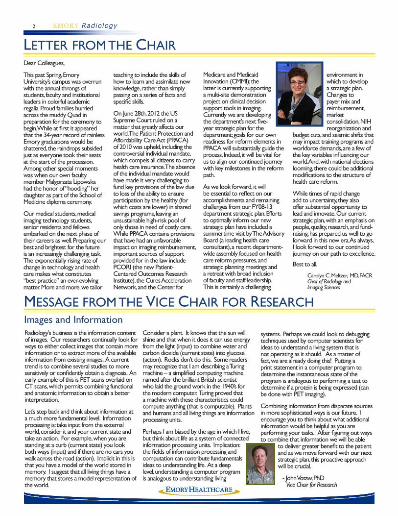
LETTER FROM THE CHAIRDear Colleagues,
This past Spring, Emory University’s campus was overrun with the annual throngs of students, faculty and institutional leaders in colorful academic regalia. Proud families hurried across the muddy Quad in preparation for the ceremony to begin. While at first it appeared that the 34-year record of rainless Emory graduations would be shattered, the raindrops subsided just as everyone took their seats at the start of the procession. Among other special moments was when our own faculty member Malgorzata Lipowska had the honor of “hooding” her daughter as part of the School of Medicine diploma ceremony.
Our medical students, medical imaging technology students, senior residents and fellows embarked on the next phase of their careers as well. Preparing our best and brightest for the future is an increasingly challenging task. The exponentially rising rate of change in technology and health care makes what constitutes “best practice” an ever-evolving matter. More and more, we tailor
teaching to include the skills of how to learn and assimilate new knowledge, rather than simply passing on a series of facts and specific skills.
On June 28th, 2012 the US Supreme Court ruled on a matter that greatly affects our world. The Patient Protection and Affordability Care Act (PPACA) of 2010 was upheld, including the controversial individual mandate, which compels all citizens to carry health care insurance. The absence of the individual mandate would have made it very challenging to fund key provisions of the law due to loss of the ability to ensure participation by the healthy (for which costs are lower) in shared savings programs, leaving an unsustainable high-risk pool of only those in need of costly care. While PPACA contains provisions that have had an unfavorable impact on imaging reimbursement, important sources of support provided for in the law include PCORI (the new Patient-Centered Outcomes Research Institute), the Cures Acceleration Network, and the Center for
Medicare and Medicaid Innovation (CMMI); the latter is currently supporting a multi-site demonstration project on clinical decision support tools in imaging. Currently we are developing the department’s next five-year strategic plan for the department; goals for our own readiness for reform elements in PPACA will substantially guide the process. Indeed, it will be vital for us to align our continued journey with key milestones in the reform path.
As we look forward, it will be essential to reflect on our accomplishments and remaining challenges from our FY08-13 department strategic plan. Efforts to optimally inform our new strategic plan have included a summertime visit by The Advisory Board (a leading health care consultant), a recent department-wide assembly focused on health care reform pressures, and strategic planning meetings and a retreat with broad inclusion of faculty and staff leadership. This is certainly a challenging
environment in which to develop a strategic plan. Changes to payer mix and reimbursement, market consolidation, NIH reorganization and
budget cuts, and seismic shifts that may impact training programs and workforce demands, are a few of the key variables influencing our world. And, with national elections looming, there could be additional modifications to the structure of health care reform.
While times of rapid change add to uncertainty, they also offer substantial opportunity to lead and innovate. Our current strategic plan, with an emphasis on people, quality, research, and fund-raising, has prepared us well to go forward in this new era. As always, I look forward to our continued journey on our path to excellence.
Best to all,
Carolyn C. Meltzer, MD, FACR Chair of Radiology and Imaging Sciences
MESSAGE FROM THE VICE CHAIR FOR RESEARCH
systems. Perhaps we could look to debugging techniques used by computer scientists for ideas to understand a living system that is not operating as it should. As a matter of fact, we are already doing this! Putting a print statement in a computer program to determine the instantaneous state of the program is analogous to performing a test to determine if a protein is being expressed (can be done with PET imaging).
Combining information from disparate sources in more sophisticated ways is our future. I encourage you to think about what additional information would be helpful as you are performing your tasks. After figuring out ways to combine that information we will be able
to deliver greater benefit to the patient and as we move forward with our next strategic plan, this proactive approach will be crucial.
- John Votaw, PhD Vice Chair for Research
Images and InformationRadiology’s business is the information content of images. Our researchers continually look for ways to either collect images that contain more information or to extract more of the available information from existing images. A current trend is to combine several studies to more sensitively or confidently obtain a diagnosis. An early example of this is PET scans overlaid on CT scans, which permits combining functional and anatomic information to obtain a better interpretation.
Let’s step back and think about information at a much more fundamental level. Information processing is: take input from the external world, consider it and your current state and take an action. For example, when you are standing at a curb (current state) you look both ways (input) and if there are no cars you walk across the road (action). Implicit in this is that you have a model of the world stored in memory. I suggest that all living things have a memory that stores a model representation of the world.
Consider a plant. It knows that the sun will shine and that when it does it can use energy from the light (input) to combine water and carbon dioxide (current state) into glucose (action). Rocks don’t do this. Some readers may recognize that I am describing a Turing machine – a simplified computing machine named after the brilliant British scientist who laid the ground work in the 1940’s for the modern computer. Turing proved that a machine with these characteristics could compute anything (that is computable). Plants and humans and all living things are information processing units.
Perhaps I am biased by the age in which I live, but think about life as a system of connected information processing units. Implication: the fields of information processing and computation can contribute fundamentals ideas to understanding life. At a deep level, understanding a computer program is analogous to understanding living

3
us. Two ongoing examples come to mind. First, we have had physicians, physicists, and technologists working together to improve quality, efficiency, and safety at our MRI facilities for several years now. Second, many of us are under the impression that RadNet is a fixed system and nothing can be changed. Yet RadNet is regularly being upgraded and we can ask for and sometimes make considerable changes. We have more opportunities with computerized order entry (CPOE) and the wireless transmission of patient images. In the face of inefficient or substandard patient care, know that your ideas can and do help to make significant improvements.
As we move forward, take time after each milestone to appreciate what we created and to show appreciation to one another. Let’s try to remember that we are strong individually, but we are far stronger together. Then work together to provide the best care and
compassion for our patients, and our coworkers, support research efforts, and provide a learning environment for all who work with and within our department. - Chuck Powell, Interim Administrator
RADIOLOGY UPDATE
This has been a very successful year for the Department of Radiology and Imaging Sciences. As I reviewed our numerous accomplishments in fiscal year 2012, it became quickly apparent that many people and many areas deserve recognition; it would take the whole newsletter and more to capture all of your positive efforts. Because of extraordinary teamwork, some achievements were most notable. Both the EUHM and the EUH Breast Imaging Centers received Breast Center of Excellence designation. The CR/DR Committee, comprised of Healthcare and University employees, partnered with Dr. Terk and Dr. Berkowitz and many other radiologists to focus on and improve general diagnostic image quality. A similar story transpired in CT where Dr. Duong worked collaboratively with technologists and managers to improve CT quality and safety across the system. We had successful Joint Comission inspections at EUH, Wesley Woods and TEC. Just recently, we found out that EUH is now ranked number two and EUHM is ranked number six for quality in the University Healthcare Consortium. Possibly our most encompassing and far reaching shared endeavor arose
from a Radiology Leadership Academy (RLA) project that was proposed three years ago by Mike Armstrong, Dr. Lee, Marcus Foster, and Mariana Teodorescu – the Service Excellence Institute (SEI). SEI came about due to the combined and dedicated efforts of researchers, educators, administrators and clinicians. SEI demonstrated how well people from diverse backgrounds can work together to achieve a meaningful outcome. Like the RLA, SEI has furnished us with more staff-inspired ideas to improve care and service.
The future will look different than our current care model. The Affordable Care Act (healthcare reform), an aging population, and government debt will require us to modify our practice. For us to navigate this new world, continue our research and academic success, and best serve our patients, we must make use of our two greatest gifts: our people and our technology. By seeking out others, asking for input, and joining together, we can meet and overcome our collective challenges. By making best use of existing and new technology, we can offset the perceived limitations placed upon
One Year Ends and Another Year Begins
AWARDS & RECOGNITIONFaisal Khosa, MDAssistant Professor
Radiology and Imaging Sciences
Medal of Excellence (Tamgha-i-Imtiaz)In recognition of his meritorious services to the medical and dental institutions in Pakistan, Dr. Faisal Khosa has
received the Medal of Excellence (Tamgha-i-Imtiaz), one of the highest awards conferred to a civilian. The award is the fourth-highest decoration given to any civilian in Pakistan based on their achievements. Over the years, Dr. Khosa has been invited to lecture and mentor students at medical and dental universities across Pakistan. The award conferring ceremony will be held in Islamabad, Pakistan on March 23, 2013.
Southeastern Chapter of the Society of Nuclear MedicineMarshall Brucer AwardThe Southeastern Chapter of the Society of Nuclear Medicine (SECSNM) honored Raghuveer K. Halkar, MD, Chief, General Nuclear Medicine at Emory University Hospital and Professor of Radiology and Imaging Sciences, with the presentation of the Marshall Brucer Award, the highest honor that the SECSNM can bestow upon a member. The Brucer Award was presented to Dr. Halkar at the September 21-24, 2012 SECSNM Annual Meeting, recognizing his many years of service to nuclear medicine as well as his leadership in organized medicine at the state, chapter and national level.

4
Kollengode S, Terk M. Atypical bisphosphonate-related subtrochanteric femoral fracture. ACR Case in Point. May 31, 2012.
Tahvildari AM, Atnafu A, Cosco D, Acosta A, Gupta D, Hudgins PA. Global Health and Radiology: A New Paradigm for US Radiology Resident Training. J Am Coll Radiol. 2012 Jul;9(7):516-9.
Lipowska M, Klenc J, Marzilli GM, Taylor AT. Preclinical Evaluation of 99mTc(CO)3-Aspartic-N-Monoacetic Acid, a Renal Radiotracer with Pharmacokinetic Properties Comparable to 131I-o-Iodohippurate. J Nucl Med 2012 53:1277-1283 published ahead of print June 20, 2012 (10.2967/jnumed.111.102236).
AWARDS & RECOGNITION
CHECK IT OUT
Mark Goodman, PhDEndowed Chair of Imaging Sciences and Hematology and Oncology
The Society of Nuclear Medicine Paul C. Aebersold Award
Dr. Goodman has been selected to receive the prestigious 2012 Paul C.
Aebersold Award. Every year, The Society of Nuclear Medicine (SNM) Paul C. Aebersold Awards Committee selects an individual as the recipient of the Paul C. Aebersold Award for Outstanding Achievement in Basic Science applied to Nuclear Medicine. The Aebersold Award is named for Paul C. Aebersold, a pioneer in the biologic and medical application of radioactive materials and the first director of the Atomic Energy Commission’s Division of Isotopes Development at Oak Ridge.
John Oshinski, PhDAssociate Professor Radiology and Imaging Sciences
Outstanding Postdoc Mentor AwardDr. Oshinski was inducted into the “One in a Hundred” Outstanding Postdoc Mentor Award Club. Each year at the Annual Postdoc Research Symposium, the Emory Post doctoral fellows nominate and choose outstanding mentors for the Club. A postdoc or an entire lab of postdocs nominates his/her mentor for this award, and
then a committee of postdocs chooses six mentors from the nominations to receive the “One in a Hundred” award. Dr. Oshinski was one of six mentors awarded at the School of Medicine and Yerkes.
Louis Martin, MDProfessor of Radiology and Imaging Sciences
2012 SIR Dotter Lecture and Gold Medal
Dr. Martin was awarded the 2012 SIR Dotter Lecture and Gold Medal. This award
honors Dr. Martin’s extraordinary contributions to the field, dedicated service to the Society and distinguished career achievements in interventional radiology. As Honoree, Dr. Austin delivered a lecture at SIR’s Annual Scientific Meeting.
Dr. Martin has been very active in SIR leadership, having served on the FDA Device Forum since 1997 (which he chaired from 2001-2005), the Standards of Practice Committee since 1995 and several other committees for SIR, the American College of Radiology, American Board of Radiology, American Heart Association and American College of Cardiology.
NEW GRANTS
Femoral Artery Plaque Imaging using the 3-Point Dixon MRI Technique Co-Investigators: Khusrow Niazi, MD John Oshinski PhD
Significance: The goal of this proposal is to accurately determine if the constituents of femoral artery plaque can be determined using a novel MRI technique. Validation will be done via radiology pathology correlation. We intend to determine plaque components in the femoral artery with various degrees of stenosis. This would help determine not only the natural history of peripheral arterial disease (PAD) but also help assess predominance
of plaque subtypes at varying levels of stenosis. Once this can be established, effects of pharmacologic treatment on the plaque can be studied which so far have been elusive. The information that is derived from this pilot study will help us to better understand the evolution of plaque and the role of inflammation and set the stage for a larger scale study to develop a more sophisticated biomarker of plaque progression and the response to therapy.
Arthur Stillman MD, PhD William Lewis, MD
Principal Investigator: Faisal Khosa, BSc, MD

5
STRIVING FOR EXCELLENCETo TrustCan you think of any relationship that can survive without trust? I can – but I would not want to be in it! Positive, healthy relationships need many things to remain viable, but in my opinion, not many are as important as trust. Many of us can agree on this to some extent in our personal relationships, but what about our professional relationships? Many of the essential elements that help sustain personal relationships do not exist in most professional relationships – making trust that much more valuable.
Creating and sustaining a trusting relationship will require an investment of time on the part of all involved individuals. This is time well spent when we consider the benefits of trust in our working relationships. Just as empowerment increases the speed of decision making, trust will promote an environment of timely accomplishment. In Stephen M.R. Covey’s book The Speed of Trust he tells us that “low trust slows everything – every
decision, every communication, and every relationship”.
Let’s apply this principle to our everyday working environment. How many people do you interact with every day? What is their role in relationship to yours? What is your role in relationship to theirs? How much of what you do everyday can you accomplish totally on your own? Does anything you do at work require a team effort?
Now let’s look at the importance of trust in sustaining these essential relationships. Take a moment to actually answer the questions in the paragraph above. Most of the time we are relying on others more than we are consciously aware of. In his book The 17 Essential Qualities of a Team Player, John Maxwell says that, “without trust we are (and function as) a group of individuals – not a team!”. Most of us are in positions
where our professional survival causes us to rely on the contribution of others – how unsettling would it be if that reliance was on someone that we did not trust!
Extending earned trust is equally as important as being trusted in our working relationships. To accomplish both, we must value the results that come from our diligence in establishing trust.
How do we go about establishing trust in our working relationships? To start, treat all co-workers with dignity and respect. Always communicate with honesty. Be open to others and their contribution. Make sure that your actions match your words. Make group, team, and organizational decisions – do not be personal agenda driven or self serving. Above all, value honesty, value trust, value others. Over time, some people have a tendency to take for granted that what they love – not so much what they value!
- Mike Armstrong Associate Director of ImagingGROWING OUR TALENT
Last September, twelve Radiology Leadership Academy (RLA) Fellows met as a group for the first time. Many of them did not know each other at this point, but quickly discovered ways that their work connected to outcomes of our patients. Through these connections they developed projects that exemplified their work at their RLA Graduation on June 8. Over the course of the program the fellows were exposed to many leadership tools, read several thought provoking books, participated in elective courses, and spent many extra hours developing their projects.
RLA Class of 2012Being our third group to become RLA Alumni we heard many of the lessons that this group is taking with them: Bobby Burrow, who has 35 years of Emory experience, commented, “This was a lot of work, but I learned so much.” Erica Campbell-Brown, our first fellow from Grady expressed, “I really enjoyed getting to know people I never would have met.” Dr. Ashley Aiken, who helped the group to drill down into the finer details of the department budgets, articulated, “I learned so much about the department and how everything works together. There are so many decisions that make more sense now.”
Thank you to the fellows who made the third year of RLA a success. Thank you to the various presenters who contributed to the growth of our fellows and enriched the quality of the program. Thank you to the Radiology administration for your vision and dedication to making this program possible. And finally, thank you to my co-facilitators Chuck Powell, Habib Tannir, Greg Pennington, Dr. Mimi Newell and Dale Walker, whose insight into leadership and devotion to the development of an engaging program fueled its success.
- Moncia Salama Sr. Assoc. Director of Programs
RLA Class of 2012Ted Brzinski – Medical Imaging Program Instructor
Bobbie Burrow– Manager of MRI Services
Erica Campbell-Brown– Nurse Practitioner
Cory Ivins– Reimbursement Manager
Hiroumi Kitajima, PhD– Assistant Professor
Jessie Knighton– Business Analyst II
Chris Kubik– Manager
Brent Little, MD– Assistant Professor
Keisa McGlathery– Project Manager, IS
Ashley Aiken, MD– Assistant Professor
Roger Williams, MD– Assistant Professor RLA Class of 2012, stands with fellow alumni, administration
and RLA facilitators at the RLA graduation.

6
The Adopt-a-Resident Committee is pleased to announce that Dr. Thomas Loehfelm, R2. This past summer he was awarded a grant to develop a tablet-computer application to facilitate the sharing of medical images for teaching purposes among faculty and residents at Emory and beyond. His proposal nicely fulfilled the major goals of an “Adopt” proposal: originality, value to the residency program, as well as significant value to the national radiology community as a whole.
Tom’s idea for his project stemmed from the realization
Adopt-a-Resident Continues to Take Leapsthat radiologists love to save images from interesting cases and share them with their colleagues. A typical radiologist has a USB thumb drive, shared network folder, and/or external hard drive filled with hundreds or thousands of images- an interesting chest x-ray, a rare tumor or infection, or a classic radiographic finding. In an academic setting, radiologists will fill in a lull in the workday by calling a resident or medical student over to their workstation to review some of these interesting cases. This can be the most useful teaching approach because the cases have some personal meaning to the
teacher, and usually demonstrate the relevant finding clearly and memorably (which is what led the radiologist to save the case in the first place). As he wrote his adopt-a-resident proposal, he realized that the same system could
be used for much more, such as administering home-grown tests to residents or medical students. In addition to quickly sharing a case, he could create and share more comprehensive content as well, similar to a journal article or text book, with rich text and images that the user could interact with. So far he has developed a PC program to create these Tests, Cases, Teaching Files and an app for Android tablets. The next step is to translate it for the iPad and share it with the faculty and other residents.
Tom is grateful to the Adopt-a-Resident program for supporting his process. The Adopt- a- Resident committee encourages current R1’s (as well as R2’s who may have developed an idea in the interval since the last deadline) to strongly consider submitting a proposal. The deadline is February 15, 2013. Please email Mimi Newell ([email protected]) for additional information.
Tom sits with a fellow colleague explaining the new image tablet application.
ADOPT- A- RESIDENT
-Thomas Loehfelm, MD, R2-Camille Dingle Communications Specialist
Adopt-A-ResidentPersonalize the giving experience when you fund a resident’s scholarship. Your generosity may enable a resident to carry out a novel idea or attend a national radiology conference and ultimately will shape the next generation of radiologists.
You can participate in the Adopt-A-Resident Program through a $2,500 annual commitment over the span of four years. If you would like to explore this or other giving opportunities that will benefit Emory Imaging, contact:
Stacia [email protected]
A few years ago, our department’s Adopt-a-Resident grant gave Dr. Tahvildari and his mentor Dr. Hudgins’s a chance to establish a partnership with the Addis Ababa University and the Radiology Department at Black Lion Hospital in Ethiopia. After successful trips to Ethiopia, the tables have turned, and now Drs. Tequam Debebe and Getachew Assefa, from Addis Ababa University have come to Emory University to observe and shadow the Neuroradiology division. The doctors are here for one month working closely with Dr. Pat Hudgins, who will play a vital role in helping them prepare and finalize the curriculum for their new Neuroradiology fellowship program at Addis Ababa University.
In June 2011, a team from Emory Radiology was visiting Addis Ababa University, they were able to meet with the deans of
postgraduate and undergraduate training, to discuss incorporating radiology into medical student education. From the beginning of this project, it has been the Department’s goal to develop subspecialization and fellowship training. As the partnerships grows between Addis Ababa University and Emory Radiology, they will continue to exchange knowledge in residency training, including formation of subspecialty fellowships and web-based learning and research collaboration.
Bringing the Department’s Global Health efforts back to Emory University allows visiting doctors to study and observe procedures, the application of advance technology, residency program, our residency curriculums and sub-specialties. These
Dr. Pat Hudgins stands with, Dr. Tequam Debebe and Dr. Getachew Assefa, visiting professors from Addis Ababa University in Ethiopia.
GLOBAL HEALTH
doctors are able to take their experience back to their departments to educate and improve on processes, procedures and educational programs.
-Camille Dingle, Communications Specialist

7
Quality CornerWe CareThe Joint Commission (TJC), Center for Medicare/Medicaid Services (CMS), Department of Community Health (DCH); whatever you call them, they call on us regularly, frequently and unannounced. They survey us, review us, inspect us and cause trepidation in the hearts and minds of some. Many find it to be a challenge when these “people” show up on-site. Then the question is, “Who are these people?” They are the healthcare industry inspection agencies.
Many believe we have our processes and procedures in place just to please this group. The answer to that is: No. We provide care to our patients, their families and ourselves. Faculty and staff provide high quality care not because we know that we will be inspected but because we do care. We are proud of the quality of the care we provide and we show it in many ways. We show it when we are surveyed and everyone shines a little brighter and stands a little taller when the surveyors are around. We show it in the Quality Improvement projects we present at the annual EHC Quality Conference. This past year we presented eleven projects ranging from collaborating with the Emergency Department on Community Acquired Pneumonia pathway to CT Radiation Dose Reduction.
Throughout our department we are involved in several programs that continue to perpetuate us
IN THE KNOW
as a leader in quality improvements. The Radiology Leadership Academy (RLA) emphasizes a commitment to Quality. The fellows commit their time for one year to the program. The projects that spring from this incubator of ideas have been indicative of the energy, time and thoughtfulness of bringing a project from concept to reality. Employees attend the Emory Healthcare Quality Academy. This program builds an internal core of staff who understands the tools of Quality Improvement (QI). The students then go back to their departments and independently conduct QI projects. We have modality-specific Quality and Safety Committees. Digital Radiography, CT, MRI and Ultrasound, just to name a few, have all been active in improving quality at the modality level by reviewing procedures and standardizing these across Emory Healthcare.
What’s my favorite quality project? I think it is the export of Emory quality to far flung places. For this you do not have to look any further than our Adopt-A-Resident program. Drs. Pat Hudgins and Ali Tahvildari have traveled to Ethiopia twice to help with the education of Ethiopian radiologists.
So, I will say it again. We provide quality health care not because we are surveyed, inspected and reviewed. We provide it because we care. All of us. This column gives me the opportunity to say thanks. Thank you for all you do every day.
-Dale Walker, Director of Strategic Initiatives
ENGAGE IN EDUCATIONOn March 17, 2012 Interventional Radiology and Radiation Oncology presented the Symposium on Image-guided Cancer Therapies at the Ritz Carlton in Buckhead.
Dr. Kevin Kim, Director of Interventional Radiology and Image-guided Medicine, Dr. Ian Crocker and Dr. Tim Fox organized and directed the symposium. Dr. Walter J. Curran, Executive Director of Winship Cancer Institute, and Dr. Carolyn Meltzer, Chair of Radiology and Imaging Sciences opened the event by welcoming the attendees. Dr. Kim followed with an introduction on the state-of –the-art imaged-guided targeted cancer therapies. A total of 22 faculty members presented throughout the day on topics such as: advancements in interventional oncology and radiation oncology for lung cancers, liver cancers, kidney cancer, spine and bone cancers. Molecular targeted therapies and Robotic therapies, as well as advanced imaging techniques were also presented. The attendees were also able to view emerging technology presentations from two visiting professors. Dr. Reed Omary is a professor at Northwestern University and presented Interventional
Oncology: Opportunities for Innovation. Dr. Samuel Ryu, a visiting professor from Henry Ford Hospital, he presented on Spine Radiosurgery. Tiffany Deaton, Administrative Assistant for the Interventional Radiology and Image-guided Medicine division worked with the Emory CME office to coordinate the symposium.
The event was a wonderful success and the Department of Radiology and Imaging Sciences looks to the future for more of this type CME credit courses. -Dr. Kevin Kim Director of Interventional Radiology and Image-guided Medicine
Radiologists attended several presentations that touched on the latest developments in Image-guided Cancer therapies.

8
Would You Like to Receive the Rad Report?The Rad Report is the perfect opportunity to involve Emory Radiology Alumni in our current radiology happenings. Within the department there is a new issue of the Rad Report available each month. This has proven to be a great vehicle to communicate across our divisions and help everyone to have a better understanding of how each of us contributes to the department as a whole.
Twice a year a compilation of the radiology department highlights are fused into a special issue of the Rad Report, with the Radiology Alumni in mind. We hope that, as a Radiology Alumni, you take pride in the accomplishments of today.
If you are interested in receiving the Rad Report on a monthly or semi-annual basis, you can register on the home page of radiology.emory.edu or follow these easy steps:
Step One
Step TwoYES! I would like to receive the Emory Rad Report.
Prepare your information:NameE-mail address and/or phone numberSubscription Choice* - Monthly or Semi-Annual * Subscriptions are only available in electronic format.
Submit your request to [email protected] or call 404.712.7912
Step Three
You can help us unite the past and present for a promising future by building upon the contributions you have made to our successful department. Become “our” Facebook friend and use Emory Radiology’s profile to stay connected. Follow the directions below to register for your own Facebook page and add Emory Radiology as your friend:
Step 1 - Visit www.facebook.com and complete the information on the home page including your name, e-mail address, password and birthday. Then click the “sign up” button to submit your information.
Step 2 - Check your e-mail. A confirmation will arrive with a link to confirm your e-mail. If you don’t see it right away, check your spam or junk mail folder.
Step 3 - Set up your profile with as much information as you would like other Alumni to know about what you have done and where you are now.
Step 4 - Add us as a friend by typing in “Emory Radiology - Alumni” in the top right hand corner search box.
Step 5 - Under our profile picture, you will see a link to click to “request Emory Radiology as a friend”
Step 6 - Use our friends list to find others that have been a part of Emory Radiology and stay connected.
“Friend” Request Radiology
Look for a next semi-annual issue of the Rad Report in Aril 2013.
STAY CONNECTED
GET INVOLVEDMerrill’s CommitteeAfter an intense review process and close results, the Merrill’s Committee is pleased to announce its very first winner, Edwin Arias. Edwin is a Diagnostic Technologist at Emory University Hospital. He earned this award based on the submission of a Portable KUB performed on a critically ill ICU patient. Edwin chose his reward: a pair of movie tickets. Please congratulate him on his exceptional attention to image quality and high standard of patient care.
The vision of the Merrill’s Committee is to empower and inspire diagnostic technologists to seek maximum levels of image quality through positive reinforcement. This commitment to image quality can be recognized by their peers, imaging students, supervisors, radiologists or radiology residents. Remember: you can be the next Merrill’s winner!
Be sure to recognize your own or others’ stellar work by submitting a nomination for the Merrill’s Award! Blue Merrill’s Committee folders are located in each diagnostic work area. Inside are blank submission forms as well as a sample of the evaluation criteria utilized by the committee. At the end of each month, submissions will be collected and reviewed by the committee.
Technologist Edwin Arias is the first winner of the Merrill’s Committee Award.
- Katy Day Imaging Workflow Adminstrator