advances in molecular diagnostics new diagnostic...
TRANSCRIPT

The U.S. Centers for Disease Control and Preven-
tion estimate antibiotic-resistant pathogens cause
over 2 million infections annually, contribute to 23,000
deaths and add $20 billion in excess direct U.S. health-
care costs, with another $35 billion a year lost in social
costs. One of the hotly contested questions surrounding
the issue has been — and continues to be — the contri-
bution of agricultural and veterinary use of antibiotics
to the problem. Recent diagnostic innovations put to
use in research by CSU’s Microbial Ecology Group — a
collaboration between our Department of Microbiol-
ogy, Immunology and Pathology, and Clinical Sciences,
Animal Sciences, Computer Sciences and the School of
Education — have uncovered some important insights
about the source and epidemiology of some resistance
in the food chain.
testing Freed From culture constraintsThese new diagnostic innovations based on next gen-
eration sequencing (NGS) capture genome-specific
information and take advantage of the speed and robust
amount of data generated. Culture-independent NGS
provides an increased resolution for characterizing
pathogens without targeted enrichment or a precon-
ceived idea of what the pathogen may be, making it an
excellent tool for undiagnosed diseases or cases in which
the clinical picture does not completely match the diag-
nostic test result. This tool also strengthens our unique
public mission to improve our understanding of patho-
gen evolution, adaptation and virulence determinants in
the veterinary community.
For those reasons, NGS lends itself to understanding
the often complex and pan-microbial genetic dynamics
of antimicrobial resistance. With its ability to combine
many samples in a single sequencing run and obtain
high sequence coverage per sample, NGS-based metage-
nomic sequencing can detect members of the microbial
community in very low abundance that may be missed
or are too expensive to identify using other methods.
Such “shotgun metagenomics,” the study of whole-com-
munity DNA extracted directly from samples, allows us
to comprehensively sample all genes in all organisms
present in a given complex sample.
new applications, new insightsThat diagnostic capability freed from the need to culture
has led to several discoveries at CSU:
n A study just published in December 2015 used shotgun
metagenomics in to provide the first description of
the resistome of North American dairy and beef pro-
duction effluents. Sequencing produced over 1.05 bil-
lion sequencing reads across all samples, at an average
of 30.8 million reads per sample. The study identified
34 mechanisms of antimicrobial resistance — mostly
tetracycline-resistance mechanisms — within 34 soil,
manure and wastewater samples from feedlot, ranch
and dairy operations. Ranch samples contained sig-
nificantly fewer mechanisms than dairy and feedlot
samples, and the dairy resistome differed significantly
from feedlots. Soil, manure and wastewater resistomes
differed from one another, suggesting each should be
managed differently. Further, by sampling a conven-
tional against organic dairy, a U.S. against a Canadian
feedlot and cow/calf ranch against a feedlot, we were
Diagnostic news and trends from the Colorado State University Veterinary Diagnostic Laboratories Volume 21, Number 1 Spring/Summer 2016
Advances in Molecular Diagnostics
New Diagnostic Applications to Enlighten Antimicrobial Resistance
Veterinary Diagnostic Laboratories
supporting scientific efforts to mitigate the rising incidence of antimicrobial resistant infections is just one of the public-mission responsibilities of the colorado state university Veterinary diagnostic labs. as a level 1 laboratory in the national animal health laboratory network, the labs play a direct role in protecting a safe, stable and nutritious food supply for the nation. see page 13 for details. See SHOTGUN, page 2

2 Volume 21, Number 1
able to baseline resistomes based on differences in,
respectively, antimicrobial use, diet, and intensive vs.
extensive systems.
n In another study, a metagenomic approach and
shotgun sequencing technology detected pathogenic
bacteria in environmental samples collected from
the same groups of cattle at different processing steps
in the beef-production chain: At feedlot entry and
exit, in transport trucks, in slaughter holding pens
and at the end of the fabrication system. It found
some surprising results. Log read counts classified as
pathogens per million reads for Salmonella enterica,
Listeria monocytogenes, E. coli, Staphylococcus aureus,
Clostridium botulinum and perfringens and Campy-
lobacter jejuni, coli and fetus all decreased over each
sequential processing step. In fact, normalized read
counts for S. enterica, E. coli, and C. botulinum were
greater in the final product than at the feedlots.
Although some limitations of shotgun metagenom-
ics makes this approach impractical for regulatory
and confirmation purposes, its ability to characterize
the microbiome and shifts in pathogen populations
during production does shed important light on the
potential role of resistome dynamics in the environ-
ment and the general products in the chain.
n Recognizing that treating antimicrobial-resistant
infections is, in its current state, a purely reaction-
ary therapeutic process, another pilot study identi-
fied novel antimicrobial drug resistance markers that
could lead to more clinically applicable detection and
therapeutic tools. Such assays capable of rapidly iden-
tifying phenotypic expressions of resistant infections
would not only improve therapeutic efficiency but
also enhance surveillance. This study grew multi-drug
resistant isolates of Salmonella typhimurium contain-
ing similar integron profiles in a liquid nutrient broth
both with and without an ampicillin/chlorampheni-
col/streptomycin/sulfisoxazole/tetracycline drug
panel. It then subjected the isolates to protein and
metabolite extraction, followed by non-targeted pro-
teomic analysis via liquid chromatography coupled
with tandem mass spectrometry, ultra-performance
liquid chromatography-mass spectrometry and gas
chromatography-mass spectrometry. Proteins were
annotated using the Uniprot database, and metabo-
lites annotated after screening against several spectral
libraries and the Golm database. Results indicate dis-
tinct biomarker patterns in both protein and metabo-
lite expression levels between isolates grown with
and without the specific antimicrobials. This better
understanding of biomolecule expression key to
antibiotic resistance should open avenues for future
larger studies to develop novel surveillance and diag-
nostic tools. ▲
antimicrobial residue testing at csu
in partnership with the csu department of animal science, the Veterinary diagnostic laboratory is now offering food-animal residue testing using randox Food diagnostics’ evidence investigatortm and biochip array technology. the elisa-based screening test is faster and less expensive than similar analyzers using mass spectrometry. also, the biochip array technology allows for simultaneous testing of multiple analytes from a single sample.For information about sample submission and cost, including volume discounts, call us at (970) 297-1281 or email [email protected]
reFerence
mcconnel cs, stenkamp-strahm cm, rao s, linke lm, magnuson rJ, hyatt dr. antimicrobial resistance profiles in escherichia coli o157 isolates from northern colorado dairies. J Food Prot. 2016 mar;79(3):484-7.
csu Vdl bacteriology section head doreene hyatt collaborated on a just-published study using conventional pcr to document antimicrobial-resistance patterns in enterohemorrhagic shiga toxin-producing E. coli o157 isolated from colorado dairies. individual-cow fecal samples collected monthly for a year from three dairies representing 2,750 lactating cows were tested using multiplex pcr assay to identify shiga-toxin producing E. coli. the 75 positives recovered were then tested for resistance to 10 gram-negative antimicrobials: amoxicillin–clavulanic acid, ampicillin, ceftiofur, enrofloxacin, florfenicol, streptomycin, sulfisoxazole, tetracycline, trimethoprim-sulfamethoxazole and tulathromycin.the study found only six isolates, or 8%, resistant to at least one of the 10 tested antimicrobial agents, all from a single dairy. the study detected no
significant effects on resistance profiles due to virulence genes, parity or previous antimicrobial treatments within the current lactation period.a follow-up study performing logistic regression analysis on o157 outcomes found humidity a risk factor for shedding enteropathogenic E. coli and an increase in temperature and recent antibiotic treatment a positive risk factor associated with enterohaemorrigic E. coli shedding.
Resistantto tetracycline
Resistantto sulfonamidesResistant
to streptomycin
E. COLI RESISTANCE PROFILES IN ONE DAIRY
shotgun (continued from page 1)

Spring/Summer 2016 3 LABLINES
Advances in Parasitology
Tracking the Ominous Rise of Anthelmentic-Resistant ParasitesCSU Veterinary Diagnostic Labs’ new polymerase
chain reaction test for parasites can now suc-
cessfully differentiate the five major bovine stron-
gyle genera — Cooperia, Haemonchus, Ostertagia,
Trichostrongylus and Oesophagostomum — within
just two to three days. Although generalized bench-
marking of parasite levels was always possible with
fecal egg counts, this new option makes creating
baselines for different geographic regions, herds
and animal classes within herds more specific to the
parasites present.
With that ability to quickly and accurately speciate
parasites, researchers now have available a powerful
tool for parasite surveillance, as well as for tracking the
effect of longterm anthelmentic programs and man-
agement systems on individual parasite populations.
These improvements will be important in helping
keep a tab on the development of resistance to chemi-
cal anthelmentics in particular genera.
eVidence oF resistanceCSU’s VDL recently participated in a multi-state study
to help assess the possible extent of that emerging resis-
tance. During USDA’s National Animal Health Moni-
toring System’s 2007–2008 beef study, producers from
24 states were offered the opportunity to evaluate their
animals for internal parasites and overall response to
anthelmentics. Fresh fecal samples were collected from
20 animals, or from the entire group if less than 20, and
then randomly assigned to either CSU VDL or one of
two other participating labs for exam.
Samples in which strongyle egg counts exceeded 30
per gram by flotation were then subjected to pooled
PCR analysis for the presence of Ostertagia, Cooperia,
Haemonchus, Oesophagostomum, and Trichostrongylus.
Results from 72 producers in 19 States indicated that
in over a third of the operations tested, anthelmentic
treatment resulted in 90% or less reduction in fecal egg
counts at around two weeks after treatment. The 90%
threshold is important because egg-count data are key
components of licensing new anthelmentics, and aver-
age egg-count reduction values for all trials submitted
must be greater than 90%.
All operations exhibiting less than a 90% reduc-
tion had used pour-on macrocyclic lactones as the
anthelmentic treatment. While some of these reduc-
tions could have been the result of improper drug
application, PCR analyses of the parasite populations
surviving treatment, coupled with follow-up studies
at a limited number of sites, indicated they were most
likely due to anthelmentic resistance in Cooperia spp.
and possibly Haemonchus spp. The PCR data indicated
lack of efficacy is biased toward members of those two
genera.
PCR results also argued against another alternate
explanation: That some animals were simply missed
during anthelmentic treatment. But PCR results con-
firmed reductions were typically observed in all genera
except Cooperia and Haemonchus — a phenomenon
that wouldn’t have occurred in missed animals.
It is clear that the efficiencies of treating with macro-
cyclic lactones today in U.S. commercial operations are
not the same as the efficacies generated when the drugs
were first licensed. Our results should be a warning to
cattle producers: They can no longer assume treatment
is synonymous with successful control, they should
rotate their anthelmentics, they should consider the
given that pour-on formulations offer the best oppor-
tunity to encourage resistance, and they must remem-
ber that successful long-term and sustainable control
can’t be obtained by relying on anthelmentic treatment
alone. ▲
— Lora R. Ballweber, DVM, MS, CSU VDL Parasitology Section Head
© Don Lane Photos. All rights reserved. Used by permission.
parasite pcr testing at csu
samplesn at least 3 grams
of feces from each individual as or immediately after being passed.
n select samples from no more than five individuals for pooled sampling.
n include fecal egg counts if known.
n package in clean, enclosable container, like a sealable plastic bag, devoid of air.
n refrigerate for no more than 24 hours. ship cold, but protect from freezing and direct contact with ice.
n ship to arrive within 48 hours.
costn $70 per sample
turnaroundn two to three days
4 Volume 21, Number 1
Zoo Corner
Tularemia in Zoo PrimatesCSU’s Veterinary Diagnostic Labs have confirmed a
noticeable increase in the number of positive tula-
remia cases in both wild and domestic animals in the
past few years. Environmental conditions have favored
significant expansion of both wildlife reservoirs and
insect vectors, increasing the incidence in domestic ani-
mals and posing an important zoonotic risk for people.
Caused by the Gram-negative coccobacillus Fran-
cisella tularensis, tularemia is endemic in North Ameri-
can wildlife, particularly in south-central United
States. Among domestic animals, cats are particularly
sensitive, with cases commonly occurring in dogs, pigs,
horses and sheep. U.S. human cases also occur every
year; 16 human cases were confirmed in Colorado in
2014 and over 20 in 2015.
Localized disease manifests as ulceroglandular or
oropharyngeal disease involving the skin or mucous
membranes, with lymphadenitis of draining nodes. Oro-
pharyngeal disease is particularly common in cats, which
capture and consume infected prey. Localized disease can
progress to septicemia with generalized lymphadenitis
and severe inflammation and major-organ necrosis.
Tularemia has been well described in both new- and
old-world non-human captive primates, in which the
disease can be rapidly fatal, often involving multiple
animals. CSU’s Veterinary Diagnostic Labs have con-
firmed tularemia in three different species of non-
human primates in two south-central facilities in 2015
and 2016. All were housed in outdoor/indoor exhibits
with the same species.
siamang A 24 year-old female Siamang found acutely moribund,
severely weak and stumbling received supportive treat-
ment but continued to rapidly decline. Bloodwork and
physical exam suggested severe septicemia with advanced
organ failure. Necropsy following euthanasia showed
dehydration, fibrino-hemorrhagic peritonitis, marked
hepatic swelling and segmental hemorrhagic serositis of
the small intestine. Histopathology found severe hepa-
titis and splenitis that was suppurative and necrotizing
and effaced up to 30% of the parenchyma. Though Gram
and silver stains identified no bacteria, PCR confirmed
tularemia. This female’s offspring was also found listless
and febrile within days of its death. Treated aggressively
with doxycycline, the male made a full recovery, and no
other primate in the enclosure was affected.
black-crested mangabey:A 28 year-old male black-crested mangabey was exam-
ined following four days of clinical lethargy and weak-
ness. Exam showed fever, hypoglycemia and leukopenia
suggestive of septicemia. The monkey was managed
aggressively with antibiotics and supportive therapy,
but continued to decline over the next day, remaining
hypoglycemic and developing ascites. Necropsy con-
firmed ascites, mild thoracic effusion and swelling of
the liver and spleen with generalized lymphadenopathy.
Lymph nodes bulged and hemorrhaged on the cut sec-
tion, suggestive of active lymphadenitis. Histopathology
confirmed fibrino-necrotizing hepatitis, splenitis and
severe suppurative, necrotizing lymphadenitis involving
most internal lymph nodes and the tonsils. The lesions
yielded no microorganisms upon Gram and silver stains,
but PCR confirmed tularemia. No additional animals in
this group developed clinical signs.
common marmosetsA 7-year-old female common marmoset found acutely
depressed, weak and listless with possible neurologi-
cal deficits was treated with supportive care under the
assumption it may have suffered head trauma. Despite
treatment, it rapidly deteriorated in 24 hours and died.
Five days later, a second 7 year-old female marmoset with
no prior clinical signs was found dead in the same enclo-
sure. Gross findings in both females showed marked
dehydration, prominent generalized peripheral and
internal lymphadenopathy, and moderate hepatospleno-
megaly with fine tan-white pinpoint foci throughout the
parenchyma. The lungs were diffusely and moderately
congested and edematous on the cut section.
Histology demonstrated severe suppurative and nec-
rotizing hepatitis, splenitis and lymphadenitis, intersti-
tial pneumonia and moderate pulmonary edema. One
marmoset also had necrosis of the bone marrow. The
other had moderate lymphoplasmacytic meningitis. In
one marmoset, large numbers of fine Gram-negative
coccobacilli were associated with necrotizing lesions,
though no organisms were detected in the other marmo-
set. Fresh liver submitted for PCR showed tularemia in
both animals, but no plague, toxoplasmosis or lympho-
cytic choriomeningitis virus. No additional animals were
affected.
search For the inFection sourceAnimals in these four cases, and all published cases of
natural infection, had access to outdoor enclosures.
Tularemia is endemic in several wildlife reservoirs.
In the south-central United States, it is commonly
reported in prairie dogs and other rodents, including
— Sushan Han, DVM, PhD, DACVP, CSU VDL Pathologist
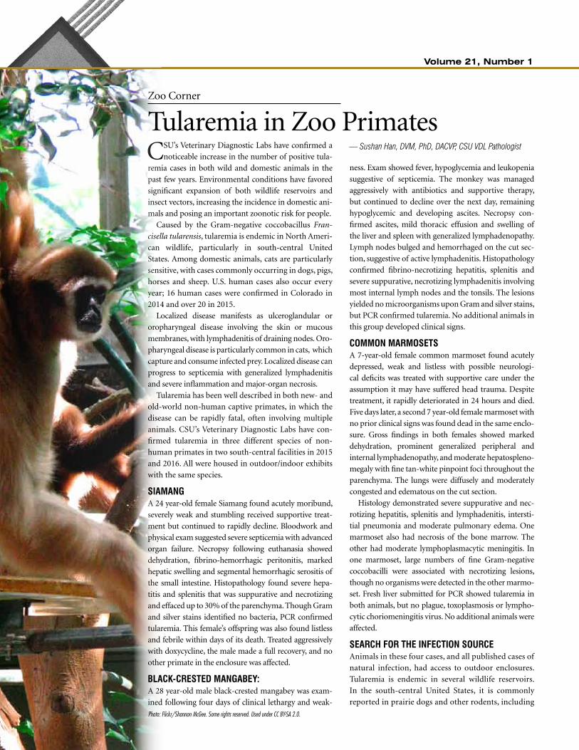
Photo: Flickr/Shannon McGee. Some rights reserved. Used under CC BY-SA 2.0.

Spring/Summer 2016 5 LABLINES
Chemistry and Toxicology
Old Stored Toxicants Can Still KillThe VDL received a call from a practitioner report-
ing about 20 cattle found dead in an area of old
farm buildings. The cattle had broken through a gate
and entered one of the buildings; inside was found
some green material. When one thinks of green colored
material that might be toxic, one first suspects prod-
ucts that are currently available. The most common
substance that comes to mind is some type of rat bait,
likely an anticoagulant.
A sample of the green material and tissue from one
of the dead animals was submitted to the laboratory
for testing. The green material was 10.1% arsenic and
11.3% copper, by dry weight. The arsenic concentra-
tion by dryweight of the liver was 290 ppm; of the
kidney, 340 ppm.
Equipped with that information, our consultation
with the veterinarian brought to mind a product avail-
able many years ago and used in agriculture as a pesti-
cide: Paris Green. Finding high concentrations of both
copper and arsenic confirmed the toxicity was caused
by old insecticide and rodenticide, whose chemi-
cal name is copper(II)
acetoarsenite. But it also
reminds us that when deal-
ing with an old building,
one needs to consider what
was available in years past,
not just today. ▲
— Dwayne Hamar, PhD, CSU VDL Chemistry and Toxicology Section Head
A 7 year-old female common marmoset showed marked caseous and necrotizing submandibular and mesenteric lymphadenitis, confirmed by PCR to be positive for Tularemia.
Photomicrographs of a mesenteric lymph node (left) and the liver (right) showed marked necrotizing lymphadenitis and random hepatic necrosis, respectively, consistent with embolic showering by bacteria.
mice. Disease occurs from early April to late Octo-
ber, when vectors are prevalent. The bacteria are gen-
erally transmitted via ticks, fleas and deer flies, but
they can also be transmitted via inhalation, wound
or mucous-membrane contamination or ingestion
of bacteria from contaminated water, environmental
sources or infected tissues of dead hosts.
Though a source of tularemia was not definitively
determined in any of these four cases, it is presumed
to be due to exposure to infected rodents or lago-
morphs within the enclosures, resulting in transfer of
infected ticks and fleas, contamination of water and
food sources, and possibly hunting and consumption
of infected prey by the primates.
Captive non-human primates of several species are
very sensitive to tularemia. Each of these four reported
cases progressed to septicemia rapidly, with few non-
specific and even presumptive neurological signs,
prior to severe morbidity and death. Early recognition
of tularemia-like signs and aggressive treatment with
effective antibiotics and supportive care can be suc-
cessful, though septicemic cases, as demonstrated, can
progress rapidly despite proper treatment. ▲
sourcessammak rl, rejmanek dd, roth tm, christe kl, chomel bb, Foley Je. investigation of tularemia outbreak after natural infection of outdoor-housed rhesus macaques (macaca mulatta) with Francisella tularensis. Comp Med. 2013 apr;63(2):183-90.
Ferrecchia ce, colgin lm, andrews kr, lewis ad. an outbreak of tularemia in a colony of outdoor-housed rhesus macaques (macaca mulatta). Comp Med. 2012 aug;62(4):316-21.
guthrie al, gailbreath kl, cienava ea, bradway ds, munoz gutierrez JF. septic tularemia in 2 cottontop tamarins(sanguinus oedipus). Comp Med. 2012 Jun;62(3):225-8.
mätz-rensing k1, Floto a, schrod a, becker t, Finke eJ, et al. epizootic of tularemia in an outdoor housed group of cynomolgus monkeys (macaca fascicularis). Vet Pathol. 2007 may;44(3):327-34.
Photo
: Wiki
pedia
/Chri
s Gou
let. S
ome r
ights
reserv
ed. U
sed u
nder
BY-SA
3.0
.

6 Volume 21, Number 1
Serology
Get the Most from Your Bovine SerologyThe most common reason the CSU Veterinary Diag-
nostic Lab sees clinicians submitting bovine serol-
ogy is to deduce the etiologic agent for either bovine
respiratory disease or bovine abortion. Serological
assays — which by definition involve not only detection
of specific changes in the serum, such as antibodies to
the pathogen produced by the host, but also antigens of
the infecting agent itself or its components — have com-
monly been used to diagnose viral infections. However,
today’s ubiquitous exposure of cattle in the United States,
both naturally and by vaccination, has made serology dif-
ficult to interpret.
Here are a few ideas to help you get more out of
this traditional diagnostic tool. Keeping these ideas in
mind will help maximize your use of diagnostic test-
ing, allowing for the time to make a reasonable diag-
nosis and easing the frustration of limited information
you receive from a single titer.
test selection is paramount. If a practitioner is
going to embark on the journey of using serology to
help diagnose disease, it’s prudent to make an appro-
priate selections of animals based on diseases affecting
the herd and their timing.
It is imperative to remember the type of test per-
formed depends on the herd history. For example, if
type II BVDV was isolated from a fetus or calf in the
herd, type II BVDV serum-neutralization tests might
be indicated. An animal infected with type II BVDV
generally will have lower antibody titers to type I BVDV.
In regard to timing, screening of young, unvacci-
nated cattle between 6 and 12 months of age may be
useful in determining if viral pathogens have recently
been or are currently circulating in a herd. Serological
assays are also available to help differentiate exposure.
paired sera still matter. In individual cattle thought
to have acute exposure in a herd, there’s still no com-
parison to the traditional uses of serological assays for
diagnosing viral infections through paired acute and
convalescent sera. In cases of acute illness, paired acute
and convalescent serum samples are more likely to be
useful information than a single sample. A single SN
titer is unlikely to yield definitive information since it
does not distinguish between current infection, previ-
ous exposure or vaccination. If a virus is involved in
the disease, a four-fold increase in titer will be observed
over time, which can be benchmarked against.
It’s important to remember that because the serum
neutralization assay, the most commonly used method
for detecting viral antibodies, measures the ability of
antibodies in test serum to neutralize a reference viral
isolate, some within-lab titer variations may exist over
time. For that reason, it is important to submit those
paired acute and convalescent sera simultaneously.
Titers to different pathogens can differ substantially
depending on the antigenic exposure.
look for additional comparisons. Even paired acute
and convalescent serum samples from an aborting cow
rarely will show a change in titer, because cows infected
with BVDV and BHV-1 abort weeks to months after
infection. Therefore, it may be more useful to compare
SN titers from cows with healthy calves to the titers of
cows that have aborted. Fetal serum samples can be
helpful in some cases. If the fetus was infected during
the last half of gestation, it may have made antibodies
to the infecting virus. ▲
— Christie Mayo, DVM, PhD, CSU VDL Virology Section Head
boVine serology Quick tips
n consider the presenting complaint: abortion, respiratory disease, duration of illness
n record type and age of cattle — beef, dairy, cow/calf, feedlot, calves, lactating cows
n include number of animals in the herd, ill and deadn consider vaccinations — brand of vaccine, modified live
or killed, age at vaccinationn note date of last vaccination and product usedn account for colostrum intake: serology tests do not
distinguish between passive immunity via maternal antibodies and active immunity induced by infection or vaccination

Spring/Summer 2016 7 LABLINES
— Barbara Powers, DVM, PhD, DACVP, CSU VDL Director
Guardians of Public Health
National Animal Health Lab Network More Critical than EverThe Veterinary Diagnostic Laboratories at CSU
are a Level 1 laboratory in the USDA’s National
Animal Health Laboratory Network. NAHLN-mem-
ber labs may be involved in surveillance for early detec-
tion of foreign animal disease, surge testing during an
outbreak, and testing samples during the outbreak
recovery phase. As such, there must be a high degree of
confidence in the quality of the laboratories and asso-
ciated test results. USDA recognizes the value of qual-
ity management systems and requires that all NAHLN
laboratories have a functional quality management
system. Our full accreditation by the American Asso-
ciation of Veterinary Laboratory Diagnosticians allows
admittance to the NAHLN without additional require-
ments related to documentation of a quality manage-
ment system.
NAHLN plays a direct role in protecting a safe,
stable and nutritious food supply. It will play an essen-
tial role in responding to a biological attack affecting
animals or people. An example of the critical surveil-
lance role NAHLN plays was evident during the 2015
highly pathogenic avian influenza outbreak. NAHLN
laboratories operated 24 hours a day, seven days a week
to test poultry samples. Those quick, reliable tests per-
mitted rapid depopulation of infected flocks, surveil-
lance testing to halt spread of the virus and testing to
permit repopulation of farms and resumption of trade.
NAHLN was developed in response to the Public
Health Security and Bioterrorism Preparedness and
Response Act of 2002. During the past 12 years the
NAHLN, composed of federal, university and state vet-
erinary diagnostic labs, has established the framework
of a surveillance and emergency response system that
provides critical and ongoing resources for laboratory
testing, surveillance and information management,
including data analysis and sharing, quality assurance
with an auditing system and the development and vali-
dation of new tests.
Today, it requires increased funding to improve
compliance with 2012’s Homeland Security Presi-
dential Directive, which required the department to
“develop nationwide laboratory networks for food,
veterinary, plant health and water quality that inte-
grate existing Federal and State laboratory resources,
are interconnected, and utilize standardized diagnostic
protocols and procedures,” according to the directive.
That improved compliance with the directive will
require expanding surveillance and surge capacity of
the NAHLN by increasing the number and level of par-
ticipating state laboratories. It will require additional
development of the infrastructure for electronic trans-
mission of data between sample collectors, laborato-
ries and state and federal databases. It will also call for
increasing efficiency and effectiveness of lab personnel
training nationwide.
NAHLN is essential to the health of U.S. animal
agriculture, bioterrorism surveillance and the U.S.
economy. USDA estimates the cash receipts of the U.S.
animal industries at $185.68 billion; therefore, even a
fully funded level of $30 million would represent only
a 0.016 percent federal investment for disease surveil-
lance to protect essential agriculture. ▲
nahln lab designations
level 1 lablevel 1 branch lablevel 2 lablevel 2 branch lablevel 3 labaffiliate labnational Veterinary services labs

8 Volume 21, Number 1
CSU VDL in the Field: Case Studies
The Many and Varied Presentations of LocoWeed ToxicosisOver the last two years, CSU VDL’s Rocky Ford labo-
ratory has been involved with several cases involv-
ing locoweed toxicosis:
n In summer 2015, we became involved with a case of
suspected locoism in which one horse, while being
checked, acted very nervous. When approached, it
reared up, fell backward, struggled to get up and
died within minutes. A second horse, pastured in
the same remote location, was not approached and
considered normal by the owner; however, it later
died in a similar manner while being trailered. Nei-
ther horse was necropsied, but the southeastern
Colorado pasture they were in is known for heavy
locoweed contamination.
n At the same time, a rancher in the same area reported
a group of stocker calves exhibiting a variety of clin-
ical signs ultimately associated with locoweed:
l Several died from right heart failure. Cattle graz-
ing pastures above 6,500 feet containing locoweed
frequently exhibit right heart failure. The calves in
this case were at an altitude of 4,000 feet.
l Many calves performed poorly, with progressively
worsening dull, rough hair coats.
l Compared to previous years, they experienced an
increase in the number of foot problems, joint
abscess and miscellaneous injection-site abscesses.
l The number of total pneumonias and non-
responsive pneumonias were increased. Although
death loss was higher than previous years, none
of the calves tested were diagnosed with BVD. No
PIs were found.
Necropsies along with liver trace-mineral profiles were
performed on the majority of the animals
that died, and no common underlying cause
was identified, other than ingestion of loco-
weeds. Three samples of wooly loco
were taken from different pad-
docks on the ranch, and a
sample of 2014 hay
was analyzed
for swainso-
nine, the agent
in locoweed
responsible for
locoism. Swain-
sonine inter-
feres with
glycoprotein metabolism in tissues throughout the
body. The samples contained 0.18%, 0.21%, 0.27% and
0.15% swainsonine, respectively. These values are 3.5 to
more than five times above the recognized toxic level of
0.05%.
n Over the last two years, we have had several eastern
and southeastern Colorado cattle herds with poor
reproductive performance, abortions and the birth
of small, weak calves that fail to thrive in which we
were unable to determine an infectious or nutri-
tional cause. In one of these herds, the cows exhib-
ited neurological signs of locoism the following
summer, leading to rough-coated, poor-doing calves
this spring and a second round of poor conception
rates, abortions, hydrops amnii, weak calves that fail
to nurse and aborted calves with joint laxity and
very high liver selenium values. In two other herds,
degenerative vacuolation compatible with swain-
sonine toxicosis was documented in fetal tissues.
Congenital alpha-mannosidases deficiency was not
ruled out in either herd — both of which were black-
hided — but neither had a history of storage disease.
Varied clinical signs in cattle and horsesAll these cases demonstrate some of the varied clini-
cal presentations of locoweed toxicity. Five clinical
syndromes are associated with locoweed toxicosis.
Four occur in North America which include locoism,
— Gene Niles, DVM, Director, CSU VDL Rocky Ford Branch; and Charlie Davis, DVM, CSU VDL Lab Coordinator
Non-specific clinical signs of locoweed toxicity in cattle can include (left) poor-doing calves with dull, rough haircoats, indicative of immunosuppressive effects or reduced repro-ductive performance of dams, and (right) submandibular/brisket edema related to swainsonine’s effect on pulmonary vascular resistance and hypertension.

Spring/Summer 2016 9 LABLINES
in the next issue oF lablines:
high blood and tissue selenium values and other clinical syndromes that occur with locoweed toxicosis.
selenosis, photosensitization and nitrotoxicosis. The
fifth, thiamine-related neurotoxicosis, affects sheep in
Morocco.1
Clinical signs of locoism in horses include depres-
sion, apparent blindness, blank stare, stiff-legged
exaggerated leg movements, nibbling lip movement
and difficulty eating. When startled, horses may rear
up and fall over backward. Belligerence and violent
behavior may follow. Surviving horses may have dif-
ficulty seeing and hearing along with incoordination
due to an exaggerated gait making them unreliable for
future use.1
Neurological signs seen with cattle, sheep and goats
are similar to horses but progressively less violent.
Nervousness and belligerence, weakness, depression,
difficulty eating, progressive weight loss and rough
hair coats occur. Cattle may have exaggerated gaits,
isolate themselves and jump for no apparent reason.
Sheep become ataxic and high-headed and tend to iso-
late themselves. Goats may have temporary hind-limb
paralysis that can become permanent.
Swainsonine suppresses the immune system, leading
to increased respiratory disease and mortality in feed-
lot calves. Days on feed were increased by more than 60
days in one study.3
Ingestion of swainsonine causes poor reproductive
performance in all livestock. An increase in non-preg-
nant animals, ovarian dysfunction, lengthened estrus
cycles, increased calving intervals, abortions, muscu-
loskeletal defects, birth of small weak offspring that
fail to suck and loss of mothering ability and bond-
ing instinct are seen with locoism. Increased numbers
of hydrops amnii cases may occur.3,4 Swainsonine has
an effect on all stages of female reproduction with a
significant drop in serum alpha-mannosidase and
increase in serum swainsonine levels within 24 hours
of exposure to locoweed. Lengthened estrus cycles and
reduced conception rates have been observed in ewes
and cows fed 15% to 20% locoweeds in their diets for
20 and 30 days, respectively.3 Males have poor libido,
decreased sperm motility and increased morphologi-
cal defects.
It is recommended that breeding females should
not be allowed to graze locoweed for more than two
to three weeks at a time, and pregnant animals should
not be allowed to graze locoweed at any time.3 Breed-
ing males should not gaze locoweed pastures during
breeding season or for 90 days pre-breeding. The
reproductive effects on both female and male animals
are self-limiting and return to normal within 60 to 70
days after removal from locoweed.3,4
Swainsonine is found in all parts of the plant with
the highest amount in the seeds and pods and least
in the foliage. Flowers contain intermediate levels of
swainsonine. Although 0.05% swainsonine is consid-
ered the toxic dose with routine lengths of exposure,
swainsonine is cumulative and concentrations of as
little as 0.001% can cause disease with prolonged
consumption of the plant. Clinical signs occur when
the threshold dose reaches 0.3 mg/Kg of body weight.
At this dose, all of the available alpha-mannosidase
is inhibited and larger doses do not cause additional
effects.1,2 ▲
Cattle grazing high-altitude pastures containing loco-weed frequently exhibit right heart failure (top). Other pathologies include (above) congested liver and ascites, as well as (right) liver abscesses.
reFerences1 borrows ge, tyrl rJ. editors. toxic
plants of north america. 2nd edition. iowa: wiley-blackwell 2013.
2 James lynn F, panter kip e, stegelmeier bryan l, et al, astragulus and oxytropis poison livestock with different toxins, in: toxicology locoweed research: updates and highlights 1999 available at: http://aces.nmsu.edu/pubs/research/livestock range/rr730/toxicology.pdf
3 panter kp, James lF, gardner dr ,et al, reproductive losses to poisonous plants: influence of management strategies, J range manage 2002 55:301-308
4 ralphs michael h, stegelmeier brian l, locoweed toxicity, ecology, control and management in international journal of poisonous plant research: Vol 1, fall 2001; 47-58 available at: http://www.ars.usda.gov/pandp/people/publication.htm?personil=5392.
5. stegelmeier bryan l. poisonous plants that contaminate hay and forages in the western united states. proceedings, 2013 western states alfalfa and Forage symposium, reno nV. uc cooperative extension, plant sciences department, university of california, davis, ca. 2013. available at: http://alflafa.ucdavis.edu/+symposium/proceeding/2013/13was-2_29_stegelmeier_poisonousplants.pdf.

10 Volume 21, Number 1
Innovations in Histopathology
Chronic Wasting Disease Diagnostic Discoveries from Ground ZeroHere at “ground zero” of the outbreak of Chronic
Wasting Disease, a geographically expanding trans-
missible spongiform encephalopathy occurring naturally
in captive and free-ranging cervids, CSU’s Veterinary
Diagnostic Laboratory collaborates with researchers from
various governmental organizations and CSU faculty
across the globe to explore many aspects of such unusual
and enigmatic prion diseases. One of the groundbreaking
aspects of this work has been development and refine-
ment of an extremely sensitive, reproducible antemor-
tem prion diagnostic test: An immunohistochemistry
staining technique of recto-anal mucosa-associated lym-
phoid tissue capable of identifying the misfolded variant
of native prion protein, termed “PrPCWD,” which leads to
neurodegeneration and ultimately death. Because PrPCWD
accumulates in tissues of the lymphatic system early in
the infection process, particularly in lymphoid follicles, it
can be detected in live animals.
Using this test, several important findings about the
disease have been published:
n Biopsy of rectal mucosa–associated lymphoid tissue
provides a useful live-animal test for chronic wast-
ing disease in mule deer, white-tailed deer and
Rocky Mountain elk. It is difficult and expensive to
complete these tests on free-ranging animals, but
wildlife health managers will benefit from methods
that can accommodate test results of varying qual-
ity. To this end, researchers with the CSU Depart-
ment of Wildlife developed a hierarchical Bayesian
model to estimate the probability that an individual
is infected based on test results. Using data on 210
adult female mule deer, they demonstrated at least
five follicles were needed in a biopsy to assure a 95%
accurate test.
n Using the progressive accumulation of the abnormal
conformer of PrPCWD and spongiform degeneration in
a single section of brain stem in Rocky Mountain elk,
we were able to create a formula to generate an overall
obex score. This scoring technique using a single sec-
tion of obex may prove useful in future work for esti-
mating the presence and abundance of PrPCWD in elk
peripheral tissues and the nervous system.
n Although no one has yet documented natural cross-
species CWD transmission, we do know infectious
prion material can be passed in the feces of crows.
Another study demonstrated CWD-infected elk
brain material could similarly pass through the gas-
trointestinal tract of coyotes and be infectious for
at least three days in a cervidized transgenic mouse
model. Coyotes and other common scavengers from
CWD-enzootic areas may play a an indirect role in
disease transmission. ▲
reFerences
nichols ta, Fischer Jw, spraker tr, kong Q, Vercauteren kc. cwd prions remain infectious after passage through the digestive system of coyotes (canis latrans). Prion. 2015 sep 3;9(5):367-75.geremia c, hoeting Ja, wolfe ll, galloway nl, antolin mF, spraker tr, miller mw, hobbs nt. age and repeated biopsy influence antemortem prp(cwd) testing in mule deer (odocoileus hemionus) in colorado, usa. J Wildl Dis. 2015 oct;51(4):801-10. spraker tr, gidlewski t, powers Jg, nichols t, balachandran a, cummings b, wild ma, Vercauteren k, o’rourke ki. progressive accumulation of the abnormal conformer of the prion protein and spongiform encephalopathy in the obex of nonsymptomatic and symptomatic rocky mountain elk (cervus elaphus nelsoni) with chronic wasting disease. J Vet Diagn Invest. 2015 Jul;27(4):431-41.
— Terry Spraker, DVM/PhD/DACVP, CSU VDL Pathologist
normal protein
disease-causing prion

Spring/Summer 2016 11 LABLINES
Lab Updates
VDL Members Promote ‘Girls & Science’Two CSU VDL faculty members participated at
the Denver Museum of Nature & Science’s Girls
& Science event in March, designed to help young
women meet successful women scientists and ignite
a passion for science.
Five faculty members and eight students from
CSU participated in this effort. In this new spin on
the traditional career fair, attendees got the chance
to explore “Science Clubhouses” throughout the
museum, where girls met role-model women scien-
tists and participated in hands-on activities to expe-
rience the many diverse opportunities in science,
technology, engineering, art and math. The full-day
event this year nearly doubled the number of attend-
ees of last year’s inaugural year, with a reported total
attendance of 11,569.
CSU participants represented areas of expertise
in veterinary medicine, human medicine, geology,
archaeology, entomology, genetics and even scien-
tific illustration.
Outreach efforts like the Girls & Science program
are important because women remain under-repre-
sented in college majors, graduate school programs
and the professoriate in mathematically intensive
fields like geoscience, engineering, economics, com-
puter science and the physical sciences. Even in
veterinary science, where women enrollees now out-
number men by 80% to 20%, professional women
still earn 18% to 21% less than men. ▲
— Deanna Daily, DVM, DACVP, PhD, CSU VDL Pathologist; and Christie Mayo, DVM, PhD, CSU VDL Virology Section Head
89%
20%
80%
1970 1975 1980 1985 1990 1995 2000 2005 2010 2015
Men
Women
ENROLLMENT IN THE US VETERINARY MEDICAL COLLEGES
DOCTORATES GRANTED (YEAR 2013)
10000
20000
30000
40000
50000
60000
WomenMen
Otherbiologicalsciences
ZoologyMicrobiologyEnvironmentallife sciences
Cell/molecularbiology
Biochemistry/biophysics
Agricultural/food sciences
Young women from a group of about 80 high-school science students from around Fort Collins line up to view Mycobacterium tuberculosis during March’s World TB Day, sponsored by the CSU Department of Microbiology, Immunology and Pathology.Photo: John Eisele/CSU Photog-raphy

12 Volume 21, Number 1
CSU VDL In Press
A Roundup of VDL Faculty ResearchbVd immunosuppressionVilander ac, niles ga, Frank cb. cerebral candidal abscess and bovine Viral diarrhoea Virus infection in an aborted bovine Fetus. J Comp Pathol. 2016 Feb-apr;154(2-3):161-4. doi: 10.1016/j.jcpa.2016.01.005.
VDL Rocky Ford Branch Director Gene Niles and
Pathologist Chad Frank collaborated with CSU Vet-
erinary Resident Allison Vilander in this study, only
the third ever to report an infection of the brain in an
animal by the opportunistic fungi Candida.
The case study of a 250-day-gestated calf submitted
without placenta for necropsy showed about 40% of
the cerebrum was effaced by a soft, fluctuant, mottled
red to white exudative mass. GMS stain revealed sev-
eral fungal pseudohyphae within the areas of necrosis
and inflammation consistent with Candida spp. organ-
isms. Fungal culture revealed heavy Candida growth,
confirmed by our fungal polymerase chain reaction. C.
etchellsii, which was identified, has never been associ-
ated with bovine abortion and has never been reported
to be pathogenic in any species.
The exact route of exposure to C. etchellsii in this
case is unknown. However, invasive candidiasis occurs
secondary to immunocompromising condi-
tions, and BVDV capture
ELISA and FA testing
on the calf ’s brain,
liver and lung tissue were all positive, consistent with
persistent infection.
BVDV is a pestivirus that causes persistent infection
in calves infected between day 40 and day 120 of gesta-
tion or that are born to a persistently infected mother.
It has also been associated with some cases of bovine
mycotic abortion. Since C. etchellsii has not previously
been considered a pathogenic fungus, it is unlikely fetal
invasion would have occurred in the absence of the co-
infection or immunosuppression.
retinal lymphoma in dogs and catsmalmberg Jl, garcia t, dubielzig rr, ehrhart eJ. canine and feline retinal lymphoma: a retrospective review of 12 cases. Vet Ophthalmol. 2016 Feb 12. doi: 10.1111/vop.12356.
VDL Resident Jennifer Malmberg and VDL Patholo-
gist EJ Ehrhart collaborated on this study that que-
ried the databases of CSU’s Veterinary Diagnostic Lab
and Wisconsin’s Comparative Ocular Pathology Lab to
identify cases between 1996 and 2013 of retinal lym-
phoma in dogs and cats, an uncommon and previously
uncharacterized entity of adult dogs and cats.
Eight canine and six feline cases were ultimately
incorporated into this retrospective study, based on
explicit inclusion criteria. Their findings demonstrate
the typical morphologic and immunophenotypic fea-
tures parallel the malignant counterpart in humans.
While the characteristic dissemination pattern of
retinal lymphoma in people is largely confined to the
central nervous system, the frequency and extent of
neoplastic involvement of extraocular tissues in the
dog and cat remains unclear. Early enucleation and
clinical staging may improve the prognosis. .
uniQue Viral species in two Feline hosts malmberg Jl, lee J, templin-hladky s, troyer rm, Vandewoude s. evidence for frequent lentiviral transmission from bobcats to mountain lions in california and Florida: implications for emergence of lentiviral epidemics. proceedings of the 17th annual research day; 2016 Jan 30; Fort collins, co. p. 35.
Malmberg also collaborated with colleagues from
CSU’s Department of Microbi-
ology, Immunology and Pathol-
ogy to document frequent
natural cross-species transmission
of a subtype of feline immunodefi-
ciency virus (FIV) between bobcats
and mountain lions in Califor-
nia and Florida. In this study they
investigated host selection pres-
sures, estimated within-host viral

Spring/Summer 2016 13 LABLINES
fitness, and examined phylogenetic relationships of
this FIV subtype in free-ranging bobcats and moun-
tain lions. By identifying ongoing selection pres-
sures and low viral fitness in the mountain lion, they
demonstrated that infections in mountain lions are
largely dependent on contact with infected sympat-
ric bobcats. The results provide empirical evidence
that the virus is host adapted to the bobcat, less fit
in the mountain lion, and therefore under intense
selection pressure to adapt and emerge in the novel
mountain lion host.
necropsy oF the horseFrank c, madden dJ, duncan c. Field necropsy of the horse.Vet clin north am equine pract. 2015 aug;31(2):233-45. doi: 10.1016/j.cveq.2015.04.002.
CSU VDL Pathologist Colleen Duncan co-edited
the first issue of Veterinary Clinics of North Amer-
ica: Equine Practice that has been entirely devoted
to pathology and diagnostics. While some equine
pathology subjects had been covered in individual
issues, this single issue devoted to the broad cat-
egory of diagnostic pathology was intended as a
useful single resource for equine practitioners. The
need for such an issue highlights both the continu-
ous stream of new information in the field of diag-
nostics and the emergence and evolution of equine
diseases that can provide a diagnostic challenge for
veterinarians.
In a contribution to the issue, Duncan and VDL
Pathologist Chad Frank detail a generalized over-
view of the equine necropsy that can be used by vet-
erinarians in the field. Use of a systematic process
enables the practitioner to develop a familiarity with
normal anatomic positioning and tissue appearance
such that abnormalities are quickly identified, Nec-
ropsy is an invaluable diagnostic tool that can be
used not only to determine cause of death, but also
answer a range of clinical questions, provide legal
documentation, and serve as a source of education
to the client and practitioner. By following the sys-
tematic steps outlined in this article the practitioner
can confidently perform a thorough field necropsy
to enhance the chances of achieving a diagnosis and
to gain a better understanding of the case. ▲

14 Volume 21, Number 1
— Barbara Powers, DVM, PhD, DACVP, CSU VDL Director
Vdl molecular diagnostics and avian diagnostics section head kristy pabilonia (center, back row) developed and conducted a training course on avian influenza virus diagnostic testing, march 20 through 24, at Jordan university of science and technology in irbid, Jordan. participants from Jordan, egypt, algeria and morocco consisted of veterinarians, laboratory veterinarians, public health vets and virologists, all involved in the diagnosis of avian influenza virus. the course was coordinated by crdF global, an independent nonprofit organization that promotes international scientific and technical collaboration through grants, technical resources, training and services.
Vdl Virology section head christie mayo participated in a computational biology workshop at the csu campus in todos santos in april, the first computational biology and genomics workshop for regional researchers in todos santos. participants attended from biotech del norte, centro de investigaciones biológicas del noroeste, centro de investigación científica y de educación superior de ensenada, centro interdisciplinario de ciencias marinas, dalhousie university, university of california at irvine and universidad autónoma de baja california sur.
Lab Updates
VDL in the FieldCSU VDL personnel stay in touch with Colorado
animal industries by make periodic tours of opera-
tions. Recent tours included:
n Circle Ranches, Ed Hansen and Sons, Livermore, Colo., in
March. Cow/Calf operation toured during calving. Attending
were Terry Spraker, Anna Fagre and Lora Ballweber, technicians
Denise Bolte, Mike Russell, building monitor Michelle Miller,
IT tech, Carrie Schmer and transcriptionist, Lori Bowker.
n Spence and Connie Rule and Family Livestock Operation,
Brush, Colo., in March. Participants included Dwayne
Hamar, Terry Spraker, transcriptionist, Julie Wright, Michelle
Miller, building monitor, Jessica Alex, sample receiving, Mike
Russell and Denise Bolte, bacteriology technicians.

Spring/Summer 2016 15 LABLINES
Parasitology Section Head Lora Ballweber will attend the
American Association of Veterinary Parasitologists 61st
annual meeting Aug. 5 through 9 in San Antonio.
VDL Director Barb Powers, Avian Diagnostics and BSL3
Operations Section Head Kristy Pabilonia, Virology Section Head
Christie Mayo and Chemistry and Toxicology Section Head
Dwayne Hamar are scheduled to attend this year’s 59th annual
meeting of the American Association of Veterinary Laboratory Diagnosticians, Oct. 13 to 19 in Greensboro, N.C.
Pabilonia will also be at the National Poultry Improvement Plan Biennial Conference, Aug. 30 to Sept 1 in Seattle.
VDL Pathologists Chad Frank and Paula Schaffer will be in
attendance at the annual meeting of the American College of Veterinary Pathologists, Dec. 3 through 7 in New
Orleans. Schaffer attended the 47th annual conference of the
International Association for Aquatic Animal Medicine in May
in Virginia Beach.
Rocky Ford Branch Director Gene Niles will present on plants
poisonous to horses and cattle at a Las Animas County Extension meeting in July, will attend the Academy of Veterinary Consultants summer meeting, Aug. 4 to 6 in Kansas City, and
the group’s winter meeting Dec. 1 to 3 in Denver, and will present
on polioencephalomalacia and bovine abortion cases seen at
the Rocky Ford lab at this year’s annual Colorado Veterinary Medical Association convention, Sept. 25 in Loveland.
Powers, Pabilonia and Mayo will also be on hand at this year’s
annual Colorado Veterinary Medical Association convention,
Sept. 25 in Loveland.
VDL Pathologist Colleen Duncan will return this fall to Alaska for
an adult fur seal capture trip to continue field research. She will
also travel to Alaska in August to attend a meeting of the science
panel of the North Pacific Research Board, to which she was
appointed in May.
VDL Pathologist Sushan Han will present on emerging diseases
in zoo and wildlife pathology during a workshop at the American Association of Zoo Veterinarians Annual Conference, July 16
through 22, in Atlanta.
VDL Pathologist Terry Spraker will attend the 12th European Multicolloquium of Parasitology, July 20 to 24, in Turku,
Finland. He will also participate in a workshop on chronic wast-
ing disease in reindeer and moose beginning in the end of July, in
Trondheim, Norway. Spraker is also scheduled to attend the 12th Conference of the European Wildlife Disease Association,
Aug. 27 to 31 in Berlin.
csu Vdl on the road: upcoming conFerences, symposia and appearances
Lab Updates
Next-Gen Equine Neurology StudyThree researchers from CSU’s Veterinary Diagnos-
tic Labs have received a Young Investigator Grant
to pursue improvements in diagnosing central nervous
system dysfunction in horses. They will apply next-
generation sequencing, a technology currently used in
basic research that could become a powerful diagnostic
tool, to refine CNS diagnostics in equines.
A significant number of equine neurologic cases
presenting with CNS dysfunction consistent with
encephalitis remain undiagnosed, despite clinical
laboratory testing. Conventional diagnostic methods
require the clinician to have prior knowledge of what
they are testing for. For instance, current PCR test-
ing for viral agents causing encephalitis requires you
to know what to test for in the first place, in order to
select the appropriate test primers specific to the virus.
In contrast, next-generation sequencing has the
potential to find any infectious agent with nucleic acids,
including viruses, bacteria, and fungi, without the clini-
cian or researcher having to anticipate what to test for.
It has successfully been used to diagnose human illness
when other conventional techniques have failed.
The CSU researchers hope this method will allow
them to detect any unknown pathogen that could
be causing these undiagnosed encephalitis cases. It is
also possible the method will detect viral, bacterial or
fungal agents already known to cause disease in horses,
but are not being detected by current testing methods.
Coordinated by CSU’s Center for Companion Animal
Studies, the Young Investigator Grant program distributes
money donated by corporate sponsors to support com-
panion-animal research that involves veterinary students.
Tissues from neurologic horses that come through
the VDL’s necropsy service or through equine service
at the Veterinary Teaching Hospital will be eligible for
this study. If you are interested in knowing more about
this study or have any questions, please contact the
Young Investigator Award grant recipients:
n Veterinary student Teresa Garcia at Teresamg@rams.
colostate.edu
n VDL Pathologist Chad Frank at chad.frank@
colostate.edu
n VDL Virology Section Head Christie Mayo at
new staFF member
katherine wadsworth, originally from saint louis, moved to grand Junction to join Vdl’s western slope lab as a new lab technician. she recently graduated with a bachelor’s degree in cellular and molecular biology from tulane university in new orleans. she gained lab experience working for louisiana state university’s health sciences center. outside of work, she enjoys live music, painting and hiking.

p
barbara powers, dVm, phd, dacVp
director
Update from the Director
Nonprofit OrganizationUS Postage
PAIDFort Collins, Colorado 80523
Permit Number 19
inside this issueinside this issueregular columns
Veterinary Diagnostic Laboratories College of Veterinary Medicine and Biomedical Sciences Fort Collins, CO 80523-1644
shotgun metagenomics p. 1new technology helps track the trail of antimicrobial-resistant bacteria in the food chain.
Zoo primate tularemia p. 4Vdl pathologist reports on several cases in colorado in the last year.
better boVine sero . . . p. 6tips to get better results from your bovine serology.
why we need nahln . . . p. 7the system of level 1 labs like csu’s Vdl remains more criti-cal than ever. here’s why.
locoweed toxocosis . . p. 8csu’s rocky Ford lab has taken on several cases.
girls & science . . . . . . p. 11what faculty are doing to promote science education to young women, and why.
This Spring certainly went by fast, and
summer has fully arrived.
We hope you enjoy this fact-filled issue
of LabLines with pertinent current infor-
mation and ongoing laboratory activi-
ties. We have had a very busy year, with
an increase in over 10% of laboratory use
in all sections.
In January, we had an excellent meet-
ing of our External Advisory Commit-
tee, and in July our new residents have
arrived.
Meanwhile, we are having a number of
faculty changes. We are sad to see them go:
n Doreene Hyatt, our Bacteriology Section Head, has
left the laboratory to concentrate on undergraduate
teaching in the Department of Microbiology, Immu-
nology and Pathology
n Lora Ballweber, the Section Head of Parasitology, has
done the same to concentrate on DVM education.
n Don Kitchen, Director of the Western Slope Diagnos-
tic Laboratory, has retired after many years of excel-
lent service.
Searches are under way for the replacements for Dr.
Hyatt and Dr. Kitchen, while Dr. Ashley McGrew will
be stepping in as the new Section Head of Parasitology.
More information on the new faculty mem-
bers will be in the next issue.
On page 7, we remind everyone of the
importance of the need for the continued
funding of the National Animal Health
Laboratory Network. We were proud to
be recently designated as a Level 1 Labora-
tory. NAHLN plays a direct role in protect-
ing a safe, stable and nutritious food supply.
We work with congress and USDA to try to
expand the funding to the needed $30 mil-
lion per year for the entire country.
We look forward to seeing many of you at
various state and national meetings this coming year. We
always welcome the chance to meet with customers of the
VDL, our colleagues and other stakeholders in the field.
Please take a look at our “VDL On the Road” section on
page 15, which lists the conferences, meetings, symposia
and other appearances we are schedule to attend. If you
find you’ll be sharing attendance at one and would like
to share some thoughts on our performance, please drop
us a line and let’s take a few minutes to talk.
Sincerely,
n parasitology p. 3 dewormer resistance rises.
n toxicology p. 5 beware old stored poisons.
n histopathology p. 10 chronic wasting disease.
n Vdl in press p. 12 recent faculty publications.
n on the road p. 15 your chance to meet us.
to contact barb powers:
call (970) 297-1281 or email [email protected]