advanced studies on the topochemistry of softwood fibres ......refining effect on fibres separately,...
TRANSCRIPT
PEER-REVIEWED ARTICLE bioresources.com
Mou et al. (2013). “Softwood fiber topochemistry,” BioResources 8(2), 2325-2336. 2325
Advanced Studies on the Topochemistry of Softwood Fibres in Low-Consistency Refining as Analyzed by FE-SEM, XPS, and ToF-SIMS
Hongyan Mou,a,b,
*, Eduardo Iamazaki,b Huaiyu Zhan,
a Elina Orblin,
b and
Pedro Fardim b,
*
The influence of low-consistency refining on the surface chemical and morphological properties of softwood chemical pulp was investigated using a special laboratory refining station and advanced topochemical analyses. Refined pulp was fractionated in order to investigate the refining effect on fibres separately, without fines. The morphological properties of whole pulp and fibre fraction were studied by field-emission-SEM. X-ray photoelectron spectroscopy and time-of-flight secondary ion mass spectrometry (ToF-SIMS) were used to analyze the surface chemistry of the pulp fibres before and after refining. As a result of refining, fibre shape changed from tubular to flat. The surface coverage by extractives increased during refining together with increasing refining energy both in the whole pulp and in the fibre fraction; the increase was more significant in the whole pulp. This is probably due to leakage of hydrophobic components from the pulp fines. In the fibre fraction, surface coverage by lignin increased in the course of refining, but in the whole pulp the trend was the opposite. Similar trends were detected by observing the ToF-SIMS peaks of polysaccharides, lignin, and extractives. Refining modifies the surface chemistry and morphology of fibres, presumably by making structural changes in the fibre cell wall composition. Eventually, these changes induce increased fibre-to-fibre bonding capability and decreased scattering of light.
Keywords: Low-consistency refining; Pine pulp fibres; Surface chemistry; FE-SEM; XPS; ToF-SIMS
Contact information: a: State Key Laboratory of Pulp and Paper Engineering, South China University of
Technology, Guangzhou, China 510640; b: Laboratory of Fibre and Cellulose Technology, Ǻbo Akademi,
Turku, Finland, 20500; *Corresponding authors: [email protected]; [email protected]
INTRODUCTION
Low-consistency refining of pulp is a mechanical treatment extensively used in
stock preparation in papermaking, and the pulp consistency in the refiner is usually
between 2% and 6%. In this paper, the term refining refers to the low-consistency process.
During refining, mechanical and hydraulic forces are employed to change the fibre
characteristics, and this affects both the morphological structure of fibres and the
macroscopic properties of paper. It is well known that most of the strength properties of
paper increase with pulp refining (Miles and May 1990; Stationwala et al. 1991).
Different fibre structural changes caused by refining have been reported (Molin and
Miller 1992; Molin and Daniel 2004; Page 1989; Laine et al. 2004).
There are many theories that explain the refining effects on the fibre cell wall
structure and surface morphology (Laine et al. 2004), and a classification of primary and
secondary effects has been proposed (Page 1989). Fibre straightening is a primary effect
PEER-REVIEWED ARTICLE bioresources.com
Mou et al. (2013). “Softwood fiber topochemistry,” BioResources 8(2), 2325-2336. 2326
of refining (Molin and Miller 1992; Molin and Daniel 2004). Refining increases the fibre
flexibility, resulting in a denser paper; this means that bulk, opacity, and porosity values
decrease during the process.
The most significant effects of the mechanical treatment performed at low
consistency can be both favourable and detrimental. Effects include, for example, the
release of secondary fines, external fibrillation, curling, straightening or shortening of
fibres, internal fibrillation, and delamination of cell wall structures (Page 1989). External
fibrillation causes delamination of the surface layers, which contributes to the fibre-to-
fibre bonding potential and also the retention of filler, pigments, and colloidal particles in
papermaking. The fibre shortening also produces fines that fill up the empty spaces in the
sheet network and contribute to formation and bonding (Clark 1957). Secondary effects
comprise the fibre longitudinal compression, mainly when beating is performed in
dispersions with a low solid content, as well as changes in shape and damage to fibre
surface and length due to external fibrillation and shortening. Refining also affects the
fibre swelling and interaction with water as measured by Schopper-Riegler drainability
(SR), freeness, and water retention value (Berthold and Salmén 1997). Bonding and
surface specific area increase by refining, but this effect depends on the equipment used
and levels-off after a certain time or applied energy load (Baker 2003; Touzinsky et al.
1977).
Other effects such as releasing of colloidal and dissolved substances into the
liquid media and abrasion of the surface at a molecular level to produce a gelatinous layer
have also been proposed (Laine et al. 2004). However, reports regarding the effects of
mechanical treatment on the fibre chemistry and the surface structure of fibres are very
limited. Usually, it is considered that refining has no effect on fibre chemistry. Moreover,
reports regarding the effects of refining on the surface chemistry of fibres are very scarce.
Our hypothesis is that during refining, a multitude of surface chemical interactions takes
place.
The investigation of fibre surface chemistry and morphology require surface-
sensitive techniques. Applications of scanning electron microscopy (SEM) have been
performed to evaluate the effects of beating. Fibre fracture and shape flattening
(Touzinsky et al. 1977), fibre curling (Fardim and Duran 2005), fibrillation (Buchanan
and Washburn 1962), delamination of the secondary cell wall S2 (Molin and Daniel
2004), and added swelling (Enomae and Lepoutre 1998) have been reported. X-ray
photoelectron spectroscopy (XPS) has been established in pulp and paper research as a
tool for detecting the elemental composition and the carbon boundary state in the surface
within a range of 3 to 10 nm deep (Brinen 1993; Orblin et al. 2011). Also, applications of
time-of-flight secondary ion mass spectrometry (ToF-SIMS) have been reported for paper
and pulp (Fardim and Durán 2000; Kleen 2000; Fardim et al. 2005; Orblin and Fardim
2010).
Combining SEM, XPS, and ToF-SIMS to investigate the refining effects on the
surface of eucalyptus kraft pulp samples (Fardim and Durán 2003), the chemical
composition of fibre surfaces were modified during refining, and the changes in surface
chemical composition with beating were suggested to be included in the refining theories.
In this work, to further research the influence of refining, the surface chemistry of the
whole pulp as well as separated fibres of softwood pine were investigated before and
after refining. A state of the art refining station with controllable refining energy input
was used, and the effects were studied with advanced surface analysis by FE-SEM, XPS,
and ToF-SIMS.

PEER-REVIEWED ARTICLE bioresources.com
Mou et al. (2013). “Softwood fiber topochemistry,” BioResources 8(2), 2325-2336. 2327
EXPERIMENTAL
Materials Bleached pine elemental chlorine free (ECF) pulp from a Finnish pulp mill was
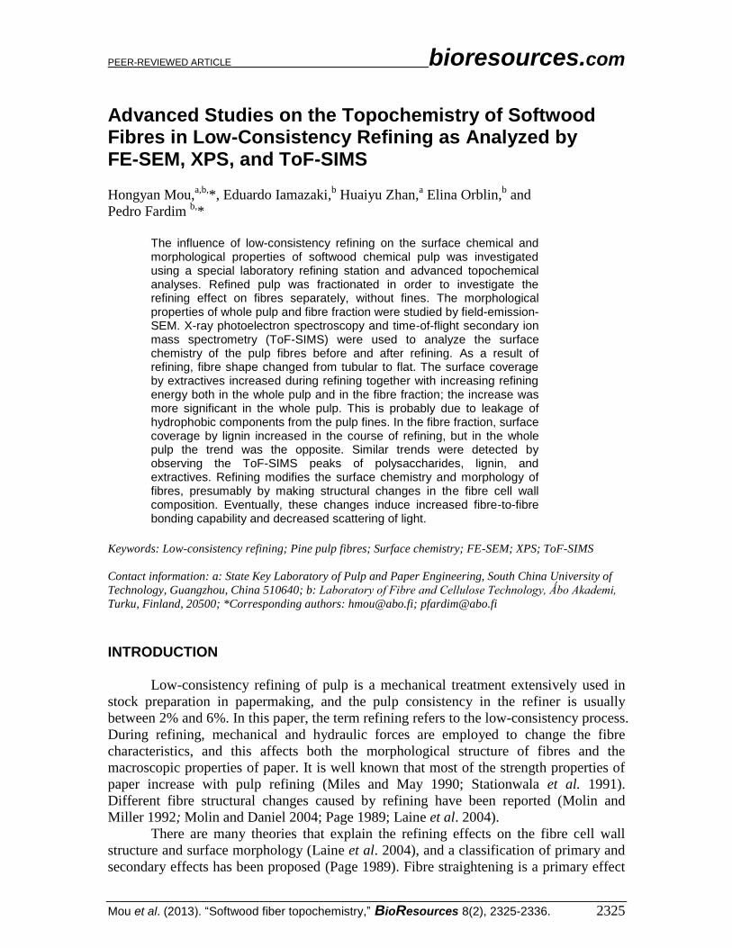
used in refining experiments in a ProLabTM
laboratory station (Metso Paper, see Fig. 1).
Technical details of the refining station can be found in Lundin et al. (2008).
Fig. 1. ProLab
TM refiner
Methods The pine pulp was refined at a specific edge load of 2 J/m and a consistency of
5%. The specific energy consumption (SEC) levels were 0, 75, 150, and 250 kWh/t,
respectively, with a long medium conical filling. Two series of samples were made:
refined whole pulps that included pulp fines, and fibre fraction samples where the pulp
fines were removed after refining. The separation was done with a dynamic drainage jar,
equipped with a 200-mesh wire and propeller stirring (TAPPI T261cm00). Pulp fines
mean the fraction passing a 200-mesh screen. SR was measured after each refining level
(ISO5267-1 1999). Fibre length and fines content were measured by Kajaani Fibrelab.
Handsheets were prepared in a Rapid Köthen apparatus using deionised water (ISO5269-
2 1988), and used for measuring optical properties (L&W Elrepho Spectrophotometer
Routine SE070R according to SCAN-CM 27:00) and for surface chemical analyses
without any further sample preparation.
The samples were identified as 13-P, 19-P, 37-P, and 72-P for the whole pulp
sheets, and 13-F, 19-F, 37-F, and 72-F for the fibre fraction sheets, according to the SR
values produced by the applied SEC levels (0, 75, 150, and 250 kWh/t, respectively). FE-
SEM images were obtained using a JEOL JSMT 300 microscope, operated in secondary
electron mode at a beam current of 100 μA and accelerating voltage of 20 kV. Samples
were previously coated with Pt for 20 s with an Agar scientific sputter coating system
equipped with a rotating base. Images were obtained in magnifications of 500×, 2000×,
and 20,000×.
XPS spectra of the whole pulp and the fibre fraction sample surfaces were
obtained with a Physical Electronics PHI2000 ESCA instrument equipped with a
monochromatic Al Kα X-ray source, operated at 200 W, and charge compensation. The
analysis area was 1 mm2, and the take-off angle was 45° relative to the sample surface. At
least three different spots were measured on each sample. Low resolution scanning was
done using the pass energy of 187 eV in 3 min, and the high resolution C1s scanning was
PEER-REVIEWED ARTICLE bioresources.com
Mou et al. (2013). “Softwood fiber topochemistry,” BioResources 8(2), 2325-2336. 2328
conducted using the pass energy of 23 eV in 5 min. A curve fitting program provided by
the instrument manufacturer was used to obtain the oxygen-to-carbon ratios (O/C) of the
samples and to interpret the C1s signal carrying information about the chemical binding
state of carbon. The following binding energies, relative to C-C (or C-H) position were
employed for the respective groups: (1.7±0.2) eV for C-O, (3.1±0.3) eV for C=O or O-C-
O, and (4.6±0.3) eV for O=C-O groups. The aliphatic carbon (C-C or C-H) is denoted
C1, and the other states are denoted C2, C3, and C4, in the order of the increasing binding
energies.
Surface coverage by lignin (Slig) and extractives (Sext) were determined using the
O/C ratio of the untreated and acetone-extracted (Soxhlet, over-night) samples using the
equations by Ström and Carlsson (1992), where the O/C ratio of the sample is compared
to the theoretical O/C ratios of pure cellulose (0.83) and of a model extractive (0.11).
Secondary ion mass spectra were obtained using a Phi Trift II ToF-SIMS
instrument (Physical Electronics, USA) using a primary beam of 69
Ga liquid metal ion
source and charge compensation by electron flood. Spectra were acquired for 3 min using
a 25 kV acceleration voltage under static conditions. At least three different spots were
analyzed on each sample. The peak intensities of the characteristic peaks from hexosans
(cellulose and glucomannans), pentosans (xylan), lignin, and various wood extractives in
the samples were compared. The characteristic fragments were chosen according to
previously published literature (Fardim and Durán 2000; Kleen 2000; Kangas 2007). In
order to discount analytical variations, the peak counts were first normalised, dividing by
the total counts of the spectrum being interpreted.
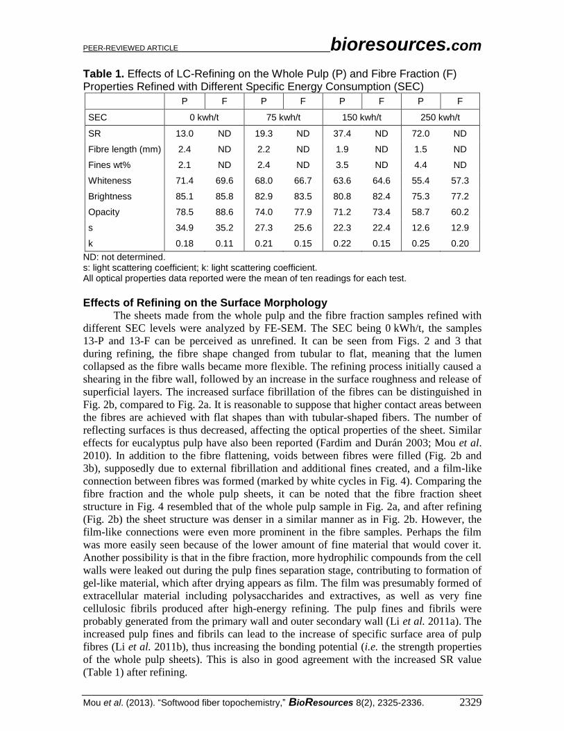
RESULTS AND DISCUSSION Effects of Refining on Pulp and Fibre Properties The effect of refining on the optical properties of the pulp and the fibre samples
was investigated. The whiteness, the brightness, the light scattering coefficient (s), and
the light absorption coefficient (k) in relation to SEC are shown in Table 1. As can be
seen, both for the whole pulp samples and the fibre fraction samples, the whiteness, the
brightness, opacity, and the light scattering coefficient were decreased at high refining
power, while the light absorption coefficient slightly increased. Decrease of brightness
after refining has also been reported for hardwood of eucalyptus (Fardim and Durán
2000).
It is well known that pulp drainage is reduced by refining due to creation of fines,
fibre fibrillation, and delamination, as shown by a fibre length reduction and a pulp fines
weight percentage increase in Table 1. This reduction in the drainage rate is one major
drawback of the refining process.
The optical properties of the whole pulp sheets did not obviously differ from
those of the fibre fraction sheets at the same SEC level, but the fibre fraction maintained
the optical properties in harsh refining (high SEC level) better than the whole pulp. The
brightness of the fibre fraction was not reduced as much as that of the whole pulp.
The role of the fine material in the optical properties of paper is known to be
related to the high specific surface area as well as the eventual lignin content (Asikainen
et al. 2010), whereas the fibres' contribution can be speculated to be mainly due to the
properties of the fibre cell wall, that is, the light reflecting surfaces of the external cell
wall layer.
PEER-REVIEWED ARTICLE bioresources.com
Mou et al. (2013). “Softwood fiber topochemistry,” BioResources 8(2), 2325-2336. 2329
Table 1. Effects of LC-Refining on the Whole Pulp (P) and Fibre Fraction (F) Properties Refined with Different Specific Energy Consumption (SEC)
P F P F P F P F
SEC 0 kwh/t 75 kwh/t 150 kwh/t 250 kwh/t
SR 13.0 ND 19.3 ND 37.4 ND 72.0 ND
Fibre length (mm) 2.4 ND 2.2 ND 1.9 ND 1.5 ND
Fines wt% 2.1 ND 2.4 ND 3.5 ND 4.4 ND
Whiteness 71.4 69.6 68.0 66.7 63.6 64.6 55.4 57.3
Brightness 85.1 85.8 82.9 83.5 80.8 82.4 75.3 77.2
Opacity 78.5 88.6 74.0 77.9 71.2 73.4 58.7 60.2
s 34.9 35.2 27.3 25.6 22.3 22.4 12.6 12.9
k 0.18 0.11 0.21 0.15 0.22 0.15 0.25 0.20
ND: not determined. s: light scattering coefficient; k: light scattering coefficient. All optical properties data reported were the mean of ten readings for each test.
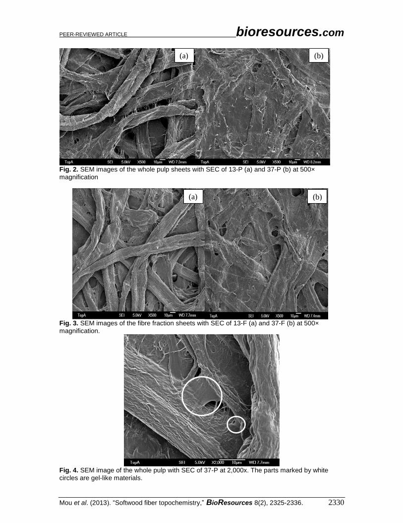
Effects of Refining on the Surface Morphology The sheets made from the whole pulp and the fibre fraction samples refined with
different SEC levels were analyzed by FE-SEM. The SEC being 0 kWh/t, the samples
13-P and 13-F can be perceived as unrefined. It can be seen from Figs. 2 and 3 that
during refining, the fibre shape changed from tubular to flat, meaning that the lumen
collapsed as the fibre walls became more flexible. The refining process initially caused a
shearing in the fibre wall, followed by an increase in the surface roughness and release of
superficial layers. The increased surface fibrillation of the fibres can be distinguished in
Fig. 2b, compared to Fig. 2a. It is reasonable to suppose that higher contact areas between
the fibres are achieved with flat shapes than with tubular-shaped fibers. The number of
reflecting surfaces is thus decreased, affecting the optical properties of the sheet. Similar
effects for eucalyptus pulp have also been reported (Fardim and Durán 2003; Mou et al.
2010). In addition to the fibre flattening, voids between fibres were filled (Fig. 2b and
3b), supposedly due to external fibrillation and additional fines created, and a film-like
connection between fibres was formed (marked by white cycles in Fig. 4). Comparing the
fibre fraction and the whole pulp sheets, it can be noted that the fibre fraction sheet
structure in Fig. 4 resembled that of the whole pulp sample in Fig. 2a, and after refining
(Fig. 2b) the sheet structure was denser in a similar manner as in Fig. 2b. However, the
film-like connections were even more prominent in the fibre samples. Perhaps the film
was more easily seen because of the lower amount of fine material that would cover it.
Another possibility is that in the fibre fraction, more hydrophilic compounds from the cell
walls were leaked out during the pulp fines separation stage, contributing to formation of
gel-like material, which after drying appears as film. The film was presumably formed of
extracellular material including polysaccharides and extractives, as well as very fine
cellulosic fibrils produced after high-energy refining. The pulp fines and fibrils were
probably generated from the primary wall and outer secondary wall (Li et al. 2011a). The
increased pulp fines and fibrils can lead to the increase of specific surface area of pulp
fibres (Li et al. 2011b), thus increasing the bonding potential (i.e. the strength properties
of the whole pulp sheets). This is also in good agreement with the increased SR value
(Table 1) after refining.
PEER-REVIEWED ARTICLE bioresources.com
Mou et al. (2013). “Softwood fiber topochemistry,” BioResources 8(2), 2325-2336. 2330
Fig. 2. SEM images of the whole pulp sheets with SEC of 13-P (a) and 37-P (b) at 500× magnification
Fig. 3. SEM images of the fibre fraction sheets with SEC of 13-F (a) and 37-F (b) at 500× magnification.
Fig. 4. SEM image of the whole pulp with SEC of 37-P at 2,000x. The parts marked by white circles are gel-like materials.
(a) (b)
(b) (a)
PEER-REVIEWED ARTICLE bioresources.com
Mou et al. (2013). “Softwood fiber topochemistry,” BioResources 8(2), 2325-2336. 2331
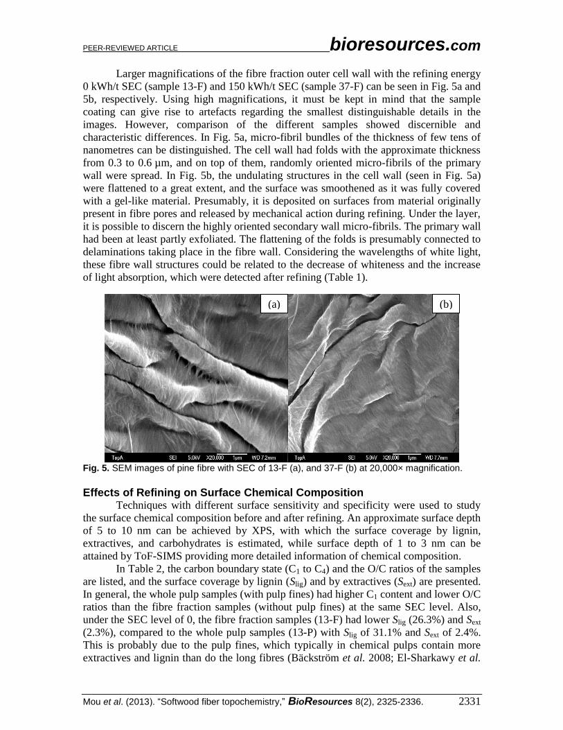
Larger magnifications of the fibre fraction outer cell wall with the refining energy
0 kWh/t SEC (sample 13-F) and 150 kWh/t SEC (sample 37-F) can be seen in Fig. 5a and
5b, respectively. Using high magnifications, it must be kept in mind that the sample
coating can give rise to artefacts regarding the smallest distinguishable details in the
images. However, comparison of the different samples showed discernible and
characteristic differences. In Fig. 5a, micro-fibril bundles of the thickness of few tens of
nanometres can be distinguished. The cell wall had folds with the approximate thickness
from 0.3 to 0.6 µm, and on top of them, randomly oriented micro-fibrils of the primary
wall were spread. In Fig. 5b, the undulating structures in the cell wall (seen in Fig. 5a)
were flattened to a great extent, and the surface was smoothened as it was fully covered
with a gel-like material. Presumably, it is deposited on surfaces from material originally
present in fibre pores and released by mechanical action during refining. Under the layer,
it is possible to discern the highly oriented secondary wall micro-fibrils. The primary wall
had been at least partly exfoliated. The flattening of the folds is presumably connected to
delaminations taking place in the fibre wall. Considering the wavelengths of white light,
these fibre wall structures could be related to the decrease of whiteness and the increase
of light absorption, which were detected after refining (Table 1).
Fig. 5. SEM images of pine fibre with SEC of 13-F (a), and 37-F (b) at 20,000× magnification.
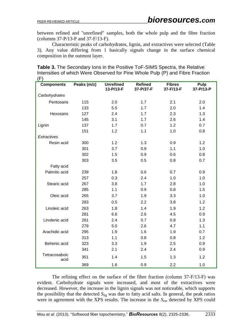
Effects of Refining on Surface Chemical Composition Techniques with different surface sensitivity and specificity were used to study
the surface chemical composition before and after refining. An approximate surface depth
of 5 to 10 nm can be achieved by XPS, with which the surface coverage by lignin,
extractives, and carbohydrates is estimated, while surface depth of 1 to 3 nm can be
attained by ToF-SIMS providing more detailed information of chemical composition.
In Table 2, the carbon boundary state (C1 to C4) and the O/C ratios of the samples
are listed, and the surface coverage by lignin (Slig) and by extractives (Sext) are presented.
In general, the whole pulp samples (with pulp fines) had higher C1 content and lower O/C
ratios than the fibre fraction samples (without pulp fines) at the same SEC level. Also,
under the SEC level of 0, the fibre fraction samples (13-F) had lower Slig (26.3%) and Sext
(2.3%), compared to the whole pulp samples (13-P) with Slig of 31.1% and Sext of 2.4%.
This is probably due to the pulp fines, which typically in chemical pulps contain more
extractives and lignin than do the long fibres (Bäckström et al. 2008; El-Sharkawy et al.
(a) (b)
PEER-REVIEWED ARTICLE bioresources.com
Mou et al. (2013). “Softwood fiber topochemistry,” BioResources 8(2), 2325-2336. 2332
2008). Thus, a higher O/C ratio of the fibre fraction samples is probably because of the
removal of pulp fines.
Table 2. Effects of Refining on Surface Chemical Composition of Pine as Analyzed by XPS
SR-Sample C1% C2% C3% C4% O/C Slig% Sext%
13-F 18.3 60.4 19.8 1.5 69.6 26.3 2.3 19-F 21.3 58.1 18.1 2.4 67.3 27.4 2.8 37-F 19.8 60.1 18.2 1.9 67.9 28.6 3.3 72-F 19.9 59.8 18.5 1.8 66.1 33.4 5.2 13-P 20.2 66.6 11.3 2.1 67.3 31.1 2.4 19-P 22.4 63.6 11.4 2.7 63.8 27.3 9.2 37-P 20.9 62.4 13.6 3.1 64.1 25.3 10.3 72-P 22.9 62.4 12.2 2.5 65.8 23.5 10.5
13-F, 19-F, 37-F, 72-F of fibre fraction samples, and 13-P, 19-P, 37-P, 72-P of whole pulp samples were with the SEC of 0, 75, 150, and 250 kWh/t, respectively.
Table 2 also shows that Slig of the fibre fraction samples increased with the
increase of SEC level, whereas the Slig of the whole pulp samples decreased when the
SEC level was increased. A decrease in Slig was also previously reported for eucalyptus
kraft pulp during refining (Fardim and Durán 2003). It has also been suggested that
hemicelluloses, particularly xylan, arabinan, and galactan, are deposited on fibre surfaces
during kraft pulping, as they have been detected clearly enriched in the primary wall of
pine kraft pulp, compared to the outer secondary wall (Kibblewhite and Brookes 1976).
The pulp fines generated from primary wall with the increase of SEC level presumably
contain deposited hemicelluloses. The opposite phenomenon observed in the fibre
fraction could be explained by the creation of fibrils, which involves the outer layers of
cell walls. Even if the lignin-rich middle lamellae are assumed to be practically all
dissolved in a bleached chemical pulp, removal of deposited hemicelluloses in refining
and subsequent fractionation stage could reveal lignin remains. Another possibility is that
the detected carbon content perceived as lignin with this technique is comprised of fatty
acid salts that were not removed in acetone extraction and therefore appear as lignin
(Fardim and Durán 2003). In addition, with the increasing of the SEC level, the Sext
increased for both of fibre fraction and whole pulp samples. Probably this is due to the
fact that hydrophobic compounds that are released from the fibre wall during refining
become distributed and re-deposit on the cellulosic surfaces. More extractives may re-
deposit on fines, as fines have larger specific surface area. Thus, Sext of pulp samples
increased obviously with increasing SEC, as presented in Table 2. However, the Sext
increases observed in the case of the fibre fraction samples is possibly due to the
deposition of extractives on the surface of fibrils, as pulp fines were already removed
after refining.
The ToF-SIMS spectra can give more detailed information on the chemical
composition despite the limitations in quantitative application of the technique. In order
to compare the relative contents of components on the surfaces, the relative peak
intensities were considered. Ratios between the normalised peaks of the whole pulp and
the fibre fraction samples with the SEC 0 kWh/t (column 13-P/13-F), as well as ratios
between the refined the whole pulp (SEC 150 kWh/t) and fibre fraction samples (column
37-P/37-F) were worked out. Further, the refining effect was studied inspecting the ratios
PEER-REVIEWED ARTICLE bioresources.com
Mou et al. (2013). “Softwood fiber topochemistry,” BioResources 8(2), 2325-2336. 2333
between refined and "unrefined" samples, both the whole pulp and the fibre fraction
(columns 37-P/13-P and 37-F/13-F).
Characteristic peaks of carbohydrates, lignin, and extractives were selected (Table
3). Any value differing from 1 basically signals change in the surface chemical
composition in the outmost layer.
Table 3. The Secondary Ions in the Positive ToF-SIMS Spectra, the Relative Intensities of which Were Observed for Pine Whole Pulp (P) and Fibre Fraction (F)
Components Peaks (m/z) Unrefined Refined Fibres Pulp 13-P/13-F 37-P/37-F 37-F/13-F 37-P/13-P
Carbohydrates
Pentosans 115 2.0 1.7 2.1 2.0
133 5.5 1.7 2.0 1.4
Hexosans 127 2.4 1.7 2.3 1.3
145 3.1 1.7 2.6 1.4
Lignin 137 1.7 0.7 1.2 0.7
151 1.2 1.1 1.0 0.8
Extractives
Resin acid 300 1.2 1.3 0.9 1.2
301 3.7 0.9 1.1 1.0
302 1.5 0.9 0.6 0.8
303 3.5 0.5 0.8 0.7
Fatty acid
Palmitic acid 239 1.8 0.6 0.7 0.9
257 0.3 2.4 1.0 1.0
Stearic acid 267 3.8 1.7 2.8 1.0
285 1.1 0.9 0.8 1.5
Oleic acid 265 3.7 1.9 3.3 1.0
283 0.5 2.2 3.8 1.2
Linoleic acid 263 1.8 1.4 1.9 1.2
281 6.6 2.6 4.5 0.9
Linolenic acid 261 2.4 0.7 0.8 1.3
279 5.0 2.6 4.7 1.1
Arachidic acid 295 1.9 1.6 1.9 0.7
313 1.1 0.8 0.8 1.2
Behenic acid 323 3.3 1.9 2.5 0.9
341 2.1 2.4 2.4 0.9
Tetracosaboic acid
351 1.4 1.5 1.3 1.2
369 1.6 0.9 2.2 1.0
The refining effect on the surface of the fibre fraction (column 37-F/13-F) was
evident. Carbohydrate signals were increased, and most of the extractives were
decreased. However, the increase in the lignin signals was not noticeable, which supports
the possibility that the detected Slig was due to fatty acid salts. In general, the peak ratios
were in agreement with the XPS results. The increase in the Sext detected by XPS could
PEER-REVIEWED ARTICLE bioresources.com
Mou et al. (2013). “Softwood fiber topochemistry,” BioResources 8(2), 2325-2336. 2334
be due to free fatty acids, mainly oleic, linoleic, and arachidic acid. Also for the whole
pulp (column 37 P/13 P), the changes after refining as detected by XPS were in good
correspondence with ToF-SIMS peak ratios. However, the remarkable increase of Sext
detected by XPS was not accompanied by any clear increase in the extractive peak
intensities. The detection depth of ToF-SIMS is restricted on the outmost monolayer,
whereas XPS collects information slightly deeper, and a difference in the component
distribution is plausible. Further, the signal intensities in ToF-SIMS depend not only on
the component content of interest, but also on various electronic, physical, and chemical
states during the measurement.
Mechanical action during refining clearly contributes to the release of components
entrapped in fibre wall pores to the external liquid phase, as previously suggested
(Fardim and Durán 2003). Xylan (pentosan) was likely re-adsorbed rather than exposed
on the surface during refining, based on the difference between the surface carbohydrate
contents evaluated with ToF-SIMS. It seems reasonable to suppose that cellulose
(hexosan) was exposed by a peeling action on the fibre surfaces and that part of the xylan
was adsorbed after being released from the cell wall pores in a similar way to the fatty
acids. Hydrogen or dispersion bonds are possibly the driving forces for the adhesion of
xylan and extractive aggregates onto external fibre surfaces. Fibre chemistry has been
shown to affect paper strength properties (Fardim and Duran 2005; Sundberg et al. 2000),
and thus the chemical changes along with the morphological modifications caused by
refining are assumed to be of importance in papermaking.
CONCLUSIONS
1. The effect of low consistency refining on the morphology and the surface chemical
composition of pine pulp fibres was investigated, and significant changes were
detected. With refining, fibre shape changed from tubular to flat, and external fibril-
lation was introduced.
2. Refining increased the surface coverage by extractives, mainly fatty acids. The results
from XPS and ToF-SIMS were in good agreement with respect to the surface
chemical composition. Evidence supporting the release of xylan and its subsequent
adherence to the fibre surfaces was found.
3. Refining affects not only the morphology but also the surface chemical composition
of the whole pulp in a way that possibly has significance for the final paper properties.
ACKNOWLEDGMENTS
This project was financially supported by Kemira. We would also like to thank
Top Analytica Ltd. for providing surface analytical instruments. We thank our colleague
Goran Kuzmanovski for his kind help in the work, as well as Dr. Joakim Järnström and
Dr. Bin Li for help to improve this manuscript.
PEER-REVIEWED ARTICLE bioresources.com
Mou et al. (2013). “Softwood fiber topochemistry,” BioResources 8(2), 2325-2336. 2335
REFERENCES CITED
Asikainen, S., Fuhrmann, A., Ranua, M., and Robertsen, L. (2010). “Effect of birch kraft
pulp primary fines on bleaching and sheet properties,” BioResources 5(4), 2173-2183.
Bäckström, M., Kolar, M.-C., and Htun, M. (2008). “Characterisation of fines from
unbleached kraft pulps and their impact on sheet properties,” Holzforschung 62, 546-
552.
Baker, C. F. (2003) “Refining and improved paper machine runnability,” In: Proceedings
of 7th
PIRA International Refining Conference & Exhibition, 25-26 March,
Stockholm, Sweden, March 25-26, paper 12, 2003.
Berthold, J. and Salmén, L. (1997). “Effects of mechanical and chemical treatments on
the pore-size distribution in wood pulps examined by inverse size-exclusion
chromatography,” J. Pulp Pap. Sci. 23, 245-253.
Brinen, J. S. (1993). “The observation and distribution of organic additives on paper
surfaces using surface spectroscopic techniques,” Nord. Pulp Pap. Res. J. 8, 123-129.
Buchanan, J. G., and Washburn, O. V. (1962). “The surface and tensile fractures of
chemical fiber handsheets as observed with the scanning electron microscope,” Pulp.
Paper Mag. Can. 63, T485-T493.
Clark, J. d’A. (1957). “The evaluation of beating equipment,” Tappi J. 40, 548-552.
El-Sharkawy, K., Koskenhely, K., and Paulapuro, H. (2008). “The fractionation and
refining of eucalyptus kraft pulps,” Nord. Pulp Pap. Res. J. 23(2), 172-180.
Enomae, T., and Lepoutre, P. (1998). “Observation of the swelling behavior of kraft
fibers and sheets in the environmental scanning electron microscope,” Nord. Pulp
Pap. Res. J. 13(4), 280-284.
Fardim, P., and Durán, N. (2003). “Modification of fibre surfaces during pulping and
refining as analysed by SEM, XPS, and ToF-SIMS,” Colloids Surf. A 223, 263-276.
Fardim, P., and Durán, N. (2005). “Influences of surface chemical composition on the
mechanical properties of pulp as investigated by SEM, XPS, and multivariate data
analysis,” J. Braz. Chem. Soc. 16(2), 163-170.
Fardim, P., and Durán, N. (2000). “Surface characterisation of unbleached eucalyptus
kraft pulp using XPS and ToF-SIMS,” Proc. 6th
European Workshop Lignocellulosics
Pulp, Bordeaux, September 3-6, p. 307.
Fardim, P., Gustafsson, J., and Schoultz, S. (2005) “Extractives on fiber surfaces
investigated by XPS, ToF-SIMS, and AFM,” Colloids Surf. A 255, 91-103.
Kangas, H. (2007). “Surface chemical and morphological properties of mechanical pulps,
fibres and fines,” Doctoral thesis, 1.12. Helsinki University of Technology, Finland.
Kibblewhite, R. P., and Brookes, D. (1976). “Distribution of chemical components in the
walls of kraft and bisulfite pulp fibers,” Wood Sci. Technol. 10, 39-46.
Kleen, M. (2000) “Surface chemistry of kraft pulp fibres during TCF bleaching studied
by ToF-SIMS,” Proc. 6th
European Workshop Lignocellulosics Pulp, Bordeaux,
September 3-6, p. 41-44.
Laine, C., Wang, X., Tenkanen, M., and Varhimo, A. (2004). “Changes in the fibre wall
during refining of bleached pine kraft pulp,” Holzforschung 58(3), 233-240.
Li, B., Bandekar, R., Zha, Q., Alsaggaf, A., and Ni, Y. (2011a). “Fiber quality analysis:
OpTest fiber quality analyser versus L&W Fiber Tester,” Ind. Eng. Chem. Res.
50(22), 12572-12578.
PEER-REVIEWED ARTICLE bioresources.com
Mou et al. (2013). “Softwood fiber topochemistry,” BioResources 8(2), 2325-2336. 2336
Li, B., Li, H., Zha, Q., Bandekar, R., Alsaggaf, A., and Ni, Y. (2011b). “Review: Effects
of wood quality and refining process on TMP pulp and paper quality,” BioResources
6(3), 3569-3584.
Lundin, T., Batchelor, W., and Fardim, P. (2008). “Fiber trapping in low-consistency
refining: New parameters to describe the refining process,” Tappi J. 7(7), 15-21.
Miles, K. B., and May, W. D. (1990). “The flow of pulp in chip refiners,” J. Pulp Pap.
Sci. 16(2), 63-72.
Molin, U.-B., and Daniel, G. (2004). “Effects of refining on the fibre structure of kraft
pulps as revealed by FE-SEM and TEM: Influence of alkaline degradation,”
Holzforschung 58(3), 226-232.
Molin, U.-B., and Miller, J. (1992). “Influence of industrial beating on fibre swelling and
fibre shape,” AICELCA, May 19-22, Bologna, Italy, 1992.
Mou, H., Zhan, H., Iamazaki, E., and Fardim, P. (2010). “Effects of LC-refining on fiber
surface chemistry of eucalyptus,” Nov. 8th
, 4th
ISETPP, Guangzhou.
Orblin, E., and Fardim, P. (2010), “Surface chemistry of deinked pulps as analysed by
XPS and ToF-SIMS,” Surf. Interface Anal. 42, 1712-1722.
Orblin, E., Eta, V., and Fardim, P. (2011). “Surface chemistry of vessel elements by FE-
SEM, μ-XPS and ToF-SIMS,” Holzforschung 65(5), 681-688.
Page, D. H. (1989). “The beating of chemical pulps - the action and the effects,” 9th
Fundamental Research Symposium, London,Vol. 1, p.1-38.
Stationwala, M. I., Atack, D., Wood, J. R., Wild, D. J., and Karnis, A. (1991). “The effect
of control variables on refining zone conditions and pulp properties,” Pap. Puu 73(1),
62-69.
Ström, G., and Carlsson, G. (1992). “Wettability of kraft pulps: Effect of surface
composition and oxygen plasma treatment,” J. Adhesion Sci. Technol. 6, 745-761.
Sundberg, A., Holmbom, B., Willför, S., and Pranovich, A. (2000). “Weakening of paper
strength by wood resin,” Nord. Pulp Pap. Res. J. 15(1), 46-53.
Touzinsky, G., Baker, F., Cunningham, R., and Bagby, M. (1977). “Scanning electron
microscopy of kenaf paper structures,” J. Agric. Food Chem. 25, 734-738.
Article submitted: November 22, 2012; Peer review completed: January 11, 2013;
Revised version received and accepted: March 5, 2013; Published: March 21, 2013.