adsorption of components of the plasma kinin-forming system on the surface of porphyromonas
TRANSCRIPT

INFECTION AND IMMUNITY, Feb. 2011, p. 797–805 Vol. 79, No. 20019-9567/11/$12.00 doi:10.1128/IAI.00966-10Copyright © 2011, American Society for Microbiology. All Rights Reserved.
Adsorption of Components of the Plasma Kinin-Forming System onthe Surface of Porphyromonas gingivalis Involves Gingipains as
the Major Docking Platforms�
Maria Rapala-Kozik,1 Grazyna Bras,1 Barbara Chruscicka,1 Justyna Karkowska-Kuleta,1 Aneta Sroka,2Heiko Herwald,3 Ky-Anh Nguyen,4,5 Sigrun Eick,6 Jan Potempa,2,7 and Andrzej Kozik1*
Department of Analytical Biochemistry1 and Department of Microbiology,2 Faculty of Biochemistry, Biophysics and Biotechnology,Jagiellonian University, Krakow, Poland; Department of Clinical Sciences, Division of Infection Medicine, Biomedical Center (BMC),
Lund University, Lund, Sweden3; Institute of Dental Research, Westmead Centre for Oral Health, Sydney, Australia4;Faculty of Dentistry, University of Sydney, Sydney, New South Wales, Australia5; Department of Periodontology,
Laboratory of Oral Microbiology, School of Dentistry, University of Bern, Bern, Switzerland6; and Oral Health andSystemic Disease Research Facility, University of Louisville School of Dentistry, Louisville, Kentucky7
Received 5 September 2010/Returned for modification 6 October 2010/Accepted 15 November 2010
Enhanced production of proinflammatory bradykinin-related peptides, the kinins, has been suggested tocontribute to the pathogenesis of periodontitis, a common inflammatory disease of human gingival tissues. Inthis report, we describe a plausible mechanism of activation of the kinin-generating system, also known as thecontact system or kininogen-kallikrein-kinin system, by the adsorption of its plasma-derived components suchas high-molecular-mass kininogen (HK), prekallikrein (PK), and Hageman factor (FXII) to the cell surface ofperiodontal pathogen Porphyromonas gingivalis. The adsorption characteristics of mutant strains deficient inselected proteins of the cell envelope suggested that the surface-associated cysteine proteinases, gingipains,bearing hemagglutinin/adhesin domains (RgpA and Kgp) serve as the major platforms for HK and FXIIadhesion. These interactions were confirmed by direct binding tests using microplate-immobilized gingipainsand biotinylated contact factors. Other bacterial cell surface components such as fimbriae and lipopolysac-charide were also found to contribute to the binding of contact factors, particularly PK. Analysis of kininrelease in plasma upon contact with P. gingivalis showed that the bacterial surface-dependent mechanism iscomplementary to the previously described kinin generation system dependent on HK and PK proteolyticactivation by the gingipains. We also found that several P. gingivalis clinical isolates differed in the relativesignificance of these two mechanisms of kinin production. Taken together, these data show the importance ofthis specific type of bacterial surface-host homeostatic system interaction in periodontal infections.
Periodontitis is a common infectious disease in humans,characterized by gingival inflammation, followed by loss ofconnective tissue and bone around the root of the tooth, whichmay eventually lead to its exfoliation (59). The progression ofperiodontal disease depends on the interactions between dif-ferent bacteria colonizing the gingival sulcus and the host im-mune system (46). Approximately 15% of the population isaffected by severe forms of this disease (27), and, moreover,the occurrence of periodontal infection is often implicated as arisk factor for other pathological conditions, including cardio-vascular diseases, adverse pregnancy outcomes, preterm lowbirth weight, diabetes, and rheumatoid arthritis (82, 84).
Porphyromonas gingivalis, an anaerobic black-pigmentedGram-negative bacterium, is found predominantly in the peri-odontal pocket, and its presence in large numbers is stronglycorrelated with periodontal destruction (80). Arguably, cysteineendopeptidases, referred to as gingipains, are the main virulencefactors of P. gingivalis (20). They not only directly neutralize theantibacterial activity of the innate immune system by degrading
defensins (14) and corrupting the complement system (40), butthe gingipains also significantly contribute to the pathogenicity ofP. gingivalis via (i) usurping control of local proteolysis (35), (ii)scavenging of iron and heme (44), (iii) stimulating expression ofproinflammatory cytokines by host cells (18), and (iv) facilitatingbacterial adherence to extracellular matrix proteins (63, 39) andhost cells (3, 15) through their hemagglutinin/adhesin (HA) do-mains (HA1 to HA4) (67).
Apart from surface-bound gingipains, a variety of other bacte-rial surface proteins mediate the adhesion of P. gingivalis to hosttissue constituents, with fimbriae and hemagglutinins being themajor adhesins (17, 21). Fimbriae, the filamentous structures lo-calized to the P. gingivalis cell surface, have been shown to interactwith fibronectin (51) and to adhere to epithelial and endothelialcells and monocytes, subsequently triggering inflammation andexpression of cell adhesion mediators by these host cells (2). TheP. gingivalis hemagglutinins enable the bacterium to bind to hosterythrocytes to access the heme/iron within (42, 58).
The host kinin-forming cascade is one of the major homeo-static systems involved in the regulation of many physiologicalprocesses. The cascade also plays an important role in the inflam-matory process (for selected reviews, see references 16, 33, and36). As a part of host defense against microbial infection, thesystem is often overactivated (25). Kinins, including bradykininand a few related peptides, are produced in large amounts at local
* Corresponding author. Mailing address: Department of AnalyticalBiochemistry, Faculty of Biochemistry, Biophysics, and Biotechnology,Jagiellonian University, Gronostajowa 7, 30-387 Krakow, Poland.Phone: 4812 664-6525. Fax: 4812 664-6902. E-mail: [email protected].
� Published ahead of print on 22 November 2010.
797
Dow
nloa
ded
from
http
s://j
ourn
als.
asm
.org
/jour
nal/i
ai o
n 17
Dec
embe
r 20
21 b
y 16
0.23
8.75
.129
.
inflammatory foci to induce vasodilation, vascular permeabilityenhancement, cell migration, and pain (13). A major pathway forkinin production at these locations depends on the adsorption(the contact system) and activation of the Hageman factor (FXII)on the surfaces of endothelium and other host cells (5, 6, 23, 34,56). Within a ternary complex assembled on the cell surface, theactivated FXII converts plasma prekallikrein (PK) into the activeenzyme which subsequently releases bradykinin from high-molec-ular-mass kininogen (HK).
The kinin-generating pathway can be hijacked by severalbacterial and fungal pathogens which take advantage of thekinin-induced increase in vascular permeability for dissemina-tion and/or delivery of plasma-derived nutrients to the site ofinfection (19). These organisms produce proteases that caneither directly release kinins from kininogens (24, 31, 69) oractivate FXII and/or prekallikrein directly (49, 50, 73). In thecase of P. gingivalis, the arginine-specific gingipains (RgpA,RgpB) generate bradykinin via PK activation, while the lysine-specific gingipain (Kgp) releases kinin directly from its precur-sor (28, 30, 77). Although bacterial proteases have evolveddifferent mechanisms to evoke the release of kinins (64), theassembly of contact factors at the microbial surface can alsolead to activation of the contact system and the generation ofkinins (7, 8, 9, 37, 48, 61, 70).
In the current work, we characterized a previously unexploredadsorption of contact system proteins on the P. gingivalis cellsurface. Based on analysis of several P. gingivalis isogenic mutantsand purified proteins, we have concluded that hemagglutinin/adhesin domain-bearing forms of gingipains (RgpA and Kgp) inthe bacterial cell envelope constitute the major docking platformfor this adsorption and kinin production.
MATERIALS AND METHODS
Bacterial strains and growth conditions. The P. gingivalis strains listed inTable 1 (except strain 381-derived strains) and clinical isolates (Table 2) weregrown on an anaerobe blood agar plate (3% soy tryptone, 1.5% agar, 0.5% yeastextract, 0.05% L-cysteine, 0.01% dithiothreitol, 0.00005% vitamin K, 0.0005%hemin, 5% defibrinated sheep blood). The 381 strain and its Dpg3 mutant weregrown on a brain heart infusion agar plate (1.85% brain heart infusion, 1.5%agar, 0.5% yeast extract, 0.00005% vitamin K, 0.0005% hemin, 5% defibrinatedsheep blood). All strains were incubated for 7 days at 37°C in an anaerobicchamber (MACS500; Don Whitley Scientific Limited, Frederick, MD) in anatmosphere of 80% N2, 10% CO2, and 10% H2. Afterwards, bacteria wereinoculated into 40 ml of appropriate broths and grown overnight under the sameconditions. For growth selection of mutants on solid medium, 1 �g/ml tetracy-cline or 5 �g/ml erythromycin was used.
Creation of Sov and gingipain mutants. Deletional inactivation of Sov wasconstructed using a technique similar to that described previously (53, 54).Briefly, two 1-kb regions flanking the sov gene were amplified by PCR using twosets of primers (set 1, CTGAGCTCGCCTACAATCTCAAGCGTATGC andTTAACCCGGGCGTGATCCTTCTTTGTTTGAGA; set 2, ACTCTAGATAGCCTAACGGCATTACCCAC and ATTAGTCGACCTTGTATGCAGGAAGCAGGATAG) and inserted into pUC19 at SacI-SmaI and XbaI-SalI sites(boldface nucleotides), respectively, with an intervening tetQ antibiotic resistancecassette (54). The resultant plasmid was electroporated into P. gingivalis W83competent cells to facilitate homologous recombination into the genome, and trans-formants were selected on tetracycline-selective blood agar. The correct genotypewas confirmed with DNA sequencing of the pertinent region, and the mutantsdisplayed a phenotype that has been reported previously (71).
Creation of single gingipain mutants has been reported previously (53, 81).The RgpA-RgpB inactivation mutant was created by truncation of RgpB atamino acid position 410 to remove the Ig C-terminal domain (CTD) usingdeletional mutagenesis as previously described (53). To create the gingipain-nullmutant W83/K/RgpAB, the ermF/AM cassette of the plasmid construct pREC-Kgp�Ig/Cterm/HA (81) was swapped for a tetQ cassette (54) and transformedinto the RgpA-RgpB mutant described above. The pertinent regions of allmutants were sequenced to confirm the correct construction, and gingipainbiochemical assays were used to confirm the absence of gingipain activity.
Isolation of bacterial surface components. Fimbriae from the P. gingivalis 381strain were isolated by the method of Arai et al. (4), while the gingipains HRgpA,RgpB, and Kgp were purified from P. gingivalis strain HG66 as previouslydescribed (62, 65). Lipopolysaccharide (LPS) was obtained from the P. gingivalisW83 strain using phenol-water extraction according to the published procedure(83), and after dialysis against deionized water, the purity of the LPS preparationwas checked by SDS-PAGE and zinc-imidazole staining (22).
Biotinylation of kinin-generating proteins. Low-molecular-mass kininogen(LK), purified from human plasma according to the method of Johnson et al.(32), human HK, human plasma PK, and human FXII (all from Enzyme Re-search Laboratories, South Bend, IN) were incubated at 2 to 3 �M in 0.1 Mcarbonate buffer, pH 8.3 (total volume 500 �l), with biotin N-hydroxysuccinimideester solution in dimethylformamide (40 �g/4 �l) for 4 h at 4°C in the presenceof 2 �M aprotinin (for PK) or 0.1 mM phenylmethylsulfonyl fluoride (PMSF)(for FXII). After conjugation, the biotinylated (Bt) proteins (LK-Bt, HK-Bt,FXII-Bt, PK-Bt) were dialyzed overnight against phosphate-buffered saline(PBS) and stored frozen until use in binding experiments.
Binding of kinin-generating proteins to bacterial cell surface. P. gingivalis cellsfrom early-stationary-phase cultures (optical density, 0.6 to 0.8) were harvestedby centrifugation (30 min, 3,500 � g), washed three times with PBS, and resus-pended in PBS (108 cells per 200 �l) containing 1 mM N�-p-tosyl-L-lysinechloromethyl ketone (TLCK) for inactivation of the gingipains. Bacterial cellsuspensions were incubated for 1 h at 37°C with biotinylated kinin-generatingproteins at concentrations of 0 to 80 nM in Eppendorf tubes which had beenpretreated with 1% bovine serum albumin (BSA) overnight at 4°C to minimizenonspecific binding of biotinylated proteins to the plastic surface during thebinding experiments. After incubation, the cells were washed three times withPBS containing 0.1% BSA before bound biotinylated proteins were probed withhorseradish peroxidase (HRP)-streptavidin conjugate (1:4,000 dilution, 1 h atroom temperature). For HRP detection, 3,3�,5,5�-tetramethylbenzidine (TMB)was used as the substrate. After the reaction was stopped with 2 M HCl, theabsorbance at 450 nm was determined in a Power Wave X Select microplatereader (BioTek Instruments, Winooski, VT) and the amounts of bound biotin-ylated proteins were estimated from the appropriate standard curves preparedfor each of the analyzed proteins.
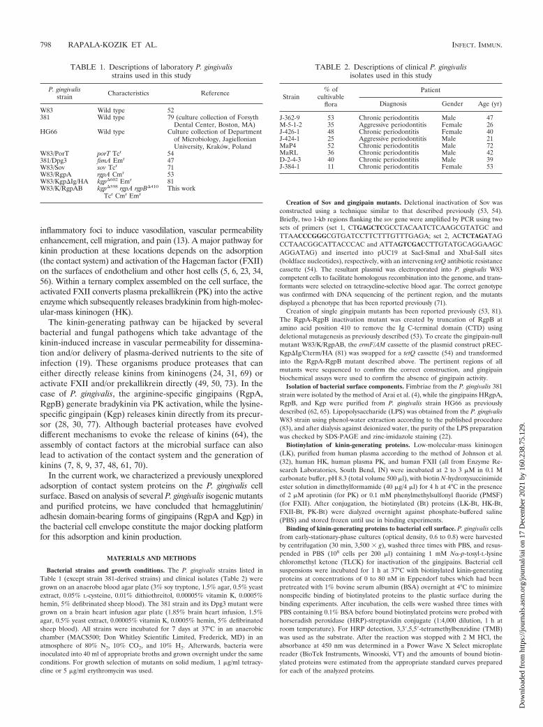
TABLE 1. Descriptions of laboratory P. gingivalisstrains used in this study
P. gingivalisstrain Characteristics Reference
W83 Wild type 52381 Wild type 79 (culture collection of Forsyth
Dental Center, Boston, MA)HG66 Wild type Culture collection of Department
of Microbiology, JagiellonianUniversity, Krakow, Poland
W83/PorT porT Tcr 54381/Dpg3 fimA Emr 47W83/Sov sov Tcr 71W83/RgpA rgpA Cmr 53W83/Kgp�Ig/HA kgp�602 Emr 81W83/K/RgpAB kgp�598 rgpA rgpB�410
Tcr Cmr EmrThis work
TABLE 2. Descriptions of clinical P. gingivalisisolates used in this study
Strain% of
cultivableflora
Patient
Diagnosis Gender Age (yr)
J-362-9 53 Chronic periodontitis Male 47M-5-1-2 35 Aggressive periodontitis Female 26J-426-1 48 Chronic periodontitis Female 40J-424-1 25 Aggressive periodontitis Male 21MaP4 52 Chronic periodontitis Male 72MaRL 36 Chronic periodontitis Male 42D-2-4-3 40 Chronic periodontitis Male 39J-384-1 11 Chronic periodontitis Female 53
798 RAPALA-KOZIK ET AL. INFECT. IMMUN.
Dow
nloa
ded
from
http
s://j
ourn
als.
asm
.org
/jour
nal/i
ai o
n 17
Dec
embe
r 20
21 b
y 16
0.23
8.75
.129
.

For the competition assay, cell suspensions (108 cells per 200 �l) were incu-bated with one of the biotinylated contact factors (3.8 pmol per tube) in thepresence of increasing amount of selected, nonbiotinylated protein (0 to 38 pmolper tube). The incubation and detection conditions were the same as thosedescribed above, except that 1 �M aprotinin in the samples containing PK and0.1 mM PMSF in the samples containing FXII were additionally applied toprevent the contact system activation.
Binding of purified bacterial surface components to contact system proteins.To test the binding of contact system proteins to purified components of the P.gingivalis cell surface, a ligand binding microplate assay was applied. The surfacecomponents—FimA (4 �g per well), the gingipains (6 pmol per well), or LPS (4�g per well)—were bound overnight at 4°C onto the wells of MaxiSorp micro-plates for enzyme-linked immunosorbent assays (ELISAs; Nunc, Roskilde, Den-mark) in PBS or in 0.1 M bicarbonate buffer, pH 9.6 (for LPS only). Wells werewashed three times with PBS and blocked for 2 h at 37°C with PBS containing3% BSA, followed by three washes with 1% BSA in PBS. The high concentrationof BSA was necessary to prevent the nonspecific binding of contact factors,especially of HK. Subsequently, solutions of biotinylated contact factors at con-centrations between 0 and 160 nM were added and the microplates were incu-bated for 1.5 h at 37°C. For experiments on gingipain binding, all solutionsadditionally contained 1 mM TLCK. Bound proteins were detected with HRP-streptavidin conjugate (1:4,000 dilution, 1 h at room temperature) and TMB asthe color-generating substrate.
Assay of gingipain activity on the surface of P. gingivalis W83 and clinicalstrains. The proteolytic activities of Rgp and Kgp on the surface of P. gingivalis cellswere determined by monitoring the hydrolysis of the chromogenic substrates ben-zoyl-L-arginine-p-nitroanilide (BApNA) and N-(p-tosyl)-Gly-Pro-Lys-p-nitroanilide(TGPKpNA), respectively, as previously described (65). Briefly, 108 cells were sus-pended in a Sarstedt microplate in 150 �l of assay buffer (200 mM Tris-HCl, 100 mMNaCl, 5 mM CaCl2, pH 7.6) supplemented with fresh 10 mM L-cysteine. Themicroplate was incubated at 37°C for 15 min prior to the addition of 50 �l 2 mMsubstrate in assay buffer. Formation of p-nitroanilide was measured as an increase inoptical density at 405 nm over a 30-min period using the Power Wave X Selectmicroplate reader. Each analysis was performed in triplicate.
Detection of kinin production from human plasma proteins adsorbed on P.gingivalis surface. PBS-washed P. gingivalis W83 cells (109 cells per analyzed sam-ple) were resuspended in 500 �l of 15 mM HEPES buffer, pH 7.0, containing 135mM NaCl and 50 �M ZnCl2 with or without 2 mM TLCK. Bacteria were then mixedwith 500 �l of human plasma and incubated at 37°C for 0 to 30 min. After incuba-tion, the suspensions were centrifuged (2,000 � g) for 5 min at 4°C and the super-natants were diluted 2-fold with 1% trifluoroacetic acid (TFA) in water. After anadditional centrifugation step, the supernatants were applied onto a C18 Sep column(Peninsula Laboratories) for extraction of the kinin peptides. Elution was performedwith 1% TFA in 60% acetonitrile. After lyophilization of the samples, the amount ofkinins liberated from human plasma was determined with an ELISA kit, accordingto the manufacturer’s instructions (Peninsula Laboratories).
Data analysis. All points in the figures represent the means � standarddeviations (SDs) from at least three independent experiments with duplicatemeasurements. For the analysis of both the saturable binding and the competi-tion plots, a one-site model was fitted using GraphPad Prism software.
RESULTS
As shown in Fig. 1, biotinylated human kininogens (LK andHK) and the zymogens of the human plasma kinin-generatingsystem (FXII and PK) were capable of tight binding to the cellsurface of the P. gingivalis W83 strain. The saturable bindinglevels were the highest for FXII, reaching a value of 2 pmol boundprotein per 108 P. gingivalis cells, followed by PK � HK � LK.Notably, LK binding reached saturation at a level as low as 0.5pmol, which is much less than the levels for the three contactfactors.
Unlabeled contact system proteins FXII, HK, and PK wereable to compete with their corresponding biotinylated tracersat the cell surface of the W83 strain with 95%, 80%, and 70%efficiencies, respectively (Fig. 2). Moreover, all tested proteinscould compete with each other to comparable extents, suggest-
FIG. 1. Binding of biotinylated contact factors and LK to the cellsurface of P. gingivalis wild-type strain W83. FXII, PK, HK, and LKwere incubated with 108 bacterial cells for 1 h at 37°C in PBS contain-ing 1 mM TLCK. Bound proteins at the bacterial surface were de-tected with streptavidin-HRP conjugate. Data represent the means ofthree experiments; error bars denote the SDs.
FIG. 2. Competition of biotinylated contact factors with the nativeproteins at the bacterial surface. The suspension of P. gingivalis W83(108 cells) was incubated with a mixture of biotinylated (3.8 pmol in a200-�l total volume) FXII (A), HK (B), or PK (C) and increasingconcentrations of nonbiotinylated competitors (HK, FXII, and PK).For the samples containing PK or FXII, the reaction mixture wassupplemented with the selective inhibitors 1 �M aprotinin and 0.1 mMPMSF, respectively. The binding was detected with a streptavidin-HRP/TMB system.
VOL. 79, 2011 BINDING OF KININ SYSTEM TO PORPHYROMONAS GINGIVALIS 799
Dow
nloa
ded
from
http
s://j
ourn
als.
asm
.org
/jour
nal/i
ai o
n 17
Dec
embe
r 20
21 b
y 16
0.23
8.75
.129
.

ing that they share most of the binding sites on the bacterialsurface.
Another P. gingivalis strain 381 bound contact factors to2-fold lower capacity (Fig. 3A) than the W83 strain. The iso-genic FimA-negative Dpg3 strain (47) was found to have abinding capacity for FXII comparable to that for wild-typestrain 381 (Fig. 3B). However, there was a 2-fold increasedbinding for HK in Dpg3 compared with that in 381, and thelevel of PK binding was reduced by half.
Our results show that FimA is one ligand for the contactfactors, but the data do not exclude a possibility that othersurface proteins might be involved. To address this experimen-tally, an array of isogenic W83-derived mutant strains lackinggingipains and other cell envelope proteins was employed (Ta-ble 1). These strains were tested in binding assays with biotin-ylated contact factors. Notably, the single rgpA and kgp mutantstrains showed reduced binding capacities for HK and FXII incomparison to those of the parental strain, and the binding wasalmost entirely abolished by the elimination of all gingipaingenes (Fig. 4A and B). The binding of HK was totally elimi-nated in the P. gingivalis mutants deficient in any of two essen-tial proteins (PorT and Sov) of the gingipain secretion system(54, 71). In contrast to HK and FXII, the lack of a singlegingipain, RgpA or Kgp, did not markedly affect the bindingcapacity for PK, and the triple-gingipain-knockout strain stillexhibited residual PK binding (Fig. 4C). In contrast to theSov-deficient strain, the PorT mutant strain showed reduced,but still significant, binding capacity for FXII and, to a lesserdegree, for PK (Fig. 4B and C, respectively). Taken together,the analysis of gingipain-deficient strains clearly indicated thatRgpA and Kgp are essential for adsorption of the contactsystem proteins on the P. gingivalis cell envelope.
In another series of experiments, the binding of contactfactors to FimA isolated from P. gingivalis 381 strain and im-mobilized to a microplate was tested (Fig. 5A). These resultsshow that all contact factors have an affinity for FimA withdecreasing binding affinity in the order of PK � FXII � HK.Microplate-immobilized LPS purified from P. gingivalis W83was shown to also bind PK (Fig. 5B) but only at half of the levelcompared with that of immobilized FimA. Similarly, the bind-ing of FXII and HK to LPS was weak, albeit detectable, sug-gesting that LPS does not significantly contribute to the ad-
FIG. 4. Binding of biotinylated contact factors to P. gingivalis W83wild-type strain and isogenic mutant strains. Biotinylated HK (A),FXII (B), and PK (C) were incubated with (108 per analytical point) P.gingivalis W83 wild-type strain (�) and isogenic mutant strains defi-cient in RgpA (Œ), Kgp (�), RgpA, RgpB, and Kgp (●), PorT (f), andSov (}) for 1 h at 37°C in PBS containing 1 mM TLCK. Boundproteins were detected with streptavidin-HRP conjugate.
FIG. 3. Binding of biotinylated contact factors to the cell surface of P. gingivalis wild-type strain 381 (A) and isogenic FimA-deficient mutant(Dpg3) (B). The binding experiments were performed as described in Materials and Methods. Bound proteins were detected with streptavidin-HRP conjugate.
800 RAPALA-KOZIK ET AL. INFECT. IMMUN.
Dow
nloa
ded
from
http
s://j
ourn
als.
asm
.org
/jour
nal/i
ai o
n 17
Dec
embe
r 20
21 b
y 16
0.23
8.75
.129
.

sorption of the last two contact factors to the P. gingivalis cellenvelope.
Similarly, to elucidate the role of the individual gingi-pains, we investigated the binding of HK, FXII, and PK tomicroplate-immobilized purified Kgp, HRgpA, and RgpB.RgpB is essentially identical to HRgpA, but it does notcontain the hemagglutinin/adhesin domains (68). As pre-sented in Fig. 6, immobilized Kgp and HRgpA bound FXIIand HK to comparable levels which were 50- to 120-foldhigher than the level of binding to immobilized FimA (re-calculated according to an equimolar amount of immobi-lized 42-kDa FimA subunit). In comparison to FXII andHK, the level of PK binding to immobilized gingipains waslower (66 to 75%). Finally, in contrast to the high-molecu-lar-mass gingipains, immobilized RgpB bound contact sys-tem proteins rather poorly (Fig. 6C), arguing that the majorbinding determinant(s) is located within the hemagglutinin/adhesin domain of the gingipains.
To test whether the interaction between contact factorsand gingipains influences activation of the contact system,we compared P. gingivalis-induced kinin generation in hu-man plasma in the presence or absence of a specific gingi-pain protease inhibitor (TLCK). When human plasma sam-ples were mixed with a suspension of wild-type P. gingivalisW83 cells in the absence of TLCK, a momentary release oflarge amounts of kinins was observed within the first minuteof incubation (Fig. 7). Subsequently, the kinin level droppedquickly, probably owing to the actions of numerous kinin-degrading enzymes (kininases) present in plasma (10). Bac-terial pretreatment with TLCK markedly lowered the levelsof kinin production. This residual gingipain activity-inde-pendent kinin generation was most likely to be from theendogenous contact system activation on the bacterial cellenvelope. This result further corroborates our hypothesisthat P. gingivalis uses both gingipain activity and cell surfacebinding of contact system components to generate kinins.
From previously published data and results presented here, it isclear that gingipains contribute to increased levels of kinins atsites of infections by a dual mechanism, either through PK acti-vation and direct bradykinin release from soluble HK (29) orthrough adsorption of contact system proteins on the bacterialsurface. To evaluate which mechanism of the kinin generation isused by clinical isolates of P. gingivalis (Table 2), gingipain activ-ities and the ability to bind contact factors were compared. Thisanalysis revealed a general trend showing that strains with lower
total enzymatic activities of Kgp and Rgp were more active in thecontact system attraction, and vice versa (Fig. 8). Hence, the twopathways of the activation of the kinin release at the surface of P.gingivalis cells seemed to be complementary.
FIG. 5. Binding of biotinylated contact factors to immobilized FimA protein from strain 381 (A) and immobilized LPS from P. gingivalis W83(B). Purified FimA and LPS were immobilized onto a MaxiSorb ELISA microplate at 4 �g per well in 200 �l PBS, and after the plate was blockedthey were probed with various concentrations of biotinylated contact factors.
FIG. 6. Binding of biotinylated contact factors to isolated and mi-croplate-immobilized gingipains. The MaxiSorp microplate was coatedovernight at 4°C with solutions of purified gingipains (6 pmol per well)Kgp (A), HRgpA (B), and Rgp B (C) in PBS, and after the plate wasblocked, it was probed with biotinylated HK, FXII, and PK. All ex-periments were performed in the presence of 1 mM TLCK.
VOL. 79, 2011 BINDING OF KININ SYSTEM TO PORPHYROMONAS GINGIVALIS 801
Dow
nloa
ded
from
http
s://j
ourn
als.
asm
.org
/jour
nal/i
ai o
n 17
Dec
embe
r 20
21 b
y 16
0.23
8.75
.129
.

DISCUSSION
Kinins are powerful proinflammatory mediators, and theyhave been implicated in the pathogenesis of periodontitis dueto their potential to strongly upregulate bone resorption (11,12, 43, 45). By facilitating P. gingivalis dissemination across thevascular barrier (26), kinins may also contribute to the patho-genesis of systemic diseases that associate with periodontitis.Although the role of the contact system in the pathology of P.gingivalis infections has been known for some decades, thepathways of kinin production in the inflamed gingival tissue arepoorly characterized. For instance, only the proteolytic actionof P. gingivalis gingipains on host kininogens and on the kal-likrein zymogen has been reported so far (28, 30, 77). In thisstudy, we aimed to investigate a more specific mechanism ofperiodontitis-associated enhancement of kinin productionwhich depends on the adsorption of the kinin-forming systemof the host on the pathogen cell surface.
Our data show that all investigated strains of P. gingivalis,including reference strains W83 and 381 as well as severalclinical isolates, have remarkable affinities for contact factors,with the binding capacities being in the order FXII � PK �HK. Under the experimental conditions used, 30% and 13% ofthe total amount of added HK was adsorbed by the W83 and381 strains, respectively. Comparable yields of HK immobili-zation were reported for Staphylococcus aureus, Escherichiacoli, and Salmonella spp. (9). The low level of binding of LK(Fig. 1) suggests that the specific surface-binding domain of theHK light chain is critically involved in this protein adsorption,which is in agreement with similar results reported for othermicrobial pathogens (8).
The competitive binding of all three contact system proteinsonto the P. gingivalis cell surface (Fig. 2) suggests that theylargely share affinity to the same structures on the bacterial cellenvelope with some minor preferences. In this respect, PKseems to partially occupy unique sites (such as LPS; Fig. 5B)which do not interact with HK and FXII.
Three groups of major P. gingivalis cell surface components(fimbriae, LPS, and gingipains) were tested for their ability to
bind the contact system proteins. To assess the role of fimbriaein binding to these components, we compared two referencestrains, strains W83 and 381 (see Fig. 1 versus Fig. 3A), whichhave been reported to differ in the level of fimbriation. Theformer strain is considered to be sparsely fimbriated, with theminor fimbriae not being expressed, while the latter strain hasbeen reported to be heavily decorated with both major andminor fimbriae (2, 55). Since markedly higher levels of FXII,HK, and PK were bound by W83 than by 381, this suggestedthat fimbriae are not the main surface structure interactingwith the contact system proteins on the P. gingivalis surface.This conclusion was supported by the fact that fimA inactiva-tion did not affect FXII binding but, instead, enhanced 2-foldthe HK binding capacity of the mutant Dpg3 (Fig. 3B). Theincreased HK binding capacity of the fimA mutant could arisefrom exposure of additional HK binding sites in the absence offimbrial structures. In contrast, binding of PK to the fimAmutant was markedly impaired in comparison to that to thewild-type strain, indicating that among contact system proteins,mainly PK has affinity for FimA. These findings were furthersupported by the binding of PK to immobilized, purified FimA(Fig. 5A), which adds PK to a list of proteins, including lami-nin, fibronectin, fibrinogen, and vitronectin, that have beenreported to interact with P. gingivalis fimbriae (1).
Tight binding of HK to E. coli LPS has been reported re-cently (60). In this study, however, we have found that LPSisolated from P. gingivalis did not show any remarkable affinityto HK or FXII (Fig. 5B). The lack of interaction is most likelydue to significant differences in the structures of LPSs derivedfrom P. gingivalis and E. coli (41, 57, 76). Again, unequivocalbinding was noted mainly for PK, but only at half the capacityobserved for FimA.
FIG. 7. Kinin generation by P. gingivalis W83 strain cells uponcontact with human plasma. A mixture of human plasma and P. gin-givalis W83 strain cells (109 cells) in 7.5 mM HEPES buffer, pH 7.0,containing 75 mM NaCl, 25 �M ZnCl2, and 1 mM TLCK was incu-bated at 37°C. After the specified times of incubation, the supernatantwas applied on a C18 Sep column. Eluted kinins were quantified witha Peninsula Laboratories ELISA kit (specific to the kinin 5-amino-acidC-terminal sequence), according to the manufacturer’s instructions.For control samples, plasma was incubated for 30 min without anygingipain added.
FIG. 8. Relation between binding of the contact factors (A) andthe gingipain proteolytic activity (B) in clinical isolates of P. gingivalis.The binding experiments were performed as described in Materialsand Methods with a 100 nM concentration of the contact factors.Gingipain activity was determined for 108 cells of P. gingivalis clinicalstrains in the assay buffer (200 mM Tris-HCl, pH 7.6, 100 mM NaCl,5 mM CaCl2) supplemented with cysteine (10 mM) in the presence ofchromogenic substrate BApNA for Rgp and the TGPKpNA substratefor Kgp. The release of p-nitroanilide was quantified at 405 nm. Theproteolytic activity of the clinical strains was expressed relative to theactivity of the wild-type W83 strain.
802 RAPALA-KOZIK ET AL. INFECT. IMMUN.
Dow
nloa
ded
from
http
s://j
ourn
als.
asm
.org
/jour
nal/i
ai o
n 17
Dec
embe
r 20
21 b
y 16
0.23
8.75
.129
.

In most P. gingivalis strains, the gingipains RgpA and Kgpare tightly associated with the bacterial cell surface (66) andtheir hemagglutinin/adhesin domains mediate the adhesion ofbacteria to the cells of host gingival tissue (3, 15); extracellularmatrix proteins such as fibrinogen, fibronectin, and laminin(63); and hemoglobin (58). Gingipain translocation across theouter membrane to the cell surface is dependent on a novelsecretion system composed of several proteins (74), includingintegral outer membrane �-barrel proteins PorT and Sov (54,72). Inactivation of any of these proteins resulted in the failurein gingipain maturation and the accumulation of inactive pro-gingipains in the periplasm (71, 75). Here, we find that the sovmutant is not able to bind any of the contact system proteins,while deletion of the porT gene eliminated HK binding andlowered FXII and PK adsorption by 70% and 50%, respec-tively (Fig. 4). These results argue that HK and FXII interactwith bacterial proteins transported to the cell surface throughthis secretion system but that other surface components, likeFimA, LPS, and possibly Sov itself, additionally contribute tothe PK adherence.
Although several P. gingivalis proteins bearing a conservativeC-terminal domain (54, 78) are apparently secreted by thePorT/Sov-dependent pathway (38), only the surface exposureof RgpA and Kgp was shown to be essential for HK and FXIIbinding by P. gingivalis. Interestingly, deficiency of a singlegingipain, either RgpA or Kgp, exerted only a modest effect,suggesting that neither gingipain alone is absolutely essentialfor binding of the contact system proteins but that together,RgpA, RgpB, and Kgp seem to cooperate for maximal sorptionof the contact system components (Fig. 4). Similarly, the rel-atively modest reduction in PK immobilization in the gingipainmutants clearly indicates the presence of alternative bindingsites for this protein on the P. gingivalis cell envelope. Resultsobtained using whole P. gingivalis cells, including single- andtriple-gingipain-knockout mutants, were fully corroborated bythe finding that both purified Kgp and RgpA, but not RgpB,strongly bind HK and FXII (Fig. 6). Further, the affinities ofFXII and HK to immobilized gingipains were found to bemuch higher than those to immobilized FimA. Taken together,it can be concluded that hemagglutinin/adhesin domains ofRgpA and Kgp provide the major binding sites for HK andFXII, whereas PK binding is less specific and may also includeFimA and LPS.
Our analysis of kinin release from human plasma on contactwith P. gingivalis clearly demonstrates that the activation of thecontact system after adsorption of its components on the bac-terial surface complement kinin generation by the proteolyticaction of the gingipains (28, 30, 77) (Fig. 7). We also found thatin the case of P. gingivalis clinical isolates with low gingipainactivity compared to that of reference strain W83, the contactactivation pathway on the bacterial surface was a significantsource of kinins (Fig. 8). These data suggest that these twosystems operate concurrently to produce kinins under patho-physiological conditions.
In conclusion, the current work describes the ability of P.gingivalis to concentrate the host plasma-derived kinin-formingsystem on its cell surface to trigger kinin production. Theadhesive contact system components HK (the kinin precursor)and FXII (the PK activator) were found to dock primarily to
the cell-bound gingipains, with their hemagglutinin/adhesindomains being the most likely binding determinants.
ACKNOWLEDGMENTS
This work was supported in part by the Ministry of Science andHigher Education, Poland (grant nos. 2P04C 035 29 and 1642/B/P01/2008/35 to A.K. and J.P., respectively), the Jagiellonian University(statutory funds DS/15/WBBiB and DS/9/WBBiB), the National Insti-tutes of Health (grant DE 09761), and the Foundation for PolishScience (Team/2009-4/8). H.H. was supported by a grant from theSwedish Research Council (project 7480). The Faculty of Biochemis-try, Biophysics, and Biotechnology of Jagiellonian University is a ben-eficiary of structural funds from the European Union (grant no.POIG.02.01.00-12-064/08, Molecular biotechnology for health).
REFERENCES
1. Amano, A. 2007. Disruption of epithelial barrier and impairment of cellularfunction by Porphyromonas gingivalis. Front. Biosci. 12:3965–3974.
2. Amano, A., I. Nakagawa, N. Okahashi, and N. Hamada. 2004. Variations ofPorphyromonas gingivalis fimbriae in relation to microbial pathogenesis. J.Periodontal Res. 39:136–142.
3. Andrian, E., D. Grenier, and M. Rouabhia. 2006. Porphyromonas gingivalis-epithelial cell interactions in periodontitis. J. Dent. Res. 85:392–403.
4. Arai, M., N. Hamada, and T. Umemoto. 2000. Purification and character-ization of a novel secondary fimbrial protein from Porphyromonas gingivalisstrain 381. FEMS Microbiol. Lett. 193:75–81.
5. Barbasz, A., I. Guevara-Lora, M. Rapala-Kozik, and A. Kozik. 2008. Kinino-gen binding to the surfaces of macrophages. Int. Immunopharmacol. 8:211–216.
6. Barbasz, A., and A. Kozik. 2009. The assembly and activation of kinin-forming systems on the surface of human U-937 macrophage-like cells. Biol.Chem. 390:269–275.
7. Bengtson, S. H., et al. 2009. Activation of TAFI on the surface of Strepto-coccus pyogenes evokes inflammatory reactions by modulating the kallikrein/kinin system. J. Innate Immun. 1:18–28.
8. Ben Nasr, A., et al. 1997. Absorption of kininogen from human plasma byStreptococcus pyogenes is followed by the release of bradykinin. Biochem. J.326:657–660.
9. Ben Nasr, A., A. Olsen, U. Sjobring, W. Muller-Esterl, and L. Bjorck. 1996.Assembly of human contact phase protein and release of bradykinin at thesurface of curli-expressing Escherichia coli. Mol. Microbiol. 20:927–935.
10. Bhoola, K. D., C. D. Figueroa, and K. Worthy. 1992. Bioregulation of kinins:kallikreins, kininogens, and kininases. Pharmacol. Rev. 44:1–80.
11. Brechter, A. B., and U. H. Lerner. 2007. Bradykinin potentiates cytokine-induced prostaglandin biosynthesis in osteoblasts by enhanced expression ofcyclooxygenase 2, resulting in increased RANKL expression. ArthritisRheum. 56:910–923.
12. Brechter, A. B., E. Persson, I. Lundgren, and U. H. Lerner. 2008. Kinin B1and B2 receptor expression in osteoblasts and fibroblasts is enhanced byinterleukin-1 and tumour necrosis factor-alpha. Effects dependent on acti-vation of NF-kappaB and MAP kinases. Bone 43:72–83.
13. Calixto, J. B., A. Cabrini, J. Ferreira, and M. M. Campos. 2000. Kinins inpain and inflammation. Pain 87:1–5.
14. Carlisle, M. D., R. N. Srikantha, and K. A. Brogden. 2009. Degradation ofhuman �- and �-defensins by culture supernatants of Porphyromonas gingi-valis strain 381. J. Innate Immun. 1:118–122.
15. Chen, T., and M. J. Duncan. 2004. Gingipain adhesin domains mediatePorphyromonas gingivalis adherence to epithelial cells. Microb. Pathog. 36:205–209.
16. Colman, R. W., and A. H. Schmaier. 1997. Contact system: a vascular biologymodulator with anticoagulant, profibrinolytic, antiadhesive, and proinflam-matory attributes. Blood 90:3819–3843.
17. Cutler, C. W., J. R. Kalmar, and C. A. Genco. 1995. Pathogenic strategies ofthe oral anaerobe, Porphyromonas gingivalis. Trends Microbiol. 3:45–51.
18. Fitzpatrick, R. E., et al. 2009. High molecular weight gingipains from Por-phyromonas gingivalis induce cytokine responses from human macrophage-like cells via a nonproteolytic mechanism. J. Innate Immun. 1:109–117.
19. Frick, I. M., L. Bjorck, and H. Herwald. 2007. The dual role of the contactsystem in bacterial infectious disease. Thromb. Haemostasis 98:497–502.
20. Guo, Y., K. A. Nguyen, and J. Potempa. 2010. Dichotomy of gingipain actionas virulence factors: from cleaving substrates with the precision of a sur-geon’s knife to a meat chopper-like brutal degradation of proteins. Peri-odontol. 2000 54:1–12.
21. Hamada, S., et al. 1998. The importance of fimbriae in the virulence andecology of some oral bacteria. Oral Microbiol. Immunol. 13:129–138.
22. Hardy, E., E. Pupo, L. Castellanos-Serra, J. Reyes, and C. Fernandez-Patron. 1997. Sensitive reverse staining of bacterial lipopolysaccharides onpolyacrylamide gels by using zinc and imidazole salts. Anal. Biochem. 244:28–32.
VOL. 79, 2011 BINDING OF KININ SYSTEM TO PORPHYROMONAS GINGIVALIS 803
Dow
nloa
ded
from
http
s://j
ourn
als.
asm
.org
/jour
nal/i
ai o
n 17
Dec
embe
r 20
21 b
y 16
0.23
8.75
.129
.

23. Henderson, L., C. D. Figueroa, W. Muller-Esterl, and K. D. Bhoola. 1994.Assembly of contact-phase factors on the surface of the human neutrophilmembrane. Blood 84:474–482.
24. Herwald, H., M. Collin, W. Muller-Esterl, and L. Bjorck. 1996. Streptococcalcysteine proteinase releases kinins: a virulence mechanism. J. Exp. Med.84:665–673.
25. Herwald, H., M. Morgelin, and L. Bjorck. 2003. Contact activation by patho-genic bacteria: a virulence mechanism contributing to the pathophysiology ofsepsis. Scand. J. Infect. Dis. 36:604–607.
26. Hu, S. W., C. H. Huang, H. C. Huang, Y. Y. Lai, and Y. Y. Lin. 2006.Transvascular dissemination of Porphyromonas gingivalis from a sequesteredsite is dependent upon activation of the kallikrein/kinin pathway. J. Peri-odontal Res. 41:200–207.
27. Hugoson, A., and O. Norderyd. 2008. Has the prevalence of periodontitischanged during the last 30 years? J. Clin. Periodontol. 35:338–345.
28. Imamura, T., J. Potempa, R. N. Pike, and J. Travis. 1995. Dependence ofvascular permeability enhancement on cysteine proteinases in vesicles ofPorphyromonas gingivalis. Infect. Immun. 63:1999–2003.
29. Imamura, T., J. Travis, and J. Potempa. 2003. The biphasic virulence activ-ities of gingipains: activation and inactivation of host proteins. Curr. ProteinPept. Sci. 4:443–450.
30. Imamura, T., R. N. Pike, J. Potempa, and J. Travis. 1994. Pathogenesis ofperiodontitis: a major arginine-specific cysteine proteinase from Porphy-romonas gingivalis induces vascular permeability enhancement through acti-vation of the kallikrein/kinin pathway. J. Clin. Invest. 94:361–367.
31. Imamura, T., et al. 2005. Induction of vascular leakage through release ofbradykinin and a novel kinin by cysteine proteinases from Staphylococcusaureus. J. Exp. Med. 201:1669–1676.
32. Johnson, D. A., G. Salvesen, M. A. Brown, and A. J. Barrett. 1987. Rapidisolation of human kininogens. Thromb. Res. 48:187–193.
33. Joseph, K., and A. P. Kaplan. 2005. Formation of bradykinin: a majorcontributor to the innate inflammatory response. Adv. Immunol. 86:159–208.
34. Joseph, K., B. Ghebrehiwet, and A. P. Kaplan. 2001. Activation of thekinin-forming cascade on the surface of endothelial cells. Biol. Chem. 382:71–75.
35. Kantyka, T., et al. 2009. Elafin is specifically inactivated by RgpB fromPorphyromonas gingivalis by distinct proteolytic cleavage. Biol. Chem. 390:1313–1320.
36. Kaplan, A. P., K. Joseph, and M. Silverberg. 2002. Pathways for bradykininformation and inflammatory disease. J. Allergy Clin. Immunol. 109:195–209.
37. Karkowska-Kuleta, J., A. Kozik, and M. Rapala-Kozik. 2010. Binding andactivation of human plasma kinin-forming system on the cell walls of Can-dida albicans and Candida tropicalis. Biol. Chem. 391:97–103.
38. Kondo, Y., et al. 2010. Tetratricopeptide repeat protein-associated proteinscontribute to the virulence of Porphyromonas gingivalis. Infect. Immun. 78:2846–2856.
39. Kontani, M., et al. 1996. Cysteine protease of Porphyromonas gingivalis 381enhances binding of fimbriae to cultured human fibroblast and matrix pro-teins. Infect. Immun. 64:756–762.
40. Krauss, J. L., J. Potempa, J. D. Lambris, and G. Hajishengallis. 2010.Complementary Tolls in the periodontium: how periodontal bacteria modifycomplement and Toll-like receptor responses to prevail in the host. Peri-odontol. 2000 52:141–162.
41. Kumada, H., Y. Haishima, T. Umemoto, and K. Tanamoto. 1995. Structuralstudy on the free lipid A isolated from lipopolysaccharide of Porphyromonasgingivalis. J. Bacteriol. 177:2098–2106.
42. Lepine, G., R. P. Ellemn, and A. Progulske-Fox. 1996. Construction andpreliminary characterization of three hemagglutinin mutants of Porphyro-monas gingivalis. Infect. Immun. 64:1467–1472.
43. Lerner, U. H. 1995. Stimulation of bone resorption by the kallikrein-kininsystem and the coagulation cascade. Acta Orthop. Scand. Suppl. 266:45–50.
44. Lewis, J. P. 2010. Metal uptake in host-pathogen interactions: role of iron inPorphyromonas gingivalis interactions with host organisms. Periodontol. 200052:94–116.
45. Ljunggren, O., and U. H. Lerner. 1988. Stimulation of bone resorption incultured mouse calvaria by Met-Lys-bradykinin. J. Periodontal Res. 23:75–77.
46. Madianos, P. N., Y. A. Bobetsis, and D. F. Kinane. 2005. Generation ofinflammatory stimuli: how bacteria set up inflammatory responses in thegingiva. J. Clin. Periodontol. 32(Suppl. 6):57–71.
47. Malek, R., et al. 1994. Inactivation of the Porphyromonas gingivalis fimA geneblocks periodontal damage in gnotobiotic rats. J. Bacteriol. 176:1052–1059.
48. Mattson, E., et al. 2001. Staphylococcus aureus induces release of bradykininin human plasma. Infect. Immun. 69:3877–3882.
49. Miyoshi, S., H. Watanabe, T. Kawas, H. Yamada, and S. Shinoda. 2004.Generation of active fragments from human zymogens in the bradykinin-generating cascade by extracellular proteases from Vibrio vulnificus and V.parahaemolyticus. Toxicon 44:887–893.
50. Molla, A., T. Yamamoto, T. Akaike, S. Miyoshi, and H. Maeda. 1989. Acti-vation of Hageman factor and prekallikrein and generation of kinin byvarious microbial proteinases. J. Biol. Chem. 264:10589–10594.
51. Murakami, Y., et al. 1996. Porphyromonas gingivalis fimbrillin is one of thefibronectin-binding proteins. Infect. Immun. 64:2571–2576.
52. Nelson, K. E., et al. 2003. Complete genome sequence of the oral pathogenicbacterium Porphyromonas gingivalis strain W83. J. Bacteriol. 185:5591–5601.
53. Nguyen, K. A., J. Travis, and J. Potempa. 2007. Does the importance of theC-terminal residues in the maturation of RgpB from Porphyromonas gingi-valis reveal a novel mechanism for protein export in a subgroup of Gram-negative bacteria? J. Bacteriol. 189:833–843.
54. Nguyen, K. A., et al. 2009. Verification of a topology model of PorT as anintegral outer-membrane protein in Porphyromonas gingivalis. Microbiology155:328–337.
55. Nishiyama, S., et al. 2007. Involvement of minor components associated withthe FimA fimbriae of Porphyromonas gingivalis in adhesive functions. Micro-biology 153:1916–1925.
56. Oehmcke, S., M. Morgelin, and H. Herwald. 2009. Activation of the humancontact system on neutrophil extracellular traps. J. Innate Immun. 1:225–230.
57. Ogawa, T. 1993. Chemical structure of lipid A from Porphyromonas (Bac-teroides) gingivalis lipopolisaccharide. FEBS J. 332:197–201.
58. Olczak, T., W. Simpson, X. Liu, and C. A. Genco. 2005. Iron and hemeutilization in Porphyromonas gingivalis. FEMS Microbiol. Rev. 29:119–144.
59. Page, R. C. 2002. The etiology and pathogenesis of periodontitis. Compend.Contin. Educ. Dent. 23(Suppl. 5):11–14.
60. Perkins, R., M. D. Ngo, F. Mahdi, and Z. Shariat-Madar. 2008. Identifica-tion of lipopolysaccharide binding site on high molecular weight kininogen.Biochem. Biophys. Res. Commun. 366:938–943.
61. Persson, K., et al. 2000. Severe lung lesions caused by Salmonella are pre-vented by inhibition of contact system. J. Exp. Med. 192:1415–1424.
62. Pike, R., W. McGraw, J. Potempa, and J. Travis. 1994. Lysine- and arginine-specific proteinases from Porphyromonas gingivalis. Isolation, characteriza-tion, and evidence for the existence of complexes with hemagglutinins.J. Biol. Chem. 269:406–411.
63. Pike, R. N., J. Potempa, W. McGraw, T. H. Coetzer, and J. Travis. 1996.Characterization of the binding activities of proteinase-adhesin complexfrom Porphyromonas gingivalis. J. Bacteriol. 178:2876–2882.
64. Potempa, J., and R. N. Pike. 2009. Corruption of innate immunity by bac-terial proteases. J. Innate Immun. 1:70–87.
65. Potempa, J., et al. 1998. Comparative properties of two cysteine proteinases(gingipains R), the products of two related but individual genes of Porphy-romonas gingivalis. J. Biol. Chem. 273:21648–21657.
66. Potempa, J., R. N. Pike, and J. Travis. 1995. The multiple forms of trypsin-like activity present in various strains of Porphyromonas gingivalis are due tothe presence of either Arg-gingipain or Lys-gingipain. Infect. Immun. 63:1176–1182.
67. Potempa, J., A. Sroka, T. Imamura, and J. Travis. 2003. Gingipains, themajor cysteine proteinases and virulence factors of Porphyromonas gingivalis:structure, function and assembly of multidomain protein complexes. Curr.Protein Pept. Sci. 4:397–407.
68. Potempa, J., A. Banbula, and J. Travis. 2000. Role of bacterial proteinasesin matrix destruction and modulation of host responses. Periodontol. 200024:153–192.
69. Rapala-Kozik, M., et al. 2010. Degradation of human kininogens with therelease of kinin peptides by extracellular proteinases of Candida spp. Biol.Chem. 391:823–830.
70. Rapala-Kozik, M., et al. 2008. Kininogen adsorption to the cell surface ofCandida spp. Int. Immunopharmacol. 8:237–241.
71. Saiki, K., and K. Konishi. 2007. Identification of a Porphyromonas gingivalisnovel protein Sov required for the secretion of gingipains. Microbiol. Im-munol. 51:483–491.
72. Saiki, K., and K. Konishi. 2010. The role of Sov protein in the secretion ofgingipain protease virulence factors of Porphyromonas gingivalis. FEMS Mi-crobiol. Lett. 302:166–174.
73. Sakata, Y., et al. 1996. Bradykinin generation triggered by Pseudomonasproteases facilitates invasion of the systemic circulation by Pseudomonasaeruginosa. Microbiol. Immunol. 40:415–425.
74. Sato, K., et al. 2010. A protein secretion system linked to bacteroidete glidingmotility and pathogenesis. Proc. Natl. Acad. Sci. U. S. A. 107:276–281.
75. Sato, K., et al. 2005. Identification of a new membrane-associated proteinthat influences transport/maturation gingipains and adhesion of Porphyro-monas gingivalis. J. Biol. Chem. 280:8668–8677.
76. Sawada, N., T. Ogawa, Y. Asai, Y. Makimura, and A. Sugiyama. 2007.Toll-like receptor 4-dependent recognition of structurally different forms ofchemically synthesized lipid as of Porphyromonas gingivalis. Clin. Exp. Im-munol. 148:529–536.
77. Scott, C. F., E. J. Whitaker, B. F. Hammond, and R. W. Colman. 1993.Purification and characterization of a potent 70-kDa thiol lysyl-proteinase(Lys-gingivain) from Porphyromonas gingivalis that cleaves kininogens andfibrinogen. J. Biol. Chem. 268:7935–7942.
78. Seers, C. A., et al. 2006. The RgpB C-terminal domain has a role in attachment ofRgpB to the outer membrane and belongs to a novel C-terminal-domain familyfound in Porphyromonas gingivalis. J. Bacteriol. 188:6376–6386.
79. Slots, J., and R. J. Gibbons. 1978. Attachment of Bacteroides melaninogeni-
804 RAPALA-KOZIK ET AL. INFECT. IMMUN.
Dow
nloa
ded
from
http
s://j
ourn
als.
asm
.org
/jour
nal/i
ai o
n 17
Dec
embe
r 20
21 b
y 16
0.23
8.75
.129
.

cus subsp. asaccharolyticus to oral surfaces and its possible role in coloniza-tion of the mouth and of periodontal pockets. Infect. Immun. 19:254–264.
80. Socransky, S. S., and A. D. Haffajee. 2005. Periodontal microbial ecology.Periodontol. 2000 38:135–187.
81. Sztukowska, M., et al. 2004. The C-terminal domains of the gingipain Kpolyprotein are necessary for assembly of the active enzyme and expressionof associated activities. Mol. Microbiol. 54:1393–1408.
82. Wegner, N., et al. 2010. Peptidylarginine deiminase from Porphyromonas
gingivalis citrullinates human fibrinogen and alpha-enolase: implicationsfor autoimmunity in rheumatoid arthritis. Arthritis Rheum. 62:2662–2672.
83. Westphal, O., and K. Jann. 1965. Bacterial lipopolysaccharides. Extractionwith phenol-water and further applications of the procedure. Methods Car-bohydr. Chem. 5:83–91.
84. Wiliams, R. C., and S. Offenbacher. 2000. Periodontal medicine: the emer-gence of a new branch of periodontology. Periodontol. 2000 23:9–12.
Editor: R. P. Morrison
VOL. 79, 2011 BINDING OF KININ SYSTEM TO PORPHYROMONAS GINGIVALIS 805
Dow
nloa
ded
from
http
s://j
ourn
als.
asm
.org
/jour
nal/i
ai o
n 17
Dec
embe
r 20
21 b
y 16
0.23
8.75
.129
.