aditi thesis submission version 2016.03.14 the roles of an essential mrna regulatory factor gle1 in...
TRANSCRIPT

Uncovering the roles of an essential mRNA regulatory factor Gle1 in stress response and disease
By
Aditi
Dissertation
Submitted to the Faculty of the
Graduate School of Vanderbilt University
in partial fulfillment of the requirements
for the degree of
DOCTOR OF PHILOSOPHY
in
Cell and Developmental Biology
December, 2016
Nashville, Tennessee
Approved:
Susan R. Wente, Ph.D. (Advisor)
David M. Miller, Ph.D. (Chair)
James R. Goldenring, Ph.D.
Katherine L. Friedman, Ph.D.
Nicholas J. Reiter, Ph.D.

ii
To my loving husband and my parents

iii
ACKNOWLEDGMENTS
I would like to start by thanking my mentor Dr. Susan Wente for her guidance and
support. She taught me to think independently and critically by giving me the freedom to pursue
my ideas. She always encouraged me during the difficult times and helped me to look at the
‘bigger picture’. I feel that I have grown, both personally and professionally, under her tutelage. I
would not be here today without her. Over the years she has become my role model who I
always look up to.
I would also like to thank my committee members Dr. David Miller, Dr. James Goldenring, Dr.
Katherine Friedman and Dr. Nicholas Reiter. They guided me through all these years. Their
advice and feedback was very influential in shaping my research and I could not have asked for a
better committee.
I am also very fortunate to work with amazing Wente lab members who have helped me
enormously. Thank you Renee for your encouragement and guidance during all these years. This
work could not have been possible without you. Thank you Andrew for helping me in starting
out with my projects and for stimulating scientific discussions. Thank you Laura Glass, Manisha
and Aaron for your valuable contributions toward various projects related to this thesis. I enjoyed
working with you immensely. Thank you Becky, Amanda, Chris, Li-En, Barbara, Xiaoyan,
Laura and Kristen. I was very nervous starting out graduate school in US. However, I felt very
welcome here. So, thank you all for making it my ‘home away from home’. I will always cherish
my experiences here.
I would also like to thank Dr. Jim Patton and Dr. Roger Chalkely for their help and
support during the first year of graduate school. I would like to thank Dr. Kathy Gould, Dr. Irina
Kaverina and Dr. Jason Jessen for allowing me to rotate in their labs. It was a very enriching

iv
experience for me. I would also like to thank Dr. Mark Boothby for encouraging me to join
graduate school.
I would also like to thank my friends Liz, Qiuyan, Nathan, Ashley, Sung Hoon, Chris and
Reshma for their support and good times.
Last but not least, I would like to thank my family for their love and unconditional
support that made it possible to finish my graduate school. I am forever indebted to my parents
for believing in me and encouraging me to follow my own dreams. You both were always there
when I needed the most. I could not expect any more and I just want to say that I am so grateful
to you. Thank you Tanu and Manu for being such uplifting little brothers to me. I am also
thankful to my in-laws for their patience and support. Finally, I would like to thank my husband
Parimal who is my biggest critic and supporter. Thank you for being my pillar of strength and
keeping me sane throughout this journey.

v
TABLE OF CONTENTS Page DEDICATION…………………………………………………………………………………....ii
ACKNOWLEDGMENTS……………………………………………………………………......iii
LIST OF TABLES……………………………………………………………………………....vii
LIST OF FIGURES…………………………………………………………………………… viii
ABBREVIATIONS……………………………………………………………………………...xi
Chapter ........................................................................................................................................... 1
1. Introduction .............................................................................................................................. 1
Overview of cellular stress response ....................................................................................... 1
Mechanisms of stress response ............................................................................................... 2
Regulation of gene expression in response to stress ............................................................... 5
Regulation of gene expression at the level of mRNA synthesis and processing .................... 5
Regulation of gene expression at the level of mRNA export ................................................. 8
Regulation of gene expression at the level of translation ..................................................... 12
Regulation of gene expression by stress granules ................................................................. 22
Regulation of mRNA life cycle by DEAD-box RNA helicases ........................................... 29
Regulation of DEAD-box proteins by Gle1 .......................................................................... 33
Concluding remarks ........................................................................................................... 40
2. Cytoplasmic hGle1A regulates stress granules by modulation of translation ......................... 41
Abstract ................................................................................................................................. 41
Introduction ........................................................................................................................... 42
Results ................................................................................................................................... 45
Discussion ............................................................................................................................. 74

vi
Materials and Methods .......................................................................................................... 81
3. An amyotrophic lateral sclerosis-linked mutation in GLE1 alters the cellular pool of human
Gle1functional isoforms ......................................................................................................... 87
Abstract ................................................................................................................................. 87
Introduction ........................................................................................................................... 88
Results ................................................................................................................................... 92
Discussion ........................................................................................................................... 104
4. Conclusions and future directions .......................................................................................... 112
Molecular mechanisms underlying the distinct roles of hGle1 isoforms ........................... 113
Investigating the role of hGle1 isoforms in translation ...................................................... 116
Investigating the role of hGle1 in translation and SG formation under stress .................... 119
Role of hGle1 self-association in SG formation and translation ........................................ 122
Regulation of hGle1 function by post translational modification ....................................... 123
Exploring hGle1 connections with ALS ............................................................................. 125
Global questions related to SG biology .............................................................................. 127
APPENDIX ................................................................................................................................. 131
A. Analysis of hGle1 phosphorylation in response to stress .................................................... 131
B. Identification of a novel isoform of GLE1 in HeLa cells ..................................................... 147
REFERENCES ........................................................................................................................... 154

vii
LIST OF TABLES
Table Page 1. ALS-linked hGle1-IVS14-2A>C exhibits behavior mimicking both hGle1B and hGle1A
isoforms. .................................................................................................................. 107
viii
LIST OF FIGURES Figure Page
1.1 Mechanisms of stress response in eukaryotes. .......................................................................... 3
1.2 A schematic of mRNA export in mammalian cells. ............................................................... 10
1.3 A schematic of cap-mediated translation initiation in eukaryotes. ......................................... 14
1.4 Regulation of global translation under stress conditions. ....................................................... 17
1.5 SGs are assembled upon translation arrest under stress conditions. ....................................... 23
1.6 GLE1 is alternatively spliced to generate two isoforms in human cells. ................................ 36
1.7 Mutations in GLE1 have been linked with ALS. .................................................................... 39
2.1 hGle1B is required for mRNA export. .................................................................................... 46
2.2 hGle1A is not required for mRNA export. ............................................................................. 49
2.3 Endogenous hGle1 and exogenously expressed hGle1A and hGle1B are recruited to SGs
upon stress. ............................................................................................................................ 51
2.4 hGle1 is recruited specifically to SGs upon heat shock. ......................................................... 52
2.5 hGle1 is required for SG assembly and SG disassembly. ....................................................... 56
2.6 hGle1-dependent SG defects are not stress or cell type specific. ........................................... 58
2.7 SG defects or translation defects in hGle1-depleted cells are not due to mRNA export
defects. .................................................................................................................................. 59
2.8 hGle1-dependent SG defects are not linked with microtubules. ............................................. 63
2.9 hGle1 modulates SG assembly by regulating translation. ...................................................... 66
2.10 AHA incorporation in hGle1-depleted cells is due to nascent protein synthesis. ................. 67
2.11 Phosphorylation of eIF2α is reduced in hGle1-depleted cells. ............................................. 69
2.12 hGle1 regulates balance between active and stalled translation upon stress. ....................... 71

ix
2.13 Puromycin does not rescue microtubule-dependent SG defects. .......................................... 72
2.14 DDX3 rescues hGle1-dependent SG and translation defects. .............................................. 76
2.15 Expression of DDX3 in hGle1-depleted cells under non-stress conditions. ......................... 77
3.1 ALS-linked hGle1-IVS14-2A>C is localized to SGs upon heat shock. ................................. 93
3.2 ALS-linked hGle1-IVS14-2A>C rescues SG assembly defects in hGle1-depleted cells similar
to that of hGle1A. ................................................................................................................. 95
3.3 Expression of ALS-linked hGle1-IVS14-2A>C results in larger SGs similar to that of
hGle1A. ................................................................................................................................. 97
3.4 Overexpression of ALS-linked hGle1-IVS14-2A>C and hGle1A result in formation of
cytoplasmic aggregates. ........................................................................................................ 98
3.5 Overexpression of ALS-linked hGle1-IVS14-2A>C and hGle1A result in formation of
cytoplasmic aggregates that do not co-stain with SG components, DDX3 and G3BP. ...... 100
3.6 Overexpression of ALS-linked hGle1-IVS14-2A>C and hGle1A result in formation of
cytoplasmic aggregates that co-stain with TDP-43. ........................................................... 101
3.7 Overexpression of ALS-linked hGle1-IVS14-2A>C and hGle1A result in formation of
cytoplasmic aggregates that co-stain with Hsp90. .............................................................. 103
3.8 ALS-linked hGle1-IVS14-2A>C rescues mRNA export defects in hGle1-depleted cells
similar to hGle1B. ............................................................................................................... 105
A.1 hGle1 is phosphorylated in response to stress. .................................................................... 133
A.2 hGle1 is phosphorylated at multiple sites. ........................................................................... 137
A.3 Cytosolic pool of hGle1 is phosphorylated during stress. ................................................... 140
A.4 hGle1 phosphorylation is not dependent upon SG formation. . ........................................... 141
A.5 MAPK kinases phosphorylate hGle1 ................................................................................... 144

x
A.6 Serine 88 residue is contributing towards altered mobility of hGle1 on phos-tag SDS PAGE
............................................................................................................................................. 145
B.1 Prediction of mRNA variants of GLE1 gene by AceView database .................................... 148
B.2 Sequence comparison of amino-terminal region of hGle1A, hGle1B and hGle1C isoforms.
............................................................................................................................................. 150
B.3 GLE1 gene encodes hGle1C isoform. .................................................................................. 151
B.4 hGle1C is localized to stress granules upon heat shock. ...................................................... 153
xi
LIST OF ABBREVIATIONS
4E-BP eIF4E-binding protein AHA L-azidohomoalanine ALS Amyotrophic lateral sclerosis Dbp DEAD-box protein eIF2α Eukaryotic initiation factor alpha 2 ER Endoplasmic reticulum ERK Extracellular regulated kinase H Human HSF1 Heat shock factor 1 IP6 Inositol-hexakisphosphate IRES Internal ribosome entry sites JNK c-Jun NH2-terminal kinase LCCS1 Lethal congenital contracture syndrome 1 MAPK Mitogen-activated protein kinases mRNP mRNA-protein complexes MT Microtubule NPC Nuclear pore complexes Nup Nucleoporin P-TEFb Positive transcription elongation factor b S. cerevisiae Saccharomyces cerevisiae SG Stress granule uORF Upstream open reading frame UPR Unfolded-protein response

1
CHAPTER 1
Introduction
Overview of cellular stress response
All living organisms experience a variety of stresses throughout their life. These insults
must be appropriately dealt with to maintain proper cellular homeostasis. Therefore, cells have
evolved diverse adaptive mechanisms to mount the optimal stress response and ensure cell
survival. Gene expression changes are the central components of stress response along with
alterations in metabolism, cell cycle progression and protein homeostasis (López-Maury et al.,
2008; de Nadal et al., 2011). These stress responses allow for considerable fine-tuning, and the
type of stress response depends upon the nature and intensity of stimulus. There are two types of
pathways; core stress response pathways elicited in response to various stresses and specific
stress pathways that are activated in response to a specific stress. Core response pathways are
highly conserved among various organisms and provide cross-protection, such that cells exposed
to one type of mild stress will show resistant to a different type of stress. In general, stress
response pathways utilize post-translational control to provide an immediate response, followed
by rewiring of the gene expression programs that deliver slower but long-term adaptation (Fulda
et al., 2010; de Nadal et al., 2011). However, under conditions where these survival strategies
fail, signaling pathways are activated to initiate cell death by various mechanisms. Thus, there
exists a fine balance between these cell decisions- to live or to die- and this balance is influenced
by the type and severity of stress (Fulda et al., 2010; Kültz, 2005; López-Maury et al., 2008; de
Nadal et al., 2011; Richter et al., 2010).

2
Mechanisms of stress response Sensing: To execute an appropriate response under stress, a cell needs to detect stress. Diverse
and highly specialized stress sensors are founds in cells to sense and communicate signals in
response to various types of stresses (Lamech and Haynes, 2015; Török et al., 2014) (Figure1.1).
For instance, a very well characterized molecule IRE1α acts as a sensor of endoplasmic
reticulum (ER) stress. IRE1α is a trans-membrane ER-resident protein consisting of a
bifunctional kinase and RNase domain. It senses the misfolded proteins generated due to ER
stress and activates the unfolded-protein response (UPR) pathway. Under basal conditions,
IRE1α is ubiquitylated and degraded by the ERAD complex. ER stress triggers release of the
IRE1α-ERAD complex thus reducing ubiquitylation of IRE1α (Sun et al., 2015). In addition,
accumulation of misfolded proteins also promotes IRE1α disassociation from the Bip chaperone
leading to IRE1α oligomerization and activation. Activated IRE1α initiates the UPR pathway
resulting in activation of many ER chaperones, lipogenic and ERAD genes (Chen and Brandizzi,
2013).
Signal transduction: Signals received by a cell need to be effectively transmitted to ensure a
rapid and appropriate stress response. Signal transduction pathways integrate, amplify and relay
incoming signals through a signaling cascade using a network of enzymes that act upon one
another (Armbruster et al., 2014) (Figure1.1). Mitogen-activated protein kinases (MAPKs) are
one of the best-characterized evolutionarily conserved signal transduction pathways. Various
types of stresses including heat shock, oxidative stress and ultraviolet light activate these
pathways (Morrison, 2012; Zhang and Liu, 2002). In mammals, the MAPK family consists of
p38, extracellular-regulated kinase (ERK) and c-Jun NH2- terminal kinase (JNK). Each MAPK

3
Figure 1.1: Mechanisms of stress response in eukaryotes.
Under stress conditions, cells sense signals through sensor molecules and communicate signals to transducers. Transducers amplify and relay signals to effector molecules. Gene expression changes are central to the stress response and are regulated at multiple levels under stress conditions as detailed in the text.

4
kinase cascade consists of three components: MAPK kinase kinase (MAPK3), MAPK kinase
(MAPK2) and MAPK. MAPK3 phosphorylates MAPK2, which in turn phosphorylates MAPK.
Phosphorylated MAPKs have diverse substrates that regulate a variety of cellular processes such
as translation, cell growth, metabolism, cell division and others (Kim and Choi, 2010; Kyriakis
and Avruch, 2001; Morrison, 2012; Zhang and Liu, 2002). Taken together, signal transduction
pathways integrate and relay information from sensors to target molecules for a proper stress
response.
Cellular responses: Once a signal is relayed, effector molecules execute programs that allow
cells to initiate efficient adaptive responses and ensure cell survival (Figure1.1). Initial effector
processes are rapid and provide very short-term protection. These initial responses do not depend
upon new RNA or protein synthesis, but are mainly executed at the level of post-translational
control. This includes utilizing pre-existing molecules for cellular defense, modulation of
enzyme activity, and permeability of ion channels or transporters (Bensaude et al., 1996;
Hohmann et al., 2007; Luyten et al., 1995; Proft and Struhl, 2004). Regulation at transcriptional
and post-transcriptional levels such as inhibition of protein synthesis, formation of stress
granules and inhibition of mRNA export occur after few minutes of the given stress (Figure1.1).
This regulation provides long-term adaptation and is crucial for cell survival. In addition, gene
expression changes are reversible. This reversibility allows cells to return to their basal level
after removal of stress (Buchan and Parker, 2009; Gasch et al., 2000; López-Maury et al., 2008;
de Nadal et al., 2011; Sørensen et al., 2005). For example, heat shock proteins are immediately
synthesized after heat shock. These proteins restore cellular homeostasis by facilitating folding
and synthesis of proteins. In addition, they participate in trafficking, protein degradation and
regulation of transcription factors. Interfering with heat shock protein functions result in

5
impaired stress response and apoptosis (Feder and Hofmann, 1999; Nollen and Morimoto, 2002;
Richter et al., 2010).
Regulation of gene expression in response to stress
Gene expression reprogramming is essential for adaptive stress response and thus
regulated tightly through modulation of each step in mRNA metabolism- from transcription to
export to translation (Figure1.1). Discussed below are various mechanisms that allow cells to
modulate their gene expression programs in response to stress.
Regulation of gene expression at the level of mRNA synthesis and processing
Synthesis of an mRNA represents the first step in the flow of information –DNA to
RNA to protein- and is one of the principal control points for gene expression. In eukaryotes, the
majority of protein-coding genes are transcribed by RNA polymerase II (Hahn, 2004). At the
beginning of transcription, DNA binding proteins and chromatin modifying enzymes remodel
chromatin to allow access to the transcription machinery. RNA polymerase II, along with general
transcription factors, is recruited to the promoter region of a gene and forms a closed complex
known as the pre-initiation complex. In the subsequent steps, melting of two DNA strands occurs
allowing positioning of the template strand to the active site of RNA polymerase. Using the
template strand as a guide, RNA polymerase II begins synthesizing RNA. After successful
synthesis of an pre-mRNA, RNA polymerase II falls off, marking the end of a transcription cycle
(Shandilya and Roberts, 2012; Weake and Workman, 2010). Pre-mRNA processing occurs
simultaneously with transcription in a 5ʹ to 3ʹ direction. These processing reactions, such as
addition of 5ʹ cap, splicing, editing and 3ʹ end processing, are performed by factors associated
6
with the carboxy-terminal domain of RNA polymerase II. Thus, transcription and RNA
processing events are interdependent upon each other (Bentley, 2014).
Modulation of mRNA synthesis and processing during stress
Inducible gene expression is required for mounting an appropriate stress response. Thus,
transcription and mRNA processing machineries are redirected to the synthesis of genes involved
in the stress response, while transcription of housekeeping genes is inhibited. In this way a cell
ensures that limited resources are not improperly utilized during stress (Lelli et al., 2012; Weake
and Workman, 2010). This regulation occurs at multiple levels. First, access to DNA by
transcription machinery is inhibited. In eukaryotes, DNA is packaged into nucleosomes and is
further condensed into chromatin. Therefore, nucleosome positioning and accessibility of
chromatin have profound effects on transcription in response to stress. This regulation is
exemplified by the work done in various laboratories demonstrating that stress alters chromatin
dynamics, modulates the accessibility of RNA polymerase II to genes, and ultimately influences
transcription (Huebert et al., 2012; Shivaswamy and Iyer, 2008; Smith and Workman, 2012;
Zanton and Pugh, 2006). Notably, some of the chromatin modifying enzymes do not necessarily
assume the same function under normal conditions as they do in stress. For example, Set1
methyltransferase adds a methyl group to histone H3 at lysine 4 (H3K4) and this modification is
usually associated with transcriptional activation. In contrast, Set1-dependent H3K4 methylation
represses transcription during stress, especially of genes involved in ribosome biogenesis
(Weiner et al., 2012). Additional controls are exerted at the level of recruitment of various
transcriptional factors. Synthesis of an mRNA by RNA polymerase II depends largely upon the
precise orchestrated recruitments of transcription factors and is also modulated by co-activators,

7
co-repressors and mediator complex. Thus, combinatorial action of various transcription factors,
cofactors and co-repressors determines transcriptional response under stress conditions (Estruch,
2000; Weake and Workman, 2010).
Since mRNA synthesis and mRNA processing events are coupled, it is not surprising that
various mRNA processing events are also regulated during stress (Yost et al., 1990). For
instance, a transcriptome wide study demonstrated that splicing is inhibited globally during heat
stress with unspliced transcripts are retained in the nucleus (Shalgi et al., 2014). Furthermore,
various splicing factors including SR proteins and hnRNPs are re-localized to nuclear stress
bodies and thus modulate splicing (Biamonti and Vourc’h, 2010).
Selective synthesis and processing of mRNAs during stress
As mentioned, rapid induction of gene expression is needed to promote synthesis of
mRNAs involved in stress survival. One of the widely employed mechanisms is regulation of
chromatin dynamics by kinase signaling pathways (Miotto, 2013; Whitmarsh, 2007). For
instance, in Saccharomyces cerevisiae (S. cerevisiae), Hog1 is phosphorylated upon osmotic
stress and translocates from the cytoplasm to the nucleus (Brewster and Gustin, 2014; Ferrigno et
al., 1998). The Hot1 transcription factor recruits Hog1 to the promoters of osmoresponsive stress
genes, which stimulates rapid transcription of osmoresponsive genes by recruiting mediator
complex, general transcriptional machinery and members of RISC complex (Brewster and
Gustin, 2014; Whitmarsh, 2007). Similarly, in human cells, p38 MAPK binds to and stimulates
transcription of stress-responsive genes upon various stresses (Ferreiro et al., 2010).
RNA polymerase II pausing at the promoters of stress-responsive genes is an additional
mechanism to allow for rapid induction of gene expression. Extensive work in flies and human

8
cells shows that heat shock-responsive genes are bound to RNA polymerase II under non-stress
conditions (Adelman and Lis, 2012; Brannan et al., 2012; Jonkers and Lis, 2015; Maxwell et al.,
2014; Sawarkar et al., 2012; Zhou et al., 2012). However, RNA polymerase II pauses after
transcribing 20-40 nucleotides. This RNA polymerase II pausing is mediated by two factors:
negative elongation factor and DRB sensitivity-inducing factor. Under stress conditions, paused
RNA polymerase II is released, resulting in synthesis of heat shock-responsive transcripts.
Notably, this RNA polymerase II release is dependent upon activation and recruitment of the
transcription factor heat shock factor 1(HSF1). Under basal conditions, HSF1 is found as a
monomer associated with Hsp70 and Hsp90 proteins that negatively regulates HSF1 activity.
Upon stress, HSF1 dissociates from the chaperones, homo-trimerizes and binds to DNA. After its
promoter accumulation, it recruits mediator complex and positive transcription elongation factor
b (P-TEFb). PTEF-b phosphorylates the carboxy-terminal domain of RNA polymerase II and
DRB sensitivity-inducing factor, enabling the release of negative elongation factor and thus
allowing elongation of RNA polymerase II (Anckar and Sistonen, 2011; Vihervaara and
Sistonen, 2014).
Taken together, these studies highlight the regulation of transcription and pre-mRNA
processing events to mediate the expression of stress-responsive genes during changing
conditions.
Regulation of gene expression at the level of mRNA export
In eukaryotes, genetic information is compartmentalized in the nucleus, which enables
mRNA and protein synthesis in two distinct compartments in the cell. Although this provides
another complex means of regulation and ensures that gene expression is spatially and

9
temporally regulated, it poses a physical barrier for the movement of mRNA(Hatch and Hetzer,
2014). In the nuclear envelope, the presence of specialized channels known as nuclear pore
complexes (NPCs) allows the regulated movement of most RNA and proteins between the
nucleus and cytoplasm. NPCs are highly conserved large proteinaceous assemblies consisting of
multiple copies of ~30 nucleoporins (Nups). The NPC structure consists of a central channel and
peripheral structures, namely nuclear basket and cytoplasmic filaments. Selective, bidirectional
transport of molecules through the central channel is established through specific required
interactions between cargo receptors and unstructured phenylalanine-glycine (FG) domain-
containing nucleoporins known as FG Nups. Unidirectional transport through the NPC is
achieved by regulated association and dissociation of transport factors on both sides of NPC
(Grünwald et al., 2011; Köhler and Hurt, 2007; Strambio-De-Castillia et al., 2010).
In case of mRNAs, export competency is achieved only after completion of pre-mRNA
processing events and association with export factors. Bulk mRNAs are exported by the mRNA
export factors consisting of a heterodimer NXF1/NXT1 (also known as TAP/p15), and this
process is not dependent upon Ran gradient needed for protein transport (Herold et al., 2000).
The NXF1/NXT1 complex is recruited to the mRNA through an adaptor protein ALY
(Hautbergue et al., 2008)(Figure 1.2). Notably, a subset of transcripts do not use the NXF1
pathway, but instead rely on the karyopherin Crm1 for their export (Kimura et al., 2004).
Following the formation of an export-competent mRNA, export reeptors interact with FG Nups,
leading to docking and translocation through NPC (Bonnet and Palancade, 2014; Carmody and
Wente, 2009; Terry and Wente, 2007). The unidirectionality of mRNA export is primarily
maintained by the actions of two essential conserved factors known as Gle1 and DEAD-box
protein Dbp5. Work in S. cerevisiae shows that Gle1 in association with inositol-

10
Figure 1.2: A schematic of mRNA export in mammalian cells.
RNA pol II transcripts are co-transcriptionally assembled into pre-mRNP complexes followed by their association with the NXF1/NXT1 (labeled as TAP/p15 in the figure) mRNA export factors as described in detail in text. These mRNPs are ready to be exported through the NPCs. This figure is adapted from (Köhler and Hurt, 2007).
11
hexakisphosphate (IP6) stimulates the ATPase activity of Dbp5, which triggers the removal of a
subset of proteins from mRNAs, including the export receptor Mex67 (S. cerevisiae homologue
of NXF1) (Figure 1.2). Thus selective removal of export factors changes the composition of
mRNA-protein complexes (mRNPs), preventing mRNA translocation back into the nucleus
(Alcázar-Román et al., 2006; Lund and Guthrie, 2005; Tran et al., 2007; Weirich et al., 2006a).
Once in the cytoplasm, mRNPs can be translated. Interestingly, Gle1, Dbp5 and IP6 also function
in translation (Alcázar-Román et al., 2010; Bolger et al., 2008). This suggests a close coupling
between mRNA export and protein synthesis processes that ensures proper gene expression.
Modulation of mRNA export during stress
To prevent protein synthesis of housekeeping genes during stress, mRNA export of bulk
poly (A)+ mRNA is inhibited. Work in S. cerevisiae provides important insight into this
mechanism. Bulk poly (A)+ mRNA export is inhibited in yeast following ethanol and heat stress
(Saavedra et al., 1996). It is proposed that the heterogeneous nuclear protein, Npl3p, dissociates
from mRNAs, making them export incompetent under stress conditions (Krebber et al., 1999).
Interestingly, the block of mRNA export is also facilitated by Slt2 kinase in yeast. Slt2
phosphorylates mRNA export adaptor Nab2 upon stress. Following stress, Nab2 association with
Mex67 is reduced. In contrast, Nab2’s interaction is enhanced with Yra1 and Mlp1, causing
relocalization of these proteins to nuclear foci and promoting retention of bulk mRNAs in the
nucleus (Carmody et al., 2010). In addition to these mechanisms, post-translation modifications
of NPC components and their compositions affect mRNA export during stress (Izawa et al.,
2004; Regot et al., 2013; Takemura et al., 2004).

12
Selective export of stress-responsive mRNAs during stress
While bulk export is inhibited, export of stress specific transcripts is supported under
stress. In yeast, the export of heat shock mRNAs requires Nup42 and Mex67 and is independent
of Npl3 and Crm1(Hurt et al., 2000; Saavedra et al., 1996, 1997). This mechanism allows
selective export of heat shock mRNAs. Likewise, in mammalian cells, translation factor eEF1A1
facilitates export of HSP70 mRNAs from the nucleus to translationally active polysomes.
Depletion of eEF1A1 results in poor heat shock response and reduced thermo-tolerance (Vera et
al., 2014).
Together, these studies indicate that selective export of mRNAs from the nucleus to
cytoplasm is an important mechanism for the regulation of gene expression during stress. This
importance is further highlighted by a series of recent studies that implicate altered
nucleocytoplsmic transport as one of the potential mechanisms for neurological diseases,
especially amyotrophic lateral sclerosis (ALS) (Freibaum et al., 2015; Jovičić et al., 2015;
Zhang et al., 2015a). However, we lack a clear picture of how this process is regulated during
stress in higher eukaryotes. Defining the molecular aspects of import/export mechanisms in
mammalian cells will be instrumental in understanding how gene expression is regulated during
stress and diseases.
Regulation of gene expression at the level of translation
Translation of an mRNA into a protein represents the final step in gene expression.
Protein synthesis is a dynamic process and is the primary level of control to alter the proteome of
a cell. As such, translational control allows for rapid and reversible regulation of gene expression
by altering the composition and abundance of proteins (Sonenberg and Hinnebusch, 2009).

13
The translation process consists of four steps –initiation, elongation, termination and
recycling. Initiation begins with the formation of a ternary complex containing eIF2-GTP and the
initiator Met-tRNAiMet. This ternary complex binds to the 40S small ribosomal subunit with the
help of initiation factors eIF1, eIF1A, eIF3 and eIF5 to form the 43S pre-initiation complex
(Kong and Lasko, 2012; Sonenberg and Hinnebusch, 2009) (Figure 1.3). Meanwhile, mRNA is
bound to the cap-recognition complex known as eIF4F and PABP. The eIF4F complex consists
of cap binding protein eIF4E, eIF4G and RNA helicase eIF4A. Next, mRNA bound to eIF4F
complex and PABP is recruited to the 43S pre-initiation complex through an association between
eIF4G and eIF3 (Figure 1.3) (Aitken and Lorsch, 2012; Gebauer and Hentze, 2004; Kong and
Lasko, 2012; Sonenberg and Hinnebusch, 2009). After binding to mRNA at the 5ʹ end, 43S pre-
initiation complex scans for an initiator AUG codon. Recognition of the start codon triggers
release of eIF1. eIF5 mediates binding of the large ribosomal subunit 60S, resulting in formation
of 80S initiation complex. Start codon recognition and large subunit joining trigger GTP
hydrolysis of eIF2 and eIF5B, respectively. eIF5 is released and the 80S initiation complex is
ready to enter into the elongation phase (Figure 1.3) (Aitken and Lorsch, 2012; Gebauer and
Hentze, 2004; Kong and Lasko, 2012; Sonenberg and Hinnebusch, 2009).
Elongation begins with the delivering of amino-acyl tRNA by the elongation factor eEF1A-GTP
to the A-site of the ribosome. When the correct tRNA is deposited, GTP is hydrolyzed and
eEF1A-GDP dissociates. Following this, peptide bond formation occurs and the nascent peptide
is transferred onto the A-site tRNA. The reaction is catalyzed by a peptidyltransferase center
consisting mainly of conserved ribosomal RNA of the 60S subunit. The eEF2 shifts the peptidyl-
tRNA into the P-site and deacylated tRNA into A-site. With this, the ribosome is ready for
another round of elongation cycle (Dever and Green, 2012).

14
Figure 1.3: A schematic of cap-mediated translation initiation in eukaryotes.
Translation initiation is a complex process that involves assembly of a pre-initiation complex (PIC) containing the 40S subunit, initiator tRNA and initiation factors. The mRNA associated with initiation factors is recruited to the pre-initiation complex, followed by start site recognition. Finally, the 60S subunit joins and ribosomes are primed for translation initiation. Detailed steps are discussed in the text. TC= ternary complex IC= initiation complex. Figure is reprinted from (Aitken and Lorsch, 2012).

15
Translation termination occurs when the ribosome encounters a stop codon (UAA, UAG
or UGA). Two termination factors, eRF1 and eRF3, are involved in this process. Encounter of a
stop codon results in binding of a eRF1-eRF3-GTP ternary complex. This stimulates the transfer
of peptide from the tRNA in the P-site to H2O, coupled with hydrolysis of GTP. Following GTP
hydrolysis, polypeptide is released from the ribosome (Dever and Green, 2012; Jackson et al.,
2012; Mitkevich et al., 2006).
After the release of polypeptide, 80S ribosomes are still bound to mRNA. The recycling
process involves release of mRNA and disassociation of 80S subunit so that ribosomes can start
another round of translation. This process is very well studied in bacteria and ribosome-recycling
factors are found in bacteria (Hirokawa et al., 2005; Janosi et al., 1994; Kiel et al., 2007).
However, how ribosomes are recycled in eukaryotes is not well understood. Studies by various
laboratories suggest that ABCE1 family members are likely players involved in the recycling
process (Barthelme et al., 2011; Pisarev et al., 2010; Shoemaker and Green, 2011). In summary,
ribosome recycling connects translation termination and initiation together.
Modulation of translation during stress
Translation is a complex and highly energy consuming process. To meet the demands of
changing conditions, translation is reprogrammed to selectively synthesize proteins needed for
defense mechanisms, while global protein synthesis is downregulated. This regulation occurs at
various steps of translation (discussed below).
Initiation step: One of the mechanisms widely employed by cells to globally inhibit protein
synthesis is by phosphorylation of initiation factor eIF2α at serine 51 (Wek et al., 2006). As
mentioned above, eIF2-GTP is a part of the ternary complex. After GTP hydrolysis, eIF2-GDP is

16
released from the complex (Kong and Lasko, 2012; Sonenberg and Hinnebusch, 2009). eIF2B, a
guanine nucleotide exchange factor, recycles eIF2 by recharging it with GTP. The eIF2 protein
consists of three subunits- α, β, γ. Phosphorylation of eIF2α at serine 51 serves as a competitive
inhibitor of eIF2B and prevents exchange of GDP to GTP. This inhibition results in an overall
reduction of ternary complex formation and therefore global protein synthesis is reduced (Figure
1.4) (Rowlands et al., 1988). Phosphorylation of eIF2α at serine 51 is reliant upon four kinases –
GCN2, PERK, HRI, and PKR. These kinases are usually activated in a stress specific manner.
Thus, various pathways converge on eIF2α phosphorylation to regulate protein synthesis (Haro
et al., 1996; Wek et al., 2006).
A second mechanism to reduce general translation is by interfering with the cap recognition
complex. eIF4E, as a part of eIF4F complex, recognizes the m7G cap of mRNA. Several eIF4E-
binding proteins (4E-BPs) hinder eIF4F binding to the cap by sequestering eIF4E (Richter and
Sonenberg, 2005). The strength of the binding depends upon the phosphorylation state of 4E-
BPs. In a hypophosphorylated state, 4E-BPs bind to eIF4E strongly. However, in a
hyperphosphorylated state, a conformational switch releases eIF4E, allowing the formation of
eIF4F (Brunn et al., 1997; Gingras et al., 1998; Lin et al., 1994; Pause et al., 1994; Richter and
Sonenberg, 2005) (Figure 1.4). Notably, the phosphorylation status of 4E-BPs is regulated by
various stress and growth signals (Richter and Sonenberg, 2005; Teleman et al., 2005).
Furthermore, eIF4E activity is controlled by phosphorylation. Phosphorylation by MAPK kinase
Mnk1 at serine 209 promotes cap binding and translation efficiency (Pyronnet et al., 1999).
Proteolytic cleavage of translation factors is another way to regulate translation. eIF4G and
PABP are cleaved by caspase 3 during stress and this cleavage event interferes with the
translation process.

17
Figure 1.4: Regulation of global translation under stress conditions.
(A) Global control of protein synthesis by phosphorylation of eIF2α at serine 51 position. Phosphorylated eIF2α sequesters eIF2B resulting in limited ternary complex formation. (B) 4E-BPs inhibits translation by sequestering eIF4E and interfering with eIF4F complex formation. Figure is reprinted from (Gebauer and Hentze, 2004).

18
However, cleavage is only observed during apoptosis (Marissen and Lloyd, 1998; Marissen et
al., 2004).
Elongation step: In addition to extensive regulation at the initiation step, translation is also
modulated at the elongation stage. For example, elongation factor eEF2 is phosphorylated at
threonine 56 in response to oxidative stress (Ryazanov, 1987; Ryazanov and Davydova, 1989).
This residue lies in the GTP binding domain and phosphorylation prevents eEF2 binding to the
ribosomes, thus inhibiting ribosome translocation. Atypical calmodulin-dependent kinase eEF2
phosphorylates eEF2 at threonine 56 (Ryazanov, 1987; Ryazanov and Davydova, 1989). eEF2
kinase activity itself is regulated extensively by its phosphorylation at various residues. For
instance, mTORC kinase inhibits its activity in response to growth signals (Browne and Proud,
2004). In contrast, phosphorylation by AMPK kinase promotes its activation under hypoxic, ER
and oxidative stress conditions (Browne et al., 2004; Horman et al., 2002). During recovery
phase from stress, eEF2K is degraded by the ubiquitin-proteasome system, thus allowing
resumption of translation. Therefore, eEF2 activity is tightly regulated during growth and stress
conditions (Kruiswijk et al., 2012; Wiseman et al., 2013).
In addition to regulation of elongation factors, growing evidence also suggests that
ribosomes pause during heat oxidative and proteotoxic stress. Using ribosome profiling, it was
shown that pausing generally happens in the first 100 bases of the codon. Moreover, association
of Hsp70 with the ribosomes and translation machinery is reduced in the presence of stress
(Shalgi et al., 2013). Based on these observations, it is proposed that ribosome-associated
chaperones assist in nascent peptide folding near the exit tunnel, but this association is reduced
during stress. Thus, exposed nascent peptides emerging from the ribosomes signal translation
pausing (Shalgi et al., 2013). Although the molecular basis for translation elongation pausing is

19
not fully understood, widespread elongation pausing helps to maintain protein homeostasis under
stress.
RNA modifications: Most tRNAs and rRNAs are extensively post-transcriptionally modified on
the four canonical bases. Although most of these modifications are not essential, increasing
evidence suggests that they play regulatory roles, especially during stress (Karijolich and Yu,
2015; Kirchner and Ignatova, 2015; Yacoubi et al., 2012). In tRNA, the greatest diversity is
found at position 34 (the wobble position) and position 37 (3ʹ side of the anticodon). These
positions are critical for codon-anticodon pairing and thus modifications at these sites alter
translation accuracy (Gu et al., 2014; Hou et al., 2015). For instance, upon oxidative stress, the
distribution of 5-methylcytosine modification increases at the wobble position of tRNA Leu
(CAA). This modification allows for selective translation of mRNAs from genes enriched in the
TTG codon (Chan et al., 2012). In addition to tRNA modifications, stress-induced fragmentation
of tRNA is also observed. Stress induced nucleases such as angiotensin specifically cleave
tRNAs within the conserved 3ʹ CCA termini, thus inhibiting translation. Cleaved -CCA ends are
repaired by a CCA-adding enzyme during the stress recovery phase (Czech et al., 2013).
Similar to tRNA, rRNAs are dynamically modified in response to stress. Methylation of
28S by methytransferase NSUN5 results in more efficient translation of stress responsive
mRNAs under oxidative conditions (Schosserer et al., 2015). Together, these studies highlight
the stress induced reprogramming of RNA modifications and their role in translation control.
Selective translation during stress
Eukaryotic cells preferentially translate proteins needed for survival and cellular
homeostasis during stress. Discussed below are various mechanisms that allow cells to

20
synthesize proteins during stress conditions.
Non-canonical cap independent translation: All cellular mRNAs do not contain a 5’ cap. These
mRNAs are preferentially translated in a cap-independent mode of translation initiation known
as IRES mediated translation, where 40S ribosome is directly recruited to the vicinity of the start
codon without the requirement of some or all initiation factors (Hellen and Sarnow, 2001; Komar
and Hatzoglou, 2011). The mRNA regions required for this 40S ribosome recruitment are known
as internal ribosome entry sites (IRES). IRES sequences form complex secondary and tertiary
structures that allow them to interact with a subset of canonical translation initiation factors and
mediate cap-independent translation. In addition, a number of RNA-binding proteins known as
IRES trans-acting factors also modulate IRES-mediated translation (Hellen and Sarnow, 2001;
Komar and Hatzoglou, 2011; Martínez-Salas et al., 2012). A growing body of evidence indicates
that IRES mediated translation is required for the synthesis of key regulatory proteins in
situations when cap-dependent translation is impaired, such as mitosis, apoptosis or cellular
stress. In support of this, stress-responsive transcripts coding for Hsp70, p53, c-Myc, Bcl2,
XIAP, VEGF, HIFα etc. contain IRES elements in their 5ʹ UTR and their translation is believed
to be IRES-dependent (Bornes et al., 2007; Holčík et al., 2003; Lang et al., 2002; Sherrill et al.,
2004; Spriggs et al., 2008; Yang et al., 2006a). Collectively, IRES-mediated translation provides
a mean to escape global decline in protein synthesis by allowing selective synthesis of key
regulatory genes involved in cell cycle, stress response and apoptosis.
Regulatory upstream ORFs: Many cellular mRNAs contains one or many upstream open reading
frames (uORFs) that precede the initiation codon of the main coding regions. These uORFs have
been shown to impact gene expression by either regulating the translation efficiency of the main
ORF or by modulating mRNA decay (Barbosa et al., 2013; Morris and Geballe, 2000). One of

21
the best-studied examples is the yeast transcription factor Gcn4. The 5’UTR of GCN4 contains
four uORFs that directly regulate translation of GCN4 mRNA. In the absence of stress, uORFs
are efficiently translated due to rapid loading of translation initiation factors and ribosome
recruitment to the mRNA. However, ribosomes fail to reinitiate at the main ORF due to
termination at these uORFs and thus Gcn4 protein is not made. Under stress condition, due to
limitation of ternary complex, ribosomes scan and bypass through uORFs and reach the GCN4
initiator codon (Abastado et al., 1991; Dever et al., 1992; Holcik and Sonenberg, 2005; Mueller
and Hinnebusch, 1986). This mechanism allows rapid translation of GCN4 mRNA in response to
stress. In addition to GCN4, transcripts coding for ATF4, GADD34 and CHOP (All proteins
involved in stress response) have uORFs and their protein synthesis is regulated by eIF2α
phosphorylation (Barbosa et al., 2013).
Ribosome heterogeneity: Ribosomes are traditionally viewed as protein synthesis machines with
little or any regulatory roles. However, emerging evidence challenges this view and suggests that
ribosomes can regulate gene expression by modulating the initiation and elongation rate of
specific mRNA in response to various stimuli. Ribosome heterogeneity is thought to be
generated by altering the composition as well as post-transcriptional modification of ribosomal
protein and/or rRNA (Filipovska and Rackham, 2013; Slavov et al., 2015; Xue and Barna, 2012).
For instance, a recent study found that mitochondrial ribosomal protein MRPL-18 contains a
hidden CUG start codon downstream of the main initiation codon. Under heat stress conditions, a
cytosolic isoform of MRPL-18 is generated by alternative translation initiation at CUG codon.
This cytosolic isoform is incorporated into the 80S ribosome complex and facilitates synthesis of
Hsp70 protein (Zhang et al., 2015b). Thus ‘specialized ribosomes’ ensure efficient translation of
stress-responsive mRNAs under unfavorable conditions.

22
In summary, these studies reveal that translation is reprogrammed at various levels to
ensure that proteins are synthesized at the right time and at the right place and that cellular
homeostasis is maintained during changing conditions.
Regulation of gene expression by stress granules
Stress reprograms translation to inhibit global protein synthesis and to selectively
translate mRNAs necessary for adaptation and damage repair. This global inhibition of protein
synthesis results in translationally stalled mRNA and protein complexes that are redirected to
cytoplasmic foci known as stress granules (SGs) (Buchan and Parker, 2009; Kedersha and
Anderson, 2009). SGs are cytoplasmic, non-membrane-bound reversible aggregates of mRNA
and protein complexes that are observed in a wide variety of cells and organisms (Figure 1.5A
and 1.5B). SGs assemble when translation is impaired either due to stress, drugs that inhibit
translation initiation or due to overexpression of certain RNA-binding proteins that act as
translation repressors (Anderson, 2006; Buchan and Parker, 2009; Kedersha and Anderson,
2002, 2009) (Figure 1.5A and 1.5B). SG formation is initiated by and dependent upon
phosphorylation of eIF2α at serine 51 in most cases. Thus, expression of a phosphomimetic
mutant of eIF2α (S51D) induces SG assembly, while expression of a phosphodead eIF2α (S51A)
prevents translation suppression and SG assembly (Kedersha et al., 1999; McEwen et al., 2005).
However, drugs such as pateamine and hippuristanol, that suppress translation by inhibiting
eIF4A, independent of eIF2α, also initiate SG assembly (Mazroui et al., 2006). Thus, SG
formation requires a non-translating stalled pool of mRNPs.

23
Figure 1.5: SGs are assembled upon translation arrest under stress conditions.
(A) Global inhibition of translation initiation results in accumulation of stalled translation initiation complexes. These complexes with the help of RNA-binding proteins are redirected to cytoplasmic foci known as SGs. SG are dynamic in nature and they disassemble after the removal of stress. (B) Assembly of SGs in Hela cells upon heat stress. HeLa cells were heat shocked for 60 min at 450C and processed for immunofluorescence using anti-hGle1 antibodies. Arrows point to SGs.

24
Compositions of SGs
The prototypical SG constituents are mainly poly (A)+ mRNA, 40S ribosomal subunit,
translation initiation complexes (eIF2, eIF3, eIF4A, eIF4E and eIF4G) and RNA-binding
proteins involved in mRNA structure and function (Buchan and Parker, 2009; Kedersha and
Anderson, 2002, 2009; Kedersha et al., 1999). In addition, several cell signaling molecules such
as TRAF2, mTORC, stress-activated JNK kinase, HDAC6 etc. are found in SGs (Kedersha et al.,
2013) . Transcripts coding for housekeeping genes are retained in SGs, while HSP70 mRNA
encoding for stress-specific chaperones are excluded from SGs (Kedersha and Anderson, 2002;
Zurla et al., 2011). However, the molecular composition of SGs is variable and is dependent
upon the type of stress. For example, TTP and Hsp27 are found in SGs upon heat shock but are
excluded during oxidative stress (Cuesta et al., 2000; Kedersha et al., 1999; Stoecklin et al.,
2004). Interestingly, recent studies also suggest that SG morphology and composition change
during the course of the stress response (Yang et al., 2006b).
Regulation of SGs assembly and disassembly
SG assembly process begins as numerous small microscopically visible foci, which
progressively coalesce together to form several large complexes. The complexes that comprise
these SGs disperse once the stress is over. SGs are highly dynamic structures and their assembly
and disassembly is regulated by several mechanisms. One of the mechanisms is by regulation
through posttranslational modifications of proteins, as it provides a rapid response and new
protein synthesis is not required. Recent studies from various laboratories show that SG
components undergo various types of protein modifications such as acetylation, phosphorylation,
ubiquitylation and methylation, and these modifications influence the recruitment and function of

25
SG components (De Leeuw et al., 2007; Kwon et al., 2007; Stoecklin et al., 2004). For example,
phosphorylation of RNA-binding proteins TTP and G3BP reduce their localization in SGs
(Stoecklin et al., 2004; Tourrière et al., 2003). Importantly, protein modifications were also
shown to determine a protein’s propensity to remain soluble or assemble into the SGs and hence
regulate granule assembly in a spatial and temporal manner (Kato et al., 2012).
A second aspect that influences assembly and disassembly of SGs is the dynamic protein-
protein and RNA-protein interactions. Recent studies suggest that SGs are liquid droplets and
assemble by liquid-liquid phase transition. Many of the RNA-binding proteins have conserved
prion or QN rich domains that allow proteins to self-aggregate and promote phase separation,
thus facilitating SGs assembly (Lin et al., 2015; Molliex et al., 2015; Patel et al., 2015). Loss of
these domains result in inhibition of SG assembly (Gilks et al., 2004; Kedersha et al., 1999).
Similarly, a recent study identified low complexity amino-acid sequences that are necessary and
sufficient for reversible assembly and disassembly of granules (Kato et al., 2012). Notably, this
observation suggests that differential homotypic and heterotypic interactions could promote
partitioning of components into granules with distinct compositions that coexist in the
cytoplasm.
Another regulatory mechanism to control SG formation is via cytoskeletal elements.
Microtubules facilitate fusion of smaller granules into larger complexes by actively transporting
SG components. Therefore, disruption of either microtubules or knockdown of microtubule
motor proteins like kinesin and dynein inhibit SG assembly and disassembly (Ivanov et al., 2003;
Loschi et al., 2009; Nadezhdina et al., 2010). Taken together, the nature of the stress response
and the interactions of various mRNPs likely define a granule and determine the assembly order
for SGs.
26
SGs are reversible complexes and disassemble after removal of stress (Figure 1.5). SG
disassembly is thought to be promoted by re-initiation of translation (Kedersha et al., 2000).
Certain mRNAs in the SGs enter into translation during stress recovery and thus promotes SG
disassembly. Moreover, mRNAs can also be exchanged with P bodies for decay. This
mechanism is supported by observations that inhibiting mRNA decay machinery led to an
increased number of SGs in yeast and various mRNA decay components such as TTP and XRN1
are found in SGs (Buchan et al., 2008; Kedersha et al., 2005; Stoecklin et al., 2004). Therefore,
there exists a dynamic exchange of mRNPs between SGs and P bodies. Furthermore, various
chaperones influence SG disassembly (Cherkasov et al., 2013; Gilks et al., 2004; Kedersha et al.,
1999; Mazroui et al., 2007). Finally, recent studies suggest that SG clearance could be promoted
by autophagy. Several components that play a role in autophagy are found in SGs. Moreover, SG
disassembly is inhibited by the silencing of these components or the expression of altered
proteins arising from disease-linked mutations, suggesting that autophagy plays a critical role in
disassembly of SGs (Buchan et al., 2013; Seguin et al., 2014).
Functions of SGs
The SGs are proposed to function in global translation repression by storing
translationally repressed mRNAs since their formation correlates with global reduction of protein
synthesis. However, recent studies suggest that SG formation is not required for global inhibition
of protein synthesis and could be uncoupled from translation by disruption of SG formation
(Hofmann et al., 2012; Loschi et al., 2009; Ohn et al., 2008). Thus, translation regulates SG
formation but SGs are not necessary for translation arrest. This raises the question as to why
mRNPs aggregate into SGs? SG formation results in higher local concentration of various SG

27
proteins. SGs also contain stalled mRNAs- translation initiation complexes. This is thought to
facilitate mRNP assembly or rearrangement driven by these factors. Therefore, SGs could
promote assembly of translation initiation complexes, thus enabling efficient translation re-
initiation after the stress is over (Moeller et al., 2004). Another consequence of mRNP
aggregation is to stabilize mRNAs during stress. Several RNA binding proteins that stabilize
mRNAs such FMRP, Staufen1, HuR and TIA1 are found in SGs and it is proposed that
sequestration in SGs may prevent decay of mRNAs (Gallouzi et al., 2000; Gilks et al., 2004;
Mazroui et al., 2002; Thomas et al., 2009). For instance, recruitment of p21 mRNA and its co-
activator CUGBP1 to SGs stabilizes p21 during oxidative stress. (Lian and Gallouzi, 2009).
Despite this, SGs are dynamic entities whose components move in and out of SGs. Various
studies have demonstrated that SG proteins like TTP, TIA-1, G3BP and poly (A)+ mRNAs have
a short half-life inside SGs (Kedersha et al., 2005). Moreover, SGs are in dynamic equilibrium
with the translation complexes (Kedersha et al., 2000). Thus, SGs do not just store mRNA, but
are proposed to act as mRNA triage centers where mRNPs are remodeled, packaged and sorted
for storage, decay and re-initiation for translation.
SGs are also recently proposed as RNA-centric signaling hubs analogous to receptor
mediated multi-protein signaling networks. Various cell-signaling molecules are found in SGs
including adaptor/scaffold proteins, kinases, phosphatses, helicases, ribonucleases, GTPases,
nucelocytoplasmic shuttling proteins, and ubiquitin modifying enzymes (Kedersha et al., 2013).
Localization of these signaling molecules in SGs reduces their cytosolic/nuclear concentration
and likely alters the cellular signaling. For example, mTORC localization in SGs inhibits cell
growth (Sahoo et al., 2012; Wippich et al., 2013) while Wnt recruitment influences cell polarity.

28
Therefore, by sequestering various signaling molecules, SGs also serve to modulate signaling
pathways and impact growth, metabolism and cell polarity.
Finally, SG formation is linked to cell survival during a stress response. For example,
sequestration of TRAF2 and RACK1 molecules in the SGs inhibits apoptosis. TRAF2 is an
adaptor protein that links the TNFα signaling cascade to NF-kB activation in the nucleus,
resulting in an inflammatory response and apoptosis. In response to heat stress, eIF4G recruits
TRAF2 to the SGs and thus prevents NF-kB activation (Kim et al., 2005). Similarly, RACK1
acts as a scaffold and facilitates activation of MTK1. MTK1 is a mitogen-activated kinase that
acts upstream of JNK and promotes apoptosis. Sequestration of RACK1 in SGs prevents
activation of MTK1 and cell survival is promoted (Arimoto et al., 2008). Thus, SGs link mRNP
regulation to cell survival by sequestering and nullifying key apoptosis-promoting factors.
Aberrant SG formation links with diseases
SGs are highly dynamic structures that play an important role in RNA metabolism, cell
growth and survival. Therefore, it is not surprising that SGs are also observed in various disease
states. For example, certain viruses like HTLV exploit SGs to support chronic infection by
switching SG assembly on and off with the help of the viral protein Tax (Legros et al., 2011).
Similarly, cancer cells form SGs in response to hypoxia to sequester HIF1α regulated transcripts.
Upon reoxygenation, these transcripts are rapidly translated and promote radio-resistance in
cancer cells upon irradiation (Moeller et al., 2004). Moreover, SG formation in cancer cells
promotes cell survival against chemotherapeutic agents (Gareau et al., 2011; Kaehler et al., 2014;
Somasekharan et al., 2015). In addition to above-mentioned diseases, SGs have also gained
considerable attention due to their link with various neurological diseases (Li et al., 2013;

29
Ramaswami et al., 2013; Vanderweyde et al., 2013). Mutations in genes encoding for several SG
components like FMRP, FUS, ataxin, VCP and TDP43 are associated with various neurological
diseases (Bosco et al., 2010; Elden et al., 2010; Johnson et al., 2010; Millecamps et al., 2010;
Sreedharan et al., 2008; Verkerk et al., 1991). It is proposed that aberrant or persistent SG
formation is one of the underlying causes of these diseases. This hypothesis is supported by
observations that 1) Cytoplasmic foci or aggregates are often seen in affected cells that resemble
compositionally to SGs (Johnson et al., 2009; Wolozin, 2012). 2) RNA binding proteins linked
with neurodegenerative diseases such as hnRNPA1 or TDP-43 promote a hyper-aggregated state
and aberrant SG assembly (Johnson et al., 2009; Kim et al., 2013). 3) Expression of altered
proteins arising from disease-linked mutations, that are required for clearance of SGs, lead to
failure of SG clearance in cells and promotes neurodegeneration (Buchan et al., 2013). 4)
Knockdown of SG assembly factors reduce the toxic effects caused by exogenous expression of
disease linked-proteins (Ritson et al., 2010; Sun et al., 2011a). Therefore, aberrant SG formation
interferes with the normal RNA metabolism, cell signaling and growth, and is toxic to cells.
Determining the underlying molecular mechanisms of SG assembly and disassembly will be
crucial to decipher the roles of SG in stress and diseases.
Regulation of mRNA life cycle by DEAD-box RNA helicases
For proper gene expression, an mRNA should contain information for its export,
localization, protein synthesis and stability. In addition to the coding sequence of an mRNA, the
information is provided by the association of specific RNA-binding proteins. Each mRNA has a
unique complement of RNA-binding proteins and this ‘mRNP code’ determines mRNA fate and
function (Mitchell and Parker, 2014). The association of RNA-binding proteins is dynamic and

30
thus the protein composition changes during mRNA life cycle. This ensures that each step of
gene expression is coupled and provides quality control (Glisovic et al., 2008; Lunde et al., 2007;
Mitchell and Parker, 2014). A class of RNA-binding proteins, known as DEAD-box proteins
(Dbps), plays crucial roles in maintaining a proper and specific mRNP code and thus regulates
every aspect of RNA metabolism from transcription and translation to mRNA decay (Linder and
Jankowsky, 2011; Rocak and Linder, 2004).
Dbps are ATP-dependent RNA helicases. They unwind inter-or intra-molecular RNA
structures and/or dissociate RNA- protein complexes in an ATP dependent manner. Dbps are an
evolutionary conserved family of proteins and found in animal and plant kingdom. Some viruses
such as HTLV also encode for these proteins for their own replication. Dbps belong to
superfamily II, the largest family of RNA helicases (Jarmoskaite and Russell, 2014; Linder and
Jankowsky, 2011; Rocak and Linder, 2004). These proteins consist of a helicase core surrounded
by divergent amino and carboxy-terminal regions. The core is made up of two identical domains
that resemble the bacterial recombinase protein RecA. Within this core, there are at least 12
conserved motifs. Motif II is known as DEAD motif (Asp-Glu-Ala-Asp) and is conserved at
similar positions in various members of this family (Caruthers and McKay, 2002; Linder et al.,
1989; Singleton et al., 2007). The two Rec A domains are flexible and the ATP binding pocket is
located within between both domains. This ATP binding pocket is generally available when the
protein is bound to RNA. The RNA binding motif is lined by positively charged amino acids and
can bind six nucleotides of single-strand RNA. The recognition of RNA is mediated by the sugar
–phosphate backbone of the RNA thus explaining how Dbps usually bind RNA in a sequence-
independent manner (Jarmoskaite and Russell, 2014; Linder and Jankowsky, 2011; Rocak and
Linder, 2004). Discussed below are two modes of action through which Dbps function.

31
Non-processive RNA unwinding: Dbps usually do not translocate along the RNA but contact the
RNA duplex directly, suggesting very low processivity. Recent work with DEAD-box Mss116p
from S.cerevisiae sheds light about the molecular mechanism of RNA unwinding by Dbps. In
this model, at the beginning of cycle, RNA and ATP bind to two separate RecA domains.
Simultaneously binding of two ligands results in closure of the domain and hydrolysis of ATP.
Domain closure also results in exclusion of one strand of RNA, bending of other, and thus local
unwinding of RNA. Importantly, ATP hydrolysis is not required for RNA unwinding but release
of RNA from the helicase, thus recycling of the enzyme (Mallam et al., 2012). In addition to
unwinding, some Dbps can anneal two strands of RNA to form a duplex, as has been shown by
Rok1 enzyme (Young et al., 2013).
Remodelling of RNP complexes: In addition to local unwinding duplexes, Dbps also remove
protein from assembled mRNPs. One such example is Dbp5. Dbp5, in association with Gle1-IP6,
functions in the terminal step of mRNA export by removing selective export factors from the
exporting mRNA. The selective removal of proteins ensures unidirectional export of mRNA
from the nucleus. (Alcázar-Román et al., 2006; Folkmann et al., 2011; Tran et al., 2007; Weirich
et al., 2006a) Similarly another Dbp, Ded1/DDX3, displaces proteins from the mRNA.
Interestingly, Ded1 also shows selectivity (Bowers et al., 2006). Importantly, these
rearrangements of RNA brought about by Dbps are tightly regulated.
DEAD-box proteins in stress response
As discussed above, an ‘mRNP code’ determines fate and function of an mRNA. Not
surprisingly, extensive remodeling of mRNPs occurs during stress and various Dbps are involved
this process (Linder, 2006; Linder and Jankowsky, 2011; Owttrim, 2006) . One of the best-

32
studied examples is DDX3 protein. DDX3 is a multifunctional Dbp that is ubiquitously
expressed in metazoans. Similar to other Dbps, it exhibits ATP-dependent RNA helicase activity
and can unwind long (~ 50 nucleotides) stretches of dsRNA. Notably, flanking carboxy and
amino- terminal regions are required for full ATPase activity and may regulate its catalytic
activity (Sharma and Jankowsky, 2014; Shih et al., 2012; Yedavalli et al., 2004). DDX3 is a
nucleocytoplasmic shuttling protein and interacts with nuclear export factors such as Crm1 (a
receptor for protein containing the nuclear export signal) and Tip-associated protein (TAP, an
mRNA export factor) (Lai et al., 2008). Work from various laboratories shows that DDX3 is
involved in various aspects of RNA metabolism such as transcription, splicing, mRNA export
and translation initiation (Geissler et al., 2012; Merz et al., 2007; Soto-Rifo and Ohlmann, 2013;
Soto-Rifo et al., 2012; Yedavalli et al., 2004).
Under stress conditions, DDX3 is recruited to SGs and regulates the stress response.
DDX3 is believed to be a core assembly factor of SGs. Depletion of DDX3 inhibits SG assembly
under heat stress (Shih et al., 2012). However, one study found that DDX3 depletion did not
affect SG assembly under arsenite condition (Lai et al., 2008). This difference suggests that
DDX3 may function in a stress-specific manner. In addition, overexpression of DDX3 in
mammalian cells causes constitutive SG formation similar to the yeast orthologue Ded1
((Hilliker et al., 2011; Lai et al., 2008), suggesting that DDX3 could nucleate SG assembly.
Surprisingly, alterations in DDX3 protein that impair its ATPase or helicase activity do not affect
SG assembly. Instead the amino-terminal region containing an eIF4E-binding motif is required
for its incorporation into the SGs. Furthermore, an eIF4E-binding-defective DDX3 protein is
impaired in SG assembly, suggesting that interactions with eIF4E are critical for its functions in
SGs (Shih et al., 2012). Notably, Ded1 modulates translation by assembling and remodeling the

33
eIF4F-mRNA complex in translation initiation. Similar to DDX3, Ded1 assembles the eIF4F-
mRNA complex in an ATP-dependent manner in yeast. However, ATPase activity is needed
during the disassembly of the mRNPs and initiation of translation (Hilliker et al., 2011). Thus, it
will be interesting to know if DDX3 disassembles SGs in an ATP-dependent manner. Depletion
of DDX3 from human cells also results in reduced survival demonstrating the critical role of
DDX3 in coordinating protein synthesis, SG formation and cell survival.
Considering regulation of RNA processing is a critical step during gene expression,
DEAD-box helicases have emerged as key players in coordinating the various steps of RNA
metabolism under stress. We have just begun to understand the role of these proteins in stress
response and future research will be focused on identifying the biological targets of these
proteins and how their activities are regulated in response to stress. With the advent of new
technology in RNA biology, this will shed light on the diverse functions of these helicases in
growth and survival.
Regulation of DEAD-box proteins by Gle1
Dbps play critical roles in most, if not all, aspects of RNA metabolism and regulation.
Since they use energy derived from ATP hydrolysis to mediate RNA rearrangements, their
ATPase activity must be spatially and temporally regulated to ensure proper execution of gene
expression. Various co-factors have been identified that modulate the ATPase activity of Dbps
(Feoktistova et al., 2013; Gustafson and Wessel, 2010; Linder and Jankowsky, 2011; Young et
al., 2013). Gle1 is one such co-factor.
Gle1 is an essential multifunctional protein first identified as an mRNA export factor in
S. cerevisiae (Murphy and Wente, 1996). It is a highly conserved protein in eukaryotic

34
organisms. Over the years, extensive work in the yeast and human model systems has provided
important insight into the functions of Gle1 in regulation of gene expression. Specifically, these
studies reveal that Gle1 functions in mRNA export and translation processes by regulating
distinct Dbps (Alcázar-Román et al., 2006; Bolger and Wente, 2011; Bolger et al., 2008;
Folkmann et al., 2011, 2013, 2014; Noble et al., 2011; Tran et al., 2007).
In yeast (y), Gle1 functions in the terminal step of mRNA export by modulating the
ATPase activity of Dbp5. Work from our laboratory and several others has provided important
insights into the molecular mechanisms of how Gle1 regulates the ATPase cycle of Dbp5 at the
NPC (Montpetit et al., 2011; Noble et al., 2011; Tran et al., 2007; Weirich et al., 2006a). Both
yGle1 and Dbp5 are targeted to the cytoplasmic face of the NPC through specific interactions
with Nup42 and Nup159, respectively. It is proposed that yGle1 together with IP6 promotes ATP
binding to Dbp5. ATP-bound Dbp5 has the highest affinity towards RNA. mRNP binding
stimulates both the release of yGle1-IP6 and ATP hydrolysis. The ATP-ADP conversion drives a
conformational change in Dbp5 facilitating mRNP remodeling and release of RNA from Dbp5.
Finally, Nup159 promotes ADP release from Dbp5, thus recycling the enzyme (Folkmann et al.,
2011).
Following mRNA export, yGle1-IP6 also positively regulates Dbp5 during translation
termination. yGle1 and Dbp5 physically interact with the eukaryotic release factor eRF1, and
dbp5 and gle1 mutants show defects in translation termination (Alcázar-Román et al., 2010;
Bolger et al., 2008). Intriguingly, yGle1 also plays a role in the initiation step of translation by
physically interacting with eukaryotic initiation factor eIF3 and negatively regulating Ded1.
Ded1 is proposed to function in translation initiation by unwinding long structured 5’UTR
regions of mRNA and by promoting start site recognition. Interestingly, yGle1 inhibits ATPase

35
activity of Ded1 in an IP6-independent manner and this regulation is required for proper
translation initiation (Bolger and Wente, 2011). Together, these studies in yeast demonstrate that
Gle1 regulates key stages of gene expression by differentially modulating Dbps (Dbp5 and
Ded1).
In human (h) cells, the GLE1 gene is alternatively spliced to generate at least two
isoforms – hGle1A and hGle1B (Kendirgi et al., 2003; Watkins et al., 1998). hGle1B is the
predominant isoform in HeLa cells and is mainly localized at the nuclear envelope through its
interactions with Nup155 and hCG1 nucleoporins. hGle1A is similar in sequence to hGle1B
except at the carboxy-terminus where it lacks the hCG1-binding site. At steady state, hGle1A is
mainly localized at the cytoplasm (Figure 1.6A and 1.6B). However, both isoforms shuttle in and
out of the nucleus and this shuttling activity is dependent upon an internal 39 amino acid residues
common to both isoforms (Figure 1.6A) (Watkins et al., 1998; Kendirgi et al., 2003, 2005;
Rayala et al., 2004). Similar to yGle1, hGle1 functions in the export of poly (A+) mRNAs and
Hsp70 mRNAs. Importantly, inhibition of the shuttling activity of hGle1 results in accumulation
of poly (A+) mRNAs in the nucleus suggesting that shuttling of hGle1 is essential for its role in
mRNA export (Watkins et al., 1998; Kendirgi et al., 2003, 2005). Using biochemical and cell
culture model systems, our recent work also shows that hGle1 self-associates through its
conserved coiled-coil domain. Notably, this self-association is required for hGle1 function in
mRNA export. Furthermore, using siRNA-add back experiments, hGle1B isoform is sufficient
and necessary for mRNA export (Folkmann et al., 2013). However, function of the hGle1A
isoform is unclear.
In addition to mRNA export, hGle1 also physically interacts with the eIF3f subunit
(Bolger et al., 2008). Although a direct role of hGle1 translation has not been shown, this data

36
Figure 1.6: GLE1 is alternatively spliced to generate two isoforms in human cells.
(A) Domain map of hGle1 isoforms. Both isoforms are highly similar in sequence except hGle1A isoform lacks hCG1-binding domain. Figure is adapted from (Kendirgi et al., 2003; Folkmann et al., 2013). (B) Steady-state localization of hGle1 isoforms. GFP-hGLE1A and GFP-hGLE1B plasmids were expressed in HeLa cells and localization of GFP-tagged hGle1A and hGle1B proteins was visualized by live cell microscopy.

37
suggests that hGle1 might have a conserved role in translation. Taken together, these studies
suggest that the Gle1 protein across different kingdom performs similar functions.
hGle1 dysfunction in neurological diseases
RNA-binding proteins are involved in various aspects of the mRNA life cycle and
influence its fate and function. Not surprisingly, mutations in genes encoding for various RNA-
binding proteins are linked with various diseases including cancer and neurodegeneration
(Lukong and Fatimy, 2012; Ramaswami et al., 2013). Interestingly, mutations in GLE1 are
linked with amyotrophic lateral sclerosis (ALS) and lethal congenital contracture syndrome
1 (LCCS-1)(Kaneb et al., 2015; Nousiainen et al., 2008).
LCCS1 is an autosomal recessive disorder characterized by lack of mobility of the fetus.
LCCS1 invariably leads to prenatal death by 32 weeks of gestation. The major mutation Finmajor
is a single nucleotide substitution that generates an illegitimate splice acceptor site in GLE1. This
aberrant splicing results in an in-frame insertion of three amino acids in hGle1 protein
(Nousiainen et al., 2008). It was believed that LCCS1 disease is defects in motor neuron
development. However, modeling of this disease in zebrafish showed that defects are both
neurogenic and non-neurogenic in nature (Jao et al., 2012). Moreover, recent work from our
laboratory shows that hGle1-self association and mRNA export are perturbed with Finmajor
protein (Folkmann et al., 2013). Taken together, this study provided the evidence for altered
hGle1 function at the mRNA export as an underlying cause of LCCS1 pathology.
Apart from the hgle1-Finmajor mutation, Kaneb and colleagues found two deleterious
mutations in GLE1 that are linked with ALS pathology (Figure 1.7) (Kaneb et al., 2015). ALS is
an adult onset, progressive neurodegenerative disease that affects motor neurons in brain and

38
spinal cord. Gradual degeneration of motor neurons results in muscular atrophy, and eventually
respiratory paralysis. There is no known cure for this disease and patients usually die within 3-5
years of diagnosis. Although most cases of ALS are sporadic (sALS), 5-10% are inherited
(familial ALS) (Kiernan et al., 2011). The first ALS mutation identified is a non-sense mutation
(c. 209C>A) that introduces a premature stop codon in exon 2 of GLE1. This results in a
truncated version of hGle1 protein (hGle1-S70X) (Figure 1.7). Furthermore, the c. 209C>A
mRNA is degraded via the non-sense mediated decay mechanism thus leading to lower levels of
hGle1 protein at the NPC and in the cytoplasm. The second mutation is recognized as a splice
site mutation in intron 14 of GLE1 (c.1965-2A>C). This mutation is predicted to result in loss of
an acceptor splice site and use of an alternative acceptor site. This results in a shift in the coding
sequence and replacement of the last 44 amino acids in hGle1B WT protein with a different 88
amino acid in the protein product (hGle1-IVS14-2A>C) (Figure 1.7) . Moreover, yeast two
hybrid data confirms the loss of interaction of hGle1-IVS14-2A>C with hCG1 protein.
Interestingly, this ALS-linked hGle1 protein is localized mainly at the cytoplasm similar to
hGle1A (Kaneb et al., 2015). Using zebrafish model system, our laboratory further showed that
depletion of zGle1 using anti-sense morpholinos results in motor neuron defects. These defects
can be rescued by injection of hGLE1B WT mRNA but not with mRNA containing c.1965-
2A>C and c. 209C>A mutations (Kaneb et al., 2015). Although the underlying mechanism of
how GLE1 mutations lead to ALS is unclear, this study suggests that ALS-linked mutations
might alter the levels or cellular pools of hGle1 isoforms.

39
Figure 1.7: Mutations in GLE1 have been linked with ALS.
Schematic comparisons of hGle1A, hGle1B and ALS-linked hGle1variants. hGle1-IVS-2A>C protein is generated by splice site mutation in intron 14 of GLE1 gene resulting in shift in reading frame of protein and replacement of hCG1 binding site with an additional novel 88 amino-acid residues. hGle1-S70X protein is a result of mutation in exon 2 of GLE1 gene that introduces a premature stop codon in the reading sequence and protein product is truncated (Kaneb et al., 2015).

40
Concluding remarks
Reprogramming of gene expression is central for mounting an appropriate stress response
and ensures cell survival. Regulation of gene expression not only provides plasticity and long-
term adaptation to stress but also contributes towards adjustment to unforeseen challenges not
encountered previously. Not surprisingly, mutations in various genes that regulate distinct steps
of gene expression have been linked with diseases including cancer, viral infections and
neurological disorders. However, there exists a large gap in our understanding about the
molecular mechanisms of stress response. Selective message sorting into distinct fates such as
storage, degradation and translation initiation is crucial for cell survival during a stress response.
However, how these sorting and remodeling events take place and how the balance between
active and stalled translation is maintained are unclear. The work presented here provides insight
into the mechanisms by which Gle1, which is thought to be a crucial regulator of the mRNA
lifecycle, is involved in assembly of SGs by mediating the transition of mRNPs between SG and
translation, and how mutations in GLE1 alter the cellular pools of isoforms and contribute
towards ALS.

41
CHAPTER 2
Cytoplasmic hGle1A regulates stress granules by modulation of translation
Abstract
When eukaryotic cells respond to stress, gene expression pathways change to selectively
export and translate subsets of mRNAs. Translationally-repressed mRNAs accumulate in
cytoplasmic foci known as stress granules (SGs). SGs are in dynamic equilibrium with the
translational machinery, but mechanisms controlling this are unclear. Gle1 is required for
DEAD-box protein function during mRNA export and translation. Here we document that
human (h) Gle1 is a critical regulator of translation during stress. hGle1 is recruited to SGs, and
hGLE1 siRNA-mediated knockdown perturbs SG assembly resulting in increased numbers of
smaller SGs. The rate of SG disassembly is also delayed. Furthermore, SG hGle1-depletion
defects correlate with translation perturbations and the hGle1 role in SGs is independent of
mRNA export. Interestingly, we observe isoform specific roles for hGle1 wherein SG function
requires hGle1A versus mRNA export requires hGle1B. We find that the SG defects in hGle1-
depleted cells are rescued by puromycin or DDX3 expression. Together with recent links of
hGLE1 mutations in ALS patients, these results uncover a paradigm for hGle1A modulating the
balance between translation and SGs during stress and disease.
This chapter is adapted from “Cytoplasmic hGle1A regulates stress granules by modulation of translation. Aditi, Andrew W. Folkmann, and Susan R. Wente. Mol Biol Cell. 2015, 26(8):1476-90” and “Gle1 functions during mRNA export in an oligomeric complex that is altered in human disease. Andrew W. Folkmann, Scott E. Collier, Xiaoyan Zhan, Aditi, Melanie D. Ohi, Susan R. Wente. Cell, 2013 155:582-593."

42
Introduction
Eukaryotic cells modulate gene expression in order to mount optimal stress responses and
ensure cell survival. Translation is one such mechanism that is rapidly and reversibly regulated
during stress to inhibit global protein synthesis and selectively translate mRNAs necessary for
adaptation and damage repair (Kedersha and Anderson, 2009; Spriggs et al., 2010). The global
decrease in protein synthesis is mediated by phosphorylation of the eukaryotic initiation factor 2
alpha subunit (eIF2 α), which results in stalled translation initiation (Gebauer and Hentze, 2004;
Sonenberg and Hinnebusch, 2009; Spriggs et al., 2010). Moreover, translationally-stalled,
mRNA-protein complexes (mRNPs) are redirected to non-membrane-bound reversible
aggregates in cytoplasmic foci known as stress granules (SGs)(Buchan and Parker, 2009;
Kedersha and Anderson, 2009; Thomas et al., 2011). Current working models view SGs as sites
for sequestering stalled mRNPs from active translation machinery, with specific mechanisms
controlling storage, decay, or re-intiation of translation (Buchan and Parker, 2009; Kedersha and
Anderson, 2002; Kedersha et al., 2005; Thomas et al., 2011). Whether common factors modulate
SG dynamics and active translation is not fully resolved.
Several pieces of evidence support the hypothesis that mRNPs in SGs are in dynamic
equilibrium with active translation complexes. Treatment of cells with drugs that immobilize
polysomes (such as emetine and cycloheximide) prevents SG assembly. In contrast, puromycin
induced disassembly of polysomes promotes SG assembly (Kedersha et al., 2000). A recent
study also showed that hepatitis C virus-infected cells oscillate between active and repressed
phases of translation, defined by the absence and presence of SGs, respectively (Ruggieri et al.,
2012). Aberrant SG formation is linked with various neurological diseases including
amyotrophic lateral sclerosis (ALS)(Bosco et al., 2010; Dewey et al., 2011; Vanderweyde et al.,

43
2013; Wolozin, 2012). In addition, diverse viruses hijack SG machinery to effectively bias active
translation for their own protein synthesis (Bosco et al., 2010; Khaperskyy et al., 2012; Lloyd,
2012, 2013). Further work is required to better understand how interplay between translation and
SG dynamics are regulated at the molecular level.
Importantly, the molecular composition of SGs is variable and is dependent upon the type
of stress (Balagopal and Parker, 2009; Cuesta et al., 2000; Kedersha et al., 1999; Stoecklin et al.,
2004). Prototypical SG constituents include poly(A)+ mRNA, 40S ribosomal subunits,
translation initiation complexes, RNA binding proteins, RNA-dependent ATPases known as
DEAD-box proteins (Dbps), motor proteins and cell signaling molecules (Anderson and
Kedersha, 2009; Buchan and Parker, 2009; Kedersha and Anderson, 2009). mRNA-protein
rearrangements are likely required for changes in mRNPs necessary for SG association versus
active translation, and such mRNP remodeling is potentially facilitated by Dbps. However, the
molecular mechanisms of how Dbps are regulated to modulate SG exchange of mRNPs require
further investigation.
Gle1 is a conserved essential protein required for regulation of Dbp function during both
mRNA export and translation. Originally identified in the budding yeast Saccharomyces
cerevisiae (y) (Murphy and Wente, 1996), yGle1 functions in mRNA export in association with
inositol hexakisphosphate to stimulate the ATPase activity of Dbp5 for mRNP remodeling that
confers export directionality at the nuclear pore complex (NPC) (Alcázar-Román et al., 2006;
Montpetit et al., 2011; Tran et al., 2007; Weirich et al., 2006b). In the cytoplasm, yGle1 interacts
with translation initiation factor eIF3 and modulates the function of a different Dbp, Ded1 (for
which the human orthologue is DDX3) (Bolger and Wente, 2011; Bolger et al., 2008; Yedavalli
et al., 2004). In contrast, during translation termination, yGle1-inositol hexakisphosphate

44
interacts with Sup45 (eRF1) to again regulate Dbp5 (Bolger and Wente, 2011; Bolger et al.,
2008). Given its regulation of multiple Dbps at distinct steps in the gene expression pathway, we
speculated that Gle1 function might be linked to SG dynamics.
In human (h) cells, the GLE1 gene is alternatively spliced to generate at least two protein
isoforms – hGle1A and hGle1B (Kendirgi et al., 2003). The hGle1A and hGle1B isoforms
shuttle between nucleus and cytoplasm and differ by only a 39 amino acid carboxy-terminal
extension in hGle1B. These additional residues contain the binding domain for the NPC protein
(nucleoporin, Nup) hCG1 (Kendirgi et al., 2003, 2005), and at steady state, hGle1B localizes
predominantly at the NPC (Folkmann et al., 2013; Kendirgi et al., 2003, 2005; Rayala et al.,
2004). We showed that hGle1B is necessary and sufficient for mRNA export in human tissue
culture cells (Folkmann et al., 2013). In contrast, hGle1A is mainly localized in the cytoplasm
(Kendirgi et al., 2003), and discrete cellular functions for hGle1A have not been directly tested.
Here we define a specific role for hGle1A during SG formation and translation regulation
during environmental stress responses. hGle1 localizes to SGs and hGle1 depletion causes SG
assembly and disassembly defects. We also find that SG defects in hGle1-depleted cells strongly
correlate with deregulation of translation and are only rescued by expression of hGle1A.
Additionally, hGle1 interacts with DDX3 and DDX3 is linked to the hGle1 role in SG dynamics.
We propose that hGle1A acts to modulate the distribution of mRNPs between active translation
and repressed translation in SGs through regulation of DDX3. We also recently reported
unexpected links between hGLE1 mutations and ALS(Kaneb et al., 2015), wherein ALS-linked
hGLE1 mutations alter the cellular pools of hGle1A and hGle1B. Thus, defining critical hGle1A
functions in modulating the balance between active translation and SGs reveals connections
between pathophysiology and cellular stress responses.

45
Results hGle1B functions in mRNA export
Our work in S. cerevisiae has shown that Gle1 play a role in mRNA export. To test if hGle1
function is conserved, we established a siRNA knockdown-add back system in HeLa cells. To
deplete endogenous hGle1, siRNAs targeting GLE1 were employed and Gle1 depletion was
confirmed at the protein level by western blot (Figure 2.1A). As a control, siRNA that lack
homology to any known mammalian gene were used (CTRL). We next measured the bulk poly
(A)+ mRNA accumulation by in situ hybridization using Cy3-labeled oligo-dT probe. Control
cells showed a widespread distribution of poly (A)+ mRNA in the nucleus and cytoplasm. In
contrast, hGLE1 siRNA-treated cells showed robust nuclear accumulation of poly (A)+ mRNA
(Figure 2.1B and 2.1C). Interestingly, expression of siRNA resistant GFP-hGLE1BR but not
GFP alone (mean N/C ratio~ 1.6) rescued the mRNA export defect of hGle1-depleted cells
(mean N/C ratio ~ 1.2) (Figure 2.1B and 2.1C). These results argue that mRNA export defect
observed in hGLE1 siRNA-treated cells is due to specific knockdown of hGle1 and further
support the conserved role of hGle1 in mRNA export.
Having established the mRNA export assay, we next determined the ability of LCCS1-
linked GFP-hgle1B- FinMajor protein to rescue the mRNA export defect in hGle1-depleted cells.
Interestingly, addition of siRNA resistant GFP-hgle1B- FinMajor (GFP-hgle1B- FinMajor R)
construct failed to rescue mRNA export defects as evident by higher N/C ratio of poly (A)+
mRNA (1.44) compared to GFP-hGLE1BR (1.21) in hGle1-depleted cells (Figure 2.1B and
2.1C). Taken together, these results suggested that hGle1B is required for mRNA export but
FinMajor perturbs its essential function in mRNA export.

46
Figure 2.1: hGle1B is required for mRNA export.
(A) hGLE1 siRNA treatment depletes endogenous hGle1 protein levels. Immunoblot analysis of hGle1 and actin protein levels in control or hGLE1 siRNA-treated HeLa cells transfected with the indicated GFP-tagged proteins. (B) Nuclear poly(A)+ RNA accumulation in hGLE1 siRNA-treated cells expressing the indicated GFP-tagged proteins, detected by in situ oligo-dT hybridization and direct fluorescence microscopy. (C) Quantification of the nucleocytoplasmic distribution of poly(A)+RNA in CTRL and hGLE1 siRNA treated cells transfected with plasmids expressing GFP, GFP-hGLE1BR, or GFP-FinMajor
R. Total poly(A)+RNA was detected by in situ oligo (dT) hybridization. The mean Cy3 intensity was determined for the nuclear and cytoplasmic compartment of individual GFP positive cells. Nuclear/cytoplasmic (N/C) ratios were calculated. Error bars represent mean ± 95% confidence interval with n ≥ 175 cells from three independent experiments.

47
hGle1A and hGle1B isoforms are differentially required during mRNA export
To follow up on our work showing hGle1B rescues mRNA export defects in hGle1-
depleted cells (Folkmann et al., 2013), we investigated if hGle1A is sufficient. Immunoblotting
confirmed the hGle1 depletion and respective expression of EGFP-hGle1A or EGFP-hGle1B
(Figure 2.2A). To assay for mRNA export, the nuclear versus cytoplasmic (N/C) distribution of
bulk poly (A)+ mRNA was detected and quantified by oligo-dT in situ hybridization. Consistent
with our published report (Folkmann et al., 2013), poly (A)+ RNA accumulated in the nucleus of
hGLE1 siRNA-treated cells (Figure 2.2B and 2.2C). Expression of EGFP alone, EGFP-tagged
siRNA-resistant (R) hGLE1A (EGFP-hGLE1AR), or EGFP-tagged siRNA-resistant hGLE1B
(EGFP-hGLE1BR) plasmids in control siRNA cells showed a widespread distribution of poly
(A)+ RNA in the nucleus and cytoplasm with similar mean N/C ratios of ~ 0.8 (Figure 2.2B and
2.2C). As reported (Folkmann et al., 2013), expression of EGFP- hGLE1BR in hGLE1 siRNA-
treated cells rescued the nuclear poly (A)+ mRNA accumulation (mean N/C ratios ~ 1.0). In
contrast, neither EGFP-hGLE1AR (mean N/C ratios ~1.25) nor EGFP alone (mean N/C ratios
~1.25) were sufficient to rescue the nuclear poly (A)+ RNA accumulation in hGLE1 siRNA cells
(Figure 2.2B and 2.2C).
The steady-state enrichment of hGle1 at the NPC requires hGle1 self-association and
interaction with both hNup155 and hCG1 (Folkmann et al., 2013; Kendirgi et al., 2003; Kendirgi
et al., 2005; Rayala et al., 2004). We speculated that hGle1A might not rescue mRNA export
defects because it lacks the hCG1-binding region and is not recruited to the NPCs (Kendirgi et
al., 2003; Kendirgi et al., 2005). To test this hypothesis, the localization of EGFP-hGle1AR and
EGFP-hGle1BR in CTRL and hGLE1 siRNA-treated cells was monitored using 3D-structural
illumination microscopy in living cells co-expressing the NPC protein Pom121-mCherry.

48

49
Figure 2.2: hGle1A is not required for mRNA export.
(A) Endogenous hGle1 protein levels are reduced upon hGLE1 depletion in HeLa cells. hGLE1 or scrambled CTRL siRNA-treated cells were transfected with indicated EGFP-tagged plasmids and cell lysates were analyzed by immunoblot using anti-hGle1, GFP and actin antibodies. (B) Expression of EGFP-hGle1B but not EGFP-hGle1A rescues mRNA export defects in hGle1-depleted HeLa cells. Nuclear poly (A)+ mRNA accumulation was detected by Cy3 labeled oligo-dT in situ hybridization in the CTRL or hGLE1 siRNA-treated samples expressing EGFP, EGFP-hGLE1AR, or EGFP- hGLE1BR plasmids. Scale bar =10µm. (C) Quantification of N/C ratio of poly (A)+ RNA in CTRL and hGLE1 siRNA-treated samples expressing indicated plasmids. Error bars represent mean +/- 95% confidence interval. (D) Both hGle1A and hGle1B localize to the cytoplasmic face of NPC in hGle1-depleted cells. hGLE1 or CTRL siRNA-treated HeLa cells were transfected with indicated EGFP-tagged plasmids and cells were imaged live using super resolution structural microscopy with Pom121-mCherry. Scale bar =1µm.
50
Interestingly, in CTRL siRNA cells, EGFP-hGle1B localized at the cytoplasmic face of nuclear
envelope, whereas EGFP-hGle1A was not enriched at nuclear envelope (Figure 2.2D). However,
in hGLE1 siRNA cells, both the EGFP-hGle1A and EGFP-hGle1B isoforms were localized at the
cytoplasmic face of nuclear envelope (Figure 2.2D). This indicated that endogenous hGle1B
competes with EGFP-hGle1A for recruitment to the NPC in CTRL siRNA cells. Furthermore,
hGle1A localization at the NPC was not sufficient for efficient mRNA export, suggesting a
specific role for the hGle1-hCG1 interaction during the export mechanism. Overall, we
concluded that hGle1B and hGle1A play distinct cellular roles.
hGle1 is recruited to stress granules
Given the yGle1 roles in translation initiation and termination, we speculated that
cytoplasmically-localized hGle1A was involved in regulating translation. Since SGs and
translation are in dynamic equilibrium, and Dbps such as DDX3, DDX1 and eIF4A that play a
role in translation are recruited to SGs (Lai et al., 2008; Low et al., 2005; Mazroui et al., 2006;
Onishi et al., 2008; Shih et al., 2012), we asked if hGle1A and hGle1B are recruited to SGs upon
heat shock stress. To test this, HeLa cells co-expressing mCherry-G3BP and either exogenous
EGFP, EGFP-hGLE1AR, or EGFP-hGLE1BR were subjected to heat shock at 45°C for 60 min
and imaged by live cell microscopy. EGFP-hGle1A and EGFP-hGle1B, but not EGFP, were co-
localized to cytoplasmic foci with SG marker mCherry-G3BP (Tourrière et al., 2003) (Figure
2.3A). Indirect immunofluorescence microscopy with anti-hGle1 and anti-G3BP antibodies was
also used to evaluate endogenous hGle1 localization. At 37°C in HeLa cells, endogenous hGle1
localization was pancellular with distinct nuclear rim staining, whereas G3BP was uniformly
distributed throughout the cytoplasm (Figure 2.4A). Upon heat shock, hGle1 was detected at

51
Figure 2.3: Endogenous hGle1 and exogenously expressed hGle1A and hGle1B are recruited to SGs upon stress.
(A) Both hGle1A and hGle1B are recruited to SGs upon heat shock. Plasmids expressing EGFP, EGFP-hGle1AR or EGFP-hGle1BR and mCherry-G3BP proteins were co-expressed in HeLa cells. Cells were heat shocked at 45°C for 45 min and imaged live by confocal microscopy. Scale bar =10µm. (B) hGle1 localization in SGs is not cell type or stress specific. U2OS and RPE-1 cells were heat shocked at 45°C for 60 min and processed for immunofluorescence using anti-hGle1 and G3BP antibodies. Scale bar =10µm. (C) hGle1 localization in SGs is not stress type specific. HeLa cells were treated with 10 µM thapsigargin for 60 min at 37°C and processed for immunofluorescence using anti-hGle1 and G3BP antibodies. Scale bar =10µm.

52
Figure 2.4: hGle1 is recruited specifically to SGs upon heat shock.
(A) Endogenous hGle1 is localized to SGs. HeLa cells were either left untreated or exposed to heat shock at 45°C for 60 min. Cells were processed for immunofluorescence using anti-hGle1 and G3BP antibodies. Scale bar =10µm. (B) hGle1 is not a component of P bodies. Immunofluorescence of hGle1 and P body marker, Dcp1a in HeLa cells treated with either heat shock or left untreated. The white line in Figure 2A and B indicates the position of the line-scan to assess co- localization in merged images using ImageJ. Arrow points toward the position of white line showing co-localization. Scale bar =10µm.

53
cytoplasmic foci that co-labeled with G3BP (Figure 2.4A). Line-scan analysis of individual SGs
indicated that anti-hGle1 staining overlapped with anti-G3BP (Figure 2.4A). Moreover, the
hGle1 cytoplasmic foci also co-localized with other SG components including DDX3, HuR and
FMRP (Gallouzi et al., 2000; Lai et al., 2008; Mazroui et al., 2002; Shih et al., 2012) (Figure
2.6A). HeLa cells were treated with thapsigargin for 60 min at 37°C which induces SGs by
causing endoplasmic reticulum stress, and hGle1 localization to SGs was observed (Figure
2.3C). Furthermore, hGle1 was also recruited to SGs in U2OS and RPE-1 cells upon heat shock
(Figure 2.3B). Thus, hGle1 localization in SGs was a general phenomenon.
Processing bodies (P bodies) are another type of RNA granule found in cells under
normal and stress conditions (Anderson and Kedersha, 2009; Balagopal and Parker, 2009).
Although P bodies share many components with SGs, (Kedersha and Anderson, 2009; Kedersha
et al., 2005), P bodies are linked with mRNA decay (Decker and Parker, 2012; Parker and Sheth,
2007). To investigate if hGle1 is also a component of P bodies, HeLa cells were subjected to heat
shock and indirect immunofluorescence with anti-hGle1 and anti-Dcp1a (a P body marker) was
performed. P bodies were detected under normal growth conditions in HeLa cells (Figure 2.4B),
and upon heat shock, P bodies were localized near SGs. (Figure 2.4B). However, anti-hGle1
staining did not overlap with anti-Dcp1a, indicating that hGle1 is not a component of P bodies
(Figure 2.4B). Together, these data suggested that hGle1 is a novel component of SGs.
hGle1A specifically functions in SG assembly in response to cellular stress
To determine whether hGle1 plays an active role modulating SGs and/or is sequestered in
SGs to regulate its own activity during stress, the effect of hGle1 depletion on SGs was assayed.
If hGle1 is involved in SG formation, the absence of hGle1 should perturb SG dynamics;
however, if hGle1 is simply sequestered, differences in SG dynamics would not be expected (as

54
is found for sequestered signaling molecules (Arimoto et al., 2008; Kim et al., 2005)). The
presence of SGs was monitored after 60 min of heat shock in CTRL and hGLE1 siRNA-treated
HeLa cells by indirect immunofluorescence with anti-G3BP antibodies. The majority of the
hGle1-depleted cells exhibited an increased number of small G3BP-positive SGs compared to
CTRL cells (Figure 2.5A). Other hGle1-depleted cells showed either diffuse cytoplasmic
distribution of G3BP or localization to a few disorganized foci (Figure 2.5A). Similar
phenotypes were observed when other SG markers were analyzed (DDX3, HuR and FMRP)
(Figure 2.6A). As controls for off-targets effects, two independent siRNA sets were employed
that target different regions of the hGLE1 gene. A similar increase perturbation of SGs was
observed with both, indicating that the phenotype is specific to hGle1 depletion (Figure 2.6D).
SG changes were also observed in hGle1-depleted HeLa cells treated with thapsigargin and in
hGle1-depleted U20S and RPE-1 cells upon heat shock. (Figure 2.6B and 2.6C). Thus, hGle1
depletion altered SG assembly and the effects were not limited to stress or cell types.
To analyze if the SG defects observed with hGle1 depletion were due to altered mRNA
export, knockdown experiments were conducted for two other NPC-associated essential mRNA
export factors: DDX19B (yDbp5 homolog) and NXF1 (Grüter et al., 1998; Herold et al., 2000;
Hodge et al., 2011; Kang and Cullen, 1999). siRNA-mediated knockdown of NXF1 or DDX19B
resulted in nuclear accumulation of bulk poly (A)+ RNA indicative of an mRNA export defect
(Figure 2.7A). However, neither NXF1 siRNA nor DDX19B siRNA-treated cells showed
perturbations of SGs like that in hGle1-depleted cells (Figure 2.7A and 2.7B). Thus, inhibition of
mRNA export was not sufficient for perturbing SG formation. Next, the hGLE1-siRNA
knockdown and add-back approach was used to test for hGle1A versus hGle1B roles at SGs.

55

56
Figure 2.5: hGle1 is required for SG assembly and SG disassembly.
(A) hGle1-depleted cells show SG assembly defects. HeLa cells transfected with CTRL or hGLE1 siRNAs were subjected to heat shock at 45°C for 60 min and processed for immunofluorescence using anti-hGle1 and G3BP antibodies. hGle1-depleted cells show either increased numbers of SGs or fail to assemble SGs. Scale bar =10µm. (B) Expression of EGFP-hGle1A but not EGFP-hGle1B rescues SG assembly defects in hGle1-depleted cells. CTRL or hGLE1 siRNA-treated HeLa cells were transfected with EGFP, EGFP-hGLE1AR, or EGFP- hGLE1BR plasmids, heat shocked and processed for immunofluorescence detection of G3BP and hGle1. Scale bar =10µm. (C) Quantification of SG numbers in CTRL and hGLE1 siRNA cells expressing indicated plasmids. (D-E) Analysis of SG formation in CTRL and hGLE1 siRNA-treated samples. hGLE1 siRNA or CTRL siRNA-treated HeLa cells were heat shocked at 45°C. Samples were fixed across a time course of 0 to 60 min, and processed for immunofluorescence detection of G3BP and hGle1. p< 0.000001 for each data pair. (F) SG disassembly is delayed in hGle1-depleted cells. Following heat shock at 45°C for 60 min, cells were incubated at 37°C for the indicated time and processed for immunofluorescence using anti-G3BP and hGle1 antibodies. Error bar represents mean +/- standard error from three independent experiments. ** indicates p< 0.001.

57

58
Figure 2.6: hGle1-dependent SG defects are not stress or cell type specific.
(A) CTRL or hGLE1 transfected HeLa cells were heat shocked at 45°C for 60 min and processed for immunofluorescence with anti-hGle1 and DDX3, HuR, or FMRP. Scale bar =10µm. (B) hGle1-depleted cells exhibit SG defects in response to thapsigargin treatment. CTRL or hGLE1 siRNA-treated HeLa cells were incubated with 10 µM thapsigargin for 60 min at 37°C. Following treatment, cells were processed for immunofluorescence using anti-hGle1 and G3BP antibodies. Scale bar =10µm. (C) hGle1-dependent SG defects are not limited to HeLa cells. U20S and RPE-1 cells were transfected with hGLE1 and CTRL siRNAs. After 72 h siRNA transfection, cells were heat shocked at 45°C for 60 min and processed for immunofluorescence with anti-hGle1 and G3BP antibodies. Scale bar =10µm. (D) SG defects in hGle1-depleted cells are not due to off targets effects of siRNAs. HeLa cells transfected with either CTRL, hGLE1 siRNA No.4 or hGLE1 siRNA No.6. Cells were heat shocked at 45°C for 60 min and processed for immunofluorescence with anti-hGle1 and G3BP antibodies. Scale bar=10µm.

59
Figure 2.7: SG defects or translation defects in hGle1-depleted cells are not due to mRNA export defects.
(A) HeLa cells were treated with CTRL, hGLE1, NXF1, or DDX19B siRNAs for 48 h. After siRNA treatments, cells were heat shocked at 45°C for 60 min and processed for immunofluorescence with anti-G3BP antibody followed by in situ hybridization using Cy3 oligo-dT probe. Scale bar =10µm. (B) NXF1 knockdown does not cause deregulation in translation similar to hGle1-depleted cells. HeLa cells were transfected with either CTRL or NXF1 siRNAs. Cells were either left untreated or heat shocked at 45°C for 15 min followed by metabolically labeling with AHA at either 37°C or 45°C for 30 min. After fixation, CTRL and NXF1 siRNA-treated cells were processed for detection of AHA–labeled proteins using Alkyne-488 followed by immunofluorescence with anti- NXF1 antibodies. Scale bar =10µm.

60
CTRL siRNA or hGLE1 siRNA cells were transfected with either EGFP, EGFP-hGLE1AR, or
EGFP-hGLE1BR plasmids. After 24 h, cells were subjected to heat shock and G3BP localization
was assessed as an indicator of SG formation. Expression of EGFP, EGFP-hGLE1AR, or EGFP-
hGLE1BR plasmids had no impact on the number of SGs formed in CTRL siRNA-treated cells
(Figure 2.5B and 2.5C), and the number of SGs formed was significantly higher in hGLE1
siRNA-treated cells expressing EGFP (Figure 2.5B and 2.5C). Strikingly, the expression of
EGFP-hGLE1AR but not EGFP-hGLE1BR led to a significant rescue of the SGs defect induced
upon hGle1 depletion (Figure 2.5B and 2.5C). These results further supported the fact that the
SG defects in hGLE1 siRNA-treated cells were not due to off-target effects (based on the
hGle1A alone rescue) or mRNA export defects (based on hGle1B being sufficient for rescuing
mRNA export). Overall, we concluded that the hGle1A isoform specifically functions in SG
assembly.
hGle1 is required for early assembly and disassembly of SGs
SGs first assemble as numerous small cytoplasmic foci, and in response to prolonged
stress, the foci coalesce to form a smaller number of large SGs (Kedersha et al., 2000). To
pinpoint the role for hGle1 in early or later stages of SG assembly, time course analysis was
conducted by heat shocking in 10 min increments, followed by fixation and indirect
immunofluorescence with anti-G3BP antibodies to assess SG formation. CTRL siRNA cells
assembled many small SGs after 20 min of heat shock. With increasing time of heat shock
treatment, the SGs were larger in size and fewer in number (Figure 2.5D and 2.5E).
Quantification of SG size and number at each time point revealed several important distinctions
between CTRL and hGLE1 siRNA-treated cells. First, the number of SGs detected in hGLE1

61
siRNA-treated cells were significantly higher compared to CTRL siRNA-treated cells at all time
points during heat shock (Figure 2.5D). Second, the average SG size was significantly smaller in
hGle1-depleted cells compared to CTRL cells at all time points (Figure 2.5E). Finally, SG
numbers or size did not change much over the entire course of heat shock for the hGle1-depleted
cells compared to CTRL cells (where SGs became bigger and fewer over time) (Figure 2.5D and
2.5E). These results indicated that hGle1 is involved in early SG assembly steps that regulate SG
number and granule size.
We further investigated whether hGle1 is also required during SG disassembly.
Following heat shock for 60 min, CTRL siRNA or hGLE1 siRNA cells were allowed to recover
for 10, 20, 30, 40 and 60 min at 37°C and SG disassembly was monitored by indirect
immunofluorescence with anti-G3BP antibodies. Cells were scored as having SGs if G3BP foci
were detected. Interestingly, hGle1-depleted cells disassembled SGs. However, SG disassembly
occurred more slowly than in CTRL cells, with significantly higher percentages of cells still
containing SGs at later time points of recovery (Figure 2.5F). Collectively, hGle1 activity was
required for proper SG assembly and disassembly.
hGle1 depletion-induced SG defects persist upon microtubule perturbation
Intact and dynamic microtubule (MT) networks are required for proper SG assembly;
thus, MT disruptions by drugs that depolymerize (nocodazole) or stabilize (taxol) MTs result in
numerous smaller SGs (Chernov et al., 2009; Ivanov et al., 2003; Kolobova et al., 2009;
Nadezhdina et al., 2010). Given the similar phenotype, we tested if the SG phenotypes in hGle1-
depleted cells were linked to changes in the MT network. If SG defects in hGle1-depleted cells
were dependent upon MTs, we predicted that disrupting MT dynamics in hGle1- depleted cells

62
would not have an additive effect. Conversely, if hGle1-dependent SG defects were independent
of MTs, a further increase in the number of SGs should result from both hGle1-depletion and
disrupted MT networks. CTRL and hGLE1 siRNA cells were treated with vehicle alone, taxol or
nocodazole and the number of SGs was determined based on G3BP localization. Indirect
immunofluorescence staining with anti-alpha tubulin antibodies confirmed that MT networks
were disrupted after treatments with taxol or nocodazole compared to vehicle alone (Figure
2.8A, 2.8B and 2.8C). Similar to previous studies (Chernov et al., 2009), treatment of CTRL
cells with either taxol or nocodazole led to smaller and numerous SGs. Addition of taxol and
nocodazole with hGLE1 siRNA treatment led to a further increase in number of SGs (Figure
2.8A, 2.8B and 2.8C). Interestingly, quantification of SG numbers revealed that a significantly
higher level in hGLE1 siRNA cells compared to CTRL cells in the presence of either taxol or
nocodazole (Figure 2.8D). These results supported the conclusion that hGle1-dependent SG
assembly defects are independent of an effect on MTs.
The function of hGle1 in SG assembly is linked to translation
SG assembly is regulated by the available pool of free non-translated mRNPs (Kedersha
et al., 2000). Thus, altering translation could lead to perturbations in SG assembly. Given this
and that yGle1 plays a role in translation, we next asked if hGle1 modulates translation and if
this function is linked with SG formation. To measure translation in CTRL and hGLE1 siRNA-
treated HeLa cells under non-stress and stress conditions, metabolic labeling with L-
azidohomoalanine (AHA), a methionine analogue, was conducted for 30 min and newly
synthesized AHA-containing proteins were detected with alkyne-488 using click chemistry
(Dieterich et al., 2006, 2007).

63
Figure 2.8: hGle1-dependent SG defects are not linked with microtubules.
(A-C) HeLa cells transfected with CTRL and hGLE1 siRNAs were treated with A) vehicle alone, B) 5 µM nocodazole or C) 100 nM taxol for 120 min at 37°C followed by heat shock at 45°C for 60 min. Cells were processed for immunofluorescence with anti-G3BP, α tubulin and hGle1 antibodies. Scale bar =10µm.(D) Quantification of SG numbers in hGLE1 and CTRL siRNA cells treated with indicated drugs after heat shock.

64
Following the labeling reaction, indirect immunofluorescence microscopy was used to assess
hGle1 and G3BP. In CTRL siRNA cells, there was strong signal for AHA-labeled proteins under
non-stress conditions (Figure 2.9A). Strikingly, hGLE1 siRNA-treated cells exhibited reduced
AHA-488 signal as compared to CTRL siRNA-treated cells. A 35S metabolic labeling assay was
used as an alternative measure of protein synthesis, and again a reduction in nascent protein
synthesis was observed in hGLE1 siRNA-treated cells compared to CTRL siRNA cells (Figure
2.9D). Reduced protein synthesis under non-stress conditions was expected due to the role of
hGle1 in mRNA export. Therefore, we tested if depletion of NXF1 also led to reduced protein
synthesis under non-stress conditions using AHA labeling. However, we did not observe
significant reduction in AHA signal in NXF1 siRNA -treated cells compared to control cells
(Figure 2.7C). To further test if hGle1 has a role in translation, polysome profiles were
performed in hGle1-depleted cells. Strikingly, we observed increased monosome (80S) peak and
reduced polysomes compared to CTRL siRNA cells (Figure 2.9E). This result was consistent
with yGle1 suggesting that hGle1 plays a conserved role in translation initiation (Bolger et al.,
2008).
We next examined the effect of hGle1 depletion on translation during heat shock.
Following heat shock of CTRL siRNA cells, the AHA-488 labeling decreased (Figure 2.9B) in
agreement with previous reports of global translation downregulation upon heat shock (Panniers,
1994). In comparison, the AHA-488 signal dramatically increased in heat-shocked hGle1-
depleted cells (Figure 2.9B and 2.9C). Moreover, cycloheximide treatment led to a loss of AHA-
488 signal in hGLE1-siRNA cells (Figure 2.10). Thus, the AHA signal detected was due to
nascent protein synthesis. As a control, NXF1 siRNA-treated cells were tested and there was no
increased AHA labeling compared to CTRL siRNA-treated cells upon heat shock (Fig 2.7C).

65

66
Figure 2.9: hGle1 modulates SG assembly by regulating translation.
(A-B) Nascent protein synthesis is deregulated in hGle1-depleted cells. HeLa cells treated with CTRL and hGLE1 siRNAs were subjected to heat shock at 45°C or left untreated. After 15 min, AHA is added to the incubations and heat shock treatment was continued for additional 30 min. Samples were processed Alkyne-488 staining followed by immunofluorescence with anti-G3BP and hGle1 antibodies. Scale bar =10µm. (C) Quantification of AHA-488 staining. Mean fluorescent intensity of AHA-488 staining in individual cells was calculated in CTRL and hGLE1 siRNA cells using ImageJ. (D) CTRL or hGLE1 siRNA-treated HeLa cells were either heat shocked at 45°C or left untreated followed by metabolic labeling with 100µCi/ml 35S methionine/cysteine for 30 min at either 37°C or 45°C. Cells were lysed and 35S incorporation was measured by liquid scintillation counter. Counts per minutes (cpm) are shown for hGLE1 and CTRL siRNA-treated cells. (E) hGle1-depleted cells have polysome profile defects under normal conditions. CTRL or hGLE1 siRNA cells were lysed and polysome profiles were generated by subjecting cells to 7%-47% sucrose gradient centrifugation. The 40S, 60S, 80s and polysome peaks are labeled. (F) Expression of hGle1A but not hGle1B rescues translation defect in hGle1-depleted cells. CTRL or hGLE1 siRNA-treated HeLa cells were transfected with mCherry, mCherry-hGLE1AR, or mCherry- hGLE1BR plasmids, heat shocked and processed for metabolic labeling using AHA. AHA incorporation was detected with alkyne-488 using click chemistry. Scale bar =10µm. (G) Quantification of AHA-488 staining in CTRL and hGLE1 siRNA cells expressing indicated plasmids.
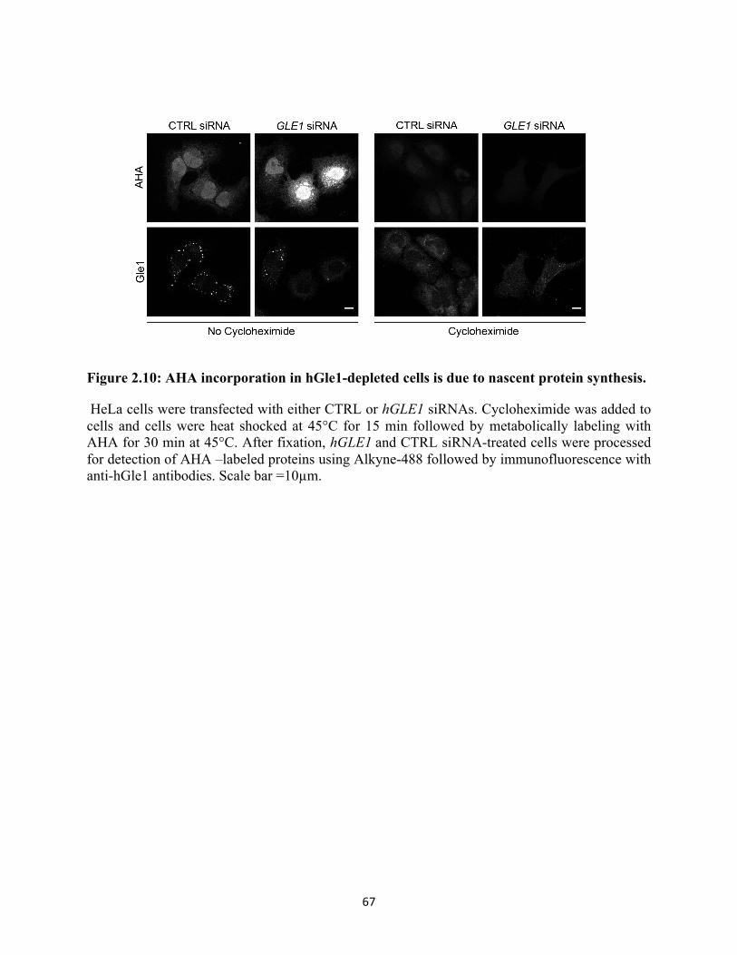
67
Figure 2.10: AHA incorporation in hGle1-depleted cells is due to nascent protein synthesis.
HeLa cells were transfected with either CTRL or hGLE1 siRNAs. Cycloheximide was added to cells and cells were heat shocked at 45°C for 15 min followed by metabolically labeling with AHA for 30 min at 45°C. After fixation, hGLE1 and CTRL siRNA-treated cells were processed for detection of AHA –labeled proteins using Alkyne-488 followed by immunofluorescence with anti-hGle1 antibodies. Scale bar =10µm.

68
Therefore, the de-regulation of translation during heat shock in hGle1-depleted cells was not due
to global perturbations in mRNA export. To confirm AHA labeling results by alternative
method, we measured nascent protein synthesis upon heat shock in CTRL and hGLE1 siRNA
cells by a 35S metabolic assay. Consistent with AHA labeling results, we observed increased
incorporation of 35S-methionine/cysteine in hGle1-depleted cells (Figure 2.9D).
Finally, by evaluating the SG defects in the hGle1-depleted cells, the de-regulation in
translation strongly correlated with the SG defects. Approximately 70% of hGle1-depleted cells
with increased AHA-488 labeling either did not assemble SGs or had an increased number of
SGs (Figure 2.9B). Since hGle1 depletion resulted in defects in translation, we next investigated
if the hGle1A and/or hGle1B isoform rescues translation defects in hGle1-depleted cells. CTRL
or hGLE1 siRNA-treated cells were transfected with mCherry alone, mCherry-hGle1AR,or
mCherry-hGLE1BR plasmids. 24h after plasmid transfection, cells were heat shocked and
processed for AHA labeling. Interestingly, expression of mCherry-hGle1A but not mCherry-
hGle1B or mCherry rescued translation defects in hGle1-depleted cells upon heat shock (Figure
2.9F and 2.9G). Together, these results suggested that hGle1A modulates SG formation by
regulating translation upon heat shock.
During cell stress conditions, various signaling pathways converge to result in the
phosphorylation of eIF2α at serine 51, which inhibits eIF2 recycling and results in a global
reduction in translation (Spriggs et al., 2010; Wek et al., 2006). To gain insight into the
mechanisms for how hGle1 depletion alters translation, we analyzed the phosphorylation levels
of eIF2α. Immunoblotting with anti-eIF2α P [Ser51] antibody revealed an increase in eIF2α
phosphorylation in CTRL siRNA-treated cells after 30 min of heat shock, which persisted to the
60 min time point (Figure 2.11A and 2.12B). Strikingly, the relative eIF2α phosphorylation level

69
Figure 2.11: Phosphorylation of eIF2α is reduced in hGle1-depleted cells.
(A-B) HeLa cells transfected with either CTRL or hGLE1 siRNAs were left untreated or heat shocked at 45°C for 30 min or 60 min. Lysates were analyzed by immunoblotting using anti-eIF2α and anti-Phospho-eIF2α (Ser51) antibodies. Phospho- eIF2α levels were quantified by densitometry and normalized to total eIF2α protein levels. Error bar represents standard deviation from mean from four independent experiments.

70
was reduced modestly (~36%) but significantly in hGle1-depleted cells compared to CTRL
siRNA cells (Figure 2.11A and 2.11B). These results indicated that reduced eIF2α
phosphorylation might be linked to translation deregulation in hGle1-depleted cells upon heat
shock.
hGle1 modulates the dynamic balance between translation and SGs upon stress
Several pharmacological agents are known to change the dynamic equilibrium between
SGs and translation. For example, cycloheximide treatment prevents SG formation whereas
puromycin results in larger SGs upon stress (Kedersha et al., 2000). If hGle1 regulates the
equilibrium between SGs and active translation, hGle1 depletion might shift the balance of
mRNPs towards active translation, limiting the free mRNP pool and inhibiting SG assembly. In
this model, SG assembly defects induced by hGle1-depletion should be rescued by shifting the
mRNP balance towards SG formation by addition of puromycin. The number of SGs in CTRL or
hGLE1 siRNA-treated cells was determined in the presence of either vehicle alone or puromycin.
Treatment of CTRL siRNA cells with either puromycin or vehicle alone did not change the
number of SGs significantly (Figure 2.12A and 2.12B). Notably, treatment of hGle1-depleted
cells with either 0.1mg/ml puromycin or 0.5mg/ ml puromycin significantly reduced the number
of SGs (Figure 2.12A and 2.12B). Thus, puromycin partially rescued the SG defects in hGle1-
depleted cells upon heat shock. As a control, we tested if puromycin rescues the increase in SGs
observed in nocodazole-treated cells and found it did not (Figure 2.13A and 2.13B). Since
puromycin failed to rescue MT-dependent SG defects, puromycin and nocodazole were affecting
SG formation at distinct steps. We concluded that puromycin rescues the SG phenotype in
hGle1- depleted cells by increasing the free mRNP pool.

71
Figure 2.12: hGle1 regulates balance between active and stalled translation upon stress.
(A-B) Puromycin rescues SG assembly defects in hGle1-depleted cells. CTRL or hGLE1 siRNA HeLa cells were treated with vehicle alone, 0.1 mg/ml, or 0.5 mg/ml puromycin for 60 min at 45°C. Cells were processed for immunofluorescence using anti-hGle1 and G3BP antibodies. Scale bar =10µm. (C-D) EGFP-hGle1A but not EGFP-hGle1B overexpression results in bigger stress granules: HeLa cells were transfected with either EGFP, EGFP-hGLE1AR, or EGFP- hGLE1BR and either C) left untreated or D) heat shocked for 60 min at 45°C. After heat shock, cells were processed for immunofluorescence using anti-hGle1 and G3BP antibodies. Data represent mean +/- standard error from three independent experiments. Scale bar =10µm.
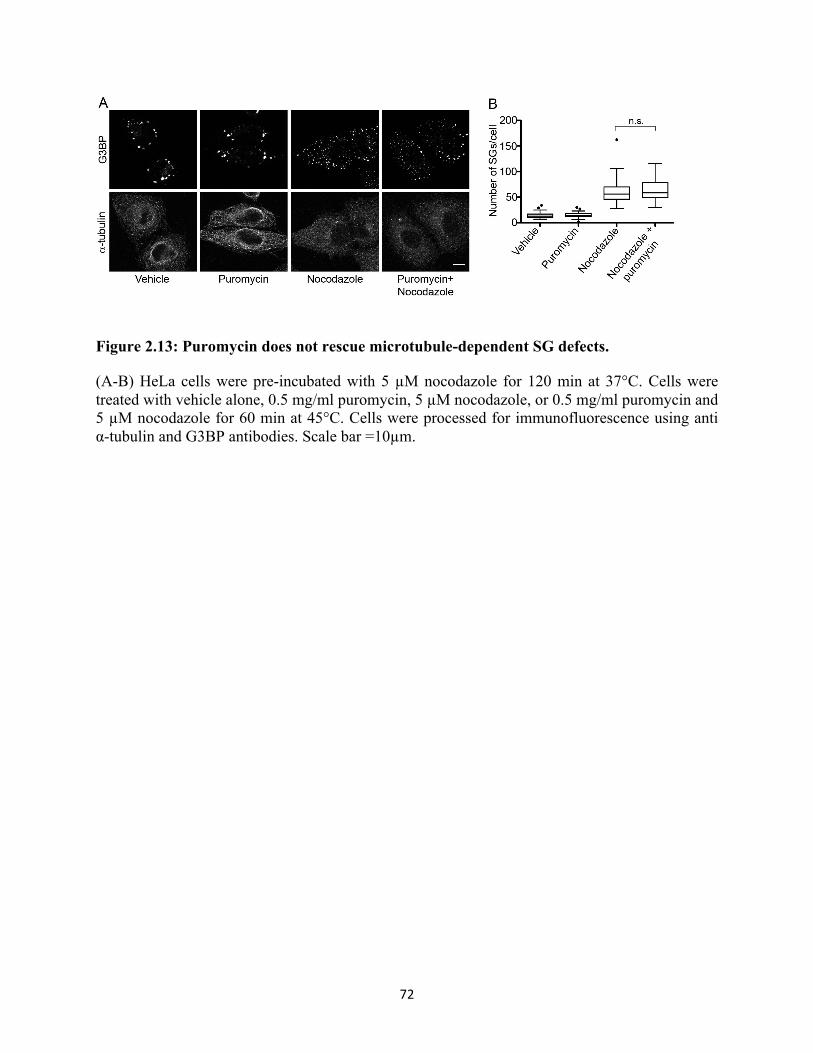
72
Figure 2.13: Puromycin does not rescue microtubule-dependent SG defects.
(A-B) HeLa cells were pre-incubated with 5 µM nocodazole for 120 min at 37°C. Cells were treated with vehicle alone, 0.5 mg/ml puromycin, 5 µM nocodazole, or 0.5 mg/ml puromycin and 5 µM nocodazole for 60 min at 45°C. Cells were processed for immunofluorescence using anti α-tubulin and G3BP antibodies. Scale bar =10µm.

73
If hGle1 levels impact the free mRNP pool by changing the distribution of mRNPs
between active translation and SGs, then overexpression of hGle1 should increase the free
mRNP pool and result in larger SGs (similar to puromycin treatment alone). HeLa cells
transfected with either EGFP, EGFP-hGLE1AR, or EGFP-hGLE1BR plasmids were heat shocked
and analyzed for SG formation. G3BP-positive SGs assembled in cells expressing either EGFP,
EGFP-hGLE1AR, or EGFP-hGLE1BR plasmids (Figure 2.12D). Overexpression of hGle1A
caused a modest but significant increase in SG size (mean=1.34µm2) compared to EGFP
(mean=1.16µm2) or hGle1B (mean=1.15µm2) (Figure 2.12D). Thus, hGle1A but not hGle1B
overexpression resulted in larger SG formation. TTP, TIA-1 and G3BP proteins play a role in
early assembly of SGs and their overexpression drives constitutive SG formation under non-
stress conditions(Gilks et al., 2004; Tourrière et al., 2003). However, in non-stress conditions,
expression of either EGFP, EGFP-hGLE1AR, or EGFP-hGLE1BR plasmids in HeLa cells showed
pan cellular cytoplasmic staining for G3BP suggesting that hGle1 cannot nucleate SG formation
in the absence of stress (Figure 2.12C).
DDX3 suppresses loss of hGle1 function
Based on the published connections between yGle1 and Ded1 during translation initiation
(Alcázar-Román et al., 2006; Bolger and Wente, 2011; Bolger et al., 2008), the roles for Ded1 in
releasing mRNA from SGs and promoting translation (Hilliker et al., 2011), and the recruitment
of the Ded1 human homologue DDX3 to SGs (Hilliker et al., 2011; Lai et al., 2008; Shih et al.,
2012), we evaluated whether hGle1 function in SGs is mediated through interaction with DDX3.
First, co-immunoprecipitation experiments were conducted with HeLa cell lysates from
untreated or heat shocked cultures. hGle1 and DDX3 were co-isolated in the presence or absence
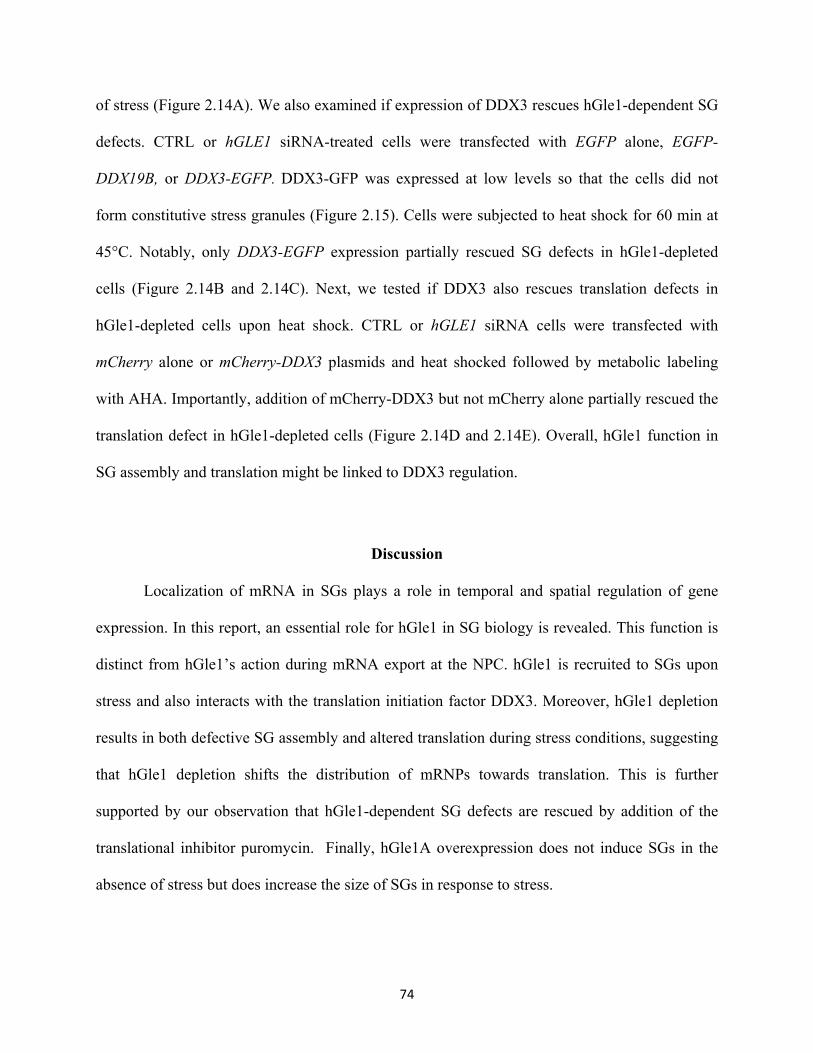
74
of stress (Figure 2.14A). We also examined if expression of DDX3 rescues hGle1-dependent SG
defects. CTRL or hGLE1 siRNA-treated cells were transfected with EGFP alone, EGFP-
DDX19B, or DDX3-EGFP. DDX3-GFP was expressed at low levels so that the cells did not
form constitutive stress granules (Figure 2.15). Cells were subjected to heat shock for 60 min at
45°C. Notably, only DDX3-EGFP expression partially rescued SG defects in hGle1-depleted
cells (Figure 2.14B and 2.14C). Next, we tested if DDX3 also rescues translation defects in
hGle1-depleted cells upon heat shock. CTRL or hGLE1 siRNA cells were transfected with
mCherry alone or mCherry-DDX3 plasmids and heat shocked followed by metabolic labeling
with AHA. Importantly, addition of mCherry-DDX3 but not mCherry alone partially rescued the
translation defect in hGle1-depleted cells (Figure 2.14D and 2.14E). Overall, hGle1 function in
SG assembly and translation might be linked to DDX3 regulation.
Discussion
Localization of mRNA in SGs plays a role in temporal and spatial regulation of gene
expression. In this report, an essential role for hGle1 in SG biology is revealed. This function is
distinct from hGle1’s action during mRNA export at the NPC. hGle1 is recruited to SGs upon
stress and also interacts with the translation initiation factor DDX3. Moreover, hGle1 depletion
results in both defective SG assembly and altered translation during stress conditions, suggesting
that hGle1 depletion shifts the distribution of mRNPs towards translation. This is further
supported by our observation that hGle1-dependent SG defects are rescued by addition of the
translational inhibitor puromycin. Finally, hGle1A overexpression does not induce SGs in the
absence of stress but does increase the size of SGs in response to stress.
75

76
Figure 2.14: DDX3 rescues hGle1-dependent SG and translation defects.
(A) DDX3 co-immunoprecipitates with hGle1. HeLa cells were either left untreated or heat shocked for 30 min or 60 min and cell lysates were immunoprecipitated using anti-hGle1 or IgG control antibodies and immunoblotted with anti-hGle1 or DDX3 antibodies. (B) DDX3 partially rescues SG defects in hGle1-depleted cells. CTRL or hGLE1 siRNA-treated cells were transfected with EGFP, EGFP-DDX19B, or DDX3-EGFP plasmids and heat shocked. Cells were processed for immunofluorescence detection of G3BP and hGle1. Scale bar =10µm. (C) SG numbers in CTRL and hGLE1 siRNA cells expressing indicated plasmids were quantified. (D) DDX3 partially rescues translation defects in hGle1-depleted cells. CTRL or hGLE1 siRNA-treated HeLa cells transfected with mCherry or mCherry-DDX3 plasmids were heat shocked at 45°C. After 15 min of heat shock, AHA was added to the incubations and heat shock treatment was continued for additional 30 min. Samples were processed for detection of AHA labeled proteins with alkyne 488 using click chemistry. Scale bar =10µm. (E) Quantification of AHA-488 staining. Mean fluorescent intensity of AHA-488 staining in individual cells was calculated in CTRL and hGLE1 siRNA cells expressing indicated plasmids.

77
Figure 2.15: Expression of DDX3 in hGle1-depleted cells under non-stress conditions.
CTRL or hGLE1 siRNA-treated cells were transfected with EGFP, EGFP-DDX19B, or DDX3-EGFP plasmids. Cells were processed for immunofluorescence detection of G3BP and hGle1. Scale bar =10µm.

78
Similarly, puromycin is not sufficient alone to nucleate SGs but induces larger granule formation
in the presence of stress. Thus, hGle1A induces the formation of larger SGs by modulating
translation. In sum, hGle1 plays a key role in the exchange of mRNPs between SGs and the
active translation machinery, thereby serving as a critical factor during the cellular stress
response.
Previously, we defined functions for Gle1 as a regulator of Dbps during mRNA export
and translation (Bolger and Wente, 2011; Bolger et al., 2008; Murphy and Wente, 1996; Noble et
al., 2011; Tran et al., 2007). Given the effects here on translation in the hGle1-depleted cells and
the interactions between hGle1 and DDX3, a role for Gle1 in translation is likely conserved
between S. cerevisiae and human cells. Multiple lines of evidence suggest that the impact of
hGle1 on SGs is not due to its role in mRNA export. Depletion of the mRNA export factor
DDX19B or NXF1 does not result in SG defects. Moreover, hGle1A rescues SG defects yet fails
to rescue the mRNA export defects. It is clear that the hGleA and hGle1B isoforms play distinct
separable functions in the cell: hGle1A in cytoplasmic SG function and hGle1B in mRNA export
at the NPC.
This work supports a model wherein hGle1 exists in at least two distinct pools in the cell,
allowing for a repertoire of independent functions and an ability to simultaneously regulate
multiple steps in the gene expression pathway. It is possible that isoform-specific interactions
with their unique protein partners might determine localization and/or functions. For instance,
hGle1B but not hGle1A interacts with hCG1. Interestingly, only EGFP-hGle1B localizes to the
NPC when transiently expressed in HeLa cells. EGFP-Gle1A does not localize to the NPC in the
presence of endogenous hGle1. However, we find here that EGFP-hGle1A can in fact localize to
NPCs when endogenous hGle1 is depleted; yet, mRNA export is not efficient with hGle1A

79
alone. We conclude that hGle1B binding to hCG1 is required for a step in mRNA export that is
distinct from strictly localization at NPCs. Indeed, our very recent studies support this conclusion
as interactions between the carboxy-terminal domain of Nup42 (human homologue hCG1) and
yGle1 are required for mRNP remodeling in yeast (Adams et al., 2014)
How mRNPs move in and out of SGs is long standing question in the field (Anderson and
Kedersha, 2008; Kedersha et al., 2005; Zhang et al., 2011). To transition between these different
states, mRNP rearrangements potentially occur and thus RNA binding proteins and mRNP
remodelers are expected to play a role in directing mRNPs for storage in SG or translation. In
yeast, the DEAD-box protein Ded1 regulates release of mRNA from SGs and promotes
translation (Hilliker et al., 2011). We propose that hGle1 might function in distribution of
mRNPs between SGs and translation through its regulation of DDX3. We find hGle1 interacts
with DDX3, and the hGle1-dependent SG and translation defects are rescued by expression of
DDX3. Interestingly, Ded1 and DDX3 assemble SGs in an ATP independent manner (Hilliker et
al., 2011; Shih et al., 2012). However, Ded1’s ATPase activity is required for disassembly of
SGs and re-entry into translation (Hilliker et al., 2011). Here we show that hGle1 depletion
results in SG assembly defects and slows disassembly of SGs. Moreover, hGle1 is involved in
regulating the exchange between SGs and translation in a manner similar to that reported for
Ded1. Thus, lack of proper hGle1-mediated control of DDX3 could lead to improper SG
dynamics.
Surprisingly, eIF2α phosphorylation at Ser51 is modestly reduced in hGle1-depleted cells
upon stress. This is consistent with the continued translation observed upon heat shock in hGle1-
depleted cells. As expression of non-phosphorylatable serine to alanine mutant at residue 51 of
eIF2α allows continued protein synthesis upon heat shock in CHO cells (Murtha-Riel et al.,

80
1993), eIF2α phosphorylation is likely one of the primary mechanisms to inhibit global protein
synthesis upon stress (Gebauer and Hentze, 2004; Sonenberg and Hinnebusch, 2009; Wek et al.,
2006). It is intriguing that hGle1 depletion affects eIF2α phosphorylation and cells maintain
translation under stress. The eiF2 kinase, PKR is often activated by its association with dsRNAs,
and upon heat shock, Alu RNA can form stable complexes with PKR that result in its activation
(Chu et al., 1998; Williams, 1999). Thus, as hGle1 regulates Dbps for mRNP remodeling, this
could be a link to a potential activation of PKR kinase. Alternatively, there might be other
mechanisms that play a role in regulating eIF2α phosphorylation. Additionally, translation
perturbations observed in hGle1-depleted cells could be due to other regulatory pathways that
may or may not be linked with eIF2α. Further studies are required to determine precisely how
hGle1 levels influence eIF2α and modulate translation.
Deregulation of SG assembly, disassembly and their clearance is associated with various
neurological diseases. An emerging theme is that hyperassembly of SGs acts as an intermediate
for disease progression. For instance, ALS-linked TDP-43 and FUS mutants form larger and
stable SGs under normal stress conditions (Buchan et al., 2013; Dewey et al., 2011; Parker et al.,
2012; Vance et al., 2013; Wolozin, 2012)(Buchan et al., 2013; Dewey et al., 2011; Parker et al.,
2012; Vance et al., 2013; Wolozin, 2012). The SG phenotype during hGle1A overexpression is
remarkably similar to that for the ALS-linked TDP-43 and FUS mutants. Our very recent work
has linked mutations in hGLE1 with ALS (Kaneb et al., 2015). These ALS linked hGLE1
mutants lack the hCG1 binding site and fail to localize at the NPC in a manner similar to the
hGle1A isoform. Thus, the ALS linked hGLE1 mutations alter the cellular pools of hGle1A and
hGle1B isoforms and this may contribute to disease phenotypes. Our work here presents further
evidence that depleting specific hGle1 isoforms differentially affect mRNA export and

81
translation. Additionally, mutations in hGLE1 linked with inherited human diseases LCCS1 and
LAAHD (Nousiainen et al., 2008) are due to disregulated mRNA export (Folkmann et al., 2013,
2014).
Taken together, we speculate that the ALS hGLE1 mutations could impact both mRNA
export (by decreasing the pool of hGle1B available) and translation (by altering the pool of
hGle1A and SG dynamics). Moreover, other disease states could arise from alterations in
specifically hGle1A function that impact translational regulation. Investigating the hGle1
mechanisms that drive the assembly and disassembly of SGs under normal conditions will guide
the understanding of how pathological aggregates form.
Materials and Methods Cell culture, treatments and plasmids
HeLa, U2OS and RPE-1 cells were grown in complete DMEM media or DMEM-F12 (Gibco)
supplemented with 10% fetal bovine serum (Atlanta Biologicals) at 37°C in 5% CO2. To induce
heat stress, cells were incubated at 45°C in a non-C02 air incubator for 60 min unless indicated
otherwise. For ER induced stress, cells were exposed to 10 µM thapsigargin (Sigma) for 60 min
at 37°C. Puromycin (Sigma) was used at either 0.1 mg/ml or 0.5 mg/ml for 60 min.
Cycloheximide (Sigma) was used at 10ug/ml. For MT perturbations, cells were incubated with 5
µM nocodazole (Sigma) or 500 nM Taxol (Sigma) for 120 min at 37°C followed by heat shock
at 45°C for 60 min. Plasmids expressing EGFP-C1, EGFP-hGle1BR (pSW3908, Folkmann et al.,
2013), EGFP-hGLE1A (pSW1409, Kendirgi et al., 2003) and Pom121-mCherry (Mor et al.,
2010) were described previously. siRNA resistant EGFP-hGLE1AR, mCherry-hGLE1AR and
mCherry-hGLE1BR were generated by introduction of three silent mutations at the hGLE1

82
siRNA targeting regions of EGFP-hGLE1A (pSW3909), mCherry-hGLE1A and mCherry-
hGLE1B plasmids by site directed mutagenesis, respectively. DDX3-EGFP plasmid was PCR
amplified from a cDNA (MGC:20129) purchased from GeneCopoeia Inc. and cloned into
pEGFP-N3 vector using XhoI and BamHI sites( pSW4105). mCherry-DDX3 was generated by
PCR amplification of DDX3 cDNA and cloned into pmCherry-C1 vector.
siRNAs and plasmids transfections
Negative control siRNA and hGLE1 siRNAs were purchased from Qiagen. NXF1 and DDX19B
Smart pool siRNAs were purchased from Dharmacon. Cells were reverse transfected with
indicated 20 nM siRNAs using HiPerFect (Qiagen) following manufacturer’s instructions.
Plasmid transfections were performed using Fugene 6(Promega) following manufacturer’s
instructions. For rescue of SG and translation phenotype, siRNA resistant (“R”) EGFP, EGFP-
hGLE1BR and EGFP-hGLE1AR expression constructs were transfected after 48 h of siRNA
transfections.
Co-immunoprecipitation
HeLa cells were plated in 100 mm dishes (Fisher Scientific) and either left untreated or heat
shocked for indicated time at 45°C. After treatments, cells were washed with 1X PBS and lysed
in buffer containing 0.5% NP-40, 50 mM Tris-Cl pH 7.5, 100 mM NaCl, 1 mM EDTA, 50 mM
NaF, 1 mM PMSF and protease inhibitors cocktail (Roche). Cell lysates were spun at 13000 rpm
for 10 min at 4°C and the supernatants were incubated with control IgG or affinity-purified
guinea pig polyclonal antibodies raised against recombinant amino- terminus of hGle1 amino
acids 1-360 (ASW48, Covance laboratory) for 5 h at 4°C. Protein A-sepharose (GE Healthcare)

83
beads were added and lysates were further incubated for additional 1 h at 4°C. Beads were
washed with lysis buffer five times and immune complexes were eluted with 50 µl 2X SDS-
laemmli buffer. The eluted complexes were resolved on 7.5% SDS PAGE and subjected to
immunoblotting using anti-hGle1, 1:1000 (ASW48) and anti-DDX3,1:1000 (Bethyl
Laboratories) antibodies.
Immunoblotting and Immunofluorescence
For immunoblotting, cells were plated in 60 mm dishes (Fisher Scientific) and reverse
transfected with 20 nM indicated siRNAs. After 72 h post siRNA transfections, cells were lysed
in buffer containing 1% NP-40, 150 mM NaCl, 50mM Tris-Cl (pH 7.5), and protease and
phosphatase inhibitor cocktail. For add back experiments, indicated plasmids were transfected
after 24 h of siRNA treatments. Proteins were resolved on 7.5% SDS PAGE and immunoblotted
with anti-hGle1, 1:1000 (Folkmann et al., 2013), anti-GFP, 1:1000 (Molecular probes), anti-
actin,1:5000 (Sigma), anti- eIF2α, 1:1000(Cell Signaling Technology), and anti-Phospho-eIF2α
(Ser51),1:1000 (Cell Signaling Technology). Far-red dyes-conjugated secondary antibodies were
used for detection and blots were scanned using LI-COR Odyssey scanner. For
immunofluorescence, cells were plated on 1.5 mm round coverslips in a 24 well plate (Fisher
Scientific). Following treatments, cells were fixed with 4% paraformaldehyde for 15 min and
permeabilized with 0.2% triton X-100 for 5 min. Coverslips were blocked with 10%FBS/PBS for
1 h at room temperature. Following primary antibodies were used: anti-hGle1, 1:300 (ASW48),
anti-G3BP, 1:300 (BD Transduction), anti-DDX3, 1:300 (Bethyl Laboratories), anti-HuR,1:100
(Santa Cruz), anti- FMRP, 1:300 (Milipore) and anti-α tubulin, 1:500 (Abcam) for
approximately 3 h at RT. Alexa-fluor conjugated secondary antibodies (Life Technologies) were

84
used and slides were mounted using Prolong Gold AntiFade (Life Technologies). Cells were
imaged using 63X (1.4 NA) oil-immersion objective on confocal microscope (Leica TCS SP5).
In Situ hybridization
HeLa cells were fixed with 4% paraformaldehyde for 15 min. Cells were washed with 1XPBS
followed by permeablization with 0.2% tritonX100/1XPBS for 5 minutes. Cells were incubated
with 1ng/ul of Cy3-conjugated oligo dT in hybridization buffer containing 125 µg/ml tRNA, 1
mg/ml ssDNA and 1%BSA, 10% dextran sulfate, 50% formamide, 5X SSC, for 1h 30 min at
37°C. After staining, coverslips were washed with 2X SSC two times followed by washes with
1X SSC. Coverslips were rinsed finally with 1X PBS and slides were mounted using Prolong
Gold AntiFade. When combined with immunofluroscence, in situ hybridization was performed
following secondary antibody incubations. Images were acquired using 63X (1.4 NA) oil-
immersion objective on Leica TCS SP5 confocal microscope. Images were processed using
ImageJ. For each EGFP positive cell, mean Cy3 intensity was determined for cytoplasmic and
nuclear compartment, and N/C ratios were calculated.
Live cell microscopy
HeLa cells were co-transfected with plasmids encoding for mCherry-G3BP and either EGFP,
EGFP-hGle1BR, and EGFP-hGle1AR using Fugene 6. Before imaging, culture medium was
replaced with phenol free DMEM supplemented with 25 mM HEPES and 10% FBS. Cells were
imaged live on a heated stage at 45°C using 63X (1.4NA) oil objective lens on Leica SP5
microscope. For 3D structural illumination microscopy, CTRL or hGLE1 siRNAs cells were co
transfected with Pom121-mCherry and either EGFP, EGFP-hGLE1BR, or EGFP-hGLE1AR

85
plasmids. Images were acquired in 3D structural illumination microscopy mode on a heated stage
at 37°C using a Delta Vision OMX microscope (Applied Precision).
Metabolic labeling for protein synthesis
F and hGLE1 siRNAs transfected cells were washed with DMEM media without cysteine and
methionine and incubated in the same medium for 45 min at 37°C. Cells were either left
untreated or subjected to heat shock at 45°C for 15 min. 50 µM methionine analogue AHA (Life
Technologies) was added to incubations and cells were either left untreated or heat shocked at
45°C for additional 30 min. Following treatments, cells were fixed with 4% paraformaldehyde
and permeabilized with 0.2% triton X 100. Cells were washed with 2% BSA/PBS three times
and AHA-labeled proteins were detected with 1 µM Alkyne-tagged Alexa Fluor-488 (Life
Technologies) using Click-iT cell detection reagent (Life Technologies) for 45 min at room
temperature. Cells were further processed for immunofluorescence as described above. For 35S
metabolic labeling, CTRL and hGLE1 siRNA-treated cells were washed with DMEM media
without cysteine and methionine and incubated in the same medium for 45 min at 37°C.
100µCi/ml radioactive 35S methionine and cysteine (Perkin Elmer) was added to the medium and
cells were incubated for additional 30 min at 37°C. Cells were lysed in RIPA buffer and protease
and phosphatase inhibitor cocktail. Protein concentrations were determined using DC protein
assay kit (Bio-Rad). For liquid scintillation counts, equal amounts of labeled cell lysates were
precipitated with 1ml of 10% TCA and BSA as a carrier protein for 30 min on ice. TCA
precipitates were filtered onto GC/F filter disks in a filtration apparatus under vacuum. Disks
were washed with 10% TCA followed by 100% ice-cold ethanol. Filters were air-dried and then
counts were determined using liquid scintillation counter (Beckman Coulter).

86
Measurement of SG size and number
Post-image processing was done using ImageJ software (NIH). ImageJ plug-in “3D objects
counter” was used to calculate size and numbers of SGs of individual cell. 3D objects counter
measurement parameters were set to ‘surface’ and minimum size filter was set to 3. Surface
measurements were used to calculate size of SGs. SG size and numbers were represented using
box plot generated in Prism6 (GraphPad). The percentages of cells with SGs were calculated as
100 X [(No. of cells with G3BP positive foci)/ ( Total No. of cells)]. All data are result of at
least three independent experiments. Statistical significance was determined using Student’s t
test (Excel) and Fisher exact test (GraphPad) where appropriate.
Polysome profiles
Cells were grown in 10cm dishes and lysed in 10mM Tris-Cl pH 7.5, 100mM NaCl, 30mM
MgCl2, 200µg heparin/ml, cycloheximide 100µg/ml and 1% tritonX 100. The lysates were
cleared by centrifugation at 5000rpm for 5 min. Cleared lysates were layered on 7%-47%
sucrose gradients cast in 50mM Tris pH7.5, 50mM NH4Cl, 12mM MgCl2, 100ug/ml
cycloheximide in ultracentrifuge tubes (Beckman). Gradients were centrifuged in a Beckman
SW-41 rotor for 3h at 28800 rpm. An absorbance profile was collected from the gradients at 254
nm.

87
CHAPTER 3
An amyotrophic lateral sclerosis-linked mutation in GLE1 alters the cellular pool of human
Gle1 functional isoforms
Abstract
Amyotrophic lateral sclerosis (ALS) is a lethal late onset motor neuron disease with
underlying cellular defects in RNA metabolism. In prior studies, two deleterious heterozygous
mutations in the gene encoding human (h)Gle1 were identified in ALS patients. hGle1 is an
mRNA processing modulator that requires inositol hexakisphosphate (IP6) binding for function.
Interestingly, one hGLE1 mutation (c.1965-2A>C) results in a novel 88 amino acid carboxy-
terminal insertion, generating an altered protein. Like hGle1A, at steady state, the altered protein
termed hGle1-IVS14-2A>C is absent from the nuclear envelope rim and localizes to the
cytoplasm. hGle1A performs essential cytoplasmic functions in translation and stress granule
regulation. Therefore, we speculated that the ALS disease pathology results from altered cellular
pools of hGle1 and increased cytoplasmic hGle1 activity. GFP-hGle1-IVS14-2A>C localized to
stress granules comparably to GFP-hGle1A, and rescued stress granule defects following siRNA-
mediated hGle1 depletion. As described for hGle1A, overexpression of the hGle1-IVS14-2A>C
protein also induced formation of larger SGs. Interestingly, hGle1A and the disease associated
hGle1-IVS14-2A>C overexpression induced the formation of distinct cytoplasmic protein
aggregates that appear similar to those found in neurodegenerative diseases.
This chapter is adapted from “An amyotrophic lateral sclerosis-linked mutation in GLE1 alters the cellular pool of human Gle1 functional isoforms. Aditi, Laura Glass, T. Renee Dawson and Susan R. Wente. Adv Biol Regul. 2015 Nov 11 pii: S2212-4926 (15) 30031-2”.

88
Strikingly, the ALS-linked hGle1-IVS14-2A>C protein also rescued mRNA export defects upon
depletion of endogenous hGle1, acting in a potentially novel bi-functional manner. We conclude
that the ALS-linked hGle1-c.1965-2A>C mutation generates a protein isoform capable of both
hGle1A- and hGle1B-ascribed functions, and thereby uncoupled from normal mechanisms of
hGle1 regulation.
Introduction
The regulation of RNA metabolism is a critical facet of the eukaryotic cell’s capacity to
react to its environment and modulate gene expression. At each stage in the mRNA lifecycle,
RNA-binding proteins associate and disassociate with a transcript, forming messenger
ribonucleoprotein complexes (mRNPs) with specific protein compositions that direct association
of the message with the proper cellular machinery. This tightly orchestrated association of
transcripts with specific RNA-binding proteins is a primary mediator of an mRNA’s fate
(Mitchell and Parker, 2014). For example, in Saccharomyces cerevisiae, association of the RNA-
binding proteins Nab2 with an mRNP in the nucleus mediates binding of the mRNA export
receptor Mex67-Mtr2 (vertebrate NXF1/TAP) for transport through the nuclear pore complex
(NPC, (Carmody et al., 2010; Iglesias et al., 2010) ; then release of Nab2 and Mex67 at the
cytoplasmic face of the NPC confers directionality to the export process and primes the mRNP
for its cytoplasmic fate (Brockmann et al., 2012; Lund and Guthrie, 2005; Tran et al., 2007).
Neurons are particularly vulnerable to misregulation of mRNP composition given the additional
hurdle of transporting mRNPs to the synapse for localized translation (Alami et al., 2014; Hutten
et al., 2014; Liu-Yesucevitz et al., 2014). Indeed, a recent surge in genetic studies has firmly
implicated the disregulation of mRNP metabolism in numerous neurodegenerative diseases,

89
including Alzheimer’s disease, Parkinson’s disease, and the late onset motor neuron disease
amyotrophic lateral sclerosis (ALS; (Fox and Tibbetts, 2015; Nussbacher et al., 2015; Philips and
Rothstein, 2015; Ramaswami et al., 2013).
ALS is a devastating disease characterized by rapid degeneration of the motor neurons.
The most prevalent form of motor neuron disease, diagnosis with ALS brings with it a prognosis
of paralysis and fatality within two to five years. The list of ALS-linked mutations in genes
encoding mRNP components and modulators is rapidly expanding, and includes TAR DNA-
binding protein 43 (TDP-43; (Sreedharan et al., 2008)), Fused in Sarcoma (FUS; (Kwiatkowski
et al., 2009; Vance et al., 2009)), hnRNP1 and hnRNPA2 (Kim et al., 2013), and, more recently,
Gle1 (Kaneb et al., 2015). Gle1 is a highly conserved and essential modulator of RNA-dependent
DEAD-box ATPases during mRNA export (Alcázar-Román et al., 2006; Folkmann et al., 2013;
Murphy and Wente, 1996) translation (Alcázar-Román et al., 2010; Bolger and Wente, 2011;
Bolger et al., 2008) and stress granule dynamics (Aditi et al., 2015). By inducing the release of
select RNA-binding proteins, DEAD-box proteins facilitate changes in the mRNP protein
composition through a process termed mRNP remodeling, and these mRNP changes play critical
roles in an mRNA’s processing (Jarmoskaite and Russell, 2014). At least two isoforms of human
(h)Gle1 result from the hGLE1 pre-mRNA transcript (Kendirgi et al., 2003). The more highly
expressed isoform, hGle1B, contains a 39 amino acid extension at its carboxy-terminus that
binds to the NPC component hCG1 and localizes at steady-state primarily to the nuclear rim
(Folkmann et al., 2013; Kendirgi et al., 2003, 2005; Rayala et al., 2004). The molecular
mechanism of Gle1-stimulated DEAD-box activation at the NPC is largely based on the S.
cerevisiae model system (Folkmann et al., 2011); however, our recent work shows that only the
highly conserved Gle1B isoform, presumably in complex with the cofactor inositol

90
hexakisphosphate (IP6), is capable of activating the DEAD-box protein for mRNP remodeling
during mRNA export (Folkmann et al., 2013). Conversely, the shorter hGle1A isoform lacking
the hCG1-binding domain localizes primarily to the cytoplasm at steady state and is required for
proper stress granule (SG) function (Aditi et al., 2015). Thus, the hGle1A and hGle1B isoforms
exist in separate cellular pools and play multiple independent roles in regulating mRNPs, any
one of which might be impacted in hGle1-associated ALS pathogenesis.
A c.1965-2A>C ALS-linked mutation in intron 14 of hGLE1 destroys a splice acceptor
site and results in the expression of a protein hGle1-IVS14-2A>C, in which the 44 amino acid
hCG1 binding site is replaced with a novel 88 amino acid carboxy-terminal domain (Kaneb et
al., 2015). Similar to hGle1A, the hGle1-IVS14-2A>C isoform localizes at steady state in the
presence of endogenous hGle1 to primarily the cytoplasm, and, by two-hybrid assays, does not
bind hCG1 (Kaneb et al., 2015). These properties suggest that the ALS hGle1-IVS14-2A>C
protein may function like hGle1A in SG biology, yet perhaps in a disregulated fashion.
Considering that a defining hallmark of ALS motor neuron pathology is cytoplasmic inclusions
of aggregated RNA-binding proteins (Leigh et al., 1991), aberrant SG function is plausible as a
potential molecular mechanism in ALS pathogenesis.
The most prevalent neuronal inclusions reported in ALS patient samples are those
containing aggregates of mutant TDP-43 or FUS (Arai et al., 2006; Neumann et al., 2006). TDP-
43 and FUS are highly conserved DNA and RNA-binding proteins with roles in transcriptional
repression, pre-mRNA splicing and localized translation (Bentmann et al., 2013); and like hGle1,
TDP-43 also facilitates stress granule assembly (McDonald et al., 2011). Many of the mutations
catalogued for such genes encode proteins with low complexity glycine-rich, prion-like domains
that promote their cytoplasmic aggregation and loss of nuclear functions (Dormann and Haass,

91
2011). Although these aggregates are distinct from normal SGs in that they are not reversible
(Bentmann et al., 2013), it has been suggested that SGs may play a role in forming the inclusions
by sequestering mutant TDP-43 or FUS and ‘seeding’ the initial aggregation step (King et al.,
2012; Parker et al., 2012). Reports differ on whether stress granule markers colocalize with
neuronal inclusions (Colombrita et al., 2009; Dormann et al., 2010; Liu-Yesucevitz et al., 2010);
however, several lines of evidence support the hypothesis. Repeated episodes of severe stress can
induce the formation of wild-type TDP-43 aggregates in SGs (Parker et al., 2012), and mutant
forms of FUS increase the size and number of SGs (Baron et al., 2013). Furthermore, expression
of mutant stress granule components alters the formation of FUS aggregates in Saccharomyces
cerevisiae (Sun et al., 2011b) and an inhibitor of stress granule formation reduces TDP-43
cytotoxicity in Drosophila and mammalian neurons (Kim et al., 2014). These observations
suggest that SGs might function in an early step of ALS progression by promoting the formation
of insoluble protein aggregates.
With regard to ALS pathogenesis, it remains unclear whether the loss of TDP-43 and
FUS nuclear functions or the accumulation of cytoplasmic inclusions are causative for
neurodegeneration. Similar obstacles exist for discerning the pathogenicity of hGle1 in ALS
since it functions both in mRNA export across the nuclear envelope and in cytoplasmic functions
of translation and stress granule biology. Thus, in this study, we sought to gain insight by
identifying the functional consequences of the c.1965-2A>C mutation on hGle1 biology.

92
Results
The ALS-linked hGle1-IVS14-2A>C protein is recruited to stress granules.
The emerging model that neuronal inclusions are related to SGs suggests that ALS
pathogenicity may arise from defects in the formation or clearance of these granules. Previously,
we reported that hGle1 is a component of SGs and is required for SG assembly and disassembly
(Aditi et al., 2015). Interestingly, hIPK1, the kinase required for producing hGle1’s cofactor IP6,
also relocalizes to SGs upon stress (Brehm et al., 2007). We speculated that the carboxy-terminal
alterations in the ALS-linked hGle1-IVS14-2A>C protein might perturb its recruitment to SGs
and/or functional capacity upon stress. To address this, subcellular localization experiments were
conducted with HeLa cells coexpressing mCherry-G3BP and either GFP, GFP-hIPK1, or GFP-
hGle1-IVS14-2A>C (the protein encoded by GFP-hGLE1-c.1965-2A>CR). The cells were
subjected to heat shock at 45oC for 45 minutes or left untreated at 37oC, and imaged by live cell
microscopy. At 37oC, both GFP-hGle1-IVS14-2A>C and GFP-hIPK1 exhibited cytoplasmic
steady state localization, with no apparent localization at the nuclear envelope or nucleoplasm
(Figure 3.1). The cytoplasmic distribution of GFP-hGle1-IVS14-2A>C with exclusion from the
nucleus was indistinguishable from previously reported hGle1A localization (Aditi et al., 2015;
Kaneb et al., 2015; Kendirgi et al., 2003), and had no similarity to hGle1B localization
(Folkmann et al., 2013; Kendirgi et al., 2003). Interestingly, as previously observed for both
hGle1A and hGle1B, GFP-hGle1-IVS14-2A>C was recruited to SGs upon heat shock at 45oC
(Figure 3.1). GFP-hIPK1 also colocalized to SGs with mCherry-G3BP, confirming the
previously published report (Brehm et al., 2007). Thus, the ALS hGle1 variant exhibits steady
state localization and SG recruitment comparable to hGle1A.

93
Figure 3.1: ALS-linked hGle1-IVS14-2A>C is localized to SGs upon heat shock.
The mCHERRY-G3BP plasmid and either GFP, hGLE1 siRNA resistant GFP-hGle1-c.1965-2A>CR, or GFP-hIPK1 plasmids were co-transfected in HeLa cells. After 24h post-transfection, HeLa cells were heat shocked at 45°C for 45 min and imaged live by confocal microscopy. Both hGle1-IVS14-2A>C and hIPK1 are localized to SGs upon heat shock. Scale bar represents 10 µm.

94
The ALS-linked hGle1-IVS14-2A>C is capable of SG function
We recently found that hGle1A is required for proper SG assembly, disassembly and
translational modulation (Aditi et al., 2015). The depletion of endogenous hGle1 from HeLa cells
results in a striking decrease in the size and increase in the total population of SGs formed upon
heat shock, as compared to control siRNA-treated cells. This defect is rescued by expression of
hGle1A, but not hGle1B (Aditi et al., 2015). Since GFP-hGle1-IVS14-2A>C localized to SGs,
we next investigated whether this variant was functionally competent to rescue SG defects in
hGle1-depleted cells. To test this, CTRL siRNA or hGLE1 siRNA-treated cells were transfected
with either GFP or GFP-hGLE1-c.1965-2A>CR plasmids. After 24h, cells were heat shocked at
45oC for 60 minutes, fixed and SG formation was assessed by indirect immunofluorescence of
the SG protein G3BP (Tourrière et al., 2003). In CTRL siRNA-treated cells, no change in SG
numbers was observed in cells expressing either GFP or GFP-hGLE1-c.1965-2A>CR plasmids
(Figure 3.2A). Consistent with our previous report (Aditi et al., 2015), in hGLE1 siRNA-treated
cells transfected with GFP, there was a significant increase in the number of SGs and apparent
decrease in individual size (Figure 3.2A and 3.2B; Aditi et al., 2015). However, strikingly,
expression of GFP-hGLE1-c.1965-2A>CR led to a significant reduction in SG numbers in hGle1-
depleted cells (Figure 3.2B). This data suggested that the ALS-linked hGle1 variant is capable of
functioning in SGs similarly to hGle1A, at least at a gross morphological level. Future studies
are required to determine whether assembly and disassembly rates are impacted by the hGLE1-
c.1965-2A>C mutation.
Overexpression of the ALS-linked hGle1-IVS14-2A>C alters stress granule morphology
The mRNPs in SGs and those engaged by translation machinery are in dynamic

95
Figure 3.2: ALS-linked hGle1-IVS14-2A>C rescues SG assembly defects in hGle1-depleted cells similar to that of hGle1A.
(A) CTRL or hGLE1 siRNA-treated cells were transfected with either GFP or GFP-hGLE1-c.1965-2A>CR plasmids. Cells were heat shocked at 45°C for 60 minutes and processed for indirect immunofluorescence using anti-G3BP antibodies. Scale bar represents 10 µm. (B) Quantification of SG numbers in CTRL and hGLE1 siRNA cells expressing indicated plasmids. ImageJ plug-in “3D objects counter” was used to count SG numbers of an individual cell. Statistical significance was determined using Student’s t test (Excel).

96
equilibrium (Aditi et al., 2015; Kedersha et al., 2000). Previously, we demonstrated that hGle1A
modulates this equilibrium and thus influences SG formation. Overexpression of hGle1A but not
hGle1B alters the cell’s translation activity and increases the pool of free mRNPs, resulting in the
formation of larger SGs (Aditi et al., 2015). Since the ALS-linked hGle1 variant rescues an SG
defect in hGle1-depleted cells, we hypothesized that it might also impact SG size in a fashion
similar to hGle1A overexpression. To test this, SG morphology was examined in cells
transfected with GFP, GFP-hGle1A or GFP-hGLE1-c.1965-2A>CR plasmids and subjected to
heat shock stress. G3BP staining was used to assess SG formation. As reported previously,
expression of GFP-hGle1A resulted in a modest but significant increase in mean SG size
compared to GFP alone (Figure 3.3; Aditi et al., 2015). Interestingly, we observed that
expression of GFP-hGle1-IVS14-2A>C also led to an increase in size of SGs (Figure 3.3). In
fact, no significant difference was detected for SG morphology in cells expressing hGle1A
versus hGle1-IVS14-2A>C (Figure 3.3). This data further argued that the general capacity for
hGle1 functions in SG assembly are unaffected by the ALS-linked hGle1 variant.
Overexpression of hGle1A or the ALS-linked hGle1-IVS14-2A>C results in cytoplasmic
aggregates that do not co-localize with SGs components
Our analysis of SG morphology following overexpression of hGle1 isoforms also
revealed the presence of cytoplasmic GFP-hGle1A or GFP-hGle1-IVS14-2A>C aggregates in
approximately 10% of the transfected cell population. We categorized these aggregates into two
groups. In one group, the cells formed small “foci” aggregates reminiscent of SGs (Figure 3.4,
center column). In the second group, the cells exhibited large “fibrous” aggregates that appeared
to form from long extended fibers of hGle1 protein (Figure 3.4, right column).

97
Figure 3.3: Expression of ALS-linked hGle1-IVS14-2A>C results in larger SGs similar to that of hGle1A.
HeLa cells were transfected with GFP, hGLE1 siRNA resistant GFP-hGLE1AR, or GFP-hGLE1-c.1965-2A>CR plasmids. Cells were heat shocked for 60 minutes at 45°C. After heat shock, cells were processed for indirect immunofluorescence using anti-G3BP antibodies. Size of SGs was determined using ImageJ plug-in “3D objects counter”. Data are mean +/- standard error from three independent experiments. Statistical significance was determined using Student’s t test (Excel); whiskers represent 10-90 percentile. Scale bar represents 10 µm.

98
Figure 3.4: Overexpression of ALS-linked hGle1-IVS14-2A>C and hGle1A result in formation of cytoplasmic aggregates.
GFP, GFP-hGle1AR or GFP-hGle1-c.1965-2A>CR plasmids were transfected in HeLa cells. After 24h, cells were fixed and processed for indirect immunofluorescence. Approximately 90% of cells showed uniform cytoplasmic localization of GFP-hGle1A and GFP-hGle1-IVS14-2A>C. However, ~10% of cells formed either “foci” or “fiber-like” aggregates. Scale bar represents 10 µm.

99
To examine whether these aggregates contain SG components, indirect immunofluorescence was
performed for G3BP and DDX3 in cells transfected with GFP, GFP-hGLE1AR or GFP-hGLE1-
c.1965-2A>CR plasmids. No co-localization was observed between G3BP or DDX3 and the
hGle1A or hGle1-IVS14-2A>C aggregates (Figure 3.5A and 3.5B). On the contrary, in cells
exhibiting “fibrous” hGle1 aggregates, G3BP formed independent cytoplasmic foci resembling
SGs. This data suggests that the stress response pathway was provoked by the presence of hGle1
aggregates and likely does not initiate the aggregation. However, it cannot be ruled out that the
SG components might be co-recruited in a sequential or temporal manner, or that other SG
components might be preferentially recruited to these foci.
hGle1A and ALS-linked hGle1-IVS14-2A>C aggregates co-localize with TDP-43
It is proposed that the aggregation of ALS-linked TDP-43 and FUS altered proteins
sequester several other RNA-binding proteins to promote pathological formation of the neuronal
inclusions (Bentmann et al., 2013). Therefore, indirect immunofluorescence microscopy was
used to test whether the hGle1A or hGle1-IVS14-2A>C positive aggregates sequester wild type
TDP-43 or FUS. “Foci-like” hGle1 aggregates did not co-localize with either FUS or TDP-43
proteins (Figure 3.6A and 3.6B). However, TDP-43 did colocalize with the “fibrous” GFP-
Gle1A and GFP-hGle1-IVS14-2A>C aggregates (Figure 3.6A and 3.6B). This data indicated that
overexpression of hGle1A or ALS-linked hGle1 variant can sequester wild type TDP-43 protein
in this cell model system.
Hsp90 colocalizes with hGle1A and hGle1-IVS14-2A>C aggregates
Hsp90 is a molecular chaperone and master regulator of the stress response program,

100
Figure 3.5: Overexpression of ALS-linked hGle1-IVS14-2A>C and hGle1A result in formation of cytoplasmic aggregates that do not co-stain with SG components, DDX3 and G3BP.
HeLa cells were transfected with GFP, GFP-hGLE1AR, or GFP-hGLE1-c.1965-2A>CR plasmids. 24 hours after transfection, cells were fixed and processed for indirect immunofluorescence using (A) anti-DDX3 and (B) anti-G3BP antibodies. Scale bar represents 10 µm.
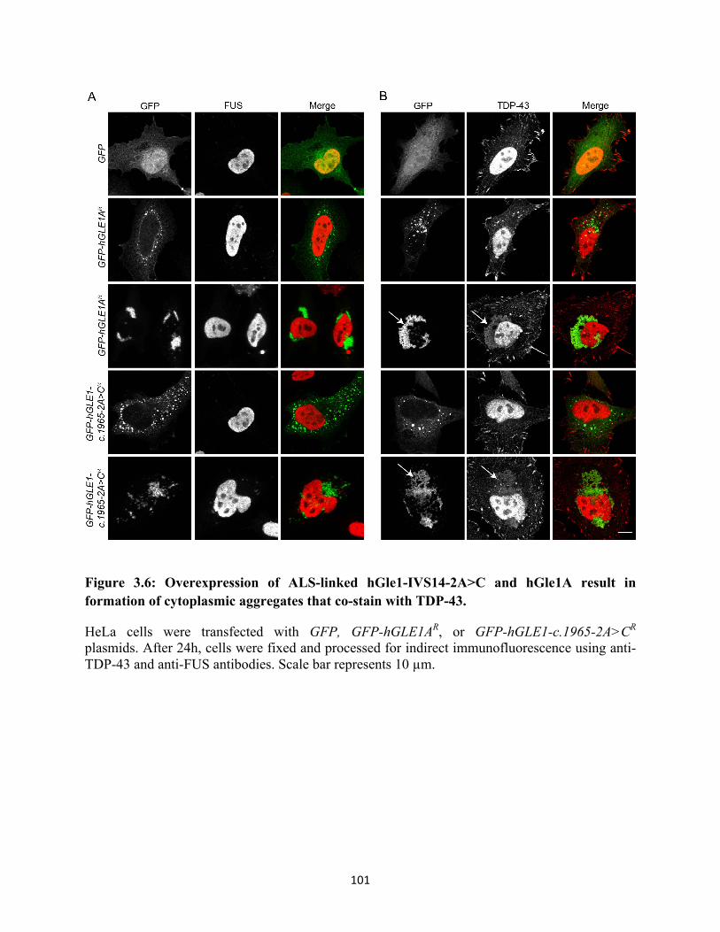
101
Figure 3.6: Overexpression of ALS-linked hGle1-IVS14-2A>C and hGle1A result in formation of cytoplasmic aggregates that co-stain with TDP-43.
HeLa cells were transfected with GFP, GFP-hGLE1AR, or GFP-hGLE1-c.1965-2A>CR plasmids. After 24h, cells were fixed and processed for indirect immunofluorescence using anti-TDP-43 and anti-FUS antibodies. Scale bar represents 10 µm.

102
which has been suggested to modulate the stability and degradation of mutant protein aggregates
in neuronal inclusions (Luo et al., 2010). To determine whether Hsp90 might be involved in
regulating hGle1 inclusions, we tested whether hGle1A or hGle1-IVS14-2A>C aggregates co-
localize with Hsp90 protein. As described, HeLa cells expressing GFP, GFP-hGle1A or GFP-
hGle1-IVS14-2A>C were fixed and processed for indirect immunofluorescence of Hsp90.
Although no colocalization was observed in “foci-like” hGle1 aggregates, Hsp90 co-localized
with “fibrous” hGle1 aggregates in both GFP-Gle1A and GFP-hGle1-IVS14-2A>C expressing
cells (Figure 3.7A). This data suggests that Hsp90 might be recruited to hGle1-IVS14-2A>C
cytoplasmic aggregates in ALS motor neurons. Since deregulation of RNA metabolism is also
linked to ALS (Ramaswami et al., 2013), in situ hybridization was performed to determine if
these aggregates also contain poly(A)+ RNA. No nuclear accumulation of poly(A)+ RNA was
observed in GFP-Gle1A or GFP-hGle1-IVS14-2A>C aggregates (Figure 3.7B), however, we
cannot exclude the possibility that specific types of RNA are present in these aggregates.
The ALS-linked hGle1-IVS14-2A>C protein rescues mRNA export defects of hGle1-
depleted cells
Efficient and expedient mRNA export from the nucleus is essential for proper gene
expression and cellular function. In complex with IP6, hGle1B plays an integral role in this
process by regulating mRNP remodeling at the NPC cytoplasmic face (Folkmann et al., 2011).
Based on the lack of nuclear rim localization for the ALS-linked hGle1-IVS14-2A>C and its
functional similarity with hGle1A in SG biology, we predicted that hGle1-IVS14-2A>C would

103
Figure 3.7: Overexpression of ALS-linked hGle1-IVS14-2A>C and hGle1A result in formation of cytoplasmic aggregates that co-stain with Hsp90.
HeLa cells were transfected with GFP, GFP-hGLE1AR, or GFP-hGLE1-c.1965-2A>CR plasmids. Cells were fixed and processed for either indirect immunofluorescence using anti-Hsp90 antibodies or Cy3-conjugated oligo-dT in situ hybridization for poly(A)+ RNA. Scale bar represents 10 µm.
104
not support mRNA export. Using our previously validated knockdown:add-back approach of
hGle1 depletion and exogenous expression (Folkmann et al., 2013; Aditi et al., 2015), we
investigated whether the hGle1-IVS14-2A>C protein was capable of mediating mRNA export.
Following CTRL siRNA or hGle1 siRNA treatment, HeLa cells were transfected with GFP,
GFP-hGLE1BR, GFP-hGLE1AR or GFP-hGLE1-c.1965-2A>CR plasmids. A small, yet
statistically significant, increase in the nucleocytoplasmic ratio of poly(A)+ RNA was observed
in hGle1-depleted cells expressing GFP, as compared to CTRL siRNA-treated cells (Figure 3.8).
The mRNA export defect was even more pronounced in hGle1-depleted cells expressing
exogenous hGle1A. However, expression of exogenous hGle1B or, surprisingly, exogenous
ALS-linked hGle1-IVS14-2A>C rescued the mRNA export defect caused by endogenous hGle1
depletion (Figure 3.8). Thus, hGle1-IVS14-2A>C supports some level of poly(A)+ RNA export.
Discussion
hGle1 is a multifunctional regulator of DEAD-box proteins that is essential for proper
mRNP dynamics at several stages in the mRNA lifecycle (Bolger and Wente, 2011; Bolger et al.,
2008; Murphy and Wente, 1996). Although the use of a single factor to control multiple
processes can provide the cell a mechanism for rapid and coordinated changes in gene
expression, it also comes with a fair degree of risk for catastrophic effects in the event of hGle1
dysfunction. We recently uncovered one mechanism by which the cell compartmentalizes hGle1,
elucidating that the two hGle1 isoforms hGle1A and hGle1B localize to different cellular pools
and perform non-overlapping critical functions (Table 1; Folkmann et al., 2013; Aditi et al.,
2015). Mutations that impact the balance in the functional pools of hGle1 or alter the function of
both hGle1 isoforms should have far-reaching impact on mRNA metabolism.
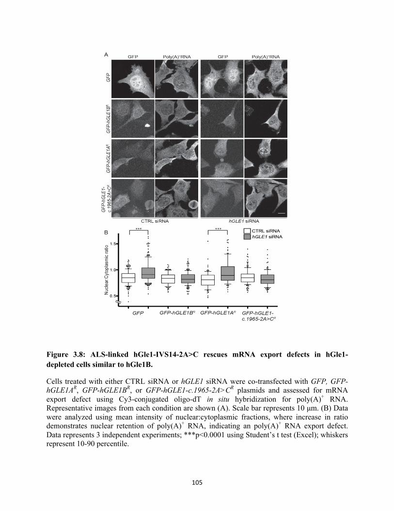
105
Figure 3.8: ALS-linked hGle1-IVS14-2A>C rescues mRNA export defects in hGle1-depleted cells similar to hGle1B.
Cells treated with either CTRL siRNA or hGLE1 siRNA were co-transfected with GFP, GFP-hGLE1AR, GFP-hGLE1BR, or GFP-hGLE1-c.1965-2A>CR plasmids and assessed for mRNA export defect using Cy3-conjugated oligo-dT in situ hybridization for poly(A)+ RNA. Representative images from each condition are shown (A). Scale bar represents 10 µm. (B) Data were analyzed using mean intensity of nuclear:cytoplasmic fractions, where increase in ratio demonstrates nuclear retention of poly(A)+ RNA, indicating an poly(A)+ RNA export defect. Data represents 3 independent experiments; ***p<0.0001 using Student’s t test (Excel); whiskers represent 10-90 percentile.
106
The lethal inherited diseases LCCS1 and LAAHD are prime examples of this, wherein mutations
that decrease hGle1’s ability to homo-oligomerize or destabilize its structural integrity lead to
mRNA export defects that are linked to the arrest of proper development ((Folkmann et al.,
2013; Nousiainen et al., 2008).
In this study, we characterized the functional capacity of a rare ALS-linked hGLE1-
c1965-2A>C mutation that alters the carboxy-terminal domain of hGle1 to produce a protein
with a novel 88 amino acid domain in place of the predicted hCG1-binding interface. Based on
the loss of the NPC-anchoring hCG1 binding domain, we previously surmised that this mutation
probably results in haploinsufficiency by reducing the pool of hGle1 supporting mRNA export
(Kaneb et al., 2015). Surprisingly, we discovered here that the ALS-linked hGle1-IVS14-2A>C
is functional for both stress granule homeostasis and mRNA export, making it the first known bi-
functional hGle1 isoform capable of functions assigned to both hGle1A and hGle1B.
In Table 1, the functional capacities of the hGle1-IVS14-2A>C protein is compared to the
wild type isoforms hGle1A and hGle1B. The novel hGle1-IVS14-2A>C protein functioned in all
aspects of stress granule modulation that we examined and also rescued mRNA defects under the
conditions tested. The only known molecular characteristic that hGle1-IVS14-2A>C appears to
lack is its reported inability to bind hCG1 (Kaneb et al., 2015) and therefore weak association
with the NPC (demonstrated herein). This lack of hGle1-IVS14-2A>C interaction with hCG1 has
only been tested using the qualitative yeast-two-hybrid. It is possible that the hGle1-IVS14-
2A>C protein has reduced affinity for hCG1 compared to hGle1B, but is not as compromised as
hGle1A. Alternatively, the extended carboxy-terminal domain in hGle1-IVS14-2A>C may
support another interaction that compensates for loss of hCG1 binding. In either case, such a bi-
functional isoform would impact the balance between the hGle1B and hGle1A cellular pools and

107
Table 1: ALS-linked hGle1-IVS14-2A>C exhibits behavior mimicking both hGle1B and hGle1A isoforms.
†(Kaneb et al., 2015), *Rescues defect in hGle1-depleted cells
Isoform Localizes to
SGs Rescues SG
defects* Forms larger
SGs Forms
aggregates Rescues mRNA export defects*
Binds hCG1†
GFP-hGle1A + + + + - - GFP-hGle1B + - - Not tested + + GFP-hGle1-IVS14-2A>C + + + + + -

108
could limit the cell’s ability to properly regulate hGle1 activity.
Several caveats must be considered in drawing conclusions from this data with regard to
mechanisms of ALS disease pathogenesis. First, the ability of hGle1-IVS14-2A>C to function in
mRNA export was observed in the absence of any other endogenous hGle1 protein, whereas the
ALS patients were heterozygous. Second, the expression level for the ALS-linked hGle1-IVS14-
2A>C in HeLa cells is likely not at a level representative of its endogenous steady state levels in
ALS patients; nor are HeLa cells a model for the neuronal disease. Moreover, hGle1-IVS14-
2A>C does not localize to the nuclear rim in the presence of endogenous hGle1B. Therefore, in a
heterozygous background, some reduction in mRNA export competent hGle1 function is
expected. More work will be required to determine if an mRNA export defect or another
nucleocytoplasmic transport defect is exhibited by the patient cell lines isolated from patients
carrying the hGLE1-c1965-2A>C mutation. Nevertheless, the data shown here suggests that this
mutation does not result in a complete loss of hGle1 function, but rather is more likely to impact
ALS pathology by confounding the carefully balanced separation of hGle1 functions.
The potential disregulation of mRNP dynamics by the ALS-linked hGle1-IVS14-2A>C
represents one of several recently discovered links between ALS and Gle1-associated functions.
Several studies have implicated disregulation of the hGle1-cofactor IP6 in neurodegenerative
disease. First, activation of the caspase-independent apoptosis pathway is associated with
familial ALS, and is provoked through phosphorylation of IP6 following cytoplasmic
translocation of IP6 kinase (reviewed in Ghavami et al., 2014). Second, IPK1 is recruited to
stress granules and increased levels of IP6 are neuroprotective, suggesting that IP6 production in
SG might facilitate disaggregation of proteins sequestered there (Anekonda et al., 2011; Brehm
et al., 2007; Lv et al., 2015; Xu et al., 2008). Importantly, general disruption of NPC function is

109
also highly associated with ALS. A clustering of ALS-linked mutations in sequence encoding the
nuclear localization sequence (NLS) in FUS directly demonstrated the importance of nuclear
import in ALS pathogenesis (Dormann et al., 2010). Furthermore, very recently, the most
common form of familial, inherited ALS, C9ORF72 repeat expansion, was shown to induce
pathogenesis by disrupting nucleocytoplasmic transport through the NPC (Freibaum et al., 2015;
Jovičić et al., 2015; Zhang et al., 2015a). Finally, irregular Nup62 localization/nuclear envelope
shape, loss of nuclear importins or cytoplasmic redistribution of importins are observed in the
anterior horns of ALS patients and in mouse models of ALS (Kinoshita et al., 2009; Zhang et al.,
2006).
Given that the ALS-linked hGLE1-c.1965-2A>C mutation results in an altered balance of
hGle1 activity rather than a complete loss of function, we speculate that a second pathogenic
trigger might induce neurodegeneration in combination with expression of hGle1-IVS14-2A>C.
A complete genetic profile was not obtained for patients carrying the hGLE1-c.1965-2A>C
mutation. Furthermore, analyses of other ALS-linked mutations as well as the frequency of
sporadic ALS suggest this to be a disease of multiple contributing factors. A major contributing
factor in TDP-43, FUS, and hGle1 biology is the effect of subcellular distribution on their
aggregation potential (worked presented here; (Dormann and Haass, 2011; Johnson et al., 2008;
Vance et al., 2013). Thus, any mutations or conditions that increase their cytoplasmic
distribution, when combined with an altered state of nucleocytoplasmic transport, could shift the
cellular equilibrium toward pathogenesis. Such a combinatorial pathway to the disease state is
consistent with the late onset of ALS pathology. One major change in RNA metabolism that
would also contribute to late onset is the detrimental effect of aging NPCs on nucleocytoplasmic
transport. Numerous reports have implicated decreased efficiency of nucleocytoplasmic transport

110
in neurodegenerative diseases (Patel and Chu, 2011), and our laboratory recently discovered
specific defects in transport pathways that occur as a result of ageing-related Nup deterioration
(Lord et al., 2015). This must be accounted for in models of ALS pathogenesis, particularly with
respect to sporadic ALS where aging-related transport defects might combine with another ALS
risk factor such as repeated stress (Fidler et al., 2011) to induce disease progression.
In recent years, several studies have highlighted the connections between deregulation in
RNA metabolism, protein aggregation, and neurodegenerative diseases including ALS. For
example, FUS and TDP-43 are found in protein aggregates. These FUS and TDP-43 inclusions
occur independently and are distinct from stress granules (SGs) in that they not reversible, but
rather are insoluble (Bentmann et al., 2013). Here, we have demonstrated that hGle1 is capable
of forming protein aggregates upon its overexpression. Strikingly, these hGle1 aggregates
sequestered both endogenous TDP-43 and Hsp90 proteins. Although the precise pathological
effects of hGle1 protein aggregates are unknown, we propose that hGle1 aggregates sequester
native RNA-binding proteins such as TDP-43 and thereby promote ALS by the gain- and/or loss
of native RNA-binding protein functions. This idea is further supported by recent studies
demonstrating that the lariat debranching enzyme Dbr1 modifies TDP-43 toxicity by capturing
TDP-43, preventing its aggregation and thus the sequestration of other RNA-binding proteins
with it (Armakola et al., 2012).
The seeding of cytoplasmic aggregates due to delayed or nonexistent SG disassembly is
another attractive model for how the characteristic insoluble neuronal inclusions of ALS motor
neurons form. This model is supported by the cellular pathology of ALS-linked mutations in the
gene encoding ubiquitin segregase Valosin-containing protein (VCP). VCP is required for
clearance of SGs after the stress response, and mutations in VCP result in constitutive SG

111
formation. Strikingly, though, these mutations also result in sequestration of wild type TDP-43
within the constitutive SGs (Buchan et al., 2013; Johnson et al., 2010). We observed no
functional alterations in the ability of hGle1-IVS14-2A>C to support SG homeostasis; however,
there are certainly other potential aspects of SG function that might be perturbed by hGle1-
IVS14-2A>C expression. Our future studies will need to examine the capacity of this hGle1-
IVS14-2A>C in the kinetics of SG function, such as the rate of SG disassembly.
Taken together, this data provides evidence that the hGle1-IVS14-2A>C protein is a bi-
functional isoform capable of mediating both mRNA export, like hGle1B at the NPC, and SG
function, like hGle1A in the cytoplasm. This surprising discovery highlights the critical nature
of the carboxy-terminal domain in regulating hGle1, and calls for further investigation to
determine its mechanistic role in hGle1 function, interactions and regulation. We also found that
hGle1 assembles into cytoplasmic aggregates that harbor TDP-43 and Hsp90, both of which are
found in the neuronal inclusions of ALS patients. It is intriguing that hGle1, like TDP-43 and
FUS, plays roles at multiple stages of the mRNA life-cycle and that all three are linked to
devastating human diseases with motor neuron pathologies. Previous studies do not report any
direct functional links between hGle1 and either TDP-43 or FUS; however all three play roles in
regulating both SGs and translation activity. Thus, one molecular mechanism of ALS pathology
might be the combined impact of their misregulation on translational activity. Alternatively,
hGle1 misregulation might alter the nuclear export of specific mRNAs that interact with TDP-43
and FUS for localized translation. Future studies are needed to shed light on these molecular
connections and further characterize the effect of hGle1 misregulation on motor neuron health
and in ALS pathogenesis.

112
CHAPTER 4
Conclusions and future directions
Modulation of mRNA metabolism has a central role in the cellular adaptive response
against changing conditions (de Nadal et al., 2011). This regulation occurs at both
transcriptional and post-transcriptional levels and is mediated by RNA-binding proteins that
determine the fate of mRNAs. Extensive rearrangements of mRNPs occur during stress
conditions, altering their downstream fate during RNA processing, export, translation and decay
(Keene, 2007; de Nadal et al., 2011; Nakaminami et al., 2012; Shih and Lee, 2014). However,
we do not understand the mechanisms by which messages are sorted for their distinct fates. Our
data suggests that an mRNA regulatory factor, hGle1, plays a critical role in the stress response
by mediating mRNP rearrangements. In chapter 2, We found that hGle1 regulates SGs by
modulating the level of active translation. Interestingly, only the hGle1A isoform plays a role in
SG formation, whereas only hGle1B functions in mRNA export. We propose that hGle1A
modulates SGs by regulating the activity of DDX3, since DDX3 suppresses hGle1-mediated SG
and translation phenotypes, and interacts with hGle1 (Aditi et al., 2015). Taken together, this
work defines an isoform-specific role for hGle1A in mediating the stress response by modulating
the trafficking of mRNPs between an actively translating pool and a repressed pool sequestered
in SGs.
In the second part of this work (chapter 3), we determined that the ALS-linked hGle1
variant protein (hGle1-IVS14-2A>C) is capable of functioning in both mRNA export and SG
formation, in essence behaving as both hGle1A and hGle1B isoforms (Aditi et al., 2015).
Collectively, this work suggests that an ALS-linked GLE1 mutation leads to the loss of isoform

113
specific hGle1 function, leading to deregulation of normal mRNA metabolism. We speculate that
the resulting loss of proper gene expression in response to stress is a major contributing factor in
the pathogenesis of ALS.
In summary, this work elucidated that hGle1A and hGle1B perform distinct functions and
also suggested a potential mechanism underlying ALS pathogenesis. However, these studies did
not offer mechanistic insights into the distinct non-overlapping functions of hGle1 isoforms.
Given that the ALS-linked hGle1-IVS14-2A>C can complement the functions of both hGle1A
and hGle1B isoforms, understanding the mechanistic underpinnings of each activity will be
crucial to tease out the disregulation underlying ALS pathology. In the following sections, I
discuss potential future avenues.
Molecular mechanisms underlying the distinct roles of hGle1 isoforms
In human cells, the hGle1A and hGle1B isoforms are generated by alternative splicing of
transcripts from a single hGLE1 locus. These isoforms are very similar in sequence except that
hGle1A lacks 39 amino acids at its carboxy-terminal (Kendirgi et al., 2003). Our work reveals
that despite sharing more than 90% of amino-acid sequence, these two isoforms perform distinct
functions in a cell; hGle1B is required for mRNA export (Folkmann et al., 2013), while hGle1A
functions in SGs (Aditi et al., 2015). Interestingly, this functional specificity is not due solely to
subcellular localization. Similar to hGle1B, hGle1A is localized at NPCs in hGLE1 siRNA-
treated cells. Likewise, hGle1A and hGle1B are recruited to SGs in response to stress. This raises
the question as to how the functional specificity of each hGle1 isoforms is achieved? There are at
least two possible reasons for this. The additional residues in hGle1B could allow it to bind to
unique protein partners necessary for modulating mRNA export. Indeed, we previously showed

114
that the additional 39 residues in hGle1B are required for its interaction with the nucleoporin
hCG1 (Kendirgi et al., 2005). This observation raises the possibility that hGle1B function during
mRNA export is modulated by hCG1. In fact, this conclusion is supported by recent work from
the Wente laboratory demonstrating that interaction between yGle1 and the carboxy-terminal
domain of Nup42 (a yeast homologue of hCG1) is required for mRNP remodeling (Adams et al.,
2014). Moreover, there could be additional factors that differentially influence functions of each
hGle1 isoforms. Therefore, future studies could be focused on defining the unique protein
interaction partners of hGle1A and hGle1B. We have generated stable HeLa cell lines expressing
low levels of either GFP, GFP-hGle1A or GFP-hGle1B. These cell lines are ideal for performing
immunoprecipitation since the expression of hGle1 isoforms is homogenous on a cell-to-cell
basis in contrast to transient transfection. Immunoprecipitation using GFP-antibodies coupled to
magnetic beads could be performed with cell lysates prepared under normal and stress
conditions, followed by liquid chromatography tandem mass spectrometry to identify interacting
partners. Novel interactions could be validated and studied by various methods including co-
immunoprecipitation, FRET and in vitro binding assays. However, an important caveat of using
this method is that these isoforms are not expressed from their endogenous promoter. The GFP
tag might hinder the binding to some protein partners, thus biasing the identification of interactor
proteins. A complementary approach could be to manipulate the levels of hGle1 isoforms
endogenously. hGle1A contains a unique 4 amino acids at its carboxy-terminus. Therefore,
siRNAs that only target either hGLE1A or hGLE1B isoforms could be designed followed by
immunoprecipitation of isoforms using hGle1 antibodies and mass spectrometry.
An alternative explanation, but not mutually exclusive from above, for separate functions
of hGle1 isoforms could be that the isoforms regulate different Dbps. Based on work in yeast,

115
Gle1 regulates mRNA export and translation termination by stimulating the ATPase activity of
Dbp5 (human homologue DDX19) (Alcázar-Román et al., 2006, 2010; Bolger et al., 2008). In
contrast, yGle1 inhibits ATPase activity of Ded1 (human homologue DDX3) during translation
initiation (Bolger and Wente, 2011). Therefore, it is likely that different Dbps dictate the distinct
functions of hGle1 isoforms. In human cells, DDX3 is a critical component of SGs and siRNA
knockdown of DDX3 perturbs SG assembly (Shih et al., 2012). DDX3 is also linked with the
export of viral RNA but not with export of bulk poly (A)+ mRNA (Yedavalli et al., 2004). On
the other hand, we have observed that depletion of DDX19B in HeLa cells results in mRNA
export defects, but not SG defects (Aditi et al., 2015). Therefore, I speculate that hGle1A and
hGle1B specifically regulate DDX3 and DDX19 ATPase activity, respectively. Several pieces of
evidence support this model. 1) Endogenous hGle1 co-immunoprecipitates with DDX19B and
DDX3 proteins in HeLa cells (Aditi et al., 2015 and unpublished results). 2) mRNA export is
dependent upon both hGle1B and DDX19, as their depletion results in accumulation of poly (A+)
mRNA in the nucleus (Aditi et al., 2015). Moreover, expression of the dominant-negative mutant
of DDX19B, ddx19-R372G also results in mRNA export defects, by acting as a competitive
inhibitor of wild-type DDX19B and sequestering hGle1 (Hodge et al., 2011). 3) hGle1A-
dependent SGs and translation defects are rescued by expression of DDX3 but not by DDX19B
(Aditi et al., 2015). Taken together, these results suggest that, similar to yGle1, hGle1 regulates
the ATPase activity of DDX3 and DDX19. However, this hypothesis needs to be directly
verified with regard to the isoform specificity for each Dbp. This could be tested by investigating
whether each hGle1 isoform directly interacts with DDX3 or DDX19 using an in vitro soluble
binding assay. Next, an in vitro ATPase assay could be performed with the recombinant proteins
to test if hGle1 modulates the ATPase activity of DDX3 and DDX19. However, the

116
interpretation of the experimental outcome might be limited due to the absence of post-
translation modifications or additional cofactors that might dictate the endogenous specificity of
hGle1 modulation of Dbps. Therefore, if purified bacterially expressed-recombinant proteins do
not yield any clear results, insect cell lines could be used to purify these proteins. Alternatively,
human lysates could be fractionated and tested for additional cofactors.
An important remaining aspect of this research is to examine Dbp modulation by hGle1
isoforms. In this regard, structure determination of hGle1A-DDX3 and hGle1B-DDX19 protein
complexes could very helpful. It is challenging to purify full-length hGle1A and hGle1B at a
high concentration, however, the Wente laboratory has shown that the carboxy-terminus of
yGle1 is sufficient to modulate the ATPase activity of Dbps (Alcázar-Román et al., 2006; Bolger
and Wente, 2011). In fact, the Weis laboratory has reported the crystal structure of Dbp5 in
complex with the carboxy-terminus of yGle1-IP6 (Montpetit et al., 2011). Therefore, solving the
structure of carboxy-terminus of hGle1A and hGle1B proteins could be very helpful to
understand the basis of distinct functions of each isoform.
In summary, elucidation of the mechanisms underlying hGle1 regulation of Dbps and the
identification of additional interaction partners could provide great insights into the mechanism
by which hGle1 isoforms achieve functional specificity. These investigations would aid in our
understanding of misregulation of altered hGle1 protein arising from hgle1 disease-linked
mutations.
Investigating the role of hGle1 isoforms in translation
To date, we have shown that hGle1A regulates stress granules by modulating translation
during stress. However, it is unclear whether hGle1A also regulates translation under basal
conditions. In S. cerevisiae, yGle1 physically interacts with translation initiation factor eIF3

117
subunits, Prt1 and Ded1, and modulates the ATPase activity of Ded1 during AUG start site
selection. Likewise, yGle1 physically interacts with the translation termination factor Sup45 and
is required to regulate the ATPase activity of Dbp5 for efficient translation termination (Bolger
and Wente, 2011; Bolger et al., 2008). Our work in human cells shows that the siRNA-
knockdown of hGLE1 also results in translation defects (Aditi et al., 2015). Using sucrose-
density centrifugation, hGle1-depleted cells show an increase in the monosome 80S peak and
reduction in polysomes. This phenotype is similar to the polysome profile defects observed with
the ygle1 temperature-sensitive alleles, arguing that hGle1 too plays a role in translational
control. In addition, we observe reduced 35S methionine incorporation in protein extracts from
hGle1-depleted cells, indicative of overall reduction in protein synthesis (Aditi et al., 2015).
However, these studies did not address the mechanisms behind the role of hGle1 in translation.
To investigate the roles of hGle1A and/or hGle1B in translational control, in vitro
translation assays using ribosome enriched extracts and in vitro-transcribed luciferase mRNA
with recombinant hGle1A and hGle1B proteins could be performed. Additionally, siRNA
knockdown of endogenous hGLE1 and add-back of either hGLE1A or hGLE1B could be
performed to test if either isoform rescues polysome defects in hGle1-depleted cells. To test for a
specific role in start site selection (similar to yGle1), in vivo ATF4 reporter based assays could
be performed in control and hGLE1 siRNA-treated cells (Bolger et al., 2008; Hinnebusch, 2011;
Wagner et al., 2014). This assay measures leaky scanning, which results when the pre-initiation
complex fails to recognize the start codon and continues scanning. An ATF4 construct is
designed such that it lacks uORF2, 3 and 4 but retains the stimulatory uORF1. This reporter is
translationally active and serves as a control. A second reporter is designed where uORF2 of
ATF4 is followed by a sequence that encodes for luciferase. This product is only synthesized if

118
the start site in uORF2 is bypassed through leaky scanning, thus providing a measure of an
estimate of scanning defects during start site selection. An alternative assay to assess defects in
start site recognition is to test for start site selection fidelity. Start site fidelity defects are
characterized by the use of a near cognate codon for initiation (Hinnebusch, 2011; Wagner et al.,
2014). In this assay, a set of reporters coding for firefly-luciferase are expressed in control and
hGle1-depeleted cells. One set of reporters contains a consensus AUG start codon, whereas the
other set of reporters has a mutated UUG site. An increase in the ratio of UUG to AUG indicates
start site fidelity defects. If hGle1-depleted cells exhibit a defect in start site selection, rescue
assays could be performed by adding-back siRNA resistant hGLE1A and/or hGLE1B constructs
to determine which isoforms functions in translation initiation.
Similarly, translation termination defects could be assessed by transfecting control and
hGle1-depleted cells with tandem constructs expressing luciferase and renilla proteins. Three
types of constructs are used- a construct encoding a stop codon between luciferase and renilla, a
second construct encoding no stop codon, and a third construct encoding a stem-loop upstream
of the stop codon. Following their transfection and expression, read through efficiency will be
measured in control and hGle1-depleted cells. Following this analysis, constructs expressing
each isoform could be added back to determine if rescue is specific.
To understand the context of hGle1’s actions in translation, it would be informative to
know the mRNA targets of hGle1. With the advent of ribosome profiling (Ingolia et al., 2012), it
is possible to identify the mRNAs that are being actively translated. Therefore, ribosome
profiling in control and hGle1-depleted cells could be used to examine whether hGle1 is required
for translation of all or a specific subset of mRNAs. One could speculate that hGle1 targets of
translation would overlap with the subset of mRNAs identified as DDX3 targets (Lai et al.,

119
2010). However, previous studies have generated conflicting results about the role of DDX3 in
translation. Some studies suggest that DDX3 is involved in translation of mRNAs containing
long unstructured 5’ UTRs. One of the better known targets is cyclin E1 (Lai et al., 2010).
Therefore, comparing the ribosome profiling results between hGle1 and DDX3-depleted cells
would allow us to test more definitively if hGle1 and DDX3 share similar mRNA targets.
To further delve into the mechanism of hGle1 regulation, it would be informative to
understand how hGle1 interacts with translation factors. My preliminary data suggests that hGle1
binds to eIF4E cap complex (data not shown). Since yGle1 also physically and genetically
interacts with translation initiation and termination factors (Bolger and Wente, 2011; Bolger et
al., 2008), future experiments could explore how hGle1 works with translation machinery to
modulate protein synthesis.
Taken together, these assays would provide important clues into the mechanisms
underlying the role of hGle1 in translation under normal and disease conditions.
Investigating the role of hGle1 in translation and SG formation under stress
Global translation is down regulated under stress conditions (Holcik and Sonenberg,
2005). Surprisingly, we noticed that hGle1-depleted cells fail to down-regulate their translation
and exhibit SG defects. We rescued SG defects by treating cells with puromycin, which
disassembles polysomes (Aditi et al., 2015). Based on these observations, we propose that hGle1
modulates the equilibrium between SGs and translation. Since reprogramming of translation is
essential for survival of cells during stress conditions, this raised several important questions. For
instance, why do hGle1-depleted cells failed to downregulate translation even under continuous
stress conditions? Interestingly, we also noticed that eIF2α phosphorylation is reduced in hGle1-

120
depleted cells compared to control cells (Aditi et al., 2015). As mentioned earlier,
phosphorylation of eIF2α at serine 51 is one of the primary mechanisms to inhibit global protein
synthesis under stress conditions. In fact, expression of a non-phosphorylatable serine to alanine
substitution at residue 51 of eIF2α results in continued protein synthesis under heat stress
(Murtha-Riel et al., 1993). Therefore, the continued translation observed in hGle1-depleted cells
could be due to reduced eIF2 α phosphorylation. This potential regulatory loop should be
investigated by examining the regulation of eIF2α phosphorylation in hGle1-depleted cells. We
also anticipate that additional mechanisms, independent of eIF2α, might be involved by which
hGle1 regulates translation under stress conditions.
Another avenue of further investigation would be to identify the nature of mRNAs that
are being synthesized during stress condition in hGle1-depdelted cells. It is known that the IRES-
containing mRNAs such as HSP70 and ATF4, but not the housekeeping genes, are preferentially
translated (Blais et al., 2004; Holcik and Sonenberg, 2005; McGarry and Lindquist, 1985). In
order to investigate if translation is globally altered or specific to a subset of mRNAs in hGle1-
depleted cells, we could biotin label newly synthesized proteins using click-chemistry reaction
(Dieterich et al., 2006, 2007). These biotin-labeled peptides can be immunoprecipitated and
analyzed by mass spectrometry to identify the nature of the nascent synthesized proteins under
stress conditions. The identified targets could be further validated either by western blotting or
by in vivo reporter translation assays performed in control and hGLE1 siRNA-treated cells.
Another complementary approach would be to perform ribosome profiling with control and
hGle1-depleted cells under stress conditions (Ingolia et al., 2012). It is possible that hGle1 have
different mRNA targets during basal and stress conditions and this may explain why hGle1-
depletion have an opposing effects on translation under stress and non-stress conditions.

121
SGs formation is intimately linked with translation. Our work reveals that hGle1
modulates the distribution of mRNPs between those sequestered in SGs and those associated
with the active translational machinery. Interestingly, in yeast, Ded1 is shown to regulate release
of mRNA from SGs, promoting translation (Hilliker et al., 2011). Our work also suggests that
DDX3 plays similar role as Ded1, however, the mechanistic details are not clear. Therefore,
future work could be focused on determining the steps by which hGle1, through its regulation of
DDX3, influences the movement of mRNPs between SG and translation. For this purpose, it is
important to know which mRNAs reside in SGs. Roy Parker’s laboratory has recently published
a protocol to purify SGs from cells (Jain et al., 2016). Based on this protocol, we could purify the
SGs from control, hGle1 and DDX3-depleted cells. Following SGs purification, the identity of
proteins and mRNAs could be investigated by mass spectrometry and RNA-Seq, respectively. If
hGle1 and DDX3 have common targets, further validation could be performed by sucrose
density centrifugation followed by isolation of mRNAs in the free mRNP fraction versus
mRNPs-bound in the polysome fractions. If hGle1 and DDX3 are involved in exchange of
mRNPs between SGs and translation, we would expect to see a change in the distribution pattern
of mRNAs in control versus hGle1 and DDX3-depleted cells.
The reprogramming of translation is important during stress conditions. However, we do
not fully understand the molecular mechanism by which stress redirects the translation
machinery. Since hGle1 depletion has a dramatic effect on translation and SGs, these studies are
designed to understand the mechanisms by which hGle1 regulates translation and SG formation
would be informative in deciphering the steps involved in translation regulation under stress
conditions.

122
Role of hGle1 self-association in SG formation and translation
hGle1 self-associates in human cells. This self-association is mediated through
interaction between the coiled-coil domains and is required during mRNA export (Folkmann et
al., 2013). Notably, mutations that disrupt the sequence encoding the coiled-coil domain of
hGle1 (hgle1-Finmajor) alter hGle1 self-association and result in mRNA export defects.
Interestingly, Finmajor mimic insertions in ygle1 also exhibit genetic interactions with mRNA
export mutants but not with translation mutants (Folkmann et al., 2013). These data suggest that
yGle1-self association is not required during translation. However, since hGle1-depleted cells
show opposite effects on translation under basal and stress conditions, it is possible that there
might be differential requirements for hGle1 self-association under normal and stress conditions.
Future experiments could be focused on testing the requirement of hGle1 self-association during
SG formation and translation. It is known that hGle1B homo-oligomerizes at the NPC and in the
cytoplasm in living cells. But, we do not know if hGle1A also self-associates. As a starting point,
Fluorescence Resonance Energy Transfer (FRET) experiments could be performed with
mCerulean-hGLE1A and mVenus-hGle1A expressing FRET-pair constructs. In addition, in vitro
binding experiments could be performed with recombinant MBP-hGle1A and 35S labeled-
hGle1A. To test for the requirement for hGle1 self-association during SG formation, deletion
constructs lacking the coiled-coil region (ΔCC) could be tested for their ability to rescue SG
defects in hGle1-depleted cells. An important caveat of this experiment is that a major portion of
protein is missing and thus lack of rescue could be due to other reasons. Alternatively, the GFP-
hgle1-Finmajor, which is compromised for self-association, could be tested for its ability to rescue
SG defects in hGle1-depeleted cells. Similarly for translation, the GFP-hgle1-FinmajorR and GFP-
hGLE1ΔCC constructs could be tested for their ability to rescue translation defects in hGle1-

123
depeleted cells. It is unknown if a hGle1A-hGle1B heterodimer complex exists in a cell.
Therefore, FRET experiments could be performed to test this hypothesis. Additionally,
immunoprecipitation of hGle1A followed by immunoblotting with hGle1B could give
information if this pair exists. Given that ALS-linked hGle1-IVS14-2A>C functions as both
hGle1A and hGle1B (Aditi et al., 2015), it is possible that this variant may have greater tendency
to form a heterodimer and thus could serve as a bifunctional isoform. Therefore, if hGle1
isoforms form heterodimers, it will be important to decipher the functional consequence of this
heterodimer association.
Regulation of hGle1 function by post translational modification
hGle1 is an essential mRNA regulatory factor that controls various steps of gene
expression. Importantly, mutations in GLE1 have been linked with various diseases and thus
hGle1 function must be tightly regulated (Kaneb et al., 2015; Nousiainen et al., 2008). However,
we do not know how hGle1 activity is regulated inside a cell. One possibility already discussed
is that its protein partners/localization modulates its function. A second mechanism could be
regulation by post-translation modifications. Indeed, several large-scale mass spectrometry
studies have identified phosphorylated hGle1 peptides (Mayya et al., 2009; Olsen et al., 2010;
Sharma et al., 2014; Stuart et al., 2015). Furthermore, a recent study also showed that hGle1 is
ADP-ribosylated at the NPC (Carter-O’Connell et al., 2016). Interestingly, we have also
observed that hGle1 is phosphorylated at various residues upon stress (unpublished work;
discussed in appendix A section, Figure A.2). We noticed that the cytosolic but not the nuclear
pool of hGle1 is phosphorylated. Moreover, inhibition of SG formation did not affect hGle1
phosphorylation suggesting that hGle1 phosphorylation is not dependent upon SGs (unpublished
work; discussed in appendix A section). Thus, phosphorylation could be one of the possible

124
mechanisms to regulate hGle1 function. Future efforts will be focused on understanding the role
of hGle1 phosphorylation. To test if phosphorylation of hGle1A plays any role in SGs and
translation, the siRNA-knockdown add-back assay could be performed with phospho-dead or
phospho-mimetic hgle1 constructs and assayed for their effect on SG assembly, disassembly and
translation. Similarly, constructs expressing for phospho-dead or phospho-mimetic hGle1B
proteins could be tested for the rescue of mRNA export in hGle1-depleted cells. These assays
would give insights into the requirement of phosphorylation at various steps of gene expression.
Interestingly, we have identified four phosphorylated residues of hGle1 that are clustered
together and lie in a low complexity region near to the coil-coil domain (unpublished work;
discussed in appendix A section). This observation raises the possibility that hGle1
phosphorylation might affect its self-association. FRET experiments along with soluble binding
assay with recombinant phospho-dead and phospho-mimetic hGle1 proteins could be performed
to test if phosphorylation has any impact on hGle1 self-association. Recent studies have also
highlighted the importance of low complexity domains to help phase-separate proteins into
liquid-like droplets. Importantly, SG formation is also thought to be mediated by a phase-
separation process (Hyman and Simons, 2012; Lin et al., 2015; Molliex et al., 2015; Patel et al.,
2015). Therefore, it would be interesting to test whether hGle1 phase-separates in vitro using
recombinant proteins, and moreover if phosphorylation influences this property.
Finally, it will be informative to know how hGle1 phosphorylation influences its
association with Dbps and their activity. To address this, ATPase assays performed with
recombinant phospho-dead or phospho-mimetic hGle1 could be done. The Roy Parker laboratory
has shown that Ded1 ATPase activity is required during stress recovery for disassembly of SGs

125
and resumption of translation. Therefore, it would be informative to know if phosphorylated
hGle1 modulates the ATPase activity of Dbps differently than the non-phosphorylated hGle1.
In summary, the proposed studies would give an indication how phosphorylation of
hGle1 regulates its activity and impacts various steps of gene expression.
Exploring hGle1 connections with ALS
A recent surge of papers has linked altered nucleocytoplasmic transport as an underlying
cellular mechanism in neurological diseases, especially ALS. Although 90% of ALS cases are
spontaneous, 5-10% of cases are familial in nature (Kiernan et al., 2011). A GGGCC
hexanucleotide repeat expansion (HRE) in C9ORF72 gene has been associated with about half of
the fALS cases (Van Blitterswijk et al., 2012). Three laboratories independently showed that
nuclear export and import pathways are affected as a result of HRE expansion in C9ORF72 gene
in yeast, fly and iPSC-derived neuron model systems (Freibaum et al., 2015; Jovičić et al., 2015;
Zhang et al., 2015). Although, there is not a clear overlap in the protein players identified by
these studies, they all converge on defective NPC transport as a contributing factor in
neurodegenerative diseases.
Notably, mutations in GLE1 have been linked with ALS disease (Kaneb et al., 2015).
Therefore, an exciting area of research would be to understand if mutations in GLE1 also result
in altered nucleocytoplasmic transport defects similar to ALS-linked C9ORF72 mutations.
Interestingly, siRNA knockdown-add back experiments performed with the construct expressing
ALS-linked hGle1-IVS14-2A>C in HeLa cells suggest that this protein variant is capable of
function in mRNA export similar to wild-type hGle1B protein (Aditi et al., 2015). Further
supporting this conclusion, patient derived lymphoblastoid cells also showed no obvious defects

126
in mRNA export (unpublished work). This is reasonable given that patients develop disease at
later stages of their life. However, we do not know if export of a specific subset of mRNAs
might still be affected. Intriguingly, expression array data comparing the neural tissues derived
from LCCS1 post mortem fetuses and control tissues show deregulation of specific mRNA
involved in synapses and neuron development (Pakkasjärvi et al., 2007). Thus, it is likely that
neuronal cells have differential requirements for hGle1 and mutations in GLE1 lead to
perturbation in the fate of specific mRNAs.
Recently, several studies have shown that patient-derived induced-pluripotent stem cells
can be differentiated into motor neuron cell types for use as a model system for ALS (Lee and
Huang., 2015). Therefore, RNA-Seq experiments could be performed using patient-derived iPSC
motor neurons to identify if export of a specific set of mRNAs is inhibited/ promoted. Similar to
mRNA export, it is likely that translation of certain transcripts is inhibited or promoted. The
translational landscape could also be tested by ribosome profiling of these control and patient-
derived iPSC motor neurons. Taken together, these studies would suggest if mRNA
export/translation of a specific subset of mRNAs is perturbed in patient cells.
A second possibility is that mutations in GLE1 might impact transport of molecules
between nucleus and cytoplasm indirectly. Intriguingly, a recent study showed that cytoplasmic
aggregates observed in neurodegenerative diseases interfere with the nucleocytoplasmic
transport of RNA and protein in the cells by sequestering nuclear transport factors (Woerner et
al., 2016). Interestingly we also notice that over-expression of ALS-linked hGle1-IVS14-2A>C
protein results in protein aggregates that differentially sequester wild-type TDP-43 and Hsp90
(Aditi et al., 2015). We did not look at the co-localization of various transport factors in the
hGle1 aggregates. Thus, it is possible that mutations in GLE1 do not affect its function in mRNA

127
export but may promote cytoplasmic aggregation of hGle1 protein in neurons over time, which
sequester various proteins including, transport factors, and ultimately impact nucleocytoplasmic
transport pathway. Therefore, it would be interesting to test if ALS-linked hGle1-IVS14-2A>C
aggregates-containing HeLa cells also exhibit nculeocytoplasmic defects. Furthermore, iPSC
neuronal model system could be use to test if patient cells show any cytoplasmic aggregates and
if these aggregates sequester proteins including, transport factors. Also, it would be informative
to know if aggregates found in other type of ALS models sequester wild type hGle1.
Finally, it would be interesting to investigate how ALS-linked hGle1-IVS14-2A>C
protein behaves as bifunctional hGle1 isoform. It is shown that ALS-linked hGle1-IVS14-2A>C
does not interact with hCG1 using yeast two hybrid assay (Kaneb et al., 2015). However, the
expression levels of yeast two-hybrid constructs were not determined. Therefore, the lack of
yeast two-hybrid interaction could be due to low expression of ALS-linked hGle1-IVS14-2A>C
protein compared to wild-type hGle1B. In fact, ALS-linked hGle1-IVS14-2A>C protein is
expressed at lower levels in human cells compared to hGle1B (Kaneb et al., 2015). Therefore,
soluble binding assays with recombinant proteins could be done to test the interactions of ALS-
linked hGle1-IVS14-2A>C with hCG1. Furthermore, determining the structure of ALS-linked
hGle1-IVS14-2A>C protein and examining its impact on Dbps ATPase activity would be
insightful towards understanding its function.
Global questions related to SG biology
SGs are highly dynamic structures that play important roles in RNA metabolism and cell
survival. Although more than 200 proteins are identified as SG components, we still do not fully
understand the rules that govern their assembly and disassembly process (Buchan, 2014; Buchan

128
and Parker, 2009; Kedersha et al., 2013). Below, two key unresolved questions associated with
SG biology are discussed.
1) What is the fate of mRNA in SGs?
SGs are reversible complexes whose components move in and out of SGs. This
observation has led to a model where SGs serve as a site to sort mRNPs for storage, decay and
re-initiation for translation. However, this model has never been tested due to limitation of
available technologies. In recent years, various tools have been developed to either track single
mRNA species or visualize translation at a specific location. Approaches to test this model by
using recently developed tools are discussed below.
The Doudna laboratory has recently described the RCas9 system where Cas9 protein can
be specifically directed to bind or cut RNA targets (O’Connell et al., 2014). Using this system,
nuclease-insensitive fluorescently tagged-RCas9 system could be used to track single or multiple
mRNAs under stress. An important advantage of this method is that multiple endogenous mRNA
species can be tracked without introducing any additional sequences to mRNAs. A recent study
has identified several mRNA species that reside in SGs (Jain et al., 2016). Therefore, we could
label multiple mRNA species that reside in SGs and track them live, in cells that co-express SG
and P body markers, during a stress response. This way, it would be possible to get an idea if
mRNAs are exchanged between SG and P bodies during a stress response. To further test if
mRNAs that reside in SGs could also enter into translation, a modified version of proximity
specific ribosome profiling protocol developed by the Weissman laboratory, could be used (Jan
et al., 2014). Using this protocol, the Weissman laboratory biotinylated ER ribosomes in intact
cells by co-expressing an ER restricted biotin ligase fusion protein (BirA) along with ribosomes
containing an AviTag, which makes ribosomes substrate for BirA. The in vivo biotinylation of

129
ER-bound ribosomes followed by ribosome profiling provided information regarding the
messages that were translated specifically at the ER (Jan et al., 2014). In the modified approach
to test for translation targets at SGs, BirA will be redirected to translation machinery by
expressing it as a fusion protein with a translation factor such as 60S subunit protein (This
translation factor should not localize to SGs). Nuclease-insensitive RCas9 with an Avi tag will
be expressed in cells that can be targeted to a specific transcript. A short biotin pulse will be
applied during stress and biotin-streptavidin pull down protocol will be followed. If an mRNA is
translated, RCas9 will be biotinylated. Thus, we could test if an mRNA enters into translation or
follows a different path during stress. Although this method offers labeling of endogenous
mRNAs, RCas9 system has not been tested extensively in mammalian cells and it may have off-
targets effects.
2) Is SG disassembly required for translation-reinitiation?
The SGs are proposed to function in global translation repression by storing
translationally-repressed mRNAs (Buchan and Parker, 2009; Kedersha and Anderson, 2009).
However, there are conflicting reports that suggest that SG formation is not needed for
translation repression (Buchan, 2014; Kedersha et al., 2013). Furthermore, it is unclear if SG
disassembly is prerequisite for translation re-initiation after stress. There are several
pharmacological reagents that interfere with SG assembly and disassembly. For example, K252a
addition prevents SG disassembly, while ISRIB promotes disassembly (Sidrauski et al., 2015;
Wippich et al., 2013). Using these drugs we could investigate the effects of SG
assembly/disassembly on translation. Cells will be treated with either K252a, ISRIB and vehicle
alone under stress and non-stress conditions. Following treatments, cells will be processed for

130
ribosome profiling to determine the translational landscape in control and drug-treated cells
under normal and stress conditions. This analysis would give us some clues whether translation
is affected if cells fail to assemble or disassemble granules. However, it is possible that stalled
polysome-bound mRNAs are enriched rather than active polysomes involved in translation.
In conclusion, to investigate the involvement of SGs in diseases, it is imperative to
decipher the nature and function of these granules. The proposed studies would give us some
clues regarding the formation and function of these organelles and their deregulation in diseases.

131
APPENDIX A
Analysis of hGle1 phosphorylation in response to stress
Introduction
The human genome consists of approximately 20,000 protein-coding genes. However,
the proteome of humans is very complex with additional diversity and complexity generated by
post-translational modifications (PTMs), in addition to changes during the transcription, splicing
and translation levels (Di Giammartino et al., 2011; Nilsen and Graveley, 2010; Shabalina et al.,
2014; Walsh et al., 2005). PTMs usually refer to the covalent addition of a functional group to
proteins or the proteolytic processing of protein subunits. A variety of PTMs occur in cells,
including phosphorylation, methylation, acetylation, ubiquitylation, and glycosylation. PTMs
often influence the function of a protein by modulating its activity, localization or interaction
with other binding partners, and thus are capable of regulating various aspects of biological
processes (Beltrao et al., 2013; Lothrop et al., 2013; Prabakaran et al., 2012; Seo and Lee, 2004).
Under stress conditions, PTMs also provide a rapid and reversible response, as no new
protein synthesis is required. Interestingly, PTMs of some SG components regulate the
interaction with other proteins and affect SG functions (De Leeuw et al., 2007; Kwon et al.,
2007; Stoecklin et al., 2004; Tourrière et al., 2003). For example, phosphorylation of growth
factor receptor-bound protein 7 (Grb7) by focal adhesion kinases influences SG assembly and
disassembly. Under stress conditions, hypo-phosphorylated Grb7 is recruited to SGs through its
interaction with the RNA-binding protein HuR. Grb7 stabilizes TIA-1 aggregation and promotes
SG assembly. During recovery phase, Grb7 is phosphorylated by FAK1 kinases, resulting in
reduced interaction with HuR and SG components, thus promoting SG assembly (Tsai et al.,

132
2008). Similarly, several SG components including ribosomal proteins are O-linked N-
acetylglucosamine (O-GlcNac) modified and depletion of O-GlcNac transferases inhibits SG
assembly (Ohn et al., 2008). Additionally, PTMs can determine the partitioning of a protein into
an aggregated or a soluble state and thereby influence its localization in granules. For instance,
phosphorylation of FUS protein by DNA-PK enzyme impedes its recruitment to in vitro
hydrogels compared to hypo-phosphorylated FUS (Han et al., 2012) . Thus, PTMs of proteins
have a major influence on RNA granule dynamics.
Interestingly, our preliminary results show that hGle1 is post-translationally modified
under stress conditions. Therefore, we sought to determine if posttranslational modification of
hGle1 regulates its activity during the stress response. Below a summary of these findings is
presented.
Results and discussion
hGle1 is phosphorylated during stress
Under stress conditions, we discovered that hGle1 protein exhibited a reduced
electrophoretic mobility compared with untreated control samples on SDS-PAGE. This was
detected by immunoblotting with antibody raised against the amino-terminal region of hGle1. To
test if this slower migration is due to phosphorylation, cell lysates from control, heat shocked and
sodium arsenite-treated samples were incubated with lambda phosphatase in the presence or
absence of phosphatase inhibitors. Lambda phosphatase treatment resulted in the collapse of the
higher migrating band of hGle1 in samples prepared from stressed cells, while no change was
observed in the presence of phosphatase inhibitors (Figure A.1A). This data suggested that the
altered

133
Figure A.1: hGle1 is phosphorylated in response to stress.
(A) HeLa cells were either left untreated, exposed to heat shock at 45°C or treated with sodium aresenite for 60 min. After treatments, cell lysates were prepared and lysates were either incubated with phosphatase buffer alone, lambda phosphatase (500UI) or lambda phosphatase (500UI) and phosphatase inhibitors together for 30 min. Samples were resolved on SDS-PAGE and immunoblotted with anti-hGle1 antibodies. (B) Untreated or heat shocked cell lysates were resolved on a phos-tag gel and immunoblotted with anti-Gle1 antibody. (C and D) HeLa cells exressing GFP-hGLE1A or GFP-hGLE1B plasmids were either left untreated at 37°C, heat shocked at 45°C or treated with 0.5mM sodium arsenite for 60 min at 37°C. Following treatments, cell lysates were resolved by (C) SDS-PAGE, or (D) phos-tag SDS-PAGE, and immunoblotted with anti-GFP antibodies.

134
mobility of hGle1 protein occurred as a result of hGle1 phosphorylation under stress. To
examine hGle1 phosphorylation by an alternative method, we utilized the phos-tag gel method.
Phos-tag is a functional molecule that binds specifically to phosphorylated ions. Phosphorylated
proteins migrate more slowly within phos-tag SDS-PAGE due to their interaction with the phos-
tag molecule. This results in a separation of phosphorylated proteins from non-phosphorylated
proteins (Kinoshita et al., 2006). We observed three distinct bands for hGle1 on phos-tag SDS
PAGE in heat shocked samples (Figure A.1B). The lowermost band corresponded to non-
phosphorylated hGle1 (Figure A.1B; lane 2), while middle and upper bands represented
phosphorylated hGle1 (Figure A.1B; lane 2). This data suggested that hGle1 is phosphorylated to
varying degrees upon stress. Importantly, we also observed a basal level of phosphorylation of
hGle1 in lysates prepared from control cells (Figure A.1B; lane 1). However, the proportion of
phosphorylated hGle1 was much higher in heat shock-treated samples compared to controls.
Thus, hGle1 is basally phosphorylated under normal conditions and this phosphorylation is
enhanced during stress.
We next sought to determine if both hGle1A and hGle1B isoforms are phosphorylated
under stress conditions. We did not observe any electrophoretic mobility shift for exogenously
expressed GFP-hGle1A and GFP-hGle1B proteins by a SDS-PAGE gel and immunoblotting of
lysates (Figure A.1C). This was not surprising since the addition of a GFP tag changes the
molecular weight of hGle1 protein by ~27 kD, potentially masking the addition of
phosphorylated group’s mass. Therefore, we turned to the phos tag gel system to examine the
phosphorylated status of hGle1 isoforms. HeLa cells expressing GFP-hGle1A or GFP-hGle1B
proteins were either left untreated or heat shocked at 45oC for 60 min and cell lysates were
prepared in RIPA buffer. The proteins were resolved on phos-tag SDS-PAGE. Both GFP-
135
hGle1A and GFP-hGle1B showed two distinct bands in phos-tag SDS-PAGE for the heat
shocked sample (Figure A.1D; lane 4). To test if the higher migrating band represented
phosphorylated hGle1A or hGle1B, cell lysates were treated with lambda phosphatase. The
higher migrating band collapsed into a single band upon lambda phosphatase treatment in both
GFP-hGle1A and GFP-hGle1B samples under stress conditions, while there was no change in
the migration pattern in the presence of lambda phosphatase and phosphatase inhibitors (Figure
A.1D; lane 5 and lane 6). Thus, both GFP-hGle1A and GFP-hGle1B isoforms are
phosphorylated. Taken together, these results demonstrated that hGle1 is phosphorylated.
Identification of phosphorylated residues by mass spectrometry In order to identify phosphorylated amino-acid residues of hGle1, we took a proteomic
approach. Hela cells transfected with GFP-hGLE1AR plasmid were either left untreated or
treated with 0.5mM sodium arsenite for 60 min. Following treatment, cell lysates were prepared
in RIPA buffer and GFP-hGle1A was immunoprecipitated and proteins were separated on a
SDS-PAGE gel. We treated cells with sodium arsenite instead of heat shock because GFP-
hGle1A and GFP-hGle1B protein levels were reduced upon heat shock (Figure A.1C and A.1D).
There were also technical difficulties in immunoprecipitating enough material from heat-shocked
cells to identify phosphorylation sites by mass spectrometry. A band corresponding to GFP-
hGle1A was excised from the gel, trypsin digested and samples were processed for mass
spectrometry. The peptide spectra data was aligned with a theoretical peptide database,
identifying a total of thirteen putative phosphorylation sites under stress conditions compared to
six phosphorylation sites under normal conditions (Figure A.2A, A.2B and A.2D). Interestingly,

136
the six phosphorylation sites identified in GFP-hGle1A under normal conditions were also
identified during stress

137
Figure A.2: hGle1 is phosphorylated at multiple sites.
(A-C) Identification of phosphorylated residues by mass spectrometry. Sequence coverage map of (A) GFP-hGle1A protein from untreated cells (B) GFP-hGle1A and (C) GFP-hGle1B proteins from arsenite-treated cells. The yellow region indicates the peptide sequence identified by mass spectrometry and green color highlights the amino acid residues that are posttranslationally modified. (D) A list of phosphorylated amino acids that are identified by mass spectrometry.

138
conditions (Figure A.2A, A.2B and A.2D). We also tested if similar amino -acid residues were
phosphorylated in hGle1B as observed in hGle1A. Only seven phosphorylated sites were
identified in GFP-hGle1B under stress conditions (Figure A.2C and A.2D). The sequence
coverage for GFP-hGle1B (51%) was lower compared to GFP-hGle1A sample (74%) (Figure
A.2C and A.2D). Thus, this could be one of the potential reasons for lower number of
phosphorylated sites identified in GFP-hGle1B compared to GFP-hGle1A. Importantly, all the
putative phosphorylation sites identified in GFP-hGle1B were also present in GFP-hGle1A
isoform (Figure A.2D). Thus, both isoforms are phosphorylated at the same sites. However, it is
very likely that we might have missed phosphorylated amino acids in the unique sites of GFP-
hGle1B due to lower sequence coverage. Additionally, it is possible that these isoforms might
have specific post-translation modifications other than phosphorylation. Collectively, mass
spectrometry data confirmed the results obtained with small-scale immunoblotting analysis,
demonstrating that hGle1 is phosphorylated under basal and stress conditions.
Phosphorylated hGle1 is enriched in the cytosol during stress conditions
Endogenous hGle1 is localized at the cytoplasm, nuclear rim and in the nucleus at steady-
state (Aditi et al., 2015; Kendirgi et al., 2003). Since phosphorylation can alter the localization
of proteins, we investigated if phosphorylated hGle1 is enriched in one cellular compartment
compared to others. To test this, nuclear and cytosolic fractions were prepared from untreated
and heat-shocked cells, and hGle1 phosphorylation was analyzed by immunoblotting. Lamin B1
and GAPDH were used as markers for nuclear and cytosolic fractions, respectively. The
isolations were relatively pure as determined by the lack of GAPDH in the nuclear fractions and
vice-versa (Figure A.3). Surprisingly, we observed that the cytosolic fraction but not the nuclear

139
fraction contained phosphorylated hGle1 as determined by the higher migrating band on
immunoblot (Figure A.3). The nuclear fractions, prepared under normal or stress conditions,
contained hGle1 but did not show any mobility shift (Figure A.3). This data suggested that
phosphorylated hGle1 is enriched at the cytoplasm. hGle1 dynamically shuttles in and out of
nucleus and it is possible that phosphorylation might alter these shuttling dynamics and restrict
localization to the cytoplasm.
SG formation is not required for hGle1 phosphorylation
Since the cytosolic pool of hGle1 is phosphorylated, and hGle1 is recruited to
cytoplasmic SGs, we next tested if hGle1 phosphorylation is dependent upon SG formation. SG
assembly is inhibited by the addition of cycloheximide, a translation inhibitor, which prevents
disassembly of polysomes (Kedersha et al., 2000, 2005; Mollet et al., 2008). Therefore, we
examined the status of hGle1 phosphorylation during stress in the presence of cycloheximide. In
agreement with published results (Kedersha et al., 2000, 2005; Mollet et al., 2008), we noticed
that cells incubated with cycloheximide failed to assemble SGs in response to heat shock, as
assessed by immunofluorescence staining with SG marker G3BP (Figure A.4A). However,
immunoblotting analysis of cell lysates prepared from these treatments still showed reduced
mobility of hGle1 protein (Figure A.4B). Thus, hGle1 phosphorylation is not dependent upon SG
assembly. Interestingly, we also noticed that hGle1 was phosphorylated when cell lysates were
prepared under normal conditions in the presence of cycloheximide (Figure A.4B). Since
cycloheximide is a translation inhibitor and its addition resulted in hGle1 phosphorylation,

140
Figure A.3: Cytosolic pool of hGle1 is phosphorylated during stress.
Hela cells were either left untreated at 37°C or heat shocked at 45°C for 60 min. Cell lysates were fractionated into nuclear and cytosolic fractions. These fractions were separated by SDS-PAGE and immunoblotted with anti-hGle1, anti-Lamin b1 and anti- GAPDH antibodies.

141
Figure A.4: hGle1 phosphorylation is not dependent on SG fromation.
Cells were either left untreated or heat shocked at 45°C for 60 min in the presence or absence of cycloheximide (CHX). SG formation was assessed by immunofluorescence using anti-G3BP antibodies. (B) Cell lysates were prepared and immunoblotted with anti hGle1 and anti-actin antibodies. (C) Hela cells were treated with various translation inhibitors as indicated and lysates were resolved by SDS-PAGE and immunoblotted with anti-hGle1, anti-phospho-p38, anti-phospho-JNK and anti-actin antibodies.

142
we next determined if translation inhibition is a signal for hGle1 phosphorylation.We used five
different translation inhibitors – cycloheximide, puromycin, emetine, anisomycin and blasticidin
that target various steps of translation. Intriguingly, hGle1 was phosphorylated when cells were
treated with cycloheximide, anisomycin and blasticidin, but not with emetine and puromycin
(Figure A.4C). Thus, these results argue that translation inhibition alone is not sufficient for
hGle1 phosphorylation. However, it is possible that blocking translation at a specific step might
be a signal for hGle1 phosphorylation. Alternatively, these three inhibitors might activate a
specific cell-signaling pathway that may result in hGle1 phosphorylation.
hGle1 is phosphorylated by MAPK kinases
Cycloheximide and anisomycin have been shown to activate MAPK kinases (Zinck et al.,
1995). In fact, we observed that the MAPK kinases p38 and JNK were activated when cells were
treated with cycloheximide, anisomycin or blasticidin (Figure A.4C). Activation of p38 and JNK
kinases is determined by immunoblotting analysis using phospho-p38 and phospho-JNK
antibodies (Roux and Blenis, 2004; Xing, 2000). Since MAPK kinases are activated under stress
conditions and play critical roles in a stress response, we next investigated if MAPK kinases
were required for hGle1 phosphorylation. In order to test the involvement of p38, JNK and ERK
kinases, a series of inhibitors were employed that target each of these MAPKs. Inhibitors
targeting each individual MAPK did not affect hGle1 phosphorylation (Figure A.5A). However,
inhibition of JNK and ERK together resulted in the collapse of the higher migrating hGle1 band
under stress conditions (Figure A.5A). Inhibition of either JNK and p38 or p38 and ERK
together did not influence hGle1 phosphorylation (Figure A.5A). These results suggested that
ERK and JNK kinase might be responsible for hGle1 phosphorylation.

143
To further test the involvement of ERK and JNK kinases, we performed an in vitro
phosphorylation assay with purified JNK and ERK kinases. Bacterially expressed recombinant
MBP-hGle1A or MBP alone were incubated with purified active ERK and/or JNK kinases in the
presence of radioactive (gamma-32P)-ATP and radioactivity incorporation was measured by
autoradiography. JNK kinases phosphorylated both MBP protein and MBP-hGle1to a similar
degree, and thus, this analysis was inconclusive. However, incubation of MBP-hGle1A with
ERK kinase resulted in a significant (gamma-32P)-ATP incorporation compared to MBP alone
sample (Figure A.5B). Thus, this data suggested that ERK kinase is responsible for hGle1A
phosphorylation. (Figure A.5B).
MAPK kinases are categorized as proline (P)-directed kinases that usually phosphorylate
proteins on a serine (S) residue followed by a proline (SP) or a threonine (T) residue followed by
a proline (TP) (Roux and Blenis, 2004). hGle1 has three SP and one TP sites. Moreover, mass
spectrometry results also identified two SP sites (serine 88 and serine 99) that are phosphorylated
(Figure A.2) . Therefore, we mutated the codon for serine 88, serine 92, serine 96, serine 99 and
threonine 102 to alanine (A) and tested whether these changes influenced hGle1 migration.
Interestingly, expression of a plasmid encoding for GFP-hGle1A (S88A) resulted in loss of gel
shift on a phos-tag gel. However, expression of GFP-hGle1A(S92A), GFP-hGle1A( S96A),
GFP-hGle1A( S99A) and GFP-hGle1A(T102A) did not affect the gel shift (Figure A.6). These
results suggested that the serine 88 residue contributes towards altered mobility of hGle1 on a
phos tag gel. Currently, I am testing if recombinant, bacterially expressed MBP-hGle1A (S88A)
or MBP-hGle1A (S88A/S92A/S96A/S99AT102A) are phosphorylated in vitro by ERK and/or
JNK kinases.

144
Figure A.5: MAPK kinases phosphorylate hGle1
(A) Cells were either left untreated or heat shocked at 45°C for 60 min in the presence of indicated kinase inhibitors. Cell lysates were prepared and immunoblotting was performed with anti-hGle1, anti-phospho-p38, anti-phospho-JNK, anti-phospho ERK and anti-actin antibodies. (B) MBP or MBP-hGle1A proteins expressed in bacteria were purified and incubated with recombinant active ERK and JNK kinases in the presence of radioactive (gamma-32P)-ATP. Reactions were terminated by addition of 6X SDS dye and resolved on a SDS-PAGE gel. Radioactivity incorporation was measured by autoradiography. The gel was also stained with coomassie stain to confirm equal loading of proteins.

145
Figure A.6: Serine 88 residue is contributing towards altered mobility of hGle1 on phos-tag SDS-PAGE
Hela cells transfected with plasmids expressing the indicated EGFP-hGle1A phospho-dead proteins were either left untreated at 37°C (Cont), heat shocked at 45°C (HS) or treated with sodium arsenite (Ars) for 60 min at 37°C. Cell lysates were resolved by phos-tag SDS-PAGE and immunoblotted with anti-Gle1 antibodies.

146
In summary, this work has demonstrated that hGle1 is phosphorylated potentially at
several sites. Moreover, we showed that hGle1 phosphorylation is not dependent upon SG
formation. However, it is unclear if hGle1 phosphorylation is required for SG formation and how
phosphorylation coordinates with inhibition of translation in response to stress. We also do not
understand the contribution of each phosphorylation site versus phosphorylation at multiple sites
and how it regulates hGle1 biological function. Future studies should be focused on investigating
how phosphorylation modulates hGle1 activity to alter gene expression during stress.
Investigating this aspect of hGle1 function will provide valuable insight into the mechanisms by
which PTMs modulate mRNP dynamics and influence the flow of information in response to
stress.

147
APPENDIX B
Identification of a novel isoform of GLE1 in HeLa cells
Introduction
Eukaryotic cells rarely follow a one gene-one polypeptide concept. The majority of
human genes encode more than one isoform of a protein by utilizing alternative transcription
start sites, alternative splicing or alternative polyadenylation. This diversity is key in accounting
for the evolution of proteome complexity in higher eukaryotes despite having relatively fewer
protein-coding genes (Di Giammartino et al., 2011; Nilsen and Graveley, 2010; Shabalina et al.,
2014). The GLE1 gene is also alternatively spliced to generate at least two known isoforms-
hGle1A and hGle1B (Kendirgi et al., 2003; Watkins et al., 1998). Interestingly, cDNA sequences
available from public databases indicate that additional isoforms exist for hGle1. One such
database (AceView) predicts nine isoforms for hGle1. The AceView database is a curated,
comprehensive annotation of non-redundant cDNA-supported gene and transcripts. It aligns
experimental cDNA sequences available from the public databases (GenBank, RefSeq and
dbEST) onto the genome sequence and then clusters good cDNA sequences into mRNA models
(Thierry-Mieg and Thierry-Mieg, 2006). According to this database, transcription of the GLE1
gene produces eleven mRNA products (nine alternatively spliced and two unspliced forms). The
nine spliced mRNAs are predicted to code for six complete and three partial proteins (Figure
B.1). Interestingly, we noticed that one mRNA variant labeled as ‘aAug10’ is predicted to
encode a 701 amino acid long protein that is very similar in sequence to the hGle1B isoform
(Figure B.1 and B.2). This mRNA variant sequence is constructed from thirty-five cDNA clones
isolated from various tissues and the difference in amino acid sequence is predicted to be

148
Figure B.1: Prediction of mRNA variants of GLE1 gene by AceView database
The AceView database predicts eleven mRNA variants of GLE1 gene. Nine alternatively spliced mRNAs are shown here and two mRNA variants encode for the known hGle1A and hGle1B isoforms. These mRNA variants shown are aligned from 5’ to 3’ on a virtual genome. Exon size is proportional to its length and intron size has been shrunken to a minimum length. 5’ cap and 3’ polyA tail flags show completeness of the transcripts.

149
generated by alternative splicing. The resulting protein product contains a unique 38 amino acid
domain at its amino-terminus, while the rest of protein sequence is very similar to hGle1B
(Figure B.1 and B.2B). Since hGle1A and hGle1B isoforms perform distinct non-overlapping
functions despite sharing more than 90% sequence homology, we sought to investigate if this
predicted hGle1 isoform also exists in cells and whether it performs a distinct function in the
cell. These findings are discussed below.
Results and discussion
To determine if the predicted hGle1 isoform exists, we investigated if full-length mRNA
sequence corresponding to the predicted protein is present in cells. We purified RNA from HeLa
cells and prepared total cDNA by reverse transcription using oligo (dT)16 primers. Next, we
used gene specific primers to amplify the coding sequence of this putative mRNA from the
cDNA. Using a forward primer specific to the new isoform and a reverse primer that is common
to the new variant and GLE1B isoform, we detected a ~ 2.1 kb product on the gel (Figure B.3B).
This matched well with predicted size of the putative mRNA variant. The amplified PCR product
was gel purified and cloned into a MultiSite Gateway vector with a CMV/SP6 promoter and an
EGFP tag at the amino- terminus (pCMV/SP6-EGFP-hGLE1C). This construct can be used to
express hGle1 protein in zebrafish and mammalian cells. Importantly, plasmid DNA sequencing
results confirmed that the cloned cDNA sequence matched exactly to the predicted mRNA
variant coding sequence (Figure B.2A). Expression of pCMV/SP6-EGFP-hGLE1C in HeLa cells
was further confirmed by immunoblotting using anti-hGle1 antibody. We observed a ~ 106 kD
protein product on the SDS gel that matched with the predicted size of the protein (Figure B.3B).

150
Figure B.2: Sequence comparison of amino-terminal region of hGle1A, hGle1B and hGle1C isoforms.
(A) mRNA and (B) protein sequences alignments of hGle1C with hGle1A and hGle1B isoforms. * identical residues, : or., conserved residues.
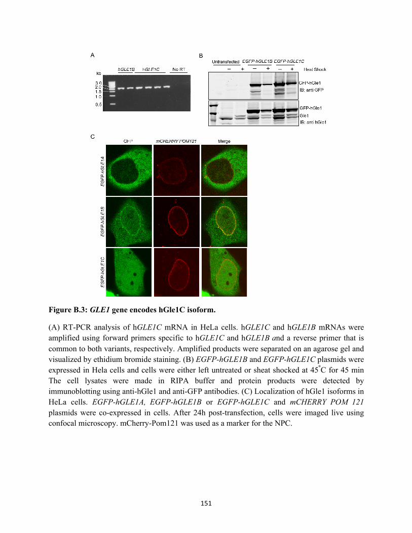
151
Figure B.3: GLE1 gene encodes hGle1C isoform.
(A) RT-PCR analysis of hGLE1C mRNA in HeLa cells. hGLE1C and hGLE1B mRNAs were amplified using forward primers specific to hGLE1C and hGLE1B and a reverse primer that is common to both variants, respectively. Amplified products were separated on an agarose gel and visualized by ethidium bromide staining. (B) EGFP-hGLE1B and EGFP-hGLE1C plasmids were expressed in Hela cells and cells were either left untreated or sheat shocked at 45°C for 45 min The cell lysates were made in RIPA buffer and protein products were detected by immunoblotting using anti-hGle1 and anti-GFP antibodies. (C) Localization of hGle1 isoforms in HeLa cells. EGFP-hGLE1A, EGFP-hGLE1B or EGFP-hGLE1C and mCHERRY POM 121 plasmids were co-expressed in cells. After 24h post-transfection, cells were imaged live using confocal microscopy. mCherry-Pom121 was used as a marker for the NPC.

152
These results confirmed that an additional mRNA isoform of hGle1 is expressed in HeLa cells.
We designated this new isoform as hGle1C protein.
We next investigated the localization of this protein in HeLa cells. Plasmids expressing
either EGFP-hGLE1A, EGFP-hGLE1B, or EGFP-hGLE1C and mCherry POM121 were
expressed in HeLa cells and visualized by live microscopy. Pom121 was used as a marker for the
nuclear rim. Interestingly, EGFP-hGle1C did not localize to nuclear rim, but appeared to localize
in the nucleus and at the cytoplasm at the steady state levels. Importantly, we observed a
noticeable difference in the hGle1C localization pattern compared to hGle1A (present in the
cytoplasm) and hGle1B isoforms (present at the nuclear envelope, cytoplasm and nucleus)
(Figure B.3C)(Aditi et al., 2015; Kendirgi et al., 2003). Since the sequence of the first 38 amino
acids of hGle1C protein is different compared to hGle1A and hGle1B isoforms, this dissimilarity
could account for its distinct localization (Figure B.2B). Notably, the amino-terminal 29 amino
acids of hGle1A and hGle1B proteins interact with Nup155 protein (Rayala et al., 2004), and this
hGle1B domain is required for NPC localization. Therefore, we anticipate that hGle1C might not
be capable of binding to Nup155. We also tested the localization of this isoform in SGs and
observed that it is recruited to SGs similar to other hGle1 isoforms (Figure B.4).
Taken together, these results indicate that an additional isoform of hGle1 is expressed in
human cells. This suggests that, by having multiple isoforms, hGle1 could regulate various steps
of gene expression in a spatial and temporal manner. Moreover, these isoforms may have tissue -
specific functions. Future experiments could be focused on understanding the biological roles of
various hGle1 isoforms in a cell and tissue-specific manner and how they influence gene
expression.

153
Figure B.4: hGle1C is localized to stress granules upon heat shock.
Plasmids expressing either GFP-hGle1B or GFP-hGle1C and mCherry-G3BP proteins were co-expressed in HeLa cells. Cells were heat shocked at 45°C for 45 min and imaged live by confocal microscopy.
154
REFERENCES Abastado, J.P., Miller, P.F., Jackson, B.M., and Hinnebusch, A.G. (1991). Suppression of ribosomal reinitiation at upstream open reading frames in amino acid-starved cells forms the basis for GCN4 translational control. Mol. Cell. Biol. 11, 486–496.
Adams, R.L., Terry, L.J., and Wente, S.R. (2014). Nucleoporin FG domains facilitate mRNP remodeling at the cytoplasmic face of the nuclear pore complex. Genetics 197, 1213–1224.
Adelman, K., and Lis, J.T. (2012). Promoter-proximal pausing of RNA polymerase II: emerging roles in metazoans. Nat. Rev. Genet. 13, 720–731.
Aditi, Folkmann, A.W., and Wente, S.R. (2015). Cytoplasmic hGle1A regulates stress granules by modulation of translation. Mol. Biol. Cell 26, 1476–1490.
Aitken, C.E., and Lorsch, J.R. (2012). A mechanistic overview of translation initiation in eukaryotes. Nat. Struct. Mol. Biol. 19, 568–576.
Alami, N.H., Smith, R.B., Carrasco, M.A., Williams, L.A., Winborn, C.S., Han, S.S.W., Kiskinis, E., Winborn, B., Freibaum, B.D., Kanagaraj, A., et al. (2014). Axonal transport of TDP-43 mRNA granules is impaired by ALS-causing mutations. Neuron 81, 536–543.
Alcázar-Román, A.R., Tran, E.J., Guo, S., and Wente, S.R. (2006). Inositol hexakisphosphate and Gle1 activate the DEAD-box protein Dbp5 for nuclear mRNA export. Nat. Cell Biol. 8, 711–716.
Alcázar-Román, A.R., Bolger, T.A., and Wente, S.R. (2010). Control of mRNA export and translation termination by inositol hexakisphosphate requires specific interaction with Gle1. J. Biol. Chem. 285, 16683–16692.
Anckar, J., and Sistonen, L. (2011). Regulation of HSF1 function in the heat stress response: implications in aging and disease. Annu. Rev. Biochem. 80, 1089–1115.
Anderson, P. (2006). RNA granules. J. Cell Biol. 172, 803–808.
Anderson, P., and Kedersha, N. (2008). Stress granules: the Tao of RNA triage. Trends Biochem. Sci. 33, 141–150.
Anderson, P., and Kedersha, N. (2009). RNA granules: post-transcriptional and epigenetic modulators of gene expression. Nat. Rev. Mol. Cell Biol. 10, 430–436.
Anekonda, T.S., Wadsworth, T.L., Sabin, R., Frahler, K., Harris, C., Petriko, B., Ralle, M., Woltjer, R., and Quinn, J.F. (2011). Phytic acid as a potential treatment for alzheimer’s pathology: evidence from animal and in vitro models. J. Alzheimers Dis. JAD 23, 21–35.
Arai, T., Hasegawa, M., Akiyama, H., Ikeda, K., Nonaka, T., Mori, H., Mann, D., Tsuchiya, K., Yoshida, M., Hashizume, Y., et al. (2006). TDP-43 is a component of ubiquitin-positive tau-

155
negative inclusions in frontotemporal lobar degeneration and amyotrophic lateral sclerosis. Biochem. Biophys. Res. Commun. 351, 602–611.
Arimoto, K., Fukuda, H., Imajoh-Ohmi, S., Saito, H., and Takekawa, M. (2008). Formation of stress granules inhibits apoptosis by suppressing stress-responsive MAPK pathways. Nat. Cell Biol. 10, 1324–1332.
Armakola, M., Higgins, M.J., Figley, M.D., Barmada, S.J., Scarborough, E.A., Diaz, Z., Fang, X., Shorter, J., Krogan, N.J., Finkbeiner, S., et al. (2012). Inhibition of RNA lariat debranching enzyme suppresses TDP-43 toxicity in ALS disease models. Nat. Genet. 44, 1302–1309.
Armbruster, D., Nagy, J., and Young, J. (2014). Three level signal transduction cascades lead to reliably timed switches. J. Theor. Biol. 361, 69–80.
Balagopal, V., and Parker, R. (2009). Polysomes, P bodies and stress granules: states and fates of eukaryotic mRNAs. Curr. Opin. Cell Biol. 21, 403–408.
Barbosa, C., Peixeiro, I., and Romão, L. (2013). Gene expression regulation by upstream open reading frames and human disease. PLoS Genet 9, e1003529.
Baron, D.M., Kaushansky, L.J., Ward, C.L., Sama, R.R.K., Chian, R.-J., Boggio, K.J., Quaresma, A.J.C., Nickerson, J.A., and Bosco, D.A. (2013). Amyotrophic lateral sclerosis-linked FUS/TLS alters stress granule assembly and dynamics. Mol. Neurodegener. 8, 30.
Barthelme, D., Dinkelaker, S., Albers, S.-V., Londei, P., Ermler, U., and Tampé, R. (2011). Ribosome recycling depends on a mechanistic link between the FeS cluster domain and a conformational switch of the twin-ATPase ABCE1. Proc. Natl. Acad. Sci. 108, 3228–3233.
Beltrao, P., Bork, P., Krogan, N.J., and van Noort, V. (2013). Evolution and functional cross-talk of protein post-translational modifications. Mol. Syst. Biol. 9, 714.
Bensaude, O., Bellier, S., Dubois, M.F., Giannoni, F., and Nguyen, V.T. (1996). Heat-shock induced protein modifications and modulation of enzyme activities. EXS 77, 199–219.
Bentley, D.L. (2014). Coupling mRNA processing with transcription in time and space. Nat. Rev. Genet. 15, 163–175.
Bentmann, E., Haass, C., and Dormann, D. (2013). Stress granules in neurodegeneration--lessons learnt from TAR DNA binding protein of 43 kDa and fused in sarcoma. FEBS J. 280, 4348–4370.
Biamonti, G., and Vourc’h, C. (2010). Nuclear stress bodies. Cold Spring Harb. Perspect. Biol. 2.
Blais, J.D., Filipenko, V., Bi, M., Harding, H.P., Ron, D., Koumenis, C., Wouters, B.G., and Bell, J.C. (2004). Activating transcription factor 4 is translationally regulated by hypoxic stress. Mol. Cell. Biol. 24, 7469–7482.

156
Bolger, T.A., and Wente, S.R. (2011). Gle1 is a multifunctional DEAD-box protein regulator that modulates Ded1 in translation initiation. J. Biol. Chem. 286, 39750–39759.
Bolger, T.A., Folkmann, A.W., Tran, E.J., and Wente, S.R. (2008). The mRNA Export Factor Gle1 and Inositol Hexakisphosphate Regulate Distinct Stages of Translation. Cell 134, 624–633.
Bonnet, A., and Palancade, B. (2014). Regulation of mRNA trafficking by nuclear pore complexes. Genes 5, 767–791.
Bornes, S., Prado-Lourenco, L., Bastide, A., Zanibellato, C., Iacovoni, J.S., Lacazette, E., Prats, A.-C., Touriol, C., and Prats, H. (2007). Translational induction of VEGF internal ribosome entry site elements during the early response to ischemic stress. Circ. Res. 100, 305–308.
Bosco, D.A., Lemay, N., Ko, H.K., Zhou, H., Burke, C., Kwiatkowski, T.J., Sapp, P., McKenna-Yasek, D., Brown, R.H., and Hayward, L.J. (2010). Mutant FUS proteins that cause amyotrophic lateral sclerosis incorporate into stress granules. Hum. Mol. Genet. 19, 4160–4175.
Bowers, H.A., Maroney, P.A., Fairman, M.E., Kastner, B., Lührmann, R., Nilsen, T.W., and Jankowsky, E. (2006). Discriminatory RNP remodeling by the DEAD-box protein DED1. RNA 12, 903–912.
Brannan, K., Bentley, D.L., Brannan, K., and Bentley, D.L. (2012). Control of transcriptional elongation by RNA Polymerase II: a retrospective Genet. Res. Int, 2012, e170173.
Brehm, M.A., Schenk, T.M.H., Zhou, X., Fanick, W., Lin, H., Windhorst, S., Nalaskowski, M.M., Kobras, M., Shears, S.B., and Mayr, G.W. (2007). Intracellular localization of human Ins(1,3,4,5,6)P5 2-kinase. Biochem. J. 408, 335–345.
Brewster, J.L., and Gustin, M.C. (2014). Hog1: 20 years of discovery and impact. Sci Signal 7, re7–re7.
Brockmann, C., Soucek, S., Kuhlmann, S.I., Mills-Lujan, K., Kelly, S.M., Yang, J.-C., Iglesias, N., Stutz, F., Corbett, A.H., Neuhaus, D., et al. (2012). Structural basis for polyadenosine-RNA binding by Nab2 Zn fingers and its function in mRNA nuclear export. Struct. Lond. Engl. 1993 20, 1007–1018.
Browne, G.J., and Proud, C.G. (2004). A novel mTOR-regulated phosphorylation site in elongation factor 2 kinase modulates the activity of the kinase and its binding to calmodulin. Mol. Cell. Biol. 24, 2986–2997.
Browne, G.J., Finn, S.G., and Proud, C.G. (2004). Stimulation of the AMP-activated protein kinase leads to activation of eukaryotic elongation factor 2 kinase and to its phosphorylation at a novel site, serine 398. J. Biol. Chem. 279, 12220–12231.
Brunn, G.J., Hudson, C.C., Sekulić, A., Williams, J.M., Hosoi, H., Houghton, P.J., Lawrence, J.C., and Abraham, R.T. (1997). Phosphorylation of the translational repressor PHAS-I by the mammalian target of rapamycin. Science 277, 99–101.

157
Buchan, J.R. (2014). mRNP granules. Assembly, function, and connections with disease. RNA Biol. 11, 1019–1030.
Buchan, J.R., and Parker, R. (2009). Eukaryotic stress granules: the ins and out of translation. Mol. Cell 36, 932.
Buchan, J.R., Muhlrad, D., and Parker, R. (2008). P bodies promote stress granule assembly in Saccharomyces cerevisiae. J. Cell Biol. 183, 441–455.
Buchan, J.R., Kolaitis, R.-M., Taylor, J.P., and Parker, R. (2013). Eukaryotic stress granules are cleared by autophagy and Cdc48/VCP function. Cell 153, 1461–1474.
Carmody, S.R., and Wente, S.R. (2009). mRNA nuclear export at a glance. J. Cell Sci. 122, 1933–1937.
Carmody, S.R., Tran, E.J., Apponi, L.H., Corbett, A.H., and Wente, S.R. (2010). The mitogen-activated protein kinase Slt2 regulates nuclear retention of non-heat shock mRNAs during heat shock-induced stress. Mol. Cell. Biol. 30, 5168–5179.
Carter-O’Connell, I., Jin, H., Morgan, R.K., Zaja, R., David, L.L., Ahel, I., and Cohen, M.S. (2016). Identifying family-member-specific targets of mono-ARTDs by using a chemical genetics approach. Cell Rep. 14, 621–631.
Caruthers, J.M., and McKay, D.B. (2002). Helicase structure and mechanism. Curr. Opin. Struct. Biol. 12, 123–133.
Chan, C.T.Y., Pang, Y.L.J., Deng, W., Babu, I.R., Dyavaiah, M., Begley, T.J., and Dedon, P.C. (2012). Reprogramming of tRNA modifications controls the oxidative stress response by codon-biased translation of proteins. Nat. Commun. 3, 937.
Chen, Y., and Brandizzi, F. (2013). IRE1: ER stress sensor and cell fate executor. Trends Cell Biol. 23, 547–555.
Cherkasov, V., Hofmann, S., Druffel-Augustin, S., Mogk, A., Tyedmers, J., Stoecklin, G., and Bukau, B. (2013). Coordination of translational control and protein homeostasis during severe heat stress. Curr. Biol. CB 23, 2452–2462.
Chernov, K.G., Barbet, A., Hamon, L., Ovchinnikov, L.P., Curmi, P.A., and Pastré, D. (2009). Role of microtubules in stress granule ssembly microtubule dynamical instability favors the formation of micrometric stress granules in cells. J. Biol. Chem. 284, 36569–36580.
Chu, W.M., Ballard, R., Carpick, B.W., Williams, B.R., and Schmid, C.W. (1998). Potential Alu function: regulation of the activity of double-stranded RNA-activated kinase PKR. Mol. Cell. Biol. 18, 58–68.
Colombrita, C., Zennaro, E., Fallini, C., Weber, M., Sommacal, A., Buratti, E., Silani, V., and Ratti, A. (2009). TDP-43 is recruited to stress granules in conditions of oxidative insult. J. Neurochem. 111, 1051–1061.

158
Cuesta, R., Laroia, G., and Schneider, R.J. (2000). Chaperone Hsp27 inhibits translation during heat shock by binding eIF4G and facilitating dissociation of cap-initiation complexes. Genes Dev. 14, 1460–1470.
Czech, A., Wende, S., Mörl, M., Pan, T., and Ignatova, Z. (2013). Reversible and rapid transfer-RNA deactivation as a mechanism of translational repression in stress. PLoS Genet 9, e1003767.
Decker, C.J., and Parker, R. (2012). P-Bodies and Stress Granules: Possible roles in the control of translation and mRNA degradation. Cold Spring Harb. Perspect. Biol. 4.
De Leeuw, F., Zhang, T., Wauquier, C., Huez, G., Kruys, V., and Gueydan, C. (2007). The cold-inducible RNA-binding protein migrates from the nucleus to cytoplasmic stress granules by a methylation-dependent mechanism and acts as a translational repressor. Exp. Cell Res. 313, 4130–4144.
Dever, T.E., and Green, R. (2012). The elongation, termination, and recycling phases of translation in eukaryotes. Cold Spring Harb. Perspect. Biol. 4.
Dever, T.E., Feng, L., Wek, R.C., Cigan, A.M., Donahue, T.F., and Hinnebusch, A.G. (1992). Phosphorylation of initiation factor 2α by protein kinase GCN2 mediates gene-specific translational control of GCN4 in yeast. Cell 68, 585–596.
Dewey, C.M., Cenik, B., Sephton, C.F., Dries, D.R., Mayer, P., Good, S.K., Johnson, B.A., Herz, J., and Yu, G. (2011). TDP-43 is directed to stress granules by sorbitol, a novel physiological osmotic and oxidative stressor. Mol. Cell. Biol. 31, 1098–1108.
Dieterich, D.C., Link, A.J., Graumann, J., Tirrell, D.A., and Schuman, E.M. (2006). Selective identification of newly synthesized proteins in mammalian cells using bioorthogonal noncanonical amino acid tagging (BONCAT). Proc. Natl. Acad. Sci. 103, 9482–9487.
Dieterich, D.C., Lee, J.J., Link, A.J., Graumann, J., Tirrell, D.A., and Schuman, E.M. (2007). Labeling, detection and identification of newly synthesized proteomes with bioorthogonal non-canonical amino-acid tagging. Nat. Protoc. 2, 532–540.
Di Giammartino, D.C., Nishida, K., and Manley, J.L. (2011). Mechanisms and consequences of alternative polyadenylation. Mol. Cell 43, 853–866.
Dormann, D., and Haass, C. (2011). TDP-43 and FUS: a nuclear affair. Trends Neurosci. 34, 339–348.
Dormann, D., Rodde, R., Edbauer, D., Bentmann, E., Fischer, I., Hruscha, A., Than, M.E., Mackenzie, I.R.A., Capell, A., Schmid, B., et al. (2010). ALS-associated fused in sarcoma (FUS) mutations disrupt Transportin-mediated nuclear import. EMBO J. 29, 2841–2857.
Elden, A.C., Kim, H.-J., Hart, M.P., Chen-Plotkin, A.S., Johnson, B.S., Fang, X., Armakola, M., Geser, F., Greene, R., Lu, M.M., et al. (2010). Ataxin-2 intermediate-length polyglutamine expansions are associated with increased risk for ALS. Nature 466, 1069–1075.

159
Estruch, F. (2000). Stress-controlled transcription factors, stress-induced genes and stress tolerance in budding yeast. FEMS Microbiol. Rev. 24, 469–486.
Feder, M.E., and Hofmann, G.E. (1999). Heat-shock proteins, molecular chaperones, and the stress response: evolutionary and ecological physiology. Annu. Rev. Physiol. 61, 243–282.
Feoktistova, K., Tuvshintogs, E., Do, A., and Fraser, C.S. (2013). Human eIF4E promotes mRNA restructuring by stimulating eIF4A helicase activity. Proc. Natl. Acad. Sci. U. S. A. 110, 13339–13344.
Ferreiro, I., Barragan, M., Gubern, A., Ballestar, E., Joaquin, M., and Posas, F. (2010). The p38 SAPK is recruited to chromatin via its interaction with transcription factors. J. Biol. Chem. 285, 31819–31828.
Ferrigno, P., Posas, F., Koepp, D., Saito, H., and Silver, P.A. (1998). Regulated nucleo/cytoplasmic exchange of HOG1 MAPK requires the importin beta homologs NMD5 and XPO1. EMBO J. 17, 5606–5614.
Fidler, J.A., Treleaven, C.M., Frakes, A., Tamsett, T.J., McCrate, M., Cheng, S.H., Shihabuddin, L.S., Kaspar, B.K., and Dodge, J.C. (2011). Disease progression in a mouse model of amyotrophic lateral sclerosis: the influence of chronic stress and corticosterone. FASEB J. Off. Publ. Fed. Am. Soc. Exp. Biol. 25, 4369–4377.
Filipovska, A., and Rackham, O. (2013). Specialization from synthesis: How ribosome diversity can customize protein function. FEBS Lett. 587, 1189–1197.
Folkmann, A.W., Noble, K.N., Cole, C.N., and Wente, S.R. (2011). Dbp5, Gle1-IP6 and Nup159: A working model for mRNP export. Nucl. Austin Tex 2, 540–548.
Folkmann, A.W., Collier, S.E., Zhan, X., Aditi, Ohi, M.D., and Wente, S.R. (2013). Gle1 functions during mRNA export in an oligomeric complex that is altered in human disease. Cell 155, 582–593.
Folkmann, A.W., Dawson, T.R., and Wente, S.R. (2014). Insights into mRNA export-linked molecular mechanisms of human disease through a Gle1 structure-function analysis. Adv. Biol. Regul. 54, 74–91.
Fox, B.W., and Tibbetts, R.S. (2015). Neurodegeneration: Problems at the nuclear pore. Nature 525, 36–37.
Freibaum, B.D., Lu, Y., Lopez-Gonzalez, R., Kim, N.C., Almeida, S., Lee, K.-H., Badders, N., Valentine, M., Miller, B.L., Wong, P.C., et al. (2015). GGGGCC repeat expansion in C9orf72 compromises nucleocytoplasmic transport. Nature 525, 129–133.
Fulda, S., Gorman, A.M., Hori, O., Samali, A., Fulda, S., Gorman, A.M., Hori, O., and Samali, A. (2010). Cellular stress responses: cell survival and cell death. Int. J. Cell Biol. Int. J. Cell Biol. 2010, 2010, e214074.

160
Gallouzi, I.-E., Brennan, C.M., Stenberg, M.G., Swanson, M.S., Eversole, A., Maizels, N., and Steitz, J.A. (2000). HuR binding to cytoplasmic mRNA is perturbed by heat shock. Proc. Natl. Acad. Sci. 97, 3073–3078.
Gareau, C., Fournier, M.-J., Filion, C., Coudert, L., Martel, D., Labelle, Y., and Mazroui, R. (2011). p21WAF1/CIP1 Upregulation through the stress granule-associated protein CUGBP1 confers resistance to bortezomib-mediated apoptosis. PLoS ONE 6, e20254.
Gasch, A.P., Spellman, P.T., Kao, C.M., Carmel-Harel, O., Eisen, M.B., Storz, G., Botstein, D., and Brown, P.O. (2000). Genomic expression programs in the response of yeast cells to environmental changes. Mol. Biol. Cell 11, 4241–4257.
Gebauer, F., and Hentze, M.W. (2004). Molecular mechanisms of translational control. Nat. Rev. Mol. Cell Biol. 5, 827–835.
Geissler, R., Golbik, R.P., and Behrens, S.-E. (2012). The DEAD-box helicase DDX3 supports the assembly of functional 80S ribosomes. Nucleic Acids Res. 40, 4998–5011.
Ghavami, S., Shojaei, S., Yeganeh, B., Ande, S.R., Jangamreddy, J.R., Mehrpour, M., Christoffersson, J., Chaabane, W., Moghadam, A.R., Kashani, H.H., et al. (2014). Autophagy and apoptosis dysfunction in neurodegenerative disorders. Prog. Neurobiol. 112, 24–49.
Gilks, N., Kedersha, N., Ayodele, M., Shen, L., Stoecklin, G., Dember, L.M., and Anderson, P. (2004). Stress granule assembly is mediated by prion-like aggregation of TIA-1. Mol. Biol. Cell 15, 5383–5398.
Gingras, A.C., Kennedy, S.G., O’Leary, M.A., Sonenberg, N., and Hay, N. (1998). 4E-BP1, a repressor of mRNA translation, is phosphorylated and inactivated by the Akt(PKB) signaling pathway. Genes Dev. 12, 502–513.
Glisovic, T., Bachorik, J.L., Yong, J., and Dreyfuss, G. (2008). RNA-binding proteins and post-transcriptional gene regulation. FEBS Lett. 582, 1977–1986.
Grünwald, D., Singer, R.H., and Rout, M. (2011). Nuclear export dynamics of RNA–protein complexes. Nature 475, 333–341.
Grüter, P., Tabernero, C., von Kobbe, C., Schmitt, C., Saavedra, C., Bachi, A., Wilm, M., Felber, B.K., and Izaurralde, E. (1998). TAP, the human homolog of Mex67p, mediates CTE-dependent RNA export from the nucleus. Mol. Cell 1, 649–659.
Gu, C., Begley, T.J., and Dedon, P.C. (2014). tRNA modifications regulate translation during cellular stress. FEBS Lett. 588, 4287–4296.
Gustafson, E.A., and Wessel, G.M. (2010). DEAD-box Helicases: Posttranslational regulation and function. Biochem. Biophys. Res. Commun. 395, 1–6.
Hahn, S. (2004). Structure and mechanism of the RNA Polymerase II transcription machinery. Nat. Struct. Mol. Biol. 11, 394–403.

161
Han, T.W., Kato, M., Xie, S., Wu, L.C., Mirzaei, H., Pei, J., Chen, M., Xie, Y., Allen, J., Xiao, G., et al. (2012). Cell-free formation of RNA granules: bound RNAs identify features and components of cellular assemblies. Cell 149, 768–779.
Haro, C. de, Méndez, R., and Santoyo, J. (1996). The eIF-2alpha kinases and the control of protein synthesis. FASEB J. 10, 1378–1387.
Hatch, E., and Hetzer, M. (2014). Breaching the nuclear envelope in development and disease. J. Cell Biol. 205, 133–141.
Hautbergue, G.M., Hung, M.-L., Golovanov, A.P., Lian, L.-Y., and Wilson, S.A. (2008). Mutually exclusive interactions drive handover of mRNA from export adaptors to TAP. Proc. Natl. Acad. Sci. U. S. A. 105, 5154–5159.
Hellen, C.U.T., and Sarnow, P. (2001). Internal ribosome entry sites in eukaryotic mRNA molecules. Genes Dev. 15, 1593–1612.
Herold, A., Suyama, M., Rodrigues, J.P., Braun, I.C., Kutay, U., Carmo-Fonseca, M., Bork, P., and Izaurralde, E. (2000). TAP (NXF1) belongs to a multigene family of putative RNA export factors with a conserved modular architecture. Mol. Cell. Biol. 20, 8996–9008.
Hilliker, A., Gao, Z., Jankowsky, E., and Parker, R. (2011). The DEAD-box protein Ded1 modulates translation by the formation and resolution of an eIF4F-mRNA complex. Mol. Cell 43, 962–972.
Hinnebusch, A.G. (2011). Molecular mechanism of scanning and start codon selection in eukaryotes. Microbiol. Mol. Biol. Rev. 75, 434–467.
Hirokawa, G., Nijman, R.M., Raj, V.S., Kaji, H., Igarashi, K., and Kaji, A. (2005). The role of ribosome recycling factor in dissociation of 70S ribosomes into subunits. RNA N. Y. N 11, 1317–1328.
Hodge, C.A., Tran, E.J., Noble, K.N., Alcazar-Roman, A.R., Ben-Yishay, R., Scarcelli, J.J., Folkmann, A.W., Shav-Tal, Y., Wente, S.R., and Cole, C.N. (2011). The Dbp5 cycle at the nuclear pore complex during mRNA export I: dbp5 mutants with defects in RNA binding and ATP hydrolysis define key steps for Nup159 and Gle1. Genes Dev. 25, 1052–1064.
Hofmann, S., Cherkasova, V., Bankhead, P., Bukau, B., and Stoecklin, G. (2012). Translation suppression promotes stress granule formation and cell survival in response to cold shock. Mol. Biol. Cell 23, 3786–3800.
Hohmann, S., Krantz, M., and Nordlander, B. (2007). Yeast osmoregulation. Methods Enzymol. 428, 29–45.
Holcik, M., and Sonenberg, N. (2005). Translational control in stress and apoptosis. Nat. Rev. Mol. Cell Biol. 6, 318–327.

162
Holčík, M., Gordon, B.W., and Korneluk, R.G. (2003). The internal ribosome entry site-mediated translation of antiapoptotic protein XIAP is modulated by the heterogeneous nuclear ribonucleoproteins C1 and C2. Mol. Cell. Biol. 23, 280–288.
Horman, S., Browne, G., Krause, U., Patel, J., Vertommen, D., Bertrand, L., Lavoinne, A., Hue, L., Proud, C., and Rider, M. (2002). Activation of AMP-activated protein kinase leads to the phosphorylation of elongation factor 2 and an inhibition of protein synthesis. Curr. Biol. CB 12, 1419–1423.
Hou, Y.-M., Gamper, H., and Yang, W. (2015). Post-transcriptional modifications to tRNA—A response to the genetic code degeneracy. RNA 21, 642–644.
Huebert, D.J., Kuan, P.-F., Keleş, S., and Gasch, A.P. (2012). Dynamic changes in nucleosome occupancy are not predictive of gene expression dynamics but are linked to transcription and chromatin regulators. Mol. Cell. Biol. 32, 1645–1653.
Hurt, E., Sträßer, K., Segref, A., Bailer, S., Schlaich, N., Presutti, C., Tollervey, D., and Jansen, R. (2000). Mex67p mediates nuclear export of a variety of RNA Polymerase II transcripts. J. Biol. Chem. 275, 8361–8368.
Hutten, S., Sharangdhar, T., and Kiebler, M. (2014). Unmasking the messenger. RNA Biol. 11, 992–997.
Hyman, A.A., and Simons, K. (2012). Beyond oil and water—Phase transitions in cells. Science 337, 1047–1049.
Iglesias, N., Tutucci, E., Gwizdek, C., Vinciguerra, P., Von Dach, E., Corbett, A.H., Dargemont, C., and Stutz, F. (2010). Ubiquitin-mediated mRNP dynamics and surveillance prior to budding yeast mRNA export. Genes Dev. 24, 1927–1938.
Ingolia NT, Brar GA, Rouskin S, McGeachy AM, Weissman JS. (2012). The ribosome profiling strategy for monitoring translation in vivo by deep sequencing of ribosome-protected mRNA fragments. Nature protocols. 7(8):1534-1550. Ivanov, P.A., Chudinova, E.M., and Nadezhdina, E.S. (2003). Disruption of microtubules inhibits cytoplasmic ribonucleoprotein stress granule formation. Exp. Cell Res. 290, 227–233.
Izawa, S., Takemura, R., and Inoue, Y. (2004). Gle2p is essential to induce adaptation of the export of bulk poly(A)+ mRNA to heat shock in Saccharomyces cerevisiae. J. Biol. Chem. 279, 35469–35478.
Jackson, R.J., Hellen, C.U.T., and Pestova, T.V. (2012). Termination and post-termination events in eukaryotic translation. Adv. Protein Chem. Struct. Biol. 86, 45–93.
Jain, S., Wheeler, J.R., Walters, R.W., Agrawal, A., Barsic, A., and Parker, R. (2016). ATPase-modulated stress granules contain a diverse proteome and substructure. Cell 164, 487–498.

163
Jan, C.H., Williams, C.C., and Weissman, J.S. (2014). Principles of ER cotranslational translocation revealed by proximity-specific ribosome profiling. Science 346, 1257521.
Janosi, L., Shimizu, I., and Kaji, A. (1994). Ribosome recycling factor (ribosome releasing factor) is essential for bacterial growth. Proc. Natl. Acad. Sci. U. S. A. 91, 4249–4253.
Jao, L.-E., Appel, B., and Wente, S.R. (2012). A zebrafish model of lethal congenital contracture syndrome 1 reveals Gle1 function in spinal neural precursor survival and motor axon arborization. Dev. Camb. Engl. 139, 1316–1326.
Jarmoskaite, I., and Russell, R. (2014). RNA helicase proteins as chaperones and remodelers. Annu. Rev. Biochem. 83, 697–725.
Johnson, B.S., McCaffery, J.M., Lindquist, S., and Gitler, A.D. (2008). A yeast TDP-43 proteinopathy model: Exploring the molecular determinants of TDP-43 aggregation and cellular toxicity. Proc. Natl. Acad. Sci. U. S. A. 105, 6439–6444.
Johnson, B.S., Snead, D., Lee, J.J., McCaffery, J.M., Shorter, J., and Gitler, A.D. (2009). TDP-43 is intrinsically aggregation-prone, and amyotrophic lateral sclerosis-linked mutations accelerate aggregation and increase toxicity. J. Biol. Chem. 284, 20329–20339.
Johnson, J.O., Mandrioli, J., Benatar, M., Abramzon, Y., Van Deerlin, V.M., Trojanowski, J.Q., Gibbs, J.R., Brunetti, M., Gronka, S., Wuu, J., et al. (2010). Exome sequencing reveals VCP mutations as a cause of familial ALS. Neuron 68, 857–864.
Jonkers, I., and Lis, J.T. (2015). Getting up to speed with transcription elongation by RNA polymerase II. Nat. Rev. Mol. Cell Biol. 16, 167–177.
Jovičić, A., Mertens, J., Boeynaems, S., Bogaert, E., Chai, N., Yamada, S.B., Paul, J.W., Sun, S., Herdy, J.R., Bieri, G., et al. (2015). Modifiers of C9orf72 dipeptide repeat toxicity connect nucleocytoplasmic transport defects to FTD/ALS. Nat. Neurosci. 18, 1226–1229.
Kaehler, C., Isensee, J., Hucho, T., Lehrach, H., and Krobitsch, S. (2014). 5-Fluorouracil affects assembly of stress granules based on RNA incorporation. Nucleic Acids Res. 42, 6436–6447.
Kaneb, H.M., Folkmann, A.W., Belzil, V.V., Jao, L.-E., Leblond, C.S., Girard, S.L., Daoud, H., Noreau, A., Rochefort, D., Hince, P., et al. (2015). Deleterious mutations in the essential mRNA metabolism factor, hGle1, in amyotrophic lateral sclerosis. Hum. Mol. Genet. 24, 1363–1373.
Kang, Y., and Cullen, B.R. (1999). The human Tap protein is a nuclear mRNA export factor that contains novel RNA-binding and nucleocytoplasmic transport sequences. Genes Dev. 13, 1126–1139.
Karijolich, J., and Yu, Y.-T. (2015). The new era of RNA modification. RNA 21, 659–660.
Kato, M., Han, T.W., Xie, S., Shi, K., Du, X., Wu, L.C., Mirzaei, H., Goldsmith, E.J., Longgood, J., Pei, J., et al. (2012). Cell-free formation of RNA granules: low complexity sequence domains form dynamic fibers within hydrogels. Cell 149, 753–767.

164
Kedersha, N., and Anderson, P. (2002). Stress granules: sites of mRNA triage that regulate mRNA stability and translatability. Biochem. Soc. Trans. 30, 963–970.
Kedersha, N., and Anderson, P. (2009). Regulation of translation by stress granules and processing bodies. In Progress in Molecular Biology and Translational Science, (Elsevier), pp. 155–185.
Kedersha, N., Cho, M.R., Li, W., Yacono, P.W., Chen, S., Gilks, N., Golan, D.E., and Anderson, P. (2000). Dynamic shuttling of TIA-1 accompanies the recruitment of mRNA to mammalian stress granules. J. Cell Biol. 151, 1257–1268.
Kedersha, N., Stoecklin, G., Ayodele, M., Yacono, P., Lykke-Andersen, J., Fritzler, M.J., Scheuner, D., Kaufman, R.J., Golan, D.E., and Anderson, P. (2005). Stress granules and processing bodies are dynamically linked sites of mRNP remodeling. J. Cell Biol. 169, 871–884.
Kedersha, N., Ivanov, P., and Anderson, P. (2013). Stress granules and cell signaling: more than just a passing phase? Trends Biochem. Sci. 38, 494–506.
Kedersha, N.L., Gupta, M., Li, W., Miller, I., and Anderson, P. (1999). RNA-binding proteins TIA-1 and TIAR link the phosphorylation of eIF-2 alpha to the assembly of mammalian stress granules. J. Cell Biol. 147, 1431–1442.
Keene, J.D. (2007). RNA regulons: coordination of post-transcriptional events. Nat. Rev. Genet. 8, 533–543.
Kendirgi, F., Barry, D.M., Griffis, E.R., Powers, M.A., and Wente, S.R. (2003). An essential role for hGle1 nucleocytoplasmic shuttling in mRNA export. J. Cell Biol. 160, 1029–1040.
Kendirgi, F., Rexer, D.J., Alcázar-Román, A.R., Onishko, H.M., and Wente, S.R. (2005). Interaction between the shuttling mRNA export factor Gle1 and the nucleoporin hCG1: a conserved mechanism in the export of Hsp70 mRNA. Mol. Biol. Cell 16, 4304–4315.
Khaperskyy, D.A., Hatchette, T.F., and McCormick, C. (2012). Influenza A virus inhibits cytoplasmic stress granule formation. FASEB J. Off. Publ. Fed. Am. Soc. Exp. Biol. 26, 1629–1639.
Kiel, M.C., Kaji, H., and Kaji, A. (2007). Ribosome recycling: An essential process of protein synthesis. Biochem. Mol. Biol. Educ. Bimon. Publ. Int. Union Biochem. Mol. Biol. 35, 40–44.
Kim, E.K., and Choi, E.-J. (2010). Pathological roles of MAPK signaling pathways in human diseases. Biochim. Biophys. Acta BBA - Mol. Basis Dis. 1802, 396–405.
Kim, H.J., Kim, N.C., Wang, Y.-D., Scarborough, E.A., Moore, J., Diaz, Z., MacLea, K.S., Freibaum, B., Li, S., Molliex, A., et al. (2013). Mutations in prion-like domains in hnRNPA2B1 and hnRNPA1 cause multisystem proteinopathy and ALS. Nature 495, 467–473.
Kim, H.-J., Raphael, A.R., LaDow, E.S., McGurk, L., Weber, R.A., Trojanowski, J.Q., Lee, V.M.-Y., Finkbeiner, S., Gitler, A.D., and Bonini, N.M. (2014). Therapeutic modulation of

165
eIF2α phosphorylation rescues TDP-43 toxicity in amyotrophic lateral sclerosis disease models. Nat. Genet. 46, 152–160.
Kim, W.J., Back, S.H., Kim, V., Ryu, I., and Jang, S.K. (2005). Sequestration of TRAF2 into stress granules interrupts tumor necrosis factor signaling under stress conditions. Mol. Cell. Biol. 25, 2450–2462.
Kimura, T., Hashimoto, I., Nagase, T., and Fujisawa, J.-I. (2004). CRM1-dependent, but not ARE-mediated, nuclear export of IFN-alpha1 mRNA. J. Cell Sci. 117, 2259–2270.
King, O.D., Gitler, A.D., and Shorter, J. (2012). The tip of the iceberg: RNA-binding proteins with prion-like domains in neurodegenerative disease. Brain Res. 1462, 61–80.
Kinoshita, E., Kinoshita-Kikuta, E., Takiyama, K., and Koike, T. (2006). Phosphate-binding Tag, a new tool to visualize phosphorylated proteins. Mol. Cell. Proteomics 5, 749–757.
Kinoshita, Y., Ito, H., Hirano, A., Fujita, K., Wate, R., Nakamura, M., Kaneko, S., Nakano, S., and Kusaka, H. (2009). Nuclear contour irregularity and abnormal transporter protein distribution in anterior horn cells in amyotrophic lateral sclerosis. J. Neuropathol. Exp. Neurol. 68, 1184–1192.
Kirchner, S., and Ignatova, Z. (2015). Emerging roles of tRNA in adaptive translation, signalling dynamics and disease. Nat. Rev. Genet. 16, 98–112.
Kiernan, MC., Vucic S., Cheah BC., Turner MR., Eisen A., Hardiman O., Burrell JR., Zoing MC. (2011). Amyotrophic lateral sclerosis. The Lancet. 377, 9769, 942 - 955 Köhler, A., and Hurt, E. (2007). Exporting RNA from the nucleus to the cytoplasm. Nat. Rev. Mol. Cell Biol. 8, 761–773. Kolobova, E., Efimov, A., Kaverina, I., Rishi, A.K., Schrader, J.W., Ham, A.-J., Larocca, M.C., and Goldenring, J.R. (2009). Microtubule-dependent association of AKAP350A and CCAR1 with RNA stress granules. Exp. Cell Res. 315, 542–555.
Komar, A.A., and Hatzoglou, M. (2011). Cellular IRES-mediated translation. Cell Cycle 10, 229–240.
Kong, J., and Lasko, P. (2012). Translational control in cellular and developmental processes. Nat. Rev. Genet. 13, 383–394.
Krebber, H., Taura, T., Lee, M.S., and Silver, P.A. (1999). Uncoupling of the hnRNP Npl3p from mRNAs during the stress-induced block in mRNA export. Genes Dev. 13, 1994–2004.
Kruiswijk, F., Yuniati, L., Magliozzi, R., Low, T.Y., Lim, R., Bolder, R., Mohammed, S., Proud, C.G., Heck, A.J.R., Pagano, M., et al. (2012). Coupled activation and degradation of eEF2K regulates protein synthesis in response to genotoxic stress. Sci. Signal. 5, ra40–ra40.

166
Kültz, D. (2005). Molecular and evolutionary basis of the cellular stress response. Annu. Rev. Physiol. 67, 225–257.
Kwiatkowski, T.J., Bosco, D.A., Leclerc, A.L., Tamrazian, E., Vanderburg, C.R., Russ, C., Davis, A., Gilchrist, J., Kasarskis, E.J., Munsat, T., et al. (2009). Mutations in the FUS/TLS gene on chromosome 16 cause familial amyotrophic lateral sclerosis. Science 323, 1205–1208.
Kwon, S., Zhang, Y., and Matthias, P. (2007). The deacetylase HDAC6 is a novel critical component of stress granules involved in the stress response. Genes Dev. 21, 3381–3394.
Kyriakis, J.M., and Avruch, J. (2001). Mammalian mitogen-activated protein kinase signal transduction pathways activated by stress and inflammation. Physiol. Rev. 81, 807–869.
Lai, M.-C., Lee, Y.-H.W., and Tarn, W.-Y. (2008). The DEAD-Box RNA helicase DDX3 associates with export messenger ribonucleoproteins as well as Tip-associated protein and participates in translational control. Mol. Biol. Cell 19, 3847–3858.
Lai, M.-C., Chang, W.-C., Shieh, S.-Y., and Tarn, W.-Y. (2010). DDX3 regulates cell growth through translational control of cyclin E1. Mol. Cell. Biol. 30, 5444–5453.
Lamech, L.T., and Haynes, C.M. (2015). The unpredictability of prolonged activation of stress response pathways. J. Cell Biol. 209, 781–787.
Lang, K.J.D., Kappel, A., and Goodall, G.J. (2002). Hypoxia-inducible factor-1alpha mRNA contains an internal ribosome entry site that allows efficient translation during normoxia and hypoxia. Mol. Biol. Cell 13, 1792–1801.
Lee, S., and Huang, E.J. (2015) Modeling ALS and FTD with iPSC-derived neurons. Brain Res.
Legros, S., Boxus, M., Gatot, J.S., Van Lint, C., Kruys, V., Kettmann, R., Twizere, J.C., and Dequiedt, F. (2011). The HTLV-1 Tax protein inhibits formation of stress granules by interacting with histone deacetylase 6. Oncogene 30, 4050–4062.
Leigh, P.N., Whitwell, H., Garofalo, O., Buller, J., Swash, M., Martin, J.E., Gallo, J.M., Weller, R.O., and Anderton, B.H. (1991). Ubiquitin-immunoreactive intraneuronal inclusions in amyotrophic lateral sclerosis. Morphology, distribution, and specificity. Brain J. Neurol. 114 ( Pt 2), 775–788.
Lelli, K.M., Slattery, M., and Mann, R.S. (2012). Disentangling the many layers of eukaryotic transcriptional regulation. Annu. Rev. Genet. 46, 43–68.
Li, Y.R., King, O.D., Shorter, J., and Gitler, A.D. (2013). Stress granules as crucibles of ALS pathogenesis. J. Cell Biol. 201, 361–372.
Lian, X.J., and Gallouzi, I.-E. (2009). Oxidative stress increases the number of stress granules in senescent cells and triggers a rapid decrease in p21waf1/cip1 translation. J. Biol. Chem. 284, 8877–8887.

167
Lin, T.A., Kong, X., Haystead, T.A., Pause, A., Belsham, G., Sonenberg, N., and Lawrence, J.C. (1994). PHAS-I as a link between mitogen-activated protein kinase and translation initiation. Science 266, 653–656.
Lin, Y., Protter, D.S.W., Rosen, M.K., and Parker, R. (2015). Formation and maturation of phase-separated liquid droplets by RNA-binding proteins. Mol. Cell 60, 208–219.
Linder, P. (2006). Dead-box proteins: a family affair—active and passive players in RNP-remodeling. Nucleic Acids Res. 34, 4168–4180.
Linder, P., and Jankowsky, E. (2011). From unwinding to clamping — the DEAD box RNA helicase family. Nat. Rev. Mol. Cell Biol. 12, 505–516.
Linder, P., Lasko, P.F., Ashburner, M., Leroy, P., Nielsen, P.J., Nishi, K., Schnier, J., and Slonimski, P.P. (1989). Birth of the D-E-A-D box. Nature 337, 121–122.
Liu-Yesucevitz, L., Bilgutay, A., Zhang, Y.-J., Vanderwyde, T., Citro, A., Mehta, T., Zaarur, N., McKee, A., Bowser, R., Sherman, M., et al. (2010). Tar DNA binding protein-43 (TDP-43) associates with stress granules: analysis of cultured cells and pathological brain tissue. PLoS ONE 5, e13250.
Liu-Yesucevitz, L., Lin, A.Y., Ebata, A., Boon, J.Y., Reid, W., Xu, Y.-F., Kobrin, K., Murphy, G.J., Petrucelli, L., and Wolozin, B. (2014). ALS-linked mutations enlarge TDP-43-enriched neuronal RNA granules in the dendritic arbor. J. Neurosci. Off. J. Soc. Neurosci. 34, 4167–4174.
Lloyd, R.E. (2012). How do viruses interact with stress-associated RNA granules? PLoS Pathog 8, e1002741.
Lloyd, R.E. (2013). Regulation of stress granules and P-bodies during RNA virus infection. Wiley Interdiscip. Rev. RNA 4, 317–331.
López-Maury, L., Marguerat, S., and Bähler, J. (2008). Tuning gene expression to changing environments: from rapid responses to evolutionary adaptation. Nat. Rev. Genet. 9, 583–593.
Lord, C.L., Timney, B.L., Rout, M.P., and Wente, S.R. (2015). Altering nuclear pore complex function impacts longevity and mitochondrial function in S. cerevisiae. J. Cell Biol. 208, 729–744.
Loschi, M., Leishman, C.C., Berardone, N., and Boccaccio, G.L. (2009). Dynein and kinesin regulate stress-granule and P-body dynamics. J Cell Sci 122, 3973–3982.
Lothrop, A.P., Torres, M.P., and Fuchs, S.M. (2013). Deciphering post-translational modification codes. FEBS Lett. 587, 1247–1257.
Low, W.-K., Dang, Y., Schneider-Poetsch, T., Shi, Z., Choi, N.S., Merrick, W.C., Romo, D., and Liu, J.O. (2005). Inhibition of eukaryotic translation initiation by the marine natural product pateamine A. Mol. Cell 20, 709–722.

168
Lukong, K.E., and Fatimy, R.E. (2012). Implications of RNA-binding proteins for human diseases. In eLS, John Wiley & Sons, Ltd, ed. (Chichester, UK: John Wiley & Sons, Ltd)
Lund, M.K., and Guthrie, C. (2005). The DEAD-Box protein Dbp5p is required to dissociate Mex67p from exported mRNPs at the nuclear rim. Mol. Cell 20, 645–651.
Lunde, B.M., Moore, C., and Varani, G. (2007). RNA-binding proteins: modular design for efficient function. Nat. Rev. Mol. Cell Biol. 8, 479–490.
Luo, W., Sun, W., Taldone, T., Rodina, A., and Chiosis, G. (2010). Heat shock protein 90 in neurodegenerative diseases. Mol. Neurodegener. 5, 24.
Luyten, K., Albertyn, J., Skibbe, W.F., Prior, B.A., Ramos, J., Thevelein, J.M., and Hohmann, S. (1995). Fps1, a yeast member of the MIP family of channel proteins, is a facilitator for glycerol uptake and efflux and is inactive under osmotic stress. EMBO J. 14, 1360–1371.
Lv, Y., Zhang, Z., Hou, L., Zhang, L., Zhang, J., Wang, Y., Liu, C., Xu, P., Liu, L., Gai, X., et al. (2015). Phytic acid attenuates inflammatory responses and the levels of NF-κB and p-ERK in MPTP-induced Parkinson’s disease model of mice. Neurosci. Lett. 597, 132–136.
Mallam, A.L., Del Campo, M., Gilman, B., Sidote, D.J., and Lambowitz, A.M. (2012). Structural basis for RNA-duplex recognition and unwinding by the DEAD-box helicase Mss116p. Nature 490, 121–125.
Marissen, W.E., and Lloyd, R.E. (1998). Eukaryotic translation initiation factor 4G is targeted for proteolytic cleavage by caspase 3 during inhibition of translation in apoptotic cells. Mol. Cell. Biol. 18, 7565–7574.
Marissen, W.E., Triyoso, D., Younan, P., and Lloyd, R.E. (2004). Degradation of poly(A)-binding protein in apoptotic cells and linkage to translation regulation. Apoptosis Int. J. Program. Cell Death 9, 67–75.
Martínez-Salas, E., Piñeiro, D., and Fernández, N. (2012). Alternative mechanisms to initiate translation in eukaryotic mRNAs. Comp. Funct. Genomics 2012, 1–12.
Maxwell, C.S., Kruesi, W.S., Core, L.J., Kurhanewicz, N., Waters, C.T., Lewarch, C.L., Antoshechkin, I., Lis, J.T., Meyer, B.J., and Baugh, L.R. (2014). Pol II docking and pausing at growth and stress genes in C. elegans. Cell Rep. 6, 455–466.
Mayya, V., Lundgren, D.H., Hwang, S.-I., Rezaul, K., Wu, L., Eng, J.K., Rodionov, V., and Han, D.K. (2009). Quantitative phosphoproteomic analysis of T cell receptor signaling reveals system-wide modulation of protein-protein interactions. Sci. Signal. 2, ra46.
Mazroui, R., Huot, M.-E., Tremblay, S., Filion, C., Labelle, Y., and Khandjian, E.W. (2002). Trapping of messenger RNA by Fragile X Mental Retardation protein into cytoplasmic granules induces translation repression. Hum. Mol. Genet. 11, 3007–3017.

169
Mazroui, R., Sukarieh, R., Bordeleau, M.-E., Kaufman, R.J., Northcote, P., Tanaka, J., Gallouzi, I., and Pelletier, J. (2006). Inhibition of ribosome recruitment induces stress granule formation independently of eukaryotic initiation factor 2α phosphorylation. Mol. Biol. Cell 17, 4212–4219.
Mazroui, R., Marco, S.D., Kaufman, R.J., and Gallouzi, I.-E. (2007). Inhibition of the ubiquitin-proteasome system induces stress granule formation. Mol. Biol. Cell 18, 2603–2618.
McDonald, K.K., Aulas, A., Destroismaisons, L., Pickles, S., Beleac, E., Camu, W., Rouleau, G.A., and Velde, C.V. (2011). TAR DNA-binding protein 43 (TDP-43) regulates stress granule dynamics via differential regulation of G3BP and TIA-1. Hum. Mol. Genet. 20, 1400–1410.
McEwen, E., Kedersha, N., Song, B., Scheuner, D., Gilks, N., Han, A., Chen, J.-J., Anderson, P., and Kaufman, R.J. (2005). Heme-regulated inhibitor kinase-mediated phosphorylation of eukaryotic translation initiation factor 2 inhibits translation, induces stress granule formation, and mediates survival upon arsenite exposure. J. Biol. Chem. 280, 16925–16933.
McGarry, T.J., and Lindquist, S. (1985). The preferential translation of Drosophila hsp70 mRNA requires sequences in the untranslated leader. Cell 42, 903–911.
Merz, C., Urlaub, H., Will, C.L., and Lührmann, R. (2007). Protein composition of human mRNPs spliced in vitro and differential requirements for mRNP protein recruitment. RNA N. Y. N 13, 116–128.
Millecamps, S., Salachas, F., Cazeneuve, C., Gordon, P., Bricka, B., Camuzat, A., Guillot-Noël, L., Russaouen, O., Bruneteau, G., Pradat, P.-F., et al. (2010). SOD1, ANG, VAPB, TARDBP, and FUS mutations in familial amyotrophic lateral sclerosis: genotype-phenotype correlations. J. Med. Genet. 47, 554–560.
Miotto, B. (2013). Kinases and chromatin structure. Epigenetics 8, 1008–1012.
Mitchell, S.F., and Parker, R. (2014). Principles and properties of eukaryotic mRNPs. Mol. Cell 54, 547–558.
Mitkevich, V.A., Kononenko, A.V., Petrushanko, I.Y., Yanvarev, D.V., Makarov, A.A., and Kisselev, L.L. (2006). Termination of translation in eukaryotes is mediated by the quaternary eRF1•eRF3•GTP•Mg2+ complex. The biological roles of eRF3 and prokaryotic RF3 are profoundly distinct. Nucleic Acids Res. 34, 3947–3954.
Moeller, B.J., Cao, Y., Li, C.Y., and Dewhirst, M.W. (2004). Radiation activates HIF-1 to regulate vascular radiosensitivity in tumors: role of reoxygenation, free radicals, and stress granules. Cancer Cell 5, 429–441.
Mollet, S., Cougot, N., Wilczynska, A., Dautry, F., Kress, M., Bertrand, E., and Weil, D. (2008). Translationally repressed mRNA transiently cycles through stress granules during stress. Mol. Biol. Cell 19, 4469–4479.

170
Molliex, A., Temirov, J., Lee, J., Coughlin, M., Kanagaraj, A.P., Kim, H.J., Mittag, T., and Taylor, J.P. (2015). Phase separation by low complexity domains promotes stress granule assembly and drives pathological fibrillization. Cell 163, 123–133.
Montpetit, B., Thomsen, N.D., Helmke, K.J., Seeliger, M.A., Berger, J.M., and Weis, K. (2011). A conserved mechanism of DEAD-box ATPase activation by nucleoporins and InsP6 in mRNA export. Nature 472, 238–242.
Morris, D.R., and Geballe, A.P. (2000). Upstream open reading frames as regulators of mRNA translation. Mol. Cell. Biol. 20, 8635–8642.
Morrison, D.K. (2012). MAP Kinase Pathways. Cold Spring Harb. Perspect. Biol. 4, a011254.
Mueller, P.P., and Hinnebusch, A.G. (1986). Multiple upstream AUG codons mediate translational control of GCN4. Cell 45, 201–207.
Murphy, R., and Wente, S.R. (1996). An RNA-export mediator with an essential nuclear export signal. Nature 383, 357–360.
Murtha-Riel, P., Davies, M.V., Scherer, B.J., Choi, S.Y., Hershey, J.W., and Kaufman, R.J. (1993). Expression of a phosphorylation-resistant eukaryotic initiation factor 2 alpha-subunit mitigates heat shock inhibition of protein synthesis. J. Biol. Chem. 268, 12946–12951.
de Nadal, E., Ammerer, G., and Posas, F. (2011). Controlling gene expression in response to stress. Nat. Rev. Genet.
Nadezhdina, E.S., Lomakin, A.J., Shpilman, A.A., Chudinova, E.M., and Ivanov, P.A. (2010). Microtubules govern stress granule mobility and dynamics. Biochim. Biophys. Acta BBA - Mol. Cell Res. 1803, 361–371.
Nakaminami, K., Matsui, A., Shinozaki, K., and Seki, M. (2012). RNA regulation in plant abiotic stress responses. Biochim. Biophys. Acta 1819, 149–153.
Neumann, M., Sampathu, D.M., Kwong, L.K., Truax, A.C., Micsenyi, M.C., Chou, T.T., Bruce, J., Schuck, T., Grossman, M., Clark, C.M., et al. (2006). Ubiquitinated TDP-43 in frontotemporal lobar degeneration and amyotrophic lateral sclerosis. Science 314, 130–133.
Nilsen, T.W., and Graveley, B.R. (2010). Expansion of the eukaryotic proteome by alternative splicing. Nature 463, 457–463.
Noble, K.N., Tran, E.J., Alcázar-Román, A.R., Hodge, C.A., Cole, C.N., and Wente, S.R. (2011). The Dbp5 cycle at the nuclear pore complex during mRNA export II: nucleotide cycling and mRNP remodeling by Dbp5 are controlled by Nup159 and Gle1. Genes Dev. 25, 1065–1077.
Nollen, E.A.A., and Morimoto, R.I. (2002). Chaperoning signaling pathways: molecular chaperones as stress-sensing `heat shock’ proteins. J. Cell Sci. 115, 2809–2816.

171
Nousiainen, H.O., Kestilä, M., Pakkasjärvi, N., Honkala, H., Kuure, S., Tallila, J., Vuopala, K., Ignatius, J., Herva, R., and Peltonen, L. (2008). Mutations in mRNA export mediator GLE1 result in a fetal motoneuron disease. Nat. Genet. 40, 155–157.
Nussbacher, J.K., Batra, R., Lagier-Tourenne, C., and Yeo, G.W. (2015). RNA-binding proteins in neurodegeneration: Seq and you shall receive. Trends Neurosci. 38, 226–236.
O’Connell, M.R., Oakes, B.L., Sternberg, S.H., East-Seletsky, A., Kaplan, M., and Doudna, J.A. (2014). Programmable RNA recognition and cleavage by CRISPR/Cas9. Nature 516, 263–266.
Ohn, T., Kedersha, N., Hickman, T., Tisdale, S., and Anderson, P. (2008). A functional RNAi screen links O-GlcNAc modification of ribosomal proteins to stress granule and processing body assembly. Nat. Cell Biol. 10, 1224–1231.
Olsen, J.V., Vermeulen, M., Santamaria, A., Kumar, C., Miller, M.L., Jensen, L.J., Gnad, F., Cox, J., Jensen, T.S., Nigg, E.A., et al. (2010). Quantitative phosphoproteomics reveals widespread full phosphorylation site occupancy during mitosis. Sci. Signal. 3, ra3.
Onishi, H., Kino, Y., Morita, T., Futai, E., Sasagawa, N., and Ishiura, S. (2008). MBNL1 associates with YB-1 in cytoplasmic stress granules. J. Neurosci. Res. 86, 1994–2002.
Owttrim, G.W. (2006). RNA helicases and abiotic stress. Nucleic Acids Res. 34, 3220–3230.
Pakkasjärvi, N., Kerosuo, L., Nousiainen, H., Gentile, M., Saharinen, J., Suhonen, S., Sariola, H., Peltonen, L., Kestilä, M., and Wartiovaara, K. (2007). Neural precursor cells from a fatal human motoneuron disease differentiate despite aberrant gene expression. Dev. Neurobiol. 67, 270–284.
Parker, R., and Sheth, U. (2007). P Bodies and the control of mRNA translation and degradation. Mol. Cell 25, 635–646.
Parker, S.J., Meyerowitz, J., James, J.L., Liddell, J.R., Crouch, P.J., Kanninen, K.M., and White, A.R. (2012). Endogenous TDP-43 localized to stress granules can subsequently form protein aggregates. Neurochem. Int. 60, 415–424.
Patel, V.P., and Chu, C.T. (2011). Nuclear transport, oxidative stress, and neurodegeneration. Int. J. Clin. Exp. Pathol. 4, 215–229.
Patel, A., Lee, H.O., Jawerth, L., Maharana, S., Jahnel, M., Hein, M.Y., Stoynov, S., Mahamid, J., Saha, S., Franzmann, T.M., et al. (2015). A liquid-to-solid phase transition of the ALS Protein FUS accelerated by disease mutation. Cell 162, 1066–1077.
Pause, A., Belsham, G.J., Gingras, A.-C., Donzé, O., Lin, T.-A., Lawrence, J.C., and Sonenberg, N. (1994). Insulin-dependent stimulation of protein synthesis by phosphorylation of a regulator of 5’-cap function. Nature 371, 762–767.
Philips, T., and Rothstein, J.D. (2015). Rodent models of amyotrophic lateral sclerosis. Curr. Protoc. Pharmacol. Editor. Board SJ Enna Ed.--Chief Al 69, 5.67.1–5.67.21.

172
Pisarev, A.V., Skabkin, M.A., Pisareva, V.P., Skabkina, O.V., Rakotondrafara, A.M., Hentze, M.W., Hellen, C.U.T., and Pestova, T.V. (2010). The role of ABCE1 in eukaryotic posttermination ribosomal recycling. Mol. Cell 37, 196–210.
Prabakaran, S., Lippens, G., Steen, H., and Gunawardena, J. (2012). Post-translational modification: nature’s escape from genetic imprisonment and the basis for dynamic information encoding. Wiley Interdiscip. Rev. Syst. Biol. Med. 4, 565–583.
Proft, M., and Struhl, K. (2004). MAP kinase-mediated stress relief that precedes and regulates the timing of transcriptional induction. Cell 118, 351–361.
Pyronnet, S., Imataka, H., Gingras, A.C., Fukunaga, R., Hunter, T., and Sonenberg, N. (1999). Human eukaryotic translation initiation factor 4G (eIF4G) recruits mnk1 to phosphorylate eIF4E. EMBO J. 18, 270–279.
Ramaswami, M., Taylor, J.P., and Parker, R. (2013). Altered “Ribostasis”: RNA-protein granule formation or persistence in the development of degenerative disorders. Cell 154.
Rayala, H.J., Kendirgi, F., Barry, D.M., Majerus, P.W., and Wente, S.R. (2004). The mRNA export factor human Gle1 interacts with the nuclear pore complex protein Nup155. Mol. Cell. Proteomics 3, 145–155.
Regot, S., de Nadal, E., Rodríguez-Navarro, S., González-Novo, A., Pérez-Fernandez, J., Gadal, O., Seisenbacher, G., Ammerer, G., and Posas, F. (2013). The Hog1 stress-activated protein kinase targets nucleoporins to control mRNA export upon stress. J. Biol. Chem. 288, 17384–17398.
Richter, J.D., and Sonenberg, N. (2005). Regulation of cap-dependent translation by eIF4E inhibitory proteins. Nature 433, 477–480.
Richter, K., Haslbeck, M., and Buchner, J. (2010). The heat shock response: life on the verge of death. Mol. Cell 40, 253–266.
Ritson, G.P., Custer, S.K., Freibaum, B.D., Guinto, J.B., Geffel, D., Moore, J., Tang, W., Winton, M.J., Neumann, M., Trojanowski, J.Q., et al. (2010). TDP-43 mediates degeneration in a novel drosophila model of disease caused by mutations in VCP/p97. J. Neurosci. Off. J. Soc. Neurosci. 30, 7729–7739.
Rocak, S., and Linder, P. (2004). DEAD-box proteins: the driving forces behind RNA metabolism. Nat. Rev. Mol. Cell Biol. 5, 232–241.
Roux, P.P., and Blenis, J. (2004). ERK and p38 MAPK-activated protein kinases: a family of protein kinases with diverse biological functions. Microbiol. Mol. Biol. Rev. 68, 320–344.
Rowlands, A.G., Panniers, R., and Henshaw, E.C. (1988). The catalytic mechanism of guanine nucleotide exchange factor action and competitive inhibition by phosphorylated eukaryotic initiation factor 2. J. Biol. Chem. 263, 5526–5533.

173
Ruggieri, A., Dazert, E., Metz, P., Hofmann, S., Bergeest, J.-P., Mazur, J., Bankhead, P., Hiet, M.-S., Kallis, S., Alvisi, G., et al. (2012). Dynamic oscillation of translation and stress granule formation mark the cellular response to virus infection. Cell Host Microbe 12, 71–85.
Ryazanov, A.G. (1987). Ca2+/calmodulin-dependent phosphorylation of elongation factor 2. FEBS Lett. 214, 331–334.
Ryazanov, A.G., and Davydova, E.K. (1989). Mechanism of elongation factor 2 (EF-2) inactivation upon phosphorylation. Phosphorylated EF-2 is unable to catalyze translocation. FEBS Lett. 251, 187–190.
Saavedra, C., Tung, K.S., Amberg, D.C., Hopper, A.K., and Cole, C.N. (1996). Regulation of mRNA export in response to stress in Saccharomyces cerevisiae. Genes Dev. 10, 1608–1620.
Saavedra, C.A., Hammell, C.M., Heath, C.V., and Cole, C.N. (1997). Yeast heat shock mRNAs are exported through a distinct pathway defined by Rip1p. Genes Dev. 11, 2845–2856.
Sahoo, P.K., Murawala, P., Sawale, P.T., Sahoo, M.R., Tripathi, M.M., Gaikwad, S.R., Seshadri, V., and Joseph, J. (2012). Wnt signalling antagonizes stress granule assembly through a Dishevelled-dependent mechanism. Biol. Open 1, 109–119.
Sawarkar, R., Sievers, C., and Paro, R. (2012). Hsp90 globally targets paused RNA polymerase to regulate gene expression in response to environmental stimuli. Cell 149, 807–818.
Schosserer, M., Minois, N., Angerer, T.B., Amring, M., Dellago, H., Harreither, E., Calle-Perez, A., Pircher, A., Gerstl, M.P., Pfeifenberger, S., et al. (2015). Methylation of ribosomal RNA by NSUN5 is a conserved mechanism modulating organismal lifespan. Nat. Commun. 6, 6158.
Seguin, S.J., Morelli, F.F., Vinet, J., Amore, D., De Biasi, S., Poletti, A., Rubinsztein, D.C., and Carra, S. (2014). Inhibition of autophagy, lysosome and VCP function impairs stress granule assembly. Cell Death Differ. 21, 1838–1851.
Seo, J., and Lee, K.-J. (2004). Post-translational modifications and their biological functions: proteomic analysis and systematic approaches. J. Biochem. Mol. Biol. 37, 35–44.
Shabalina, S.A., Ogurtsov, A.Y., Spiridonov, N.A., and Koonin, E.V. (2014). Evolution at protein ends: major contribution of alternative transcription initiation and termination to the transcriptome and proteome diversity in mammals. Nucleic Acids Res. 42, 7132–7144.
Shalgi, R., Hurt, J.A., Krykbaeva, I., Taipale, M., Lindquist, S., and Burge, C.B. (2013). Widespread regulation of translation by elongation pausing in heat shock. Mol. Cell 49, 439–452.
Shalgi, R., Hurt, J.A., Lindquist, S., and Burge, C.B. (2014). Widespread inhibition of posttranscriptional splicing shapes the cellular transcriptome following heat shock. Cell Rep. 7, 1362–1370.

174
Shandilya, J., and Roberts, S.G.E. (2012). The transcription cycle in eukaryotes: From productive initiation to RNA polymerase II recycling. Biochim. Biophys. Acta BBA - Gene Regul. Mech. 1819, 391–400.
Sharma, D., and Jankowsky, E. (2014). The Ded1/DDX3 subfamily of DEAD-box RNA helicases. Crit. Rev. Biochem. Mol. Biol. 49, 343–360.
Sharma, K., D’Souza, R.C.J., Tyanova, S., Schaab, C., Wiśniewski, J.R., Cox, J., and Mann, M. (2014). Ultradeep human phosphoproteome reveals a distinct regulatory nature of Tyr and Ser/Thr-based signaling. Cell Rep. 8, 1583–1594.
Sherrill, K.W., Byrd, M.P., Van Eden, M.E., and Lloyd, R.E. (2004). BCL-2 translation is mediated via internal ribosome entry during cell stress. J. Biol. Chem. 279, 29066–29074.
Shih, J.-W., and Lee, Y.-H.W. (2014). Human DExD/H RNA helicases: Emerging roles in stress survival regulation. Clin. Chim. Acta 436, 45–58.
Shih, J., Wang, W., Tsai, T., Kuo, C., Li, H., and Wu Lee, Y. (2012). Critical roles of RNA helicase DDX3 and its interactions with eIF4E/PABP1 in stress granule assembly and stress response. Biochem. J. 441, 119–129.
Shivaswamy, S., and Iyer, V.R. (2008). Stress-dependent dynamics of global chromatin remodeling in yeast: dual role for SWI/SNF in the heat shock stress response. Mol. Cell. Biol. 28, 2221–2234.
Shoemaker, C.J., and Green, R. (2011). Kinetic analysis reveals the ordered coupling of translation termination and ribosome recycling in yeast. Proc. Natl. Acad. Sci. 108, E1392–E1398.
Sidrauski, C., McGeachy, A.M., Ingolia, N.T., and Walter, P. (2015). The small molecule ISRIB reverses the effects of eIF2α phosphorylation on translation and stress granule assembly. eLife 4, e05033.
Singleton, M.R., Dillingham, M.S., and Wigley, D.B. (2007). Structure and mechanism of helicases and nucleic acid translocases. Annu. Rev. Biochem. 76, 23–50.
Slavov, N., Semrau, S., Airoldi, E., Budnik, B., and van Oudenaarden, A. (2015). Differential stoichiometry among core ribosomal proteins. Cell Rep. 13, 865–873.
Smith, K.T., and Workman, J.L. (2012). Chromatin proteins: key responders to stress. PLoS Biol 10, e1001371.
Somasekharan, S.P., El-Naggar, A., Leprivier, G., Cheng, H., Hajee, S., Grunewald, T.G.P., Zhang, F., Ng, T., Delattre, O., Evdokimova, V., et al. (2015). YB-1 regulates stress granule formation and tumor progression by translationally activating G3BP1. J. Cell Biol. 208, 913–929.

175
Sonenberg, N., and Hinnebusch, A.G. (2009). Regulation of translation initiation in eukaryotes: mechanisms and biological targets. Cell 136, 731–745.
Sørensen, J.G., Nielsen, M.M., Kruhøffer, M., Justesen, J., and Loeschcke, V. (2005). Full genome gene expression analysis of the heat stress response in Drosophila melanogaster. Cell Stress Chaperones 10, 312–328.
Soto-Rifo, R., and Ohlmann, T. (2013). The role of the DEAD-box RNA helicase DDX3 in mRNA metabolism. Wiley Interdiscip. Rev. RNA 4, 369–385.
Soto-Rifo, R., Rubilar, P.S., Limousin, T., de Breyne, S., Decimo, D., and Ohlmann, T. (2012). DEAD-box protein DDX3 associates with eIF4F to promote translation of selected mRNAs. EMBO J. 31, 3745–3756.
Spriggs, K.A., Stoneley, M., Bushell, M., and Willis, A.E. (2008). Re-programming of translation following cell stress allows IRES-mediated translation to predominate. Biol. Cell 100, 27–38.
Spriggs, K.A., Bushell, M., and Willis, A.E. (2010). Translational tegulation of gene expression during conditions of cell stress. Mol. Cell 40, 228–237.
Sreedharan, J., Blair, I.P., Tripathi, V.B., Hu, X., Vance, C., Rogelj, B., Ackerley, S., Durnall, J.C., Williams, K.L., Buratti, E., et al. (2008). TDP-43 mutations in familial and sporadic amyotrophic lateral sclerosis. Science 319, 1668–1672.
Stoecklin, G., Stubbs, T., Kedersha, N., Wax, S., Rigby, W.F.C., Blackwell, T.K., and Anderson, P. (2004). MK2-induced tristetraprolin:14-3-3 complexes prevent stress granule association and ARE-mRNA decay. EMBO J. 23, 1313–1324.
Strambio-De-Castillia, C., Niepel, M., and Rout, M.P. (2010). The nuclear pore complex: bridging nuclear transport and gene regulation. Nat. Rev. Mol. Cell Biol. 11, 490–501.
Stuart, S.A., Houel, S., Lee, T., Wang, N., Old, W.M., and Ahn, N.G. (2015). A Phosphoproteomic Comparison of B-RAFV600E and MKK1/2 inhibitors in melanoma cells. Mol. Cell. Proteomics MCP 14, 1599–1615.
Sun, S., Shi, G., Sha, H., Ji, Y., Han, X., Shu, X., Ma, H., Inoue, T., Gao, B., Kim, H., et al. (2015). IRE1α is an endogenous substrate of endoplasmic-reticulum-associated degradation. Nat. Cell Biol. 17, 1546–1555.
Sun, Z., Diaz, Z., Fang, X., Hart, M.P., Chesi, A., Shorter, J., and Gitler, A.D. (2011). Molecular determinants and genetic modifiers of aggregation and toxicity for the ALS disease protein FUS/TLS. PLoS Biol 9, e1000614.
Takemura, R., Inoue, Y., and Izawa, S. (2004). Stress response in yeast mRNA export factor: reversible changes in Rat8p localization are caused by ethanol stress but not heat shock. J. Cell Sci. 117, 4189–4197.

176
Teleman, A.A., Chen, Y.-W., and Cohen, S.M. (2005). 4E-BP functions as a metabolic brake used under stress conditions but not during normal growth. Genes Dev. 19, 1844–1848.
Terry, L.J., and Wente, S.R. (2007). Nuclear mRNA export requires specific FG nucleoporins for translocation through the nuclear pore complex. J. Cell Biol. 178, 1121–1132.
Thierry-Mieg, D., and Thierry-Mieg, J. (2006). AceView: a comprehensive cDNA-supported gene and transcripts annotation. Genome Biol. 7, 1–14.
Thomas, M.G., Tosar, L.J.M., Desbats, M.A., Leishman, C.C., and Boccaccio, G.L. (2009). Mammalian Staufen 1 is recruited to stress granules and impairs their assembly. J. Cell Sci. 122, 563–573.
Thomas, M.G., Loschi, M., Desbats, M.A., and Boccaccio, G.L. (2011). RNA granules: The good, the bad and the ugly. Cell. Signal. 23, 324–334.
Török, Z., Crul, T., Maresca, B., Schütz, G.J., Viana, F., Dindia, L., Piotto, S., Brameshuber, M., Balogh, G., Péter, M., et al. (2014). Plasma membranes as heat stress sensors: From lipid-controlled molecular switches to therapeutic applications. Biochim. Biophys. Acta BBA - Biomembr. 1838, 1594–1618.
Tourrière, H., Chebli, K., Zekri, L., Courselaud, B., Blanchard, J.M., Bertrand, E., and Tazi, J. (2003). The RasGAP-associated endoribonuclease G3BP assembles stress granules. J. Cell Biol. 160, 823–831.
Tran, E.J., Zhou, Y., Corbett, A.H., and Wente, S.R. (2007). The DEAD-Box protein Dbp5 controls mRNA export by triggering specific RNA:protein remodeling events. Mol. Cell 28, 850–859.
Tsai, N.-P., Ho, P.-C., and Wei, L.-N. (2008). Regulation of stress granule dynamics by Grb7 and FAK signalling pathway. EMBO J. 27, 715–726.
Van Blitterswijk, M., DeJesus-Hernandez, M., & Rademakers, R. (2012). How C9ORF72 repeat expansions cause ALS and FTD: can we learn from other non-coding repeat expansion disorders? Current Opinion in Neurology, 25(6), 689–700. Vance, C., Rogelj, B., Hortobágyi, T., Vos, K.J.D., Nishimura, A.L., Sreedharan, J., Hu, X., Smith, B., Ruddy, D., Wright, P., et al. (2009). Mutations in FUS, an RNA processing protein, cause familial amyotrophic lateral sclerosis type 6. Science 323, 1208–1211.
Vance, C., Scotter, E.L., Nishimura, A.L., Troakes, C., Mitchell, J.C., Kathe, C., Urwin, H., Manser, C., Miller, C.C., Hortobágyi, T., et al. (2013). ALS mutant FUS disrupts nuclear localization and sequesters wild-type FUS within cytoplasmic stress granules. Hum. Mol. Genet. 22, 2676–2688.
Vanderweyde, T., Youmans, K., Liu-Yesucevitz, L., and Wolozin, B. (2013). Role of stress granules and RNA-binding proteins in neurodegeneration: A Mini-Review. Gerontology 59, 524–533.
177
Vera, M., Pani, B., Griffiths, L.A., Muchardt, C., Abbott, C.M., Singer, R.H., and Nudler, E. (2014). The translation elongation factor eEF1A1 couples transcription to translation during heat shock response. eLife 3, e03164.
Verkerk, A.J.M.H., Pieretti, M., Sutcliffe, J.S., Fu, Y.-H., Kuhl, D.P.A., Pizzuti, A., Reiner, O., Richards, S., Victoria, M.F., Zhang, F., et al. (1991). Identification of a gene (FMR-1) containing a CGG repeat coincident with a breakpoint cluster region exhibiting length variation in fragile X syndrome. Cell 65, 905–914.
Vihervaara, A., and Sistonen, L. (2014). HSF1 at a glance. J Cell Sci 127, 261–266.
Wagner, S., Herrmannová, A., Malík, R., Peclinovská, L., and Valášek, L.S. (2014). Functional and biochemical characterization of human eukaryotic translation initiation factor 3 in living cells. Mol. Cell. Biol. 34, 3041–3052.
Walsh, C.T., Garneau-Tsodikova, S., and Gatto, G.J. (2005). Protein posttranslational modifications: the chemistry of proteome diversifications. Angew. Chem. Int. Ed. 44, 7342–7372.
Watkins, J.L., Murphy, R., Emtage, J.L.T., and Wente, S.R. (1998). The human homologue of Saccharomyces cerevisiae Gle1p is required for poly(A)+ RNA export. Proc. Natl. Acad. Sci. 95, 6779–6784.
Weake, V.M., and Workman, J.L. (2010). Inducible gene expression: diverse regulatory mechanisms. Nat. Rev. Genet. 11, 426–437.
Weiner, A., Chen, H.V., Liu, C.L., Rahat, A., Klien, A., Soares, L., Gudipati, M., Pfeffner, J., Regev, A., Buratowski, S., et al. (2012). Systematic dissection of roles for chromatin regulators in a yeast stress response. PLoS Biol 10, e1001369.
Weirich, C.S., Erzberger, J.P., Flick, J.S., Berger, J.M., Thorner, J., and Weis, K. (2006a). Activation of the DExD/H-box protein Dbp5 by the nuclear-pore protein Gle1 and its coactivator InsP6 is required for mRNA export. Nat. Cell Biol. 8, 668–676.
Wek, R.C., Jiang, H.Y., and Anthony, T.G. (2006). Coping with stress: eIF2 kinases and translational control. Biochem. Soc. Trans. 34, 7–11.
Whitmarsh, A.J. (2007). Regulation of gene transcription by mitogen-activated protein kinase signaling pathways. Biochim. Biophys. Acta BBA - Mol. Cell Res. 1773, 1285–1298.
Williams, B.R. (1999). PKR; a sentinel kinase for cellular stress. Oncogene 18, 6112–6120.
Wippich, F., Bodenmiller, B., Trajkovska, M.G., Wanka, S., Aebersold, R., and Pelkmans, L. (2013). Dual specificity kinase DYRK3 couples stress granule condensation/dissolution to mTORC1 signaling. Cell 152, 791–805.

178
Wiseman, S.L., Shimizu, Y., Palfrey, C., and Nairn, A.C. (2013). Proteasomal degradation of eukaryotic elongation factor-2 kinase (EF2K) is regulated by cAMP-PKA signaling and the SCFβTRCP ubiquitin E3 Ligase. J. Biol. Chem. 288, 17803–17811.
Woerner, A.C., Frottin, F., Hornburg, D., Feng, L.R., Meissner, F., Patra, M., Tatzelt, J., Mann, M., Winklhofer, K.F., Hartl, F.U., et al. (2016). Cytoplasmic protein aggregates interfere with nucleocytoplasmic transport of protein and RNA. Science 351, 173–176.
Wolozin, B. (2012). Regulated protein aggregation: stress granules and neurodegeneration. Mol Neurodegener 7, 56–56.
Xing, H. (2000). 14-3-3 proteins block apoptosis and differentially regulate MAPK cascades. EMBO J. 19, 349–358.
Xu, Q., Kanthasamy, A.G., and Reddy, M.B. (2008). Neuroprotective effect of the natural iron chelator, phytic acid in a cell culture model of Parkinson’s disease. Toxicology 245, 101–108.
Xue, S., and Barna, M. (2012). Specialized ribosomes: a new frontier in gene regulation and organismal biology. Nat. Rev. Mol. Cell Biol. 13, 355–369.
Yacoubi, B.E., Bailly, M., and Crécy-Lagard, V. de (2012). Biosynthesis and function of posttranscriptional modifications of transfer RNAs. Annu. Rev. Genet. 46, 69–95.
Yang, D.-Q., Halaby, M.-J., and Zhang, Y. (2006a). The identification of an internal ribosomal entry site in the 5’-untranslated region of p53 mRNA provides a novel mechanism for the regulation of its translation following DNA damage. Oncogene 25, 4613–4619.
Yang, F., Peng, Y., Murray, E.L., Otsuka, Y., Kedersha, N., and Schoenberg, D.R. (2006b). Polysome-bound endonuclease PMR1 Is targeted to stress granules via stress-specific binding to TIA-1. Mol. Cell. Biol. 26, 8803–8813.
Yedavalli, V.S.R.K., Neuveut, C., Chi, Y.-H., Kleiman, L., and Jeang, K.-T. (2004). Requirement of DDX3 DEAD box RNA helicase for HIV-1 Rev-RRE export function. Cell 119, 381–392.
Yost, H.J., Petersen, R.B., and Linquist, S. (1990). RNA metabolism: strategies for regulation in the heat shock response. Trends Genet. 6, 223–227.
Young, C.L., Khoshnevis, S., and Karbstein, K. (2013). Cofactor-dependent specificity of a DEAD-box protein. Proc. Natl. Acad. Sci. U. S. A. 110, E2668–E2676.
Zanton, S.J., and Pugh, B.F. (2006). Full and partial genome-wide assembly and disassembly of the yeast transcription machinery in response to heat shock. Genes Dev. 20, 2250–2265.
Zhang, W., and Liu, H.T. (2002). MAPK signal pathways in the regulation of cell proliferation in mammalian cells. Cell Res. 12, 9–18.

179
Zhang, J., Ito, H., Wate, R., Ohnishi, S., Nakano, S., and Kusaka, H. (2006). Altered distributions of nucleocytoplasmic transport-related proteins in the spinal cord of a mouse model of amyotrophic lateral sclerosis. Acta Neuropathol. (Berl.) 112, 673–680.
Zhang, J., Okabe, K., Tani, T., and Funatsu, T. (2011). Dynamic association-dissociation and harboring of endogenous mRNAs in stress granules. J Cell Sci 124, 4087–4095.
Zhang, K., Donnelly, C.J., Haeusler, A.R., Grima, J.C., Machamer, J.B., Steinwald, P., Daley, E.L., Miller, S.J., Cunningham, K.M., Vidensky, S., et al. (2015a). The C9orf72 repeat expansion disrupts nucleocytoplasmic transport. Nature 525, 56–61.
Zhang, X., Gao, X., Coots, R.A., Conn, C.S., Liu, B., and Qian, S.-B. (2015b). Translational control of the cytosolic stress response by mitochondrial ribosomal protein L18. Nat. Struct. Mol. Biol. 22, 404–410.
Zhou, Q., Li, T., and Price, D.H. (2012). RNA Polymerase II elongation control. Annu. Rev. Biochem. 81, 119–143.
Zinck, R., Cahill, M.A., Kracht, M., Sachsenmaier, C., Hipskind, R.A., and Nordheim, A. (1995). Protein synthesis inhibitors reveal differential regulation of mitogen-activated protein kinase and stress-activated protein kinase pathways that converge on Elk-1. Mol. Cell. Biol. 15, 4930–4938.
Zurla, C., Lifland, A.W., and Santangelo, P.J. (2011). Characterizing mRNA interactions with RNA granules during translation initiation inhibition. PLoS ONE 6.