adhesive luting of new cad/cam materials · mikroskop „verschwinden“ (abb. 4). dieses problem...
TRANSCRIPT
Seediscussions,stats,andauthorprofilesforthispublicationat:https://www.researchgate.net/publication/274309727
AdhesivelutingofnewCAD/CAMmaterials
ArticleinInternationaljournalofcomputerizeddentistry·March2015
CITATIONS
25
READS
1,949
7authors,including:
Someoftheauthorsofthispublicationarealsoworkingontheserelatedprojects:
LasertechnologiesViewproject
EndodonticsViewproject
RolandFrankenberger
PhilippsUniversityofMarburg
416PUBLICATIONS5,340CITATIONS
SEEPROFILE
VeraHartmann
PhilippsUniversityofMarburg
2PUBLICATIONS25CITATIONS
SEEPROFILE
MichaelKrech
PhilippsUniversityofMarburg
3PUBLICATIONS25CITATIONS
SEEPROFILE
NorbertKrämer
Justus-Liebig-UniversitätGießen
167PUBLICATIONS2,995CITATIONS
SEEPROFILE
AllcontentfollowingthispagewasuploadedbyRolandFrankenbergeron21September2015.
Theuserhasrequestedenhancementofthedownloadedfile.

International Journal of Computerized Dentistry 2015;18(1): 9–20 9
SCIENCE
Zusammenfassung
Ziel: Im Rahmen der vorliegenden Arbeit sollte die adhäsive Performance neuer zahnfarbener CAD/CAM-Materialien nach unterschiedlicher Vorbehandlung und bei unterschied-licher Befestigung evaluiert werden. Methoden: Untersuch-te CAD/CAM-Materialien waren e.max CAD (Lithiumdisili-katkeramik/Ivoclar Vivadent, Schaan, Liechtenstein), Celtra Duo (zirkonverstärkte Lithiumsilikatkeramik/Dentsply DeTrey, Konstanz), Lava Ultimate (Resin Nano Keramik/3M Espe, Neuss) und Enamic (kompositinfiltrierte Keramik/Vita Zahnfabrik, Bad Säckingen). 240 Blöcke (n = 5) wurden unterschiedlich vorbehandelt (keine Vorbehandlung, Silan, Sandstrahlen, Sandstrahlen + Silan, Flusssäure, Flusssäure + Silan) und danach mit unterschiedlichen Klassen adhäsiver Befestigungsmaterialien verklebt (Adhäsiv: Prime&Bond XP + SCA + Calibra / selbstadhäsiv: RelyX Unicem). Nach 24 Stunden Wasserlagerung und 10.000 Thermozyklen (5°C/55°C) wurden die Proben in Stäbchen geschnitten und ein Mikrozugversuch durchgeführt. Ergebnisse: Das Bonding neuer CAD/CAM-Materialien zeigte eine klare Abhängigkeit von der Vorbehandlung (p < 0,05). Für die
a Prof. Dr. med. dent. Roland Frankenberger
b Dr. med. dent. Vera E. Hartmann
c Dr. med. dent. Michael Krech
d Prof. Dr. med. dent. Norbert Krämer
e Prof. Dr. Sven M. Reich
f Prof. Dr. med. dent. Andreas Braun
g Dr. med. dent. Matthias J. Roggendorf
a, b, c, f, g Abteilung für Zahnerhaltungskunde, Med. Zentrum für ZMK, Universität Marburg und Universitätsklinikum Gießen und Marburg, Campus Marburg, Marburg
d Poliklinik für Kinderzahnheilkunde, ZMK-Klinik, Universi-tät Gießen und Universitätsklinikum Gießen und Marburg, Campus Gießen, Gießen
e Klinik für Prothetik, Implantologie und Biomaterialien, Universitätsklinikum der RWTH, Aachen
Abstract
Objective: To evaluate the adhesive bonding performance of recently introduced tooth-colored CAD/CAM materials after different pretreatment protocols and using different luting materials. Materials and methods: The CAD/CAM materials under investigation were e.max CAD (lithium disilicate glass ceramic; Ivoclar Vivadent, Schaan, Liechtenstein), Celtra Duo (zirconia-reinforced lithium disilicate ceramic; Dentsply DeTrey, Konstanz, Germany), Lava Ultimate (resin nano ceramic; 3M ESPE, Neuss, Germany), and Enamic (resin infil-trated ceramic; Vita, Bad Säckingen, Germany). A total of 240 blocks (n = 5) received various pretreatments (no pre-treatment, silane, sandblasting, sandblasting + silane, hydro-fluoric acid, hydrofluoric acid + silane), and then different classes of adhesive luting composites were applied (adhesive: Prime&Bond XP + SCA + Calibra; Dentsply DeTrey; self adhesive: RelyX Unicem; 3M ESPE). After 24 h water storage and 10,000 thermocycles (5°C/55°C), specimens were cut into beams and microtensile bond strengths were recorded. Results: Bonding performance of recent CAD/CAM mater-ials was clearly influenced by the pretreatment method
R. Frankenbergera, V. E. Hartmannb, M. Krechc, N. Krämerd, S. Reiche, A. Braunf, M. Roggendorfg
Adhesive luting of new CAD/CAM materials
Adhäsive Befestigung neuer CAD/CAM-Materialien
Roland Frankenberger

International Journal of Computerized Dentistry 2015;18(1): 9–2010
SCIENCE
(P < 0.05). In general, significantly higher µ-TBS values were recorded for the ceramic materials compared to the hybrid materials (P < 0.05). Among the hybrid materials, Enamic exhibited higher bond strengths than Lava Ultimate (P < 0.05). However, despite the differences found, all mater-ials showed a high level of bonding performance, being suf-ficient to withstand intraoral chewing forces during mastica-tion. Conclusion: When pretreated as recommended by the manufacturers, recent tooth-colored CAD/CAM materials show an encouraging bonding performance for adhesive luting.
Keywords: adhesive luting, CAD/CAM, ceramics, enamel, dentin, hybrid materials, resin composite
Introduction
Tooth-colored dental restorations are routinely used today for cavitated carious lesions.1-4 Direct approaches have gained in importance with growing excellence in clinical application and simplified use of both adhesive systems and bulk-fill resin composites.2,5,6 On the other hand, indirect restorations have also been reported to be very successful in extended restorations, primarily when it comes to re-dentistry, where restoration repair is not an option.4,7-13
CAD/CAM technologies are a growing sector among indirect restoratives, allowing for more or less semi-direct procedures, eg, with Cerec chairside processing, without contamination with temporary cements and without crack formation during provisional restoration.14 The success of tooth-colored CAD/CAM materials has been demonstrated for decades. Several clinical trials since 1990 have document-ed feldspathic ceramics and the leucite-reinforced glass ceramic IPS Empress (Ivoclar Vivadent), reporting outstand-ing clinical performance.4,7-11,13 In the case of all commer-cially available ceramics, fractures have been the main cause of clinical failures of CAD/CAM restorations.4,7-11,13 These fractures have been of a marginal nature in Class I inlays; in Class II situations, they have been dominated by chippings and bulk fractures.7,8,10,11,13 Although many authors have described a reduced marginal quality over time, the vast majority of these have not been crucial for clinical survival.7,13,15
Recently, innovative CAD/CAM ceramics were intro-duced in two main materials science areas: 1) lithium
Keramiken konnten höhere Verbundfestigkeiten gemessen werden als für die Hybridmaterialien (p < 0,05). Unter den Hybridmaterialien zeigte Enamic höhere Haftwerte als Lava Ultimate (p < 0,05). Trotz aller Unterschiede waren die Ergebnisse für alle untersuchten Materialien auf einem so hohen Niveau, dass Kaukräften in der Mundhöhle gut widerstanden werden kann. Schlussfolgerungen: Werden die Vorbehandlungsempfehlungen der Hersteller eingehal-ten, zeigen neue zahnfarbene CAD/CAM-Materialien ein vielversprechendes Verhalten im Rahmen der adhäsiven Befestigung.
Schlüsselwörter: CAD/CAM, Keramik, Komposit, Adhäsi-ve, Hybridmaterialien, adhäsives Befestigen, Schmelz, Dentin
Einleitung
Zahnfarbene Restaurationen sind heute klinischer Stan-dard für kavitierte kariöse Läsionen1-4. Direkte Füllungs-techniken werden dabei immer wichtiger, da die behand-lerische Exzellenz in diesen Techniken sowie vereinfachte Adhäsive und Bulk-Fill-Komposite hier den Weg geebnet haben2,5,6. Auf der anderen Seite zeigen auch indirekte Methoden sehr gute Resultate, gerade wenn es um exten-dierte Kavitäten in der sogenannten „Re-Dentistry“ geht und Reparaturen präexistenter direkter Restaurationen nicht mehr infrage kommen4,7-13.
Die digitale Zahnmedizin und CAD/CAM stellen einen beträchtlichen Wachstumsmarkt im Rahmen der indirekten Versorgung dar, und gerade die semi-direkten Chairsi-de-Methoden wie Cerec (Sirona, Bensheim) bergen die Vorteile, dass keine Kontaminationsproblematik mit provi-sorischem Zement besteht und auch keine non-adhäsive Befestigung von Provisorien zu Rissbildungen führt14. Zahnfarbene CAD/CAM-Materialien haben seit Jahrzehn-ten gezeigt, wie leistungsfähig sie sind – beginnend bei Feldspat bis hin zu Empress. Nach 1990 haben diverse klini-sche Studien dokumentiert, wie außergewöhnlich die klini-sche Performance dieser Restaurationsvariante ist4,7-11,13. Ein dominantes Verlustszenario für alle CAD/CAM-Restau-rationen war von jeher die Fraktur4,7-11,13. Während in der Klasse I hauptsächlich Randfrakturen auftreten, sind in der Klasse II die katastrophale Fraktur („bulk fracture“) und das „Chipping“ vorherrschend7,8,10,11,13. Obwohl bei allen kli-nischen Studien eine longitudinale Verschlechterung der Ränder dieser Restaurationen beschrieben wurde, führte
International Journal of Computerized Dentistry 2015;18(1): 9–20 11
SCIENCE
dieser nachlassende marginale Bereich in der Regel nicht zum Versagen der Restauration7,13,15.
In den letzten Jahren wurden innovative CAD/CAM-Materialien im Markt eingeführt: 1) Lithiumdisili-kat-Keramik (LS2) ist durch verbesserte Biegefestigkeit und Ermüdungsresistenz charakterisiert, was klinisch zu redu-zierten Frakturraten führen soll16-18. 2) Sogenannte Hyb-ridmaterialien bestehen sowohl aus Keramik als auch aus Komposit und sollen die positiven Eigenschaften beider Materialklassen vereinen, wobei eine Art Stoßdämpferef-fekt – wiederum zur Reduktion der klinischen Frakturanfäl-ligkeit und Schonung der Antagonisten – herbeigeführt werden soll15,19-22. Im Rahmen der adhäsiven Befestigung werden diese Materialien wie Keramiken eingesetzt, das heißt, die Haftung an Schmelz und Dentin wird ähnlich sein. Interprismatische und interkristalline Mikroretention (Abb. 1) auf Schmelz und erfolgreiche Hybridisierung auf Dentin (Abb. 2) stellen noch immer die dominanten Adhäsionseffekte an den Zahnhartsubstanzen dar23. Wäh-rend im Schmelz die „Etch-and-rinse“-Technik nach wie vor das Optimum darstellt und „Self-etch“-Adhäsive sowie selbstadhäsive Kompositzemente diese Effektivität (noch) nicht erreichen (Abb. 3)24, ist im Dentin der „self-etch“- Ansatz heute als stabil akzeptiert25,26.
disilicate ceramics, promising an enhanced flexural fatigue behavior for reduced fracture rates;16-18 and 2) so-called hybrid materials (partially ceramic and partially resin com-posite), providing a supposed cushion effect compared to the more brittle ceramics so that chewing forces are reduced, fewer fractures result, and there is presumably less damage to the enamel of antagonistic teeth.15,19-22 Regard-ing bonding to tooth hard tissue, these materials have to be adhesively luted, so there will be no influence of these recent materials on enamel/dentin bonding. Interprismatic and intercrystalline interlocking (Fig 1) on enamel, and suc-cessful hybridization (Fig 2) on dentin remain the predomi-nant retention elements in adhesive luting.23 In the case of enamel, the optimal approach is still considered to be etch-and-rinse, with self-adhesive resin cements considered to be inferior (Fig 3).24 In the case of dentin, the self-etch approach is reported to be the most favorable way of achieving durable adhesion.25,26
A special clinical problem with indirect restorations such as CAD/CAM ceramics or polymers is light transmis-sion when purely light-curing materials or components are used. Insufficient interfaces especially to dentin are char-acterized by short resin tags, which means in the clinical situation nonpolymerized adhesive in the dentin tubules
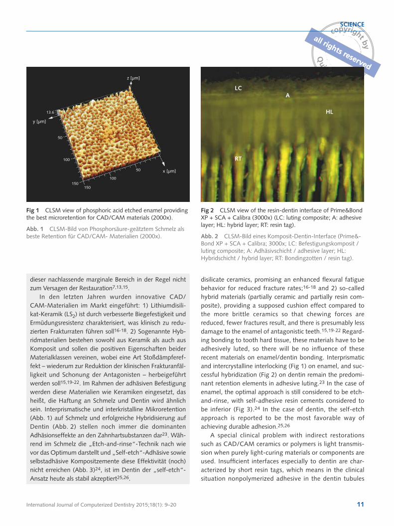
Fig 1 CLSM view of phosphoric acid etched enamel providing the best microretention for CAD/CAM materials (2000x).
Abb. 1 CLSM-Bild von Phosphorsäure-geätztem Schmelz als beste Retention für CAD/CAM- Materialien (2000x).
Fig 2 CLSM view of the resin-dentin interface of Prime&Bond XP + SCA + Calibra (3000x) (LC: luting composite; A: adhesive layer; HL: hybrid layer; RT: resin tag).
Abb. 2 CLSM-Bild eines Komposit-Dentin-Interface (Prime&-Bond XP + SCA + Calibra; 3000x; LC: Befestigungskomposit / luting composite; A: Adhäsivschicht / adhesive layer; HL: Hybridschicht / hybrid layer; RT: Bondingzotten / resin tag).
LCA
HL
RT
z [µm]
x [µm]
y [µm]
13.6
50
100
150150
100
50

International Journal of Computerized Dentistry 2015;18(1): 9–2012
SCIENCE
Ein spezielles klinisches Problem mit CAD/CAM-Mate-rialien ist die Lichttransmission, wenn lichthärtende Mate-rialien oder Komponenten verwendet werden. Insuffizient polymerisierte Verbundzonen im Dentin sind durch kurze Bondingzotten charakterisiert, welche natürlich nur im Rasterelektronenmikroskop kurz sind, in Wirklichkeit jedoch genauso lang sind wie immer (ca. 200 µm), nur eben nicht polymerisiert und daher im Rasterelektronen-mikroskop „verschwinden“ (Abb. 4). Dieses Problem bei indirekten Restaurationen kann einfach verhindert wer-den, indem man adhäsive Aufbaufüllungen macht oder komplett dual-härtende Materialien zum Einsatz kommen oder beides27. Es bleibt zuletzt jedoch die Frage, in wie-weit adhäsive Befestigungsmaterialien an den neuen Materialien haften können.
Materialien und Methoden
240 CAD/CAM-Blöcke vier verschiedener Materialien kamen in dieser Studie zum Einsatz. Materialien (Blöcke und Befestigungsmaterialien), Klassifikationen, Zusam-mensetzungen und Hersteller sind in Tabelle 1 dargestellt.
beneath esthetic restorations (Fig 4). This particular prob-lem with indirect restorations can be easily avoided by using either adhesive build-ups or completely dual-curing luting materials and procedures, or both.27 Finally, the question remains as to the quality and performance of the bonding between these novel CAD/CAM blocks and lut-ing agents.
Materials and methods
The present investigation used 240 CAD/CAM blocks of four different materials. Table 1 shows the materials (blocks and luting agents under investigation), classifications, composi-tions, and manufacturers.
Specimens measuring 10 mm × 10 mm × 10 mm were retrieved from original blocks using a slow-speed diamond saw (IsoMet, Buehler, Lake Bluff, IL, USA). One surface of each specimen was ground using 600-grit silicon carbide paper under water rinsing. Specimens were randomly assigned to six different pre-treatment groups: 1. Negative control: no pretreatment. 2. Silane.
Fig 3 SEM image of a gp formation between self-adhesive resin cement (Maxcem Elite, Kerr, CA, USA) and the enamel margin of a ceramic inlay (300x) (SALC: self-adhesive luting composite; E: enamel).
Abb. 3 REM-Bild eines Randspalts zwischen selbstadhäsivem Befestigungskomposit (Maxcem Elite Kerr, CA, USA) und dem Schmelzrand eines Keramikinlays (300x; SALC: Kompositze-ment / self-adhesive luting composite; E: Schmelz / enamel).
Fig 4 SEM view (2000x) of short resin tags in dentin when a light curing adhesive is not separately light cured. These tags are actually longer but the non-polymerized part is diminished during SEM processing.
Abb. 4 REM-Bild (2000x) kurzer Bondingzotten wenn ein lichthärtendes Adhäsiv durch die Keramik belichtet wurde. Diese „Tags” sind natürlich viel länger, aber nicht genug polymerisiert. Dadurch sind nur die polymerisierten Anteile sichtbar.
SALC
E

International Journal of Computerized Dentistry 2015;18(1): 9–20 13
SCIENCE
Table 1 Materials under investigation
Tab. 1 Materialien.
CAD/CAM material/ CAD/CAM-Material
Classification/ Klassifikation Composition (%wt)/ Zusammensetzung (%) Manu-facturer/ Hersteller
e.max CAD Lithium disilicate ceramic (LS2)Lithiumdisilikatkeramik (LS2)
57-80% SiO2, 11-19% Li2O, 0-13% K2O, 0-11% P2O5, 0-8% ZrO2, 0-8% ZnO, ZnO, 0-5% Al2O3, 0-5% MgO
Ivoclar Vivadent
Celtra Duo Zirconia- reinforced lithium silicate ceramic (ZLS)zirkonverstärkte Lithiumsilikatkeramik (ZLS)
Lithium silicate with ~10% ZrO2
Lithiumsilikat mit ~10% ZrO2
Dentsply DeTrey
Lava Ultimate Hybrid A: resin nano ceramic (RNC)Hybrid A: Resin-Nano-Keramik (RNK)
80% nano ceramic, 20% resin matrix80% Nanokeramik, 20% Kompositmatrix
3M ESPE
Enamic Hybrid B: polymer infiltrated ceramic network (PIC)Hybrid B: polymerinfiltriertes Keramiknetzwerk (PIK)
86% feldspathic ceramic, 14% polymer
86% Feldspat, 14% Polymer
Vita
Luting material/ Befestigungsmaterial
Prime&Bond XP + SCA
Etch-and-rinse adhesive with Self-Cure Activator
Etch-and-rinse-Adhäsiv mit Self-cure-Aktivator
Carboxylic acid modified dimethacrylate (TCB resin); phosphoric acid modified acrylate resin (PENTA); urethane dimethacrylate (UDMA); triethyleneglycol- dimethacrylate (TEGDMA); 2-hydroxyethylmet-hacrylate (HEMA); butylated benzenediol (stabilizer); ethyl-4-di-methylamino-benzoate; camphorquinone; functionalized amorphous silica; tert-ButanolSelf-Cure Activator: urethane dimethacrylate (UDMA); 2-hy-droxy-ethylmethacrylate (HEMA); catalyst; photoinitiators; stabilizers; acetone; waterCarboxylsäuremodifiziertes Dimethacrylat (TCB); Phosphorsäuremo-difiziertes Acrylat (PENTA); Urethandimethacrylat (UDMA); Triethyle-neglycoldimethacrylat (TEGDMA); 2-Hydroxyethylmethacrylat (HEMA); Butyliertes Benzenediol (Stabilisator); Ethyl-4-Dimethylamino-Benzo-at; Campherquinon; funkionalisiertes amorphes Silikat; Tert-ButanolSelf-Cure Aktivator: Urethandimethacrylat (UDMA); 2-Hy-droxy-ethylmethacrylat (HEMA); Katalyator; Photoinitiatoren; Stabilisatoren; Aceton; Wasser
Dentsply DeTrey
Calibra Luting resin composite
Luting resin Komposite
Base: dimethacrylate resins; camphorquinone (CQ) photoinitiator; stabilizers; glass fillers; fumed silica; titanium dioxide; pigmentsCatalyst: dimethacrylate resins; catalyst; stabilizers; glass fillers; fumed silicaBasispaste: Dimethacrylatharz; Campherquinon (CQ) Photoinitiator; Stabilisatoren; Glassfüller; Silikat; Titandioxid; PigmenteKatalysator: Dimethacrylatharz; Katalysator; Stabilisatoren; Glassfüller; Aerosol
Dentsply DeTrey
RelyX Unicem Self-adhesive luting resin com-posite
Selbstadhäsive Befestigung resin Komposite
Powder: glass powder, silica, calcium hydroxide, pigment, substituted pyrimidine, peroxy compound, initiator Liquid: methacrylated phosphoric ester, dimethacrylate, acetate, stabilizer, initiatorPulver: Glasmehl, Silikat, Calciumhydroxid, Pigmente, Pyrimidin, Peroxybestandteile, Initiator Flüssigkeit: Methacryliertes Phosphorsäureester, Dimethacrylat, Acetat, Stabilisator, Initiator
3M ESPE
International Journal of Computerized Dentistry 2015;18(1): 9–2014
SCIENCE
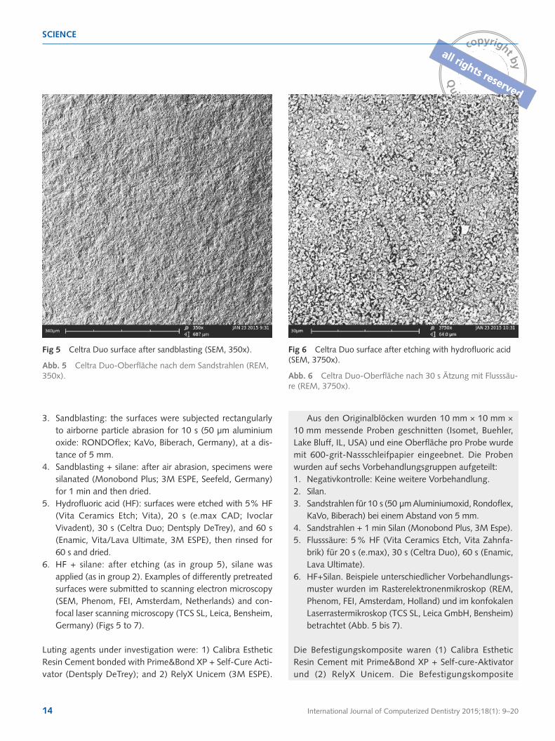
3. Sandblasting: the surfaces were subjected rectangularly to airborne particle abrasion for 10 s (50 µm aluminium oxide: RONDOflex; KaVo, Biberach, Germany), at a dis-tance of 5 mm.
4. Sandblasting + silane: after air abrasion, specimens were silanated (Monobond Plus; 3M ESPE, Seefeld, Germany) for 1 min and then dried.
5. Hydrofluoric acid (HF): surfaces were etched with 5% HF (Vita Ceramics Etch; Vita), 20 s (e.max CAD; Ivoclar Vivadent), 30 s (Celtra Duo; Dentsply DeTrey), and 60 s (Enamic, Vita/Lava Ultimate, 3M ESPE), then rinsed for 60 s and dried.
6. HF + silane: after etching (as in group 5), silane was applied (as in group 2). Examples of differently pretreated surfaces were submitted to scanning electron microscopy (SEM, Phenom, FEI, Amsterdam, Netherlands) and con-focal laser scanning microscopy (TCS SL, Leica, Bensheim, Germany) (Figs 5 to 7).
Luting agents under investigation were: 1) Calibra Esthetic Resin Cement bonded with Prime&Bond XP + Self-Cure Acti-vator (Dentsply DeTrey); and 2) RelyX Unicem (3M ESPE).
Aus den Originalblöcken wurden 10 mm × 10 mm × 10 mm messende Proben geschnitten (Isomet, Buehler, Lake Bluff, IL, USA) und eine Oberfläche pro Probe wurde mit 600-grit-Nassschleifpapier eingeebnet. Die Proben wurden auf sechs Vorbehandlungsgruppen aufgeteilt: 1. Negativkontrolle: Keine weitere Vorbehandlung. 2. Silan.3. Sandstrahlen für 10 s (50 µm Aluminiumoxid, Rondoflex,
KaVo, Biberach) bei einem Abstand von 5 mm. 4. Sandstrahlen + 1 min Silan (Monobond Plus, 3M Espe). 5. Flusssäure: 5 % HF (Vita Ceramics Etch, Vita Zahnfa-
brik) für 20 s (e.max), 30 s (Celtra Duo), 60 s (Enamic, Lava Ultimate).
6. HF+Silan. Beispiele unterschiedlicher Vorbehandlungs-muster wurden im Rasterelektronenmikroskop (REM, Phenom, FEI, Amsterdam, Holland) und im konfokalen Laserrastermikroskop (TCS SL, Leica GmbH, Bensheim) betrachtet (Abb. 5 bis 7).
Die Befestigungskomposite waren (1) Calibra Esthetic Resin Cement mit Prime&Bond XP + Self-cure-Aktivator und (2) RelyX Unicem. Die Befestigungskomposite
Fig 5 Celtra Duo surface after sandblasting (SEM, 350x).
Abb. 5 Celtra Duo-Oberfläche nach dem Sandstrahlen (REM, 350x).
Fig 6 Celtra Duo surface after etching with hydrofluoric acid (SEM, 3750x).
Abb. 6 Celtra Duo-Oberfläche nach 30 s Ätzung mit Flusssäu-re (REM, 3750x).

International Journal of Computerized Dentistry 2015;18(1): 9–20 15
SCIENCE
wurden in 3 mm-Schichten direkt appliziert, wobei die Proben mit der vorbehandelten Oberfläche nach oben in einer speziellen Silikonform eingebettet waren und mit einem isolierten transparenten Stempel auf die Oberfläche gepresst wurden, um den klinischen Anpressdruck zu simulieren. Die Photopolymerisation erfolgte mit einer Bluephase-Lampe (Ivoclar Vivadent) mit 1.200 mW/cm2 für 60 s. Nach Entfernung der Applikationshilfen wurde die Lichthärtung für 20 s von jeder Seite wiederholt.
Nach 25-stündiger Wasserlagerung bei 37°C und 10.000 Thermozyklen (5°C/55°C; THE 1100, SD Mechatronic, Feldkirchen) wurden die Proben in Scheiben geschnitten (Isomet), welche wiederum in Stäbchen gesägt wurden. Das Sägeblatt erhielt einen Abstandsmarker bei 1,3 mm, durch die Dicke des Sägeblatts (300 µm) erhielten wir sodann ~20 Stäbchen mit 1 mm × 1 mm pro Probe und 100 Stäbchen pro Gruppe. Für den Fall einer Spontan-fraktur des Stäbchens vor der Testung wurden die Prozent-sätze früh frakturierter Proben notiert und als 0 MPa in das Gesamtergebnis extrapoliert. Die Stäbchen wurden schließ-lich bei 0,5 mm/min Traversengeschwindigkeit auseinan-dergezogen (MTD 500, SD Mechatronic).
Die statistische Analyse erfolgte mit SPSS, Version 14.0 für Windows (SPSS Inc., Chicago, IL, USA). Durch die mehrheitliche Nicht-Normalverteilung (Kolmogorov-Smir-nov-Test) kamen nicht-parametrische Tests zum Einsatz (Wilcoxon matched-pairs signed-ranks Test, Mann-Whit-ney-U-Test), gerechnet auf dem 95%-Niveau.
Luting agents were directly applied in one 3 mm layer into a special silicone mold with the specimens surface-up at the bottom, and condensed using an isolated transparent block to simulate pressure, as in a clinical situation, and to guaran-tee light transmission. Light curing was performed using a Bluephase curing unit (Ivoclar Vivadent) operating at 1,200 mW/cm2 for 60 s. After removal of the mold, light curing was repeated from four sides for 20 s each.
After 24 h of water storage at 37°C and 10,000 thermocy-cles (5°C/55°C; THE1100, SD Mechatronik, Feldkirchen, Ger-many), the specimens were sectioned into slices (IsoMet), which were sectioned again to receive beams. The saw was adjusted to steps of 1.3 mm, due to the thickness of the blade (300 µm), resulting in ~20 sticks per block, with a cross-sec-tional area of 1 mm × 1 mm. In case one or more of the select-ed sticks failed due to the sectioning process, the percentage of prematurely failed specimens in relation to the total number of selected specimens was recorded. The same (or approximat-ed) percentage of the 100 final specimens per group received 0 MPa as a final µ-TBS result. The µ-TBS sticks were then frac-tured according to a well-suited protocol, with a crosshead speed of 0.5 mm/min (MTD 500, SD Mechatronik).
Statistical analysis was performed using SPSS, Version 14.0 for Windows XP (SPSS, Chicago, IL, USA). As the major-ity of groups did not exhibit normal data distribution (Kol-mogorov-Smirnov test), nonparametric tests were used (Wil-coxon matched-pairs signed-ranks test, Mann-Whitney test) for pairwise comparisons at the 95% significance level.
Fig 7 e.max CAD after sandblasting and silanating (CLSM, 300x).
Abb. 7 e.max CAD nach Sandstrahlen und Silan (CLSM, 300x).
z [µm]
5050
27.82010
100100
150150

International Journal of Computerized Dentistry 2015;18(1): 9–2016
SCIENCE
Results
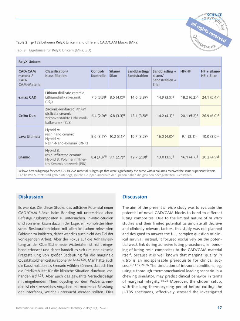
Table 2 shows the µ-TBS results for Calibra, while Table 3 shows the results for RelyX Unicem. In general, the µ-TBS results for Calibra were slightly higher compared to those for RelyX Unicem (P < 0.05); however, in the untreated speci-mens, RelyX Unicem performed better (P < 0.05). Whereas sandblasting was the most effective pretreatment method for Lava Ultimate (P < 0.05), all other materials exhibited the best bonding performance when hydrofluoric acid etching was performed, followed by silane treatment. For Calibra, statistical subgroups of best performing groups were Celtra Duo > e.max CAD > Enamic > Lava Ultimate (P < 0.05), and for RelyX Unicem, statistical subgroups of best performing groups were Celtra Duo = e.max CAD > Enamic > Lava Ulti-mate (P < 0.05). To sum up, it was shown that the manufac-turers’ instructions for use gave the best bonding results and that pretreatment of CAD/CAM blocks is a decisive factor in adhesive luting procedures.
Ergebnisse
Die Mikrozugergebnisse sind in Tabelle 2 (Calibra) und Tabelle 3 (RelyX Unicem) dargestellt. Die Ergebnisse für Calibra waren etwas höher als für RelyX Unicem (p< 0,05), aber in den unbehandelten Gruppen war wiederum RelyX Unicem besser (p< 0,05). Sandstrahlen zeigte sich als beste Methode zur Vorbehandlung von Lava Ultimate (p< 0,05), bei den anderen Materialien war HF+Silan überlegen. Für Calibra waren die statistischen Untergrup-pen mit bester Performance Celtra Duo > e.max CAD > Enamic > Lava Ultimate (p< 0,05), für RelyX Unicem Celtra Duo = e.max CAD > Enamic > Lava Ulti-mate (p< 0,05). Insgesamt zeigten die von den Herstellern empfohlenen Vorbehandlungsstrategien die besten Ergeb-nisse und es wurde deutlich, dass das Vorbehandlungs-muster einen substanziellen Einfluss auf das Haftresultat hat.
Table 2 µ-TBS between Prime&Bond XP + SCA + Calibra and different CAD/CAM blocks [MPa]
Tab. 2 Ergebnisse für Calibra [MPa](SD).
Prime&Bond XP + SCA + Calibra
CAD/CAM material/ CAD/CAM-Material
Classification/ Klassifikation
Control/ Kontrolle
Silane/ Silan
Sandblasting/ Sandstrahlen
Sandblasting + silane/ Sandstrahlen + Silan
HF/HF HF + silane/HF + Silan
e.max CADLithium disilicate ceramicLithiumdisilikatkeramik (LS2)
3.5 (8.3)A 4.5 (9.0)A 18.4 (4.5)A 18.9 (4.9)A 24.3 (8.2)B 26.3 (7.7)B
Celtra Duo
Zirconia-reinforced lithium disilicate ceramiczirkonverstärkte Lithiumsili-katkeramik (ZLS)
2.4 (7.2)A 3.8 (9.3)A 15.3 (4.6)B 16.2 (5.0)B 30.2 (8.1)A 31.2 (7.9)A
Lava Ultimate
Hybrid A: resin nano ceramicHybrid A: Resin-Nano-Keramik (RNK)
4.5 (6.7)A 4.2 (9.1)A 17.9 (4.5)A 16.8 (4.6)B 10.4 (6.9)C 11.2 (5.5)C
Enamic
Hybrid B: resin infiltrated ceramicHybrid B: Polymerinfiltrier-tes Keramiknetzwerk (PIK)
0B 3.1 (8.7)A 15.2 (3.9)B 15.7 (4.5)B 21.8 (6.2)B 23.4 (5.0)B
Yellow: best subgroups for each CAD/CAM material; subgroups that were significantly the same within columns received the same superscript letters.Die besten Subsets sind gelb hinterlegt, gleiche Gruppen innerhalb der Spalten haben die gleichen hochgestellten Buchstaben.
International Journal of Computerized Dentistry 2015;18(1): 9–20 17
SCIENCE
Diskussion
Es war das Ziel dieser Studie, das adhäsive Potenzial neuer CAD/CAM-Blöcke beim Bonding mit unterschiedlichen Befestigungskompositen zu untersuchen. In-vitro-Studien sind von jeher kaum dazu in der Lage, ein komplettes klini-sches Restaurationsleben mit allen kritischen relevanten Faktoren zu imitieren, daher war dies auch nicht das Ziel der vorliegenden Arbeit. Aber der Fokus auf die Adhäsivleis-tung an der Oberfläche neuer Materialien ist nicht einge-hend erforscht und daher handelt es sich um eine aktuelle Fragestellung von großer Bedeutung für die marginale Qualität solcher Restaurationen6,11,12,24,26. Man hätte auch die Kausimulation als Szenario wählen können, da auch hier die Prädiktabilität für die klinische Situation durchaus vor-handen ist14,28. Aber auch das gewählte Versuchsdesign mit eingehendem Thermocycling vor dem Probenschnei-den ist ein stressreiches Vorgehen mit maximaler Belastung der Interfaces, welche untersucht werden sollten. Dies
Discussion
The aim of the present in vitro study was to evaluate the potential of novel CAD/CAM blocks to bond to different luting composites. Due to the limited nature of in vitro studies and their limited potential to simulate all decisive and clinically relevant factors, this study was not planned and designed to answer the full, complex question of clin-ical survival; instead, it focused exclusively on the poten-tial weak link during adhesive luting procedures, ie, bond-ing of luting resin composites to the CAD/CAM material itself, because it is well known that marginal quality in vitro is an indispensable prerequisite for clinical suc-cess.6,11,12,24,26 The simulation of intraoral conditions, eg, using a thorough thermomechanical loading scenario in a chewing simulator, may predict clinical behavior in terms of marginal integrity.14,28 Moreover, the chosen setup, with the long thermocycling period before cutting the µ-TBS specimens, effectively stressed the investigated
Table 3 µ-TBS between RelyX Unicem and different CAD/CAM blocks [MPa]
Tab. 3 Ergebnisse für RelyX Unicem [MPa](SD).
RelyX Unicem
CAD/CAM material/ CAD/CAM-Material
Classification/ Klassifikation
Control/ Kontrolle
Silane/ Silan
Sandblasting/ Sandstrahlen
Sandblasting + silane/ Sandstrahlen + Silan
HF/HF HF + silane/HF + Silan
e.max CADLithium disilicate ceramicLithiumdisilikatkeramik (LS2)
7.5 (3.3)B 8.5 (4.0)B 14.6 (3.8)A 14.9 (3.9)B 18.2 (6.2)A 24.1 (5.4)A
Celtra Duo
Zirconia-reinforced lithium disilicate ceramiczirkonverstärkte Lithiumsili-katkeramik (ZLS)
6.4 (2.9)B 6.8 (3.3)B 13.1 (3.5)B 14.2 (4.1)B 20.1 (5.2)A 26.9 (6.0)A
Lava Ultimate
Hybrid A: resin nano ceramicHybrid A: Resin-Nano-Keramik (RNK)
9.5 (3.7)A 10.2 (3.1)A 15.7 (3.2)A 16.0 (4.0)A 9.1 (3.1)C 10.0 (3.5)C
Enamic
Hybrid B: resin infiltrated ceramicHybrid B: Polymerinfiltrier-tes Keramiknetzwerk (PIK)
8.4 (3.0)AB 9.1 (2.7)A 12.7 (2.9)B 13.0 (3.5)B 16.1 (4.7)B 20.2 (4.9)B
Yellow: best subgroups for each CAD/CAM material; subgroups that were significantly the same within columns received the same superscript letters.Die besten Subsets sind gelb hinterlegt, gleiche Gruppen innerhalb der Spalten haben die gleichen hochgestellten Buchstaben.

International Journal of Computerized Dentistry 2015;18(1): 9–2018
SCIENCE
interfaces. This was clearly recordable by analyzing pretest failures that had considerable percentages, primarily in the test groups with insufficient pretreatment of CAD/CAM blocks prior to adhesive luting.
Clinical studies and systematic reviews about CAD/CAM inlays and partial crowns show favorable annual fail-ure rates,7-11 always accompanied, however, with a con-siderable longitudinal decrease of marginal quality over time.12,13 Decreasing marginal integrity is primarily dependent on luting composite wear and the tendency of adjacent structures to loose adhesion.12,13 Exactly this aspect was the aim of the present investigation, where we tried to pre-identify potential weak links for adhesive lut-ing of recently introduced CAD/CAM materials. Elsaka investigated the bonding performance of Lava Ultimate and Enamic to Bifix SE self-adhesive resin cement (Voco)19 with mostly different µ-TBS results but a similar tendency in his conclusions: Although distinct differences between different pretreatments were found, the overall perfor-mance of the tested materials was promising, and the val-ues of adhesion made it clear that the recorded µ-TBS val-ues were high enough to withstand masticatory forces in the oral cavity. Elsaka’s methodology was not exactly the same as that used in our study – no thermocycling, but longer water storage – so direct comparisons are not appli-cable.19 It is probably due to our intensive thermocycling of 10,000 cycles that up to 100% of the specimens were lost during the processing of the beams, primarily in the negative control groups. A remarkable difference between our study results and those of Elsaka is the effect of HF on Lava Ultimate. Whereas our results clearly show a detri-mental effect compared to sandblasting or sandblasting + silane, Elsaka’s values for HF treatment of Lava Ultimate were reasonably promising.
To date, very few data have been published for Enam-ic;19,22,29 the database for Lava Ultimate is already larg-er.20,30 The same is true for Celtra Duo, with very little information in the literature, whereas e.max ceramics have been more widely investigated.17,18,31 Despite the limited data thus far, it became clear to us in this study that the manufacturers’ recommendations are feasible and led to reliable bonding performances for all CAD/CAM materials under investigation. Sandblasting is recommended only for the resin nano ceramic, Lava Ultimate. The other materials showed the best outcome after hydrofluoric acid etching. Although some differences in bonding behavior were found among the groups studied, it can be stated that the best performing subgroups for each material
zeigten nicht zuletzt die PTFs („pre-test-failures“), welche in den schwachen Gruppen durchaus vorkamen.
Sowohl klinische Studien als auch systematische Über-sichten zeigten vielversprechende jährliche Verlustquoten von CAD/CAM-gefertigten Inlays und Teilkronen7-11, jedoch wie oben beschrieben immer einhergehend mit einem gewis-sen longitudinalen Abfall der marginalen Qualität12,13. Eine abfallende marginale Integrität geht auf das Konto des Ver-schleißes beim Befestigungskomposit, ist aber auch durch Adhäsionsverluste an den Grenzflächen bedingt12,13. Genau hier setzte die aktuelle Untersuchung an, da es galt, poten-zielle Schwachstellen im Verbund Zahn – Komposit – Bon-ding – CAD/CAM-Material zu identifizieren. Elsaka unter-suchte kürzlich eine ähnliche Fragestellung bei Lava Ultimate und Enamic in Verbindung mit dem selbstadhäsiven Befesti-gungszement Bifix SE19, die Ergebnisse waren in den meisten Aspekten auf einem anderen Niveau, zeigten aber immerhin ähnliche Tendenzen in der Schlussfolgerung: Obwohl die unterschiedlichen Vorbehandlungsmuster unterschiedlich effektiv waren, war die Gesamtperformanz der getesteten Materialien vielversprechend. Es wurde deutlich, dass die Mikrozugergebnisse auf einem auch klinisch akzeptablen Niveau waren. Die Methode von Elsaka war nicht exakt gleich wie im vorliegenden Beispiel – kein Thermocycling, aber längere Wasserlagerung – daher kann man die Studien nicht beliebig vergleichen19. Vermutlich zeichnet unser subs-tanzielles Thermocycling von 10.000 Zyklen dafür verant-wortlich, dass gerade in den Kontrollgruppen bei uns bis zu 100% der Proben beim Sägen gebrochen sind. Ein bemer-kenswerter Unterschied zwischen unserer Arbeit und Elsaka’s Resultaten ist der Effekt von Flusssäure auf Lava Ultimate. In der vorliegenden Studie war HF im Vergleich zu Sandstrahlen oder Sandstrahlen und Silan deutlich schlechter, Elsaka’s Werte für HF bei Lava Ultimate jedoch ziemlich gut.
Bis zum heutigen Zeitpunkt sind für Enamic nur wenige Daten publiziert19,22,29, die Datenbank für Lava Ultimate ist da schon breiter20,30. Dasselbe gilt für Celtra Duo mit spärli-chen Informationen in der Literatur, wobei e.max-Keramiken schon wesentlich häufiger Ziel werkstoffkundlicher Untersu-chungen waren17,18,31. Unabhängig von der Zahl der Publi-kationen wurde jedoch klar, dass ein striktes Befolgen der Herstellerempfehlungen zur Vorbehandlung neuer CAD/CAM-Materialien wichtig und empfehlenswert ist. Nur für die Resin-Nano-Keramik Lava Ultimate wird Sandstrahlen klar empfohlen, die anderen Materialien profitieren am meis-ten von HF-Ätzung + Silan. Obwohl zwischen den Haftwer-ten per se gewisse Unterschiede zu detektieren waren, kann abschließend festgehalten werden, dass die jeweils

International Journal of Computerized Dentistry 2015;18(1): 9–20 19
SCIENCE
erfolgreichsten Untergruppen für alle Materialien vielverspre-chende Haftungen hervorbrachten, um damit zielführend klinisch arbeiten zu können.
Schlussfolgerungen
Unter strikter Einhaltung der Herstellerempfehlungen zur Vorbehandlung zeigen die neuen CAD/CAM-Materialien mit den gewählten Befestigungskompositen gute Ergeb-nisse in der adhäsiven Performance.
provided µ-TBS values that are completely satisfactory for clinical work.
Conclusion
Under application of the recommended pretreatment proto-cols, the novel CAD/CAM materials show promising bonding performances to different types of luting resin composites.
References
1. Frankenberger R, Lohbauer U, Roggendorf MJ, Naumann M, Tasch-ner M. Selective enamel etching reconsidered: better than etch-and-rinse and self-etch? J Adhes Dent 2008;10:339–344.
2. Frankenberger R, Reinelt C, Krämer N. Nanohybrid vs. fine hybrid composite in extended class II cavities: 8-year results. Clin Oral Investig 2014;18:125–137.
3. Opdam NJ, Bronkhorst EM, Loomans BA, Huysmans MC. 12-year sur-vival of composite vs. amalgam restorations. J Dent Res 2010;89: 1063–1067.
4. Roggendorf MJ, Kunzi B, Ebert J, Roggendorf HC, Frankenberger R, Reich SM. Seven-year clinical performance of CEREC-2 all-ceramic CAD/CAM restorations placed within deeply destroyed teeth. Clin Oral Investig 2012;16:1413–1424.
5. Opdam NJ, Bronkhorst EM, Loomans BA, Huysmans MC. 12-year sur-vival of composite vs. amalgam restorations. J Dent Res 2010;89: 1063–1067.
6. Roggendorf MJ, Krämer N, Appelt A, Naumann M, Frankenberger R. Marginal quality of flowable 4-mm base vs. conventionally layered resin composite. J Dent 2011;39:643–647.
7. Frankenberger R, Taschner M, Garcia-Godoy F, Petschelt A, Krämer N. Leucite-reinforced glass ceramic inlays and onlays after 12 years. J Adhes Dent 2008;10:393–398.
8. Otto T, De Nisco S. Computer-aided direct ceramic restorations: a 10-year prospective clinical study of Cerec CAD/CAM inlays and onlays. Int J Prosthodont 2002;15:122–128.
9. Posselt A, Kerschbaum T. Longevity of 2328 chairside Cerec inlays and onlays. Int J Comput Dent 2003;6:231–248.
10. Reiss B. Clinical results of Cerec inlays in a dental practice over a period of 18 years. Int J Comput Dent 2006;9:11–22.
11. Sjögren G, Molin M, van Dijken JW. A 10-year prospective evaluation of CAD/CAM-manufactured (Cerec) ceramic inlays cemented with a chemically cured or dual-cured resin composite. Int J Prosthodont 2004;17:241–246.
12. Taschner M, Krämer N, Lohbauer U, et al. Leucite-reinforced glass ceramic inlays luted with self-adhesive resin cement: a 2-year in vivo study. Dent Mater 2012;28:535–540.
13. Thordrup M, Isidor F, Hörsted-Bindslev P. A prospective clinical study of indirect and direct composite and ceramic inlays: ten-year results. Quintessence Int 2006;37:139–144.
14. Frankenberger R, Krämer N, Appelt A, Lohbauer U, Naumann M, Rog-gendorf MJ. Chairside vs. labside ceramic inlays: effect of temporary restoration and adhesive luting on enamel cracks and marginal integri-ty. Dent Mater 2011;27:892–898.
15. Krämer N, Kunzelmann KH, Taschner M, Mehl A, Garcia-Godoy F, Frankenberger R. Antagonist enamel wears more than ceramic inlays. J Dent Res 2006;85:1097–1100.
16. Guess PC, Vagkopoulou T, Zhang Y, Wolkewitz M, Strub JR. Marginal and internal fit of heat pressed versus CAD/CAM fabricated all-ceramic onlays after exposure to thermo-mechanical fatigue. J Dent 2014;42:199–209.
17. Schultheis S, Strub JR, Gerds TA, Guess PC. Monolithic and bi-layer CAD/CAM lithium-disilicate versus metal-ceramic fixed dental pros-theses: comparison of fracture loads and failure modes after fatigue. Clin Oral Investig 2013;17:1407–1413.
18. Wiedhahn K. The impression-free Cerec multilayer bridge with the CAD-on method. Int J Comput Dent 2011;14:33–45.
19. Elsaka SE. Bond strength of novel CAD/CAM restorative materials to self-adhesive resin cement: the effect of surface treatments. J Adhes Dent 2014;16:531–540.
20. Keul C, Müller-Hahl M, Eichberger M, et al. Impact of different adhe-sives on work of adhesion between CAD/CAM polymers and resin composite cements. J Dent 2014;42:1105–1114.
21. Peampring C. Restorative management using hybrid ceramic of a patient with severe tooth erosion from swimming: a clinical report. J Adv Prosthodont 2014;6:423–426.

International Journal of Computerized Dentistry 2015;18(1): 9–2020
SCIENCE
22. Dirxen C, Blunck U, Preissner S. Clinical performance of a new biomi-metic double network material. Open Dent J;7:118–122.
23. De Munck J, Mine A, Vivan Cardosa M, et al. Effect of dentin location and long-term water storage on bonding effectiveness of dentin adhe-sives. Dent Mater J 2011;30:7–13.
24. Frankenberger R, Lohbauer U, Schaible RB, Nikolaenko SA, Naumann M. Luting of ceramic inlays in vitro: marginal quality of self-etch and etch-and-rinse adhesives versus self-etch cements. Dent Mater 2008;24:185–191.
25. Peumans M, De Munch J, Van Landuyt K, Van Meerbeek B. Thir-teen-year randomized controlled clinical trial of a two-step self-etch adhesive in non-carious cervical lesions. Dent Mater 2015.pii:S0109-5641(15)00019-6. doi:10.1016/j.dental.2015.01.005. [epub ahead of print].
26. Van Meerbeek B, Yoshihara K. Clinical recipe for durable dental bond-ing: why and how? J Adhes Dent 2014;16:94.
27. Frankenberger R, Lohbauer U, Taschner M, Petschelt A, Nikolaenko SA. Adhesive luting revisited: influence of adhesive, temporary cement, cavity cleaning, and curing mode on internal dentin bond strength. J Adhes Dent 2007;9(suppl 2):269–273.
28. Frankenberger R, Hehn J, Hajtó J, et al. Effect of proximal box elevation with resin composite on marginal quality of ceramic inlays in vitro. Clin Oral Investig 2013;17:177–183.
29. Della Bona A, Corazza PH, Zhang Y. Characterization of a polymer-in-filtrated ceramic-network material. Dent Mater 2014;30:564–569.
30. Belli R, Geinzer E, Muschweck A, Petschelt A, Lohbauer U. Mechanical fatigue degradation of ceramics versus resin composites for dental res-torations. Dent Mater 2014;30:424–432.
31. Lührs AK, De Munck J, Geurtsen W, Van Meerbeek B. Composite cements benefit from light-curing. Dent Mater 2014;30:292–301.
Address/Adresse
Prof. Dr. Roland Frankenberger, Abteilung für Zahnerhaltungskunde,
Med. Zentrum für ZMK, Universität Marburg und Universitätsklinikum
Gießen und Marburg, Campus Marburg, Georg-Voigt-Straße 3,
35039 Marburg, Tel.: +49 (0) 6421 5863240; Fax: +49 (0) 6421 5863745;
E-Mail: [email protected]
View publication statsView publication stats