adhd a meta-analysis,2012
DESCRIPTION
articol adhdTRANSCRIPT

Reviews and Overviews
Mechanisms of Psychiatric Illness
Toward Systems Neuroscience of ADHD:A Meta-Analysis of 55 fMRI Studies
Samuele Cortese, M.D., Ph.D.
Clare Kelly, Ph.D.
Camille Chabernaud, Ph.D.
Erika Proal, Ph.D.
Adriana Di Martino, M.D.
Michael P. Milham, M.D., Ph.D.
F. Xavier Castellanos, M.D.
Objective: The authors performed a com-prehensive meta-analysis of task-basedfunctional MRI studies of attention deficithyperactivity disorder (ADHD).
Method: The authors searched PubMed,Ovid, EMBASE, Web of Science, ERIC,CINAHAL, and NeuroSynth for studiespublished through June 30, 2011. Signifi-cant differences in brain region activationbetween individuals with ADHD andcomparison subjects were detected usingactivation likelihood estimation meta-analysis. Dysfunctional regions in ADHDwere related to seven reference neuronalsystems. The authors performed a set ofmeta-analyses focused on age groups(children and adults), clinical character-istics (history of stimulant treatment andpresence of psychiatric comorbidities), andspecific neuropsychological tasks (inhibi-tion, working memory, and vigilance/attention).
Results: Fifty-five studies were included(39 for children and 16 for adults). Inchildren, hypoactivation in ADHD relativeto comparison subjects was observed
mostly in systems involved in executivefunction (frontoparietal network) and at-tention (ventral attentional network). Sig-nificant hyperactivation in ADHD relativeto comparison subjects was observedpredominantly in the default, ventral at-tention, and somatomotor networks. Inadults, ADHD-related hypoactivation waspredominant in the frontoparietal system,while ADHD-related hyperactivation waspresent in the visual, dorsal attention, anddefault networks. Significant ADHD-relateddysfunction largely reflected task featuresand was detected even in the absence ofcomorbid mental disorders or a history ofstimulant treatment.
Conclusions: A growing literature pro-vides evidence of ADHD-related dysfunc-tion inmultiple neuronal systems involvedin higher-level cognitive functions butalso in sensorimotor processes, includingthe visual system, and in the default net-work. This meta-analytic evidence extendsearly models of ADHD pathophysiologythat were focused on prefrontal-striatalcircuits.
(Am J Psychiatry 2012; 169:1038–1055)
Attention deficit hyperactivity disorder (ADHD) is oneof the most common childhood-onset psychiatric con-ditions, with an estimated worldwide-pooled prevalenceexceeding 5% in children (1). Impairing ADHD symptomspersist into adulthood in as many as 65% of cases (2).Despite a voluminous literature (3), ADHD pathophysi-ology remains incompletely understood. To gain insightinto the neural correlates of ADHD, Dickstein et al. (4)conducted a quantitative meta-analysis of 16 functionalMRI (fMRI) studies published before February 2006. Theyfound evidence suggesting significant neuronal hypo-activation in individuals with ADHD relative to comparisonsubjects, mostly in the fronto-striatal and parietal regions.A substantial number of studies included in Dickstein et al.(4) assessed response inhibition as a potential contributorto the particular dysfunctional regions identified in ADHD,reflecting the influence of a neuropsychological theory pos-iting inhibitory dysfunction as the core deficit in ADHD (5).
The ADHD fMRI literature has grown substantially since,and neuropsychological paradigms beyond response in-hibition have been more frequently investigated (6). Inaddition, the field has shifted to reporting between-groupcontrasts (i.e., between individuals with ADHD and com-parison subjects) instead of relying on qualitative compar-isons of within-group results, as was common in the earlyliterature. Finally, from a theoretical perspective, ADHD isincreasingly thought to reflect altered connectivity withinand among several neural networks rather than abnor-malities of discrete, isolated brain regions (7, 8).Accordingly, we present an updated meta-analytic
review of the ADHD fMRI literature.We included pertinenttask-based fMRI studies reporting between-group con-trasts regardless of the type of task examined. We con-ducted a set ofmeta-analyses focusing on clinically relevantissues that can now be addressed with greater precisionthanwas possible at the timeof theDickstein et al. study (4).
This article is featured in this month’s AJP Audio and is the subject of a CME course (p. 1123)
1038 ajp.psychiatryonline.org Am J Psychiatry 169:10, October 2012

In particular, the larger number of available studies al-lowed us to explore possible ADHD-related dysfunctionsin relation to specific age groups (children and adolescentsor adults), clinical characteristics (history of stimulanttreatment and presence of comorbid psychiatric disor-ders), or neuropsychological paradigms (inhibitory con-trol, working memory, and vigilance/attention).Based on the perspective that ADHD is a disorder
reflecting dysfunction of large-scale neuronal systems (7),we interpreted abnormally activated brain regions fromour meta-analysis as dysfunctional nodes of large-scalenetworks described in the current neuroscience literature.We used a set of functional networks recently derived froma large data set of resting-state functional imaging asa reference (9). As proposed in a recent qualitative review(7), we hypothesized ADHD-related dysfunctions in net-works that are involved not only in higher-level cognitive-behavioral functions, such as the frontoparietal, dorsalattention, and default networks, but also in sensorimotorprocesses, including somatomotor and visual networks.Consistent with qualitative reviews of fMRI studies inADHD (10, 11), we expected ADHD-related dysfunctions 1)to differ in adults compared with children, 2) to be presentregardless of comorbid psychiatric disorders or history ofstimulant treatment, and 3) to differ according to thespecific neuropsychological task examined.
Method
Search Strategy
We searched the following databases: PubMed, Ovid (in-cluding PsycINFO and Ovid MEDLINE), EMBASE, Web of Science(Science Citation Index Expanded, Social Sciences Citation Index,and Arts and Humanities Citation Index), ERIC, CINAHL, andNeuroSynth (www.neurosynth.org). Details of the search strategyare reported in section A1 of the data supplement that ac-companies the online edition of this article.
Study Eligibility Criteria
Studies were included if they 1) used a diagnosis of ADHDaccording to DSM-IV, DSM-IV-TR, or ICD-10, 2) used a typicallydeveloping comparison group, 3) reported data as three-dimensionalcoordinates in stereotactic space, and 4) used between-groupcontrasts.
Studies were excluded if they 1) used a neuroimaging methodother than fMRI; 2) included participants with ADHD symptomsbut without a formal diagnosis of ADHD; 3) assessed the effect ofmedication without reporting fMRI data at baseline or afterwashout; 4) reported only within-group contrasts; 5) conducteda priori region-of-interest analyses (as these violate the assump-tion, under the null hypothesis, that the likelihood of locatingactivated foci is equal at every voxel); 6) reported only deac-tivations (this occurred in only one study [12], which was thusnot comparable to the others); or 7) included adults with ADHDin partial remission, as it has not been established whether theneuronal correlates of individuals with ADHD in partial re-mission are similar to those with the full syndrome.
Data Extraction
Two authors (S.C. and C.C.) independently searched theliterature, examined the retrieved articles, and extracted and
cross-checked data. Initial disagreements on seven of 2,287screened articles were resolved by consensus. We extracteddemographic information, ADHD diagnostic criteria and subtype,psychiatric comorbidities, medication status, three-dimensionalcoordinates, tasks, and contrasts.
Meta-Analytic Technique
We conducted an activation likelihood estimation meta-analysis using GingerALE, version 2.1.1 (www.brainmap.org/ale/). Activation likelihood estimation allows the detection ofquantitative interstudy consistencies in activation by generatingmaps of activation likelihood estimates. In fMRI studies, theprecise localization of specific activation coordinates is limitedby substantial intersubject anatomical variability. Within studies,this is imperfectly addressed by Gaussian smoothing. Accordingly,activation foci are best considered as localization probabilitydistributions centered at the reported coordinates. Based on thislogic, in activation likelihood estimation meta-analysis, foci arefirst transformed into probability distributions using three-dimensional Gaussian functions with width expressed in milli-meters at half the maximum value (referred to as full width athalf maximum). Second, a whole-brain map is created byassigning each voxel a value equal to the probability that atleast one of the activation points will be found within the voxel.This value is referred to as the activation likelihood estimation foreach voxel. Third, to differentiate the voxels in the map thatrepresent signal (i.e., nonrandom clustering of foci) from thosethat represent noise (i.e., random clustering), activation likeli-hood estimation values are compared with a null hypothesisdistribution generated by permutation analysis (see www.brainmap.org/ale/).
For our meta-analysis, coordinates reported in Talairach spacewere transformed to Montreal Neurological Institute (MNI)coordinates using the icbm2tal (Lancaster) transformation (13).Moreover, since nearby coordinates cannot be assigned un-equivocally to different regions, when coordinates associatedwith multiple contrasts from the same task were less than 12 mmapart, we excluded all but one (e.g., for a set of four coordinateswithin 12 mm of each other, all but the fourth peak wereexcluded). Statistical significance was determined using a per-mutation test (5,000 permutations) of randomly generated foci,corrected for multiple comparisons. Per Eickhoff et al. (14), fullwidth at half maximum was calculated based on the numberof participants in each study. The threshold for final activationlikelihood estimation maps was set at p,0.05 using the falsediscovery rate with an extent threshold greater than 200 mm3
(the GingerALE default) and overlaid onto the MNI 152 template.As recommended (15), anatomical labels were assigned afterdirect examination of anatomy (16).
We performed focused meta-analyses of studies contrasting 1)children (age,18 years) with ADHD and comparison childrenacross all tasks; 2) adults (age$18 years) with ADHD and com-parison adults across all tasks; 3) all stimulant-naive participantswith ADHD (regardless of age) and comparison subjects (studieswere included in this subanalysis only if all participants werestimulant naive); 4) all comorbidity-free individuals with ADHD(regardless of age) and comparison subjects; all individuals withADHD (children and adults) and comparison subjects in 5)inhibition tasks, 6) working memory tasks, and 7) vigilance andattention tasks. As shown in Table 1, the number of retrievedstudies with relevant foci was insufficient to perform separatemeta-analyses of studies assessing 1) paradigms other thaninhibition, working memory, or vigilance/attention; 2) individ-ual tasks in children and adults separately; 3) individualparadigms in participants who were stimulant naive or withoutpsychiatric comorbidities; and 4) ADHD . comparison subjectsfor working memory or vigilance and attention tasks. Additionally,
Am J Psychiatry 169:10, October 2012 ajp.psychiatryonline.org 1039
CORTESE, KELLY, CHABERNAUD, ET AL.
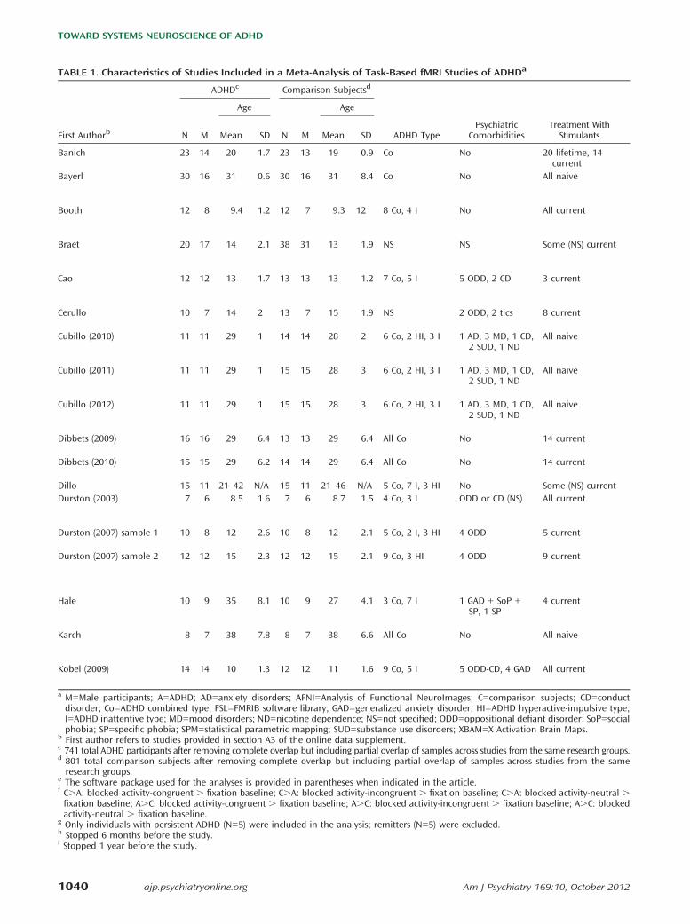
TABLE 1. Characteristics of Studies Included in a Meta-Analysis of Task-Based fMRI Studies of ADHDa
ADHDc Comparison Subjectsd
Age Age
First Authorb N M Mean SD N M Mean SD ADHD TypePsychiatric
ComorbiditiesTreatment With
Stimulants
Banich 23 14 20 1.7 23 13 19 0.9 Co No 20 lifetime, 14current
Bayerl 30 16 31 0.6 30 16 31 8.4 Co No All naive
Booth 12 8 9.4 1.2 12 7 9.3 12 8 Co, 4 I No All current
Braet 20 17 14 2.1 38 31 13 1.9 NS NS Some (NS) current
Cao 12 12 13 1.7 13 13 13 1.2 7 Co, 5 I 5 ODD, 2 CD 3 current
Cerullo 10 7 14 2 13 7 15 1.9 NS 2 ODD, 2 tics 8 current
Cubillo (2010) 11 11 29 1 14 14 28 2 6 Co, 2 HI, 3 I 1 AD, 3 MD, 1 CD,2 SUD, 1 ND
All naive
Cubillo (2011) 11 11 29 1 15 15 28 3 6 Co, 2 HI, 3 I 1 AD, 3 MD, 1 CD,2 SUD, 1 ND
All naive
Cubillo (2012) 11 11 29 1 15 15 28 3 6 Co, 2 HI, 3 I 1 AD, 3 MD, 1 CD,2 SUD, 1 ND
All naive
Dibbets (2009) 16 16 29 6.4 13 13 29 6.4 All Co No 14 current
Dibbets (2010) 15 15 29 6.2 14 14 29 6.4 All Co No 14 current
Dillo 15 11 21–42 N/A 15 11 21–46 N/A 5 Co, 7 I, 3 HI No Some (NS) currentDurston (2003) 7 6 8.5 1.6 7 6 8.7 1.5 4 Co, 3 I ODD or CD (NS) All current
Durston (2007) sample 1 10 8 12 2.6 10 8 12 2.1 5 Co, 2 I, 3 HI 4 ODD 5 current
Durston (2007) sample 2 12 12 15 2.3 12 12 15 2.1 9 Co, 3 HI 4 ODD 9 current
Hale 10 9 35 8.1 10 9 27 4.1 3 Co, 7 I 1 GAD + SoP +SP, 1 SP
4 current
Karch 8 7 38 7.8 8 7 38 6.6 All Co No All naive
Kobel (2009) 14 14 10 1.3 12 12 11 1.6 9 Co, 5 I 5 ODD-CD, 4 GAD All current
a M=Male participants; A=ADHD; AD=anxiety disorders; AFNI=Analysis of Functional NeuroImages; C=comparison subjects; CD=conductdisorder; Co=ADHD combined type; FSL=FMRIB software library; GAD=generalized anxiety disorder; HI=ADHD hyperactive-impulsive type;I=ADHD inattentive type; MD=mood disorders; ND=nicotine dependence; NS=not specified; ODD=oppositional defiant disorder; SoP=socialphobia; SP=specific phobia; SPM=statistical parametric mapping; SUD=substance use disorders; XBAM=X Activation Brain Maps.
b First author refers to studies provided in section A3 of the online data supplement.c 741 total ADHD participants after removing complete overlap but including partial overlap of samples across studies from the same research groups.d 801 total comparison subjects after removing complete overlap but including partial overlap of samples across studies from the sameresearch groups.
e The software package used for the analyses is provided in parentheses when indicated in the article.f C.A: blocked activity-congruent . fixation baseline; C.A: blocked activity-incongruent . fixation baseline; C.A: blocked activity-neutral .fixation baseline; A.C: blocked activity-congruent . fixation baseline; A.C: blocked activity-incongruent . fixation baseline; A.C: blockedactivity-neutral . fixation baseline.
g Only individuals with persistent ADHD (N=5) were included in the analysis; remitters (N=5) were excluded.h Stopped 6 months before the study.i Stopped 1 year before the study.
1040 ajp.psychiatryonline.org Am J Psychiatry 169:10, October 2012
TOWARD SYSTEMS NEUROSCIENCE OF ADHD

for comparability with the previous meta-analysis (4), weperformed a meta-analysis across all pertinent studies reportingresults of between-group contrasts, regardless of participantcharacteristics and the specific paradigm tested (an “omnibus”meta-analysis).
To test whether results of the focused meta-analyses differedstatistically, we performed subtraction analyses using the con-trast studies procedure in GingerALE. We compared ADHDadults with ADHD children; stimulant-naive individuals withstimulant-treated individuals; and participants with comorbidmental disorders with comorbidity-free participants in the con-trasts comparison subjects . ADHD and ADHD . comparison
subjects. For “all participants, inhibition tasks” compared with“all participants, working memory tasks,” “all participants,inhibition tasks” compared with “all participants, vigilance/attention tasks,” and “all participants, working memory tasks”compared with “all participants, vigilance/attention tasks,” wecould only examine the contrast comparison subjects . ADHD.
Activation Liklihood Estimation Results in Relation toNeuronal Networks
We related the ADHD hypo- and hyperactivated regions fromour meta-analysis to seven reference networks defined by Yeoet al. (9) on the basis of a data-driven analysis of resting state
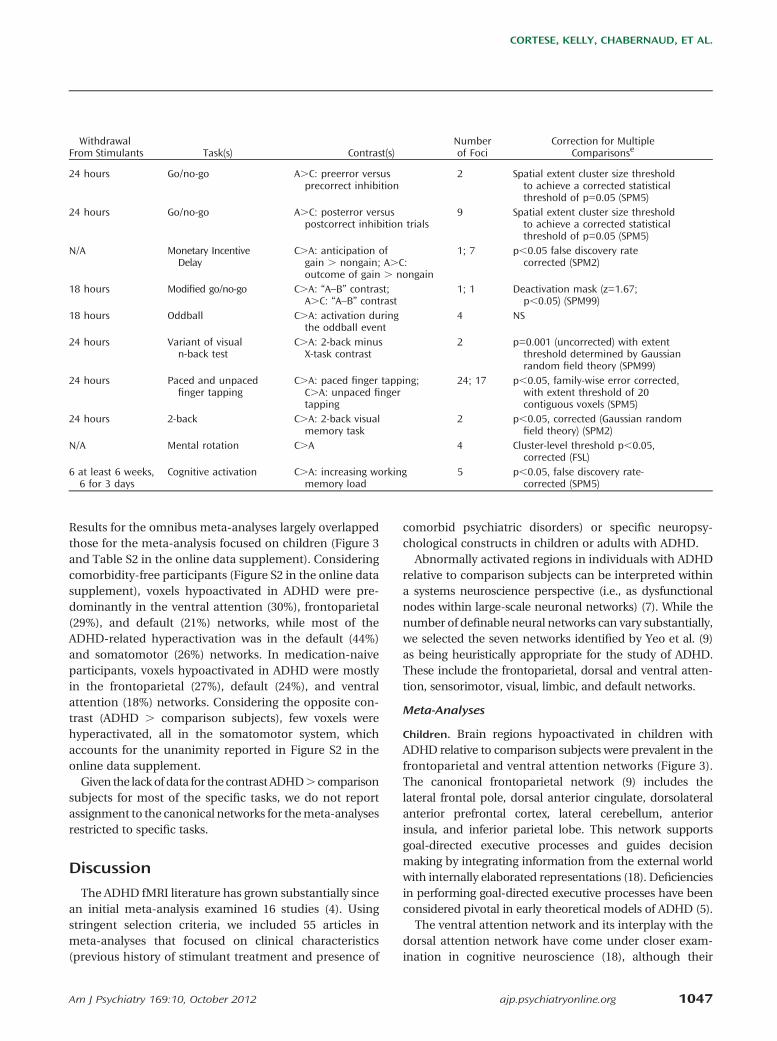
Withdrawal FromStimulants Task(s) Contrast(s)
Numberof Foci
Correction for MultipleComparisonse
24 hours Color word Stroop(variant)
Six contrasts includedin the meta-analysisf
20 (total) Cluster-wise protection; voxel levelthreshold p=0.01 (AFNI AlphaSim)
N/A 2-back workingmemory
C.A 1 Cluster-size thresholding (50 voxels);voxel level p=0.001 uncorrected(SPM99)
48 hours Go/no-go selectiveattention
C.A; C.A 17; 3 Cluster-size thresholding (10 voxels);voxel level p,0.001 uncorrected(SPM99)
24 hours Sustained Attention toResponse Test(go/no-go)
C.A: successful inhibition 5 NS for second-level analysis (AFNI)
2 weeks Cued target detection C.A: intrinsic alerting effect;C.A: phasic alerting effect;C.A: alerting effect
3; 4; 3 Cluster-size thresholding (10 voxels);voxel level p,0.001 uncorrected(SPM2)
Day of the scan Continuous PerformanceTask with stimulus=X
C.A; A.C 1; 2 Cluster-size thresholding (137 voxels);voxel-level p,0.05 uncorrected (AFNI)
N/A Tracking stop C.A: successful stop-go trials;C.A: unsuccessful stop-go trials;C.A: switch task
2; 1; 2 Cluster-mass threshold; voxel-widesignificance: p,0.05; clusterthreshold p,0.01 (XBAM)
N/A Simon task C.A: incongruent-congruent;C.A: oddball-congruent
1; 1 Cluster-mass threshold; voxel-widesignificance: p,0.05; clusterthreshold p,0.01 (XBAM)
N/A Rewarded CPT C.A: effect of attention;C.A: effect of reward
3; 2 Cluster-mass threshold; voxel-widesignificance: p,0.05; clusterthreshold p,0.01 (XBAM)
24 hours Modified go/no-go A.C: overall activation 1 Cluster-size thresholding (3 voxels);voxel level p,0.05 (SPM2)
24 hours Switch C.A; A.C 6; 6 Cluster-level thresholding (1,000iterations; p,0.05) (Brainvoyager QX)
3 weeks Go/no-go A.C 2 NS (SPM2)24 hours Go/no-go C.A: no-go . go; A.
C: no-go . go1; 8 Cluster-size thresholding (3 voxels);
voxel-level p,0.05 (NeuroImagingSoftware)
24 hours Variant go/no-go C.A: unexpected stimulus-unexpected time
1 Cluster-size thresholding (5 voxels);voxel-level p,0.05 (SPM2)
24 hours Variant go/no-go C.A: expected stimulus-unexpected time; C.A:unexpected stimulus-expected time
1; 2 Cluster-size thresholding (5 voxels);voxel-level p,0.05 (SPM2)
2 weeks WAIS forward/backwarddigit span
C.A: backward; A.C:forward; A.C: backward
5; 7; 3 Cluster-size thresholding (25 voxels);voxel level p,0.001 uncorrected(SPM2)
N/A Auditory go/no-go(modified)
C.A 7 Cluster-size thresholding; p,0.001uncorrected; threshold: 10 voxels(BrainVoyager)
24 hours n-back C.A: averaged 2- and3-back
5 Family-wise error correction (p,0.05);cluster size: 10 voxels (SPM5)
Am J Psychiatry 169:10, October 2012 ajp.psychiatryonline.org 1041
CORTESE, KELLY, CHABERNAUD, ET AL.

functional imaging data collected from 1,000 participants. Thoseseven robustly replicable networks, which are limited to corticalregions, include the frontoparietal, the dorsal and ventralattentional, the somatomotor, the visual, the limbic, and thedefault networks. We first determined the network in whicheach voxel of the ADHD-related hypo- or hyperactivated regionswas located by computing the number of significant voxels fromthe comparison subjects . ADHD and ADHD . comparisonsubjects contrasts that overlapped each of the network masks(downloaded from http://surfer.nmr.mgh.harvard.edu/fswiki/CorticalParcellation_Yeo2011). We then performed a chi-square
analysis contrasting the proportions of hypo- and hyperactivatedvoxels in the seven networks.
Results
Studies Included in the Meta-Analysis
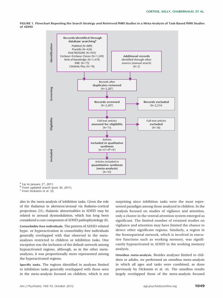
Figure 1 summarizes the search results according to thePreferred Reporting Items for Systematic Reviews andMeta-Analyses (PRISMA) flowchart (17). Details of included
TABLE 1. Characteristics of Studies Included in a Meta-Analysis of Task-Based fMRI Studies of ADHDa (Continued)
ADHDc Comparison Subjectsd
Age Age
First Authorb N M Mean SD N M Mean SD ADHD TypePsychiatric
ComorbiditiesTreatment With
Stimulants
Kobel (2010) 14 14 10 1.3 12 12 11 1.6 9 Co, 5 I 5 ODD-CD, 4 GAD All current
Konrad 16 16 10 1.9 16 16 10 1.3 9 Co, 6 I, 1 HI 5 ODD, 3 AD All naive
Kooistra 10 10 22 NS 10 10 22 NS NS N/A No after age16 years
Krauel 12 12 15 0.7 12 12 15 1.3 6 Co, 5 I, 1 HI 3 ODD, 2 CD 5 current
Mostofsky 11 8 10 1.2 11 8 10 1.4 9 Co, 2 I No 8 current
Passarotti (2010, J IntlNeuropsychol Soc)
15 12 13 2.6 14 7 14 2.4 All C No 5 current
Passarotti (2010, J Am AcadChild Adolesc Psychiatry)
14 9 13 2.3 19 9 14 3.1 All Co No NS
Passarotti (2010,Psychiatry Res)
11 6 13 2.7 15 7 14 3.1 All Co No 6 current
Prehen-Kristensen 12 NS 13 1.8 12 NS 14 2 12 Co, 2 I 5 ODD All current
Rubia (1999) 7 7 16 NS 9 9 15 NS All Co CD (NS) NS
Rubia (2005) 16 16 13 2.1 21 21 14 1.6 All Co 5 CD All naive
Rubia (2007) 17 17 14 2 18 18 13 2 All Co No All naive
Rubia (2008) 20 20 13 1.5 20 20 14 2 All Co No All naive
Rubia (2009, Am JPsychiatry)
18 18 13 1 16 16 13 3 All Co No All naive
Rubia (2009, J ChildPsychol Psychiatry)
20 20 13 1.4 20 20 14 1.9 All Co No All naive
1042 ajp.psychiatryonline.org Am J Psychiatry 169:10, October 2012
TOWARD SYSTEMS NEUROSCIENCE OF ADHD

and excluded studies are provided in section A2 of theonline data supplement. The search yielded 55 eligiblestudies, 16 of them assessing adults and 39 assessingchildren. Additional characteristics of the studies aresummarized in Table 1. While we endeavored to countindividuals from the same sample only once, the totalnumber of participants reported in Table 1 (741 withADHD and 801 comparison subjects) is approximatebecause some research groups reported results with
partially overlapping samples. References of the in-cluded studies are provided in section A3 of the onlinedata supplement.
Activation Likelihood Estimation Results
The meta-analysis focused on children (Table 2 andFigure 2) revealed significant ADHD-related hypoactiva-tion in frontal regions and putamen bilaterally and inright parietal and right temporal regions. ADHD-related
No n-back C.A: activation in3-back task
1 Correction applied but type ofcorrection NS (SPM 5)
N/A Attention Network Test(modified)
C.A: alerting; C.A:executive control; A.C:alerting; A.C: reorienting;A.C: executive control
1; 2; 1;3; 1
Cluster-size thresholding (10 voxels);p,0.001 uncorrected (SPM2)
N/A Go/no-go C.A: go; A.C: go 16; 2 Corrected cluster significancethreshold: p=0.01 (FSL)
24 hours Recognition memory C.A: neutral; A.C:emotional; A.C: neutral
1; 4; 4 Cluster-size thresholding (10 voxels);voxel-level p,0.001 uncorrected(SPM99)
24 hours Sequential fingertapping
C.A 2 Cluster-size thresholding (84 voxels);voxel-level p,0.001 uncorrected(SPM99)
Over a 3-weekperiod
Emotional valenceStroop
C.A: negative . neutral;C.A: positive . neutralwords; A.C: negative .neutral words; A.C: positive. neutral words
2; 2; 4; 5 Contiguity threshold: p,0.01uncorrected; experiment-wise type 1:p,0.02 corrected (AFNI AlphaSim)
At least 4 days Affective 2-backworking memory
C.A: angry . neutral faces;A.C: angry . neutral faces;A.C: happy . neutral faces
13; 1; 12 Contiguity threshold: p,0.01uncorrected; experiment-wise type 1:p,0.02 corrected (AFNI AlphaSim)
3 weeks Response inhibition C.A; A.C 7; 3 Contiguity threshold: p,0.01uncorrected; experiment-wise type 1:p,0.02 corrected (AFNI AlphaSim)
48 hours Delayed-match-to-sampleparadigm
C.A 15 Cluster-size thresholding (5 voxels);voxel-level p,0.05 (SPM5)
1 week Stop; delay C.A; C.A 4; 2 Permutation; voxel-wise probabilitytype I error50.05
N/A Stop C.A: successful . unsuccessfulinhibition; C.A: unsuccessfulinhibition . baseline go
2; 2 Cluster level difference; ,1 falseactivated cluster at p,0.05 (voxelcomparison); p,0.01 (clustercomparison)
N/A Oddball C.A: successful oddball .standard; C.A: standard. oddball
3; 2 Cluster level difference; ,1 falseactivated cluster at p,0.05 (voxelcomparison); p,0.01 (clustercomparison)
N/A Tracking stop C.A: successful . failed stop;C.A: failed stop . go; C.A:go . stop
1; 1; 1 Cluster level difference; ,1 falseactivated cluster at p,0.05 (voxelcomparison); p,0.03 (clustercomparison)
N/A Rewarded CPT C.A: effect of attention; C.A:effect of reward; A.C: effectof attention
2; 1; 9 Cluster level difference; ,1 falseactivated cluster at p,0.05(voxel comparison); p,0.01(cluster comparison)
N/A Simon C.A: incongruent . oddball;C.A: oddball . congruent
2; 2 Cluster level difference; ,1false activated cluster at p,0.05(both voxel and cluster comparison)
WithdrawalFrom Stimulants Task(s) Contrast(s)
Numberof Foci
Correction for MultipleComparisonse
Am J Psychiatry 169:10, October 2012 ajp.psychiatryonline.org 1043
CORTESE, KELLY, CHABERNAUD, ET AL.
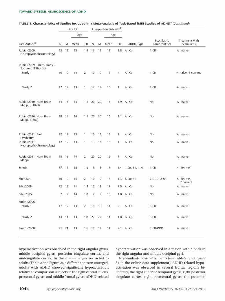
hyperactivation was observed in the right angular gyrus,middle occipital gyrus, posterior cingulate cortex, andmidcingulate cortex. In the meta-analysis restricted toadults (Table 2 and Figure 2), a different pattern emerged.Adults with ADHD showed significant hypoactivationrelative to comparison subjects in the right central sulcus,precentral gyrus, andmiddle frontal gyrus. ADHD-related
hyperactivation was observed in a region with a peak inthe right angular and middle occipital gyri.In stimulant-naive participants (see Table S1 and Figure
S1 in the online data supplement), ADHD-related hypo-activation was observed in several frontal regions bi-laterally, the right superior temporal gyrus, right posteriorcingulate cortex, right postcentral gyrus, the putamen
TABLE 1. Characteristics of Studies Included in a Meta-Analysis of Task-Based fMRI Studies of ADHDa (Continued)
ADHDc Comparison Subjectsd
Age Age
First Authorb N M Mean SD N M Mean SD ADHD TypePsychiatric
ComorbiditiesTreatment With
Stimulants
Rubia (2009,Neuropsychopharmacology)
13 13 13 1.4 13 13 13 1.8 All Co 1 CD All naive
Rubia (2009, Philos Trans RSoc Lond B Biol Sci)Study 1 10 10 14 2 10 10 15 4 All Co 1 CD 4 naive, 6 current
Study 2 12 12 13 1 12 12 13 1 All Co 1 CD All naive
Rubia (2010, Hum BrainMapp, p 1823)
14 14 13 1.1 20 20 14 1.9 All Co No All naive
Rubia (2010, Hum BrainMapp, p 287)
18 18 14 1.1 20 20 15 1.1 All Co No All naive
Rubia (2011, BiolPsychiatry)
12 12 13 1 13 13 13 1 All Co No All naive
Rubia (2011,Neuropsychopharmacology)
12 12 13 1 13 13 13 1 All Co No All naive
Rubia (2011, Hum BrainMapp)
18 18 14 2 20 20 16 1 All Co No All naive
Schulz 5g 5 18 1.3 5 5 18 1.4 1 Co, 3 I, 1 HI 1 CD 4 lifetimeh
Sheridan 10 0 15 2 10 0 15 1.3 6 Co; 4 I 2 ODD, 2 SP 5 lifetimei,2 current
Silk (2008) 12 12 11 1.5 12 12 11 1.5 All Co No All naive
Silk (2005) 7 7 14 1.8 7 7 15 1.8 All Co No All naive
Smith (2006)Study 1 17 17 13 2 18 18 14 2 All Co 5 CD All naive
Study 2 14 14 13 1.8 27 27 14 1.8 All Co 5 CD All naive
Smith (2008) 21 21 13 1.6 17 17 14 2.1 All Co 3 CD/ODD All naive
1044 ajp.psychiatryonline.org Am J Psychiatry 169:10, October 2012
TOWARD SYSTEMS NEUROSCIENCE OF ADHD

bilaterally, and the right thalamus. Only one significantcluster of ADHD-related hyperactivation was observed,with a peak located in the right superior longitudinalfasciculus underlying the insula.When considering comorbidity-free participants (see
Table S1 and Figure S1 in the online data supplement),ADHD-related hypoactivated regions were located in the
frontal regions and the putamen bilaterally, the rightsuperior temporal gyrus, and the right occipital pole. ADHD-related hyperactivation was observed in the left inferiorfrontal gyrus, left Heschl’s gyrus, and several right posteriorregions.In analyses limited to specific tasks (see Table S1 in the
online data supplement), ADHD-related hypoactivation
N/A Rewarded CPT C.A: attention; C.A: reward;A.C: reward
13; 2; 4 Cluster level difference; ,1 falseactivated cluster at p,0.05(voxel comparison); p,0.02(cluster comparison) (XBAM)
36 hours (N=6) Delay discounting C.A 6 Cluster level difference; ,1false activated cluster atp,0.05 (voxel comparison);p,0.006 (cluster comparison)
N/A Timediscrimination
C.A; A.C 1; 2 Cluster level difference; ,1false activated cluster atp,0.05 (voxel comparison);p,0.006 (cluster comparison)
N/A Switch C.A 2 Cluster level difference; ,1false activated cluster at p,0.05for both voxel and clustercomparison (XBAM)
N/A Visual trackingstop; Meiranswitch
C.A: successful-stop;C.A: failed stop; C.A
1; 2; 3 Cluster level difference;,1 false activated clusterat p,0.05 (voxel comparison);p,0.003 (cluster comparison)(XBAM)
N/A Visual trackingstop
C.A: successful inhibition;C.A: inhibition failure
13; 13 Threshold-free clusterenhancement (p,0.05)
N/A Simon C.A: Simon . oddballcondition
4 Cluster level difference; ,1 falseactivated cluster at p,0.05 (voxelcomparison); p,0.01 (clustercomparison) (XBAM)
N/A Simon C.A: oddball . congruent;C.A: incongruent . oddball
2; 2 Cluster level difference; ,1 false activatedcluster at p,0.05 (voxel comparison);p,0.01 (cluster comparison) (XBAM)
N/A Go/no-go C.A: correct no-go . correctgo; A.C: correct no-go. correct go
2; 3 Cluster-size thresholding (120voxels); voxel level p,0.01uncorrected (SPM99)
24 hours Delay match-to-sample
C.A: activation at highload period
2 Gaussian field theory; p=0.05 correctedat cluster level (fMRIstat program)
N/A Raven’s StandardProgressive Matrices
C.A 45 Clusters of voxels (z.2.33) with clusterlevel p,0.05 corrected (FSL)
N/A Mental rotation C.A; A.C 8; 4 Voxel level p,0.001 uncorrected;cluster level p,0.05 corrected
N/A Go/no-go C.A: successful no-gostimuli
1 Cluster level difference; ,1 false activatedcluster at p,0.05 (voxel comparison);p,0.01 (cluster comparison)
N/A Switch C.A: switch 9 Cluster level difference; ,1 false activatedcluster at p,0.05 (voxel comparison);p,0.01 (cluster comparison)
N/A Time discrimination C.A: time discrimination. temporal order judgment
2 Cluster level difference; ,1 false activatedcluster at p,0.05 (voxel comparison);p,0.01 (cluster comparison) (XBAM)
WithdrawalFrom Stimulants Task(s) Contrast(s)
Numberof Foci
Correction for MultipleComparisonse
Am J Psychiatry 169:10, October 2012 ajp.psychiatryonline.org 1045
CORTESE, KELLY, CHABERNAUD, ET AL.
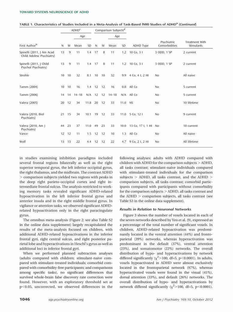
in studies examining inhibition paradigms includedseveral frontal regions bilaterally as well as the rightsuperior temporal gyrus, the left inferior occipital gyrus,the right thalamus, and themidbrain. The contrast ADHD. comparison subjects yielded two regions with peaks inthe deep right parieto-occipital cortex and right in-termediate frontal sulcus. The analysis restricted to work-ing memory tasks revealed significant ADHD-relatedhypoactivation in the left inferior frontal gyrus andanterior insula and in the right middle frontal gyrus. Invigilance or attention tasks, we observed significant ADHD-related hypoactivation only in the right paracingulategyrus.
The omnibus meta-analysis (Figure 2; see also Table S2in the online data supplement) largely recapitulated theresults of the meta-analysis focused on children, withadditional ADHD-related hypoactivations in the inferiorfrontal gyri, right central sulcus, and right posterior pa-rietal lobe and hyperactivations inHeschl’s gyrus as well asadditional loci in inferior frontal gyri.
When we performed planned subtraction analyses(adults compared with children; stimulant-naive com-pared with stimulant-treated individuals; comorbid com-pared with comorbidity-free participants; and comparisonsamong specific tasks), no significant differences thatsurvived whole-brain false discovery rate correction werefound. However, with an exploratory threshold set atp,0.05, uncorrected, we observed differences in the
following analyses: adults with ADHD compared withchildrenwith ADHD for the comparison subjects.ADHD,all tasks contrast; stimulant-naive individuals comparedwith stimulant-treated individuals for the comparisonsubjects . ADHD, all tasks contrast, and the ADHD .
comparison subjects, all tasks contrast; comorbid partic-ipants compared with participants without comorbidityfor the comparison subjects. ADHD, all tasks contrast andthe ADHD . comparison subjects, all tasks contrast (seeTable S3 in the online data supplement).
Results in Relation to Neuronal Networks
Figure 3 shows the number of voxels located in each ofthe seven networks described by Yeo et al. (9), expressed asa percentage of the total number of significant voxels. Inchildren, ADHD-related hypoactivation was predomi-nantly located in the ventral attention (44%) and fronto-parietal (39%) networks, whereas hyperactivation waspredominant in the default (37%), ventral attention(23%), and somatomotor (22%) networks. The overalldistribution of hypo- and hyperactivations by networkdiffered significantly (x2.100, df=5, p,0.0001). In adults,voxels hypoactivated in ADHD were almost exclusivelylocated in the frontoparietal network (97%), whereashyperactivated voxels were found in the visual (41%),dorsal attention (33%), and default (26%) networks. Theoverall distribution of hypo- and hyperactivations bynetwork differed significantly (x2.100, df=5, p,0.0001).
TABLE 1. Characteristics of Studies Included in a Meta-Analysis of Task-Based fMRI Studies of ADHDa (Continued)
ADHDc Comparison Subjectsd
Age Age
First Authorb N M Mean SD N M Mean SD ADHD TypePsychiatric
ComorbiditiesTreatment With
Stimulants
Spinelli (2011, J Am AcadChild Adolesc Psychiatry)
13 9 11 1.4 17 8 11 1.2 10 Co, 3 I 3 ODD, 1 SP 2 current
Spinelli (2011, J ChildPsychol Psychiatry)
13 9 11 1.4 17 8 11 1.2 10 Co, 3 I 3 ODD, 1 SP 2 current
Strohle 10 10 32 8.1 10 10 32 9.9 4 Co, 4 I, 2 HI No All naive
Tamm (2004) 10 10 16 1.4 12 12 16 0.8 All Co No 5 current
Tamm (2006) 14 14 14–18 N/A 12 12 14–18 N/A All Co No 5 current
Valera (2005) 20 12 34 11.8 20 12 33 11.0 NS No 10 lifetime
Valera (2010, BiolPsychiatry)
21 15 34 10.1 19 12 33 11.0 5 Co, 12 I No 9 current
Valera (2010, Am JPsychiatry)
44 23 37 11.0 49 23 33 10.0 13 Co, 17 I, 1 HI No 18 current
Vance 12 12 11 1.5 12 12 10 1.3 All Co No All naive
Wolf 13 13 22 4.4 12 12 22 4.7 9 Co, 2 I, 2 HI No All lifetime
1046 ajp.psychiatryonline.org Am J Psychiatry 169:10, October 2012
TOWARD SYSTEMS NEUROSCIENCE OF ADHD
Results for the omnibus meta-analyses largely overlappedthose for the meta-analysis focused on children (Figure 3and Table S2 in the online data supplement). Consideringcomorbidity-free participants (Figure S2 in the online datasupplement), voxels hypoactivated in ADHD were pre-dominantly in the ventral attention (30%), frontoparietal(29%), and default (21%) networks, while most of theADHD-related hyperactivation was in the default (44%)and somatomotor (26%) networks. In medication-naiveparticipants, voxels hypoactivated in ADHD were mostlyin the frontoparietal (27%), default (24%), and ventralattention (18%) networks. Considering the opposite con-trast (ADHD . comparison subjects), few voxels werehyperactivated, all in the somatomotor system, whichaccounts for the unanimity reported in Figure S2 in theonline data supplement.Given the lackof data for the contrast ADHD. comparison
subjects for most of the specific tasks, we do not reportassignment to the canonical networks for themeta-analysesrestricted to specific tasks.
Discussion
The ADHD fMRI literature has grown substantially sincean initial meta-analysis examined 16 studies (4). Usingstringent selection criteria, we included 55 articles inmeta-analyses that focused on clinical characteristics(previous history of stimulant treatment and presence of
comorbid psychiatric disorders) or specific neuropsy-chological constructs in children or adults with ADHD.Abnormally activated regions in individuals with ADHD
relative to comparison subjects can be interpreted withina systems neuroscience perspective (i.e., as dysfunctionalnodes within large-scale neuronal networks) (7). While thenumber of definable neural networks can vary substantially,we selected the seven networks identified by Yeo et al. (9)as being heuristically appropriate for the study of ADHD.These include the frontoparietal, dorsal and ventral atten-tion, sensorimotor, visual, limbic, and default networks.
Meta-Analyses
Children. Brain regions hypoactivated in children withADHD relative to comparison subjects were prevalent in thefrontoparietal and ventral attention networks (Figure 3).The canonical frontoparietal network (9) includes thelateral frontal pole, dorsal anterior cingulate, dorsolateralanterior prefrontal cortex, lateral cerebellum, anteriorinsula, and inferior parietal lobe. This network supportsgoal-directed executive processes and guides decisionmaking by integrating information from the external worldwith internally elaborated representations (18). Deficienciesin performing goal-directed executive processes have beenconsidered pivotal in early theoretical models of ADHD (5).The ventral attention network and its interplay with the
dorsal attention network have come under closer exam-ination in cognitive neuroscience (18), although their
24 hours Go/no-go A.C: preerror versusprecorrect inhibition
2 Spatial extent cluster size thresholdto achieve a corrected statisticalthreshold of p=0.05 (SPM5)
24 hours Go/no-go A.C: posterror versuspostcorrect inhibition trials
9 Spatial extent cluster size thresholdto achieve a corrected statisticalthreshold of p=0.05 (SPM5)
N/A Monetary IncentiveDelay
C.A: anticipation ofgain . nongain; A.C:outcome of gain . nongain
1; 7 p,0.05 false discovery ratecorrected (SPM2)
18 hours Modified go/no-go C.A: “A–B” contrast;A.C: “A–B” contrast
1; 1 Deactivation mask (z=1.67;p,0.05) (SPM99)
18 hours Oddball C.A: activation duringthe oddball event
4 NS
24 hours Variant of visualn-back test
C.A: 2-back minusX-task contrast
2 p=0.001 (uncorrected) with extentthreshold determined by Gaussianrandom field theory (SPM99)
24 hours Paced and unpacedfinger tapping
C.A: paced finger tapping;C.A: unpaced fingertapping
24; 17 p,0.05, family-wise error corrected,with extent threshold of 20contiguous voxels (SPM5)
24 hours 2-back C.A: 2-back visualmemory task
2 p,0.05, corrected (Gaussian randomfield theory) (SPM2)
N/A Mental rotation C.A 4 Cluster-level threshold p,0.05,corrected (FSL)
6 at least 6 weeks,6 for 3 days
Cognitive activation C.A: increasing workingmemory load
5 p,0.05, false discovery rate-corrected (SPM5)
WithdrawalFrom Stimulants Task(s) Contrast(s)
Numberof Foci
Correction for MultipleComparisonse
Am J Psychiatry 169:10, October 2012 ajp.psychiatryonline.org 1047
CORTESE, KELLY, CHABERNAUD, ET AL.
potential dysfunction in ADHD has been infrequentlyconsidered. The ventral attention network includes thetemporoparietal junction, the supramarginal gyrus, fron-tal operculum, and anterior insula; the dorsal system isanchored in the intraparietal sulcus and the frontal eyefields (18). While the dorsal attention network underpinsthe selection of sensory stimuli based on internal goals orpersonal expectations, the ventral network supportsattentional reorienting to salient and behaviorally relevantexternal stimuli (18). A recent preliminary study reporteddeficient ventral attention network engagement in adultswith ADHD, suggesting that this may underlie an ADHD-related deficit in adaptive switching to external salientstimuli (19). This, along with our finding of ventralattention network hypoactivation, is in line with thetheoretical framework proposed by Nigg and Casey (6),which emphasizes that learning when and in whatcontexts to expect an event is critical for planning andmaintaining appropriate behaviors. In their model, diffi-culties in detecting regularities or irregularities in theenvironment lead to problems in modulating behaviorsaccording to changes in the environment, which manifestas ADHD symptoms. We speculate that hypoactivation inthe ventral attention network underpins ADHD-relateddeficits in detecting regularities and irregularities in theenvironment. We also observed hyperactivation of re-gions in the ventral attention network. Since the sup-pression of this network is necessary to prevent shifts ofattention to irrelevant objects (20), its hyperactivationmight underpin distractibility, one of the cardinal symp-toms of ADHD.
We note that the dorsal attention system was relativelyunderrepresented among ADHD-related hypoactivatedregions, although hypofunction of this system has beenproposed in ADHD (7). In part, our results may reflect thesubstantial prevalence of inhibition-related tasks that aresubserved predominantly by the ventral, rather than thedorsal, attention network (18).
We also identified peaks of ADHD-related hypoactivationin the right somatomotor system and in the putamenbilaterally. Together with the cluster of hypoactivation inthemedial superior frontal gyrus and supplementarymotorarea, these regions are consistent with abnormal function ofthe pyramidal motor system, which would be expected inADHD given the salience of motoric hyperactivity. Re-markably, interindividual differences in locomotor activityhave rarely been examined in ADHD using neuroimagingmeasures. Using transcranial magnetic stimulation, ro-bustly abnormal intracortical inhibition has been reportedin the motor system in children with ADHD (21).
Besides hypoactivation, we also observed several re-gions of ADHD-related hyperactivation, predominantlyin the default network. This network underlies self-referential cognitive processes that are typically sup-pressed during the performance of externally orientedattentionally demanding tasks (22). Spontaneous activity
fluctuations in the default network tend to be anticor-related with fluctuations in “task positive” networks(i.e., networks that are activated during active tasks), suchas the frontoparietal and dorsal attention networks (22).According to the default-mode hypothesis of ADHD (22),the default network might be inadequately regulated byother task-active systems and might consequently intrudeon or disrupt ongoing cognitive performance, contributingto fluctuations in attention that characterize ADHD. Thestudies whose coordinates contributed to the hyper-activated default network clusters in our meta-analysisdid not systematically report whether this hyperactivationreflected weaker task-related deactivation in ADHDrelative to comparison subjects, or stronger activation inADHD relative to comparison subjects. However, amelio-ration of inadequate default network deactivation inADHD by methylphenidate was recently reported by twoseparate groups (12, 23).We also observed ADHD-related hyperactivation in the
somatomotor and visual systems. This is in line with thehypothesis that individuals with ADHD compensate forimpaired function in the prefrontal and anterior cingulatecortex by overreliance (relative to comparison subjects) onbrain regions associated with visual, spatial, or motoricprocessing (24).
Adults. Themeta-analysis restricted to adults yielded fewerregional group differences compared with that restrictedto children. This may be accounted for by the smallernumber of retained studies (N=16) and consequentlylower statistical power. Almost all the hypoactivated voxelswere found in the frontoparietal system, which is con-sistent with the persistence of executive dysfunction inadults with ADHD (25). Hypoactivation in the somatomo-tor systemwas less prominent in adults than in children, inline with clinical observations that motoric hyperactivitydecreases with age (26). In the visual and dorsal attentionsystems, adults with ADHD exhibited proportionally morehyperactivation than children, suggesting the hypothesisthat these networks may carry the bulk of the compensa-tory load in adults (24).
Stimulant-naive individuals. Although early imaging studiesof ADHD were confounded by previous stimulant treat-ment history (27), recent meta-analytic evidence hasconfirmed that brain structural changes are present instimulant-naive participants with ADHD (28) and sug-gested that treatment with stimulants may even normalizestructural abnormalities (29). Here, we extend those obser-vations byfinding significant differences between stimulant-naive individuals with ADHD and comparison subjects,which indicates that ADHD neuronal dysfunctions are alsonot likely accounted for by previous stimulant treatment.The thalamus, despite its central location inmultiple brain
circuits, has been generally overlooked in the ADHDneuroimaging literature (30). We observed thalamic hypo-activation in stimulant-naive individuals with ADHD and
1048 ajp.psychiatryonline.org Am J Psychiatry 169:10, October 2012
TOWARD SYSTEMS NEUROSCIENCE OF ADHD
also in the meta-analysis of inhibition tasks. Given the roleof the thalamus in alertness/arousal via thalamo-corticalprojections (31), thalamic abnormalities in ADHD may berelated to arousal dysmodulation, which has long beenconsidered a core component of ADHDpathophysiology (6).
Comorbidity-free individuals. The pattern of ADHD-relatedhypo- or hyperactivation in comorbidity-free individualsgenerally overlapped with that observed in the meta-analyses restricted to children or inhibition tasks. Oneexception was the inclusion of the default network amonghypoactivated regions, although, as in the other meta-analyses, it was proportionally more represented amongthe hyperactivated regions.
Specific tasks. The regions identified in analyses limitedto inhibition tasks generally overlapped with those seenin the meta-analysis focused on children, which is not
surprising since inhibition tasks were the most repre-sented paradigm among those analyzed in children. In theanalysis focused on studies of vigilance and attention,only a cluster in the ventral attention system emerged assignificant. The limited number of retained studies onvigilance and attention may have limited the chance todetect other significant regions. Similarly, a region inthe frontoparietal network, which is involved in execu-tive functions such as working memory, was signifi-cantly hypoactivated in ADHD in the working memoryanalysis.
Omnibus meta-analysis. Besides analyses limited to chil-dren or adults, we performed an omnibus meta-analysisin which all ages and tasks were combined, as donepreviously by Dickstein et al. (4). The omnibus resultslargely overlapped those of the meta-analysis focused
FIGURE 1. Flowchart Reporting the Search Strategy and Retrieved fMRI Studies in a Meta-Analysis of Task-Based fMRI Studiesof ADHD
Full-text articlesexcluded
(N=36)
Articles included inquantitative synthesis
(meta-analysis)(N=55)
Articlesincluded in qualitative
synthesis(N=37+9b+9c)
Full-text articlesassessed for eligibility
(N=73)
Records excluded(N=2,214)
Records screened(N=2,287)
Records after duplicates removed
(N=2,287)
Records identified through database searchinga
PubMed (N=889)PsycInfo (N=426)
Ovid MEDLINE (N=935)Embase+Embase Classic (N=1,249)
Web of Knowledge (N=1,419)ERIC (N=75)
CINAHAL Plus (N=78)
Additional recordsidentified through other sources (manual search)
(N=2)
Incl
ud
ed
Eligib
ilit
ySc
reen
ing
Iden
tifi
cati
on
a Up to January 27, 2011.b From updated search (June 30, 2011).c From Dickstein et al. (4).
Am J Psychiatry 169:10, October 2012 ajp.psychiatryonline.org 1049
CORTESE, KELLY, CHABERNAUD, ET AL.

on children, which was expected because 70% of the in-cluded studies were conducted in children. Still, the largernumber of studies and greater statistical power allowed usto detect a substantially larger number of significantly
different voxels than in the sum of the two age-limitedmeta-analyses (see Figure 3). This yielded additionalputatively dysfunctional regions in ADHD, including thesuperior frontal gyrus/supplementary motor area, the
TABLE 2. Regions Exhibiting Significantly Greater Activation in Comparison Subjects Relative to Individuals With ADHD andVice Versa in the Meta-Analyses Focused on Children or Adults Across All Tasks
Weighted CenteraMaximum Activation
Likelihood Estimate Valuea
Contrast, Analysis, andCluster Number
Volume(mm3) x y z
ExtremaValue x y z Anatomical Labelb
Comparison subjects .participants with ADHDChildren, all tasksc
1 1,864 20.35 15.56 48.61 0.0211 0 16 54 Medial superior frontal gyrus/supplementary motor area(BA 6) R, L (frontoparietal/ventral attention)
0.0186 0 14 44 Paracingulate gyrus (BA 32) R, L(ventral attention)
2 688 31.42 0.19 3.33 0.0148 32 0 4 Putamen R3 464 221.92 2.37 4.1 0.0119 220 4 4 Putamen L4 424 33.7 227.47 50.75 0.0136 34 228 52 Postcentral gyrus R
(somatomotor)5 400 42.23 9.52 29.36 0.0134 42 10 30 Inferior frontal sulcus; inferior
precentral sulcus R(frontoparietal)
6 384 57.29 225.00 28.42 0.0145 58 226 28 Inferior parietal lobule(supramarginal gyrus; BA 40)R (ventral attention)
7 368 55.66 12.64 26.64 0.0136 56 12 26 Superior temporal gyrus (BA 22)R (somatomotor)
8 280 241.26 31.46 23.46 0.0131 242 32 24 Middle frontal gyrus (BA 9/46) L(frontoparietal)
9 248 47.46 25.85 31.84 0.0115 48 26 32 Middle frontal gyrus (BA 9/46) R(frontoparietal)
Adults, all tasksc
1 400 21.7 227.31 50.84 0.0106 22 228 50 Central sulcus/precentral gyrus R2 232 34.38 56.96 13.62 0.0103 34 56 14 Middle frontal gyrus (BA 10) R
(frontoparietal)Participants with ADHD .comparison subjectsChildren, all tasksd
1 680 39.72 258.51 18.51 0.0135 40 258 20 Angular gyrus; middle occipitalgyrus R
0.0079 40 266 14 Angular gyrus; middle occipitalgyrus R (visual)
2 400 15.02 250.94 31.01 0.0101 16 252 32 Posterior cingulate cortex;subparietal sulcus R
3 392 31.22 27.23 16.7 0.0107 32 28 16 White matter R (suboperculum)4 352 4.67 213.72 42.61 0.0101 4 214 42 Midcingulate cortex R
(ventral attention)Adults, all tasksd
1 872 40.39 258.08 16.91 0.0152 40 258 18 Angular gyrus; middleoccipital gyrus R
a Montreal Neurological Institute coordinates.b R=right; L=left; Brodmann’s areas (BA) are indicated in parentheses when identifiable. When located unambiguously in a cortical region, theanatomic label is followed in parentheses by the neural network corresponding to the maximum activation likelihood estimation value, fromthe seven reference neuronal networks identified by Yeo et al. (9).
c After removing complete overlap but including partial overlap of samples across studies from the same research groups. For children, alltasks, number of foci=241; number of experiments=35; and total number of subjects=958. For adults, all tasks, number of foci=81; numberof experiments=12; and total number of subjects=414.
d For children, all tasks, number of foci=80; number of experiments=17; and total number of subjects=431. For adults, all tasks, number offoci=49; number of experiments=8; and total number of subjects=226.
1050 ajp.psychiatryonline.org Am J Psychiatry 169:10, October 2012
TOWARD SYSTEMS NEUROSCIENCE OF ADHD
putamen, and the superior temporal gyrus, that had notemerged in the previous meta-analysis (4).
Subtraction analyses. Despite specificities in each focusedmeta-analysis, subtraction analyses corrected for multiplecomparisons showed that age, clinical characteristics, ortype of task did not moderate our results. However, thesenegative results must be interpreted cautiously. Althoughdefinitive standards for statistical power in subtractionanalyses do not exist, the informal consensus in theGingerALE users forum (www.brainmap.org/ale/) is thatbetween-analysis subtractions with fewer than 10 studiesprovide inadequate statistical power. Therefore, while thenumber of studies was sufficient for focused meta-analyses within subgroups, we were likely underpoweredto carry out reliable subtraction analyses across thesesubgroups. Indeed, when we relaxed the statisticalthreshold and considered noncorrected results, we diddetect differences in some subtraction analyses. Thoseresults are not discussed further as they likely contain toomany type I errors. However, we report them in Table S3 in
the online data supplement since they may be useful forcomparison with future analyses.
General Overview
ADHD is increasingly being conceived as a disorderunderpinned by dysfunctions in multiple large-scale brainnetworks (7, 8). This perspective facilitates identification ofseveral broad themes that emerged across our multiplemeta-analytic instances. These include hypoactivation inthe frontoparietal executive control network, putamen,and ventral attention network, which is consistent with theclassical model of ADHD as a disorder of deficient fronto-striatal activation (32). However, we also detected substan-tial hyperactivation, particularly in the default networkand visual circuits, which supports a model of ADHDbased on the interrelationships among neural networks.Together with more recent reports (12, 23), our results ofdefault network hyperactivation are consistent with thehypothesis that the inconsistency that characterizes manyindividuals with ADHD results from faulty interregulationbetween the default network and task-positive circuits
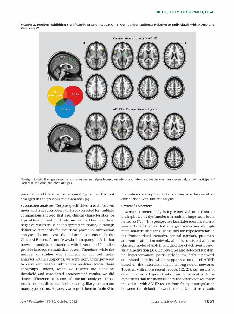
FIGURE 2. Regions Exhibiting Significantly Greater Activation in Comparison Subjects Relative to Individuals With ADHD andVice Versaa
Comparison subjects > ADHD
ADHD > Comparison subjects
R L
AdultsAdults
Children
All participants
aR=right; L=left. The figure reports results for meta-analyses focused on adults or children and for the omnibus meta-analysis. “All participants”refers to the omnibus meta-analysis.
Am J Psychiatry 169:10, October 2012 ajp.psychiatryonline.org 1051
CORTESE, KELLY, CHABERNAUD, ET AL.

such as the frontoparietal, ventral, or dorsal attentionnetworks (7). The next generation of functional imagingstudies should examine the temporal dependencies be-tween behavioral indices of attentional lapses (e.g., episod-ically prolonged response times) in relation to their brainimaging correlates with the goal of capturing the deviationsin the interplay between the default and task-positivenetworks.
Our results also provide meta-analytic support to viewspositing ADHD as a disorder characterized not only by
functional deficiencies but also by possible compensatorymechanisms, such as hyperactivation in visual regions.Such putative compensatory mechanisms can be difficultto observe through clinical measures alone, but becomeevident through neuroimaging (33). Awareness and doc-umentation of brain compensatory mechanisms mayeventually yield a clinical benefit from neuroimaging. Thiswould be analogous to the use of neurocognitive assess-ments to identify particular strengths to best formulatea comprehensive clinical treatment plan.
FIGURE 3. Proportions of ADHD-Related Hypo- or Hyperactivation in Meta-Analyses Focused on Adults or Children and forthe Omnibus Meta-Analysisa
Co
mp
ari
son
Su
bje
cts
> A
DH
DA
DH
D >
Co
mp
ari
son
Su
bje
cts
Visual Somatomotor Dorsal Attention Ventral Attention Limbic Frontoparietal Default
Children Adults All Participants
44% 39%
9%8%
3%
97%
41%30%
4%
20%5%
17%
26%33%37%
14%
22%
4%
23%
41%
39%
9%26%
8%
1%
a Number of significant voxels in the contrast comparison subjects . ADHD: children=3,320; adults=272; omnibus=6,024. Number ofsignificant voxels in the contrast ADHD . comparison subjects: children=888; adults=464; omnibus=2,720. The regions presented in theupper panel are in relation to the Yeo et al. (9) seven networks.
1052 ajp.psychiatryonline.org Am J Psychiatry 169:10, October 2012
TOWARD SYSTEMS NEUROSCIENCE OF ADHD

We failed to find support for the involvement of regionsrelated to motivation and emotion, such as the ventralstriatum (34), orbitofrontal, or amygdala/hippocampus(35), despite increasing recognition of motivational andemotional dysfunction in models of ADHD (36). Wecannot rule out type II error, as three studies assessingreward (37–39) and two on emotional processing (40, 41)did not meet our inclusion criteria. In addition, theorbitofrontal cortex and the medial temporal lobes arechallenging brain regions to examine with fMRI because ofsusceptibility artifact and signal dropout. We also note thelack of apparent involvement of the cerebellum, which hasbeen implicated in ADHD by multiple volumetric andfunctional studies and by theoretical models emphasizingthe role of cerebellar dysfunction in contributing todeficits in monitoring the frequency and timing of events(6). In our meta-analysis, several peaks in the cerebellumdid not reach our cluster size threshold for significance.We suspect that high intersubject variability in cerebellargeometry relative to stereotaxic space mitigated theemergence of cerebellar findings across studies.
Limitations
Several limitations of our meta-analysis should be takeninto account. The first relates to selection criteria. Tominimize confounding factors such as differences indiagnostic procedures or analytical approaches, we ex-cluded approximately half the screened studies. Still, thestudies included were heterogeneous, for example, withrespect to the method used to correct for multiple com-parisons. This is notable because activation likelihoodestimation does not take into account interstudy differencesin statistical thresholds. Second, separate meta-analysescould not be performed by ADHD subtype or sex, becauseseparate results for male and female participants and ADHDsubtypes are not usually reported in the literature. This isunfortunate since patterns of fMRI activation in ADHD candiffer by sex (42) and, possibly, by subtype (43). Third, themeta-analytic approach we adopted allows a quantitativesummary of positive results but cannot take into accountnegative findings. Effect sizes in fMRI may be confoundedbymany factors, such as movement covariates, and there isno agreement on how they should be handled. Thus,activation likelihood estimation should be considereda summary of the spatial distribution of positive results,rather than a true meta-analysis. Fourth, it was generallynot possible to determine the extent to which overlappingsamples were reported across studies. Thus, the totalnumber of participants represents an upper bound. Theopen sharing of fMRI data (e.g., via www.OpenfMRI.org)should obviate this problem in the future. Finally, fMRI dataare fundamentally limited. Besides only indirectly reflect-ing neuronal activity (44), fMRI data cannot define anabsolute or quantitative baseline state of activation (45), asthey always depend on the differences in signal betweentwo conditions. Positron emission tomography provides
absolute quantification (e.g., see 46, 47), but current ethicalconstraints limit its application to adults.
Conclusions
Moving beyond models of ADHD focused on a limitedset of brain regions, the maturing fMRI literature in ADHDreveals dysfunctions in regions belonging to multipleneuronal networks involved in higher-level cognitive andsensorimotor functions. Our results were not ascribable tostimulant treatment history or presence of comorbidities.The systems neuroscience perspective we adopted is inline with the NIH Research Domain Criteria framework(48), which conceptualizes mental disorders in terms ofdysfunctions of brain circuits to inform future nosologicalsystems beyond a symptoms-based approach. Futurework aimed at understanding the interplay among large-scale neural networks and their links to ADHD symptomdimensions should illuminate the pathophysiology of thiscommon and vexing disorder.
Received Oct. 15, 2011; revisions received Feb. 11, and April 14,2012; accepted April 23, 2012 (doi: 10.1176/appi.ajp.2012.11101521).From the Phyllis Green and Randolph Cowen Institute for PediatricNeuroscience, Child Study Center of the NYU Langone Medical Center,New York; the Child Neuropsychiatry Unit, G. B. Rossi Hospital,Department of Life Science and Reproduction, Verona University,Verona, Italy; UMR-S INSERM U 930, François-Rabelais University, ChildPsychiatry Center, University Hospital, Tours, France; Neuroingenia,Mexico City, Mexico; the Nathan S. Kline Institute for PsychiatricResearch, Orangeburg, N.Y.; and the Center for the Developing Brain,Child Mind Institute, New York. Address correspondence to Dr.Castellanos ([email protected]).Dr. Cortese has received financial support to attend medical
meetings from Eli Lilly and Shire Pharmaceuticals; was a co-investigator in studies sponsored by GlaxoSmithKline, Eli Lilly, andGenopharm; and has served as a consultant for Shire Pharmaceuticals.The other authors report no financial relationships with commercialinterests.Dr. Cortese is supported by the Marie Curie grant for Career
Development (Outgoing International Fellowship, POIF-253103) fromthe European Commission. This research was also supported by NIHgrants MH083246, MH081218, HD065282, and K23M087770.
References
1. Polanczyk G, de Lima MS, Horta BL, Biederman J, Rohde LA:The worldwide prevalence of ADHD: a systematic reviewand metaregression analysis. Am J Psychiatry 2007; 164:942–948
2. Mannuzza S, Klein RG, Moulton JL 3rd: Persistence of attention-deficit/hyperactivity disorder into adulthood: what have welearned from the prospective follow-up studies? J Atten Disord2003; 7:93–100
3. Wolraich M: Attention deficit hyperactivity disorder: the moststudied and yet most controversial diagnosis. Ment Retard DevDisabil Res Rev 1999; 5:163–168
4. Dickstein SG, Bannon K, Castellanos FX, Milham MP: Theneural correlates of attention deficit hyperactivity disorder:an ALE meta-analysis. J Child Psychol Psychiatry 2006; 47:1051–1062
5. Barkley RA: Behavioral inhibition, sustained attention, and ex-ecutive functions: constructing a unifying theory of ADHD.Psychol Bull 1997; 121:65–94
Am J Psychiatry 169:10, October 2012 ajp.psychiatryonline.org 1053
CORTESE, KELLY, CHABERNAUD, ET AL.

6. Nigg JT, Casey BJ: An integrative theory of attention-deficit/ hy-peractivity disorder based on the cognitive and affective neu-rosciences. Dev Psychopathol 2005; 17:785–806
7. Castellanos FX, Proal E: Large-scale brain systems in ADHD: beyondthe prefrontal-striatal model. Trends Cogn Sci 2012; 16:17–26
8. Makris N, Biederman J, Monuteaux MC, Seidman LJ: Towardsconceptualizing a neural systems-based anatomy of attention-deficit/hyperactivity disorder. Dev Neurosci 2009; 31:36–49
9. Yeo BT, Krienen FM, Sepulcre J, Sabuncu MR, Lashkari D,Hollinshead M, Roffman JL, Smoller JW, Zöllei L, Polimeni JR,Fischl B, Liu H, Buckner RL: The organization of the humancerebral cortex estimated by intrinsic functional connectivity.J Neurophysiol 2011; 106:1125–1165
10. Cubillo A, Rubia K: Structural and functional brain imaging inadult attention-deficit/hyperactivity disorder. Expert Rev Neu-rother 2010; 10:603–620
11. Paloyelis Y, Mehta MA, Kuntsi J, Asherson P: Functional MRI inADHD: a systematic literature review. Expert Rev Neurother2007; 7:1337–1356
12. Peterson BS, Potenza MN, Wang Z, Zhu H, Martin A, Marsh R,Plessen KJ, Yu S: An fMRI study of the effects of psychostimulantson default-mode processing during Stroop task performance inyouths with ADHD. Am J Psychiatry 2009; 166:1286–1294
13. Lancaster JL, Tordesillas-Gutiérrez D, Martinez M, Salinas F,Evans A, Zilles K, Mazziotta JC, Fox PT: Bias between MNI andTalairach coordinates analyzed using the ICBM-152 brain tem-plate. Hum Brain Mapp 2007; 28:1194–1205
14. Eickhoff SB, Laird AR, Grefkes C, Wang LE, Zilles K, Fox PT:Coordinate-based activation likelihood estimation meta-analysisof neuroimaging data: a random-effects approach based onempirical estimates of spatial uncertainty. Hum Brain Mapp2009; 30:2907–2926
15. Devlin JT, Poldrack RA: In praise of tedious anatomy. Neuro-image 2007; 37:1033–1041, discussion 1050–1058
16. Destrieux C, Fischl B, Dale A, Halgren E: Automatic parcellationof human cortical gyri and sulci using standard anatomicalnomenclature. Neuroimage 2010; 53:1–15
17. Liberati A, Altman DG, Tetzlaff J, Mulrow C, Gøtzsche PC, Ioan-nidis JP, Clarke M, Devereaux PJ, Kleijnen J, Moher D: ThePRISMA statement for reporting systematic reviews and meta-analyses of studies that evaluate healthcare interventions: ex-planation and elaboration. BMJ 2009; 339:b2700
18. Corbetta M, Patel G, Shulman GL: The reorienting system of thehuman brain: from environment to theory of mind. Neuron2008; 58:306–324
19. Helenius P, Laasonen M, Hokkanen L, Paetau R, Niemivirta M:Impaired engagement of the ventral attentional pathway inADHD. Neuropsychologia 2011; 49:1889–1896
20. Corbetta M, Shulman GL: Control of goal-directed and stimulus-driven attention in the brain. Nat Rev Neurosci 2002; 3:201–215
21. Gilbert DL, Isaacs KM, Augusta M, Macneil LK, Mostofsky SH:Motor cortex inhibition: a marker of ADHD behavior and motordevelopment in children. Neurology 2011; 76:615–621
22. Sonuga-Barke EJ, Castellanos FX: Spontaneous attentional fluc-tuations in impaired states and pathological conditions: a neu-robiological hypothesis. Neurosci Biobehav Rev 2007; 31:977–986
23. Liddle EB, Hollis C, Batty MJ, Groom MJ, Totman JJ, Liotti M, ScerifG, Liddle PF: Task-related default mode network modulation andinhibitory control in ADHD: effects of motivation and methyl-phenidate. J Child Psychol Psychiatry 2011; 52:761–771
24. Fassbender C, Schweitzer JB: Is there evidence for neural com-pensation in attention deficit hyperactivity disorder? a review ofthe functional neuroimaging literature. Clin Psychol Rev 2006;26:445–465
25. Seidman LJ: Neuropsychological functioning in people withADHD across the lifespan. Clin Psychol Rev 2006; 26:466–485
26. Biederman J, Mick E, Faraone SV: Age-dependent decline ofsymptoms of attention deficit hyperactivity disorder: impact ofremission definition and symptom type. Am J Psychiatry 2000;157:816–818
27. Castellanos FX, Lee PP, Sharp W, Jeffries NO, Greenstein DK,Clasen LS, Blumenthal JD, James RS, Ebens CL, Walter JM, Zij-denbos A, Evans AC, Giedd JN, Rapoport JL: Developmentaltrajectories of brain volume abnormalities in children andadolescents with attention-deficit/hyperactivity disorder. JAMA2002; 288:1740–1748
28. Frodl T, Skokauskas N: Meta-analysis of structural MRI studies inchildren and adults with attention deficit hyperactivity disorderindicates treatment effects. Acta Psychiatr Scand 2012; 125:114–126
29. Nakao T, Radua J, Rubia K, Mataix-Cols D: Gray matter volumeabnormalities in ADHD: voxel-based meta-analysis exploringthe effects of age and stimulant medication. Am J Psychiatry2011; 168:1154–1163
30. Bush G, Valera EM, Seidman LJ: Functional neuroimaging ofattention-deficit/hyperactivity disorder: a review and suggestedfuture directions. Biol Psychiatry 2005; 57:1273–1284
31. Platt B, Riedel G: The cholinergic system, EEG and sleep. BehavBrain Res 2011; 221:499–504
32. Castellanos FX: Toward a pathophysiology of attention-deficit/hyperactivity disorder. Clin Pediatr (Phila) 1997; 36:381–393
33. Rubia K: The dynamic approach to neurodevelopmental psy-chiatric disorders: use of fMRI combined with neuropsy-chology to elucidate the dynamics of psychiatric disorders,exemplified in ADHD and schizophrenia. Behav Brain Res2002; 130:47–56
34. Carmona S, Proal E, Hoekzema EA, Gispert JD, Picado M, MorenoI, Soliva JC, Bielsa A, Rovira M, Hilferty J, Bulbena A, Casas M,Tobeña A, Vilarroya O: Ventro-striatal reductions underpinsymptoms of hyperactivity and impulsivity in attention-deficit/hyperactivity disorder. Biol Psychiatry 2009; 66:972–977
35. Plessen KJ, Bansal R, Zhu H, Whiteman R, Amat J, QuackenbushGA, Martin L, Durkin K, Blair C, Royal J, Hugdahl K, Peterson BS:Hippocampus and amygdala morphology in attention-deficit/hyperactivity disorder. Arch Gen Psychiatry 2006; 63:795–807
36. Sonuga-Barke EJ: Editorial: ADHD as a reinforcement disorder:moving from general effects to identifying (six) specific modelsto test. J Child Psychol Psychiatry 2011; 52:917–918
37. Plichta MM, Vasic N, Wolf RC, Lesch KP, Brummer D, Jacob C,Fallgatter AJ, Grön G: Neural hyporesponsiveness and hyper-responsiveness during immediate and delayed reward pro-cessing in adult attention-deficit/hyperactivity disorder. BiolPsychiatry 2009; 65:7–14
38. Scheres A, Milham MP, Knutson B, Castellanos FX: Ventralstriatal hyporesponsiveness during reward anticipation in attention-deficit/hyperactivity disorder. Biol Psychiatry 2007; 61:720–724
39. Stoy M, Schlagenhauf F, Schlochtermeier L, Wrase J, Knutson B,Lehmkuhl U, Huss M, Heinz A, Ströhle A: Reward processing inmale adults with childhood ADHD—a comparison betweendrug-naive and methylphenidate-treated subjects. Psycho-pharmacology (Berl) 2011; 215:467–481
40. Brotman MA, Rich BA, Guyer AE, Lunsford JR, Horsey SE, ReisingMM, Thomas LA, Fromm SJ, Towbin K, Pine DS, Leibenluft E:Amygdala activation during emotion processing of neutral facesin children with severe mood dysregulation versus ADHD orbipolar disorder. Am J Psychiatry 2010; 167:61–69
41. Schlochtermeier L, Stoy M, Schlagenhauf F, Wrase J, Park SQ,Friedel E, Huss M, Lehmkuhl U, Heinz A, Ströhle A: Childhoodmethylphenidate treatment of ADHD and response to affectivestimuli. Eur Neuropsychopharmacol 2011; 21:646–654
42. Valera EM, Brown A, Biederman J, Faraone SV, Makris N,Monuteaux MC, Whitfield-Gabrieli S, Vitulano M, Schiller M,
1054 ajp.psychiatryonline.org Am J Psychiatry 169:10, October 2012
TOWARD SYSTEMS NEUROSCIENCE OF ADHD

Seidman LJ: Sex differences in the functional neuroanatomy ofworking memory in adults with ADHD. Am J Psychiatry 2010;167:86–94
43. Solanto MV, Schulz KP, Fan J, Tang CY, Newcorn JH: Event-related fMRI of inhibitory control in the predominantly in-attentive and combined subtypes of ADHD. J Neuroimaging2009; 19:205–212
44. Logothetis NK: What we can do and what we cannot do withfMRI. Nature 2008; 453:869–878
45. Gusnard DA, Raichle ME, Raichle ME: Searching for a baseline:functional imaging and the resting human brain. Nat RevNeurosci 2001; 2:685–694
46. Ernst M, Liebenauer LL, King AC, Fitzgerald GA, Cohen RM,Zametkin AJ: Reduced brain metabolism in hyperac-tive girls. J Am Acad Child Adolesc Psychiatry 1994; 33:858–868
47. Zametkin AJ, Nordahl TE, Gross M, King AC, Semple WE, RumseyJ, Hamburger S, Cohen RM: Cerebral glucose metabolism inadults with hyperactivity of childhood onset. N Engl J Med 1990;323:1361–1366
48. Insel T, Cuthbert B, Garvey M, Heinssen R, Pine DS, Quinn K,Sanislow C, Wang P: Research domain criteria (RDoC): towarda new classification framework for research on mental dis-orders. Am J Psychiatry 2010; 167:748–751
Am J Psychiatry 169:10, October 2012 ajp.psychiatryonline.org 1055
CORTESE, KELLY, CHABERNAUD, ET AL.