adenocarcinoma of the lung with brain and adrenal metastases
TRANSCRIPT

Lung CancerAdenocarcinoma With Sustained Remission After Brain andAdrenal Metastases - Earl Daniels’ Extraordinary Survivorship
This lung cancer victim was treated with immunotherapeutic intent. Objective evidence accumulatedduring the course of this treatment supports the operation of a host-mediated immune responsebelieved central to this patient's sustained remission status.
Earl Daniels presented in March 2002 with a swollen face, shortness ofbreath and weakness. His chest X-ray revealed a prominent mass in theright hilar region and a markedly widened upper mediastinum (areabetween the lungs). His CT scan of the chest, below left, with vascularcontrast showed delayed emptying next to the right collar bone andabsence of blood on the left side indicating nonfunction of veins on thatside, probably due to clot. These findings indicated a blockage of venousreturn to the heart. This causes what is known as superior vena cavasyndrome, because large vessels returning blood to the heart in theupper chest are blocked (purple arrow). Red arrow on the CT scan designates numerous vessels adjacent to the right collar bone (greenarrow) containing stagnant blood and not seen on the other side.
Slightly lower in the chest, a mass begins which extendsthroughout the upper chest down to the branching of thetrachea. This mass protrudes in the root of the right lung(blue arrow) where blood comes and goes from the rightlung. It was along the path of this large mass that thesuperior vena cava was compressed causing the superiorvena cava syndrome.
March 2002 - Chest CT scan at presentation shows bulky mediastinal tumor causing superior vena cava syndrome
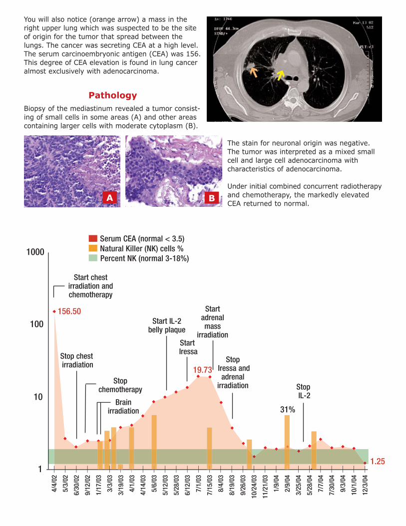
You will also notice (orange arrow) a mass in theright upper lung which was suspected to be the siteof origin for the tumor that spread between thelungs. The cancer was secreting CEA at a high level.The serum carcinoembryonic antigen (CEA) was 156.This degree of CEA elevation is found in lung canceralmost exclusively with adenocarcinoma.
PathologyBiopsy of the mediastinum revealed a tumor consist-ing of small cells in some areas (A) and other areascontaining larger cells with moderate cytoplasm (B).
The stain for neuronal origin was negative.The tumor was interpreted as a mixed smallcell and large cell adenocarcinoma with characteristics of adenocarcinoma.
Under initial combined concurrent radiotherapyand chemotherapy, the markedly elevatedCEA returned to normal.
1
10
100
1000
4/4/
02
5/3/
02
6/30
/02
9/12
/02
1/17
/03
3/3/
03
3/19
/03
4/1/
03
4/14
/03
5/6/
03
5/12
/03
5/28
/03
6/12
/03
7/1/
03
7/15
/03
8/4/
03
8/19
/03
9/26
/03
10/2
4/03
11/2
1/03
1/9/
04
2/9/
04
3/25
/04
5/28
/04
7/7/
04
7/30
/04
9/3/
04
10/1
/04
12/3
/04
__ __ __ __ _ _ _ _ __ _ __ __ __ __ _ __ __ __ __ __ __ _ __ __ __ __ __ __ __________________________
156.50
19.73
1.25
Serum CEA (normal < 3.5)Natural Killer (NK) cells %Percent NK (normal 3-18%)
31%
Start IL-2 belly plaque
StartIressa
Start adrenalmass
irradiation
StopIL-2
Start chest irradiation andchemotherapy
StopIressa andadrenal
irradiation
Stop chestirradiation
Brainirradiation
Stopchemotherapy
A B

Taxotere "consolidation" chemotherapy was poorly tolerated ("like to have killed me"), not an unusualexperience for chemotherapy recipients after repeated cytotoxic cycles. This patient, like many others,declined further chemotherapy as his independent decision. We respect this decision was prudent. Afterfurther chemotherapy was declined, the conventional treatment would have been "watchful waiting". Thisis a euphemism for doing nothing. In this case multi-agent immunotherapy including IL-2 immunotherapywas discussed with the patient and begun in earnest with his informed consent.
CT chest of July 2002, taken after radiotherapy to the chest andchemotherapy, shows stint (green arrow) in superior vena cava thathad been inserted prior to radiotherapy to relieve acute symptomsof compression of this large vein (vena cava). This mechanical compression had caused severe symptoms when Mr. Daniels firstsought medical attention. There was marked shrinkage of masseffect in the mediastinum (orange arrow) after initial treatment.Chest radiotherapy and chemotherapy were associated with CEAfalling to normal range.
Mr. Daniels’ presenting complaints with brain metastasis (yellow arrows) were headaches, confusionand incoordination.
November, 2002, three months after chemotherapy stopped, CT brain shows multiple brain metastases
July, 2002 - CT chest after radiotherapy

The dark centers in several of these tumors (green arrow) are evidence of central necrosis, which isindicative of dead tumor that has lost its blood supply. Central necrosis is commonly seen in adeno-carcinoma and is less common in small cell carcinoma. The dark area around the tumor nodules is cerebral adema (purple arrow).
These metastases required treatment with radiotherapy. The brain metastasesshrank markedly after radiotherapy. Radiotherapy to the brain consisted of 4000cGy in 4 weeks, a regimen far more protective of higher cognitive function thanthe often used 3000 cGy in two weeks. Immunotherapy with IL-2 belly plaque and other agents was continued before and after the brain irradiation.
After brain radiotherapy was complete, the CEA cancer marker began a disappointing steady rise (see graph).
July 2003 - Repeat chest CT scan shows active mass in adrenal gland
1/17
/03
3/3/
03
3/19
/03
4/1/
03
4/14
/03
5/6/
03
5/12
/03
5/28
/03
6/12
/03
7/1/
03
_ _ _ _ __ _ __ __ __ __________________
19.7

In July 2003, a PET scan was ordered in an attempt to define thesite of origin for the rising CEA. A CT scan through a solitary area ofintense PET uptake revealed a mass in the left adrenal, which haddeveloped since the CT scan taken at presentation. It was hypothe-sized that this adrenal mass was metastatic cancer largely responsiblefor the progressive rise in CEA. Note the dark center inside this mass(green arrow) similar to that seen on several of the brain metastases.Such dark areas are felt to represent central necrosis or poor bloodflow and are commonly seen in adenocarcinoma.
The adrenal gland is the source of cortisone and hydrocortisone,known immunosuppressive hormones. These hormones cause lysis (death) of lymphocytes. The brain andadrenal gland are well documented as preferred metastatic recurrence sites for adenocarcinoma of thelung. The brain is a common site for recurrences as chemotherapy does not penetrate the blood-brainbarrier in sufficient quantities to have anticancer effects. The adrenal acts as "fertile soil" for recurrenceslikely due to the immunosuppressive micro environment hostile to natural killer lymphocyte mediatedimmunotherapy.
The same CT demonstrating the adrenal mass also memorializes several prominent areas of IL-2 generatedbelly plaque (orange arrows). Contrast these areas on different cuts with the opposite side (above in redbox), and you will note the difference.
Blood determinations at around this time revealed substantial elevation of natural killer (NK) lymphocyteblood levels (see graph). This elevation supports our theory that the belly plaque is a factory for NK cells.NK cells escape the plaque into the general circulation.
Most medical oncologists would have recommended more chemotherapy at discovery of the adrenalmetastasis, the indication being systemic recurrence or relapse. This was a critical decision fork. (See endof section for other treatment decision forks.) The adrenal recurrence was treated with local radiotherapy,hopefully reducing the body burden below the IIT and blocking corticosteroid production by the adrenal.The tumor was radiosensitized with Iressa. IL-2 belly plaque was continued, but was later stopped.

The CEA fell rapidly during adrenal irradiation to normal, and CEA has remained normal for 15 months. The patient has remained physically and mentally active with an excellent quality of life (see video).
The 14-month, no-chemotherapy remission currently enjoyed by Mr. Daniels after two manifestations of distant spread (brain and adrenal) is an anomalousoccurrence under the prevailing chemotherapy centric paradigm of cancertreatment. In our opinion, this protracted delay to recurrence in associationwith other evidence of immune activity strongly supports our case that ourimmunotherapy efforts were successful in arresting tumor regrowth.
A repeat CT scan of the chest was obtained in February of 2004. A residual mass in the mediastinum maybe a fibrous tissue scar left behind after the cellular part of a tumor is destroyed leaving behind only acontracted fibrous tissue scar from the connective tissue scaffolding of the previous cancer. Such massesare often seen after local control in bulky Hodgkin's disease masses.
Based on the well established radiobiology of bulky adenocarcinomas, combined radiotherapy andchemotherapy, the treatment Mr. Daniels initially received, rarely create local control of 8 cm masses likethe one in Mr. Daniels’ chest at presentation. Another interpretation of this case, one that we favor formanagement purposes, is that immunotherapy has induced growth arrest of residual tumor in a greatlyshrunken tumor rather than total tumor eradication or cure.
Clonagens are individual cancer cells that can regrow to large masses. We believe the presumption shouldbe that cancer clonagens are typically incorporated in residual masses after standard radiotherapy andchemotherapy. Clonagens also are likely to be found at one or more distant sites, and these clonagenswill proliferate if immune surveillance is low.
CEA normal for 14 months after
marked elevation; off standard treatments
Possible cure!
February 2004 - Mediastinum CT

It has been our experience over the years that patients who decrease or termi-nate their immunotherapy based on duration of remission often relapse. Relapsesafter known systemic spread have been universally fatal. These considerationslead us to the conclusion that disciplined immunotherapy carried out by thepatient for years under physician supervision is worthwhile after initial treatmentso long as elevated serologic marker determinations remain normal and no evi-dence of disease (NED) is the clinical condition of the patient.
This patient's quality of life during immunotherapy has been as good as it wasprior to his developing cancer. This high quality survival would not likely havebeen the case if, at the first sign of dissemination, chemotherapy had been elected by the patient as further treatment. Earl Daniels
Marks, Mississippi
Keeping the Grim Reaper at Bay in a Deadly Condition
Prohibited: Immune Restorative Therapy at Work