adeno-associated virus-mediated heme oxygenase-1 g
TRANSCRIPT
BASIC RESEARCH
Adeno-associated virus-mediated heme oxygenase-1 gene transfer suppresses the progression of micronodular cirrhosis in rats
Tung-Yu Tsui, Chi-Keung Lau, Jian Ma, Gabriel Glockzin, Aiman Obed, Hans J Schlitt, Sheung-Tat Fan
Tung-Yu Tsui, Chi-Keung Lau, Jian Ma, Sheung-Tat Fan, Centre for the Study of Liver Disease and Department of Surgery, the University of Hong Kong, Pokfulam, Hong KongTung-Yu Tsui, Gabriel Glockzin, Aiman Obed, Hans J Schlitt, Department of Surgery, University of Regensburg Medical Centre, Regensburg, GermanyCorrespondence to: Dr. Tung-Yu Tsui, Department of Surgery, University of Regensburg Medical Centre, Franz-Josef-Strauss-Allee 11, 93053 Regensburg, Germany. [email protected]: +49-941-9446852 Fax: +49-941-9446802Received: 2005-09-12 Accepted: 2005-10-26
AbstractAIM: To test the hypothesis that enhancement of the activity of heme oxygenase can interfere with processes of fibrogenesis associated with recurrent liver injury, we investigated the therapeutic potential of over-expression of heme oxygense-1 in a CCl4-induced micronodular cir-rhosis model.
Methods: Recombinant adeno-associated viruses car-rying rat HO-1 or GFP gene were generated. 1×10¹² vg of adeno-associated viruses were administered through portal injection at the time of the induction of liver fi-brosis.
Results: Conditioning the rat liver with over-expres-sion of HO-1 by rAAV/HO-1 significantly increased the HO enzymatic activities in a stable manner. The develop-ment of micronodular cirrhosis was significantly inhib-ited in rAAV/HO-1-transduced animals as compared to controls. Portal hypertension was markedly diminished in rAAV/HO-1-transduced animals as compared to con-trols, whereas there are no significant changes in systolic blood pressure. This finding was accompanied with im-proved liver biochemistry, less infiltrating macrophages and less activated hepatic stellate cells (HSCs) in rAAV/HO-1-transduced livers.
ConClusIons: Enhancement of HO activity in the liv-ers suppresses the development of cirrhosis.
© 2006 The WJG Press. All rights reserved.
Key Words: Cirrhosis; Portal hypertension; Heme oxy-genase; Gene therapy; Adeno-associated virus
Tsui TY, Lau CK, Ma J, Glockzin G, Obed A, Schlitt HJ, Fan ST. Adeno-associated virus-mediated heme oxygenase-1 gene transfer suppresses the progression of micronodular cirrhosis in rats. World J Gastroenterol 2006; 12(13): 2016-2023
http://www.wjgnet.com/1007-9327/12/2016.asp
IntroductIonHeme oxygenase �H��� �s a �a�e���m���ng en�yme ��a� �H��� �s a �a�e���m���ng en�yme ��a� c�eaves p�o�ox�dan� �eme �n�o equ�mo�a� amoun�s of ca�bon monox�de �C���, b���ve�d�n/b����ub�n, and f�ee, and f�ee and f�ee fe��ous ��on [1,2]. Up �o da�e, ���ee �sofo�ms of H� �ave been �den��f�ed. Among of ��em, H��1 and H��2 a�e exp�essed �n ��ve�s. H��2 exp�esses cons���u��ve�y �n �epa��ocy�e and func��ons as an �mpo��an� en�yme �n ca�a�y��ng ��e endogenous and exogenous �eme. In con��as�, H��1 �s an �nduc�b�e fo�m of H� �n ��ve�s [3]. A���oug� exp�es�s�on of H��1 �n ��e ��ve� �s p��ma���y �es���c�ed �o a sub�popu�a��on of Kupffe� ce��s [3,4], ��e p�o�e�n �s exp�essed �n bo�� pa�enc�yma� and pa���cu�a��y non�pa�enc�yma� ��ve� ce��s, and ��e�efo�e �esu��s �n ��g�e� en�yma��c ac��v���es of H� unde� s��ess cond���ons [4]. T�e cy�op�o�ec��ve effec�s of en�anced H� ac��v���es �n ��ve� m�g�� be ���oug� ��e mec�an�sms assoc�a�ed w��� ��s ca�a�y��c p�oduc�s. A�� of ��s ���ee ca�a�y��c p�oduc�s can con���bu�e ��e p�o�ec��ve ef�fec�s of H� [5�7].
H��1 was �nduced �n ��e ��ve�s w��� c����os�s [8]. I� was ��oug�� �o be an adop��ve �esponse �o ox�da��ve s��esses, �nf�amma�o�y �nsu��s of pe�s�s�en� o� �ecu��en� ��ve� �nju�y as we�� as ��e �espons�veness �o ��e �nc�ease of �n��as�nuso�da� �es�s�ance. H��1 was ma�n�y exp�essed �n Kupffe� ce��s, s�nuso�da� endo��e��a� ce��s, s�e��a�e ce��s and �n a pa�� of �epa�ocy�e �n ��ve�s w��� c����os�s [8,9]. H��1 was a�so exp�essed �n vascu�a� sys�em ��a� m�g�� con���b�u�e �o ��e deve�opmen� of �emodynam�c c�anges �n ��e �a�s w��� c����os�s [9]. In add���on, ��g�e� �eve�s of C� p�oduc��on m�g�� be assoc�a�ed w��� ��e deve�opmen� of �epa�opu�mona�y synd�ome �n pa��en�s w��� c����os�s [10]. T�us, �� becomes con��ove�sy w�e��e� ��e exp�ess�on of H��1 o� en�ancemen� of H� ac��v���es con���bu�e �o ��e pa��op�ys�o�og�ca� c�anges �n ��e deve�opmen� of c����o�s�s, o� w�e��e� H��1 func��ons as �omeos�a��c mo�ecu�e �n con��o���ng of d�sease p�og�ess�on. To e�uc�da�e ��e �o�e of
PO Box 2345, Beijing 100023, China World J Gastroenterol 2006 April 7; 12(13):2016-2023www.wjgnet.com World Journal of Gastroenterology ISSN [email protected] © 2006 The WJG Press. All rights reserved.
www.wjgnet.com
Tsui TY et al . HO-1 suppresses liver fibrogenesis 2017
H��1 �n ��e con�ex� of �ecu��en� �epa�oce��u�a� �nju�y and ��e deve�opmen� of c����os�s, �e�e we �a�e ��e advan�ageof c����os�s, �e�e we �a�e ��e advan�agec����os�s, �e�e we �a�e ��e advan�age of adeno�assoc�a�ed v��a� vec�o� �o �nc�ease ��e ove�a�� H� en�yma��c ac��v��y of ��e ��ve� �n a s�ab�e manne� and �o �nves��ga�e ��e effec�s of H��1 �n ��e d�sease p�og�ess�on �n ca�bon �e��ac��o��de �CC�4����nduced c����os�s mode�.��nduced c����os�s mode�.�nduced c����os�s mode�.
MAtErIALS And MEtHodSGeneration of recombinant adeno-associated viral vectorss (r���sr���s�� rAAVs were produced and purified as previously described [11]. In b��ef, a fu����eng�� H��1 gene o��g�na��y c�oned f�oma fu����eng�� H��1 gene o��g�na��y c�oned f�omfu����eng�� H��1 gene o��g�na��y c�oned f�om��eng�� H��1 gene o��g�na��y c�oned f�om�eng�� H��1 gene o��g�na��y c�oned f�omH��1 gene o��g�na��y c�oned f�om gene o��g�na��y c�oned f�om ��e sp�een of a ���� �a� o� ��e �epo��e� gene ���� wasa ���� �a� o� ��e �epo��e� gene ���� was���� �a� o� ��e �epo��e� gene ���� was��e �epo��e� gene ���� was�epo��e� gene ���� was �nse��ed �n�o ��e vec�o� p�asm�d �o cons��uc� pSNAV1/���� o� pSNAV1/H��1. �AAV/���� o� �AAV/H��1 waswas gene�a�ed �n BHK�21 ce��s �Ame��can Type Cu��u�e Co��ec���on, Manassas, VA�� by ��ansfec��on of vec�o� p�asm�d and subsequen� �escue �AAVs �se�o�ype 2�� by co��nfec��on w��� recombinant HSV1-rc/∆UL2, which is essential for viron packaging. A large scale of rAAV was purified as described [12].
�nimal model and gene delivery protocolInb�ed ���� �a�s �230�250 g�� we�e pu�c�ased f�om ��eed f�om ��e f�om ��e An�ma� Ins���u�e of Med�ca� Sc�oo� Hannove�, �e�many and we�e ma�n�a�ned �n ��e �abo�a�o�y An�ma� Un�� of��e �abo�a�o�y An�ma� Un�� of�abo�a�o�y An�ma� Un�� of ��e Un�ve�s��y of Hong Kong. T�e s�udy was app�oved by ��e Comm���ee on ��e Use of ��ve An�ma�s fo� Teac��ng and Resea�c�, ��e Un�ve�s��y of Hong Kong. M�c�onodu�a� ��ve� c����os�s �n ���� �a�s was �nduced us�ng a p�o�oco� desc��bed p�ev�ous�y [13]. In b��ef, p�en�oba�b��a� sod�um (35 mg/L) was given one week before the first dose of CCl4 �n o�de� �o �nc�ease ��e sens���v��y of ��e �a� ��ve� �o CC�4 and was p�esen� �n d��n��ng wa�e� �n ��e w�o�e pe��od of��e w�o�e pe��od ofw�o�e pe��od of �nduc��on. A �o�a� of 9 doses of CC� �o�a� of 9 doses of CC��o�a� of 9 doses of CC�of 9 doses of CC�9 doses of CC�4 �0.2 m�/�g pe� w��� we�e g�ven �o ��e �a�s �n��agas���ca��y. �AAVs �1×10¹²v.g.�� was g�ven ���oug� ��e po��a� ve�n of �a�s a� ��e ��me pen�o�barbital was given in drinking water and �� d before the firstwas given in drinking water and �� d before the firstin drinking water and �� d before the firsting water and �� d before the first water and �� d before the first dose of CC�4.
Liver biochemistry, histology, immunohistochemistry and ELIS� Se�um samp�es we�e co��ec�ed a� ��e end po�n� of expe���men�s. T�e ac��v���es of a�an�ne am�no��ansfe�ase �A�T�� and �o�a� b����ub�n �n ��e p�asma we�e measu�ed �n ��e Depa��men� of C��n�ca� B�oc�em�s��y, ��e Un�ve�s��y of Hong Kong. ��ve� samp�es we�e snap f�o�en and s�o�ed a� �75 ℃ un��� fu���e� app��ca��ons. ��ve m�c�ome�e�s of f�o���ve m�c�ome�e�s of f�o� of f�o��en sec��ons we�e used fo� �ema�oxy��n and eos�n s�a�n�ng,and eos�n s�a�n�ng, eos�n s�a�n�ng, Masson’s ���c��ome s�a�n�ng, and �mmuno��s�oc�em�s��y., and �mmuno��s�oc�em�s��y. and �mmuno��s�oc�em�s��y. The area of fibrotic tissues in the cross section of liverss �n ��e c�oss sec��on of ��ve�s �n ��e c�oss sec��on of ��ve�s was measu�ed by ��e compu�e� sof�wa�e �Me�aMo�p� �m�ag�ng sys�em, Un�ve�sa� Imag�ng Co�po�a��on, ��A�� af�e� Masson’s trichrome, collagen 1α and fibronectin staining. A�� measu�emen�s we�e done �n a doub�e�b��nd manne�a doub�e�b��nd manne�doub�e�b��nd manne��b��nd manne�b��nd manne� w��� a� �eas� 20 a�eas/��ve� �f�b�o��c a�ea, o��g�na� 200×�� �n ��e 5 ��ve�s/g�oup. Mouse an����a� �D1 ��nf����a��ng mac�op�age��, an����D2 ���ssue �es�den��a� mac�op�age��, an���H��1 ��SA�111�� monoc�ona� an��bod�es and po�y�clonal anti-TGF-β1, anti-desmin, anti-collagen 1α, and, and and
an���f�b�onec��n an��bod�es �Se�o�ec ��d., �xfo�d, UK; S��essgene, V�c�o��a, B����s� Co�umb�a, Canada; C�em�con, Temecu�a, CA; San�a C�u�, CA�� we�e app��ed �n ���s s�udy fo� �mmuno��s�oc�em�s��y us�ng ��e s�anda�d �o�se�ad�s���e s�anda�d �o�se�ad�s�s�anda�d �o�se�ad�s� pe�ox�dase p�o�oco�. Se�um mac�op�age m�g�a��on �n��b���o�y fac�o� �MI��� �eve� was de�ec�ed by ��ISA acco�d�ng �o ��e �ns��uc��on of manufac�u�e� �C�em�con��.
Measurement of portal and systolic blood pressureAn�ma�s we�e anes��es��ed w��� �e�am�ne �100 mg/�g, �p��anes��es��ed w��� �e�am�ne �100 mg/�g, �p��w��� �e�am�ne �100 mg/�g, �p�� and Rompun �0.2 mg/�g, �p��. ��o��a� p�essu�e and sys�o��c b�ood p�essu�e we�e measu�ed by d��ec��y �n��oduc�ng 24� Ang�oca��® �BD B�osc�ences, San Jose, CA�� �n�o ��e po��a���e po��a�po��a� vein or abdominal aorta and connecting with a saline filleda saline filledsaline filled s��a�n gauge ��ansduce�. T�e s�gna�s we�e mon��o�ed by Co��n B���408 Ma�� III �Japan�� and we�e se� �o �e�o befo�e ��e measu�emen�.
HO enzymatic activityT�e H� en�yma��c ac��v��y was measu�ed by ��e p�oduc���e p�oduc�p�oduc���on of m�c�osoma� b����ub�n �n ��e ��ve�. ��o�en samp�es of ��ve�s we�e �omogen��ed �n �ce�co�d suc�ose and T���HC� buffe�. M�c�osoma� pe��e� was ob�a�ned af�e� cen���fu�M�c�osoma� pe��e� was ob�a�ned af�e� cen���fu��c�osoma� pe��e� was ob�a�ned af�e� cen���fu�ga��on and was ��en �e�suspended �n MgC�2�po�ass�umpo�ass�um p�osp�a�e buffe�. Samp�e p�o�e�n was fu���e� �ncuba�ed w��� ��e �eac��on m�x�u�e con�a�n�ng �a� ��ve� cy�oso�, �e�m�n, g�ucose�6�p�osp�a�e, g�ucose�6�p�osp�a�e de�yd�o�genase and NAD��H �S�gma�A�d��c�, S�. �ou�s, M��� fo� 60 m�nu�es a� 37 ℃. T�e gene�a�ed b����ub�n was measu�ed us�ng spec��op�o�ome��y. T�e �eve� of b����ub�n p�oduc���on was demons��a�ed by ��e �a��o of samp�e/no�ma� ��ve�.
RNase protection assayT�e mRNA �eve� of �a�ge� genes was de�e�m�ned bymRNA �eve� of �a�ge� genes was de�e�m�ned by RNase p�o�ec��on assay acco�d�ng �o ��e �ns��uc��on of manufac�u�e� �R�boQuan� ���, BD B�osc�ences ���a�m�n�gen, San D�ego, CA��. In b��ef, �o�a� RNA of ��ansduced and non���ansduced ��ve� samp�es we�e ex��ac�ed and pu���f�ed us�ng Rneasy ��� �Q�agen, H��den, �e�many��. T��ee m�c�og�ams of RNA/samp�es we�e �yb��d��ed w��� com�p��men�a�y [32��]UT�� �abe�ed ��bop�obes ove�n�g��. T�e p�obes we�e d�ges�ed w��� Rnase and we�e �oaded on a dena�u�ed po�yac�y�am�de ge�. T�e �ad�oac��ve s�gna�s we�e then detected by exposure to X-ray film (BioMax, Kodak, Rochester, NY) and quantified by phosphorimaging (Strom, Mo�ecu�a� Dynam�cs, Sunnyva�e, CA��.
Statistical analysisDa�a we�e demons��a�ed as mean ± S�M. �ne�way AN��VA was used �o compa�e ��e d�ffe�ence of means be�ween ��e expe��men�a� g�oups w��� ��e Bonfe��on�’s t��es�. P < 0.05 was considered statistically significant.
rESuLtS r���-mediated stable HO enzymatic activity in rat liver To en�ance H� ac��v��y �n a s�ab�e manne�, we adm�n��s�e�ed ��e �AAV ca��y�ng �a� H��1 cDNA �o ��e ��ve� ���oug� po��a� �njec��on. Af�e� ��e �njec��on, a �a�ge num�
www.wjgnet.com
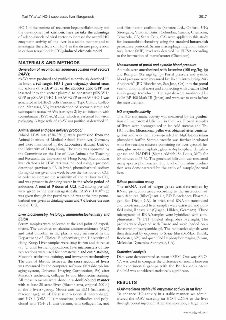
be� of H��1 pos���ve non�pa�enc�yma� ce��s we�e found �n bo�� �AAV/���� and �AAV/H��1���ansduced ��ve�s on d 7 �17.7 ± 0.5 ce��s/mm2 ve�sus 10.8 ± 0.7 ce��s/mm2, n = 3��. Howeve�, ��e numbe� of pos���ve ce��s dec�eased d�ama���ca��y �o a �eve� s�m��a� �o no�ma� ��ve�s �n �AAV/���� ��ansduced ��ve� on day 30, w�e�eas ��e�e was a s�gn�f��can��y �a�ge� numbe� of H��1�pos���ve non�pa�enc�yma� ce��s �n �AAV/H��1���ansduced ��ve�s �3.9 ± 0.8 ce��s/mm2 vs 5.8 ± 1.0 ce��s/mm2, n = 3, P < 0.05; ��gu�es 1A and 1B��. No H��1 pos���ve �epa�ocy�e was found �n bo�� �AAV/���� and �AAV/H��1 ��ea�men� g�oups on d 7. T�e ex�pression of HO-1 in hepatocyte was first detected sporadi�ca��y on d 14 af�e� po��a� �njec��on of �AAV/H��1, w�e�e�as ��e�e was no H��1 pos���ve �epa�ocy�e �n �AAV/�������ansduced ��ve�s �da�a no� s�own��. Mo�e H��1 pos���ve �epa�ocy�es cou�d be de�ec�ed �n �AAV/H��1���ansduced ��ve�s on d 30 �2.2 ± 0.3 ce��s/mm2, n = 3, P < 0.05��, w�e�eas ��ey �ema�ned unde�ec�ab�e �n �AAV/�������ansduced ��v�e�s ���gu�es 1A and 1B��. To eva�ua�e ��e b�o�og�ca� ac��v��y of ��ansduced H��1 �n ��e ��ve�s, we de�e�m�ned ��e en��yma��c ac��v��y of H� by measu��ng ��e gene�a�ed b����u�b�n of m�c�osoma� p�o�e�n �so�a�ed f�om ��e ��ve�s. T�e�e was a s�gn�f�can� �nc�ease of H� ac��v��y of ��e �AAV���ansduced ��ve�s on d 7 ���e ea���es� ��me po�n� ��a� we de�ec�ed��. Bo�� �AAV/���� and �AAV/H��1 ��ansduced ��ve�s s�owed �nc�eased amoun� of gene�a�ed b����ub�n �n �so�a�ed m�c�osoma� p�o�e�ns. Imp�ess�ve�y, ��e amoun� of gene�a�ed b����ub�n �0.9 fo�d ove� ��e basa� �eve� of no�ma� �a� ��ve��� �ema�ned e�eva�ed �n ��e �AAV/H��1 ��ea�men� g�oup and sus�a�ned fo� ove� 3 mo of obse�va��on ��me. In con��as�, ��e amoun� of gene�a�ed b����ub�n dec�eased �o ��e �eve� of no�ma� �a�s �n ��e �AAV/���� ��ea�men� group on d 14. (Figure 1C). This suggests the specificity of �AAV/H��1 �n ��e �nduc��on of s�ab�e H� ac��v��y �n �a� ��ve�.
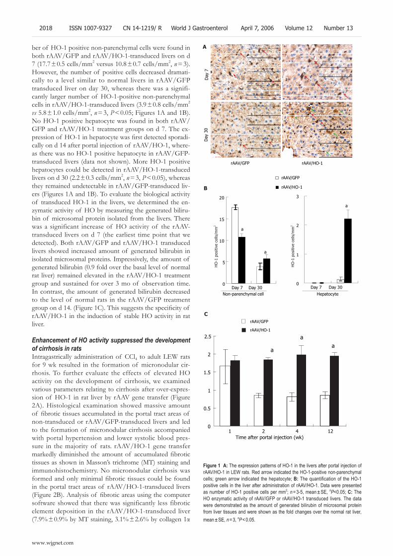
Enhancement of HO activity suppressed the development of cirrhosis in rats In��agas���ca��y adm�n�s��a��on of CC�4 �o adu�� ���� �a�s fo� 9 w� �esu��ed �n ��e fo�ma��on of m�c�onodu�a� c�����os�s. To fu���e� eva�ua�e ��e effec�s of e�eva�ed H� ac��v��y on ��e deve�opmen� of c����os�s, we exam�ned va��ous pa�ame�e�s �e�a��ng �o c����os�s af�e� ove��exp�es�s�on of H��1 �n �a� ��ve� by �AAV gene ��ansfe� ���gu�e 2A��. H�s�o�og�ca� exam�na��on s�owed mass�ve amoun� of fibrotic tissues accumulated in the portal tract areas of non���ansduced o� �AAV/�������ansduced ��ve�s and �ed �o ��e fo�ma��on of m�c�onodu�a� c����os�s accompan�ed w��� po��a� �ype��ens�on and �owe� sys�o��c b�ood p�es�su�e �n ��e majo���y of �a�s. �AAV/H��1 gene ��ansfe� markedly diminished the amount of accumulated fibrotic ��ssues as s�own �n Masson’s ���c��ome �MT�� s�a�n�ng and �mmuno��s�oc�em�s��y. No m�c�onodu�a� c����os�s was formed and only minimal fibrotic tissues could be found �n ��e po��a� ��ac� a�eas of �AAV/H��1���ansduced ��ve�s (Figure 2B). Analysis of fibrotic areas using the computer sof�wa�e s�owed ��a� ��e�e was s�gn�f�can��y �ess f�b�o��c e�emen� depos���on �n ��e �AAV/H��1���ansduced ��ve� (��.9% ± 0.9% by MT staining, 3.1% ± 2.6% by collagen 1α
Figure 1 A: The expression patterns of HO-1 in the livers after portal injection of rAAV/HO-1 in LEW rats. Red arrow indicated the HO-1-positive non-parenchymal cells; green arrow indicated the hepatocyte; B: The quantification of the HO-1 positive cells in the liver after administration of rAAV/HO-1. Data were presented as number of HO-1 positive cells per mm2; n = 3-5, mean ± SE, aP<0.05; C: The HO enzymatic activity of rAAV/GFP or rAAV/HO-1 transduced livers. The data were demonstrated as the amount of generated bilirubin of microsomal protein from liver tissues and were shown as the fold changes over the normal rat liver, mean ± SE, n = 3, aP < 0.05.
0
0.5
1
1.5
2
2.5
1 2 4 12Time after portal injection (wk)
rAAV/GFP
rAAV/HO-1
aa
a
www.wjgnet.com
2018 ISSN 1007-9327 CN 14-1219/ R World J Gastroenterol April 7, 2006 Volume 12 Number 13
rAAV/GFP rAAV/HO-1
Day
30
Day
7
0
1
2
3
Day 7 Day 30
rAAV/GFP
rAAV/HO-1
Non-parenchymal cell Hepatocyte
0
5
10
15
20
Day 7 Day 30
a
a
a
←
A
B
← ←
←
←
←
←
←
←
←←
←
←←
←
←
←
HO
-1 p
ositi
ve c
ells
/mm
2
HO
-1 p
ositi
ve c
ells
/mm
2
C
0
20
40
60
80
100
120
Normal
rat
Non-tr
ansd
uced
rAAV
/GFP
rAAV
/HO-1Sy
stol
icbl
ood
pres
sure
(mm
Hg)
0
2
4
6
8
10
12
14
16
Normal
rat
Non-tr
ansd
uced
rAAV
/GFP
rAAV
/HO-1
Port
alpr
essu
re(m
mH
g)
d
0
5
10
15
20
25
30
Non-transduced rAAV/GFP rAAV/HO-1
Fibr
oic
area
(%)
MT
Collagen 1
Fibronectin
b
bb
Figure 2 A: The model to induce micronodular cirrhosis in LEW rat. The rAAVs were injected through the portal vein shortly before the induction; B: The histology and immunohistochemistry of the livers with or without treatment at the 10th week (H-E, original magnification ×40; MT: Masson’s trichrome stiaining, the fibrotic elements were stained in green, original 100×; collagen 1 and fibronectin staining, original magnification ×100); C: The measurement of fibrotic area in the livers at the 10th week by the computer software as described in Materials & Methods. Data were shown as mean±SE, n = 5, bP < 0.001; D and E: The systolic and portal pressure of rats after induction of liver cirrhosis with or without treatments. The data were shown as mean±SE, n = 10, dP < 0.001.
s�a�n�ng, 4.4% ± 1.2% by f�b�onec��n s�a�n�ng, P < 0.001��. In con��as�, �n ��e ��ve�s of non���ea�men� o� �AAV/���� ��ea�ed g�oups, ��e�e was a ��g�e� �eve� of f�b�o��c e�e�men� depos���on �22.2% ± 5.8% o� 20.8 %± 3.8% by MT staining, 21.9 %± 5.��% or 24.5 %± 2.8% by collagen 1α s�a�n�ng, 24.1 %± 4.4% o� 21.1% ± 3.6% by f�b�onec��n staining, Figure 2C). In addition, the beneficial effects of H��1 on ��e deve�opmen� of ��ve� c����os�s cou�d be �e�flected by the reduction of portal hypertension (8.0 ± 0.8 mmHg ve�sus 13.3 ± 0.4 mmHg �n ��e non���ea�men� g�oup and 13.8 ± 0.6 mmHg �n ��e �AAV/���� ��ea�men� g�oup, P < 0.001), whereas there was no significant differ�ence a� ��e sys�o��c b�ood p�essu�e �81.7 ± 0.3 mmHg�� �n compa��son �o non���ea�men� �81.8 ± 0.6 mmHg�� o� vec�o� con��o�s �87.0 ± 0.2 mmHg, P > 0.31, ��gu�es 2D and 2���.
Stable HO activity protected against CCl4-mediated recur-rent liver injury
Repea�ed adm�n�s��a��on of CC�4 �o adu�� �a�s �ed �o c��on��c ��ve� �nju�y w��� e�eva�ed p�asma �eve� of �o�a� b����ub�n (no treatment group: 5��.25 ± 6.95 μmol/L; rAAV/GFP group: 60.5±4.8�� μmol/L) and ALT (non-treatment group: 4 522 ± 334 IU/�; �AAV/���� g�oup: 6 552 ± 1 363 IU/�, n = 5��. Inc�eas�ng H� ac��v��y �n ��e ��ve� by �AAV/H��1 s�gn�f�can��y �mp�oved ��e ��ve� func��on of �a�s unde� �ong��e�m CC�4 �ox�c��y, ��e amoun� of �o�a� b����ub�n and A�T dec�eased d�ama��ca��y �n ��e �AAV/H��1���ea�ed �a�s �n compa��son �o non���ea�men� o� �AAV/���� con�trols (total bilirubin: 6.20 ± 3.49 μmol/L; ALT: 199.6 ±80.5 IU/�, n = 5, P < 0.005, ��gu�es 3A and 3B��. To fu���e� exam�ne ��e p�o�ec��ve effec�s of ove��exp�ess�ng H��1 �n ��e ��ve�s, we de�e�m�ned ��e ��ansc��p� �eve� of apop�o��c genes and ene�gy exc�anges of ��ve�s unde� �ong��e�m CC�4 �ox�c��y. Rnase p�o�ec��on assay s�owed ��e ��ansc��p� level of Fas, caspase 3, and BAX genes significantly dimin��s�ed �n ��e �AAV/H��1���ansduced ��ve�s �n = 3, P < 0.05,
www.wjgnet.com
Tsui TY et al . HO-1 suppresses liver fibrogenesis 2019
Non-transduced rAAV/GFP rAAV/HO-1
MT
Fibr
onec
tinCo
llage
n1
H&
E
rAAVs
Phenobarbital sodium + CCl4 (10 wk)A
B
0
2
4
6
8
10
12
14
16
Normal
rat
Non-tr
ansd
uced
rAAV
/GFP
rAAV
/HO-1
Port
alpr
essu
re(m
mH
g)
d
E
D
C
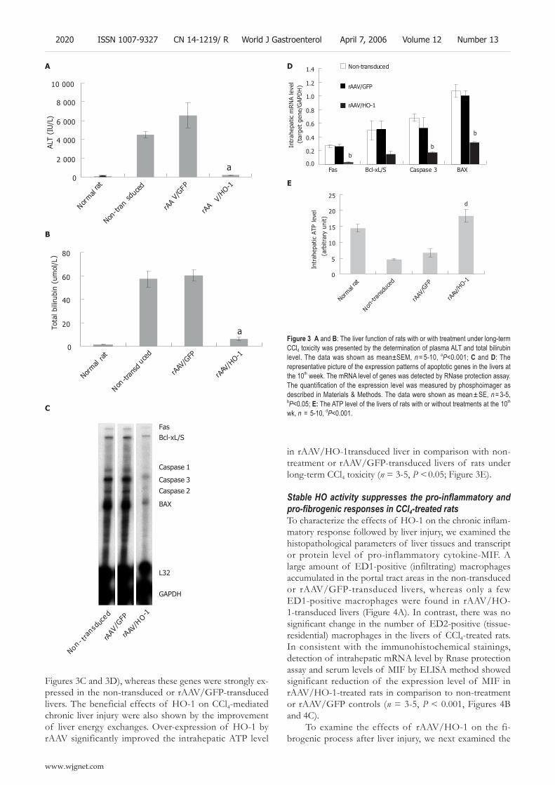
Figure 3 A and B: The liver function of rats with or with treatment under long-term CCl4 toxicity was presented by the determination of plasma ALT and total bilirubin level. The data was shown as mean±SEM, n = 5-10, aP<0.001; C and D: The representative picture of the expression patterns of apoptotic genes in the livers at the 10th week. The mRNA level of genes was detected by RNase protection assay. The quantification of the expression level was measured by phosphoimager as described in Materials & Methods. The data were shown as mean ± SE, n = 3-5, bP<0.05; E: The ATP level of the livers of rats with or without treatments at the 10th wk, n = 5-10, dP<0.001.
��gu�es 3C and 3D��, w�e�eas ��ese genes we�e s��ong�y ex�p�essed �n ��e non���ansduced o� �AAV/�������ansduced ��ve�s. T�e benef�c�a� effec�s of H��1 on CC�4�med�a�ed c��on�c ��ve� �nju�y we�e a�so s�own by ��e �mp�ovemen� of ��ve� ene�gy exc�anges. �ve��exp�ess�on of H��1 by �AAV s�gn�f�can��y �mp�oved ��e �n��a�epa��c AT�� �eve�
Bcl-xL/S
Caspase 1
Caspase 3
BAX
L32
GAPDH
Fas
Caspase 2
Non- tr
ansd
uced
rAAV
/GFP
rAAV/H
O-1
0
2 000
4 000
6 000
8 000
10 000
Normal
rat
Non-tr
ansd
uced
rAAV/GFP
rAA
V/HO-1
ALT
(IU
/L)
a
0
20
40
60
80
Normal
rat
Non-tr
ansd
uced
rAAV
/GFP
rAAV
/HO-1
Tota
lbili
rubi
n(u
mol
/L)
a
www.wjgnet.com
2020 ISSN 1007-9327 CN 14-1219/ R World J Gastroenterol April 7, 2006 Volume 12 Number 13
A
0.0
0.2
0.4
0.6
0.8
1.0
1.2
1.4
Fas Bcl-xL/S Caspase 3 BAX
Intr
ahep
atic
mR
NA
leve
l(t
arge
tge
ne/G
APD
H)
Non-transduced
rAAV/GFP
rAAV/HO-1
bb
b
0
5
10
15
20
25
Normal
rat
Non-tr
ansd
uced
rAAV
/GFP
rAAV
/HO-1
Intr
ahep
atic
ATP
leve
l
(arb
itrar
yun
it)
d
E
D
C
B
�n �AAV/H��1��ansduced ��ve� �n compa��son w��� non���ea�men� o� �AAV/�������ansduced ��ve�s of �a�s unde� �ong��e�m CC�4 �ox�c��y �n = 3�5, P < 0.05; ��gu�e 3���.
Stable HO activity suppresses the pro-inflammatory and pro-fibrogenic responses in CCl4-treated ratsTo characterize the effects of HO-1 on the chronic inflam�ma�o�y �esponse fo��owed by ��ve� �nju�y, we exam�ned ��e ��s�opa��o�og�ca� pa�ame�e�s of ��ve� ��ssues and ��ansc��p� o� p�o�e�n �eve� of p�o��nf�amma�o�y cy�o��ne�MI�. A �a�ge amoun� of �D1�pos���ve ��nf����a��ng�� mac�op�ages accumu�a�ed �n ��e po��a� ��ac� a�eas �n ��e non���ansduced o� �AAV/�������ansduced ��ve�s, w�e�eas on�y a few �D1�pos���ve mac�op�ages we�e found �n �AAV/H��1���ansduced ��ve�s ���gu�e 4A��. In con��as�, ��e�e was no significant change in the number of ED2-positive (tissue-�es�den��a��� mac�op�ages �n ��e ��ve�s of CC�4���ea�ed �a�s. In cons�s�en� w��� ��e �mmuno��s�oc�em�ca� s�a�n�ngs, de�ec��on of �n��a�epa��c mRNA �eve� by Rnase p�o�ec��on assay and se�um �eve�s of MI� by ��ISA me��od s�owed s�gn�f�can� �educ��on of ��e exp�ess�on �eve� of MI� �n �AAV/H��1���ea�ed �a�s �n compa��son �o non���ea�men� o� �AAV/���� con��o�s �n = 3�5, P < 0.001, ��gu�es 4B and 4C��.
To exam�ne ��e effec�s of �AAV/H��1 on ��e f��b�ogen�c p�ocess af�e� ��ve� �nju�y, we nex� exam�ned ��e
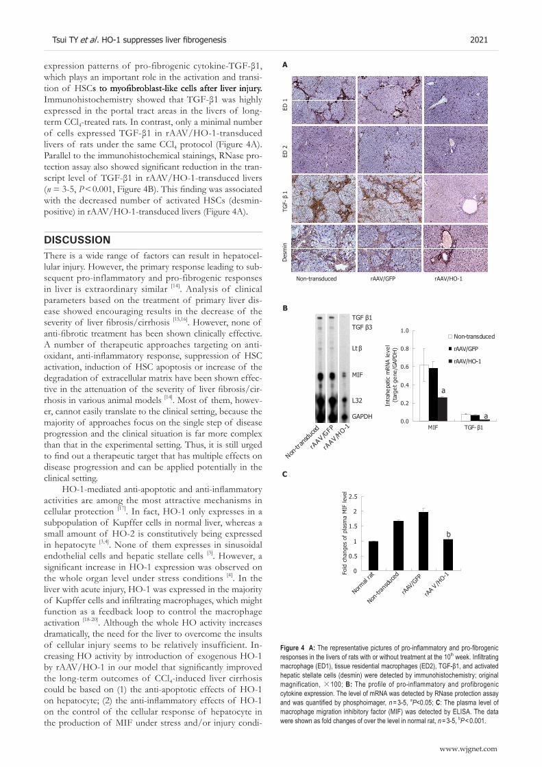
Figure 4 A: The representative pictures of pro-inflammatory and pro-fibrogenic responses in the livers of rats with or without treatment at the 10th week. Infiltrating macrophage (ED1), tissue residential macrophages (ED2), TGF-β1, and activated hepatic stellate cells (desmin) were detected by immunohistochemistry; original magnification, ×100; B: The profile of pro-inflammatory and profibrogenic cytokine expression. The level of mRNA was detected by RNase protection assay and was quantified by phosphoimager, n = 3-5, aP<0.05; C: The plasma level of macrophage migration inhibitory factor (MIF) was detected by ELISA. The data were shown as fold changes of over the level in normal rat, n = 3-5, bP < 0.001.
www.wjgnet.com
Tsui TY et al . HO-1 suppresses liver fibrogenesis 2021
expression patterns of pro-fibrogenic cytokine-TGF-β1, w��c� p�ays an �mpo��an� �o�e �n ��e ac��va��on and ��ans��tion of HSCs to myofibroblast-like cells after liver in�ury.s to myofibroblast-like cells after liver in�ury. to myofibroblast-like cells after liver in�ury. Immunohistochemistry showed that TGF-β1 was highly exp�essed �n ��e po��a� ��ac� a�eas �n ��e ��ve�s of �ong��e�m CC�4���ea�ed �a�s. In con��as�, on�y a m�n�ma� numbe� of cells expressed TGF-β1 in rAAV/HO-1-transduced ��ve�s of �a�s unde� ��e same CC�4 p�o�oco� ���gu�e 4A��. ��a�a��e� �o ��e �mmuno��s�oc�em�ca� s�a�n�ngs, RNase p�o�tection assay also showed significant reduction in the tran�script level of TGF-β1 in rAAV/HO-1-transduced livers �n = 3�5, P < 0.001, Figure 4B). This finding was associated w��� ��e dec�eased numbe� of ac��va�ed HSCs �desm�n�pos���ve�� �n �AAV/H��1���ansduced ��ve�s ���gu�e 4A��.
dIScuSSIonT�e�e �s a w�de �ange of fac�o�s can �esu�� �n �epa�oce���u�a� �nju�y. Howeve�, ��e p��ma�y �esponse �ead�ng �o sub�sequen� p�o��nf�amma�o�y and p�o�f�b�ogen�c �esponses �n ��ve� �s ex��ao�d�na�y s�m��a� [14]. Ana�ys�s of c��n�ca� pa�ame�e�s based on ��e ��ea�men� of p��ma�y ��ve� d�s�ease s�owed encou�ag�ng �esu��s �n ��e dec�ease of ��e severity of liver fibrosis/cirrhosis [15,16]. Howeve�, none of anti-fibrotic treatment has been shown clinically effective. A numbe� of ��e�apeu��c app�oac�es �a�ge��ng on an���oxidant, anti-inflammatory response, suppression of HSC ac��va��on, �nduc��on of HSC apop�os�s o� �nc�ease of ��e deg�ada��on of ex��ace��u�a� ma���x �ave been s�own effec�tive in the attenuation of the severity of liver fibrosis/cir���os�s �n va��ous an�ma� mode�s [14]. Mos� of ��em, �owev�e�, canno� eas��y ��ans�a�e �o ��e c��n�ca� se���ng, because ��e majo���y of app�oac�es focus on ��e s�ng�e s�ep of d�sease p�og�ess�on and ��e c��n�ca� s��ua��on �s fa� mo�e comp�ex ��an ��a� �n ��e expe��men�a� se���ng. T�us, �� �s s���� u�ged to find out a therapeutic target that has multiple effects on d�sease p�og�ess�on and can be app��ed po�en��a��y �n ��e c��n�ca� se���ng.
HO-1-mediated anti-apoptotic and anti-inflammatory ac��v���es a�e among ��e mos� a���ac��ve mec�an�sms �n ce��u�a� p�o�ec��on [17]. In fac�, H��1 on�y exp�esses �n a subpopu�a��on of Kupffe� ce��s �n no�ma� ��ve�, w�e�eas a sma�� amoun� of H��2 �s cons���u��ve�y be�ng exp�essed �n �epa�ocy�e [3,4]. None of ��em exp�esses �n s�nuso�da� endo��e��a� ce��s and �epa��c s�e��a�e ce��s [3]. Howeve�, a significant increase in HO-1 expression was observed on ��e w�o�e o�gan �eve� unde� s��ess cond���ons [4]. In ��e ��ve� w��� acu�e �nju�y, H��1 was exp�essed �n ��e majo���y of Kupffer cells and infiltrating macrophages, which might func��on as a feedbac� �oop �o con��o� ��e mac�op�age ac��va��on [18�20]. A���oug� ��e w�o�e H� ac��v��y �nc�eases d�ama��ca��y, ��e need fo� ��e ��ve� �o ove�come ��e �nsu��s of ce��u�a� �nju�y seems �o be �e�a��ve�y �nsuff�c�en�. In�c�eas�ng H� ac��v��y by �n��oduc��on of exogenous H��1 by rAAV/HO-1 in our model that significantly improved ��e �ong��e�m ou�comes of CC�4��nduced ��ve� c����os�s cou�d be based on �1�� ��e an���apop�o��c effec�s of H��1 on hepatocyte; (2) the anti-inflammatory effects of HO-1 on ��e con��o� of ��e ce��u�a� �esponse of �epa�ocy�e �n ��e p�oduc��on of MI� unde� s��ess and/o� �nju�y cond��
A
Non-transduced rAAV/GFP rAAV/HO-1ED
2D
esm
inTG
F-β
1ED
1
TGF β1TGF β3
Lt β
MIF
L32
GAPDH
Non-tr
ansd
uced
rAAV/G
FP
rAAV/H
O-10.0
0.2
0.4
0.6
0.8
1.0
MIF TGF-β1
Intr
ahep
atic
mR
NA
leve
l(t
arge
tge
ne/G
APD
H)
Non-transduced
rAAV/GFP
rAAV/HO-1
a
a
0
0.5
1
1.5
2
2.5
Normal
rat
Non-tr
ansd
uced
rAAV
/GFP
rAA V /H
O-1Fold
chan
ges
ofpl
asm
aM
IFle
vel
b
C
B
��ons and ��e supp�ess�on of ��e mac�op�age ac��va��on; and �3�� ��e an���f�b�ogen�c effec�s of H��1 on ��e sup�p�ess�on of ��e co��agen syn��es�s and/o� p�o��fe�a��on of ac��va�ed HSCs.
Imp�ovemen� of ��ve� func��on unde� �ong��e�m CC�4 �ox�c��y by �AAV/H��1 may �ef�ec� ��e fac� ��a� ��e ex�p�ess�on of H��1 �n ��e �epa�ocy�e was ab�e �o p�even� ��ve� damage, w��c� was suppo��ed by ��e down��egu�a��on of p�o�apop�o��c genes and en�ancemen� of ��ve� AT�� �eve� �n ou� mode�. In acu�e ��ve� �nju�y and �sc�em�a/�epe�fus�on �nju�y of ��ansp�an�ed ��ve�, �ecen� da�a sug�ges�ed ��a� ��e �es�s�ance of H��1 exp�ess�ng ce��s �o ��e p�o�apop�o��c s��mu�� m�g�� be d��ec��y ���oug� ��s en�y�ma��c p�oduc� C� and/o� �nd��ec��y ���oug� ��e �nduc��on of ��e �e2+�seques�e��ng p�o�e�n fe�����n. �xposu�e of C� �o ��e p��ma�y �epa�ocy�e cou�d p�even� ��e �umo� nec�o�sis factor-α-mediated and anti-CD95-mediated apoptotic even�s ���oug� ��e down��egu�a��on of caspase�3 ac��v���y [21], w�e�eas ��e �nduc��on of fe�����n supp�essed se�um�dep��ved o� ox�da��ve s��ess�med�a�ed �epa�ocy�e apop�os�s by modu�a��on of �n��ace��u�a� �e2+ �eve� and �n��b���on of �e2+�med�a�ed conve�s�on of �yd�ogen pe�ox�de �n�o �H− and �H ���oug� ��e �en�on �eac��on [7,22]. In add���on �o an���apop�o��c effec�s, H��1 cou�d a�so supp�ess ��e p�oduc���on of p�o��nf�amma�o�y cy�o��ne�MI� p�oduc��on �n a �uman p��ma�y �epa�ocy�e cu��u�e �Tsu� TY, e� a�. unpub���s�ed da�a��. T��s may sugges� ��a� H��1 can func��on as �omeos�a��c mo�ecu�e �n ��e p�even��on of apop�o��c even� and ��e con��o� of ce��u�a� �esponse.
H��1 can be �nduced �n va��ous ce�� �ypes �nc�ud�ng HSC ���oug� d��ec� o� �nd��ec� mec�an�sms. �u� da�a a�e cons�s�en� w��� a �ecen� f�nd�ng �n p��ma�y �uman HSC cu��u�e s�ow�ng ��a� ��e �nduc��on of H��1 exp�ess�on �n HSC suppressed the transcript level of pro-collagen 1α and se�um�med�a�ed HSC p�o��fe�a��on [23]. T��s may sugges� ��a� H��1 can se�ve as a nega��ve �egu�a�o� �n ��e con��o� of HSC ac��va��on and p�o��fe�a��on. T�us, �ess accumu�a�tion of fibrotic elements and prevention of the develop�men� of po��a� �ype��ens�on �n �AAV/H��1���ansduced animals might reflect the outcomes of cellular protection, anti-inflammatory and anti-fibrogenic responses. Although fu���e� �nves��ga��on �s needed �o c�a��fy ��e mec�an�sms �n mo�e de�a��s, �nc�eased H� ac��v��y �n ��ve� and sys�em�c effec�s of ��e p�oduc�s of H� en�yma��c ac��v��y can be ��e �ey �n ��e con��o� of ��e deve�opmen� of c����os�s and po��a� �ype��ens�on �n ou� mode�.
In conc�us�on, ou� da�a demons��a�ed ��a� en�ance�men� ��e H� ac��v��y �n a s�ab�e manne� can supp�esses ��e pa��op�ys�o�og�ca� c�anges of c����os�s. In add���on, �AAV�med�a�ed gene ��ansfe� may �ep�esen� an a���ac��ve app�oac� �n con��o���ng ��e deve�opmen� of c����os�s. Howeve�, fu���e� s�ud�es s�ou�d be ca���ed ou� �n o�de� �o answe� ��e ques��on of w�e��e� ��e �AAV app�oac� �s su��ab�e fo� ��e �epa����s v��us�med�a�ed c��on�c ��ve� �nju�y and ��e subsequen� ou�come.
rEfErEncES1 MainesMD. The heme oxygenase system: a regulator of sec-
ond messenger gases. Annu Rev Pharmacol Toxicol 1997; 37:
www.wjgnet.com
2022 ISSN 1007-9327 CN 14-1219/ R World J Gastroenterol April 7, 2006 Volume 12 Number 13
517-5542 SchullerDJ, Wilks A, Ortiz de Montellano PR, Poulos TL.
Crystal structure of human heme oxygenase-1. Nat Struct Biol 1999; 6: 860-867
3 GodaN, Suzuki K, Naito M, Takeoka S, Tsuchida E, Ishimura Y, Tamatani T, Suematsu M. Distribution of heme oxygenase isoforms in rat liver. Topographic basis for carbon monoxide-mediated microvascular relaxation. J Clin Invest 1998; 101: 604-612
4 BauerI, Wanner GA, Rensing H, Alte C, Miescher EA, Wolf B, Pannen BH, Clemens MG, Bauer M. Expression pattern of heme oxygenase isoenzymes 1 and 2 in normal and stress-exposed rat liver. Hepatology 1998; 27: 829-838
5 KatoY, Shimazu M, Kondo M, Uchida K, Kumamoto Y, Wak-abayashi G, Kitajima M, Suematsu M. Bilirubin rinse: A simple protectant against the rat liver graft injury mimicking heme oxygenase-1 preconditioning. Hepatology 2003; 38: 364-373
6 KyokaneT, Norimizu S, Taniai H, Yamaguchi T, Takeoka S, Tsuchida E, Naito M, Nimura Y, Ishimura Y, Suematsu M. Carbon monoxide from heme catabolism protects against hepatobiliary dysfunction in endotoxin-treated rat liver. Gas-troenterology 2001; 120: 1227-1240
7 FerrisCD,Jaffrey SR, Sawa A, Takahashi M, Brady SD, Bar-row RK, Tysoe SA, Wolosker H, Baranano DE, Dore S, Poss KD, Snyder SH. Haem oxygenase-1 prevents cell death by regulating cellular iron. Nat Cell Biol 1999; 1: 152-157
8 MakinoN, Suematsu M, Sugiura Y, Morikawa H, Shiomi S, Goda N, Sano T, Nimura Y, Sugimachi K, Ishimura Y. Altered expression of heme oxygenase-1 in the livers of patients with portal hypertensive diseases. Hepatology 2001; 33: 32-42
9 ChenYC, Gines P, Yang J, Summer SN, Falk S, Russell NS, Schrier RW. Increased vascular heme oxygenase-1 expression contributes to arterial vasodilation in experimental cirrhosis in rats. Hepatology 2004; 39: 1075-87
10 ArguedasMR, Drake BB, Kapoor A, Fallon MB. Carboxyhe-moglobin levels in cirrhotic patients with and without hepato-pulmonary syndrome. Gastroenterology 2005; 128: 328-33
11 TsuiTY, Wu X, Lau CK, Ho DW, Xu T, Siu YT, Fan ST. Pre-vention of chronic deterioration of heart allograft by recombi-nant adeno-associated virus-mediated heme oxygenase-1 gene transfer. Circulation 2003; 107: 2623-2629
12 WuXB, Dong XY, Wu ZJ, Ho YD. A novel method for puri-fication of recombinant adeno-associated virus vectors on a large scale. Chinese Sci Bull 2001; 46: 484-489
13 ProctorE, Chatamra K. High yield micronodular cirrhosis in the rat. Gastroenterology 1982; 83: 1183-1190
14 RockeyDC. The cell and molecular biology of hepatic fibro-genesis: Clinical and therapeutic implications. Clin Liver Dis 2000; 4: 319-355
15 DienstagJL, Goldin RD, Heathcote EJ, Hann HW, Woessner M, Stephenson SL, Gardner S, Gray DF, Schiff ER. Histological outcome during long-term lamivudine therapy. Gastroenterol-ogy 2003; 124: 105-117
16 ArifA, Levine RA, Sanderson SO, Bank L, Velu RP, Shah A, Mahl TC, Gregory DH. Regression of fibrosis in chronic hepa-titis C after therapy with interferon and ribavirin. Dig Dis Sci 2003; 48: 1425-1430
17 OtterbeinLE, Soares MP, Yamashita K, Bach FH. Heme oxy-genase-1: unleashing the protective properties of heme. Trends Immunol 2003; 24: 449-455
18 PaxianM, Rensing H, Rickauer A, Schonhofen S, Schmeck J, Pannen BH, Bauer I, Bauer M. Kupffer cells and neutrophils as paracrine regulators of the heme oxygenase-1 gene in hepato-cytes after hemorrhagic shock. Shock 2001; 15: 438-445
19 OtterbeinLE, Bach FH, Alam J, Soares M, Tao Lu H, Wysk M, Davis RJ, Flavell RA, Choi AM. Carbon monoxide has anti-inflammatory effects involving the mitogen-activated protein kinase pathway. Nat Med 2000; 6: 422-428
20 LeeTS, Tsai HL, Chau LY. Induction of heme oxygenase-1 expression in murine macrophages is essential for the anti-inflammatory effect of low dose 15-deoxy-Delta 12,14-prosta-glandin J2. J Biol Chem 2003; 278: 19325-19330
21 SassG, Soares MC, Yamashita K, Seyfried S, Zimmermann
WH, Eschenhagen T, Kaczmarek E, Ritter T, Volk HD, Tiegs G. Heme oxygenase-1 and its reaction product, carbon monoxide, prevent inflammation-related apoptotic liver damage in mice. Hepatology 2003; 38: 909-918
22 BerberatPO,Katori M, Kaczmarek E, Anselmo D, Lassman C, Ke B, Shen X, Busuttil RW, Yamashita K, Csizmadia E, Tyagi S, Otterbein LE, Brouard S, Tobiasch E, Bach FH, Kupiec-
www.wjgnet.com
Tsui TY et al . HO-1 suppresses liver fibrogenesis 2023
Weglinski JW, Soares MP. Heavy chain ferritin acts as an anti-apoptotic gene that protects livers from ischemia reperfusion injury. FASEB J 2003; 17: 1724-1726
23 LiL, Grenard P, Nhieu JT, Julien B, Mallat A, Habib A, Lot-ersztajn S. Heme oxygenase-1 is an antifibrogenic protein in human hepatic myofibroblasts. Gastroenterology 2003; 125: 460-469
S-EditorWang JL-Editor Zhang JZ E-Editor Zhang Y