adaptation of seedsnipes (aves, charadriiformes...
TRANSCRIPT

347ZOOSYSTEMA • 2009 • 31 (2) © Publications Scientifi ques du Muséum national d’Histoire naturelle, Paris. www.zoosystema.com
Korzun L. P., Érard C., Gasc J.-P. & Dzerzhinsky F. J. 2009. — Adaptation of seedsnipes (Aves, Charadriiformes, Thinocoridae) to browsing: a study of their feeding apparatus. Zoosystema 31 (2) : 347-368.
ABSTRACTTh e present study describes in detail for the fi rst time the skeleto-muscular system of the jaw and hyoid apparatus (i.e. feeding apparatus) of seedsnipes (Th inocoridae), a small (two genera, four species) South-American bird family the biology and particularly the diet of which are still barely known. Morpho-functional analyses interpreted in the light of presently available behavioural and diet data show a strongly developed adaptation to the removal of attached plant items based on the acquisition of the separate control of jaws. In order to acquire this mechanism and because they are separated in two very diff erent size-classes, large Attagis and small Th inocorus species did not modify the same osteo-muscular structures. Attagis species expanded m. pseudotemporalis superfi cialis of which they increased the intricacy of the internal aponeurotic system. Th inocorus species evolved a specifi c orbito-zygomatic process (mainly
Leonid P. KORZUNLomonossov State University of Moscow,
Vorobievi Gori 119899, B-234 Moscow (Russia)
Christian ÉRARDMuséum national d’Histoire naturelle,
Département Écologie et Gestion de la Biodiversité, UMR MNHN-CNRS 7179,
4 avenue du Petit Château, F-91800 Brunoy (France)[email protected]
Jean-Pierre GASCMuséum national d’Histoire naturelle,
Département Écologie et Gestion de la Biodiversité, Anatomie comparée, UMR MNHN-CNRS 7179,
case postale 55, 57 rue Cuvier, F-75231 Paris cedex 05 (France)
Felix J. DZERZHINSKYLomonossov State University of Moscow,
Vorobievi Gori 119899, B-234 Moscow (Russia)
Adaptation of seedsnipes (Aves, Charadriiformes, Thinocoridae) to browsing: a study of their feeding apparatus
348 ZOOSYSTEMA • 2009 • 31 (2)
Korzun L. P. et al.
INTRODUCTION
Endemic to Patagonia and the Andean zone of South America, the four species of seedsnipes (two in each of the genera Th inocorus Eschscholtz, 1829 and Attagis Geoff roy Saint-Hilaire & Lesson, 1831)
represent a lineage of grass steppe, semi-desert and alpine habitats birds (Fjeldså 1996). Th eir taxonomic position is still discussed although molecular as well as anatomical data converge in placing them among the Charadriiformes; their closest relative appears to be the Plains-wanderer Pedionomus torquatus Gould,
by expansion and fusion of postorbital and zygomatic processes by ossifi cation of aponeuroses), expanded the medial portion of the external adductor of the mandible and increased the complexity of the aponeuroses of this medial portion. Seedsnipes appear to be well specialized in the continuous removal of small plant fragments thanks to a tongue raising mechanism joint to enlarged salivary glands and crenate areas in the buccal cavity. Th inocoridae seem to have followed a morpho-functional evolutionary pathway similar to that followed by sandgrouse (Pteroclididae) in the course of the adaptation of their bill and hyoid apparatus to a vegetarian diet essentially based on the removal of small attached plant fragments in rather similar habitat conditions.
RÉSUMÉL’adaptation des attagis et thinocores (Aves, Charadriiformes, Th inocoridae) au broutage : une étude de leurs appareils du bec et hyoïdien.Dans la présente étude sont décrits en détail pour la première fois le squelette et la musculature des appareils du bec et hyoïdien des Th inocoridae, petite famille (deux genres, quatre espèces) d’oiseaux sud-américains dont la biologie demeure plutôt mal connue. Les analyses morpho-fonctionnelles, interprétées à la lumière des données actuellement disponibles sur le comportement et le régime alimentaire, montrent une profonde adaptation au détachement d’items végétaux fi xés fondée sur l’acquisition du mécanisme de contrôle séparé du mouvement des mâchoires. Pour acquérir ce mécanisme et en raison de leur séparation en deux classes de taille fort diff érentes les attagis (grandes espèces) et les thinocores (petites espèces) n’ont pas modifi é les mêmes structures ostéo-musculaires. Les premiers ont développé le m. pseudotemporalis superfi cialis dont ils ont complexifi é le système interne des aponévroses alors que les seconds ont à la fois constitué un processus orbito-zygomatique (essentiellement par développement et fusion des processus post-orbitaire et zygomatique par ossifi cation des aponévroses) et développé la portion médiale de l’adducteur externe de la mandibule dont ils ont également complexifi é la structure aponévrotique. Les Th inocoridae apparaissent bien spécialisés dans le détachement en continu de petits fragments végétaux grâce à un système de relèvement de la langue couplé à un système de glandes salivaires développées et à un dispositif de zones crénelées dans la cavité buccale. Les Th inocoridés semblent avoir suivi un cheminement évolutif morpho-fonctionnel similaire à celui des gangas (Pteroclididae) durant l’adaptation de leurs appareils du bec et hyoïdien à un régime alimentaire végétarien basé pour l’essentiel sur le prélèvement de petits fragments végétaux fi xés, dans des conditions d’habitat relativement similaires.
MOTS CLÉSAves,
Charadriiformes,Th inocoridae,
Th inocorus,Attagis,
morphologie fonctionnelle,
adaptations trophiques,bec et appareil hyoïdien,
oiseaux,attagis et thinocores,
gangas.
KEY WORDSAves,
Charadriiformes,Th inocoridae,
Th inocorus,Attagis,
functional morphology,feeding adaptations,
jaw and hyoid apparatus,birds,
seedsnipes,sandgrouse.

349
Adaptation of seedsnipes (Aves, Charadriiformes, Th inocoridae) to browsing
ZOOSYSTEMA • 2009 • 31 (2)
1841, an endemic of the lowland native grasslands of eastern Australia (Sibley et al. 1968; Strauch 1978; Olson & Steadman 1981; Sibley & Ahlquist 1990; Paton et al. 2003; Paton & Baker 2006).
Th ese birds are particularly interesting because in their shape, morphology, behaviour and habitat (see MacLean 1969; Fjeldså 1996): 1) they resemble buttonquails (Turnicidae, a family of birds usu-ally placed in the Gruiformes but which is most probably the sister group of the Lari among the Charadriiformes [Paton et al. 2003; Paton & Baker 2006]); 2) particularly Th inocorus species, they also resemble sandgrouse (Pteroclididae, a family closely allied to Columbiformes but that some authors place among the Charadriiformes or close to them in a separate order, see Sibley & Ahlquist 1990); and 3) particularly Attagis species, they also look like ptarmigans (Galliformes). Th e four species (which quite exceptionally for Charadriiformes have a crop, a gizzard and long intestinal caeca) are almost certainly strictly vegetarian, feeding on tiny bits of plants, e.g., buds, leaf tips, small green leaves and seeds. Many not to say most food items come from succulent plants: nobody ever observed them drinking in the wild although they may do that in captivity (see MacLean 1969; Fjeldså 1996, and references therein).
Although there exist some general descriptions of the skull (e.g., Olson & Steadman 1981), the anatomy of the jaw apparatus of the Th inocoridae had not been previously studied in details. Only Fjeldså (1996) mentions in Th inocorus the strong tendon, ossifi ed along much of its length, originat-ing from the postorbital and zygomatic processes and connecting the mandible with the postorbital region of the skull. He suggests that the bill might be primarily adapted for high-precision browsing. He also insists on the fact that the skull of Attagis diff ers in many respects from that of Th inocorus but that the functional implications of this are still unknown.
As we have already agreed (see e.g., Korzun et al. 2003, 2004a, b, 2008), descriptive morphology is the source of functional information and a mean to propose hypotheses. Our objective is to obtain new data on the adaptations of the studied species, these adaptations being considered as a state and as
content of evolutionary transformations. We inter-pret observed osteo-muscular characters in terms of functional units. In the present study we describe the jaw and hyoid apparatus of the Th inocoridae, based on the dissection of heads of specimens of both Th inocorus and Attagis. We examine whether the observed characters support the hypothesis that Th inocoridae are mechanically adapted to browse, bite off , attached succulent plant food items from which they also extract the water they require.
MATERIAL AND METHODS
MATERIAL EXAMINED
We dissected specimens of Th inocorus orbignyianus Geofroy Saint-Hilaire & Lesson, 1831 (one adult, in the Muséum national d’Histoire naturelle, Paris), T. rumicivorus Eschscholtz, 1829 (a young, from the Zoologisk Museum, Copenhagen) and Attagis gayi Geoff roy Saint-Hilaire & Lesson, 1831 (one adult from the American Museum of Natural History, New York, AMNH 11213).
Th e ornithological nomenclature used is that of the classical Checklist of the Birds of the World (see Dickinson 2003).
For all species we examined the skull as well as the cranial muscles and the hyoid skeleton and musculature.
Working procedureWe conducted a biomechanical analysis of the skull and dissected the musculature associated with the functioning of the jaw apparatus. Specimens were examined under magnifying binocular glasses Leica WILDM3Z and Zeiss SV11; drawings were done with a drawing tube Zeiss S. Syndesmological preparations were handled to simulate all possible movements and allowed better analyses.
We follow here the nomenclature of muscles and aponeuroses (Dzerzhinsky & Potapova 1974) which we used elsewhere (e.g., Korzun et al. 2004a, 2008). Muscles and aponeuroses originate on the braincase and insert on the mandible and other moving parts.
Th e general morpho-functional approach to the bill and hyoid apparatus that we follow here has
350 ZOOSYSTEMA • 2009 • 31 (2)
Korzun L. P. et al.
already been applied to various groups of birds (cf. Korzun et al. 2004b). It rests on methods that use anatomically-based working drawings to con-duct graphical analyses of the static balance of the forces during gripping and manipulation of items (Dzerzhinsky 1972; a method elaborated in the continuity of von Kripp 1933 and Kummer 1959). Contrasted with available biological, ecological and behavioural data, results bring precision on the mechanical functions of the jaw and hyoid appa-ratus considered as a single functional unit. Th ese functions can be interpreted in terms of feeding adaptations and generate new hypotheses testable through direct observation of living animals.
Morpho-functional analyses have been interpreted in the light of the relatively few behavioural and diet data on the species existing in the literature (see Mac-Lean 1969; Fjeldså 1996 and references therein).
ABBREVIATIONS
Muscle aponeurosesace aponeurosis caudalis externa;aci ap. caudalis interna;acg ap. ceratoglossis;ad ap. depressoris;ado ap. dorsalis originalis;ai ap. interna;ali ap. lateralis insertionis;alm ap. lateralis mandibularis;alo ap. lateralis originalis;am ap. medialis;am1-3 aponeuroses of origin of the medial por-
tion of the external adductor;ami1-3 aponeuroses of insertion of the medial
portion of the external adductor;amq ap. medioquadrata;amr ap. mediorostralis;ams ap. mediosuperfi cialis;ao ap. occipitalis;apm ap. pseudotemporalis mandibularis;app ap. pseudotemporalis profunda;apo ap. postorbitalis;apq ap. posterior quadrata;aps ap. pseudotemporalis superfi cialis;ar ap. rostralis;as ap. superfi cialis;avi ap. ventralis insertionis;avo ap. ventralis originalis.
Other abbreviationsAec m. adductor mandibulae externus profundus
caudalis;
Aem m. add. md. ext. medialis;Aer m. add. md. ext. profundus rostralis;Aes m. add. md. ext. superfi cialis;Ap m. add. md. posterior;Bas basihyale;Bm m. branchiomandibularis;Bmc m. branchiomandibularis caudalis;Bmr m. branchiomandibularis rostralis;Bpt vestigial basipterygoidal joint;Cb ceratobranchiale;Cga m. ceratoglossus anterior;Cgp m. ceratoglossus posterior;Ch m. ceratohyoideus;cl lateral condyle of the quadrate in the man-
dibular joint;cm medial condyle of the quadrate in the man-
dibular joint;Dm m. depressor mandibulae;Ent entoglossum;Fl leaf fragment;Gls salivary gland;Hp m. hypoglossus;Jg jugale (arcus jugalis);Lji lig. jugomandibulare internum;Lom lig. occipitomandibulare;Lp lig. postorbitale;Md mandibula;Mst mesethmoideum;Mstcr rostral crest of the mesethmoid;Mh m. mylohyoideus;Ns (os) nasale;NV nervus trigeminus;Oc olfactory capsule;Pdl m. pterygoideus dorsalis lateralis;Pdm m. pt. dorsalis medialis;Pl palatinum;Pim processus internus mandibulae;Ppm proc. palatinus (ossis) maxillaries;Po proc. orbitalis (ossis) quadrati;Poz processus occipito-zygomaticus;Ppo proc. postorbitalis;Pr m. protractor pterygoidei et quadrati;Pra proc. retroarticularis;Psp m. pseudotemporalis profundus;Pss m. pseudotemporalis superfi cialis;Pt (os) pterygoideum;Pvl m. pt. ventralis lateralis;Pvm m. pt. ventralis medialis;Pz proc. zygomaticus;rc row of crenellation;Sf serrated fold;Sn septum nasale;So septum orbitale;Tng tongue;Ur urohyale;Q (os) quadratum;

351
Adaptation of seedsnipes (Aves, Charadriiformes, Th inocoridae) to browsing
ZOOSYSTEMA • 2009 • 31 (2)
PozPo
Ppo
Bpt
PpmPraPim
Pim
Pim
Po
So
So
Mst
Mst
Mstcr
Mstcr
Ns
Ns
Sn
A
B
C D
E
Sn
Vm
Vm
Vm
Jg
Jg
Pl
Pl
Pt
Pz
cl
cm
cm
cl
Pl
Md
Md
Md
Md
Pt
Pt
Pra
PraPra
Lom
Q
Q
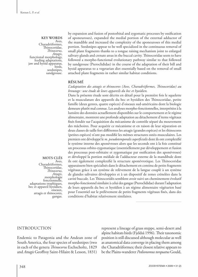
FIG. 1. — Particularities of seedsnipe’s skull: A, Thinocorus orbignyianus Geoffroy Saint-Hilaire & Lesson, 1831; B, Attagis gayi Geoffroy Saint-Hilaire & Lesson, 1831; C-E, Thinocorus Eschscholtz, 1829; C, ventral view of the mandible and palate; D, surface of the mandibular joint with location of the contact area of the lateral and caudal condyles of the quadrate; E, caudal view of the mandibular joint. Abbreviations: see text.
Sh m. serpihyoideus;Sth m. stylohyoideus;Vm (os) vomer.
RESULTS
ANATOMICAL CHARACTERISTICS OF THE BILL OF THE THINOCORIDAE
All the following descriptions apply to the genus Th inocorus as well as to the genus Attagis. When they exist, diff erences between these genera are clearly indicated.
SkullTh e skull of Th inocoridae (Fig. 1A, B) is clinorhyn-chic, i.e. the base of the upper jaw and that of the neurocranium make an obtuse angle. Bill is relatively thick. Culmen is curved but the extremity of the upper jaw, with the rhamphotheca, is not hooked and overhangs only very slightly the rounded tip
of the mandible. Th e rims of the rhamphotheca of the upper jaw overlap those of the mandible, making a kind of scissors.
Th e rounded external nares are covered with a keratinized fold of skin which leaves open just a narrow horizontal slit. Each bony naris extends almost over half the length of the upper jaw and dorso-caudad ends in an acute angle the vertex of which inserts just slightly between the maxillary and pre-maxillary processes of the nasal bone.
Th e pliable area of the upper jaw (a hinge between the upper jaw and the neurocranium) is clearly in-dicated by a very narrow slot between the caudal edge of the nasal septum and the rostral crest of the mesethmoid. It is located a little frontal relatively to the vertex of the dorso-caudal extremity of the bony naris. Th e pliable area of the maxillary process of the nasal is more caudal, located at the level of the vertex of the bony naris. Such an arrangement of the pliable areas explains well the unavoidable
352 ZOOSYSTEMA • 2009 • 31 (2)
Korzun L. P. et al.
warping of the upper jaw during protraction. Th is is clearly visible when one manipulates syndesmo-logical preparations.
Th e palate is of the schizognathous type, i.e. the contralateral halves of the upper jaw are separated by a wide slot. In Th inocorus the palatine processes of the maxillars slightly overlap the lateral edge of the palatines. On the other hand they are larger in Attagis where they extend caudad along the surface of the palatines and more clearly overlap the me-dial edge of the palatines but nevertheless leave a wide slot between them. Th e enlarged blade-like caudal part of the palatines is not oriented in the frontal plane, i.e. the lateral edges of these blades are more ventral than the medial edges which are applied to the base of the interorbital wall. Th is latter separates the palatines along most of their length; these bones join only by their caudal ex-tremity near the contact zone with the pterygoids. Sympalatiny exists but is not very well marked. Th e vomer is wide and fl attened, and its caudal processes attach to the palatines (diastasy fi de Hofer 1945). In its medial half the enlarged part of the palatines bears a ventrally oriented blade. In Attagis the medial edge of the ventral wing of the palatines is fi xed to the palatine process of the maxillar by a very narrow bridge; this link is absent in Th inocorus.
Th e pterygoids are straight. Th ere is no basiptery-goidal joint in adults: there is no contact between pterygoids and skull base. In adult Th inocorus, on the basisphenoid there is however a small bump oriented towards the pterygoids which them too possess a small bump oriented towards cranium base (Fig. 1C) whereas in the young (here a pullus of T. rumicivorus) there is a true basipterygoidal joint. We failed to fi nd any such bumps in adult Attagis.
Th e quadrate has an enlarged orbital process and three condyles in the quadrato-mandibular joint. Th e lateral and caudal condyles make a saddle, with convexity oriented downwards and connected to the concave lateral edge of the mandible in the joint area (Fig. 1D, E). In the posterior part of the latter there is a groove in which the caudal condyle of the quadrate goes down when the mandible lowers. It must be noted here that this caudal condyle does
not come into direct contact with the mandible. Th e medial condyle is medially well apart of the other two. Th e longitudinal axis of this medial condyle is almost perpendicular to the longitudinal axis of the skull. Th ere are two condyles in the quadrato-cranial joint. Th e axis passing through these two condyles is oriented almost perpendicular to the longitudinal axis of the pterygoid.
In the braincase the interorbital septum shows two windows rather small in Attagis but rather large in Th inocorus. Th e anterior wall of the orbit is constituted by the large lateral wing of the mes-ethmoid. Th e olfactory capsules fi ll a great volume in the upper jaw and even give the impression that they push the mesethmoids backwards. We did not fi nd any trace of lacrymal, even in the pullus of T. rumicivorus we examined as this bone could have been visible before an eventual fusion with the wing of the mesethmoid.
Th e greatest cranial diff erence between Attagis and Th inocorus lies on the relation between the postorbital and zygomatic processes (Fig. 1A, B). In Attagis, these processes are typical of those found in most birds whereas in Th inocorus a part of the large superfi cial aponeurosis of the head that attaches to the postorbital process is ossifi ed and fuses with the equally ossifi ed aponeurosis of the medial portion of the external adductor. Th ese two ossifi ed aponeuroses build a very long process (a fact also pointed out by Fjeldså 1996 and illustrated in Olson & Stead-man 1981) that for convenience we call thereafter “orbito-zygomatic process”. Th is process extends down almost to the dorsal edge of the mandible and its transversal section is shaped as a V point-ing towards the orbit (see below, Fig. 5A, a, B, b). At the base of this process there is a small window through which pass muscular fi bres of the rostral part of the deep portion of the external adductor of the mandible. Th e parts of the aponeuroses which contribute to form the orbito-zygomatic process of the adult are not yet ossifi ed in the pullus of T. rumicivorus.
Th e mandible shows two windows the most cau-dal one being smaller in Attagis than in Th inocorus. Th e mandible has a rather particular shape in that its height increases gradually and caudad along the fi rst three quarters of its length, and then decreases

353
Adaptation of seedsnipes (Aves, Charadriiformes, Th inocoridae) to browsing
ZOOSYSTEMA • 2009 • 31 (2)
Aes
Aes
AemGls Sth
Sth
ShSh
Ap
Ap
Q
Q
Q
A
B
C D
Bm
Aes
Aer
Aer
AerAer
Mst
Oc
as
aps
app
apo
apo
aps
PssPr
Psp
Poz
Poz
Poz
Pra
aib
as
ams
am
am2
am1
am3
ami1 ami2 ami3
ad
am
alm
as
Gls
Gls
AemBm Sth Sh
GlsBm
LjiDm
Dm
Dm
Dm
Lp
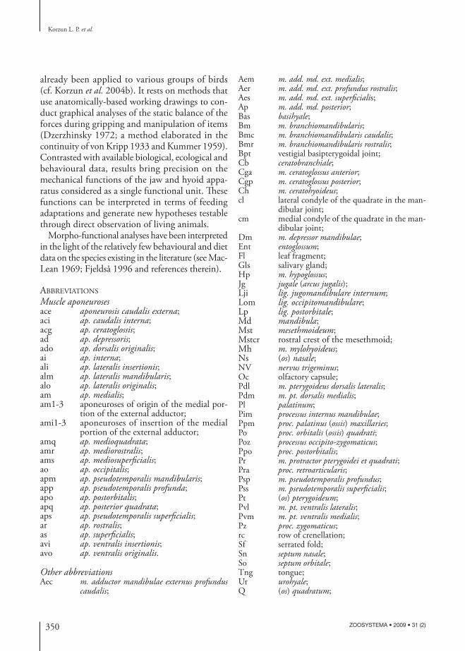
FIG. 2. — Cranial musculature of Thinocorus Eschscholtz, 1829, successive planes of the dissection: A, general view; B, lateral view through the orbit; b aponeuroses of m. pseudotemporalis superfi cialis; C, D, lateral views of the superfi cial and medial portions of the external adductor. Abbreviations: see text.
along the last quarter. Th e maximal height (marked by bumps corresponding to the attachments of the aponeuroses of insertion of the external adductor) is plumb with the rostral end of the orbito-zygomatic process. Th e mandible also shows an upward-hooked retro-articular process as well as a well-marked internal process (Fig. 1A-C).
Cranial musculature (Figs 2-6)According to the classical morphological opinion (Lakjer 1926), cranial muscles may be divided into adductors and protractors. Th e adductors of the mandible are divided into an external adductor (m. adductor mandibulae externus), a posterior adductor (m. adductor mandibulae posterior) and three internal

354 ZOOSYSTEMA • 2009 • 31 (2)
Korzun L. P. et al.
Q
A
B
asace
ace
amq
amq
amr
ams
ar
aci
aci
apo
Poz
Pra
Pra
ar
amsam
am2am1
am3
ami1 ami2
ami3am
as
FIG. 3. — Schema of the arrangement of the internal aponeuroses of external adductor in Thinocorus Eschscholtz, 1829 (A) and Attagis Geoffroy Saint-Hilaire & Lesson, 1831 (B). Abbreviations: see text.
adductors (m. pseudotemporalis superfi cialis, m. pseudotemporalis profundus and m. pterygoideus). However, from a functional viewpoint the external and posterior adductors, together with the two mm. pseudotemporales, are generally seen as constituting the group of the dorsal adductors whereas m. pterygoideus is considered as the ventral adductor.
Among the cranial muscles we fi nd also the pro-tractor of the quadrate (m. protractor quadrati), the protractor of the pterygoid (m. protractor pterygoidei) and the depressor of the mandible (m. depressor mandibulae).
M. adductor mandibulae externus. Th e external adductor of the mandible includes three portions:
one superfi cial, one medial and one deep. Th e latter is composed of two parts: one rostral and one caudal. It is useful to point out that in Th inocoridae the superfi cial portion attaches to the dorsal edge of the mandible very far forwards, passing below the eye and under the mesethmoid, almost reaching the level of the olfactory capsule. Th is superfi cial portion has a rather complex internal structure.
In Th inocorus (Figs 2A-C; 3A) the aponeuroses of origin and of insertion of this superfi cial portion are much enlarged. Th e aponeurosis of insertion as attaches along the dorsal edge of the mandible. Its central part is very thick and very elongated dorso-caudad. At the surface of this aponeurosis attach muscular fi bres originating from the lateral surface of the aponeurosis of origin am of the medial por-tion and from the surfaces of the two aponeuroses of origin of the superfi cial portion, apo and ams. Aponeurosis apo seems to be a continuation of the orbital branch of the orbito-zygomatic process, and ams to represent a short lateral lobe of am, lateral to its base. Th e aponeurosis of origin apo is also very long (like as) and wide, and it even wraps up as a little and then extends parallel to it. Aponeurosis apo is fused at its origin with the aponeurosis ams. It is important to underline here that muscular fi bres from apo also insert more medially than as on the particularly enlarged surface of the dorsal border of the mandible.
In Attagis (Figs 3B; 4B, C), the superfi cial portion diff ers from that in Th inocorus. Although extend-ing as far forwards as in Th inocorus, in Attagis the aponeurosis of insertion as shows a shorter and more localized attachment. Th e rostral part of this aponeu-rosis as folds medially, giving rise to an extension which, attaching farther caudad to the dorsal edge of the mandible, makes a long aponeurosis ar which belongs to the rostral part of the deep portion of the external adductor of the mandible. Th e aponeurosis of origin ams is in two parts: one originates from the surface of the aponeurosis am, the other is formed by the laterally folded rostral edge of am. Th ere are no signs of the aponeurosis of origin apo.
Diff erences also exist between the two genera of Th inocoridae in the medial portion of the external adductor of the mandible. In Th inocorus (Figs 2C, D; 3A) the internal structure is very particular and

355
Adaptation of seedsnipes (Aves, Charadriiformes, Th inocoridae) to browsing
ZOOSYSTEMA • 2009 • 31 (2)
Aes
Aem ApAp
Ap
Q
Q
A B
C D
E
Aem Ap
Aer
Oc
as
aps
app
apsaps
PssPr
Psp
Pssai
b
as
ams
ar
am
am
ad
ai
ar
amq
ams
amr
ar
ace
ace
aci
Gls
Gls
Aem
Mh
SthSh
Gls
Lji
Dm
Lp
FIG. 4. — Cranial musculature of Attagis Geoffroy Saint-Hilaire & Lesson, 1831, successive planes of the dissection: A, general view; B, lateral view through the orbit; b, internal aponeuroses of m. pseudotemporalis superfi cialis; C-E, lateral views of portions of the external adductor. Abbreviations: see text.
complicated. In most studied avian groups there is only a single aponeurosis of origin am and sometimes a small aponeurosis of insertion (alm) attached to the lateral surface of the mandible. In Th inocorus there exists in the medial portion of the external adductor
a system of aponeuroses of origin (am1, am2, am3) in which the aponeurosis am is separated into four aponeurotic blades attached to the orbito-zygomatic process, and a system of three aponeuroses of inser-tion (ami1, ami2, ami3) which attach to the lateral

356 ZOOSYSTEMA • 2009 • 31 (2)
Korzun L. P. et al.
surface of the mandible. Th e number of aponeuroses in this portion increases caudad: the closest is the mandibular joint the most numerous they are. In general extension and shape (superfi cial view) the medial portion in Attagis looks like that in Th inocorus however the dissection shows that the aponeurosis am is not internally divided (Figs 3A; 4C).
In both genera the most enlarged part of the deep portion of the adductor is the caudal one. Th e rostral part is smaller in Th inocorus than in Attagis.
In Th inocorus (Fig. 5D) this rostral part of the deep portion extends below the orbito-zygomatic process, a small part coming from the surface of the braincase after passing through the window located at the base of this process. Th e aponeurosis of insertion ar, bifi d at its top (one external blade and one internal) is rather thick and independent, i.e. without link with the aponeurosis of inser-tion as as in Attagis (Fig. 4C, D). Th e internal blade receives muscular fi bres coming from the aponeurosis of origin amr and from the deep niche between amr and the quadrate. Th e external blade receives muscular fi bres coming from the internal surface of the orbito-zygomatic process and from the surface of the braincase from where these fi bres pass through the window located at the base of the orbito-zygomatic process.
As described above, there is in Attagis a relation between ar (which is not bifi d) and as. Th e aponeu-rosis of origin amr attaches to the skull in front of the zygomatic process.
In Th inocoridae, there are one aponeurosis of origin and two of insertion in the caudal part of the deep portion of the adductor. Th e aponeuro-sis of insertion ace attaches near the highest point of the mandible; its rostral edge is very thick. In Th ino corus (Fig. 5C) the non-ossifi ed part of am that fringes the ossifi ed part gives rise to the aponeu-rosis of origin amq of the caudal part of the deep portion of the external adductor. Th is aponeurosis amq attaches to the skull, caudal to the orbito-zygomatic process, and to the quadrate. In Attagis too (Fig. 3D, E), amq is the caudal extension of the aponeurosis am but here am passes through the narrow slot between ace and ar before forming amq. Th e deepest aponeurosis of this caudal part is the aponeurosis of insertion aci. Th is aponeurosis
keeps the link with the aponeurosis ar in Attagis but not in Th inocorus.
M. pseudotemporalis superfi cialis. It is quite large in both genera between which diff erences exist. It is more typical in Th inocorus (Fig. 2B, b). Its aponeurosis of insertion aps makes a kind of funnel which attaches to the internal side and at the highest part of the mandible. Into this funnel enters the aponeurosis of origin ai which is attached to the crest of the posterior wall of the orbit. More medial to this crest there is a niche fi lled by muscular fi bres directed to the internal surface of aps. In Attagis (Fig. 4B, b) this muscle is larger and more complex. Its aponeurosis of insertion makes also a medially-open funnel and its point of insertion is marked by a bump on the dorsal edge of the mandible. Th e aponeurosis of origin ai begins more medially on the skull than in Th inocorus. It forms two blades. An internal one gets into the funnel and the muscular fi bres of its surface go to the internal surface of the aponeurosis aps. An external blade is located outside the funnel and the muscular fi bres which attach to its surface extend down to the internal surface of the mandible, giving rise to a voluminous additional portion of the muscle. It is important to note here the presence of a very narrow aponeurosis which originates from the lower part of the aponeurosis aps and vanishes into the additional portion (Fig. 4, b). It is very interesting that this tendinous tail resembles closely the aponeurosis of origin of the intramandibular portion in the Fulmar (Dzerzhinsky & Yudin 1979).
M. pseudotemporalis profundus. It is enlarged in both genera. It originates from the orbital process of the quadrate (its aponeurosis of origin app is attached to the rostral extremity of the process) and inserts on the internal surface of the mandible (aponeurosis of insertion apm). In Th inocorus (Fig. 5E, e) the aponeurosis app is particularly enlarged so that its inferior edge is located much lower than the dorsal edge of the mandible whereas in Attagis this inferior edge does not reach the mandible.
M. pterygoideus. Usually four portions are distin-guished: two ventral and two dorsal. We did not fi nd

357
Adaptation of seedsnipes (Aves, Charadriiformes, Th inocoridae) to browsing
ZOOSYSTEMA • 2009 • 31 (2)
ShSh
Q
Q
Q
A B
C
E
DAer
Aer
Aec
aci
Pr
Psp
Psp
PspBmc
Bmc
BmcBmc
NV
NV
NV
NVNV
Psp
Pdm
Pdl
Psp
Poz
Poz
PozPoz
b
a
e
am
amr
apq
apm
app
ar
amqace
Ap
Ap
Ap
Ap
Ap
SthSth
FIG. 5. — Lateral views of the successive planes of the dissection of the dorsal adductors in Thinocorus Eschscholtz, 1829: A-D, deep portion of the external adductor; E, m. pseudotemporalis profundus, posterior adductor and protractors; a, b, transversal sections of the orbito-zygomatic process; e, aponeuroses of m. pseudotemporalis profundus. Abbreviations: see text.

358 ZOOSYSTEMA • 2009 • 31 (2)
Korzun L. P. et al.
A B
C D
Pra Pra
avo
avi
ali
alo
alo
ado
ado
Gls
VmPpm
PimPimPdm
Pvm
Pvm
Pvl
Pvl
Pl
Pdl
Pl
Pl
Pt Pt
Sth
Gls
DmLom Lom
LomLom
FIG. 6. — A-D, ventral views of the successive planes of the dissection of m. pterygoideus in Thinocorus Eschscholtz, 1829. Abbreviations: see text.
any signifi cant diff erence between the two genera. Th e ventral portions (Fig. 6A, B) originate from the surface of the palatine and from the rostral part of the pterygoid. Th e ventro-lateral portion possesses a large aponeurosis of origin alo the superfi cial part of which attaches along the lateral edge of the palatine but which, after folding, attaches farther to the caudal edge. From its surface the muscular fi bres extend to the ventral and lateral surface of the caudal part of the mandible. Th e ventro-medial portion contains two aponeuroses: one of origin avo which attaches transversely to the palatine and one of insertion avi which attaches at the top of the internal process of the mandible. Th e medial edge of the aponeurosis avi folds up dorsally, forming an additional blade.
Th e muscular fi bres of the dorsal portions originate from the dorsal surface of the palatine and ptery-goid, and from the dorsal surface of the aponeurosis alo (Fig. 6C, D). It must be noted that the origin of these fi bres, particularly that of the most caudal ones, is more dorsal than their insertion. Th e mus-cular fi bres of the dorso-lateral portion attach to the surface of the aponeurosis of insertion ali which itself attaches to the mandible, a little in front of the base of the internal process. Th e dorso-medial portion is separated from the other portions by its more transverse orientation of fi bres. Most of its muscular fi bres originate from the pterygoid. Th e aponeurosis of origin ado of this portion attaches to the specifi c crest of the lateral surface of the ptery-goid. Th e muscular fi bres of this portion insert on the surface of the aponeurosis of insertion adi and on the mandible at the base of its internal process.
Medial to the pterygoid are muscular fi bres which make up a portion sometimes called caudal portion but which, in Th inocoridae, has no aponeurosis.
M. adductor posterior. It originates from the or-bital process of the quadrate more basally than the m. pseudotemporalis profundus behind which it is located (Figs 2C, D; 4; 5). Its aponeurosis of origin apq attaches along the inferior part of the process. We did not fi nd any signifi cant diff erence between the two genera.
M. protractor quadrati and m. protractor pterygoidei. Th ey are of the generalized type usually found in birds (Figs 2B; 4B). Th ey are diffi cult to distinguish and separate in their area of origin which is located in the lower part of the posterior wall of the orbit. Th e muscular fi bres of m. protractor quadrati insert on the quadrate near the mandibular joint whereas those of m. protractor pterygoidei attach to the caudal part of the pterygoid.
M. depressor mandibulae. It originates from the lateral surface of the occipital part of the skull and inserts on the retro-articular process of the mandible (Figs 2; 4A). Its aponeurosis of origin ao attaches along the paroccipital process. Th e external edge of this aponeurosis constitutes a ligament which links the paroccipital process to the top of the

359
Adaptation of seedsnipes (Aves, Charadriiformes, Th inocoridae) to browsing
ZOOSYSTEMA • 2009 • 31 (2)
QA
B
C
acg
aoPoz
SthBmr
Bmc
Bas
BasHp Ur
Ur
Md
Tng
Cga
Cgp
Ent
HpSf
rc
Md
Md
Fl
Pdl
Lp
Cb
Cb
Ch
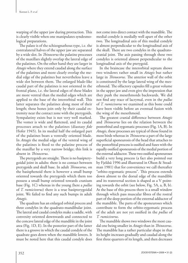
FIG. 7. — Hyoid apparatus and buccal cavity of seedsnipes (here Thinocorus Eschscholtz, 1829): A, lateral view of the hyoid apparatus displaced downward relative to the mandible; B, rostral part of the hyolingual apparatus; C, buccal cavity, the fl oor on the left, the roof on the right, a plant item present in the buccal cavity of the dissected specimen has been left in its place. Abbreviations: see text.
retro-articular process, and the internal edge fuses with the occipito-mandibular ligament. Th ere is also a small aponeurosis of insertion ad.
LigamentsLike Attagis, Th inocorus has an entirely non-ossifi ed postorbital ligament (Figs 2A; 4A). Its origin remains free above the ossifi ed orbito-zygomatic process which Attagis lacks (see above). Th is implies that the orbito-zygomatic process may result from the coalescence and ossifi cation of two aponeuroses. One is the aponeurosis of origin apo of the postorbital part of the superfi cial portion of the m. adductor mandibulae externus. Th e other is the aponeurosis am of the medial portion of the m. adductor mandibulae externus. In both genera, the postorbital ligament is bifi d at its insertion: one part attaches to the jugal bar, the other to the mandible.
In the mandibular joint there is no external jugo-mandibular ligament.
Th e internal jugo-mandibular ligament (Figs 2A; 4A) originates from the caudal extremity of the jugal bar, warps behind the mandibular joint and inserts on the base of the internal process of the mandible near the occipito-mandibular ligament. Th e internal jugo-mandibular ligament has a sesamoid ossicle behind the joint.
Th ere is also a particular ligament which joins the lateral wing of the mesethmoid and the jugal bar (Figs 2A; 4A).
Th e occipito-mandibular ligament (Fig. 6) links the internal process of the mandible to the base of the skull. Its principal peculiarity is its very slant-ing, almost horizontal orientation: it is almost in the same plane as the mandible.
As noted above, the lateral edge of the aponeu-rosis of origin of the depressor of the mandible constitutes a ligament which links the paroccipital process to the top of the retro-articular process of the mandible (Fig. 7A).
Buccal cavityTh e roof of the buccal cavity bears a transversal row of crenellations in its medial part and a serrated fold at the entrance of the oesophagus. Th e tegument of the buccal roof is rather fl exible, covering large and individualized salivary glands.
Th e tongue is relatively short and thick. It goes hardly beyond the mandibular symphysis. Its surface is rather fl exible. Its posterior edge has on each side well-marked hard serrations. Behind the glottal slot there is also a row of serrations (Fig. 7).
Salivary glandsWithout special dissection for their observation, four enlarged and individualized salivary glands were apparent during the study of the musculature (Figs 2A, B; 3A, B). One is located along the inferior part of the mandible (group of the gl. mandibularis anterior sensu Fahrenholtz 1937 and McLelland 1979), another one behind the hyoid horns (group of the gl. mandibularis posterior), a third one in the orbit along the palatines (gl. palatinae) and the

360 ZOOSYSTEMA • 2009 • 31 (2)
Korzun L. P. et al.
fourth one in the buccal roof near the entrance of the oesophagus (gl. sphenopterygoideae).
Hyoid apparatus (Fig. 7)M. mylohyoideus makes a kind of hammock caudally slung between the mandibular rami, from the symphysis to half the length of the rami (Fig. 3A). Between the two contralateral halves of the muscle there exists a longitudinal tendon: an aponeurotic band to which attach the muscular fi bres of these contralateral halves.
M. serpihyoideus originates from the top of the retro-articular process of the mandible and, slant-wise, attaches to the longitudinal tendon men-tioned above.
M. stylohyoideus is bifi d. One portion which includes most muscular fibres originates like m. serpihyoideus but a little more frontal, from the retro-articular process of the mandible. Th e other portion (a portion never observed before in any other Neognath) is smaller and originates from the base of the skull. Both portions fuse just underneath the ventral border of the mandible and then attach to the dorsal surface of the basihyal.
Th e muscular fi bres of m. ceratohyoideus originate from the caudal half of the ceratobranchial and at-tach also to the longitudinal tendon which fuses at this level with the urohyal.
M. branchiomandibularis is composed of two portions: one caudal and one rostral. Th e caudal portion originates from the caudal part of the epi-branchial, passes along the hyoid horn and inserts on the lateral surface of the mandible. Th e rostral portion originates also from the epibranchial, follows the hyoid horn wrapping it into a kind of case and attaches to the internal surface of the mandible.
M. ceratoglossus is double-bellied. Th e fi bres of m. ceratoglossus posterior originate from the surface of the ceratobranchial. Th ey end as a long tendon which inserts on the ventral surface of the entoglossal (sensu Engels 1938: internal skeleton of the tongue, i.e. paraglossal and its anterior and posterior proc-esses, see also Tomlison 2000). M. ceratoglossus anterior (Fig. 7B) is relatively complicated. Its two contralateral parts originate from the surface of the tendons of insertion of m. ceratoglossus posterior. Together with the medial part which attaches to
the basihyal, they end by a common aponeurosis which passes along and under the entoglossal and attaches near its rostral extremity.
M. hypoglossus is triangular in shape. Its base attaches to the ventral surface of the basihyal and its vertex inserts in the caudal corner of the ento-glossal.
We did not fi nd m. genioglossus.
DISCUSSION
Th ere are still relatively few detailed biological data on the Th inocoridae. Th e most signifi cant observa-tions have been synthesized by MacLean (1969) and Fjeldså (1996). Th e four species appear to be strictly vegetarian (even their pulli which feed by themselves at birth) although the ingestion of mealworms has been mentioned for captive birds. Th ey feed mainly on plant fragments such as buds, leaf tips and small leaves which they bite off green plants. But they also peck seeds on the ground, particularly during the seasons when climatic conditions reduce the avail-ability of green vegetation. Fjeldså (1996) describes how these birds forage in a crouch position, walking slowly and taking items off the plants with rapid snapping movements. Th e fragments are torn off by moving the bill towards the breast with a quick movement and are swallowed whole. Th e birds may also stretch up a little to bite off the top of grasses or to browse on taller herbs. All observers underline that the consumed plants are usually succulent ones and that in natural conditions these birds have never been seen drinking, water being obtained from succulent and green plant items.
Although it is represented without comments in Olson & Steadman (1981: fi g. 2C) who give a quite good drawing of the skull of Th inocorus ru-micivorus, Fjeldså (1996) was the fi rst to describe in Th inocorus the ossifi ed tendon that we call here “orbito-zygomatic process” (see above) and which is missing in Attagis (this might be one of the dif-ferences in relation to Th inocorus to which Fjeldså alludes without clarifying them). He also rightly mentions that the upper jaw and the muscles of the palate play an important role in bill functioning but without explaining how the system works.

361
Adaptation of seedsnipes (Aves, Charadriiformes, Th inocoridae) to browsing
ZOOSYSTEMA • 2009 • 31 (2)
A
B
Pli
PliFra
FrbF
F
h
hl
F’
F’
Ff
FIG. 8. — Forces in action when a fi xed plant item is taken off; A, a plant item is pulled along the axis of the bill; B, a plant item is torn off by moving the head downward towards the breast. Abbreviations: F (= F’ at equilibrium), clamping forces respectively exerted by the mandible and the upper jaw; Fr, resistance force of the item, labelled Fra when this item is pulled along the axis of the bill and Frb when this item is torn off by moving the head downward towards the breast (Fra = Frb); Ff, friction force which depends on the clamping forces: Ff = 2 k F (k = coeffi cient of friction); the lever arms of Fra and Frb are respectively indicated l and h, with l > h; so Fra has a less favourable lever arm than Frb: the action of these forces depending on their respective momentum, we have Fra × h < Frb x l; for the item to be maintained in the bill it is necessary to have Ff greater than or at least equal to Fr; all this means that when the item is pulled along the axis of the bill the upper jaw can more effi ciently resist the passive protraction (due to Fr) than when the item is taken off by a downwards movement of the head; Pli, plant item.
On the basis of our morpho-functional analyses and of the scanty available eco-ethological fi eld data we can propose hypotheses on the potentialities of the bill of the Th inocoridae. Th ese hypotheses will of course require further fi eld and laboratory studies, particularly to measure the actual performances of the anatomical structures we have described.
WHAT DOES IMPLY BROWSING, I.E. REMOVING AN ATTACHED ITEM OR TEARING OFF A PIECE OF IT?Usually, pecking (i.e. collecting of free items, e.g., seeds) proceeds from morpho-functional characters which are generalized among neognathous birds, and thence does not require particular adaptations (see Korzun et al. 2004b), contrary to when attached food items have to be bitten off as is the case here with the Th inocoridae. Pecking birds use the classical “catch and throw” movement (see e.g., Zweers 1982; Zweers et al. 1994; Gussekloo & Bout 2005) with or without a “slide and glue” oropharynx mechanism (Zweers 1982; Zweers et al. 1994). Th e attached item to be plucked off resists and applies a force which tends to protract the upper jaw. Th is passive protraction lessens the clamping force and thence increases the risk that the item gets out of the bill. It is however important to mention here that fi xed items can be taken off in two ways. In the fi rst one the item is pulled along the axis of the bill whereas in the second, as described by Fjeldså (1996), the bird tears off the fragment from the plant by moving the head downwards towards the breast. So the item is no more pulled along the axis of the bill. Th is causes an unfavourable condition which requires a much more important resistance of the upper jaw to the passive protraction force (Fig. 8). In both cases the success of the browsing depends on the clamping force which itself settles the friction forces between the bill and the item, friction forces which oppose the resistance force from the item.
In Neognaths there exist two main kinds of osteomuscular mechanisms for clamping in the bill an attached item in order to pluck it off (for details see Dzerzhinsky 1972; Korzun et al. 2003, 2004a, b).
Th e most widespread is the one that uses the joint muscular control of the jaws where the clamping depends entirely on the resultant of the forces pro-
duced by the dorsal adductors alone. Th is resultant has two simultaneous eff ects: one raises the mandi-ble while the other, transmitted by m. pterygoideus (which plays only this role), retracts the upper jaw. At equilibrium, the jaws exert on the item equal but opposed forces. In that case the resultant of the forces of the dorsal adductors has a well-defi ned orientation (established through a static mechanical graphical analysis; for details see Dzerzhinsky 1972 and Korzun et al. 2003, 2004a, b). By voluntarily changing the orientation of this resultant (by diff erential action on the muscles), the bird can leave the equilibrium and give the advantage either to the upper or to the lower jaw. Th e birds characterized by this joint muscular control and which have to browse fi xed items must, in order to avoid the passive protraction

362 ZOOSYSTEMA • 2009 • 31 (2)
Korzun L. P. et al.
of the upper jaw, give the advantage to this latter at the very moment when the item is bitten off .
Th e second clamping mechanism is the one based on the separate muscular control of the forces of the jaws. It is characteristic of the vegetarian birds highly adapted to pluck off fi xed items. Th e classical example is that of the Galliformes and Anseriformes (Gambaryan 1978; Dzerzhinsky 1980, 1995). In this mechanism the dorsal adductors mainly raise the mandible whereas m. pterygoideus is the inde-pendent and sole retractor of the upper jaw.
Th e instauration of this separate muscular control has been favoured by the disappearance of the ex-ternal jugo-mandibular ligament in the mandibular joint and by the very slanting orientation (i.e. much more horizontal than vertical, tending towards the plane of action of m. pterygoideus) of the occipito-mandibular ligament directly joining that muscle with the braincase. Indeed in the case of clamping by joint muscular control (where the external jugo-mandibular ligament is necessarily present) m. ptery-goideus produces two equal but opposed forces. Th e fi rst one is applied to the mandible, the second to the upper jaw. Th is second force is transmitted via the palatine and pterygoid to the quadrate which retracts. Th is stretches the external jugo-mandibular ligament which then passes the force to the mandible where it meets and annihilates the force directly applied to the mandible by m. pterygoideus. Th is is why m. pterygoideus cannot retract the upper jaw independ-ently in the case of the joint muscular control. With the disappearance of the external jugo-mandibular ligament in the case of the separate muscular con-trol, the forces produced by the m. pterygoideus do not meet and thence do not annihilate each other. Furthermore, in this case and when the occipito-mandibular ligament is almost horizontal (i.e. in the plane of action of m. pterygoideus) the force exerted on the mandible by m. pterygoideus is transmitted to the base of the skull. Th erefore in the separate muscular control m. pterygoideus applies its forces on one side to the upper jaw and on the other side to the base of the skull.
In the case of the joint muscular control, at the very moment when the item is taken off , the resistance of the upper jaw to the passive protraction is produced by a change in the balance of forces in favour of the
upper jaw. Th is is due to a change in the orientation against the equilibrium state of the resultant of the forces originating from the dorsal adductors. Th is resultant is thence very slanting and below the point z (Fig. 9). But to this new slanting resultant participate only some muscles the vector of force of which acts below the resultant characterizing the equilibrium state. Th is means that the clamping of the item will be lessened. However we underlined above that the success of the browsing does not depend only on the ability to counteract the passive protraction of the upper jaw but also on a clamping or grasping force which prevents the item from sliding along the jaws. Th ere is then a kind of contradiction which is solved by a separate muscular control, characteristic of the Th inocoridae. As indicated above, these birds lack the external jugo-mandibular ligament and, independently of the dorsal adductors, their m. pterygoideus retracts the upper jaw, with the caudal support of the base of the skull as the occipito-mandibular ligament is almost along the same axis as m. pterygoideus.
ARE SNEEDSNIPES EQUIPPED FOR AN EFFICIENT BROWSING?In Th inocoridae the upper jaw is relatively short (thus the lever arm of the force that clamps the item is relatively short) and high at base (thus the lever arm of the force exerted by m. pterygoideus is relatively long). Th is is why the force exerted by m. pterygoideus on the upper jaw acts in a quite favourable state of lever arm. Th e length of the mandible is twice that of the upper jaw (which is the lever arm of the force that clamps the item) whereas the adductors insert on the mandible close to the centre of rotation of the mandibular joint and have thence a short lever arm. Th is means that on the whole the adductors work in unfavourable conditions of lever arm. Th is is why in Th inocoridae the tendency to strengthen the effi ciency of the dorsal adductors is quite evident. Th eir main aponeuroses attach to the mandible at its highest point. Th anks to this specifi c height the axis of action of the force produced by these muscles is set away from the centre of rotation of the mandibular joint. Th is increases the length of its lever arm. Th e bumps (where attach the aponeuroses of insertion of the external adductors) located at the highest point of the mandible testify of the intense forces

363
Adaptation of seedsnipes (Aves, Charadriiformes, Th inocoridae) to browsing
ZOOSYSTEMA • 2009 • 31 (2)
A
B
F
F
a
ab
b
c
c
N
N
R
R
z
z
M
M A
AM
A
A
KR
K
K
K
Rq
q
x
x
j
j
F’
F’
A’
FIG. 9. — A, static graphical analysis of the equilibrium of forces when a food item is clamped in the bill (for more details see Dzerzhinsky 1972; Korzun et al. 2003, 2004a, b); adductors exert force A from the mandible towards the cranium and force A’, superimposed, opposite and equal to A (for the clarity of the fi gure, A has not been represented on its point of application but directly placed at j along its axis of action); force A is decomposed into F (mandibular clamping force applied along the axis going through the clamping points of the item in the bill) and K (force of the quadrate applied on the quadrato-mandibular joint q); force K is itself decomposed into M (pressure force of the quadrate on the cranium along the axis q-x) and R (retraction force of the upper jaw along the axis of the jugal bar); in turn R is decomposed into F’ (clamping force of the item by the upper jaw, opposed and equal to F) and N (pressure force of the upper jaw on the cranium at the level of the prokinetic hinge b); at the equilibrium, forces M, N and A (so A’) are applied to point z where they cancel each other; a, meeting point of the three axes of 1) clamping, 2) action on the cranium via the prokinetic joint, and 3) retraction of the upper jaw along the jugal bar; j, meeting point of the axes of 1) clamping, 2) action of the adductors, and 3) action on the cranium through the mandibular joint; x, joint of the quadrate on the neurocranium; c = meeting point of the jugal bar and the maxilla.B, the same analysis as above but with a more slanting force A from the adductors at the very moment when the attached item is taken off; then this force A passes below the point z of the equilibrium state and F’ from the upper jaw becomes greater than F from the mandible, so the advantage is given to the upper jaw and this difference between F and F’ allows to resist the passive protraction of the upper jaw.
that apply to this area. Th e fundamental specifi cities of the separate muscular control give the adductors additional possibilities to increase their effi ciency, thanks to the more vertical orientation with respect to the mandible of the resultant of the forces from the adductors.
For the external adductor, the more vertical ori-entation increasing the effi ciency of the muscle is particularly pronounced in Th inocorus. In this genus, the medial portion, the largest one, of this adductor originates from well forward on the axial skull thanks to the presence of the specifi c orbito-zygomatic proc-ess from where it spreads almost vertically to the mandible. Furthermore it possesses a very complex aponeurotic skeleton and because of that is made of a great number of relatively short muscular fi bres. It results of course mechanically from this a diminu-tion of the amplitude of the contraction but also an increase of the produced force. Field observations (e.g., MacLean 1969) of foraging Th inocoridae show that they do not need to open widely their bill dur-ing food acquisition. Th en, diminished amplitude of contraction does not penalize them and is not for them a contradiction in their trophic adaptation as they benefi t from the increase of the clamping force. Th e tendency toward a sharp increase of the number of muscular fi bres in this well-oriented medial por-tion clearly appears in the number of aponeuroses which increases as one gets closer to the mandibular joint. Th is increase is possible because the required amplitude of contraction diminishes also as one gets closer to the mandibular joint. Th anks to its V-shaped section the orbito-zygomatic process is specifi cally adapted to resist very intense bending forces coming from the extremely powerful medial portion.
In Attagis the medial portion of the external ad-ductor is of a generalized type common to many birds. But here the potentialities allowed by the separate muscular control are used by m. pseudo-temporalis superfi cialis which is a little less slanting and has an internal system of aponeuroses more complex than in Th inocorus.
In Th inocoridae the total quantity of muscular fi bres of the adductors has also been increased thanks to the great expansion of the superfi cial portion, especially in Th inocorus, which attaches to the mandible very frontal below the eye, almost
at the level of the lateral wing of the mesethmoid. Attachment of amq to the zygomatic process be-sides of quadrate is very unusual and noteworthy;

364 ZOOSYSTEMA • 2009 • 31 (2)
Korzun L. P. et al.
it represents an additional evidence of the rostral expansion of the external adductor.
In Th inocoridae an additional vertical force comes from m. pterygoideus because as described above the origin of the dorsal part of this muscle on the sur-face of the palatine and pterygoid is located much higher than its point of insertion on the mandible. Th us m. pterygoideus is not only the sole retractor of the upper jaw but it also contributes to the ad-duction of the mandible.
It is fi tting to note here that although rhynchoki-netic the bill of the Th inocoridae (which in its appear-ance recalls that of the Galliformes, see Dzerzhinsky 1980) with its robust structure (e.g., thickness of the bones of the rostral part of the upper jaw and mandible, extension of the internasal wall) fulfi ls well the requirements of resisting intense forces.
Th e hyoid apparatus of Th inocoridae is relatively typical of most Neognaths which take relatively small food items (Korzun 1978). M. branchiomandibularis is made of two portions (rostral and caudal); this in-dicates the presence of a tongue raising mechanism (Korzun et al. 2008). Th e main characteristic of this mechanism is the ability of the tongue to rise towards the palate and to support and hold there small items at the moment the bird opens its bill to take another item. In this mechanism an important role is played by the caudal portion of m. branchiomandibularis which acts like the string of the bow formed by the hyoid horn. When stretched this portion applies a pressure on the occipital surface of the head. Th is force generates from this surface a reaction force applied to each hyoid horn. Th is reaction force separates itself into two forces. Th e fi rst one is directed downwards and, with the upward force from m. mylohyoideus, constitutes a torque which turns the hyoid apparatus so that its rostral part (the tongue) is raised towards the palate. Both m. ceratoglossus anterior and m. ceratoglossus posterior allow the dorsal surface of the tongue to match the surface of the palate. Further-more it must be noted that this mechanism allows also the tongue to be raised above the dorsal edge of the mandible and thence to be maintained against the palate even when the bill opens.
Th eoretically seedsnipes could thus tear off small plant fragments without necessitating stopping browsing in order to swallow an item before taking
another one. By using their salivary secretions they could glue and clump items and thus constitute an alimentary bolus easier to swallow. Th is hypothesis is supported by the fact that, as mentioned above, there specifi cally exist several groups of voluminous and well-individualized salivary glands. Its thickness and fl exibility, together with enlarged m. ceratoglossus anterior and m. hypoglossus, show that the tongue possesses great capabilities to manipulate in the bill fl at items which usually are rather diffi cult to handle. Glued items are moved inside the buccal cavity to the oesophagus in the course of retractions of the tongue and pharynx thanks to the crenella-tions located along the caudal edge of the tongue and also caudal to the glottal slot. Th e crenellations located on the palate and on the serrated fold at the entrance of the oesophagus prevent the alimentary bolus from moving back toward bill tip during the protraction of the tongue and of the buccal fl oor. Th is mechanism of transport of the item inside the bill is similar to that described as “side-and-glue” by Zweers (1982) for the domestic pigeon.
WHICH OTHER GROUP OF BIRDS HAS THE MOST SEEDSNIPE-LIKE BILL?Th e main characters of the bill and hyoid apparatus of Th inocoridae correspond rather well with those of generalized Charadriiformes like Pluvialis squatarola (Linnaeus, 1758). However, it must also be noted that against the general arrangement and nomenclature of the aponeuroses of the external adductor established by Dzerzhinsky & Potapova (1974) and applicable to Charadriiformes, Th inocoridae possess neither the fourth aponeurosis of origin aq in the caudal part of the deep portion nor the aponeurosis of origin alt of the rostral part of this deep portion. Th inocorus is also very peculiar in having aponeurosis of origin apo which is absent in generalized Charadriiformes. However because of their strictly vegetarian diet Th i-nocoridae are quite peculiar among Charadriiformes which indeed have a wide food spectrum but which do not include any other group with a bill apparatus directly comparable to that of Th inocoridae.
On the other hand, sandgrouse (Pteroclididae) represent a most adequate group for a comparison with the seedsnipes of the morpho-functional and feeding adaptation of their bill apparatus. Both

365
Adaptation of seedsnipes (Aves, Charadriiformes, Th inocoridae) to browsing
ZOOSYSTEMA • 2009 • 31 (2)
groups have bills which look much alike in general appearance and in the location of the kinetic areas. Both use the separate muscular control of their jaws (external jugo-mandibular ligament absent, almost horizontal orientation of the occipito-mandibular ligament). Th is allows sandgrouse to feed on plant matter and, above all, while being able to peck at very small seeds they can take off fi xed items ( Korzun et al. 2008). In sandgrouse the adaptation to the removal of fi xed items corresponds to a more verti-cal orientation of the medial portion of the external adductor, with a particularly enlarged zygomatic process in Pterocles alchata (Linnaeus, 1766) and Syrrhaptes paradoxus (Pallas, 1773). In the seedsnipes there is likewise a vertical orientation of the medial portion of the external adductor thanks to the fact that the zygomatic process fuses with the ossifi ed base of the aponeurosis apo (the postorbital ligament does not participate in this fusion) forming a long orbito-zygomatic process. It is however interesting to note that between the zygomatic and postorbital processes there remains in Syrrhaptes Illiger, 1811 a tendinous link, a relict of the postorbital ligament which is totally absent in other Pteroclididae. Th ere is thus a strong functional similarity in the bill ap-paratus of these two groups but the origin of the methods of operation is not really the same. In all sandgrouse m. pseudotemporalis superfi cialis which gives an almost vertical component of the force from the external adductors is quite large as in Attagis. It can be mentioned that Pteroclididae as well as Th inocoridae lack the aponeurosis of origin aq of the caudal part of the deep portion of the external adductor which is present in many Charadriiformes. Two characters show that the adaptation to the sepa-rate muscular control is more strongly developed in sandgrouse than in seedsnipes. Th e fi rst one is the presence of a muscular portion in m. pterygoideus which links the palate to the base of the skull (so called m. retractor palatini). Th e second one is that the posterior adductor is enlarged as in certain Gal-liformes (Dzerzhinsky 1995), a particularity which allows to clamp the mandibular joint and thence to resist the passive protraction of the upper jaw at the moment of the removal of an attached item (cf. Korzun et al. 2008). It can be added that adult sandgrouse have the basipterygoidal joint that adult
seedsnipes lack, although their young have it. We did not fi nd any signifi cant diff erence between the two groups in their buccal cavity and hyoid appa-ratus, nor in the relative size of their salivary glands: apparently they use the same operating procedure to transport food items inside the buccal cavity, a “slide-and-glue” mechanism sensu Zweers (1982).
WHICH MORPHO-FUNCTIONAL PATHWAY DID SEEDSNIPES FOLLOW DURING THE ADAPTATION OF THEIR BILL APPARATUS?In the diagram of the successive changes, during their evolution, of the adaptation of the bill apparatus of pigeons and sandgrouse to a diet based on the removal and ingestion of large plant items (Korzun et al. 2008), the fi rst step of the adaptation is the disap-pearance of the internal jugo-mandibular ligament. Th is adaptation developed from an initial generalized state with: 1) a joint muscular control of the jaws; 2) a tongue raising mechanism; 3) schizorhiny; and 4) a capability to remove attached items thanks to a very slant orientation of the resultant of the forces from the dorsal adductors (resistance to the passive protraction of the upper jaw). Th e obliquity of this force from the dorsal adductors has been mainly realized in “pre-sandgrouse” by m. pseudotemporalis profundus but, in “pre-seedsnipes”, by the superfi cial portion of the external adductor which was placed in a favourable state of lever arms thanks to the shape of the mandible.
Th ere are indications that “pre-seedsnipes” pos-sessed an external jugo-mandibular ligament and thence used the joint muscular control of the jaws. Th ese indications are the existence of a quite en-larged superfi cial portion of the external adductor and the presence of a caudal condyle of the quadrate in the mandibular joint. As shown by Dzerzhinsky (1972: fi g. 28) the main role of this condyle is, with the external jugo-mandibular ligament, to lock the mandibular joint and allow the mandible and the quadrate to work together as if they were a monolith. Th is condition can be considered as an adaptation to the removal of attached items (Trounov et al. 1996). When the external jugo-mandibular ligament is missing, the articular surface of the quadrate is simplifi ed and usually there is no caudal condyle (case of Galliformes and Anseriformes, Dzerzhinsky

366 ZOOSYSTEMA • 2009 • 31 (2)
Korzun L. P. et al.
1972: fi g. 6). Extant Th inocoridae lack the external jugo-mandibular ligament but keep a caudal condyle of the quadrate. However this condyle has lost its direct application on the mandible so that the locking of the mandibular joint is no more possible.
In a second step, and diff erent from pigeons (but not from Otidiphaps Gould, 1870 and Didunculus Peale, 1848), sandgrouse lost the external jugo-mandibular ligament and thus acquired the separate muscular control of the jaws. From the same initial generalized state, seedsnipes directly went to this second stage bypassing the fi rst one. Th e change from the joint to the separate muscular control came with a change in the orientation of the result-ant of the forces from the adductors which became more vertical. Keeping the relative size of their m. pseudotemporalis profundus sandgrouse got a vertical resultant by expanding their m. pseudotemporalis su-perfi cialis and, in Pterocles alchata and Syrrhaptes, also by a more vertical orientation of the medial portion of the external adductor thanks to the enlargement of the zygomatic process. In seedsnipes the slanting superfi cial portion of the external adductor has been conserved. Th e vertical resultant has been obtained in Th inocorus thanks to the orbito-zygomatic process which insures the vertical orientation of the specifi cally enlarged medial portion of the external adductor. In Attagis the vertical resultant has been obtained through the expansion of m. pseudotemporalis super-fi cialis. In both thinocorid genera the dorsal part of m. pterygoideus has acquired the role of an eff ective additional adductor of the mandible.
Both sandgrouse and seedsnipes have specialized in the occupation of very open, steppe, even desert (particularly sandgrouse) habitats. Because they also specialized on small food items (in particular seeds and plant fragments), contrary to Didunculus and Otidiphaps, they retained the tongue raising mecha-nism. Sandgrouse developed further their adaptive radiation in habitats where their diet must be based on small, often minute, seeds and where the vegeta-tion is usually very low and succulent plants (from which water can be extracted) occur only on particu-lar substrates. So, joint to the often severe thermal conditions of their habitats, that makes necessary for them to drink regularly. Th is is why they devel-oped particular specifi c behaviours and feathers. On
the other hand, seedsnipes mainly occupy habitats where: 1) thermal conditions (at least the highest temperatures) are less constraining; 2) seed produc-tions are more seasonal (maybe with general wetter conditions less favourable to their conservation); and 3) the vegetation consists mainly in spongy or suc-culent plants so that these birds could live without drinking, although this needs confi rmation.
Th e diversifi cation of seedsnipes gave two species pairs of diff erent size: Attagis species are indeed three to six times larger than Th inocorus species (see data in Fjeldså 1996). Detailed analyses of the diet of the four seedsnipe species remain to be done. Th ough scanty, available data suggest that this diff erence in size has not aff ected the diet which remains based on the consumption of fragments removed from suc-culent plants. Th e morphological diff erences in the bill apparatus between Attagis and Th inocorus that we described above can paradoxically be explained by the similarity of their diet. It can be supposed that the vegetal matter taken by all seedsnipes has the same physical properties (particularly strength) for resisting to the removal. Th is means that the removal of a fragment of this vegetal matter requires the application of a force with a given absolute value. Th is absolute value is not here signifi cant; what is important is the fact that small as well as large birds must produce a force with the same absolute value. Th e dramatic diff erence in accessibility of this force is of primary interest here. A large bird will more easily produce this absolute force than a smaller bird. Be-cause both large and small seedsnipe species use their bill with the same mechanical system and in order to produce the same absolute force as large species, small species must have set up a proper device. So in small seedsnipes (Th inocorus), it is the zygomatic process which has created a more favourable lever arm for the medial portion of the external adduc-tor and moreover thanks to the system of internal aponeuroses it was possible to wrap up in this por-tion a great number of muscular fi bres.
CONCLUSIONS
Th e present morphological and functional study of the skeleto-muscular structures proper to the jaw

367
Adaptation of seedsnipes (Aves, Charadriiformes, Th inocoridae) to browsing
ZOOSYSTEMA • 2009 • 31 (2)
and hyoid apparatus of Th inocoridae is the fi rst one of this kind for this group of birds whose biology and particularly diet remain imperfectly known. So the conclusions that we can draw probably raise more questions than they solve and call for more exhaustive fi eld eco-ethological studies and more advanced quantitative functional analyses. It is also important to point out here that we did not neglect the few morphological particularities that we described but did not discuss. We are still unable to give a sound functional explanation for them (they may be merely neutral) but we checked that they do not contradict the morpho-functional hypotheses we develop in this paper.
A fi rst conclusion is that Th inocoridae are actu-ally quite well adapted to browsing, i.e. to remove attached plant materials. Th eir feeding apparatus is well suited as an instrument for performing pro-cedures necessary to the utilization of these food resources. Th ey solved the problem of removing fi xed items by evolving the separate control of the movements of their jaws. Th is was done through the modifi cation of the osteo-muscular structures of their bill which originally used the joint muscular control of the jaws, a mechanism characteristic of birds that take free (i.e unattached) food items.
A second conclusion is that, in order to evolve this separate control and because they belong to two quite diff erent size-classes, all seedsnipe species did not modify the same osteo-muscular structures. Th e large Attagis species expanded their m. pseudo-temporalis superfi cialis of which they also increased the intricacy of the internal aponeurotic system. On the other hand, the small Th inocorus species evolved an orbito-zygomatic process (mainly by expan-sion of the zygomatic process and its fusion with the ossifi ed aponeurosis apo), enlarged the medial portion of the external adductor of the mandible and increased the complexity of the aponeurotic structure of this medial portion.
A third conclusion is that Th inocoridae specialized on the continuous removal of small, mainly succulent, attached plant fragments thanks to a tongue raising system coupled to a system of large salivary glands and a device of crenate areas in the buccal cavity.
A fourth conclusion is that, as far as functional morphology is concerned, the birds which have a
bill directly comparable to that of Th inocoridae are the sandgrouse (Pteroclididae). Th ese two groups appear as having indeed followed a similar not to say common morpho-functional evolutionary pathway (which of course does not imply a close phyloge-netic relationship) in the course of the adaptation of their bill and hyoid apparatus to a vegetarian diet essentially based on the removal of small attached plant items in rather relatively similar conditions of habitats. We have discovered a good deal of similar-ity in basic traits of the trophic niches with external properties and facilities of the feeding apparatus, and feeding behaviour, though internal design of apparatus is somewhat (or rather) diff erent in both cases. Th is is most probably a case of ecomorpho-logical and thence functional convergence.
AcknowledgementsWe acknowledge with warmest thanks the persons and institutions who provided or helped us to obtain specimens for study: Christine Blake, Dr Joel Cracraft, Paul Sweet and Dr François Vuilleumier at the American Museum of Natural History, New York; Dr Jon Fjeldså and Dr Jan Bolding at the Zoological Museum of the University of Copen-hagen; Dr Éric Pasquet and Jacques Cuisin at the Muséum national d’Histoire naturelle, Paris. We are also grateful to the referees and editorial staff of the journal for their comments and help in the fi nalization of this paper. Th is study benefi ted from grants RFBR 03-04-48958 and Russian Universities NUR 07-03-003.
REFERENCES
DICKINSON E. C. 2003. — Th e Howard and Moore Complete Checklist of the Birds of the World. 3rd edition. Christopher Helm, London, 1039 p.
DZERZHINSKY F. J. 1972. — [Th e Biomechanics of the Bill Apparatus of the Birds]. Moscow University Press, Moscow, 156 p. (in Russian).
DZERZHINSKY F. J. 1980. — [Adaptive changes of the bill apparatus during the evolution of Galliformes], in LEBIODKINA N. S. (ed.), [Morphological Aspects of the Avian Evolution]. Nauka, Moscow: 148-158 (in Russian).
DZERZHINSKY F. J. 1995. — Evidence for common

368 ZOOSYSTEMA • 2009 • 31 (2)
Korzun L. P. et al.
ancestry of the Galliformes and Anseriformes. Courrier Forschungsinstitut Senckenberg, Frankfurt am Main 181: 325-336.
DZERZHINSKY F. J. & POTAPOVA E. G. 1974. — [Aponeu-roses system as an object of comparative myology of bill apparatus in birds]. Zoological Journal 53: 1341-1351 (in Russian).
DZERZHINSKY F. J. & YUDIN K. A. 1979. — [On the homology of the jaw muscles in Tuatara and birds]. Ornithology 14: 14-34 (in Russian).
ENGELS W. L. 1938. — Tongue musculature of passerine birds. Th e Auk 55: 642-650.
FAHRENHOLTZ C. 1937. — Drüsen der Mundhöle, in BOLK L., GÖPPERT E., KALLIUS E. & LUBOSCH W. (eds), Handbuch der vergleichenden Anatomie der Wirbeltiere. Vol. 3. Urban & Schwartzenberg, Berlin; Wien: 115-210.
FJELDSÅ J. 1996. — Family Th inocoridae (seedsnipes), in DEL HOYO J., ELLIOTT A. & SARGATAL J. (eds), Handbook of the Birds of the World. Vol. 3. Hoatzin to Auks. Lynx Edicions, Barcelona: 538-545.
GAMBARYAN G. P. 1978. — [Some features of the bill apparatus of Galliformes]. Zoological Journal 57: 1699-1705 (in Russian).
GUSSEKLOO S. W. S. & BOUT R. G. 2005. — Th e kinematics of feeding and drinking in palaeognathous birds in relation to cranial morphology. Th e Journal of Experimental Biology 208: 3395-3407.
HOFER H. 1945. — Untersuchungen über den Bau des Vögelschädels. Zoologisches Jahrbuch (Anatomie) 69: 1-158.
KORZUN L. P. 1978. — [Some aspects of the biomechanics of the hyoid apparatus and ist role in the trophic specialization of birds]. Zoological Journal 53: 1545-1554 (in Russian).
KORZUN L. P., ÉRARD C., GASC J.-P. & DZERZHINSKY F. J. 2003. — Biomechanical features of the bill and jaw apparatus of cuckoos, turacos and the hoatzin in relation to food acquisition and processing. Th e Ostrich 74: 48-57.
KORZUN L. P., ÉRARD C., GASC J.-P. & DZERZHINSKY F. J. 2004a. — Morphofunctional study of the bill and hyoid apparatus of Momotus momota (Aves, Coraciiformes, Momotidae): implications for omnivorous feeding adaptation in motmots. Comptes Rendus Biologies 327: 319-333.
KORZUN L. P., ÉRARD C., GASC J.-P. & DZERZHINSKY F. J. 2004b. — Le bec des oiseaux ou quand l’anatomiste et l’éco-éthologiste ont besoin l’un de l’autre. Alauda 72: 259-280.
KORZUN L. P., ÉRARD C., GASC J.-P. & DZERZHINSKY F. J. 2008.— Bill and hyoid apparatus of pigeons (Columbidae) and sandgrouse (Pteroclididae): a
common adaptation to vegetarian feeding? Comptes Rendus Biologies 331: 64-87.
KRIPP D. VON 1933.— Der Oberschnabelmechanismus der Vögel (nach den Methoden der graphischen Statik bearbeitet). Morphologisches Jahrbuch 71: 469-544.
KUMMER B. 1959. — Bauprinzipien des Säugerskeletes. G. Th ieme, Stuttgart, 235 p.
LAKJER T. 1926. — Studien über die Trigeminus versogte Kau-muskulatur der Sauropsiden. Copenhagen, 155 p.
MACLEAN G. L. 1969. — A study of seedsnipe in southern South America. Living Bird 8: 33-80.
MCLELLAND J. 1979. — Digestive system, in KING A. S. & MCLELLAND J. (eds), Form and Function in Birds. Vol. 1. Academic Press, London; New York; Toronto; Sydney; San Francisco: 69-181.
OLSON S. L. & STEADMAN D. W. 1981. — Th e relation-ships of the Pedionomidae (Aves: Charadriiformes). Smithsonian Contributions to Zoology 337: 1-25.
PATON T. A. & BAKER A. J. 2006. — Sequences of 14 mitochondrial genes provide a well-supported phylogeny of the Charadriiform birds congruent with the nuclear RAG-1 tree. Molecular Phylogenetics and Evolution 39: 657-667.
PATON T. A., BAKER A. J., GROTH J. G. & BARROWCLOUGH G. F. 2003. — RAG-1 sequences resolve phylogenetic relationships within Charadriiform birds. Molecular Phylogenetics and Evolution 29: 268-278.
SIBLEY C. G. & AHLQUIST J. E. 1990. — Phylogeny and Classifi cation of Birds: a Study of Molecular Evolution. Yale University Press, New Haven; London, 976 p.
SIBLEY C. G., CORBIN K. W. & AHLQUIST J. E. 1968. — Th e relationships of the seed-snipe (Th inocoridae) as indicated by their egg-white proteins and haemo-globins. Bonner zoologische Beiträge 19: 235-248.
STRAUCH J. G. 1978. — Th e phylogeny of the Charadri-iformes (Aves): a new estimate using the method of character compatibility analysis. Transactions of the Zoological Society of London 34: 263-345.
TOMLISON C. A. B. 2000. — Feeding in Paleognathous birds, in SCHWENCK K. (ed.), Feeding. Form, Function, and Evolution in Tetrapod Vertebrates. Academic Press, London: 359-394.
TROUNOV V. L., KORZUN L. P. & DZERZHINSKY F. J. 1996. — [Morpho-functional particularities of feeding adaptation in Megalaema, Capitonidae]. Bulletin of the Society of Naturalist Explorators of Moscow (MOIP) 101 (5): 39-49 (in Russian).
ZWEERS G. A. 1982. — Pecking of the pigeon (Columba livia L.). Behaviour 81: 173-230.
ZWEERS G. A., BERKHOUDT H. & VANDEN BERGE J. C. 1994. — Behavioral mechanisms of avian feeding. Advances in Comparative and Environmental Physiology 18: 241-279.
Submitted on 9 October 2007;accepted on 2 September 2008.