ad award number: w81xwh-11-1-0763 anti-ngf local … · with spinal cord injury ... using molecular...
TRANSCRIPT

AD_________________
Award Number: W81XWH-11-1-0763 TITLE: Anti-NGF Local Therapy for Autonomic Dysreflexia in Spinal Cord Injury PRINCIPAL INVESTIGATOR: Naoki Yoshimura CONTRACTING ORGANIZATION: University of Pittsburgh Pittsburgh, PA 15213-3320 REPORT DATE: October 2012 TYPE OF REPORT: Annual PREPARED FOR: U.S. Army Medical Research and Materiel Command Fort Detrick, Maryland 21702-5012 DISTRIBUTION STATEMENT: Approved for Public Release; Distribution Unlimited The views, opinions and/or findings contained in this report are those of the author(s) and should not be construed as an official Department of the Army position, policy or decision unless so designated by other documentation.

REPORT DOCUMENTATION PAGE Form Approved
OMB No. 0704-0188 Public reporting burden for this collection of information is estimated to average 1 hour per response, including the time for reviewing instructions, searching existing data sources, gathering and maintaining the data needed, and completing and reviewing this collection of information. Send comments regarding this burden estimate or any other aspect of this collection of information, including suggestions for reducing this burden to Department of Defense, Washington Headquarters Services, Directorate for Information Operations and Reports (0704-0188), 1215 Jefferson Davis Highway, Suite 1204, Arlington, VA 22202-4302. Respondents should be aware that notwithstanding any other provision of law, no person shall be subject to any penalty for failing to comply with a collection of information if it does not display a currently valid OMB control number. PLEASE DO NOT RETURN YOUR FORM TO THE ABOVE ADDRESS.
1. REPORT DATE October 1, 2012
2. REPORT TYPEAnnual
3. DATES COVERED 09/30.2011-09/29/2012
4. TITLE AND SUBTITLE Anti-NGF Local Therapy for Autonomic Dysreflexia in Spinal Cord Cord Injury
5a. CONTRACT NUMBER
5b. GRANT NUMBER W81XWH-11-1-0763
5c. PROGRAM ELEMENT NUMBER
6. AUTHOR(S)
Naoki Yoshimura 5d. PROJECT NUMBER
5e. TASK NUMBER
E-Mail: [email protected]
5f. WORK UNIT NUMBER
7. PERFORMING ORGANIZATION NAME(S) AND ADDRESS(ES) University of Pittsburgh
8. PERFORMING ORGANIZATION REPORT NUMBER
Pittsburgh, PA 15213-3320
9. SPONSORING / MONITORING AGENCY NAME(S) AND ADDRESS(ES) 10. SPONSOR/MONITOR’S ACRONYM(S)U.S. Army Medical Research and Materiel Command Fort Detrick, Maryland 21702-5012 11. SPONSOR/MONITOR’S REPORT
NUMBER(S) 12. DISTRIBUTION / AVAILABILITY STATEMENT Approved for Public Release; Distribution Unlimited
13. SUPPLEMENTARY NOTES
14. ABSTRACT Autonomic dysreflexia (AD), which induces excessive elevation of blood pressure, is a potentially life-threatening medical emergency that occurs in persons with spinal cord injury (SCI) at or above the mid-thoracic spinal cord segment. Since the most common source of stimulation that initiates AD is the genitourinary tract including bladder distention, followed by colorectal distension, elimination of activation of bladder sensory pathways during bladder distention could significantly reduce the incidence and/or degree of AD in SCI. Because previous studies have indicated that increased levels of nerve growth factor (NGF) in sensory pathways are one of the key factors to induce increased excitability of sensory pathways after SCI, anti-NGF therapy could be an attractive treatment of AD in SCI patients. However, systemic anti-NGF treatment such as the use of NGF antibodies reportedly induces some side effects. Therefore, we hypothesize that the local therapy of NGF antisense delivery using liposomes (LPs) in the bladder could reduce the activation of bladder sensory pathways, thereby suppressing AD during bladder distention after SCI. Using adult female rats with chronic spinal cord injury induced by Th4 spinal cord transection, we will investigate: (1) the contribution of hyperexcitable bladder sensory pathways in the emergence of AD in SCI (Aim 1), and (2) the effects of intravesical delivery of NGF antisense-liposome conjugate, which reduce NGF expression in the bladder, on AD in SCI (Aim 2). If successfully completed, this study directly addresses the feasibility of local NGF antisense treatment for SCI-induced AD and provides the foundation for future clinical translation of local NGF antisense therapy in military service members, their family members, and/or the U.S. veteran population, who suffer from autonomic dysreflexia due to SCI. The local anti-NGF therapy could also be extended to a general population of people with SCI or other spinal cord lesions such as multiple sclerosis. The long-term objectives of the research program are to establish new and effective therapeutic targets and/or interventions strategies for the treatment of vascular complications of SCI.
15. SUBJECT TERMS Anti-NGF Local Therapy
16. SECURITY CLASSIFICATION OF:
17. LIMITATION OF ABSTRACT
18. NUMBER OF PAGES
19a. NAME OF RESPONSIBLE PERSONUSAMRMC
a. REPORT U
b. ABSTRACT U
c. THIS PAGEU UU 17
19b. TELEPHONE NUMBER (include area code)

Table of Contents
Page
Introduction…………………………………………………………….………..….. 2
Body………………………………………………………………………………….. 2
Key Research Accomplishments………………………………………….…….. 7
Reportable Outcomes……………………………………………………………… 7
Conclusion…………………………………………………………………………… 7
References……………………………………………………………………………. 7
Appendices…………………………………………………………………………… 8

2
I. Introduction Autonomic dysreflexia (AD), which induces excessive elevation of blood pressure, is a potentially life-threatening medical emergency that occurs in persons with spinal cord injury (SCI) at or above the mid-thoracic spinal cord segment. The most common source of stimulation that initiates AD is the genitourinary tract including bladder distention. Therefore, the purpose of this project is to investigate the feasibility of local nerve growth factor (NGF) antisense treatment for SCI-induced AD during bladder distention and provides the foundation for future clinical translation of local NGF antisense therapy in people with SCI-induced AD. II. Body II.1. Timeline described in the SOW: PITT: University of Pittsburgh
Year 1 Year 2 Year 3 0-2mo 3-8mo 9-12mo 13-16mo 17-20mo 21-24mo 25-28mo 29-32mo 33-36mo
AIM 1 (PITT)
Regulatory approval for animal research
Order animals/ Prepare protocols/ documentation
Comparison of blood pressure responses during bladder distention between spinal intact and SCI rats
Comparison of blood pressure responses during bladder distention in SCI rats with or without capsaicin pretreatment
Comparison of electrophysiological properties of bladder afferent neurons from spinal intact and SCI rats
Tissue analysis ELISA histology
Animal study data analysis/ reporting
Tissue analysis Optimization and analysis of NGF expression using molecular techniques such as PCR and ELISA in spinal intact and SCI rats
Data analysis/ reporting of NGF expression data
AIM 2 (PITT)
Comparison of blood pressure responses during bladder distention in SCI rats with or without NGF antisense treatment
Comparison of dosing strategy using one-time or twice application of LP-anti NGF for reducing of blood pressure responses
Comparison of effects of cationic liposomes vs. amphoteric liposomes (charge-reversible character) on blood pressure responses
Tissue analysis ELISA histology
Comparison of electrophysiological properties of bladder afferent neurons from SCI rats with or without NGF antisense treatment
Data analysis/ reporting
Optimize formulation of NGFantisense
Manufacture LPs Analytical method development
Develop LP-NGF antisense using cationic and amphoteric liposomes
Tissue analysis Analysis of NGF expression using molecular techniques suchas PCR and ELISA in SCI rats with LP-NGF antisense treatment

3
II-2. Research Accomplishments Aim 1 (Year 1) Regulatory approval for animal research Order animals Prepare protocols/ documentation [Accomplishment] We have obtained required approvals for the animal protocol from the University of Pittsburgh IACUC and the US Army Medical Research and Materiel Command (USAMRMC) Animal Care and Use Review Office (ACURO) before starting the project and animal orders. Comparison of blood pressure responses during bladder distention between spinal intact and spinal cord
injury (SCI) rats [Accomplishment] In the first year of the project, we performed experiments to measure blood pressure responses during bladder distention using normal rats and rats with Th4-level spinal cord transection (4 weeks). In order to evaluate autonomic dysreflexia (AD) after SCI, the bladder-to-vascular responses during bladder distention was examined under urethane anesthesia. For bladder distention, intravesical pressure was increased in a stepwise manner (20–100 cmH2O with 20 cmH2O increments, for 2 min, at 2-min intervals) by connecting the urethra cannula through a three-way stopcock to a saline-filled reservoir, the height of which was adjusted to maintain a constant pressure in the bladder. We have found that: (1) arterial blood pressure was increased during bladder distention in an intravesical pressure-dependent manner in spinal intact and SCI rats (Fig. 1), and (2) the increase in arterial blood pressure during bladder distention in SCI rats started occurring at low intravesical pressure (20 cmH2O) compared with spinal intact rats, in which the arterial blood pressure increase at 20 cmH2O of intravesical pressure was negligible (Table 1). These results indicate that SCI (4 weeks) induced AD as evidenced by the earlier onset of arterial blood pressure elevation during bladder distention in SCI rats vs. spinal intact rats.
IVP
(cm
H2O
)
0
20
40
60
80
100
MB
P (
mm
Hg)
100
120
140
160
5 min
Fig. 1. Arterial blood pressure responses during passive bladder distention from 20 to 100 cmH2O with 20 cmH2O increments, for a duration of 2 min each at 2 min intervals in a spinal cord inured (SCI) rat. MBP: mean blood pressure. IVP: intravesical pressure. Arrows in the upper chart indicate the peak blood pressure responses during intravesical pressure elevation (lower chart). Note that blood pressure responses started being seen at 20 cmH2O bladder distension and increased in a bladder pressure-dependent manner up to 80 cmH2O.
TABLE 1. Correlation between arterial blood pressure and bladder pressure in spinal intact and SCI rats
Bladder pressure 20 cmH2O 40 cmH2O 60 cmH2O 80 cmH2O Blood pressure (mmHg)
Intact (n=5) SCI (n=5)
4.3 ± 1.3 15.4 ± 1.3 25.2 ± 3.8 32.5 ± 3.1
10.2 ± 0.8 * 13.2 ± 1.3 14.0 ± 1.6 19.9 ± 1.8 Data: mean ± S.E. *P<0.05 vs. Intact (spinal intact rats) Tissue analysis: Optimization and analysis of NGF expression using molecular techniques such as PCR
and ELISA in spinal intact and SCI rats

4
[Accomplishment] We successfully performed ELISA experiments to measure NGF protein levels separately in the mucosa and detrusor layer of the bladder of spinal intact and SCI rats. We have found the increased levels of NGF in the mucosa and detrusor of the bladder after SCI (Table 2). TABLE 2. NGF protein levels of the bladder (mucosa and detrusor) in spinal intact and SCI rats Intact (n=5) SCI (n=5)
Mucosa Detrusor Mucosa Detrusor NGF (pg/mg tissue protein)
1705.3 ± 134.7 1921.3 ± 195.3 3986.4 ± 630.4* 2659.2 ± 459.6*
Data: mean ± S.E. *P<0.05 vs. the corresponding sites (mucosa or detrusor) of the Intact group (spinal intact rats) Comparison of electrophysiological properties of bladder afferent neurons from spinal intact and SCI rats Tissue analysis (histology) of dorsal root ganglion (DRG) neurons [Accomplishment] We performed the electrophysiological experiments using patch-clamp recordings and histological analyses of DRG neuron including those innervating the urinary bladder. In patch clamp recordings, we used dissociated DRG neurons innervating the bladder from spinal intact and SCI rats, which were identified by axonal transport of fluorescent dyes, Fast Blue, injected into the bladder 1 week before experiments. Dissociated DRG neurons were obtained by enzymatic methods from L6-S1 DRG (Hayashi et al., 2009). We examine action potential characteristics including spike thresholds and firing pattern (current clamp conditions) in capsaicin-sensitive C-fiber afferent neurons in order to evaluate C-fiber afferent hyperexcitability after SCI.
We have found that: (1) capsaicin-sensitive bladder afferent neurons from SCI rats exhibited lower thresholds for spike activation (-26.4±1.3mV) than those from control rats (-21.8±0.9mV) and did not exhibit membrane potential relaxation during membrane depolarization (Fig. 2A), (2) the number of firing during a 800 msec depolarizing pulse was significantly increased after SCI (4.7±0.7 spikes, n=19 cells) compared to control rats (1.3±0.1 spikes, n=20 cells) (Fig. 2A), and that the peak density of A-type potassium (KA) currents during membrane depolarization to 0mV in capsaicin-sensitive B-AN of SCI rats was significantly smaller (38.1±4.6 pA/pF, n=22 cells) than that from control rats (68.6±6.3 pA/pF, n=19 cells) (Fig. 2B) (Takahashi et al., 2012 [2012 AUA abstract; PDF file included]). These results indicate that SCI induces hyperexcitability of capsaicin-sensitive C-fiber bladder afferent neurons due to reduced KA channel activity after SCI A
60
0
-60
0 20 40 60 80
80 pA
Time (ms)
mV
0control SCI 30 pA
60
0
-60
0 20 40 60Time (ms)
mV
80
0
Low threshold
Time (ms)
-60
0
60
200 400 600 800 1000
mV
0120 pA
Phasic firing
-60
0
60
200 400 600 800 1000Time (ms)
mV
050 pA
Tonic firing
High threshold
B
HP = -120 mV
HP = -40 mV
40
Cu
rre
nt
de
nsi
ty
(p
A/p
F)
0
60
80
20
100
0 100 200 300Time (ms)
-120 mV-40 mV
-60 mV
0 mV
0 100 200 300Time (ms)
HP = - 120 mV
HP = - 40 mV
40
Cu
rre
nt
de
nsi
ty
(pA
/pF
)
0
60
80
20
100
-120 mV-40 mV
-60 mV
0 mV
0
KA current-120 mV - -40 mV
40
Cu
rre
nt
de
nsi
ty
(pA
/pF
) 60
80
20
0 100 200 300
Time (ms)
100
Control SCI
KA current-120 mV - -40 mV
0
40
Cu
rre
nt
de
nsi
ty
(pA
/pF
) 60
80
20
100
0 100 200 300
Time (ms)
IA IA
IDR current
Fig. 2. Electrophysiological properties of capsaicin-sensitive bladder afferent neurons from spinal intact (Control) and SCI rats. A: Changes in firing characteristics of capsaicin-sensitive bladder afferent DRG neurons following SCI. Upper panels show action potentials evoked by 50 msec depolarizing current pulses injected through patch pipettes in a current-clamp condition. Lower panels show firing patterns during membrane depolarization (800 msec of duration). The pulse protocols are shown in the insets. Note the lower threshold for spike activation and repetitive firing pattern in the bladder afferent neuron from SCI rats compared to control rats. B: Changes in K+ currents of capsaicin-sensitive bladder afferent DRG neurons following SCI. Inward currents were suppressed by equimolar substitution of choline for Na+ and reduction of Ca2+ in the external solution. Upper panels show superimposed outward K+ currents evoked by voltage steps to 0 mV

5
from holding potentials of –120 and –40 mV. Lower panels show A-type K+ currents (IA) obtained by subtraction of the K+ currents evoked by depolarization to 0 mV from holding potentials of –40 and –120 mV. Note a reduction in KA current amplitudes in capsaicin-sensitive bladder afferent neurons from SCI rats although sustained delayed rectifier (KDR) current (IDR) was not altered. Based on these findings of patch clamp recordings, we also performed histological experiments to examine the expression of Kv4 family Kv channel subunits (Kv4.1, Kv4.2 and Kv4.3) and their auxiliary subunits because they comprise KA channel in DRG neurons (Matsuyoshi et al., 2012 [PDF file included]). Using immunohistochemistry, in situ hybridization and RT-PCR technique, we have found that: (1) the two pore-forming subunits Kv4.1 and Kv4.3 show distinct cellular distributions; that is, Kv4.3 is predominantly in isolectin b4-positive, small-sized C-fiber neurons, whereas Kv4.1 is seen in DRG neurons in various sizes although Kv4.2 was not expressed in DRG neurons and (2) the two classes of Kv4 channel auxiliary subunits are also distributed in different-sized cells; that is, KChIP3 is the only significantly expressed Ca2+-binding cytosolic ancillary subunit in DRGs and present in medium to large-sized neurons whereas the membrane-spanning auxiliary subunit DPP6 is seen in a large number of DRG neurons in various sizes, whereas DPP10 is restricted in small-sized neurons (please see the detail in the paper by Matsuyoshi et al., 2012 [PDF file included]).
These results indicate that Kv4.3 and DPP10 may contribute to A-type K+ currents in non-peptidergic, C-fiber somatic afferent neurons. Because we previously reported that the population of isolectin B4-posiitve, non-peptidergic cells among C-fiber afferent DRG neurons is numerically smaller in bladder afferent neurons compared to somatic afferent neurons (Yoshimura et al., 2003) and that Kv1.4, which is another Kv subunit comprising KA channels, is decreased to induce C-fiber afferent hyperexcitability in rat with chemical cystitis (Hayashi et al., 2009), it is assumed that KV4.3 subunit contributes to KA current in somatic afferent neurons whereas Kv1.4 subunit is involved in the formation of KA channels in visceral afferent neurons including those innervating the bladder. We will further try to characterize the properties of ion channels and their changes in bladder afferent neurons using SCI rats in the second year of the project. Aim 2 (Year 1) Optimize formulation of NGF antisense Manufacture LPs Analytical method development [Accomplishment] In the first year of the project, we optimized and manufactured liposomes (LPs) conjugated with NGF antisense as follows. The 18mer phosphorothioate oligodeoxynucleotide (ODN) with the sequence 5’-GCCCGAGACGCCTCCCGA-3’ for the experiments were made, and cationic liposomes composed of DOTAP (N-[1-(2,3-Dioleoyloxy)propyl]-N,N,N trimethylammonium methylsulfate) were made by thin film hydration method and hydrated with nuclease free water with the final lipid concentration of 7mM. The ODN were dissolved in nuclease free water at the concentration of 2mM and were complexed with liposomes in the proportion of 6l ODN solution to 1 ml liposome lipid by incubation at room temperature for 30min. Then, we perform in-vivo experiments to develop the analytical method to test the efficacy of LPs conjugated with NGF antisense. Rats were anesthetized with 2% isoflurane, and catheterized by a 24-gauge angiocatheter through the urethra into the bladder. After urine was drained from the bladder, 12M of NGF antisense or scramble OND complexed with liposome or saline in a volume of 0.5ml was infused. The bladder outlet was tied with a running suture thread for 30 minutes. Then rats were released from the tighten thread and allowed to recover from the anesthesia. The efficacy of LP-antisense treatments was assessed 24h after infusion by saline and subsequent acetic acid (AA) cystometry under urethane anesthesia (1.0 g/kg, s.c.). A polyethylene catheter (PE-50) connected by a three-way stopcock to a pressure transducer and to a syringe pump was then inserted into the bladder through the dome for recording intravesical pressure and infusing solutions into the bladder. The intravesical pressure was recorded with a data-acquisition software (sampling rate 400 Hz; Chart) on a computer system equipped with an analog-to-digital converter. A control cystometrogram (CMG) was performed by slowly filling the bladder with saline (0.04 mL/min) to elicit repetitive voiding more than for 1 hour followed by 0.25% acetic acid (AA) infusion to induce bladder irritation for more than 3 hours. The intercontractile interval (ICI) of the reflex bladder contractions during saline and AA was measured. The ICI duration was determined as the time between 2 continuing contraction cycles. The

6
average of ICI during saline and AA infusion was obtained the average of at least 3 ICIs measured more than 30 min after saline infusion and 60 min after AA infusion, respectively. We have found that; (1) LPs conjugated with NGF antisense were retained in the urothelium after
intravesical application as evidenced by histological identification of LP antisense tagged with a fluorescent dye (Fig. 3), (2) LPs-NGF antisense treatment suppressed AA-induced bladder oveactivity as evidenced by the reduction in the ICI decrease after intravesical AA application in the LPs-NGF-treated groups vs. control groups (saline or LPs-scramble oligo treatment) (Fig. 4), and (3) LPs-NGF antisense treatment reduced the NGF expression in the bladder mucosal layer (Table 3). These results indicate that the manufactured LPs-NGF antisense conjugate is effective to suppress the urothelial NGF expression and inhibit bladder overactivity induced by bladder afferent sensitization. Thus, we expect that this formula of LPs-NGF antisense conjugate will be effective to suppress AD during bladder distention in SCI animals, which will be tested in the second year of the project.
A BA B
Post instillation 8 hours Post instillation 24 hours
Fig.3. Confocal images of harvested rat bladders at 8 (panel A) and at 24 hours (panel B) after instillation of liposomal antisense with 5' tag of TYETM 563. The red fluorescence in panels A represents successful bladder distribution of ODN at 8h and the fluorescence seems to be more concentrated due to bladder folds. The bright red fluorescence was more homogenous in the urothelium at 24h with diffusion to cells in deeper layers (panel B). Localization of fluorescence in urothelium demonstrates successful uptake and retention in target cells due to binding with target mRNA. Lumen side of the section is marked by white arrow. Magnification is 40x in all sections.
A
0
20
40
60
0
20
40
60
[cmH2O] Sham
Scramble Oligo + Liposome
NGF antisense + Liposome
From 30 to 10 min before AA Infusion From 60 to 80 min after AAInfusion
5 min
0
20
40
60
B
0
50
100
Sham NGF antisense + Liposome
Scramble oligo + Liposome
**
[%]
% I
CI
afte
r AA
In
fusi
on
N = 8 N = 5 N = 6
Fig. 4. Cystometric analysis of the effects of liposome (LP)-NGF antisense treatment on bladder overactivity induced by intravesical application of acetic acid (AA; 0.25%) in rats. A: Representative cystometrograms showing the effects of intravesical application of AA in sham (saline treatment) (upper traces), LP-scramble oligo-treated (middle traces) and LP-NGF antisense treated rats (lower traces). The cystometrograms before (30 to 10 min before AA application) and after (60-90 min after AA application) are shown in left and right traces, respectively. Note that the AA-induced reduction in intercontraction intervals (ICI) was seen in sham and scramble oligo-treated rats, but not in the rat treated with LP-NGF antisense conjugates. B: Changes in ICIs after intravesical AA application expressed as the percent ratio of ICI after AA application against the ICI values prior to AA application. Note that the AA-induced reduction of ICI was significantly smaller in the LP-NGF antisense treated group (n=6) compared to sham (saline-treated) (n=8) and LP-scramble oligo-treated groups (n=5). *p<0.05 compared to sham or scramble oligo groups

7
TABLE 3. Effects of LP-NGF antisense treatment on NGF expression after acetic acid (AA) application in the mucosal layer of SCI rat bladders (ELISA) Control (no AA) (n=5) Sham (n=4) LP- NGF antisense (n=4) NGF (pg/mg tissue protein)
1043.8 ± 116.0 3847.7 ± 736.4* 1415.9 ± 175.1#
Data: mean ± S.E. *P<0.05 vs. Control, #P<0.05 vs. Sham III. Key research accomplishments Preparation and approval of animal protocols/documentation Detection of autonomic dysreflexia (AD) during bladder distention in SCI rats Detection of increased NGF expression in the bladder of SCI rats Detection of hyperexcitability of bladder afferent neurons due to the reduction of A-type K+ channel activity
in SCI rats Characterization of differential distribution of Kv4 channel subunits in DRG neurons Formulation and optimization of liposomes-NGF antisense conjugates Development of analytical methods to confirm the effectiveness of formulated liposomes-NGF antisense
conjugates IV. Reportable outcomes Published abstract:
1. Takahashi, R., Yunoki, T., Naito, S., Yoshimura, N.: Increased excitability of bladder afferent neurons in rats with spinal cord injury: a role of A-type voltage-gated potassium channels. 110th Annual Meeting AUA, Abstract No. 38, Atlanta, GA, May 19-23, 2012.
Refereed article:
1. Matsuyoshi, H., Takimoto, K., Yunoki, T., Erickson, V.L., Tyagi, P., Hirao, Y., Wanaka, A., Yoshimura, N.: Distinct cellular distributions of Kv4 pore-forming and auxiliary subunits in rat dorsal root ganglion neurons. Life Sciences, in press, 2012.
V. Conclusions In the first year of the project, we successfully completed the works listed in the SOW (Year 1). Base on the results obtained in the first year of the funding period, we observed two major implications for the SCI research. First, our results indicate that SCI induces autonomic dysreflexia (AD) during bladder distention as evidenced by enhanced arterial pressure responses during low-pressure bladder distention in SCI rats. Secondly, our formulation of liposomes conjugated with NGF antisense successfully suppresses bladder overactivity induced by AA-induced C-fiber sensitization in association with the reduction in NGF expression in the bladder urothelium. In the second year of the project, we will continue to investigate the pathogenesis of SCI-induced AD and the therapeutic effects of liposome-NGF antisense conjugates on SCI-induced AD, as proposed in the SOW (Year 2 timeline). VI. References Hayashi, Y., Takimoto, K., Chancellor, M.B., Erickson, K.A., Erickson, V.L., Kirimoto, T., Nakano, K., de Groat,
W.C., Yoshimura, N.: Bladder hyperactivity and increased excitability of bladder afferent neurons associated with reduced expression of Kv1.4 �-subunit in rats with cystitis. American Journal of Physiology Regulatory, Integrative and Comparative Physiology, 296: R1661-1670, 2009.
Matsuyoshi, H., Takimoto, K., Yunoki, T., Erickson, V.L., Tyagi, P., Hirao, Y., Wanaka, A., Yoshimura, N.: Distinct cellular distributions of Kv4 pore-forming and auxiliary subunits in rat dorsal root ganglion neurons. Life Sciences, in press, 2012.
Takahashi, R., Yunoki, T., Naito, S., Yoshimura, N.:: Increased excitability of bladder afferent neurons in rats with spinal cord injury: a role of A-type voltage-gated potassium channels. 110th Annual Meeting AUA, Abstract No. 38, Atlanta, GA, May 19-23, 2012.
Yoshimura, N., Seki, S., Erickson, K.A., Erickson, V.L., Chancellor M.B. and de Groat, W.C.: Histological and electrical properties of rat dorsal root ganglion neurons innervating the lower urinary tract. Journal of Neuroscience, 23: 4355-4561, 2003.

8
VII. Appendices PDF files of the following publications are appended.
1. Takahashi, R., Yunoki, T., Naito, S., Yoshimura, N.:: Increased excitability of bladder afferent neurons in rats with spinal cord injury: a role of A-type voltage-gated potassium channels. 110th Annual Meeting AUA, Abstract No. 38, Atlanta, GA, May 19-23, 2012.
2. Matsuyoshi, H., Takimoto, K., Yunoki, T., Erickson, V.L., Tyagi, P., Hirao, Y., Wanaka, A., Yoshimura, N.: Distinct cellular distributions of Kv4 pore-forming and auxiliary subunits in rat dorsal root ganglion neurons. Life Sciences, in press, 2012.

2012 AUA abstract
1202631 Increased excitability of bladder afferent neurons in rats with spinal cord injury: a role of A-type voltage-gated potassium channels Ryosuke Takahashi, Pittsburgh, PA; Seiji Naito, Fukuoka, Japan; Naoki Yoshimura, Pittsburgh, PA
INTRODUCTION AND OBJECTIVE: Although the etiolo gy of overactive bladder (OAB) seems to be mutifactorial, afferent sensitization is considered to contribute to OAB symptoms such as urgency. Also, increased excitability of C -fiber afferent pathways has be en proposed as an imp ortant pathophysiological basis of neurogenic detrusor overactivty (DO) in humans and animals with spinal cord injury (SCI). However, the functional mechanisms inducing hyperexcitability of C-fiber bladder afferent neurons (B-AN) after SCI are not fully elucidated. We therefore examined changes in electrophysiological properties of B-AN obtained from S CI rats, especially focusing on voltage-gated potassium channels, using patch-clamp recording techniques.
METHODS: SCI was produced by transection of the spinal cord at the level of T9-T10 in female SD rats. After 4 weeks, L6-S1 dorsal root ganglia (DRG) were removed from spinal intact and SCI rats, and freshly dissociated DRG neurons were prepared with enzymatic methods. Whole cell patch-clamp recordings were performed on individual B-AN, which were labeled by retrograde axonal transport of a fluorescent dye, Fast Blue (FB), injected into the bladder wall 7 days earlier and identified with a fluorescent microscope. Since the majority of C-fiber B-AN are sensitive to capsaicin, FB-labeled cells that exhibited inward currents in response to capsaicin (500nM) application were selected for evaluation.
RESULTS: Capsaicin-sensitive B-AN from SCI rats e xhibited lower thresholds for spike activ ation (-26.4±1.3mV) than those from control rats (-21.8±0.9mV) and did no t exhibit membrane potential relaxation during membrane depolarization. The number of firing during a 800 msec depolarizing pulse was significantly increased after SCI (4.7±0.7 spikes) compared to control rats (1.3±0.1 spikes). The peak d ensity of A-type potassium (KA) currents during membrane depolarizations to 0mV in capsaicin-sensitive B-AN of SCI rats was significantly smaller (38.1±4.6 pA/pF) than that from control rats (68.6±6.3 pA/pF), and the inactivation curve of the KA current was displaced to more hyperpolarized levels by ~10mV after SCI. On the other hand, the sustained delayed-rectifier potassium current density was not altered after SCI.
CONCLUSIONS: These results suggest that reduced KA channel activity in involved in hyperexcitability of capsaicin-sensitive C-fiber B-AN after SCI. Thus, the KA channel could be a potential target for treating OAB due to neurogenic DO. Source of funding: NIH DK57267, DK68557 and DOD SC100134

Life Sciences xxx (2012) xxx–xxx
LFS-13372; No of Pages 6
Contents lists available at SciVerse ScienceDirect
Life Sciences
j ourna l homepage: www.e lsev ie r .com/ locate / l i fesc ie
Distinct cellular distributions of Kv4 pore-forming and auxiliary subunits in rat dorsalroot ganglion neurons
Hiroko Matsuyoshi b, Koichi Takimoto c, Takakazu Yunoki a, Vickie L. Erickson a, Pradeep Tyagi a,Yoshihiko Hirao d, Akio Wanaka e, Naoki Yoshimura a,⁎a Department of Urology, School of Medicine, University of Pittsburgh; Pittsburgh, Pennsylvania 15213, USAb Department of Physiology II, Faculty of Medicine, Nara Medical University; Kashihara, Nara 634‐8521, Japanc Department of Bioengineering, Nagaoka University of Technology, Nagaoka, 940‐2188, Japand Department of Urology, Faculty of Medicine, Nara Medical University; Kashihara, Nara 634‐8521, Japane Department of Anatomy & Neuroscience, Faculty of Medicine, Nara Medical University; Kashihara, Nara 634‐8521, Japan
⁎ Corresponding author at: Department of Urology, Uof Medicine, Suite 700 Kaufmann Medical Building,Pennsylvania 15213, USA. Tel.: +1 412 692 4137; fax:
E-mail address: [email protected] (N. Yoshimura).
0024-3205/$ – see front matter © 2012 Elsevier Inc. Alldoi:10.1016/j.lfs.2012.07.007
Please cite this article as: Matsuyoshi H, et aneurons, Life Sci (2012), doi:10.1016/j.lfs.201
a b s t r a c t
a r t i c l e i n f oArticle history:
Received 18 March 2012Accepted 7 July 2012Available online xxxxKeywords:Dorsal root ganglionVoltage-gated potassium channelKv4.1KChIPDPP
Aims: Dorsal root ganglia contain heterogeneous populations of primary afferent neurons that transmit varioussensory stimuli. This functional diversitymay be correlatedwith differential expression of voltage-gated K+ (Kv)channels. Here, we examine cellular distributions of Kv4 pore-forming and ancillary subunits that are responsi-ble for fast-inactivating A-type K+ current.Main methods: Expression pattern of Kv α-subunit, β-subunit and auxiliary subunit was investigated usingimmunohistochemistry, in situ hybridization and RT-PCR technique.Key findings: The two pore-forming subunits Kv4.1 and Kv4.3 show distinct cellular distributions: Kv4.3 ispredominantly in small-sized C-fiber neurons, whereas Kv4.1 is seen in DRG neurons in various sizes. Furthermore,the two classes of Kv4 channel auxiliary subunits are also distributed in different-sized cells. KChIP3 is the onlysignificantly expressed Ca2+-binding cytosolic ancillary subunit in DRGs and present in medium to large-sized
neurons. The membrane-spanning auxiliary subunit DPP6 is seen in a large number of DRG neurons in varioussizes, whereas DPP10 is restricted in small-sized neurons.Significance: Distinct combinations of Kv4 pore-forming and auxiliary subunits may constitute A-type channelsin DRG neurons with different physiological roles. Kv4.1 subunit, in combination with KChIP3 and/or DPP6,form A-type K+ channels in medium to large-sized A-fiber DRG neurons. In contrast, Kv4.3 and DPP10 maycontribute to A-type K+ current in non-peptidergic, C-fiber somatic afferent neurons.© 2012 Elsevier Inc. All rights reserved.
Introduction
Voltage-gated K+ (Kv) currents in sensory neurons are dividedinto two major categories; sustained delayed rectifier (KDR) and tran-sient A-type K+ (KA) currents (Kostyuk et al., 1981; Hall et al., 1994;Gold et al., 1996; Yoshimura et al., 1996). KA current is activated atsubthreshold of action potential and rapidly inactivates. Thus, thiscurrent is important to determine the initiation and interval of actionpotentials. KA current in sensory neurons may be carried by a numberof Kv pore-forming subunits including Kv1.4 and any of Kv4 subunits(Kv4.1, Kv4.2, and Kv4.3). It has been shown that Kv1.4 is localized insmall-sized C-fiber DRG neurons (Rasband et al., 2001). Furthermore,KA current in small-sized C-fiber neurons exhibits slower inactivationand sensitivity to α-dendrotoxin, a blocker of Kv1-family channels. Inaddition, reduced KA current and Kv1.4 proteins are associated with
niversity of Pittsburgh School3471 Fifth Ave., Pittsburgh,
+1 412 692 4380.
rights reserved.
l, Distinct cellular distribution2.07.007
hyperexcitability of DRG neurons in animal models of bladder pain(Hayashi et al., 2009). Therefore, Kv1.4 significantly contributes tothe formation of A-type channels in a subset of C-fiber neurons. Incontrast to Kv1.4 subunits, relatively less is known about cellulardistributions of Kv4 channel subunits in DRGs. Previous studies showedthat Kv4.3 protein is predominantly expressed in non-peptidergic,small-sized DRG neurons (Chien et al., 2007). PCR analysis also detectedKv4.1 mRNA in DRG tissue and a large number of isolated, small tomedium-sized DRG neurons (Phuket and Covarrubias, 2009). Thesefindings support differential expression of the two Kv4 pore-formingsubunits in distinct DRG neurons. Yet, the cell-size distribution ofKv4.1 in the entire DRG neuronal population remains unclear.
Kv4 pore-forming proteins are known to form complexes withtwo distinct types of auxiliary subunits that markedly alter channelexpression and gating. The first type of Kv4 auxiliary subunits aresmall cytosolic Ca2+-binding proteins, namely Kv channel interactingproteins (KChIPs) (An et al., 2000), whereas the other type containsone transmembrane domain with a large extracellular portion similarto dipeptidyl peptidase (DPP6/10) (Jerng et al., 2004; Nadal et al.,2003; Ren et al., 2005). Diverse KChIPs are generated by the presence
s of Kv4 pore-forming and auxiliary subunits in rat dorsal root ganglion

2 H. Matsuyoshi et al. / Life Sciences xxx (2012) xxx–xxx
of four genes (An et al., 2000; Morohashi et al., 2002) and alternativesplicing of transcripts (Rosati et al., 2001; Takimoto et al., 2002;Holmqvist et al., 2002; Patel et al., 2002; Boland et al., 2003). How-ever, less is known about the distribution of KChIPs and DPPs inDRG neurons.
We wished to determine cellular distributions and subunit composi-tions of Kv4 channel complexes in distinct DRG neurons. We utilizedPCR analysis, in-situ hybridization and immunohistochemistry to exam-ine the expression and cellular distributions of Kv4 pore-forming andauxiliary subunits in rat DRG neurons.
Materials and methods
Experiments were performed using female Sprangue–Dawley rats(220–250 g). Care and handling of animals were in accordance withinstitutional guidelines and were approved by the Animal Care andUse Committees of the Nara Medical University and University ofPittsburgh Institutional Animal Care and Use Committees.
PCR analysis
Total RNAs were prepared from L6-S1 DRGs and total brain using acolumn-based isolation method (Qiagen, Valencia CA). Synthesis ofcDNA was performed as described previously (Takimoto et al.,2002). These primers were designed to detect splicing variants indifferent sizes (Table 1). PCR was done under the following conditions:denature at 94 °C for 5 seconds, annealing at 64 °C for 5 seconds andextension at 72 °C for 60 seconds for 28 cycles (22 cycles for GAPDH),and final extension at 72 °C for 4 minutes. PCR productswere separatedon a 5% polyacrylamide gel and stained with ethidium bromide forvisualization. Control PCRs using cDNAmadewithout reverse transcrip-tase generated no visible products.
Immunohistochemistry
Rats were perfused transcardially with saline followed by 4% para-formaldehyde in 0.1 M phosphate buffer, and L4 and L5 DRGs werethen removed. Tissues were post-fixed in 4% paraformaldehyde in0.1 M phosphate buffer overnight, and then cryoprotected in 10, 20,and 30% series of sucrose in 0.01 M phosphate buffered saline. Tissuewas cut in 5 or 10 μm thick sections for subsequent histological exami-nation. Immunohistochemical analyses were performed on tissue sec-tions obtained from different DRGs.
The general expression pattern of Kv4 subunitswere examined in L4or L5 DRG sections. After quenching of endogenous peroxidase activityby using 3% hydrodioxyde, the tissue sections were incubated with 5%of bovine serum albumin in 0.01 M phosphate-buffered saline at roomtemperature for 30 minutes. The sections were then probed with anti-body against Kv pore-forming subunits anti-Kv 4.3 antibody at 1: 200;NeuroMab Facility, Davis, California) in 5% of bovine serum albuminand 0.3% Triton X-100 in 0.01 M phosphate-buffered saline overnightat 4 °C. Kv immunoreactive proteins were detected with 5 mg L−1
(1:200) biotin-conjugated anti-mouse IgG (Jackson ImmunoResearchLaboratories, Inc., West Grove, Pennsylvania) at room temperaturefor 1 hour and then reacted with avidin–biotin peroxidase complex
Table 1Primers used in the RT-PCR study.
Gene GenBank accession no. Sequence 5′-3′
KChIP1 AY082657 atgggggccgtcatgggcac accacaccactggggcKChIP2 XM_342059 gctcctatgaccagcttacgg cctcgttgacaatcccaKChIP3 NM_032462 aggctcagacagcagtgaca gggggaagaactgggKChIP4 AF345444 agatgaacgtgagaagggtgga gtgcatatgtggtgGAPDH NM_017008 gccatcactgccactcag gtgagcttcccgttcagc
Please cite this article as: Matsuyoshi H, et al, Distinct cellular distributionneurons, Life Sci (2012), doi:10.1016/j.lfs.2012.07.007
(VECTSTAIN Elite ABCKit, Vector Laboratories, Inc., Burlingame, California)at room temperature for 30minutes. The probed sections were washedwith 0.02 M Tris–HCl pH 7.4 and incubated with diaminobenzidine(DAB Substrate kit, Vector Laboratories, Inc.). For immunofluorescencestaining, anti-Kv4.3 (1:200) or anti-KChIP3 (1: 200) antibody (NeuroMabFacility) in 5% of bovine serum albumin and 0.3% Triton X-100 in 0.01 Mphosphate-buffered saline were applied overnight at 4 °C. The sectionswere incubated with 5 mg L−1 (1:200) biotin-conjugated goat anti-mouse IgG (Jackson ImmunoResearch Laboratories, Inc.) at room tem-perature for 2 hours and then reacted with 2.7 mg L−1 Alexa Fluor488-streptavidin conjugate (1:750) (Molecular probes Inc., Eugene,Oregon) at room temperature for 2 hours.
In situ hybridization
A portion of rat Kv4.1, Kv4.3, DPP6 and DPP10 cDNAs was obtainedbyRT-PCR and cloned into a topoisomerase-based vector (pCR2.1-TOPOor pCRII-TOPO, Invitrogen) for RNA probe synthesis. Primers usedfor the cloning were as follows: Kv4.1 5’-cacagacgagctaactttcag-3′and 5′-tcacagggaagagatcttgac-3′ (GenBank ID: 116695); Kv4.3 5′-tgggttatcctatcttgtgga-3′ and 5′-ttacaagacagagaccttgac-3′ (GenBank ID:65195); DPP6, 5′-agcaatgacaacatccagtc-3′and 5′-agtaccatccaccacgagc-3′ (GenBank ID: 29272); DPP10 5′-gagcaaattacggtgcgcgact-3′ and5′-cttcatctattattagtagaagagc-3′ (GenBank accession # AY557199).Digoxigenin-labeled RNA probes were synthesized using linearizedplasmids with T7 and SP6 RNA polymerases.
In situ hybridization was performed according to the procedure de-scribed previously (Tatsumi et al., 2005) and the signal was detected bydig-NBT/BCIP system. Briefly, after rehydration with 0.1 M phosphatebuffer, the sections of L4 DRG were treated with 0.2 M HCl. The sectionswere then treatedwith 10 μg mL−1 proteinase K in 50 mMTris–HCl and5 mM EDTA and then fixed with 4% formaldehyde in 0.1 M phosphatebuffer. The sections were acetylated by 0.25% acetic anhydride in 0.1 Mtriethanolamine, dehydrated by ethanol series (70, 95 and 100%),defatted in chloroform, rinsed with ethanol, and dried. Denaturedlabeled RNA probes were applied and hybridized at 50 °C. Remainingprobes were eliminated with RNase A, followed by washing with50% formamide in sodium chloride sodium citrate. Digoxigenin-labeledprobeswere processed by anti-digoxigenin antibody-conjugated alkalinephosphatase, and visualized using nitro-blue tetrazolium chloride/5-bromo-4-chloro-3′-indolyphosphate p-toluidine salt.
Histological analysis
Sections stained by in situ hybridization technique and immuno-histochemistry with DAB were viewed under an Olympus BX51Microscope (OLYMPUS Corp., Tokyo, Japan) in bright field. Fluores-cent images were captured on an Olympus FluoView 1000 confocalmicroscope (OLYMPUS Corp., Tokyo, Japan). Randomly selected twosections from each DRG at more than 50 μm intervals were usedfor counting of positively stained cells to avoid double counting ofcells. Cross-sectional areas of all neuronal profiles, in which nucleiwere identified, were measured by using Scion Image (Scion Corp.,Frederick, Maryland). Neuronal profiles were then divided intosmall-, medium- and large-sized neuronal populations based on the
Position Product sizes
actcg 1-20 239-219 KChIP1a 239 KChIP1b 206ctgg 275-295 598-578 KChIP2a 324 KChIP2b 270 KChIP2c 174aataa 197-216 393-374 197gagtctc 376-397 753-733 KChIP4a 378 KChIP4b 276
608-625 739-756 149
s of Kv4 pore-forming and auxiliary subunits in rat dorsal root ganglion
3H. Matsuyoshi et al. / Life Sciences xxx (2012) xxx–xxx
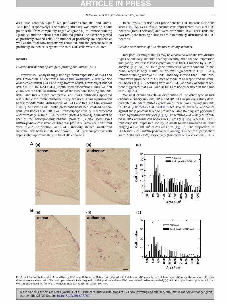
area size (areab600 μm2, 600 μm2bareab1200 μm2 and area>1200 μm2, respectively). The staining intensity was rated on a fourpoint scale, from completely negative (grade 0) to intense staining(grade 3), and the neurons that exhibited grades 2 or 3 were regardedas positively stained cells. The number of positively stained cells aswell as the total DRG neurons was counted, and the percent ratio ofpositively stained cells against the total DRG cells was calculated.
Results
Cellular distributions of Kv4 pore-forming subunits in DRGs
Previous PCR analysis suggested significant expression of Kv4.1 andKv4.3 mRNAs in DRG neurons (Phuket and Covarrubias, 2009).We alsoobserved abundant Kv4.1 and long-isoform of Kv4.3 transcripts, but notKv4.2 mRNA, in L6-S1 DRGs (unpublished observation). Thus, we firstexamined the cellular distributions of the two pore-forming subunits,Kv4.1 and Kv4.3. Since commercial anti-Kv4.1 antibodies appearedless suitable for immunohistochemistry, we used in situ hybridizationto test for differential distributions of Kv4.1 and Kv4.3 in DRG neurons(Fig. 1). Antisense Kv4.3 probe preferentially stained small-sized neu-ronal cell bodies (Fig. 1B). Kv4.3 transcript-positive cells representedapproximately 32.8% of DRG neurons (total 4 sections), equivalent tothat of the corresponding channel proteins (33.8%). Most Kv4.3mRNA-positive cells were less than 900 μm2 in cell area size. Consistentwith mRNA distribution, anti-Kv4.3 antibody stained small-sizedneuronal cell bodies (data not shown). Kv4.3 protein-positive cellsrepresented approximately 33.8% of DRG neurons.
A
B
sense antisense
sense antisense
(a) (b)
(a) (b)
Fig. 1. Cellular distributions of Kv4.1 and Kv4.3 mRNA in rat DRGs. A. Rat DRG sections staineddistributions are shown with filled and open columns indicating Kv4.1 mRNA‐positive and tocell‐size distributions (c) for Kv4.3 are shown. Scale bar, 50 μm. Bin width: 100 μm2.
Please cite this article as: Matsuyoshi H, et al, Distinct cellular distributionneurons, Life Sci (2012), doi:10.1016/j.lfs.2012.07.007
In contrast, antisense Kv4.1 probe detected DRG neurons in varioussizes (Fig. 1A). Kv4.1 mRNA-positive cells represented 59.5 % of DRGneurons (total 4 sections) and were distributed in all sizes. Thus, thetwo Kv4 pore-forming subunits are differentially distributed in DRGneurons.
Cellular distributions of Kv4 channel auxiliary subunits
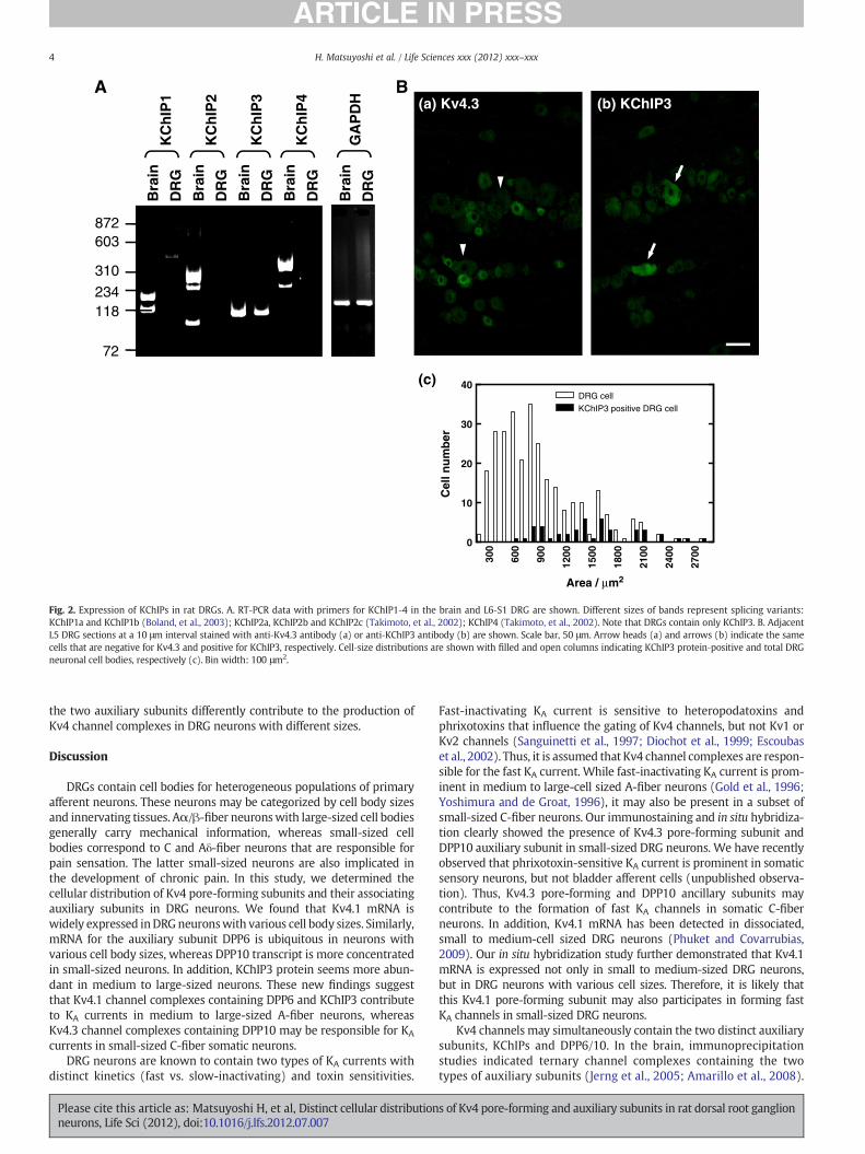
Kv4 pore-forming subunits may be associated with the two distincttypes of auxiliary subunits that significantly alter channel expressionand gating. We first tested expression of KChIP1-4 mRNAs by RT-PCRanalysis (Fig. 2A). All four gene transcripts were abundant in thebrain, whereas only KChIP3 mRNA was significant in L6-S1 DRGs.Immunostaining with anti-KChIP3 antibody showed that KChIP3 pro-teins were prominent in a subset of medium to large-sized neuronalcell bodies (Fig. 2B). Staining with anti-Kv4.3 antibody of adjunct sec-tions suggested that Kv4.3 and KChIP3 are not colocalized in the samecells (Fig. 2B).
We next examined cellular distribution of the other type of Kv4channel auxiliary subunits, DPP6 and DPP10. Our previous study dem-onstrated abundant mRNA expression of these two auxiliary subunitsin DRGs (Takimoto et al., 2006). Since several available antibodiesagainst these proteins failed to provide reliable staining, we performedin situhybridization analyses (Fig. 3). DPP6mRNAwaswidely distribut-ed in DRG neuronal cell bodies in all sizes (Fig. 3A), whereas DPP10transcript was expressed mostly in small to medium-sized neuronsranging 400–1600 μm2 of cell area size (Fig. 3B). The proportions ofDPP6 and DPP10 mRNA-positive cells among DRG neurons per sectionwere 72.8% and 27.2%, respectively (the mean of n=2 sections). Thus,
300
600
900
1200
1500
1800
2100
2400
2700
3000
0
5
10
15
20
25
30
35
DRG cellKv4.1 positive DRG cell
Cel
l nu
mb
er
Area / µm2
Area / µm2
300
600
900
1200
1500
1800
2100
2400
2700
3000
0
10
20
30
40
DRG cell
Kv4.3 positive DRG cell
Cel
l nu
mb
er
(c)
(c)
with Kv4.1‐sense RNA probe (a) or Kv4.1‐antisense RNA probe (b), are shown. Cell‐sizetal DRG neuronal cell bodies, respectively (c). B. In situ hybridization picture (a, b) and
s of Kv4 pore-forming and auxiliary subunits in rat dorsal root ganglion
(a) Kv4.3
(c)
A B
300
600
900
1200
1500
1800
2100
2400
2700
0
10
20
30
40DRG cell
KChIP3 positive DRG cell
Area / µm2
Cel
l nu
mb
er
Bra
in
DR
G
Bra
in
DR
G
Bra
in
DR
G
Bra
in
DR
G
Bra
in
DR
G
KC
hIP
1
KC
hIP
2
KC
hIP
3
KC
hIP
4
GA
PD
H
872603
310
234118
72
(b) KChIP3
Fig. 2. Expression of KChIPs in rat DRGs. A. RT‐PCR data with primers for KChIP1‐4 in the brain and L6‐S1 DRG are shown. Different sizes of bands represent splicing variants:KChIP1a and KChIP1b (Boland, et al., 2003); KChIP2a, KChIP2b and KChIP2c (Takimoto, et al., 2002); KChIP4 (Takimoto, et al., 2002). Note that DRGs contain only KChIP3. B. AdjacentL5 DRG sections at a 10 μm interval stained with anti‐Kv4.3 antibody (a) or anti‐KChIP3 antibody (b) are shown. Scale bar, 50 μm. Arrow heads (a) and arrows (b) indicate the samecells that are negative for Kv4.3 and positive for KChIP3, respectively. Cell‐size distributions are shown with filled and open columns indicating KChIP3 protein‐positive and total DRGneuronal cell bodies, respectively (c). Bin width: 100 μm2.
4 H. Matsuyoshi et al. / Life Sciences xxx (2012) xxx–xxx
the two auxiliary subunits differently contribute to the production ofKv4 channel complexes in DRG neurons with different sizes.
Discussion
DRGs contain cell bodies for heterogeneous populations of primaryafferent neurons. These neurons may be categorized by cell body sizesand innervating tissues. Aα/β-fiber neuronswith large-sized cell bodiesgenerally carry mechanical information, whereas small-sized cellbodies correspond to C and Aδ-fiber neurons that are responsible forpain sensation. The latter small-sized neurons are also implicated inthe development of chronic pain. In this study, we determined thecellular distribution of Kv4 pore-forming subunits and their associatingauxiliary subunits in DRG neurons. We found that Kv4.1 mRNA iswidely expressed inDRGneuronswith various cell body sizes. Similarly,mRNA for the auxiliary subunit DPP6 is ubiquitous in neurons withvarious cell body sizes, whereas DPP10 transcript is more concentratedin small-sized neurons. In addition, KChIP3 protein seems more abun-dant in medium to large-sized neurons. These new findings suggestthat Kv4.1 channel complexes containing DPP6 and KChIP3 contributeto KA currents in medium to large-sized A-fiber neurons, whereasKv4.3 channel complexes containing DPP10 may be responsible for KA
currents in small-sized C-fiber somatic neurons.DRG neurons are known to contain two types of KA currents with
distinct kinetics (fast vs. slow-inactivating) and toxin sensitivities.
Please cite this article as: Matsuyoshi H, et al, Distinct cellular distributionneurons, Life Sci (2012), doi:10.1016/j.lfs.2012.07.007
Fast-inactivating KA current is sensitive to heteropodatoxins andphrixotoxins that influence the gating of Kv4 channels, but not Kv1 orKv2 channels (Sanguinetti et al., 1997; Diochot et al., 1999; Escoubaset al., 2002). Thus, it is assumed that Kv4 channel complexes are respon-sible for the fast KA current. While fast-inactivating KA current is prom-inent in medium to large-cell sized A-fiber neurons (Gold et al., 1996;Yoshimura and de Groat, 1996), it may also be present in a subset ofsmall-sized C-fiber neurons. Our immunostaining and in situ hybridiza-tion clearly showed the presence of Kv4.3 pore-forming subunit andDPP10 auxiliary subunit in small-sized DRG neurons. We have recentlyobserved that phrixotoxin-sensitive KA current is prominent in somaticsensory neurons, but not bladder afferent cells (unpublished observa-tion). Thus, Kv4.3 pore-forming and DPP10 ancillary subunits maycontribute to the formation of fast KA channels in somatic C-fiberneurons. In addition, Kv4.1 mRNA has been detected in dissociated,small to medium-cell sized DRG neurons (Phuket and Covarrubias,2009). Our in situ hybridization study further demonstrated that Kv4.1mRNA is expressed not only in small to medium-sized DRG neurons,but in DRG neurons with various cell sizes. Therefore, it is likely thatthis Kv4.1 pore-forming subunit may also participates in forming fastKA channels in small-sized DRG neurons.
Kv4 channels may simultaneously contain the two distinct auxiliarysubunits, KChIPs and DPP6/10. In the brain, immunoprecipitationstudies indicated ternary channel complexes containing the twotypes of auxiliary subunits (Jerng et al., 2005; Amarillo et al., 2008).
s of Kv4 pore-forming and auxiliary subunits in rat dorsal root ganglion

300
600
900
1200
1500
1800
2100
2400
2700
3000
0
10
20
30
40
50
Cel
l nu
mb
er
DRG cellDPP10 positive DRG cell
A
B
300
600
900
1200
1500
1800
2100
2400
2700
3000
0
10
20
30
40
50
60
70
DRG cellDPP6 positive DRG cell
Cel
l nu
mb
er
Area / µm2
Area / µm2
sense antisense
sense antisense
(a) (c)(b)
(a) (c)(b)
Fig. 3. Cellular distributions of DPP6 or DPP10 mRNA in rat DRGs. A. Rat DRG sections stained with DPP6‐sense (a) or antisense RNA probe (b), are shown. Cell‐size distributions areshown with filled and open columns indicating DPP6 mRNA‐positive and total DRG neuronal cell bodies, respectively (c). B. In situ hybridization picture (a, b) and cell‐size distri-butions (c) for DPP10 are shown. Scale bar, 50 μm. Bin width: 100 μm2.
5H. Matsuyoshi et al. / Life Sciences xxx (2012) xxx–xxx
However, our RT-PCR analysis and in situ hybridization suggestthat some Kv4 channel complexes may not contain KChIPs. RT-PCRanalysis detected a high level of KChIP3 without apparent expressionof other three KChIPs in DRGs, whereas all four auxiliary subunitmRNAs were abundant in the brain. Moreover, immunohistochemis-try indicated that KChIP3 protein is present in medium to large-sizedneurons, but not in small-sized cells. A simple explanation for theseobservations is that Kv4 channel complexes in small-sized DRG neu-rons consist of Kv4.1/Kv4.3 and DPP6/10, but not any KChIPs, where-as Kv4 channel complexes in medium to large-sized DRG neuronsconsist of Kv4.1, DPP6 and KChIP3. Heterologous expression studiessuggest that KChIPs and DPP6/10 somewhat play redundant rolesin raising expression of the associated pore-forming subunits and in-ducing faster recovery from inactivation. Therefore, it is possible thata subset of small-sized C-fiber neurons contain Kv4 channel com-plexes without any KChIPs.
Sensory neuron-type selective expression of different channelsubunits may provide the basis for the development of new therapeu-tic strategy or drugs for chronic pain and other disorders. We havepreviously showed that reduced expression of Kv1.4 subunits is asso-ciated with hyperexcitability of DRG neurons in an animal model ofbladder inflammation (Hayashi et al., 2009). In contrast to visceralpain, less attention is focused on molecular correlates for Kv channelplasticity in primary afferents that transmit somatic pain, such asarthritis and chronic back pain. Further studies on alterations in theexpression of Kv4 pore-forming and auxiliary subunits and functionalproperties of Kv4-mediated KA currents could identify the molecularcorrelates that contribute to somatic pain conditions.
Please cite this article as: Matsuyoshi H, et al, Distinct cellular distributionneurons, Life Sci (2012), doi:10.1016/j.lfs.2012.07.007
Conclusion
Kv4 channel complexes in small-sized, somatic DRG neurons con-sist of Kv4.1/Kv4.3 and DPP6/10, but not any KChIPs, whereas Kv4channel complexes in medium to large-sized DRG neurons consistof.Kv4.1, DPP6 and KChIP3.
Conflict of interest statement
None.
Acknowledgements
This work was supported by grants from the National Instituteof Health (DK057267 and DK088836), the Department of Defense(SC100134 and PR110326) and theMinistry of Education, Science, Sportsand Culture of Japan (22591798).
References
Amarillo Y, De Santiago-Castillo JA, Dougherty K, Maffie J, Kwon E, Covarrubias M, et al.Ternary Kv4.2 channels recapitulate voltage-dependent inactivation kinetics ofA-type K+ channels in cerebellar granule neurons. J Physiol 2008;586:2093–106.
An WF, Bowlby MR, Betty M, Cao J, Ling H, Mendoza G, et al. Modulation of A-typepotassium channels by a family of calcium sensors. Nature 2000;403:553–6.
Boland LM, Jiang M, Lee SY, Fahrenkrug SC, Harnett MT, O'Grady SM. Functionalproperties of a brain-specific NH2-terminally spliced modulator of Kv4 channels.Am J Physiol Cell Physiol 2003;285:C161–70.
Chien L-Y, Cheng J-K, Chu D, Cheng C-F, Tsaur M-L. Reduced expression of A-typepotassium channels in primary sensory neurons inducesmechanical hypersensitivity.J Neurosci 2007;27:9855–65.
s of Kv4 pore-forming and auxiliary subunits in rat dorsal root ganglion
6 H. Matsuyoshi et al. / Life Sciences xxx (2012) xxx–xxx
Diochot S, Drici MD, Moinier D, Fink M, Lazdunski M. Effects of phrixotoxins on theKv4 family of potassium channels and implications for the role of Ito1 in cardiacelectrogenesis. Br J Pharmacol 1999;126:251–63.
Escoubas P, Diochot S, Célérier ML, Nakajima T, Lazdunski M. Novel tarantula toxins forsubtypes of voltage-dependent potassium channels in the Kv2 and Kv4 subfamilies.Mol Pharmacol 2002;62:48–57.
Gold MS, Shuster MJ, Levine JD. Characterization of six voltage-gated K+ currents inadult rat sensory neurons. J Neurophysiol 1996;75:2629–46.
Hall A, Stow J, Sorensen R, Dolly JO, Owen D. Blockade by dendrotoxin homologuesof voltage-dependent K+ currents in cultured sensory neurons from neonatalrats. Br J Pharmacol 1994;113:959–67.
Hayashi Y, Takimoto K, Chancellor MB, Erickson KA, Erickson VL, Kirimoto T, et al.Bladder hyperactivity and increased excitability of bladder afferent neurons associatedwith reduced expression of Kv1.4 α-subunit in rats with cystitis. Am J Physiol RegulIntegr Comp Physiol 2009;296:R1661–70.
Holmqvist MH, Cao J, Hernandez-Pineda R, Jacobson MD, Carroll KI, Sung MA, et al.Elimination of fast inactivation in Kv4 A-type potassium channels by an auxiliarysubunit domain. Proc Natl Acad Sci U S A 2002;99:1035–40.
Jerng HH, Qian Y, Pfaffinger PJ. Modulation of Kv4.2 channel expression and gating bydipeptidyl peptidase 10 (DPP10). Biophys J 2004;87:2380–96.
Jerng HH, Kunjilwar K, Pfaffinger PJ. Multiprotein assembly of Kv4.2, KChIP3 and DPP10produces ternary channel complexes with ISA-like properties. J Physiol 2005;568:767–88.
Kostyuk PG, Veselovsky NS, Fedulova SA, Tsyndrenko AY. Ionic currents in the somaticmembrane of rat dorsal root ganglion neurons-III. Potassium currents. Neuroscience1981;6:2439–44.
Morohashi Y, Hatano N, Ohya S, Takikawa R, Watabiki T, Takasugi N, et al. Molecularcloning and characterization of CALP/KChIP4, a novel EF-hand protein interactingwith presenilin 2 and voltage-gated potassium channel subunit Kv4. J Biol Chem2002;277:14965–75.
Please cite this article as: Matsuyoshi H, et al, Distinct cellular distributionneurons, Life Sci (2012), doi:10.1016/j.lfs.2012.07.007
Nadal MS, Ozaita A, Amarillo Y, de Miera EV-S, Ma Y-L, Mo W-J, et al. The CD26-relateddipeptidyl aminopeptidase-like protein DPPX is a critical component of neuronalA-type K+ channels. Neuron 2003;37:449–61.
Patel SP, Campbell DL, Strauss HC. Elucidating KChIP effects on Kv4.3 inactivation andrecovery kinetics with a minimal KChIP2 isoform. J Physiol 2002;545(Pt 1):5-11.
Phuket TR, Covarrubias M. Kv4 channels underlie the subthreshold-operating A-typeK-current in nociceptive dorsal root ganglion neurons. Front Mol Neurosci 2009;2:3.
Rasband MN, Park EW, Vanderah TW, Lai J, Porreca F, Trimmer JS. Distinct potassiumchannels on pain-sensing neurons. Proc Natl Acad Sci U S A 2001;98:13373–8.
Ren X, Hayashi Y, Yoshimura N, Takimoto K. Transmembrane interaction mediatescomplex formation between peptidase homologues and Kv4 channels. Mol CellNeurosci 2005;29:320–32.
Rosati B, Pan Z, Lypen S,WangHS, Cohen I, Dixon JE, et al. Regulation of KChIP2 potassiumchannel beta subunit gene expression underlies the gradient of transient outwardcurrent in canine and human ventricle. J Physiol 2001;533(Pt 1):119–25.
Sanguinetti MC, Johnson JH, Hammerland LG, Kelbaugh PR, Volkmann RA, SaccomanoNA, et al. Heteropodatoxins: peptides isolated from spider venom that blockKv4.2 potassium channels. Mol Pharmacol 1997;51:491–8.
Takimoto K, Yang EK, Conforti L. Palmitoylation of KChIP splicing variants is required forefficient cell surface expression of Kv4.3 channels. J Biol Chem 2002;277:26904–11.
Takimoto K, Hayashi Y, Ren X, Yoshimura N. Species and tissue differences in the expres-sion of DPPY splicing variants. Biochem Biophys Res Commun 2006;348:1094–100.
Tatsumi K, Haga S, Matsuyoshi H, Inoue M, Manabe T, Makinodan M, et al. Characterizationof cells with proliferative activity after a brain injury. Neurochem Int 2005;46:381–9.
Yoshimura N, de Groat WC. Characterization of voltage-sensitive Na+ and K+currents recorded from acutely dissociated pelvic ganglion neurons of the adultrat. J Neurophysiol 1996;76:2508–21.
Yoshimura N, White G, Weight FF, de Groat WC. Different types of Na+ and A-type K+
currents in dorsal root ganglion neurones innervating the rat urinary bladder.J Physiol 1996;494:1-16.
s of Kv4 pore-forming and auxiliary subunits in rat dorsal root ganglion