acute symptomatic seizures caused by electrolyte disturbances€¦ · successful management of...
TRANSCRIPT
Copyright © 2016 Korean Neurological Association 21
In this narrative review we focus on acute symptomatic seizures occurring in subjects with electrolyte disturbances. Quite surprisingly, despite its clinical relevance, this issue has received very little attention in the scientific literature. Electrolyte abnormalities are commonly encoun-tered in clinical daily practice, and their diagnosis relies on routine laboratory findings. Acute and severe electrolyte imbalances can manifest with seizures, which may be the sole present-ing symptom. Seizures are more frequently observed in patients with sodium disorders (es-pecially hyponatremia), hypocalcemia, and hypomagnesemia. They do not entail a diagnosis of epilepsy, but are classified as acute symptomatic seizures. EEG has little specificity in dif-ferentiating between various electrolyte disturbances. The prominent EEG feature is slowing of the normal background activity, although other EEG findings, including various epilepti-form abnormalities may occur. An accurate and prompt diagnosis should be established for a successful management of seizures, as rapid identification and correction of the underlying electrolyte disturbance (rather than an antiepileptic treatment) are of crucial importance in the control of seizures and prevention of permanent brain damage.Key Wordszz EEG, electrolyte, epilepsy, seizures, hyponatremia, hypernatremia, hypocalcemia.
Acute Symptomatic Seizures Caused by Electrolyte Disturbances
INTRODUCTION
Electrolyte abnormalities are commonly encountered in clinical daily practice, and their diagnosis relies on routine laboratory findings. Electrolyte disturbances may affect the brain among many other organs and tissues and need to be promptly recognized as they may lead to severe and life-threatening complications when overlooked or not appropri-ately treated. The neurological manifestations reflect the severity of acute neuronal de-rangement and therefore require emergency treatment.1-3 Acute and/or severe electrolyte imbalances can manifest with rapidly progressive neurologic symptoms or seizures, which may be the sole presenting symptom. Seizures are more frequently observed in patients with sodium disorders (especially hyponatremia), hypocalcemia, and hypomagnesaemia.3 Table 1 shows the different degrees of the electrolyte disturbances discussed in this review. An accurate and prompt diagnosis should be established for successful management of sei-zures, as rapid identification and correction of the underlying electrolyte disturbance are of crucial importance in the control of seizures and prevention of permanent brain damage.2-4
In this narrative review we focus on acute epileptic seizures occurring in subjects with electrolyte disturbances. Quite surprisingly, despite its clinical relevance, this issue has re-ceived very little attention in the scientific literature, with only a very few reviews specifical-ly dealing with electrolytes disturbances and seizures published so far.3,4
To conduct this review we selected the most relevant data from the available literature on this topic identified by searching PubMed using the search terms “seizures” or “epilepsy”
Raffaele Nardonea,b Francesco Brigoc Eugen Trinkaa,d,e
a Department of Neurology, Paracelsus Medical University Salzburg, and Christian Doppler Medical Centre, Salzburg, Austria
b Department of Neurology, Franz Tappeiner Hospital, Merano, Italy
c Department of Neurological and Movement Sciences, University of Verona, Verona, Italy
d Centre for Cognitive Neurosciences Salzburg, Salzburg, Austria
e University for Medical Informatics and Health Technology, UMIT, Hall in Tirol, Austria
pISSN 1738-6586 / eISSN 2005-5013 / J Clin Neurol 2016;12(1):21-33 / http://dx.doi.org/10.3988/jcn.2016.12.1.21
Received August 25, 2015Revised September 1, 2015Accepted September 3, 2015
CorrespondenceEugen Trinka, MD, MScDepartment of Neurology, Christian Doppler Klinik, Paracelsus Medical University Salzburg, Centre for Cognitive Neuroscience Salzburg, Ignaz Harrerstrasse 79, Salzburg A-5020, AustriaTel +4366244833000Fax +4366244833004E-mail [email protected]
cc This is an Open Access article distributed under the terms of the Creative Commons Attribution Non-Com-mercial License (http://creativecommons.org/licenses/by-nc/3.0) which permits unrestricted non-commercial use, distribution, and reproduction in any medium, provided the original work is properly cited.
JCN Open Access REVIEW

22 J Clin Neurol 2016;12(1):21-33
Electrolyte Disturbance and SeizuresJCN
combined with “electrolyte”, “hyponatremia”, “hypernatre-mia”, “hypocalcemia”, “hypercalcemia”, “hypomagnesemia”, “hypokalemia”, and “hyperkalemia”. Publications were cho-sen based on the quality of data and their relevance to the present review.
After an initial overview of this topic, which serves as gen-eral introduction, we discuss the risk of seizures according to each type of electrolyte disturbance.
A GENERAL OVERVIEW
The orderly function of the nervous system depends on its electrical excitability, which is maintained through a voltage gradient across neuronal and glial membranes by means of metabolically driven ion pumps. Alterations of electrolyte gradients across cellular membranes exert both direct and indirect effects on neuronal excitability and synchronization, and the consequent abnormal neuronal discharge may facili-tate epileptiform activities.5 Specifically, there are several clini-cal conditions, such as dehydration or renal failure, which can be associated with substantial modifications of plasma osmolality and electrolyte balance, determining marked al-terations in brain metabolism and function leading to in-creased risk of seizures.
In a recent proposal by the International League Against Epilepsy (ILAE) acute seizures were defined as “a clinical sei-zure occurring at the time of a systemic insult or in close tem-poral association with a documented brain insult.”6 Sugges-tions were made to specify the brain insult as “events occurring within 1 week of stroke, traumatic brain injury, anoxic en-cephalopathy, or intracranial surgery; at first identification of subdural hematoma; at the presence of an active central nervous system (CNS) infection; or during an active phase of multiple sclerosis or other autoimmune diseases.” In addi-tion the ILAE suggest to make a diagnosis diagnosis of an acute symptomatic seizure “in the presence of severe meta-bolic derangements (documented within 24 h by specific biochemical or hematologic abnormalities), drug or alcohol intoxication and withdrawal, or exposure to well-defined epi-leptogenic drugs.”6
Since electrolyte disturbances are, at least in the early stag-es, generally not associated with morphologic changes in CNS,
the neurologic manifestations are typically reversible.2-4,7 However, seizures and the electrolyte disturbance itself can
lead to structural alterations, so that the underlying electro-lyte disturbances should be recognized and treated before the brain tissue injury becomes permanent.
Disorders of sodium and osmolality can be responsible for an encephalopathy characterized by depression of neuronal activity, with confusion, headache, psychomotor slowing and lethargy as the major clinical manifestations, usually associ-ated with signs of irritability. Hypercalcemia and hypermag-nesemia may also produce both a neuronal depression with encephalopathy and neuronal irritability. Hypocalcemia and hypomagnesemia lead almost exclusively to CNS irritability clinically manifesting with seizures, whereas disorders of po-tassium rarely produce symptoms in the CNS, with muscle weakness being their major clinical manifestation.2,3,7
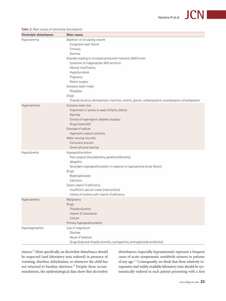
The main causes of electrolyte disturbances leading to acute seizures are reported in Table 2.
Seizures occurring in patients with sodium disorders, hy-pocalcemia, and hypomagnesemia,2,3,7 are usually general-ized tonic-clonic, but also focal (partial) seizures or other seizure types may be present. Rapidly evolving electrolyte disturbances are more likely to cause seizures than those de-veloping more gradually. It is therefore difficult to define ab-solute levels of electrolyte above or below which seizures are likely to occur.3,7
To identify the electrolyte disturbances leading to seizures, a complete serum chemistry evaluation, including measure-ments of sodium, calcium, and magnesium should be per-formed, in particular in subjects with a first-time seizures.3,8,9 Since between 15 and 30% of acute symptomatic seizures among elderly patients occur in the setting of toxic-meta-bolic causes,10 this diagnostic workup is particularly impor-tant in the elderly.
In a review of 375 adult cases of status epilepticus, 10% had a metabolic disorder as the primary etiology and mor-tality in this subset of patients was as high as 40%.11
Acute seizures due to electrolyte disorders may occur at any age, including infancy. When evaluating a first nonfe-brile seizure occurring in children, the American Academy of Neurology has recommended that laboratory screening tests should be ordered based on individual clinical circum-
Table 1. Different degrees of the electrolyte disturbances that most frequently cause seizures
Electrolyte disturbance Mild Moderate SevereHyponatremia 130–134 mEq/L 125–129 mEq/L <125 mEq/L
Hypernatremia 145–149 mEq/L 150–169 mEq/L ≥170 mEq/L
Hypocalcemia 1.9–2.2 mEq/L <1.9 mEq/L
Hypercalcemia 2.5–3 mEq/L 3–3.5 mEq/L 3.5–4 mEq/L
Hypomgnesemia 0.8–1.6 mEq/L <0.8 mEq/L
www.thejcn.com 23
Nardone R et al. JCN
stances.12 More specifically, an electrolyte disturbance should be suspected (and laboratory tests ordered) in presence of vomiting, diarrhea, dehydration, or whenever the child has not returned to baseline alertness.12 Despite these recom-mendations, the epidemiological data show that electrolyte
disturbances (especially hyponatremia) represent a frequent cause of acute symptomatic nonfebrile seizures in patients of any age.1-5 Consequently, we think that these relatively in-expensive and widely available laboratory tests should be sys-tematically ordered in each patient presenting with a first
Table 2. Main causes of electrolyte disturbances
Electrolyte disturbances Main causesHyponatremia Depletion of circulating volume
Congestive heart failureCirrhosisDiarrhea
Disorders leading to increased antidiuretic hormone (ADH) levelsSyndrome of inappropriate ADH secretionAdrenal insufficiencyHypothyroidismPregnancyRecent surgery
Excessive water intakePolydipsia
Drugs Thiazide diuretics, desmopressin, mannitol, sorbitol, glycine, carbamazepine, oxcarbazepine, eslicarbazepine
Hypernatremia Excessive water lossImpairment in access to water (infants, elderly)DiarrheaCentral of nephrogenic diabetes insipidusDrugs (mannitol)
Overload of sodiumHypertonic sodium solutions
Water moving into cellsConvulsive seizuresSevere physical exercise
Hypocalcemia HypoparathyroidismPost-surgical (thyroidectomy, parathyroidectomy)IdiopathicSecondary hyperparathyroidism in response to hypocalcemia (renal failure)
DrugsBisphosphonatesCalcitonin
Severe vitamin D deficiencyInsufficient calcium intake (malnutrition)Infants of mothers with vitamin D deficiency
Hypercalcemia MalignancyDrugs
Thiazide diureticsVitamin D intoxicationLithium
Primary hyperparathyroidism
Hypomagnesemia Loss of magnesiumDiarrheaAbuse of laxativesDrugs (loop and thiazide diuretics, cyclosporines, aminoglycoside antibiotics)

24 J Clin Neurol 2016;12(1):21-33
Electrolyte Disturbance and SeizuresJCNacute seizure.
Electrolyte disturbances may cause diffuse brain dysfunc-tion that can be assessed by means of EEG recording. In gen-eral, the most prominent feature of the EEG record in meta-bolic encephalopathies is a slowing of the normal background frequency. If serial EEGs are performed, a gradually progres-sive disorganization of the EEG recordings over the course of the disease can usually be noted. Moreover, reactivity to photic or other types of external stimulation is frequently al-tered.13 EEG evolution generally correlates well with the se-verity of encephalopathy; more specifically, the degree and severity of EEG abnormalities correlate with the rate of change of electrolyte balance rather than with the absolute level of a specific electrolyte or metabolite.14
However, EEG recordings have little specificity in differen-tiating between the various etiologies of encephalopathy. In fact, in metabolic encephalopathies, EEG patterns are usu-ally unspecific, including various degrees of diffuse slowing, epileptiform discharges, intermittent rhythmic slow activi-ty, and occurrence of triphasic waves, which are usually re-versible after treatment of the underlying causes.13,15
HYPONATRIEMIA
Hyponatremia is defined as a serum sodium level of less than 135 mEq/L and is considered severe when the serum level is below 125 mEq/L.
Clinical featuresThe clinical manifestations of hypotonic hyponatremia are largely related to CNS dysfunction and are more conspicu-ous when the decrease in serum sodium concentrations is severe or occurs rapidly (within hours). The major clinical complications from acute hyponatremia are brain cell swell-ing and herniation with neurologic symptoms being evident when hyponatremia approaches 120 mEq/L. The risk of ce-rebral edema and neurologic manifestations is minimized if the decline in serum sodium occurs slowly and gradually (≥48 h), even in case of a marked absolute reduction of serum so-dium values. Conversely, in case of a rapid decrease in serum sodium (acute hyponatremia), cerebral edema with neuro-logic symptoms are likely to occur.16,17
The neurological symptoms of hyponatremia therefore go in parallel with the severity of cerebral edema, and are less frequently induced by chronic than by acute hyponatremia: approximately half of the patients with chronic hyponatre-mia are asymptomatic, even with serum sodium concentra-tion less than 125 mEq/L.16,17 Symptoms in these patients rarely occur until the serum sodium is less than 120 mEq/L and are more usually associated with values around 110 mEq/L
or lower.3 Particularly the children are at high risk of devel-oping symptomatic hyponatremia, because of their larger brain-to-skull size ratio. Severe and rapidly evolving hypo-natremia may cause seizures, which are usually generalized tonic-clonic, and generally occur if the plasma sodium con-centration rapidly decreases to <115 mEq/L.
Age and gender of the patient as well as other several fac-tors influence the clinical outcome of neurological compli-cations of hyponatremia. Children and women in childbear-ing age (rather than postmenopausal women) are the most susceptible subjects:18,19 in a retrospective study hyponatre-mia was the only detectable cause of seizures in 70% of in-fants younger than 6 months.19
Women seem to be particularly prone to develop postop-erative hyponatremia.20 A case control study conducted in 65 adults with postoperative hyponatremic encephalopathy and 674 adult patients who had postoperative hyponatremia without encephalopathy showed a similar risk of developing hyponatremia and hyponatremic encephalopathy after sur-gery in women and men. Surprisingly, women in childbear-ing age had a 25-fold increased risk of death or permanent neurologic damage compared with either men or postmeno-pausal females.20 Consequently, it is advisable to maintain a low threshold for hyponatremia and hyponatremic encepha-lopathy in the event of headache, vomiting, nausea or lethar-gy occurring after surgery, especially in women in childbear-ing age.20
Hyponatremia represents a frequent cause of epileptic sei-zures, as shown in a recent prospective observational multi-center study where acute epileptic seizures and focal neuro-logical deficits were identified in 5% of patients with severe (<125 mEq/L) hyponatremia.21
Several etiologies may lead to hyponatremia, some of them affecting almost exclusively adults and some mostly children and infants.
In adults, generalized seizures have been reported as the first manifestation of multihormonal pituitary hormone de-ficiency causing normovolemic hyponatremia.22
Administration of some drugs, such as desmopressin23,24 or thiazide diuretics,25 may also lead to hyponatremia and sei-zures. To date, 54 cases of hyponatremia secondary to des-mopressin treatment for enuresis presenting with altered mental status or seizures have been reported. In 47 of them an intranasal formulation had been used, while excess fluid intake was documented as a contributing factor in at least 22 cases. In most cases the neurological complications devel-oped 14 days or less after starting desmopressin.24
Thiazide diuretics may cause hyponatremia in up to 14% of patients receiving these drugs (more commonly females, elderly subjects and subjects of low body weight), and may
www.thejcn.com 25
Nardone R et al. JCNcause confusion, falls and seizures.25
Although also tricyclic antidepressants cause frequently hyponatremia, seizures associated with hyponatremia are more frequently observed in subjects taking selective sero-tonin reuptake inhibitors.26-28
Several clinical conditions including fever (with true vol-ume depletion) or polydipsia may also lead to hyponatremia. A study assessed the impact of fever on sodium values in children presenting with seizures during a gastroenteritis ep-isode.29 While the presence or absence of fever did not affect seizure characteristics or duration, mild hyponatremia (so-dium levels between 126 mEq/L and 134 mEq/L) was found to affect some seizure features, particularly seizure duration, with increased risk of status epilepticus. In fact, children with hyponatremia had more prolonged seizures than pa-tients with normal serum sodium levels (6.4 minutes vs. 1.9 minutes, respectively), irrespective of body temperature. In most cases, the seizures last less than 3 minutes (range: sev-eral seconds to 20 minutes).
Polydipsia, commonly seen in patients with psychiatric dis-turbances, is another cause of hyponatremia with increased risk of seizures. A retrospective cross-sectional study was car-ried out to study the association between different levels of hyponatremia and the occurrence of epileptic seizures in pa-tients without a prior epilepsy diagnosis.30 The authors iden-tified from the database of a Swedish County hospital 363 in patients who were had serum sodium levels <125 mEq/L. Medical records were reviewed and 11 patients with seizures secondary to hyponatremia were identified. Seizures were the only neurologic manifestation of hyponatremia in the subjects with serum sodium levels >115 mEq/L. Marked in-creases in the frequency of their complex partial seizures due to hyponatremia was observed in five patients with epilepsy and polydipsia-hyponatremia with a decrease in the serum sodium level to 118–127 mEq/L.31 In all cases, patients had received antipsychotic drugs, and the serum sodium levels returned to normal through restriction of fluids with conse-quent decrease in seizure frequency. As this study shows, hy-ponatremia caused by polydipsia is a risk factor for aggrava-tion of habitual seizures in patients with epilepsy and psychiatric disorders.
Other conditions reported to be associated with hypona-tremia and seizures are the ingestion of sodium phosphate or sodium picosulfates/magnesium citrate combination, which are commonly used to evacuate the colon and rectum before colonoscopy or colorectal surgery,32 or polyethylene glycol preparation.33
Children and especially infants are particularly at risk of developing hyponatremia. A retrospective review reported 130 infants with hyponatremia (<136 mEq/L) associated with
respiratory syncytial virus bronchiolitis in infants requiring intensive care. Four infants (4%) had seizures at admission (sodium 114–123 mEq/L), and were successfully managed with hypertonic saline infusions followed by fluid restric-tion, resulting in immediate termination of seizure activity and normalization of serum sodium values over 48 hours.34
Two infants had water intoxication associated with hypo-natremic seizures (sodium levels 116 mEq/L and 121 mEq/L) after consumption of commercial bottled drinking water for infants.35
Hyponatremia should therefore be suspected in any in-fant less than 6 months old presenting with acute seizures and a body temperature of ≤36.5 degrees C.36 In these pa-tients, hyponatremia needs to be promptly recognized and treated to avoid complications, although improvement in neurologic function after correction of hyponatremia is usu-ally more rapid in children than elderly patients.37
Although the American Academy of Neurology recom-mends that laboratory screening tests for electrolytes should be ordered based on individual clinical circumstances such as vomiting, diarrhea or dehydratation,12 the epidemiological data show that hyponatremia is a frequent cause of acute symptomatic nonfebrile seizures in children. Consequently, laboratory tests should be systematically ordered in each child presenting with acute seizures.
Finally, it is noteworthy to mention that the antiepileptic drugs (AED) carbamazepine (CBZ), oxcarbazepine (OXC), and eslicarbazepine (ESL) may themselves represent a cause of hyponatremia due to syndrome of inappropriate antidi-uretic hormone.37 Possible mechanisms for this effect are an altered sensitivity to serum osmolality by the hypothalamic osmoreceptors and an increased sensitivity of the renal tu-bules to antidiuretic hormone.37
AED-induced hyponatremia is usually asymptomatic, al-though in some cases it may result in headache, confusion, general malaise, somnolence and in exacerbation of sei-zures.38-41
CBZ, OXC, and ESL may lead to hyponatremia in a rele-vant number of patients. One comparative study showed a much higher incidence of hyponatremia (defined as sodium levels ≤134 mEq/L) in patients treated with OXC compared to those receiving CBZ (29.9% vs. 13.5%; p<0.0001). Of note, sodium levels ≤128 mEq/L were found in 12.4% of patients treated with OXC and in 2.8% of those receiving CBZ (p< 0.001).42 Hyponatremia during CBZ therapy seems to be particularly common in patients with intellectual disabili-ty,43 which therefore should be considered a subset of sub-jects particularly at risk. A subsequent study found an inci-dence of severe (sodium levels ≤128 mEq/L) and symptomatic hyponatremia of 11.1% and 6.8%, respectively, in patients
26 J Clin Neurol 2016;12(1):21-33
Electrolyte Disturbance and SeizuresJCNtreated with OXC.44 Age, AED polytherapy, and the concomi-tant use of diuretics were found to be independent risk fac-tors for sever hyponatremia following OXC treatment.44 Re-cently, also ESL (the most recent AED structurally similar to CBZ to be marketed) has been shown to be associated with hyponatremia (sodium values ≤134 mEq/L) in 12.5% of pa-tients affected by post-stroke seizures; in 10% of these pa-tients, hyponatremia was symptomatic and in 3% it was as-ymptomatic.45
The frequency of AED-induced hyponatremia is therefore particularly common after OXC administration, especially in the elderly or in patients taking diuretics. Incidence of hy-ponatremia following OXC appears to be dose dependent (in one study an increase of 1 mg in the dosage of OXC was shown to increase the risk of hyponatremia by 0.2% and may be prevented by a slower and lower titration-initiation sched-ule.46 Routine plasma sodium monitoring for patients re-ceiving OXC is not usually necessary, except for patients re-ceiving AED polytherapy or sodium depleting drugs (e.g., thiazide diuretics), affected by sodium-depleting disorders, or in the elderly.44,47 Conversely, monitoring of sodium serum levels are mandatory in patients under OXV therapy devel-oping symptoms suggestive of hyponatremia (headache, confusion, general malaise, somnolence) and in those with unexplained worsening of seizures.46,47 Although no specific guidelines are available, these recommendations can be rea-sonably extended to patients receiving CBZ, ESL and other sodium depleting drugs.
EEG abnormalitiesHyponatremia usually produces nonspecific EEG slowing. A very severe hyponatremia may initially cause posterior slowing followed by diffuse delta activity. However, a vari-ety of other EEG abnormalities have been described, such as triphasic waves, burst of high-voltage rhythmic delta ac-tivity, and central high voltage 6-Hz to 7-Hz activity with stimulation-induced paroxysms of delta waves. Periodic lat-eralized epileptiform discharges may also occur, while full seizure activity is very rare.13,48
A case of polydipsia-induced hyponatremia causing de novo nonconvulsive status epilepticus was associated with focal EEG discharges.49 After recovery from status epilepticus, EEG showed some spikes in the left frontal area. In this case it is reasonable to hypothesize that hyponatremia induced by polydipsia precipitated epileptogenicity in the left frontal area, and then focal activity secondarily generalized and led to status epilepticus.
TreatmentHyponatremic seizures represent an ominous sign and hence
a medical emergency, as they are associated with high mor-tality.2 Thus, a prompt recognition and treatment of acute symptomatic hyponatremia is of utmost importance as sec-ondary brain damage may be rapid and irreversible, even in subjects with mild clinical symptoms.13 Since a small increase in the serum sodium concentration can substantially reduce cerebral edema, seizures induced by hyponatremia can be controlled by increasing the serum sodium concentration.14 However, improvement in neurologic function may occur several days after correction of the electrolyte abnormality, particularly in elderly patients.50
The most common treatment for hyponatremia consists of hypertonic saline (3%), which produces a rapid reduction in brain volume and intracranial pressure. An increase in serum sodium to values of 120 mEq/L to 125 mEq/L should be the target of therapy. Of note, more aggressive treatment of hyponatremia with hypertonic saline solution carries the risk of shrinkage of the brain leading to osmotic demyelin-ation syndrome manifesting with severe neurologic symp-toms such as quadriplegia, pseudobulbar palsy, coma, and even death.2,3,13
The sodium concentration should therefore be corrected at a rate of 0.5 mEq/L/h. Higher correction rates (a rate of 1 mEq/L to 2 mEq/L/h) have been used young patients at a risk for respiratory arrest, severe neurologic sequelae, and death14,51 and appear to be well tolerated in children.52
Hyponatremia induced by AED or other sodium-deplet-ing drugs may be managed through water restriction, reduc-tion of the dose and, if necessary, discontinuation.47
HYPERNATREMIA
Hypernatremia is defined as a serum sodium concentration in plasma >145 mEq/L.
Clinical featuresWhile hyponatremia is often the cause of seizures or status epilepticus, hypernatremia is more likely to be the conse-quence of convulsive seizure activity (especially generalized tonic-clonic seizures). In fact, during seizures intracellular glycogen is metabolized in the muscle to lactate. Since in-tracellular osmolality of muscle fibers increases (lactate is more osmotically active than glycogen), water moves into cells, causing hypernatremia. A few minutes after the onset of hypernatremia, loss of water from brain cells determines an increased intracellular brain cell osmolality and shrink-age of the brain.
The degree of CNS disturbance in hypernatremia is there-fore related mainly to the rate at which the serum sodium in-creases.53 In acute hypernatremia, the combination of hy-

www.thejcn.com 27
Nardone R et al. JCNperosmolality and shrinkage in brain volume (especially in infants) results in an encephalopathy due altered synaptic structure and function of CNS cells.3,7 Conversely, in chron-ic hypernatremic states, the risk of brain shrinkage with sub-sequent neurological symptoms is minimized. A slow, grad-ual increase in sodium values to levels as high as 170 mEq/L is often well tolerated. Severe symptoms of hypernatremia are primarily neurologic and usually result from an acute (i.e., within hours) elevation in the plasma sodium concen-tration to >158–160 mEq/L. Values >180 mEq/L are associ-ated with a high mortality rate, in adults more frequently than in children.53 In infants with hypernatremia seizures are typically absent, but can occur in case of inadvertent sodi-um loading or aggressive rehydration.3,53
In patients with hypernatremia the rupture of cerebral veins, as well as the intracerebral and subarachnoid hemorrhages, which are induced by brain shrinkage, can in turn provoke seizures.3 Although rapid sodium loading can cause seizures, convulsions are more frequently seen during rapid correction of hypernatremia, and in ≤40% of patients treated for severe hypernatremia by rapid infusion of hypotonic solutions.3,6
TreatmentThe treatment of hypernatremia consists of replenishing body water, thus restoring osmotic homeostasis and cell volume at a rate that avoids significant complications. The speed of correction depends on the speed of development of hyper-natremia and accompanying symptoms.53,54 In subjects with prolonged hypernatremia, cerebral edema may occur when the osmolality is abruptly normalized; this correction may therefore lead to convulsive seizures, coma, and death. The targeted correction of chronic hypernatremia should not ex-ceed 0.5–0.7 mEq/L/h and 10 mEq/L/day to avoid compli-cations.
In subjects with acute hypernatremia the treatment may be more rapid, and a decrease in the serum sodium concen-tration by 1 mEq/L/h can be considered appropriate.54
Patients with hypernatremia may be treated with hypoton-ic fluids, such as hypotonic saline or dextrose solutions giv-en orally or, if this approach is not feasible, intravenously.
Importantly, the volume should be restricted to that re-quired to correct hyperosmolality to prevent the risk of cere-bral edema, which increases with the volume of the infusate.53
HYPOCALCEMIA
Hypocalcemia is defined as a plasma calcium level of <8.5 mg/dL or an ionized calcium concentration <4.0 mg/dL.
Clinical featuresThe symptoms of hypocalcemia depend on the degree of hy-pocalcemia and the speed of the decrease in the serum cal-cium concentration.55 Acute hypocalcemia primarily causes increased neuromuscular excitability and tetany.
The typical CNS manifestations of acute hypocalcemia are mental status changes and seizures.2,3,7 Generalized tonic-clonic, focal motor, and (less frequently) atypical absence or even akinetic seizures may occur in patients with hypocal-cemia even without muscular tetany.2,3,7 Hence, seizures can represent the unique presenting symptom of hypercalce-mia.2,3,55 Nonconvulsive status epilepticus secondary to hy-pocalcemia has also been reported.56 Seizures are a frequent complication of acute seizures: they have been reported in 20–25% of patients with acute hypocalcemia, and in 30–70% of patients with idiopathic hypoparathyroidism.57,58
The main etiologies of hypocalcemia are hypoparathyroid-ism, severe vitamin D deficiency (VDD) and drugs (Table 2).
Hypoparathyroidism, usually as a complication of thyroid-ectomy, is a common cause of hypocalcemia. Characteristics of seizures at presentation, occurrence of subclinical seizures during follow-up, and the effect of AED withdrawal has re-cently been assessed in 70 patients with IH.59 Seizures were present in 64% of patients (87% of them being generalized tonic-clonic), and were treated with phenytoin (47%), valpro-ate (40%), and CBZ (27%). Most (69/70) patients were seizure-free during the follow-up of 6.6±4.5 years. Ten of 14 (71.4%) patients were successfully withdrawn from AED and re-mained seizure free during the follow-up period of 13.5±2.4 months (range 9–18). In few patients AEDs were restarted because of the recurrence of seizures (n=3) and poor com-pliance with calcium/vitamin D intake (n=1). The mean se-rum total calcium increased from 1.9±0.19 to 2.1±0.14 mEq/L after AED withdrawal.
Two cases with generalized tonic-clonic seizures as the first manifestation of postoperative hypoparathyroidism, ap-pearing months and years after thyroidectomy have been de-scribed.60 Thus, iatrogenic (i.e., postoperative) hypoparathy-roidism needs to be considered in the differential diagnosis of adult-onset, generalized, tonic-clonic seizures even if the thyroidectomy was performed years earlier.
Hypocalcemic seizures may also occur in subjects with severe VDD, frequently in children and during the neonatal period.61-64 Overall, seizures during the neonatal period have a broad differential diagnosis.61 Unlike in developing coun-tries, where VDD and hypocalcemia constitutes a major cause of infantile seizures, the number of neonatal seizures attributed to hypocalcemia in developed countries has de-creased dramatically due to the obligatory vitamin D supple-mentation. In developed countries, most infants presenting

28 J Clin Neurol 2016;12(1):21-33
Electrolyte Disturbance and SeizuresJCNwith hypocalcemic seizures have underlying endocrinologi-cal etiologies rather than dietary insufficiencies.61,62
The incidence of hypocalcemic seizures secondary to VDD in children in the UK and Ireland have been investi-gated in one study.62 Ninety one confirmed or probable cas-es were reported, equating to an overall annual incidence of 3.49 per million children aged 0–15 years. Incidence was sig-nificantly greater in males compared to females, in infants compared to older children, and in children of South Asian or Black ethnicity compared to children from white ethnic backgrounds. The authors concluded that current imple-mentation of public health policy in the UK is not success-ful in preventing children from developing one of the severe manifestations of VDD.
Severely-malnourished children presenting with hypocal-caemia have an increased risk of death than those without hypocalcaemia.63 In this subset of patients, the prevalence of hypocalcaemia has been reported to be as high as 26%.62 Acute diarrhea, convulsive seizures, and lethargy on admis-sion to hospital are the main clinical predictors of hypocal-cemia in these children. The presence of these features in hospitalized children with severe acute malnutrition should alert clinicians about the possibility of hypocalcemia and may help undertake potential preventive measures, in par-ticular calcium supplementation. Also infants with rickets may be particularly prone to acute convulsive seizures due to hypocalcemia.64
A possible link between acute neonatal seizures, hypocal-cemia and subsequent severe intellectual disability has been assessed in 149 adults with 22q11.2 deletion syndrome.65 A history of neonatal seizures and neonatal hypocalcemia were significant predictors of a more severe level of intellectual disability, suggesting that neonatal seizures may increase the risk for more severe intellectual deficits in 22q11.2 deletion syndrome, likely mediated by neonatal hypocalcemia.
A study conducted in UK among children presenting to the emergency department identified 89 patients with a low vitamin D level (total vitamin D levels less than 50 nmol/L), with 83% of those having very low vitamin D levels (less than 25 nmol/L).66 Seizures were present in 17% of patients, whose most common ethnic origins were Pakistani (37%) and black African (11%). Thus, acute seizures following hypocalcemia due to severe VDD should be particularly suspected in chil-dren from developing countries or with specific ethnic origins.
Furthermore, it is important to remind that maternal VDD is commonly observed in nursing mothers of infants diag-nosed with rickets and that mothers of infants presenting with hypocalcemic seizures invariably have severe VDD.67,68 Hypocalcemic seizures in infants secondary to maternal VDD might be prevented by supplementation of vitamin D.67
Although rarely, hypocalcemic seizures may also follow the administration of zoledronic acid, an aminobisphosphonate that is administered annually against osteoporosis.69
Chvostek’s and Trousseau’s signs are two physical findings resulting from a neuromuscular hyperexcitability due to la-tent tetany associated with reduced serum ionized calcium may provide clues to a diagnosis of hypocalcemia, although they have no value in differentiating among the various eti-ologies leading to this electrolyte imbalance. Chvostek’s sign is a contraction of the ipsilateral facial muscles, ranging from minimal twitching of the lip to spasm of all muscles, elicited by tapping the facial nerve just anterior to the ear. Converse-ly, Trousseau’s sign refers to the occurrence of a carpopedal spasm (i.e., adduction of the thumb, flexion of the wrist and metacarpophalangeal joints, and extension of the interpha-langeal joints) following inflation of a sphygmomanometer above systolic blood pressure for three minutes.
A study assessed the clinical utility of Chvostek’s sign in 154 patients with seizures (epilepsy, n=91; non-epileptic event, n=41; febrile convulsion, n=19; hypocalcemic seizure, n=3).70 While patients with febrile convulsions or non-epileptic sei-zures had either negative or mild Chvostek’s sign, a marked Chvostek’s sign was only found among those with the diag-nosis of epilepsy or hypocalcemia. Normocalcemic patients had no other signs of neuromuscular hyperexcitability while those with hypocalcemia manifested positive Trousseau’s sign and other signs of neuromuscular hyperexcitability.
EEG abnormalitiesEarly EEG abnormalities associated with hypocalcemia in-clude slowing of background rhythm with evolution from alpha through theta and diffuse increase in slow wave activ-ity in the theta and delta range. Generalized spikes, sharp-waves burst of delta activity with sharp components, have also been reported and associated with absence seizures.13 Neo-natal EEG recordings may show reversible 3- to 4-Hz spike-waves discharges.71
TreatmentThe indication for urgent therapy for hypercalcemia usually reflects the severity of neurological symptoms and the de-gree of hypocalcemia. Acute hypocalcemia is an emergency that requires prompt attention, and patients with symptom-atic hypocalcemia should be treated immediately because of the highly associated morbidity and mortality. Treatment with intravenous calcium is the most appropriate therapy. Doses of 100 mg to 300 mg of elemental calcium should be infused intravenously over a period of 10 min to 20 min. Cal-cium-infusion drips should be started at 0.5 mg/kg/h and continued for several hours, with close monitoring of calci-

www.thejcn.com 29
Nardone R et al. JCNum levels.55 Hypocalcemic seizures should be treated with calcium replacement, while AEDs are typically not needed. Interestingly, AEDs may abolish both overt and latent tetany, whereas hypocalcemic seizures may remain refractory.57,71,72 Obviously, the treatment of hypocalcemia should be directed at the underlying disorder, and oral calcium repletion is com-monly prescribed for outpatient treatment.
HYPERCALCEMIA
Hypercalcemia is defined as serum calcium levels of ≥2.5 mmol/L.
Clinical featuresHypercalcemia (especially that related to malignancy) is much more common than hypocalcemia,2,3,55 but is less fre-quently associated with acute seizures. The most common symptoms of severe hypercalcemia (defined as calcium lev-els higher than 3.5 mmol/L) are related to disturbances of nervous system and gastrointestinal function.2,3,60
A rapid increase to moderate (12–13.9 mg/dL) hypercal-cemia frequently results in marked neurologic dysfunction. On the contrary, chronic severe hypercalcemia (≥14 mg/dL) usually causes only minimal neurologic symptoms.73
The main neurologic manifestations of hypercalcemia are drowsiness, lethargy, weakness, and confusion. Hypercalce-mia is associated with reduced neuronal membrane excit-ability, and thus only rarely causes seizures. However, hy-percalcemia-induced hypertensive encephalopathy and vasoconstriction have been hypothesized to be responsible for seizures.3,14,74
EEG abnormalitiesIn subjects with hypercalcemia EEG abnormalities (fast ac-tivity and burst of delta and theta slowing) appear when cal-cium levels are higher than 13 mg/dL. At higher calcium levels, increased background slowing (mainly frontal), par-oxysmal theta/delta bursts, and triphasic waves can be ob-served.13,75 Diffuse and more occipital spike-slow-wave com-plexes may also appear, possibly due to a calcium-mediated vasospasm in the posterior cerebral circulation, which can be noted with very high calcium levels.76 In most cases, nor-malization of calcium levels improves, even if gradually, the EEG pattern.
TreatmentThe urgency of treatment depends on the presence of clini-cal manifestations and the underlying cause of the problem, rather than on the serum calcium levels. Severe hypercalce-mia should be treated aggressively with hydration and ad-
ministration of medications used to decrease calcium serum level such as intravenous bisphosphonate (e.g., pamidronate or zoledronate) or calcitonin.55 First, vigorous rehydration with normal saline should be initiated at a rate of 200 to 500 mL/h, monitoring fluid overload. Then 20–40 mg furose-mide may be administered intravenously, after rehydration has been achieved. If high calcium levels persist, the use of intravenous bisphosphonates (pamidronate or zoledronate), and in second line of glucocorticoids, calcitonin, mithramy-cin, gallium nitrate, may be considered.
In subjects with chronic or asymptomatic hypercalcemia the management can be limited to treatment of the underly-ing disorder with hypocalcemic diet, although oral bisphos-phonates may also be administered.
HYPOMAGNESEMIA
Hypomagnesemia is defined as a plasma concentration of magnesium <1.6 mEq/L; magnesium values lower than 0.8 mEq/L represent severe hypomagnesemia.
Clinical featuresMagnesium has membrane-stabilizing effects and interacts with N-methyl-D-aspartate glutamate receptors which, when activated, leads to a massive depolarization of neuronal net-works and bursts of action potentials.77 Magnesium also acts as a voltage-dependent calcium channel antagonist and pre-vents membrane depolarizations. Furthermore it leads to an increased production of vasodilatatory prostaglandins in the brain.78 Symptoms of hypomagnesemia usually occur at serum ionized Mg2+ levels lower than 1.2 mg/dL, although they do not always correlate with Mg2+ concentrations. The most important clinical features of hypomagnesemia are neuromuscular irritability, CNS hyperexcitability, and car-diac arrhythmias. Seizures (usually generalized tonic-clonic) can occur in neonates and adults in association with severe hypomagnesemia, usually at levels <1 mEq/L.2
HIV-seropositive patients and children are particularly prone to develop acute seizures and statu epilepticus follow-ing hypomagnesemia, although the mechanisms leading to this propensity are unclear.79 Hence, all HIV-seropositive patients with new-onset seizures should undergo metabolic screening including renal function and serum magnesium levels.79
Also children (especially infants) may experience seizures following severe (magnesium levels <0.8 mEq/L) hypomag-nesemia. In a pediatric study conducted in West Indies, 3% of children aged 6 months to 8 years presenting with fever and seizures to the emergency department had hypomag-nesaemia.80 In these patients hypomagnesemia was consid-

30 J Clin Neurol 2016;12(1):21-33
Electrolyte Disturbance and SeizuresJCNered to be the cause of seizures, as meningitis or an underly-ing bacterial infection were excluded, as well as hyponatremia, hypocalcaemia or hypoglycemia.
Hypomagnesemia has recently been recognized as an im-portant side effect of proton pump inhibitors (PPIs), and also several cases of hypomagnesemia-induced seizures in sub-jects taking PPIs have been reported.81,82
TreatmentMild (magnesium levels between 0.8 mEq/L and 1.6 mEq/L) and/or asymptomatic hypomagnesemia can be treated with oral administration of magnesium (e.g., magnesium gluco-nate), usually given in divided doses (total daily dose of 500 mg). Symptomatic or severe (<1.2 mg/dL, <1 mEq/L) hypo-magnesemia, especially if seizures are present, requires an injection of 1 to 2 g of magnesium sulfate over a 5-min peri-od, followed by an infusion of 1 to 2 g per hour for the next few hours, which may be repeated if seizures persist. In pa-tients with renal insufficiency, these dosages should be re-duced.2,83 During treatment potassium and magnesium levels should be closely monitored. In women with eclampsia/pre-eclampsia, magnesium sulfate 4-g to 6-g loading dose diluted in 100 mL fluid should be given intravenously over 15 min-utes, followed by continuous intravenous infusion at 1 to 2 g per hour, which can be discontinued 24 hours after delivery or last seizure.77,84 Intravenous magnesium has also been ad-ministered in status epilepticus, even in the absence of evi-dence of magnesium deficiency.85,86 The infusion should be given at doses that increase serum level from 0.81 mM to 3.5 mM (loading dose and then continuous infusion).
POTASSIUM ABNORMALITIES
Unlike other electrolyte abnormalities, hypokalemia or hy-perkalemia rarely cause symptoms in the CNS, and seizures do not occur. Changes in the extracellular potassium serum levels exert their effects mainly on the function of the cardio-vascular and neuromuscular systems. Severe potassium ab-normality may therefore provoke fatal arrhythmias or mus-cle paralysis before CNS symptoms appear.2,3,86
THE EPILEPTOLOGICAL POINT OF VIEW
Seizures occurring as a consequence of electrolyte imbalance do not entail a diagnosis of epilepsy, but are currently clas-sified as acute symptomatic seizures. The key point and at the same time the clinical problem is to assign a seizure to a cause. According to Shorvon87 a causal relationship is pres-ent when the following five criteria should be fulfilled.
1) Temporal association: The exposure to the risk factor, in this case electrolyte disturbance, should precede the de-velopment of a seizure.
2) Strength of association: The greater the difference on patients with or without a certain electrolyte disturbance is, the more likely it is a true association.
3) Consistency: the association should be reducible.4) Biological gradient: There should be evidence for a dose
response.5) Biological plausibility: The mechanisms of seizure gen-
eration should be related to the electrolyte abnormality.With electrolyte disturbances and acute seizures the tem-
poral relationship is most often the clue to the diagnosis. For metabolic conditions with subsequent alterations of homeo-stasis such as in the case of electrolyte imbalance, a diagno-sis of acute symptomatic seizures should be made by taking into consideration specific cutoff values having high speci-ficity in order to reduce the risk of false positive (i.e., the risk that the seizures were not caused by a metabolic derange-ment).6 To classify a seizure as acute symptomatic due to metabolic condition, the proposed cutoffs and a temporal relationship of the seizure (usually less than 24 h) need to be met.6 The greatest problem in interpreting the studies on electrolyte disturbances and seizures is the criterion of a bio-logical gradient. In acute electrolyte derangements there seems to be an association with the severity of symptoms, es-pecially with hyponatremia, but in chronic forms severe ab-normalities are tolerated without any clinical signs. To as-sign a seizure in chronic electrolyte abnormality is sometimes impossible, and prudent clinical judgment is necessary to manage acute symptomatic seizures in electrolyte distur-bance the patient appropriately.
Unlike epilepsy which is defined as “a disorder character-ized by an enduring predisposition to generate epileptic sei-zures”,88 acute symptomatic seizures are not necessarily char-acterized by such a tendency to recur,6,89 unless the underlying causal condition reoccurs. It is however of great importance to treat the underlying cause, as acute symptomatic seizures carry a higher early mortality, compared to unprovoked sei-zures.89
As a consequence, most patients experiencing acute symp-tomatic seizures may not need a prolonged antiepileptic treatment, although they necessarily require a simultaneous management aimed at resolving the underlying cause of the seizures.6,90 Obviously, when choosing an AED to treat acute symptomatic seizures, it is reasonable to prefer medications which are easily and rapidly administered and have no in-fluence themselves on electrolyte balance as it is the case with CBZ, OXC or ESL acetate.90

www.thejcn.com 31
Nardone R et al. JCNCONCLUSION
Seizures represent an important clinical manifestation of elec-trolyte disorders and are more frequently observed in pa-tients with hyponatremia, hypocalcemia, and hypomagnese-mia. In these subjects, the successful management of seizures begins with an accurate diagnosis of the underlying electro-lyte disturbances. Complete serum chemistry, including so-dium, calcium, and magnesium, should therefore be part of the initial diagnostic workup in patients with seizures. Early identification and correction of these disturbances are nec-essary to control seizures and prevent permanent brain dam-age, as AED alone are generally ineffective if the electrolyte disorder persists. In fact, treatment of seizures secondary to electrolyte imbalances is determined by the underlying cause of the disturbance, and in most cases administration of AED is not necessary as long as the underlying disturbance is rec-tified.2-4,6 The physicians should therefore be aware of the ex-istence of acute seizures due to electrolyte disturbances and have an understanding of the underlying medical conditions leading to electrolyte imbalance, for this may provide the means of controlling the disease and initiate a rapid and ap-propriate therapy.
Conflicts of InterestThere was no funding related to the preparation of this article.Raffaele Nardone has no conflicts of interests to declare.Francesco Brigo received speaker’s honoraria from UCB Pharma.Eugen Trinka has acted as a paid consultant to Eisai, Ever Neuropharma, Biogen Idec, Medtronics, Bial, and UCB and has received speakers’ hon-oraria from Bial, Eisai, GL Pharma, GlaxoSmithKline, Boehringer, Viro-pharma, Actavis, and UCB Pharma in the past 3 years. Eugen Trinka has received research funding from UCB Pharma, Biogen-Idec, Red Bull, Merck, the European Union, FWF Österreichischer Fond zur Wissenschaftsförderung, and Bundesministerium für Wissenschaft und Forschung. Eugen Trinka is also part of the investigators planning the ESET-Trial and member of the Task Force on Classification of Status Epilepticus of the ILAE.
REFERENCES1. Rose BD, Post TW. Clinical Physiology of Acid-base and Electrolyte Dis-
orders. 5th ed. New York (NY): McGraw-Hill, 2001.2. Riggs JE. Neurologic manifestations of electrolyte disturbances. Neurol
Clin 2002;20:227-239.3. Castilla-Guerra L, del Carmen Fernández-Moreno M, LÓpez-Chozas
JM, Fernández-Bolaños R. Electrolytes disturbances and seizures. Epilepsia 2006;47:1990-1998.
4. Kunze K. Metabolic encephalopathies. J Neurol 2002;249:1150-1159.5. Schwartzkroin PA, Baraban SC, Hochman DW. Osmolarity, ionic
flux, and changes in brain excitability. Epilepsy Res 1998;32:275-285.6. Beghi E, Carpio A, Forsgren L, Hesdorffer DC, Malmgren K, Sander
JW, et al. Recommendation for a definition of acute symptomatic sei-zure. Epilepsia 2010;51:671-675.
7. Victor M, Ropper AH, Adams RD. Adams and Victor’s Principles of Neurology. 7th ed. New York (NY): McGraw-Hill, 2001.
8. Browne TR, Holmes GL. Epilepsy. N Engl J Med 2001;344:1145-1151.9. Oguni H. Diagnosis and treatment of epilepsy. Epilepsia 2004;45 Suppl
8:13-16.10. LaRoche SM, Helmers SL. Epilepsy in the elderly. Neurologist 2003;9:
241-249.11. DeLorenzo RJ, Towne AR, Pellock JM, Ko D. Status epilepticus in chil-
dren, adults, and the elderly. Epilepsia 1992;33 Suppl 4:S15-S25.12. Hirtz D, Ashwal S, Berg A, Bettis D, Camfield C, Camfield P, et al.
Practice parameter: evaluating a first nonfebrile seizure in children: report of the quality standards subcommittee of the American Acad-emy of Neurology, The Child Neurology Society, and The American Epilepsy Society. Neurology 2000;55:616-623.
13. Kaplan PW. The EEG in metabolic encephalopathy and coma. J Clin Neurophysiol 2004;21:307-318.
14. Smith SJ. EEG in neurological conditions other than epilepsy: when does it help, what does it add? J Neurol Neurosurg Psychiatry 2005;76 Suppl 2:ii8-ii12.
15. Lin CC. [EEG manifestations in metabolic encephalopathy]. Acta Neu-rol Taiwan 2005;14:151-161.
16. Adrogué HJ, Madias NE. Hypernatremia. N Engl J Med 2000;342: 1493-1499.
17. Bhardwaj A. Neurological impact of vasopressin dysregulation and hyponatremia. Ann Neurol 2006;59:229-236.
18. Fraser CL, Arieff AI. Epidemiology, pathophysiology, and manage-ment of hyponatremic encephalopathy. Am J Med 1997;102:67-77.
19. Farrar HC, Chande VT, Fitzpatrick DF, Shema SJ. Hyponatremia as the cause of seizures in infants: a retrospective analysis of incidence, severity, and clinical predictors. Ann Emerg Med 1995;26:42-48.
20. Ayus JC, Wheeler JM, Arieff AI. Postoperative hyponatremic encepha-lopathy in menstruant women. Ann Intern Med 1992;117:891-897.
21. Nigro N, Winzeler B, Suter-Widmer I, Schuetz P, Arici B, Bally M, et al. Symptoms and characteristics of individuals with profound hypo-natremia: a prospective multicenter observational study. J Am Geri-atr Soc 2015;63:470-475.
22. Wójcik M, Janus D, Herman-Sucharska I, Starzyk JB. Generalized seizures as the first manifestation of multihormonal pituitary hormone deficiency causing normovolemic hyponatremia. Am J Case Rep 2013; 14:507-510.
23. Guo J, Wang D, Zeng K, Xu G, Zhao Y. Generalized tonic-clonic sei-zures in adult patients following intravenous administration of des-mopressin. Aging Clin Exp Res 2013;25:479-481.
24. Lucchini B, Simonetti GD, Ceschi A, Lava SA, Faré PB, Bianchetti MG. Severe signs of hyponatremia secondary to desmopressin treat-ment for enuresis: a systematic review. J Pediatr Urol 2013;9(6 Pt B): 1049-1053.
25. Glover M, Clayton J. Thiazide-induced hyponatraemia: epidemiolo-gy and clues to pathogenesis. Cardiovasc Ther 2012;30:e219-e226.
26. Shubrata KS, Narayanaswamy JC, Viswanath B, Rudhran V, Chan-drasekhar CR, Math SB. Sertraline-induced hyponatremia and sei-zures in old age. J Neuropsychiatry Clin Neurosci 2012;24:E47.
27. Flores G, Perez-Patrigeon S, Cobos-Ayala C, Vergara J. Severe symp-tomatic hyponatremia during citalopram therapy--a case report. BMC Nephrol 2004;5:2.
28. Fisher A, Davis M, Croft-Baker J, Purcell P, McLean A. Citalopram-induced severe hyponatraemia with coma and seizure. Case report with literature and spontaneous reports review. Adverse Drug React Toxicol Rev 2002;21:179-187.
29. Zifman E, Alehan F, Menascu S, Har-Gil M, Miller P, Saygi S, et al. Clinical characterization of gastroenteritis-related seizures in children: impact of fever and serum sodium levels. J Child Neurol 2011;26: 1397-1400.
30. Halawa I, Andersson T, Tomson T. Hyponatremia and risk of sei-zures: a retrospective cross-sectional study. Epilepsia 2011;52:410-413.
31. Okazaki M, Ito M, Kato M. Effects of polydipsia-hyponatremia on seizures in patients with epilepsy. Psychiatry Clin Neurosci 2007;61: 330-332.
32. Frizelle FA, Colls BM. Hyponatremia and seizures after bowel prepa-

32 J Clin Neurol 2016;12(1):21-33
Electrolyte Disturbance and SeizuresJCNration: report of three cases. Dis Colon Rectum 2005;48:393-396.
33. Baeg MK, Park JM, Ko SH, Min GJ, Lee KJ, Yang JH, et al. Seizures due to hyponatremia following polyethylene glycol preparation; a re-port of two cases. Endoscopy 2013;45 Suppl 2 UCTN:E269-E270.
34. Hanna S, Tibby SM, Durward A, Murdoch IA. Incidence of hypona-traemia and hyponatraemic seizures in severe respiratory syncytial virus bronchiolitis. Acta Paediatr 2003;92:430-434.
35. Bruce RC, Kliegman RM. Hyponatremic seizures secondary to oral water intoxication in infancy: association with commercial bottled drinking water. Pediatrics 1997;100:E4.
36. Farrar HC, Chande VT, Fitzpatrick DF, Shema SJ. Hyponatremia as the cause of seizures in infants: a retrospective analysis of incidence, severity, and clinical predictors. Ann Emerg Med 1995;26:42-48.
37. Van Amelsvoort T, Bakshi R, Devaux CB, Schwabe S. Hyponatremia associated with carbamazepine and oxcarbazepine therapy: a review. Epilepsia 1994;35:181-188.
38. Johannessen AC, Nielsen OA. Hyponatremia induced by oxcarbaze-pine. Epilepsy Res 1987;1:155-156.
39. Seymour JF. Carbamazepine overdose. Features of 33 cases. Drug Saf 1993;8:81-88.
40. Kuz GM, Manssourian A. Carbamazepine-induced hyponatremia: assessment of risk factors. Ann Pharmacother 2005;39:1943-1946.
41. Gumbrevicius G, Sveikata A. [A case of severe hyponatremia in a pa-tient suffering from epilepsy and using oxcarbazepine]. Medicina (Kaunas) 2006;42:649-652.
42. Dong X, Leppik IE, White J, Rarick J. Hyponatremia from oxcar-bazepine and carbamazepine. Neurology 2005;65:1976-1978.
43. Kelly BD, Hillery J. Hyponatremia during carbamazepine therapy in patients with intellectual disability. J Intellect Disabil Res 2001;45(Pt 2): 152-156.
44. Kim YS, Kim DW, Jung KH, Lee ST, Kang BS, Byun JI, et al. Frequency of and risk factors for oxcarbazepine-induced severe and symptom-atic hyponatremia. Seizure 2014;23:208-212.
45. Gupta DK, Bhoi SK, Kalita J, Misra UK. Hyponatremia following es-clicarbazepine therapy. Seizure 2015;29:11-14.
46. Lin CH, Lu CH, Wang FJ, Tsai MH, Chang WN, Tsai NW, et al. Risk factors of oxcarbazepine-induced hyponatremia in patients with epi-lepsy. Clin Neuropharmacol 2010;33:293-296.
47. Smith PE; UK Oxcarbazepine Advisory Board. Clinical recommen-dations for oxcarbazepine. Seizure 2001;10:87-91.
48. Reddy VR, Moorthy SS. Electroencephalographic and clinical corre-lation of hyponatremia induced during transurethral resection of the prostate. Ann Neurol 2001;50:554-555.
49. Azuma H, Akechi T, Furukawa TA. Absence status associated with focal activity and polydipsia-induced hyponatremia. Neuropsychiatr Dis Treat 2008;4:495-498.
50. Luckey AE, Parsa CJ. Fluid and electrolytes in the aged. Arch Surg 2003;138:1055-1060.
51. Soupart A, Decaux G. Therapeutic recommendations for management of severe hyponatremia: current concepts on pathogenesis and pre-vention of neurologic complications. Clin Nephrol 1996;46:149-169.
52. Sarnaik AP, Meert K, Hackbarth R, Fleischmann L. Management of hyponatremic seizures in children with hypertonic saline: a safe and effective strategy. Crit Care Med 1991;19:758-762.
53. Adrogué HJ, Madias NE. Hyponatremia. N Engl J Med 2000;342:1581-1589.
54. Bringhurst FR, Demay MB, Kronenberg HM. Hormones and disorders of mineral metabolism. In: Wilson JD, Foster DW, Kronen-berg HM, Larsen PR, editors. Williams Textbook of Endocrinology. 9th ed. Philadelphia (PA): WB Saunders, 1998;1155-1209.
55. Mrowka M, Knake S, Klinge H, Odin P, Rosenow F. Hypocalcemic generalised seizures as a manifestation of iatrogenic hypoparathy-roidism months to years after thyroid surgery. Epileptic Disord 2004; 6:85-87.
56. Messing RO, Simon RP. Seizures as a manifestation of systemic dis-
ease. Neurol Clin 1986;4:563-584.57. Gupta MM. Medical emergencies associated with disorders of calci-
um homeostasis. J Assoc Physicians India 1989;37:629-631.58. Modi S, Tripathi M, Saha S, Goswami R. Seizures in patients with id-
iopathic hypoparathyroidism: effect of antiepileptic drug withdrawal on recurrence of seizures and serum calcium control. Eur J Endocrinol 2014;170:777-783.
59. Levy-Shraga Y, Dallalzadeh K, Stern K, Paret G, Pinhas-Hamiel O. The many etiologies of neonatal hypocalcemic seizures. Pediatr Emerg Care 2015;31:197-201.
60. Kline CA, Esekogwu VI, Henderson SO, Newton KI. Non-convul-sive status epilepticus in a patient with hypocalcemia. J Emerg Med 1998;16:715-718.
61. Basatemur E, Sutcliffe A. Incidence of hypocalcemic seizures due to vitamin D deficiency in children in the United Kingdom and Ireland. J Clin Endocrinol Metab 2015;100:E91-E95.
62. Chisti MJ, Salam MA, Ashraf H, Faruque AS, Bardhan PK, Shahid AS, et al. Prevalence, clinical predictors, and outcome of hypocalcae-mia in severely-malnourished under-five children admitted to an ur-ban hospital in Bangladesh: a case-control study. J Health Popul Nutr 2014;32:270-275.
63. Cesur Y, Yuca SA, Kaya A, Yilmaz C, Bay A. Vitamin D deficiency rickets in infants presenting with hypocalcaemic convulsions. West Indian Med J 2013;62:201-204.
64. Cheung EN, George SR, Andrade DM, Chow EW, Silversides CK, Bassett AS. Neonatal hypocalcemia, neonatal seizures, and intellectual disability in 22q11.2 deletion syndrome. Genet Med 2014;16:40-44.
65. Kehler L, Verma S, Krone R, Roper E. Vitamin D deficiency in chil-dren presenting to the emergency department: a growing concern. Vitamin D deficiency in Birmingham’s children: presentation to the emergency department. Emerg Med J 2013;30:717-719.
66. Salama MM, El-Sakka AS. Hypocalcemic seizures in breastfed in-fants with rickets secondary to severe maternal vitamin D deficiency. Pak J Biol Sci 2010;13:437-442.
67. Mehrotra P, Marwaha RK, Aneja S, Seth A, Singla BM, Ashraf G, et al. Hypovitaminosis d and hypocalcemic seizures in infancy. Indian Pediatr 2010;47:581-586.
68. Tsourdi E, Rachner TD, Gruber M, Hamann C, Ziemssen T, Hof-bauer LC. Seizures associated with zoledronic acid for osteoporosis. J Clin Endocrinol Metab 2011;96:1955-1959.
69. Ahmed MA, Martinez A, Mariam S, Whitehouse W. Chvostek’s sign and hypocalcaemia in children with seizures. Seizure 2004;13:217-222.
70. Kossoff EH, Silvia MT, Maret A, Carakushansky M, Vining EP. Neo-natal hypocalcemic seizures: case report and literature review. J Child Neurol 2002;17:236-239.
71. Bellazzini MA, Howes DS. Pediatric hypocalcemic seizures: a case of rickets. J Emerg Med 2005;28:161-164.
72. Marx SJ. Hyperparathyroid and hypoparathyroid disorders. N Engl J Med 2000;343:1863-1875.
73. Chen TH, Huang CC, Chang YY, Chen YF, Chen WH, Lai SL. Vaso-constriction as the etiology of hypercalcemia-induced seizures. Epi-lepsia 2004;45:551-554.
74. Juvarra G, Bettoni L, Olivieri MF, Bortone E, Cavatorta A. Hypercal-cemic encephalopathy in the course of hyperthyroidism. Eur Neurol 1985;24:121-127.
75. Kaplan PW. Reversible hypercalcemic cerebral vasoconstriction with seizures and blindness: a paradigm for eclampsia? Clin Electroen-cephalogr 1998;29:120-123.
76. Euser AG, Cipolla MJ. Magnesium sulfate for the treatment of ec-lampsia: a brief review. Stroke 2009;40:1169-1175.
77. Kaplan PW. Neurologic aspects of eclampsia. Neurol Clin 2004;22: 841-861.
78. Van Paesschen W, Bodian C, Maker H. Metabolic abnormalities and new-onset seizures in human immunodeficiency virus-seropositive patients. Epilepsia 1995;36:146-150.

www.thejcn.com 33
Nardone R et al. JCN79. Donaldson D, Trotman H, Barton M, Melbourne-Chambers R. Rou-
tine laboratory investigations in infants and children presenting with fever and seizures at the University Hospital of the West Indies. West Indian Med J 2008;57:369-372.
80. Wang AK, Sharma S, Kim P, Mrejen-Shakin K. Hypomagnesemia in the intensive care unit: Choosing your gastrointestinal prophylaxis, a case report and review of the literature. Indian J Crit Care Med 2014; 18:456-460.
81. Gandhi NY, Sharif WK, Chadha S, Shakher J. A patient on long-term proton pump inhibitors develops sudden seizures and encephalopa-thy: an unusual presentation of hypomagnesaemia. Case Rep Gastro-intest Med 2012;2012:632721.
82. Dubé L, Granry JC. The therapeutic use of magnesium in anesthesi-ology, intensive care and emergency medicine: a review. Can J An-aesth 2003;50:732-746.
83. Duley L. Magnesium sulphate regimens for women with eclampsia: messages from the Collaborative Eclampsia Trial. Br J Obstet Gynaecol 1996;103:103-105.
84. Visser NA, Braun KP, Leijten FS, van Nieuwenhuizen O, Wokke JH, van den Bergh WM. Magnesium treatment for patients with refrac-
tory status epilepticus due to POLG1-mutations. J Neurol 2011;258: 218-222.
85. Robakis TK, Hirsch LJ. Literature review, case report, and expert discussion of prolonged refractory status epilepticus. Neurocrit Care 2006;4:35-46.
86. Gennari FJ. Hypokalemia. N Engl J Med 1998;339:451-458.87. Shorvon SD. Introduction to the concept of symptomatic epilepsy.
In: Shorvon SD, Andermann F, Guerrini R, editors. The Causes of Epi-lepsy: Common and Uncommon Causes in Adults and Children. Cam-bridge: Cambridge University Press, 2011;113-117.
88. Fisher RS, van Emde Boas W, Blume W, Elger C, Genton P, Lee P, et al. Epileptic seizures and epilepsy: definitions proposed by the Interna-tional League Against Epilepsy (ILAE) and the International Bureau for Epilepsy (IBE). Epilepsia 2005;46:470-472.
89. Hesdorffer DC, Benn EK, Cascino GD, Hauser WA. Is a first acute symptomatic seizure epilepsy? Mortality and risk for recurrent sei-zure. Epilepsia 2009;50:1102-1108.
90. Koppel BS. Treatment of acute and remote symptomatic seizures. Curr Treat Options Neurol 2009;11:231-241.