acute intraoperative brain herniation during elective neurosurgery
TRANSCRIPT

58ournal of Neurology, Neurosurgery, and Psychiatry 1996;61:584-590
Acute intraoperative brain herniation duringelective neurosurgery: pathophysiology andmanagement considerations
Ian R Whittle, Rajaraman Viswanathan
Department of ClinicalNeurosciences,Western GeneralHospital, EdinburghEH4 2XU, UKI R WhittleR ViswanathanCorrespondence to:Mr I R Whittle, Departmentof Clinical Neurosciences,Western General Hospital,Crewe Road, Edinburgh,EH4 2X, UK.Received 2 June 1995and in final revised form29 January 1996Accepted 9 February 1996
AbstractObjectives-To describe operative proce-dures, pathophysiological events, man-agement strategies, and clinical outcomesafter acute intraoperative brain hernia-tion during elective neurosurgery.Methods-Review of clinical diagnoses,operative events, postoperative CT find-ings, intracranial pressure, and arterialblood pressure changes and outcomes in aseries of patients in whom elective neuro-surgery had to be abandoned because ofsevere brain herniation.Results-Acute intraoperative brain her-niation occurred in seven patients. Ineach patient subarachnoid or intraven-tricular haemorrhage preceded the brainherniation. The haemorrhage occurredafter intraoperative aneurysm ruptureeither before arachnoidal dissection(three) or during clip placement (one);after resection of 70% of a recurrenthemispheric astroblastoma; after resec-tion of a pineal tumour; and after astereotactic biopsy of an AIDS lesion. Inall patients the procedure was abandonedbecause of loss of access to the intracra-nial operating site, medical measures tocontrol intracranial pressure undertaken(intravenous thiopentone), an intraven-tricular catheter or Camino intracranialpressure monitor inserted, and CT per-formed immediately after scalp closure.The patients were transferred to an inten-sive care unit for elective ventilation andmultimodality physiological monitoring.Using this strategy all patients recoveredfrom the acute ictus and no patient hadintracranial pressure > 35 mm Hg.Although one patient with an aneurysmrebled and died three days later the othersix patients did well considering the dra-matic and apparently catastrophic natureof the open brain herniation.Conclusions-There are fundamental dif-ferences in the pathophysiological mecha-nisms, neuroradiological findings, andoutcomes between open brain herniationoccurring in post-traumatic and electiveneurosurgical patients. The surprisinglygood outcomes in this series may haveoccurred because the intraoperative brainherniation was secondary to extra-axialsubarachnoid or intraventricular haem-orrhage rather than intraparenchymalhaemorrhage or acute brain oedema.Expeditious abandonment of the proce-
dure and closure of the cranium may alsohave contributed to the often very satis-factory clinical outcome.
(j Neurol Neurosurg Psychiatry 1996;61:584-590)
Keywords: brain hemiation; intraoperative aneurysmrupture; subarachnoid haemorrhage; intraventricularhaemorrhage
Profound intraoperative brain swelling andherniation through an elective craniotomy wasin most instances, before modern neuroanaes-thesia, related to either hypercarbia or to highvenous pressures. Nowadays such an event isuncommon and is only occasionally foundafter evacuation of a post-traumatic acute sub-dural haematoma. ' In this situation the hernia-tion is thought to be related to cerebralvasodilatation and hydrostatic brain oedemasecondary to brain decompression2 4 and toalterations in the biomechanics of the brainafter craniotomy. Less commonly it may berelated to the development of distant intracra-nial haematomata."
Because profound brain swelling and hernia-tion during elective neurosurgery is infre-quently reported we present seven patients, allunder the care of the senior author (IRW), inwhom such complications occurred. The aimsare to clarify the pathophysiological mecha-nisms underlying such brain swelling and tomake certain management recommendations.Pathophysiological mechanisms are impliedfrom observed intraoperative events, immedi-ate postoperative neuroimaging studies, andrecording of multiple systemic and neurophys-iological variables. Management strategieswere based on the principles of providing anoptimal systemic and intracranial milieu tominimise secondary brain insults.
MethodsThe case records of seven patients who alldeveloped profound brain herniation duringelective neurosurgical procedures were thor-oughly reviewed. This cohort comprised about0 7% of all cranial procedures performed bythe senior author over the time of the study.The clinical and surgical details, preoperativeobservation charts, intraoperative anaestheticrecords, and postoperative intensive care unitrecords were analysed and all measured physi-ological variables were plotted on appropri-ately scaled graphs so the time course of
584

Acute intraoperative brain herniation during elective neurosurgery: pathophysiology and management considerations
changes in these variables could be analysed inrelation to the apparently catastrophic intraop-erative event. Immediate postsurgical CT radi-ographs were compared with preoperativestudies.
Clinical detailsThree patients had subarachnoid haemorrhagefrom anterior communicating artery aneurysmrupture and were WFNS grade I or II preoper-atively (surgery being performed two, seven,and 11 days after aneurysmal rupture). Onepatient had subarachnoid haemorrhage from a
posterior communicating internal carotidartery aneurysm and was WFNS grade I pre-operatively (surgery performed on day 1). Onepatient had AIDS, toxoplasma retinitis, andintracerebral lesions unresponsive to twoweeks of antitoxoplasma treatment. Neuro-logically he had profound psychomotor slow-ing but neither features of raised intracranialpressure nor focal neurological deficit. Twoothers had intracerebral tumours (patient 6 a
pineoblastoma and patient 7 a recurrent lefttemporal astroblastoma) with fixed focal neu-
rological deficits despite preoperative steroidtherapy. Neither had impairment of conciousstate or papilloedema despite hydrocephalus(patient 6) and a large mass lesion (patient 7).Table 1 summarised the clinical features ofthese patients.
Anaesthetic detailsIn all patients endotracheal general anaesthe-sia was administered by an experienced con-sultant neuroanaesthetist. Premedication waswith 5 mg droperidol and 0O6 mg atropine.Thiopentone and alcuronium were used forinduction and anaesthesia was maintainedwith phenoperidine and nitrous oxide/oxygenmixture (Fio, 0 3). Neither a nor adrenergicblocking agents were given before or duringintubation, induction, or maintanance ofanaesthesia. Multiple physiological variablesincluding arterial blood pressure (radial arteryline), heart rate, central venous pressure, oxy-gen saturation (Sao,), and end tidal CO, werecontinuously monitored throughout the opera-tion. All variables were stable before the acuteintraoperative brain swelling.
Operative detailsPatients were positioned supine with the headrotated to one side with the contralateralshoulder raised for aneurysm surgery. Thepatient undergoing stereotactic biopsy was inthe supine position. The patient undergoingpineal surgery was prone, with head slightlyextended and rotated to the right. The patienthaving reoperation for brain tumour was in thelateral position. The Mayfield pin headrestwas used for all patients except the stereotacticbiopsy. Head up tilt of 150 was routine. Severebrain herniation ocurred within minutes inevery patient. In three patients (1, 2, and 4) itocurred after pterional craniotomy (with a freebone flap) and dural opening but before eitherbasal or sylvian fissure arachnoidal dissection.In these patients it was assumed to be due tointroperative aneurysmal rupture because ofthe profuse basal arterial bleeding that accom-panied the herniation. In each patient the pro-cedure was abandoned and the scalp wasclosed in a single layer over the herniatingbrain. In one patient (4) the free bone flap wasleft "floating" subcutaneously whereas in theother two patients this was physically impossi-ble. In another patient (3) rupture of theaneurysm ocurred at the time of clip place-ment and was followed by brain swelling andherniation. However, in this patient definitiveaneurysmal clip placement was possible byusing temporary anterior cerebral artery clip-ping before obliteration of the surgical field bythe swelling brain. The wound was closedrapidly in a single layer over the herniatingbrain.One patient (5), with AIDS and multiple
intracerebral lesions, had an uneventful biopsyof a parietal lesion. After a second target in thefrontal region was biopsied arterial blood wasnoted to flow from the biopsy track. It wasassumed that a small cortical arteriole at thebase of a sulcus was avulsed and rapidly there-after cerebral tissue herniated (like toothpastecoming out of a tube) through the dural open-ing and burr hole site. In patient 6 briskvenous bleeding occurred after a 2X5 cmpineoblastoma had been excised and tumourbed haemostasis obtained. The onset of bleed-ing followed withdrawal of two microretrac-tors from the deep parafalcine occipitallobe-tentorial region. Initially it was consid-
Table 1 Clinical data in seven patients with severe intraoperative brain hernia
PatientNo AgelSex Diagnosis Preoperative grade Procedure Timing of hernia
1 52/F L Post Comm A WFNS I Pterional After dural opening,aneurism craniotomy prearachnoid dissection
2 65/F Ant Comm A WFNS I Pterional After dural opening,aneurysm craniotomy prearachnoid dissection
3 39/F Ant Comm A WFNS II Pterional During clip placementaneurysm craniotomy
4 44/M Ant Comm A WFNS II Pterional After dural opening,aneurysm craniotomy prearachnoid dissection
5 41/M AIDS, cerebral GCS 15, Stereotactic After biopsy of frontaltoxoplasmosis psychomotor slowing biopsy (second) lesion
6 39/F Pineoblastoma GCS 15, Occipital After near totalpoor upgaze craniotomy excision of tumour
7 44/M Recurrent GCS 15, Temperoparietal After 70% excision ofastroblastoma dysphasia craniotomy tumour
L = left; Ant Comm = anterior communicating; Post Comm = posterior communicating; GCS = Glasgow coma score; WFVNS =World Federation of Neurological Surgeons Grading.
585

56Whittle, Viswanathan
Table 2 Physiological changes and outcome in patients with severe intraoperative brain hernia
Acute changes at the time of herniation Postoperativefindings
Patient ElectiveNo HR BP ICP control Closure Pupils CT ventilation Outcome
1 60 45 140/90 200/90 Thiopentone SC PER Diffuse 11 days ExpressiveSAH aphasia monoparesis
2 90 85 140/80 210/100 Thiopentone SC PER Diffuse 24 hours Rebleed andEVD SAH; death
IVH3 100 90 140/80 110/90 Thiopentone BFR PER Diffuse 24 hours Excellent
SAH;IVH
4 80 100 110/80 130/100 Thiopentone SC Pinpoint Diffuse 72 hours ExcellentSAH
5 90 115 100/70 220/150 Thiopentone SC Small Diffuse 24 hours Death at 10 daysunreactive SAH
6 68 65 110/60 - 100/60 EVD SC Pinpoint SAH and 24 hours GoodIVH
7 90 100 100/70 130/100 Thiopentone BFR PER SAH and 24 hours GoodIVH
HR = Heart rate; BP = blood pressure; SC = scalp closure; BFR = bone flap replaced; EVD = external ventricular drain; PER = pupils equal and reactive;SAH = subarachnoid haemorrhage; IVH = intraventricular haemorrhage.
Figure 1 Patterns ofchanges in arterial bloodpressure (BP) before andafter acute intraoperativebrain herniation duringelective neurosurgery. Thearrowheads denote onset ofswelling. BP range is50-200 mm Hg. The unitsof time intervals denotingintraoperative levels arefive minutes andpostoperatively 30 minutes.Changes in BP are shownin detailfor the first hourafter onset of swelling.Only patient 5 had anacute hypertensive episodedespite receivingthiopentone.
ered that a perigalenic vein had been avulsed.200 pV Patiet 1 However, the venous haemorrhage was rapidly150 _ f i followed by massive swelling of the occipital1500 till' IiI II lobe through the craniotomy. In this patient200 Patient 2 the brain swelling was so severe that the occip-150sj; ital pole pia ruptured and clot flowed out of100[gtIII 11 the brain. The scalp was closed in a single
I 200 - Patient 3 layer. It was not possible to even replace the1501P free bone flap in a "floating" fashion. In1i50 I patient 7, during the later stages of excision of
o 200 - Patient 4 a very vascular recurrent astroblastoma, pro-: 1500 1ll ''liii fuse bleeding from neoplastic arterioles in thea) 50 periventricular tumour bed resulted in rapidQL200 I Patient 5 and profound cerebral herniation. Haemo-
° 500-lllllllI stasis and further tumour resection werem 200 Patient 6 impossible because the herniating peritumor-
150 ous brain completely closed access to the surgi-l 11I1iiiiI iIIl111 l cal field. The scalp was closed over a
200-Pti 7 "floating" bone flap. In none of the seven0 - patients could the dura be closed and manual
100 ff¶f4111Iil1 If compression of the scalp flap was required to50
-1 0 1 3 5 reduce the brain hernia and enable scalp clo-Time (h) sure.
Figure 2 Patterns ofpostoperative meanintracranial pressure(ICP) in the first six hoursafter acute intraoperativebrain herniation duringelective neurosurgery; ICPrange in 0-50 mm Hg andthe units of time intervalsare 30 minutes. Patients 1,3, and 4 had Camino ICPmonitors and patients 2and 6 an intraventricularcatheter. Exceptforpatients I and 2 meanICP remained withinnormal ranges in the earlypostoperative period.
I
EE
ECa)-1
a)0.
cJ
50 -
40 -30 -20 -
10 _O _
5040 -
30 -20 -
10 _O _
50 -
40 -30 -20 H10 _O _
5040 -
30 -
20 H10 _O _
50 -40 -
30 -
20 -
10 H0 _ I
0 1 2 3
Time (h)4 5
Patient 1ManagementImmediately the brain herniation becameapparent a bolus injection of thiopentone, fol-lowed by intravenous infusion, was given in
Patient 2 every patient. Total thiopentone dose rangewas 1-5 g over 0 5 to two hours. There was noobvious immediate benefit in terms of reduc-
-~= tion of the brain herniation. Hypotension wasnot induced in any of our patients nor was
Patient 3 mannitol used. After scalp closure all thepatients were returned to the adjacent neuro-radiology suite under the same anaesthetic and
-- a non-contrast CT was performed. Thereafter aCamino intracranial pressure monitor was
Patient 4 placed immediately in three patients (1, 3, and4) and a ventricular catheter inserted in twopatients (2 and 6). All patients were electivelyventilated and medical measures or ventriculardrainage was employed to maintain intracra-
Patient 6 nial pressure within normal limits. Mechanicalventilation had to be continued for 11 days inone patient (1) because of pulmonary compli-cations; the other patients were extubated and
6 returned to the neurosurgical ward withinthree days.
586

Acute intraoperative brain herniation during elective neurosurgery: pathophysiology and management considerations 587
Figure 3 Matched axialpreoperative (right) and INpostoperative (left) CT of 4four of the seven patients(1 = A; 2 B; 6 = C;7 = D). The commonfeatures are extensive basalsubarachnoid orintraventricularhaemorrhage (C, D) andventricular dilatation.Although both patient 1and patient 2 hadpreoperative hydrocephalusboth were in good WFNSgrade (A, B). The pineallesion (C) andastroblastoma (D) areobvious in the preoperativescans (arrows).
A
15
U
LI

Whittle, Viswanathan
ResultsTable 2 and figs 1 and 2 show the immediateeffects of the acute brain hemiation on thephysiological variables monitored. There was atransient rise in the blood pressure in five ofthe seven patients and in these patients theblood pressure returned to the pre-event val-ues within 15-30 minutes (fig 1). Alterationsin the heart rate were also transient andinvolved minor fluctuations in rate. There wasan equal incidence of mild tachycardia andbradycardia. Intracranial pressure findings (fig2) disclosed a maximal pressure of 35 mm Hgin one patient (2), whereas in the others theintracranial pressure was either normal or onlymarginally raised (range 8-30 mm Hg). Afterremoval of the surgical drapes none of thepatients exhibited pupillary dilatation. Threepatients (4, 5, and 6) had equal but unreactivepupils and in the remainder the pupillary reac-tion to light was normal.The common findings on immediate post-
surgery CT were subarachnoid or intraventric-ular haemorrhage with some degree ofhydrocephalus. There was minimal intra-parenchymal haematoma, midline shift, orintraparenchymal low density (fig 3). Diffusebasal subarachnoid haemorrhage was found inthree patients (1, 4, and 5), whereas subarach-noid haemorrhage and intraventricular haem-orrhage were found in four patients (2, 3, 6,and 7). In several patients there was a sugges-tion of either early hydrocephalus (temporalhorn dilatation) or longer standing hydro-cephalus (fig 3). Paradoxically in patient 7 themidline shift was less than preoperatively (fig3).The overall outcomes were remarkably
good considering the dramatic and apparentlycatastrophic nature of both the open brain her-niation and the volume of the acute bleeding.Of the four patients with aneurysms, two (1and 2) had hemipareses postoperatively; theother two had no lateralising neurologicaldeficit. One patient (2) rebled from heraneurysm and died three days after the intra-operative herniation. One (4) underwent laterdefinitive aneurysmal clipping and excisionbefore retuming to his previous employment.Patient 3 also made a complete recoverybefore retuming to work. The fourth patient(1) required duraplasty and cranioplasty at thetime of her definitive aneurysm surgery and ismoderately disabled. The patient with AIDSrecovered to his preoperative neurologicalstate but died 10 days later from his immun-odeficiency disorder. The woman with thepineoblastoma had a visual field defect thatresolved over two years, and was able toresume her previous employment. The patientwith the astroblastoma, which had trans-formed to a glioblastoma, underwent furtherexcision of his tumour, duraplasty, and cranio-plasty but died three months later.
DiscussionThe most important clinical feature in thesepatients of elective neurosurgery was therapidity of onset of the brain hemiation. In
every patient the intraoperative event was sud-den, with the brain hemiating within minutesof an observed or occult deep intracranialhaemorrhage, and the brain having to be man-ually confined to effect scalp closure. Bothcerebral vasodilatation and brain oedema havebeen proposed as possible mechanisms forrapid development of brain swelling after cran-iocerebral trauma.3 4 7 28 Cerebral hyperaemiaand brain oedema are not mutually exclusiveand vascular engorgement that is severe andpersistent may result in widespread oedemaformation.7 8 In these cases of rapid brain her-niation during elective neurosurgery we pro-pose that combinations of acute intracranialhypertention caused by subarachnoid or intra-ventricular haemorrhage together with hyper-aemia, rather than brain oedema, were theprimary causes of hemiation.The proposed cerebral hyperaemia and
acute "pressure" wave intracranial pressurechanges in these patients may have occurreddirectly as a result of either the acute sub-arachnoid haemorrhage or intraventricularhaemorrhage bolus into the CSF space orindirectly by the subarachnoid haemorrhageactivating various neurovascular reflex mecha-nisms.9 1' The first hypothesis would invokethe subarachnoid haemorrhage or intraventric-ular haemorrhage being envisaged as a bolusinjection into the CSF. This would beanalagous to performing an acute experimen-tal WITH, the peak and duration of theintracranial pressure wave being related to theblood volume insult into the CSF, the rate ofbleeding, and the underlying lumped cran-iospinal compliance at the time of the sub-arachnoid or intraventricular haemorrhage.'2Acute intracranial pressure "pressure waves"and increases in the cerebral blood volumewith or without significant changes in themean systemic arterial pressure have also beendescribed in several experimental paradigmsthat involve electrical stimulation of themedullary and pontine nucli.'3 '5 It is postu-lated that excitation of these regions, perhapsdue to brainstem microcirculatory distur-bances caused by the subarachnoid haemor-rhage, results in neurogenic vasodilation andhyperaemia.16 This postulate is consistent withthe increases in cerebral blood flow of 46%recorded after intraoperative aneurysmal rup-ture in humans despite a decrease, albeit non-significant, in mean arterial pressure.22 Therelative "damping" of any systemic vascularhypertension and absence of any consistentchange in mean arterial blood pressure inthese patients may be partly attributable to theeffects of general anaesthesia.'3 17 The gener-alised nature of the subarachnoid haemor-rhage and absence of midline shift, deepintracerebral haematoma, and either focal orgeneralised brain oedema on the postoperativeCT (performed within 60 minutes of theictus), the short lived course of raised intracra-nial pressure, and the apparent benefit of bar-biturates lend support to these hypotheses.The changes in the blood pressure, heart rate,and intracranial pressure seen in our patientsare similar to those found by Grote and
588
Acute intraoperative brain herniation during elective neurosurgery: pathophysiology and management considerations
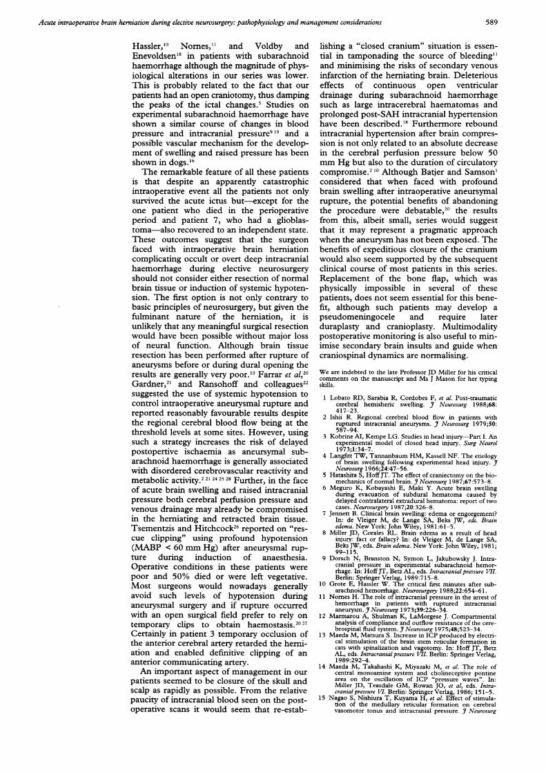
Hassler,"° Nornes," and Voldby andEnevoldsen'8 in patients with subarachnoidhaemorrhage although the magnitude of phys-iological alterations in our series was lower.This is probably related to the fact that ourpatients had an open craniotomy, thus dampingthe peaks of the ictal changes.5 Studies onexperimental subarachnoid haemorrhage haveshown a similar course of changes in bloodpressure and intracranial pressure919 and apossible vascular mechanism for the develop-ment of swelling and raised pressure has beenshown in dogs.'6The remarkable feature of all these patients
is that despite an apparently catastrophicintraoperative event all the patients not onlysurvived the acute ictus but-except for theone patient who died in the perioperativeperiod and patient 7, who had a glioblas-toma-also recovered to an independent state.These outcomes suggest that the surgeonfaced with intraoperative brain herniationcomplicating occult or overt deep intracranialhaemorrhage during elective neurosurgeryshould not consider either resection of normalbrain tissue or induction of systemic hypoten-sion. The first option is not only contrary tobasic principles of neurosurgery, but given thefulminant nature of the herniation, it isunlikely that any meaningful surgical resectionwould have been possible without major lossof neural function. Although brain tissueresection has been performed after rupture ofaneurysms before or during dural opening theresults are generally very poor.'0 Farrar et al,20Gardner,2' and Ransohoff and colleagues22suggested the use of systemic hypotension tocontrol intraoperative aneurysmal rupture andreported reasonably favourable results despitethe regional cerebral blood flow being at thethreshold levels at some sites. However, usingsuch a strategy increases the risk of delayedpostopertive ischaemia as aneursymal sub-arachnoid haemorrhage is generally associatedwith disordered cerebrovascular reactivity andmetabolic activity.22' 242528 Further, in the faceof acute brain swelling and raised intracranialpressure both cerebral perfusion pressure andvenous drainage may already be compromisedin the herniating and retracted brain tissue.Tsementzis and Hitchcock26 reported on "res-cue clipping" using profound hypotension(MABP < 60 mm Hg) after aneurysmal rup-ture during induction of anaesthesia.Operative conditions in these patients werepoor and 50% died or were left vegetative.Most surgeons would nowadays generallyavoid such levels of hypotension duringaneurysmal surgery and if rupture occurredwith an open surgical field prefer to rely ontemporary clips to obtain haemostasis.20 27Certainly in patient 3 temporary occlusion ofthe anterior cerebral artery retarded the herni-ation and enabled definitive clippnig of ananterior communicating artery.An important aspect of management in our
patients seemed to be closure of the skull andscalp as rapidly as possible. From the relativepaucity of intracranial blood seen on the post-operative scans it would seem that re-estab-
lishing a "closed cranium" situation is essen-tial in tamponading the source of bleeding"and minimising the risks of secondary venousinfarction of the herniating brain. Deleteriouseffects of continuous open ventriculardrainage during subarachnoid haemorrhagesuch as large intracerebral haematomas andprolonged post-SAH intracranial hypertensionhave been described.'8 Furthermore reboundintracranial hypertension after brain compres-sion is not only related to an absolute decreasein the cerebral perfusion pressure below 50mm Hg but also to the duration of circulatorycompromise.2 '° Although Batjer and Samson'considered that when faced with profoundbrain swelling after intraoperative aneursymalrupture, the potential benefits of abandoningthe procedure were debatable,20 the resultsfrom this, albeit small, series would suggestthat it may represent a pragmatic approachwhen the aneurysm has not been exposed. Thebenefits of expeditious closure of the craniumwould also seem supported by the subsequentclinical course of most patients in this series.Replacement of the bone flap, which wasphysically impossible in several of thesepatients, does not seem essential for this bene-fit, although such patients may develop apseudomeningocele and require laterduraplasty and cranioplasty. Multimodalitypostoperative monitoring is also useful to min-imise secondary brain insults and guide whencraniospinal dynamics are normalising.
We are indebted to the late Professor JD Miller for his criticalcomments on the manuscript and Ms J Mason for her typingskills.
1 Lobato RD, Sarabia R, Cordobes F, et al. Post-traumaticcerebral hemisheric swelling. J7 Neurosurg 1988;68:417-23.
2 Ishii R. Regional cerebral blood flow in patients withruptured intracranial aneurysms. J Neurosurg 1979;50:587-94.
3 Kobrine AI, Kempe LG. Studies in head injury-Part I. Anexperimental model of closed head injury. Surg Neurol1973;1:34-7.
4 Langfitt TW, Tannanbaum HM, Kassell NF. The etiologyof brain swelling following experimental head injury. JfNeurosurg 1966;24:47-56.
5 Hatashita S, HoffJT. The effect of craniectomy on the bio-mechanics of normal brain. JfNeurosurg 1987;67:573-8.
6 Meguro K, Kobayashi E, Maki Y. Acute brain swellingduring evacuation of subdural hematoma caused bydelayed contralateral extradural hematoma: report of twocases. Neurosurgery 1987;20:326-8.
7 Jennett B. Clinical brain swelling: edema or engorgement?In: de Vleiger M, de Lange SA, Beks JW, eds. Brainedema. New York: John Wiley, 1981:61-5.
8 Miller JD, Corales RL. Brain edema as a result of headinjury: fact or fallacy? In: de Vleiger M, de Lange SA,Beks JW, eds. Brain edema. New York: John Wiley, 1981;99-115.
9 Dorsch N, Branston N, Symon L, Jakubowsky J. Intra-cranial pressure in experimental subarachnoid hemor-rhage. In: HoffJT, Betz AL, eds. Intracranial pressure VII.Berlin: Springer Verlag, 1989:715-8.
10 Grote E, Hassler W. The critical first minutes after sub-arachnoid hemorrhage. Neurosurgery 1988;22:654-6 1.
11 Nornes H. The role of intracranial pressure in the arrest ofhemorrhage in patients with ruptured intracranialaneurysm. J Neurosurg 1973;39:226-34.
12 Marmarou A, Shulman K, LaMorgese J. Compartmentalanalysis of compliance and outflow resistance of the cere-brospinal fluid system. J Neurosurg 1975;48;523-34.
13 Maeda M, Matsura S. Increase in ICP produced by electri-cal stimulation of the brain stem reticular formation incats with spinalization and vagotomy. In: Hoff JT, BetzAL, eds. Intracranial pressure VII. Berlin: Springer Verlag,1989:292-4.
14 Maeda M, Takahashi K, Miyazaki M, et al. The role ofcentral monoamine system and cholineceptive pontinearea on the oscillation of ICP "pressure waves". In:Miller JD, Teasdale GM, Rowan JO, et al, eds. Intra-cranial pressure VI. Berlin: Springer Verlag, 1986; 151-5.
15 Nagao S, Nishiura T, Kuyama H, et al. Effect of stimula-tion of the medullary reticular formation on cerebralvasomotor tonus and intracranial pressure. J Neurosurg
589

Whittle, Viswanathan
1987;66;548-54,16 Hashimoto M, Higashi S, Kogure Y, et al. Pressure waves
and brain stem microcirculatory disturbance followingexperimental subarachnoid hemorrhage. In: Hoff JT,Betz AL, eds. Intracranial pressure VII. Berlin: SpringerVerlag, 1989: 748-53.
17 Pickard JD, Lovick AHJ, Read DH. Evidence for brainengorgement as the initial cause of brain swelling followingintraoperative aneurysm rupture in man after subarach-noid hemorrhage. Acta Physiol Scand 1986;172(suppl552):94-5.
18 Voldby B, Enevoldsen EM. Intracranial pressure changesfollowing aneurysm rupture. Part 3: recurrent hemor-rhage. Jf Neurosurg 1982;56:784-9.
19 Trojanowski T. Early effects of experimental subarachnoidhemorrhage on the cerebral circulation: part II. Regionalcerebral blood flow and cerebral microcirculation afterexperimental subarachnoid hemorrhage. Acta Neurochir1984;72:241-59.
20 Batjer H, Samson D. Intraoperative aneurysmal rupture:incidence, outcome and suggestions for surgical manage-ment. Neurosurgery 1986;18:701-1.
21 Farrar JK, Gamache FW, Ferguson GG, Barker J, VarkeyGP, Drake CG. Effects of profound hypotension on cere-
bral blood flow during surgery for intracranialaneurysms. Neurosurg 198 1;55:857-64.
22 Gardner WJ. The control of bleeding during operation byinduced hypotension. JAMA 1946;132:572-4.
23 Ransohoff J, Guy HH, Mazzia VDB, Battista A. Deliberatehypotension in surgery of cerebral aneurysms andcorrelative animal studies. NY State Med 1969;69:913-8.
24 Pickard JD, Matheson M, Patterson J, Wyper D.Prediction of late ischaemic complications after cerebralaneurysm surgery by the intraoperative measurement ofcerebral blood flow. Neurosurg 1980;53:305-8.
25 Symon L. Disordered cerbral vascular physiology inaneurysmal subarachnoid hemorrhage. Acta Neurochir1978;41 :7-22.
26 Tsementzis SA, Hitchcock ER. Outcome from "rescueclipping" of ruptured intracranial aneurysms duringinduction anaesthesia and endotracheal intubation.Neurol Neurosurg Psychiatry 1985;48: 160-3.
27 Suzuki J, Yoshimoto T. The effect of mannitol in prolonga-tion of permissible occlusion time of cerebral arteries:clinical data of aneurysm surgery. In: Suzuki J, ed.Cerebral aneurysms. Tokyo: Neuron Publishing Co, 1979:330-7.
28 Jakobsson K E, Lofgren J, Zwetnow N N. Critical thresh-olds of rebound of ICP after cerebral compression. In:Hoff JT, Betz AL, eds. Intracranial pressure VII. Berlin:Springer Verlag, 1989:853-7.
590