acute coronary syndrome - ju medicine
TRANSCRIPT

ISCHEMIC HEART DISEASE
ACUTE CORONARY SYNDROME
Prof Akram Saleh
Consultant Invasive Cardiologist
Jordan University Hospital

Case presentation
A50 year old male presented to emergency room complaining of sudden sever chest pain of 1 hour duration. It is retrosternal, compressive, and radited to left shoulder and arm.
Associated with sweating, nausea and vomiting
On examination: patient is anxious, in pain, sweaty.
BP: 100/60. PULSE: 120 BPM, RR: 26/min
Chest: basal crepitations
What is the most likely diagnosis
pathophysiology

The Spectrum of
Myocardial Ischemia
Acute Coronary Syndromes
Thrombus present in the artery
Stable
Angina
Unstable
Angina
Non-ST
Elevated MI
(NSTEMI)
ST
Elevated MI
(STEMI)
Sudden
Death

Acute Coronary Syndrome
The spectrum of clinical conditions ranging
from:
STEMI (Q-wave MI): Total occlusion
NSTEMI (non-Q wave MI): Subtotal occlusion
unstable angina: Subtotal occlusion
Characterized by the common pathophysiology of a
disrupted atheroslerotic plaque ( rupture, erosion,
or fissure)

Case presentation
A50 year old male presented to emergency room complaining of sudden sever chest pain of 1 hour duration. It is retrosternal, compressive, and radited to left shoulder and arm.
Associated with sweating, nausea and vomiting
On examination: patient is anxious, in pain, sweaty.
BP: 100/60. PULSE: 120 BPM, RR: 26/min
Chest: basal crepitations
The most likely diagnosis is Myocardial infarction
Pathophysiology??

Characteristics of Unstable( RUPTURE-PRONE PLAQUE) and
Stable Plaque
Thin
fibrous cap
Inflammatory
cells
Few
SMCs
Eroded
endotheliumActivated
macrophages
Thick
fibrous cap
Lack of
inflammatory
cells
Foam cells
Intact
endothelium
More
SMCs
Unstable Stable

platelet
Gp 11B/111A

For Internal Training Purposes Only 8

PATHOGENESIS OF ACS
Plaque rupture
THROMBOSIS
1- Primary hemostasis: Initiated by platelet
platelets adhesion, activation, and aggregation---platelet plug
2- Secondary hemostasis:
activation of the coagulation system---fibrin clot.
These two phases are dynamically interactive:
Platelet can provide a surface for coagulation enzymes
Thrombin is a potent platelet activator

ACUTE MYOCARDIAL INFARCTION
THE MOST COMMON CAUSE OF DEATH
RUPTURE ATHEROMATOUS PLAQUE---CORONARY OCCLUSION
Clinical Manifestation:
Chest pain: usually at rest, early morning
> 30 minutes ( site, radiation, severity, character, radiation, associated phenomena..)
painless MI (10-15%): DM, elderly
Present as: Hypotension, Heart failure, Arrhythmia
Physical Examination:
anxious, stressed, sweaty
vital sign: BP, Pulse, Temp
auscultation: S4,S3, Murmure, Rub


Diagnosis of Myocardial Infarction
1-History
2-ECG (Electrocardiogram): STMI and NSTMI
Hyperacute T wave
ST-segment elevation
Q- wave
T- inversion
ST-segment depresion
normal ECG will not exclude MI3-Cardiac Marker: Troponin,CPK, myoglobulin,..
Troponin T,I: 4-6 Hr (HsT 2-4 hr)
last 10-14 days
CPK:4-6 Hr, peak 17-24hr, normal 72 hr
MB(MM,BB)
MB2/MB1 >1.5

Regions of the Myocardium
Inferior
II, III, aVF
Lateral
I, AVL,
V5-V6
Anterior /
Septal
V1-V4

56 YEAR MALE, C/O: CHEST PAIN OF 2 HRS.
WHAT IS THE DIAGNOSIS?

WHAT IS THE DIAGNOSIS?

56 YEAR MALE, C/O: CHEST PAIN OF 2 HRS.
WHAT IS THE DIAGNOSIS?
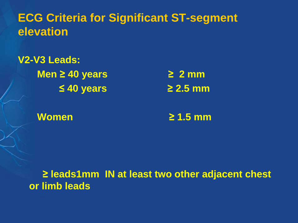
ECG Criteria for Significant ST-segment
elevation
V2-V3 Leads:
Men ≥ 40 years ≥ 2 mm
≤ 40 years ≥ 2.5 mm
Women ≥ 1.5 mm
≥ leads1mm IN at least two other adjacent chest
or limb leads

Cardiac Marker: Troponin,CPK, myoglobulin
Troponin T,I:
CPK:
Myoglobulin

Troponin:
Very specific and more sensitive than CK
Rises 4-6 hours after injury (HsT 2-4 hr)
Remains elevated for 10-14 days
Can provide prognostic information
Unable to detect re-infarction < 2 weeks

Non MI Causes of Troponin Elevation
Tachycardia
PE
Cardiac failure w/ myonecrosis
Cardiac surgery
Myocarditis
Renal failure: troponin I
Shock
Sepsis

CK/MB
Rises 4-6 hours after injury and peaks at 17- 24 hours
Remains elevated 36-48 hours
Back to normal 72 hr
CPK iso-enzymes: MM, BB, MB
MB2/MB1 >1.5
Positive if CK-MB > 5% of total CK or 2 times normal
Elevation can be predictive of mortality
False positives with exercise, trauma, muscle disease, DM, PE

Myoglobin
Rises 2-4 hours after injury and peaks at 6-12
hours
Remains elevated 24-36 hours
Not cardiac specific
Rise of 25-40% over 2 hours strongly predictive
of MI

Protein
Molecular
mass (kD)
First
detection
Duration of
detection
Sensit
ivity
Specif
icity
Myoglobin 16 1.5–2
hours
8–12 hours +++ +
CK-MB 83 2–3
hours
1–2 days +++ +++
Troponin I 33 3–4
hours
7–10 days ++++ ++++
Troponin T 38 3–4
hours
7–14 days ++++ ++++
CK 96 4–6
hours
2–3 days ++ ++
Biochemical Markers III

Biochemical Markers II

DIAGNOSIS OF MI-CONT
1-CBC: Increase WBC, ESR
2- Increase plasma glucose
3-Serum lipid (< 24 hr)
4-Echocardiogram:nonspecific changes( hypo,
akinesia, dyskinesia

Management of ACS
Primary goals: Open the blocked artery
Decrease amount of myocardial necrosis
Preserve LV function
Prevent major adverse cardiac events
Treat life threatening complications

Management of ACS
Immediate general treatment (MONA)
Morphine
Analgesia
Reduce pain/anxiety—decrease sympathetic tone, systemic vascular
resistance and oxygen demand
Careful with hypotension, hypovolemia, respiratory depression
Oxygen 2-4 liters/minute
Up to 70% of ACS patient demonstrate hypoxemia
May limit ischemic myocardial damage by increasing oxygen
delivery/reduce ST elevation

Management
Immediate general treatment(MONA)
Nitroglycerin sublingual or spray Dilates coronary vessels—increase blood flow
Reduces systemic vascular resistance and preload
Contraindications:
hypotension, RV infarction ,recent ED meds
Aspirin 160-325mg chewed and swallowed
Irreversible inhibition of platelet activation
Stabilize plaque and arrest thrombus
Reduce mortality in patients with STEMI
Careful with active PUD, hypersensitivity, bleeding disorders

TREATMENT OF MYOCARDIAL INFARCTION
IN EMERGENCY ROOM:
1-Rapid assessment
2-Establish IV access
3-12 ECG
4- Aspirin 150-300 mg Orally, Clopidogrel or ticagrelor
5-Oxygen
6-Analgesia: IV morphine, diamorphine 3-5 mg
7-Antiemetic: metoclopromide 10 mg IV
8-Sublingual nitrate: if NO hypotension, RV MI
9-ECG monitor
10-Reperfusion: PCI or Thrombolytics, (CABG)

Time is Muscle!!!

Every 30-minute delay
from onset of symptoms
to reperfusion. 1 year
mortality is increased
by 8%De Luca et al, Circulation 2004
Reduction in Long Term
Mortality

Closed Open artery
Arrival After balloon
Balloon
Primary angioplasty
• Coronary arteries: balloon angioplasty
• The European Society of Cardiology (ESC) guidelines recommend primary PCI as the preferred treatment whenever it is available within 90-120 minutes of the first medical contact

Angioplasty reduces mortality and morbidity
Primary PCI vs. Thrombolysis in ST-Elevation Myocardial Infarction:Meta-analysis (23 Randomised controlled trials, N=7,739)
Death NonfatalMI
Short-term Outcomes (4-6 weeks)
Fre
qu
en
cy
(%
)
P<.0001
P<.0001
P=.0002
P<.000
1PPCI
Thrombolytictherapy
RecurrentIschemia
Death, Nonfatal, Reinfarction,or Stroke
Based on Keeley EC, et al. Lancet. 2003;361:13-20.

Reperfusion in STEMI__________________________________

Reperfusion: PCI

ST-Segment elevation MI: Reperfusion
THROMBOLYSIS/ PCI
Time= Muscle
Early reperfusion: time dependent
-improve survival
-LV function preservation
TIMI 3 flow
-PCI: 95%, TPA:54%, STREPTO:32%
PCI: Reduce re-occlusion and recurrent thrombosis

Fibrinolysis generally preferred if:
<3 hours from onset
PCI not available/delayed
Door to balloon >90min
Door to balloon minus door
to needle > 1hr
Door to needle goal <30min
No contraindications
Invasive strategy preferred if:
>3hours from onset
PCI available
Door to balloon < 90min
Door to balloon minus door to
needle < 1hr
Fibrinolysis contraindications
High risk
STEMI dx in doubt
Age >75
ST Elevation or New LBBB
Step 2: Select Reperfusion Strategy
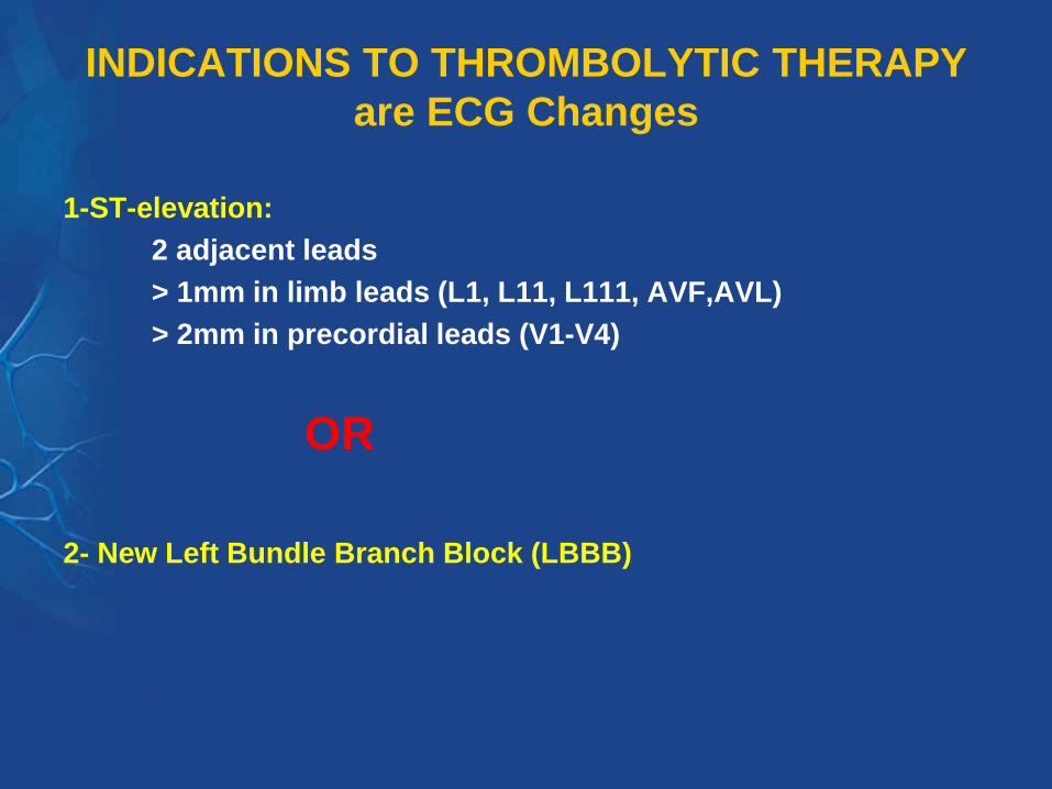
INDICATIONS TO THROMBOLYTIC THERAPY
are ECG Changes
1-ST-elevation:
2 adjacent leads
> 1mm in limb leads (L1, L11, L111, AVF,AVL)
> 2mm in precordial leads (V1-V4)
OR
2- New Left Bundle Branch Block (LBBB)

Common Thrombolytic Regimens for STEMI1
Initial treatment Co-therapy Contraindications
Streptokinase (SK) 1.5 million U in 100 mL None or iv Prior SK or
5% dextrose or 0.9% saline heparin x 2448 hours anistreplaseover 3060 min
Alteplase (tPA) 15 mg iv bolus, then iv heparin x 2448 hours
0.75 mg/kg over 30 min,
then 0.5 mg/kg iv over 60 min
Total dose not over 100 mg
Reteplase (rPA) 10 U + 10 U iv bolus given iv heparin x 2448 hours
30 min apart
Tenecteplase**** Single iv bolus iv heparin x 2448 hours
(TNK-tPA) 30 mg if <60 kg
35 mg if 60 kg to <70 kg
40 mg if 70 kg to <80 kg
45 mg if 80 kg to <90 kg
50 mg if ≥90 kg
1. Van de Werf F et al. Eur Heart J 2003; 24: 2866.
Note: acetylsalicylic acid (ASA) should be given to all patients without contraindications;
iv=intravenous
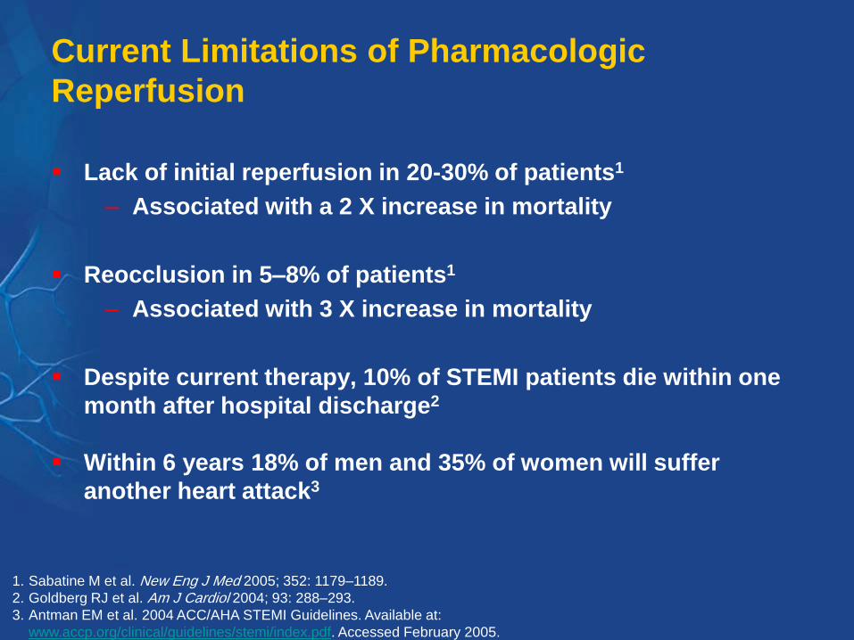
Current Limitations of Pharmacologic
Reperfusion
Lack of initial reperfusion in 20-30% of patients1
– Associated with a 2 X increase in mortality
Reocclusion in 5–8% of patients1
– Associated with 3 X increase in mortality
Despite current therapy, 10% of STEMI patients die within one
month after hospital discharge2
Within 6 years 18% of men and 35% of women will suffer
another heart attack3
1. Sabatine M et al. New Eng J Med 2005; 352: 1179–1189.
2. Goldberg RJ et al. Am J Cardiol 2004; 93: 288–293.
3. Antman EM et al. 2004 ACC/AHA STEMI Guidelines. Available at:
www.accp.org/clinical/guidelines/stemi/index.pdf. Accessed February 2005.

Contraindications to Thrombolytic Therapy
Absolute contraindication
1-Active internal bleeding
2-Suspected aortic dissection
3-Trauma or surgery < 2 weeks
4-History of hemorrhagic CVA
5-BP> 200/120 mmHg
6-Prolonged CPR
7-Recent head trauma or known
intracranial neoplasm
8-Diabetic proliferative retinopathy
9-Pregnency
10-Prvious allergy to the
thrombolytic agent
Relative contraindication
1-Trauma or surgery > 2 weeks
2-Active peptic ulcer disease
3-History CVA
4-Bleedind diathesis or current use
of anticoagulant
5-Uncontrolled hypertension
6-Previous exposure to
streptokinase
7-Pericardial friction rub
8-Significant liver dysfunction

COMPLICATION OF THROMBOLYTIC
THERAPY
1-Hemorrhage <5%
2- Systemic embolization
3-CNS bleeding
4-Allergic Reaction 1-3%, anaphylaxis 0.1%

Other Routine Therapies in Acute STEMI1
ASA 150325 mg (non-enteric coated), Clopidogrel
Beta-blockers
Angiotensin-converting enzyme (ACE) inhibitors
Oxygen
statines
Nitrates
Heparin if indicated
CCU: 24-48 hr
Word: 3-5 days
Home medication: aspirin, B-blocker, statines, ACE I, ? nitrate
1. Van de Werf F et al. Eur Heart J 2003; 24: 2866.

Complications of Myocardial Infarction
1- Arrhythmias: Any type
Ventricular: PVC, VT, Accelerated Idioventricular rhythm, VF
Atrial: AF 15% in ist 24 hr, sinus brady or tachycardia, PAC
Heart Blocks: 1st, 2nd, 3rd block, BBB
2- Heart failure ( pump failure). Killip Classification I-IV
3-Myocardial rupture: 1st 10 days
free wall, septum, papillary muscle, ventricular
pseudoaneurysm
4- Recurrent or extension of MI, Thromboembolism
5-Early pericarditis: ASA( NSAID and Steroids are contraindicated)
6-Dresslers syndrome 2-12 weeks: ASA, Ibuprofen
7- Left ventricular aneurysm
8-Sudden death

Differential Diagnosis of MI
1- Aortic Dissection
2-Massive Pulmonary Embolism
3- Acute pericarditis

PROGNOSIS of MIpre-hospital mortality:20%
hospital mortality:10-12%
1st year mortality 10%Poor prognostic featues:
1-Heart Failure
2-EF< 40%
3- Large infarction size
4-Anerior MI
5-New BBB
6- Mobits type 2 , and 3rd AV Block
7-Reinfarction or extension of MI
8-Frequent PVC
9-VF or VT
10-Atrial fibrillation
11-Post infarction angina
12-DM
13-Age> 70
14-female

TIMI Risk Score in STEMI
Risk factor Score
1- Age>65 2
2- Age>75 3
3- Hist of angina 1
4- Hist of hypertension 1
5- Hist of DM 1
6- Syst BP< 100 3
7- Heart rate> 100 2
8- Killip II-IV 2
9- Ant M or LBBB 1
10- Delay treat > 4 hr 1
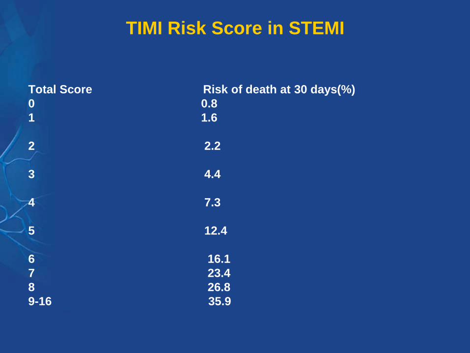
TIMI Risk Score in STEMI
Total Score Risk of death at 30 days(%)
0 0.8
1 1.6
2 2.2
3 4.4
4 7.3
5 12.4
6 16.1
7 23.4
8 26.8
9-16 35.9

Post-MI Management
1- Risk factors modification (Stop smoking, BP< 140/90, HbA1c<7,
Exercise, ..)
2-Aspirin, Clopidogrel or ticagrelor
3- B-blockers
4-Statines
5-ACE-inhibitors
6- Aldosterone antagonist( in presence of heart failure)

UNSTABLE ANGINA
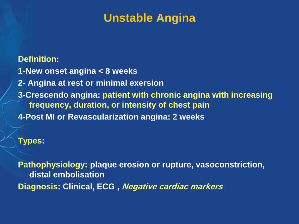
Unstable Angina
Definition:
1-New onset angina < 8 weeks
2- Angina at rest or minimal exersion
3-Crescendo angina: patient with chronic angina with increasing
frequency, duration, or intensity of chest pain
4-Post MI or Revascularization angina: 2 weeks
Types:
Pathophysiology: plaque erosion or rupture, vasoconstriction,
distal embolisation
Diagnosis: Clinical, ECG , Negative cardiac markers

Unstable Angina
Classification
1- Acute: rest pain within the last 48 hr
2- Subacute: no pain within the last 48 hr
1- primary: no secondary causes
2-Secondary: sever anemia, thyrotoxicosis, hypertension,
arrhythmias
1-High Risk
2-Low risk

HIGH RISK UNSTABLE ANGINA
1-Rest pain > 20 minutes
2-Accelerating tempo of ischemic symptoms in preceding 48 hr
3-Clinical finding of: pulmonary edema, new S3, new MR,
Hypotension, Brady or Tachycardia
3-ECG changes: transient ST segment changes, BBB, VT
4- DM

Risk Stratification
1- Age > 65
2- 3 or more cardiac risk factors
3- Prior angiographic coronary
obstruction (stenosis ≥ 50%)
4- ST segment deviation
5-More than 2 angina events within
the previous 24 hours
6-Use of aspirin within previous 7
days
7-Elevated cardiac markers
TIMI Risk ScorePredicts risk of death, new/recurrent MI, need for urgent revascularization
within 14 days

TIMI Risk Score For UA/NSTEMI
Antman EM, et al. JAMA. 2000;284:835-442. (Copyright 2000 American Medical Association. All rights reserved)
Age >65 years
>3CAD Risk Factors
Prior Stenosis >50 %
ST deviation
>2 Anginal
events <24 hours
ASA in last 7 days
Elevated Cardiac
Markers (CK-MB or
troponin)
4.78.3
13.2
19.9
26.2
40.9
0
10
20
30
40
50
0/1 2 3 4 5 6/7
D/M
I/U
rg R
evasc (
%)
Number of Risk Factors
Population (%): 4.3 17.3 32.0 29.3 13.0 3.4
C Statistic=0.65
c2 trend P<.001

Treatment of HIGH RISK UNSTABLE ANGINAAND NSTMI
1-CCU admission : Treat as MI except for thrombolytics
NO THROMBOLYTICS
2-Aspirin***, Clopidogrel
3-Anticoagulant: heparin (LMWH is superior to unfractionated
heparin)***
4- Nitrate ( S/L, oral, IV)
5-B-blocker
6-clopidogrel, GP 11b,111a--------Cath PCI(angio)
7-Statines
8- Invasive or conservative management
*** improve prognosis

Algorithm for Initial Assessment and Evaluation
of the Patient with Acute Chest Pain
Within 10 minutes
• Initial evaluatioon • 12 lead ECG
• Establish IV • Aspirin 160-325 mg - chewed
• Establish continuous ECG monitoring
• Blood for baseline serum cardiac markers
Chest pain consistent with coronary ischemia
Therapeutic/Diagnostic tracking according 12-lead ECG results
ECG suggestive of ischemia -
T wave inversion or ST depression
ST segment elevation or new
bundle branch blockNondiagnostic / normal ECG
PCI or Thrombolysis
Antithrombotic
PCI

Thank you