acute and chronic venous disease - tallahassee, fl/media/files/heart and...
TRANSCRIPT

Acute and Chronic Venous Disease
Advances in Percutaneous Intervention
William C. Dixon IV, MD, FACC, FSCAI
Outline
Chronic venous insufficiency Acute DVT Intervention Chronic DVT Intervention
Chronic Venous Insufficiency (CVI)
• Pathophysiology
• Clinical evaluation
• Role of ultrasound
• Endovascular therapy

Pathophysiology of CVI
Microcirculatory Changes
Secondary
Valve Damage/Obstruction
Skin Changes & Ulceration
Primary
Vein Degeneration
Ambulatory Venous Hypertension
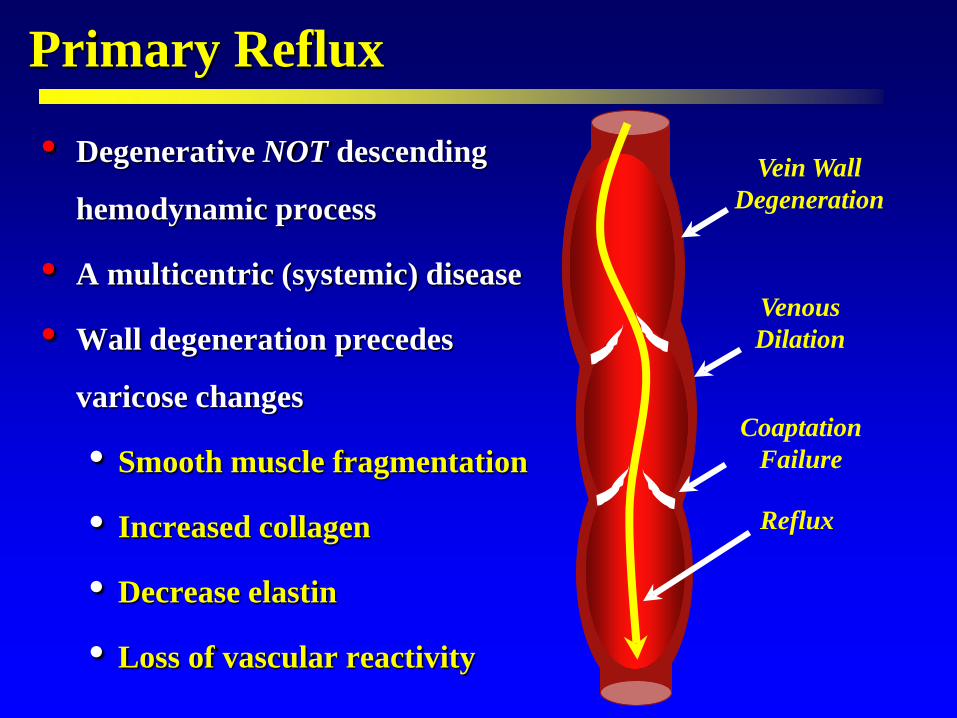
Primary Reflux
• Degenerative NOT descending
hemodynamic process
• A multicentric (systemic) disease
• Wall degeneration precedes
varicose changes
• Smooth muscle fragmentation
• Increased collagen
• Decrease elastin
• Loss of vascular reactivity
Vein Wall Degeneration
Venous Dilation
Coaptation Failure
Reflux

Secondary Reflux Markel A, J Vasc Surg 1992
• 123 limbs (103 patients) with 1st episode of DVT • Serial U/S follow-up
Days after DVT
* Saphenous reflux also present in 25% or limbs

The Pathophysiology of CVD
Reflux & Obstruction
Endothelial Activation
Increased Permeability Decreased Resistance
Leukocyte Activation, Adhesion, Migration

Leukocyte Activation The Origin of Advanced CVI
• Activated leukocytes migrate extravascularly • Release of toxic oxygen metabolites, cytokines, & proteases
• TGFß Primary mediator of dermal fibrosis Macrophage / fibroblast recruitment Fibroblast production of extracellular matrix
• Altered tissue remodeling – Imbalanced MMP-2 / TIMP
TGFß
Altered Tissue Remodeling
MMPs TIMP

Summary • Etiology of CVD
• 1º disease - A degenerative disease of the vein wall
• 2º disease - A combination of reflux and obstruction
• Anatomy is important
• Combined patterns of reflux Superficial veins
Distal deep veins
• Iliac venous obstruction
• Manifestations of venous htn mediated by the microcirculation
• Endothelial activation
• Leukocyte activation
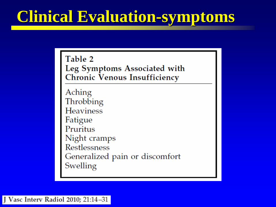
Clinical Evaluation-symptoms
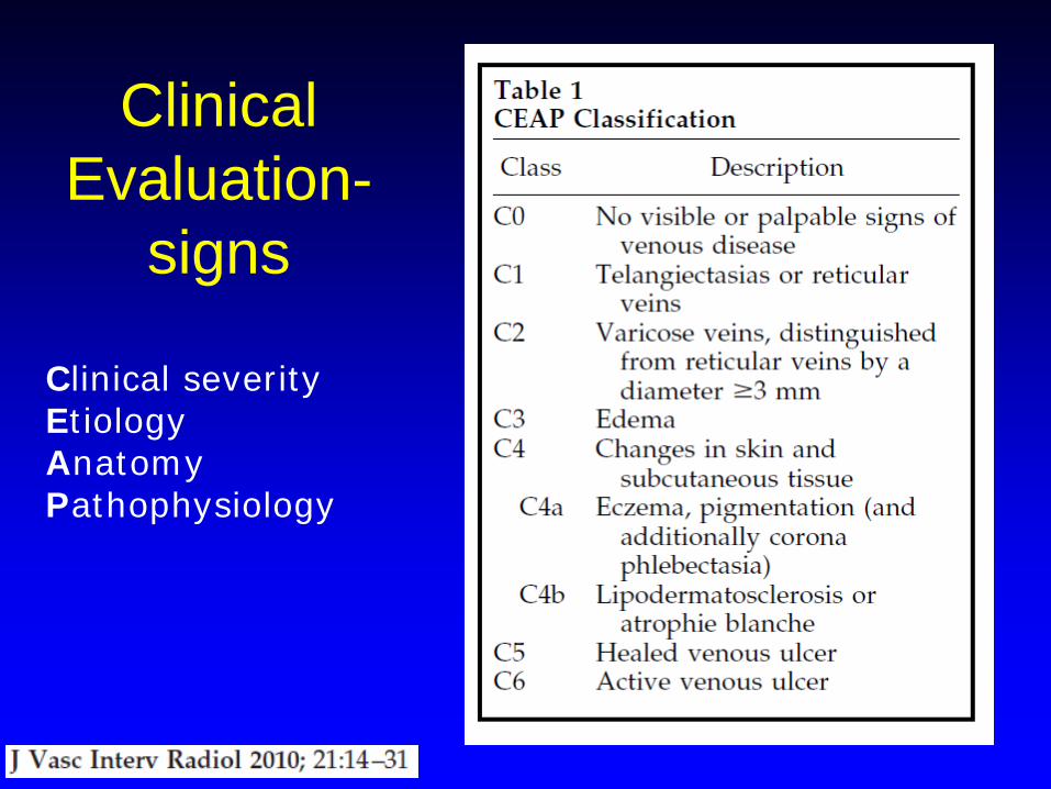
Clinical Evaluation-
signs
Clinical severity Etiology Anatomy Pathophysiology

C6
C2 C3
C4 C5
C1
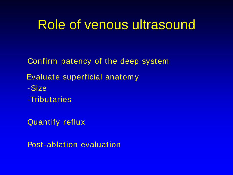
Role of venous ultrasound
Confirm patency of the deep system
Evaluate superficial anatomy -Size -Tributaries Quantify reflux Post-ablation evaluation
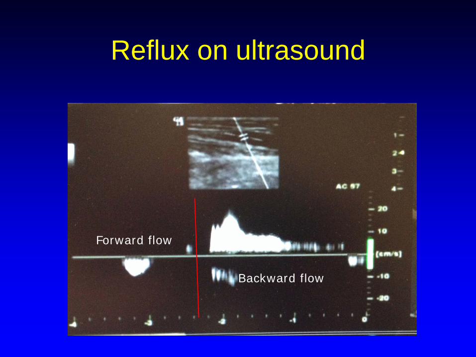
Reflux on ultrasound
Forward flow
Backward flow
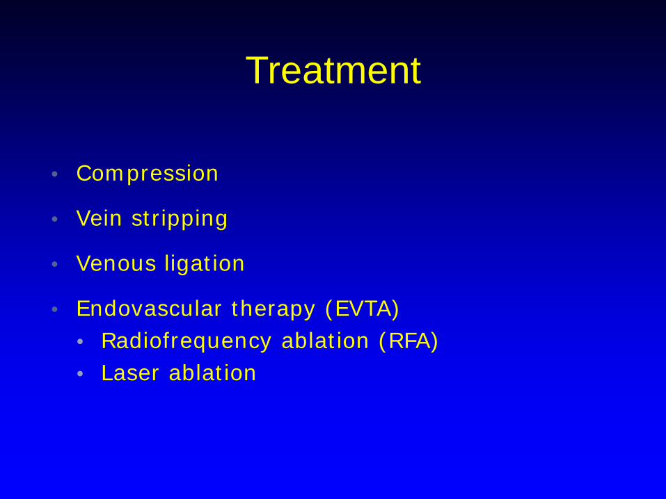
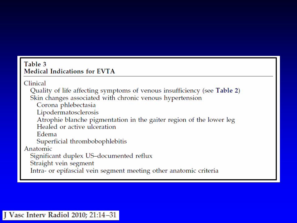
Treatment
• Compression
• Vein stripping
• Venous ligation
• Endovascular therapy (EVTA) • Radiofrequency ablation (RFA) • Laser ablation

Laser vs RFA
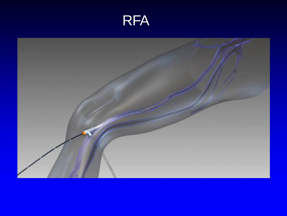
RFA

RFA
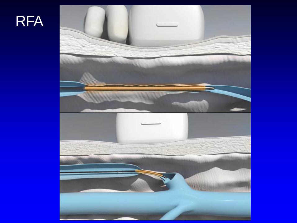
RFA

RFA
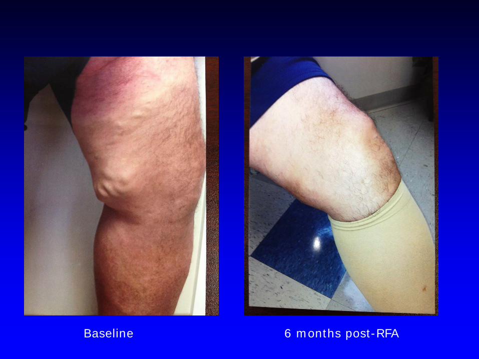
Baseline 6 months post-RFA

Deep Vein Thrombosis
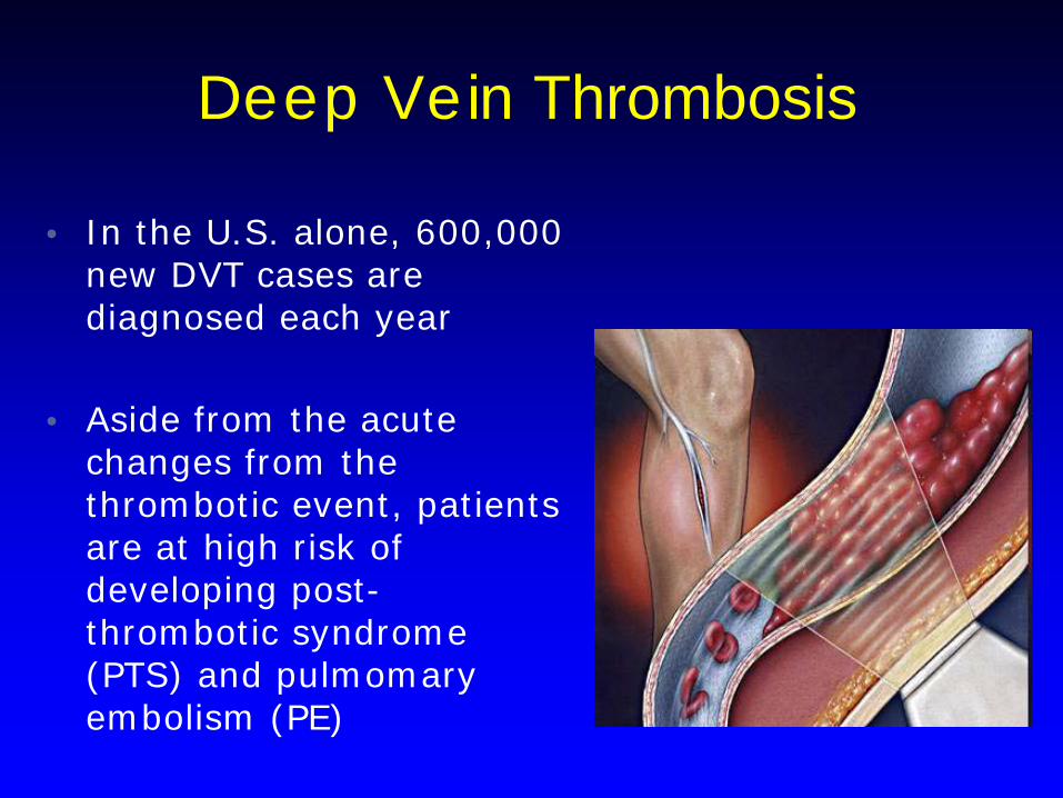
Deep Vein Thrombosis
• In the U.S. alone, 600,000 new DVT cases are diagnosed each year
• Aside from the acute changes from the thrombotic event, patients are at high risk of developing post-thrombotic syndrome (PTS) and pulmomary embolism (PE)
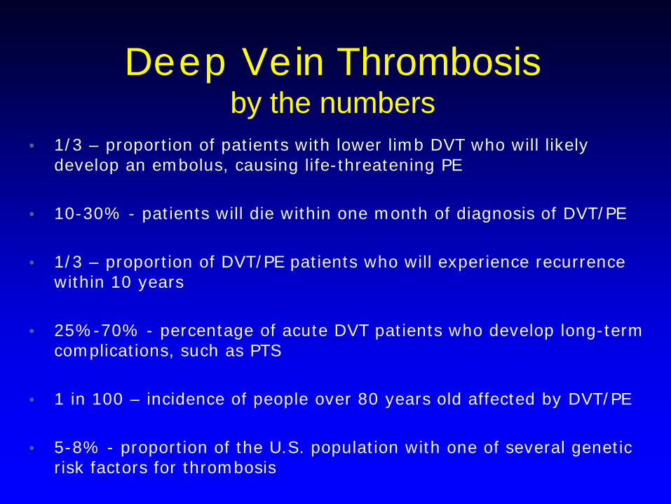
Deep Vein Thrombosis by the numbers
• 1/3 – proportion of patients with lower limb DVT who will likely
develop an embolus, causing life-threatening PE
• 10-30% - patients will die within one month of diagnosis of DVT/PE
• 1/3 – proportion of DVT/PE patients who will experience recurrence within 10 years
• 25%-70% - percentage of acute DVT patients who develop long-term complications, such as PTS
• 1 in 100 – incidence of people over 80 years old affected by DVT/PE
• 5-8% - proportion of the U.S. population with one of several genetic
risk factors for thrombosis
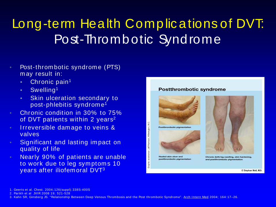
• Post-thrombotic syndrome (PTS)
may result in: • Chronic pain1
• Swelling1
• Skin ulceration secondary to post-phlebitis syndrome1
• Chronic condition in 30% to 75% of DVT patients within 2 years2
• Irreversible damage to veins & valves
• Significant and lasting impact on quality of life
• Nearly 90% of patients are unable to work due to leg symptoms 10 years after iliofemoral DVT3
1. Geerts et al. Chest. 2004;126(suppl):338S-400S 2. Parikh et al JVIR 2008 19; 521-528 3. Kahn SR, Ginsberg JS. “Relationship Between Deep Venous Thrombosis and the Post thrombotic Syndrome”. Arch Intern Med 2004; 164:17–26.
Long-term Health Complications of DVT: Post-Thrombotic Syndrome
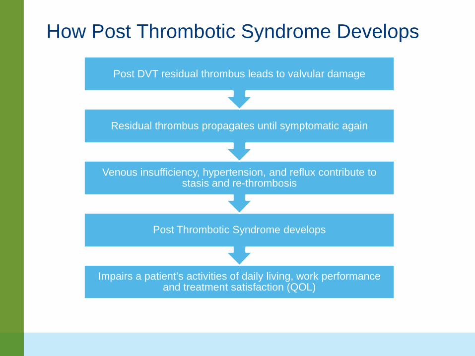
How Post Thrombotic Syndrome Develops
Impairs a patient’s activities of daily living, work performance and treatment satisfaction (QOL)
Post Thrombotic Syndrome develops
Venous insufficiency, hypertension, and reflux contribute to stasis and re-thrombosis
Residual thrombus propagates until symptomatic again
Post DVT residual thrombus leads to valvular damage

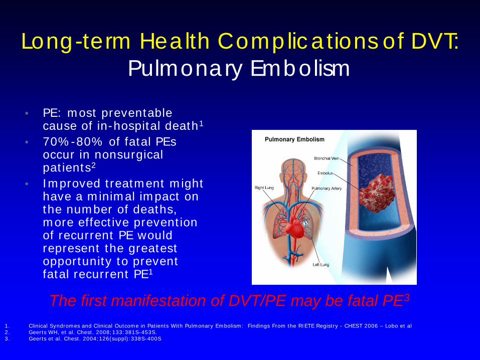
1. Clinical Syndromes and Clinical Outcome in Patients With Pulmonary Embolism: Findings From the RIETE Registry - CHEST 2006 – Lobo et al 2. Geerts WH, et al. Chest. 2008;133:381S-453S. 3. Geerts et al. Chest. 2004;126(suppl):338S-400S
• PE: most preventable cause of in-hospital death1
• 70%-80% of fatal PEs occur in nonsurgical patients2
• Improved treatment might have a minimal impact on the number of deaths, more effective prevention of recurrent PE would represent the greatest opportunity to prevent fatal recurrent PE1
The first manifestation of DVT/PE may be fatal PE3
Long-term Health Complications of DVT: Pulmonary Embolism

Conventional modes of therapy are inadequate
• Historical study on anticoagulated DVT patients
reported poor outcomes, including 95% valvular dysfunction, 90% ambulatory venous hypertension, 70% obstructive iliac vein lesion, 50% calf muscle pump dysfunction at 5 year follow up
• if thrombus resolution is not achieved within 60-90 days, the ability to restore venous patency and preserve valve function is compromised, leading to a significantly increased rate of rethrombosis

Anticoagulation Alone . . .
…does prevent clot propagation.
…does reduce risk of pulmonary embolism.
But, it typically…
…does NOT resolve clot.
…does NOT rapidly resolve symptoms.
…does NOT prevent PTS (Post Thrombotic Syndrome).
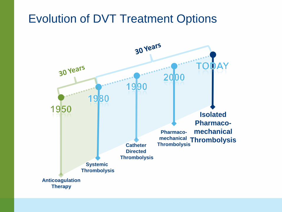
Anticoagulation Therapy
Systemic Thrombolysis
Catheter Directed
Thrombolysis
Pharmaco- mechanical
Thrombolysis
Isolated Pharmaco- mechanical
Thrombolysis
Evolution of DVT Treatment Options

Despite treatment progress…the majority of DVT patients are still being treated with anticoagulation therapy alone

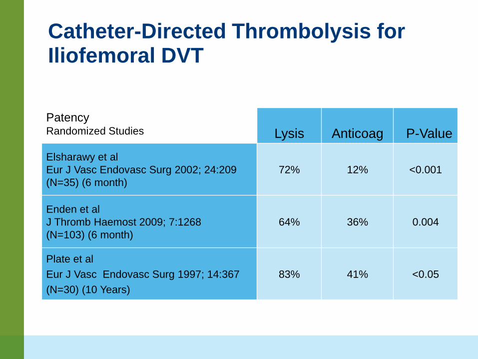
Catheter-Directed Thrombolysis for Iliofemoral DVT
Lysis Anticoag P-Value
Elsharawy et al Eur J Vasc Endovasc Surg 2002; 24:209 (N=35) (6 month)
72% 12% <0.001
Enden et al J Thromb Haemost 2009; 7:1268 (N=103) (6 month)
64% 36% 0.004
Plate et al
Eur J Vasc Endovasc Surg 1997; 14:367 (N=30) (10 Years)
83% 41% <0.05
Patency Randomized Studies
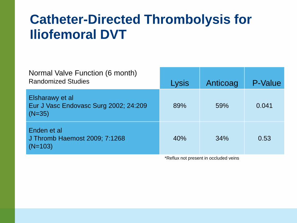
Catheter-Directed Thrombolysis for Iliofemoral DVT
Lysis Anticoag P-Value
Elsharawy et al Eur J Vasc Endovasc Surg 2002; 24:209 (N=35)
89% 59% 0.041
Enden et al J Thromb Haemost 2009; 7:1268 (N=103)
40% 34% 0.53
Normal Valve Function (6 month) Randomized Studies
*Reflux not present in occluded veins


31%38%
70%
52%45%
21%17% 18%
9%
0%
10%
20%
30%
40%
50%
60%
70%
80%
National VenousRegistry
Cleveland ClinicFoundation
Retrospective Data
EKOS
Complete clot lysisPartial clot lysisNo clot lysis
N=287 N=80
N=53
Parikh et al. J Vasc Interv Radiol. 2008 Apr;19(4):521-8.
1. Mewissen, et al. Radiology. 1999 Apr;211(1):39-49 2. Parikh et al. J Vasc Interv Radiol. 2008 Apr;19(4):521-8.
1
2
Ultrasound Accelerated Thrombolysis provides greater clot clearance than CDT
NVR – registry of DVT patients treated with CDT
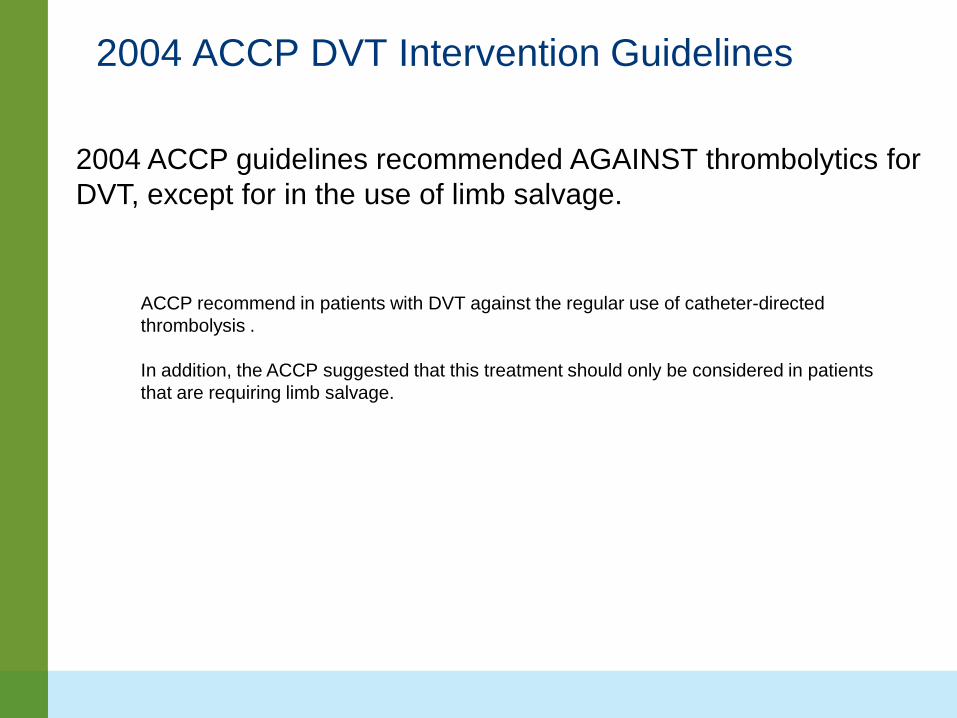
2004 ACCP DVT Intervention Guidelines
2004 ACCP guidelines recommended AGAINST thrombolytics for DVT, except for in the use of limb salvage.
ACCP recommend in patients with DVT against the regular use of catheter-directed thrombolysis . In addition, the ACCP suggested that this treatment should only be considered in patients that are requiring limb salvage.
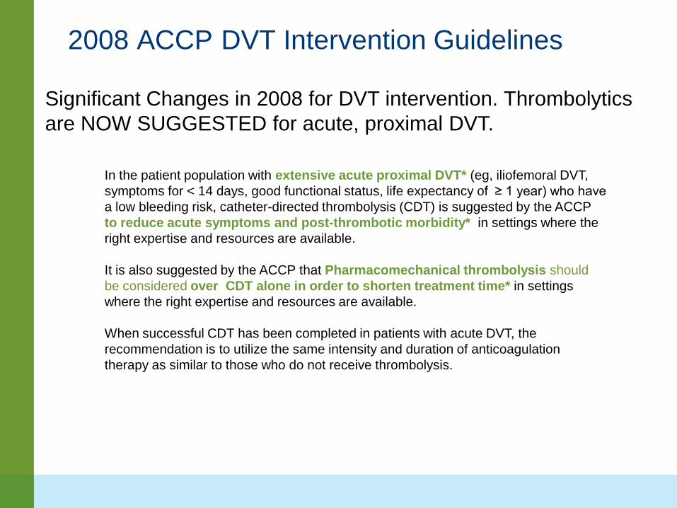
2008 ACCP DVT Intervention Guidelines
Significant Changes in 2008 for DVT intervention. Thrombolytics are NOW SUGGESTED for acute, proximal DVT.
In the patient population with extensive acute proximal DVT* (eg, iliofemoral DVT, symptoms for < 14 days, good functional status, life expectancy of ≥ 1 year) who have a low bleeding risk, catheter-directed thrombolysis (CDT) is suggested by the ACCP to reduce acute symptoms and post-thrombotic morbidity* in settings where the right expertise and resources are available. It is also suggested by the ACCP that Pharmacomechanical thrombolysis should be considered over CDT alone in order to shorten treatment time* in settings where the right expertise and resources are available. When successful CDT has been completed in patients with acute DVT, the recommendation is to utilize the same intensity and duration of anticoagulation therapy as similar to those who do not receive thrombolysis.
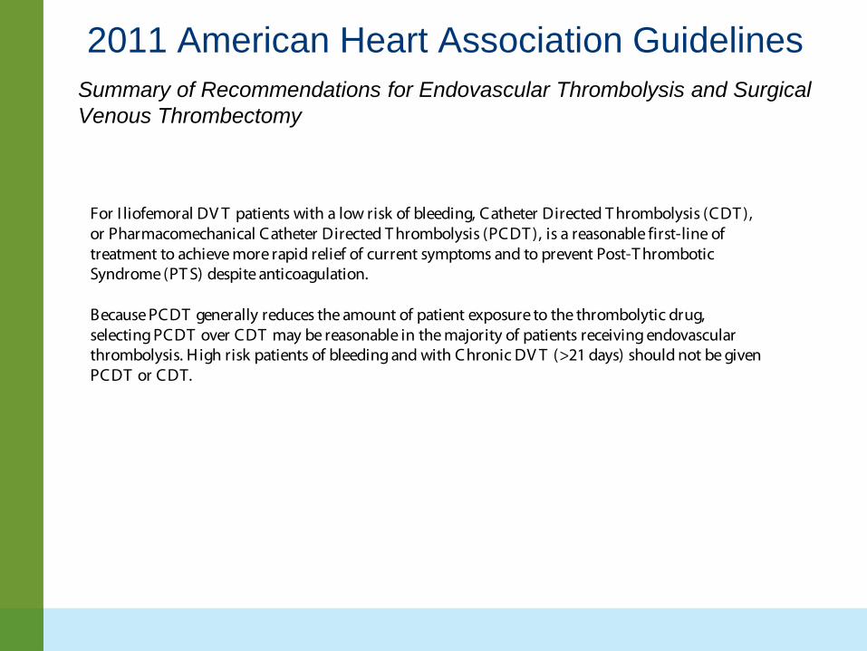
2011 American Heart Association Guidelines Summary of Recommendations for Endovascular Thrombolysis and Surgical Venous Thrombectomy
For I liofemoral DV T patients with a low risk of bleeding, Catheter Directed T hrombolysis (CDT ), or Pharmacomechanical Catheter Directed T hrombolysis (PCDT ), is a reasonable first-line of treatment to achieve more rapid relief of current symptoms and to prevent Post-T hrombotic Syndrome (PT S) despite anticoagulation. Because PCDT generally reduces the amount of patient exposure to the thrombolytic drug, selecting PCDT over CDT may be reasonable in the majority of patients receiving endovascular thrombolysis. High risk patients of bleeding and with Chronic DV T (>21 days) should not be given PCDT or CDT.
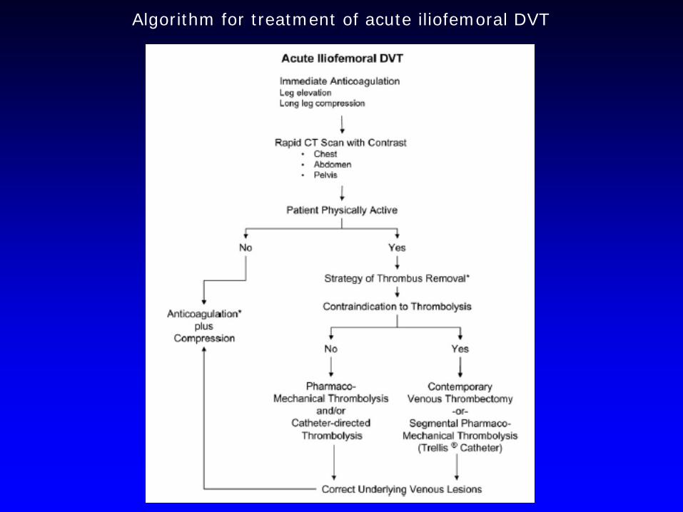
Algorithm for treatment of acute iliofemoral DVT


LLE DVT
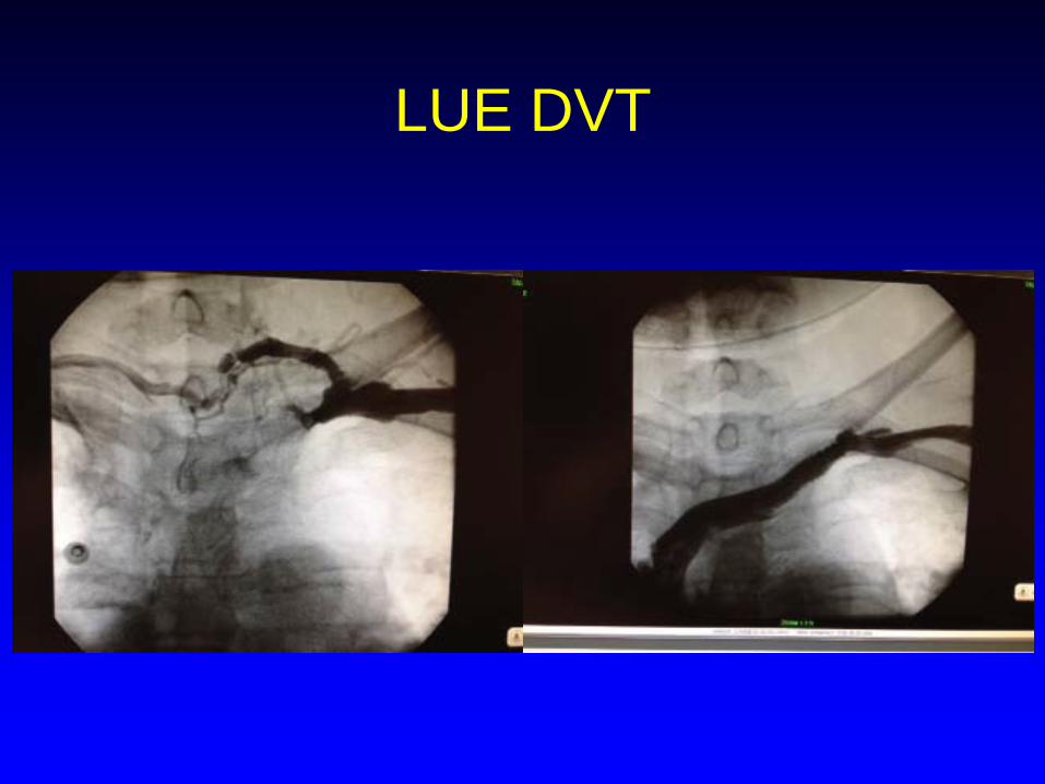
LUE DVT
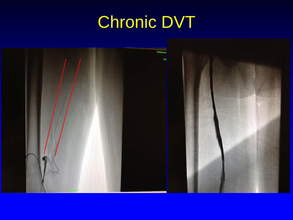
Chronic DVT

Questions?