actin at cell-cell junctions is composed of two dynamic and
TRANSCRIPT

IntroductionEpithelial cells are morphologically polarized: they arecharacterized by distinct membrane domains, a cuboidal cellshape and strong attachment to neighbouring cells andunderlying basement membrane (Nelson, 2003). As cell shapeis intrinsically related to epithelial function and actincytoskeleton organization, great effort has been put intounderstanding the actin reorganization that occurs duringmorphological differentiation. Herein, polarization is definedas the process by which epithelial cells remodel thecytoskeleton to acquire the tall, cuboidal cell shape.
In epithelia, actin bundles are present as a thickcircumferential ring around each cell, aligned with the cellborders (Owaribe et al., 1981; Yonemura et al., 1995;Zamansky et al., 1991). In addition to the circumferentialorganization, a less abundant population of actin bundles alsoappear to terminate at sites of cell-cell contacts as assessed byelectron microscopy and immunofluorescence (Green et al.,1987; Sanger et al., 1983; Vasioukhin et al., 2000; Yonemuraet al., 1995; Zamansky et al., 1991).
Surprisingly, there are currently no mechanistic insights intohow, in flat epithelial cells prior to polarization, thecytoskeleton is reorganized into the typical circumferential ringorganization for mature cells as described above. So far, theknowledge on specific epithelial actin structures and their
formation has been limited when compared to informationavailable in other cell systems (motile cell types). As themaintenance of epithelial morphology is a key factor forhomeostasis and function, it is important to identify actincytoskeletal structures required for polarization and theirmechanism of formation. Once specific actin activities andstructures are defined, the identification of their regulatorymechanisms and relevant proteins will be more focused.
What is known about the morphological polarizationprocess? During polarization, a series of steps are triggered:(1) formation of new cell-cell contacts, (2) stabilization ofthese new contacts, (3) junction maturation and (4) acquisitionof a cuboidal cell morphology. The time frame in which thesesteps occur varies according to the cell type and cultureconditions, e.g. cell confluence (McNeill et al., 1993;Vasioukhin et al., 2000). Up to now, only the early eventsrelated to junction assembly have been studied in detail (steps1 and 2 above).
In the first step, i.e. the formation of new cell-cell contacts,it is well established that adhesion mediated by cadherinreceptors is essential (Yap et al., 1997). Cadherins providecalcium-dependent cell-cell adhesion and interact indirectlywith the actin cytoskeleton (Yap et al., 1997). In the presenceof calcium ions, cells probe each other in a series of transient,weak contacts, mediated by homophilic interaction of cadherin
5549
The ability of epithelial cells to polarize requires cell-celladhesion mediated by cadherin receptors. During cell-cellcontact, the mechanism via which a flat, spread cell shapeis changed into a tall, cuboidal epithelial morphology is notknown. We found that cadherin-dependent adhesionmodulates actin dynamics by triggering changes in actinorganization both locally at junctions and within the restof the cell. Upon induction of cell-cell contacts, two spatialactin populations are distinguishable: junctional actin andperipheral thin bundles. With time, the relative positionof these two populations changes and becomesindistinguishable to form a cortical actin ring that ischaracteristic of mature, fully polarized epithelial cells.Junctional actin and thin actin bundles differ in their actindynamics and mechanism of formation, and interestingly,
have distinct roles during epithelial polarization. Whereasjunctional actin stabilizes clustered cadherin receptors atcell-cell contacts, contraction of peripheral actin bundle isessential for an increase in the maximum height at thelateral domain during polarization (cuboidal morphology).Thus, both junctional actin and thin bundles are necessary,and cooperate with each other to generate a polarizedepithelial morphology.
Key words: Microfilaments, Keratinocytes, Actin bundles,Cadherins, Cell-cell contact
Supplementary material available online athttp://jcs.biologists.org/cgi/content/full/118/23/5549/DC1
Summary
Actin at cell-cell junctions is composed of twodynamic and functional populationsJuankun Zhang1,*, Martha Betson1,*,‡, Jennifer Erasmus1, Kostas Zeikos1, Maryse Bailly2, Louise P. Cramer3
and Vania M. M. Braga1,§
1Molecular and Cellular Medicine, Faculty of Life Sciences, Imperial College London, Sir Alexander Fleming Building, London, SW7 2AZ, UK 2Institute of Ophthalmology, University College London, 11-43 Bath Street, London, EC1V 9EL, UK3Laboratory for Molecular Cell Biology and Department of Biology, University College London, Gower Street, London, WC1E 6BT, UK*These authors contributed equally to this work‡Present address: MGH Cancer Center and Harvard Medical School, Chalestown, MA, USA§Author for correspondence (e-mail: [email protected])
Accepted 15 August 2005Journal of Cell Science 118, 5549-5562 Published by The Company of Biologists 2005doi:10.1242/jcs.02639
Research Article
Jour
nal o
f Cel
l Sci
ence

5550
receptors (‘zippering’ model). These weak contacts arefollowed by clustering of cadherins into structures called‘puncta’ (Adams et al., 1996; McNeill et al., 1993).
Puncta appear within minutes of the initial cell-cell contactas discrete dots at contacting membranes, and actin recruitmentto puncta occurs concomitantly or shortly after. Actinrecruitment is predicted to restrict the mobility of clusteredcadherin receptors at the plane of the membrane and toreinforce adhesion, facilitating the progression from transientto stable contacts (step 2) (Adams et al., 1998; Adams et al.,1996). Actin-mediated stabilization of newly formed contactsis manifested as a continuous line of cadherin/actin staining atcell-cell borders (step 2) as opposed to the punctate patternobserved earlier. At this point, considerable interdigitation ofneighbouring membranes is seen in some cell types (Vaezi etal., 2002; Vasioukhin et al., 2000).
Once cell-cell contacts are stabilized, the junctionmaturation process takes place (step 3 – time frame notknown). The actin cytoskeleton in cells with newly formedjunctions (steps 1 and 2) must be remodelled in some way, asyet unknown, to produce the circumferential actin ringcoincident with the junctions (Owaribe et al., 1981). Finally,full acquisition of polarity takes place: a cuboidal morphologydevelops, with a tall lateral domain and neighbouring cellscompact towards each other (step 4, time frame unknown).
Thus, epithelial morphological polarization and actinreorganization are complex processes. Clearly, there is alarge reorganization of microfilaments, particularly during thejunction maturation and full polarization processes (steps 3 and4). However, the spatial and temporal changes and the precisemechanisms involved to complete polarization are not wellunderstood. For example, how further actin recruitmentto stable junctions is coordinated with actin filamentreorganization to form the circumferential actin ring is notclear (step 3). In addition, very little is known about whichactin reorganization process is required for the development ofa tall, cuboidal cell shape (step 4).
To begin to address some of these questions, we focusedon precise actin dynamic steps required for completemorphological polarization. We tested potential mechanismsfor the formation of actin structures that are well understoodin other model cell systems, but surprisingly have not yet beeninvestigated during epithelial polarization. Typically, theformation of diverse actin structures involves actin assembly,activity of myosin family proteins (particularly actin bundlecontractility) or both. Furthermore, a number of distinct actinassembly mechanisms can be distinguished by differences indynamics and supply of actin monomer ‘fuel’ to powerassembly (reviewed by Pollard et al., 2000). For instance,actin may assemble at the barbed end or at both the barbedand pointed end, depending on the cell type (Littlefield etal., 2001). Moreover, the supply of monomers for actinassembly in motile cells may come from previously storedor recently disassembled pools, depending on whether the cellis polarized and migrating (Cramer et al., 2002; Cramer,1999).
During epithelial polarization, new actin assembly isrequired at puncta (step 1), but the role of assembly in allsubsequent steps is unclear. Conversely, myosin functionduring the early steps is not known, but it does play a role incell compaction (step 4) (Adams et al., 1998; Collins and
Fleming, 1995). Myosin II and other myosin family membersare localized at cell-cell junctions (Krendel et al., 1999;Owaribe et al., 1981; Philip and Nachmias, 1985; Stoffler etal., 1998), and may participate in the expansion of cellmembranes as adhesive contacts are formed (step 1) (Adamset al., 1998; Gloushankova et al., 1998; Yonemura et al., 1995).Other potential additional roles for myosin II contractility, suchas in formation of the circumferential actin ring (step 3) andacquisition of a cuboidal cell shape (step 4), have yet to beformally demonstrated.
In this work, we used normal human keratinocytes, a wellestablished model for studying the formation of cadherin-dependent cell-cell contacts and epithelial polarization (Braga,2000). To exclude any potential contribution of cell migrationto the actin reorganization events, only confluent keratinocytecultures were used to induce cell-cell contacts. Using thismodel, we focus on three important questions that have notbeen addressed in the literature. (1) What is the detailedorganization of actin during the stabilization and maturationsteps? (2) What is the mechanism of formation and dynamicsof distinct actin structure during epithelial polarization? (3)What is the relative importance of these different actinpopulations for junction assembly and acquisition of a tall,cuboidal cell shape?
We found that, upon initial cell-cell contact, two spatial actinpopulations are observed: a continuous line of actin atjunctions (junctional actin) and peripheral thin bundles. Thesespatial populations can be further distinguished by their actindynamics and mechanism of formation. After cell-cell contactsare stabilized, thin bundles appear to coalesce towards thejunctional actin population to form a straight line of the thicker,continuous F-actin bundle, reminiscent of the circumferentialring observed in mature epithelia (mature junctional actin).Equally important, this process is accompanied by an increasein myosin-dependent contractility and the acquisition of apolarized cell shape in keratinocytes.
Materials and MethodsCell cultureNormal human keratinocytes (strains Kb, AEK and Sa, passages 3-7)were cultured on a mitomycin C-treated monolayer of 3T3 fibroblastsat 37°C with 5% CO2, as previously described (Hodivala and Watt,1994). To induce cell-cell contacts, confluent cultures grown in lowcalcium medium were transferred to standard calcium medium fordifferent amounts of time (Hodivala and Watt, 1994).
Antibodies and immunofluorescencePrimary antibodies used were anti-actin (C4, mouse monoclonal;MP Biomedicals, London, UK), anti-E-cadherin (HECD-1 mousemonoclonal [a gift from M. Takeichi (Shimoyama et al., 1989)] andECCD-2 rat monoclonal (Hirai et al., 1989). Anti-phosphorylatedmyosin light chain (S19, mouse monoclonal) was a kind gift from M.Matsuda (Osaka University, Japan). Secondary antibodies werepurchased from Jackson ImmunoResearch Laboratories (StratechScientific, Luton, UK). Fluorescein isothiocyanate (FITC)-phalloidinwas purchased from Sigma. Labelled actin (FITC- or Alexa Fluor568-G-actin) and DNAse1 (Alexa Fluor 488-DNAse1) werepurchased from Molecular Probes (Paisley, UK).
Unless otherwise stated, cells were fixed, permeabilized and stainedas described previously (Braga et al., 1997). To visualize labelled actinat cell-cell contacts after microinjection and in latrunculin B
Journal of Cell Science 118 (23)
Jour
nal o
f Cel
l Sci
ence
5551Actin dynamics during epithelial polarization
experiments, cells were fixed in 3% paraformaldehyde containing0.5% Triton X-100 for 10 minutes at room temperature. Staining usingC4 anti-actin antibodies was performed as described previously(Cramer, 1999). To visualize labelling of actin bundles, cells were pre-extracted with detergent before fixation in 3% paraformaldehyde(Braga et al., 1995).
Confocal images were obtained with a Leica DM IRBE microscope,containing a krypton-argon laser and TCS NT software. Cells werealso viewed using an Olympus Provis AX70 microscope and digitalimages were collected with Spot RT monochrome cooled charge-coupled (CCD) camera and software (Diagnostic Instruments) or witha KAF 1400 CCD camera (Roper Scientific) on a Nikon microscopeusing Metamorph software (Cramer et al., 2002). Images wereprocessed using Adobe Photoshop, Metamorph and Volocity Software.
Drug treatmentConfluent keratinocytes grown in low calcium medium, which doesnot induce cell-cell contacts, were transferred to standard calciummedium containing different drugs or vehicle control. Latrunculin B(Calbiochem) was carefully titrated (0.2 �M) to obtain disruption ofthe actin cytoskeleton without producing actin aggregates in thecytoplasm or cell retraction. In other experiments, latex beads (15 �m,PolySciences) coated with anti-cadherin antibodies or BSA (Braga etal., 1997) were incubated for 15-20 minutes with keratinocytes in theappropriate medium (low or standard calcium medium).
Jasplakinolide is a toxin that has two distinct activities: it stabilizesactin filaments (thereby preventing actin disassembly) and, moreslowly, induces actin polymerization (Bubb et al., 1994; Cramer,1999). To separate these two activities the jasplakinolideconcentration was carefully titrated as described previously (Cramer,1999). Keratinocytes were treated with 0.01 �M jasplakinolide(Molecular Probes; see supplementary material Fig. S1) for 60minutes or with 0.5 �M for shorter incubations (5 or 15 minutes),which did not induce actin aggregates. The same results were obtainedafter treatment with 1 �M (data not shown).
Inhibition of myosin contractility was achieved by treatment withblebbistatin, a drug that blocks myosin II ATPase activity (Merck)(Straight et al., 2003). Cells were incubated for 1 hour withblebbistatin titrated to 50 �M. In addition, Y27632 (25 �M, gift fromA. Yoshimura) or ML9 (10 �M, Calbiochem) were used.
Incorporation of labelled G-actinMicroinjection was performed essentially as described previously(Braga et al., 1997). Confluent patches of keratinocytes cultured inlow calcium medium were injected with either FITC or Alexa Fluor568-G-actin (Molecular Probes) at a pipette concentration of 3 mg/ml.Immediately after microinjection, cells were transferred to standardcalcium medium for various periods of time before fixation.
Live cells were permeabilized as described previously (Chan etal., 2000). Keratinocytes grown in low calcium medium wereswitched into standard calcium medium for 0, 5, 30 and 60 minutes.Cells were then incubated for 2 minutes in polymerization buffer[Alexa Fluor 568-actin (0.45 �M), 138 mM KCl, 10 mM PIPES pH6.9, 0.1 mM ATP, 3 mM EGTA, 4 mM MgCl2, 1% BSA and 0.025%saponin] with or without 2 �M cytochalasin B (Sigma). Nodifference in actin incorporation was seen when higherconcentrations of saponin were used. For visualization of actinbundles, pEGFP-�-actin (2 �g) was transfected into confluentkeratinocytes cultured in low calcium medium using Fugene 6(Roche). After overnight incubation, keratinocytes were transferredto standard calcium medium for 1 hour to induce cell-cell contacts.
Analyses of G- and F-actin poolsG- and F-actin pools were analysed biochemically or by
immunofluorescence. G- and F-actin pools were isolated fromkeratinocytes with newly formed cell-cell junctions essentially asdescribed previously (Cramer et al., 2002), but using CSK buffer forextraction of G-actin pool (Braga et al., 1995). SDS-PAGE wasperformed on equal amounts of protein (BCA Kit, Pierce) and the gelswere stained with Coomassie Blue. There was not much change in thetotal amount of actin during the time course (data not shown). Purifiedactin from rabbit muscle (kind gift from L. Machesky) was used as areference.
G-actin was detected by immunofluorescence after staining withDNAse1 at 5 �g/ml, and cells were co-stained with phalloidin toquantify both actin pools in the same area (Cramer et al., 2002).Confocal Z-series were collected (1 �m thick) using the same settingsfor all time points; controls were performed to ensure there was nobleed through between the two channels. A maximum confocalprojection was used for quantification of G- and F-actin (whole fieldor at the junctional actin region; see quantification below).
QuantificationTo quantify the observed pattern of F-actin organization during newcontact assembly, we classified the organization of thin bundlesaccording to their precise position relative to the interface betweenneighbouring cells, into four different categories (see Fig. 1 legend).Images were taken at the focal plane where F-actin was apparent atcell-cell contacts. At different time points the number of cells showingeach pattern was counted and expressed as a percentage of thepopulation.
Quantification using immunofluorescence images was performedusing the Leica Confocal Software. Actin dynamics were quantifiedfrom images obtained after microinjection of labelled actin. Becauseof variation in the amount of microinjected actin in different cells, thefluorescence intensity at bundles or junctional actin was divided bythe background fluorescence for each measurement made. This ratiowas plotted as fold increase over time. The best fit line was obtainedto detect the rate of actin incorporation into thin bundles(y=0.095x+1.032), and junctional actin (neighbouring injected cells,y=0.186x+1.100). Quantification of latrunculin experiments wereperformed counting as positive cells that contain either junctionalactin or any linear filaments (thin bundles remnants) at the cellperiphery. Values were expressed as percentage of the total numberof cells present in the micrograph.
The increase in P-MLC levels during induction of cell-cell contactswas quantified in two different ways. First, the overall increase wasmeasured from western blots of keratinocyte lysates probed with antiP-MLC. Values were normalized by the amount of actin present in thesame lysates and expressed relative to the P-MLC levels found in cellsmaintained in low calcium medium. Second, the sub-cellular increasein P-MLC levels was determined from confocal images by measuringthe fluorescence intensity at thin bundles or the cell body.
The lateral domain height was measured by counting the numberof confocal sections (0.3 �m thick) in which a linear staining for E-cadherin was observed at cell-cell contacts induced for 1 hour. Actinstaining was used to determine the number of sections from the top(apex) to the base of the cell. The height of the lateral domain wascalculated by multiplying the numbers of sections positive for E-cadherin staining by 0.3 �m (Z-step). Values were expressed relativeto control untreated keratinocytes, assuming the height of the lateraldomain in control cells as 100%.
G- and F-actin pools were quantified globally (biochemically andby immunofluorescence, whole optic field) or specifically at cell-cellcontact areas (by immunofluorescence). When G- and F-actin werequantified at cell-cell contacts, only images at 0 and 15 minutes wereused, as at this stage there is clear separation between junctional actinand thin bundles. At time zero, areas of close apposition ofneighbouring membranes were chosen. Quantification of G- and F-
Jour
nal o
f Cel
l Sci
ence
5552
actin pools was expressed as a percentage of the total amount of actinat each time point.
Gels and X-ray films of different exposures of were scanned (Arcus1200, AGFA) and relevant bands were quantified using Scionor TotalLab software (Nonlinear Dynamics, Mortsel, Belgium).Statistical analysis was performed using Student’s t-test.
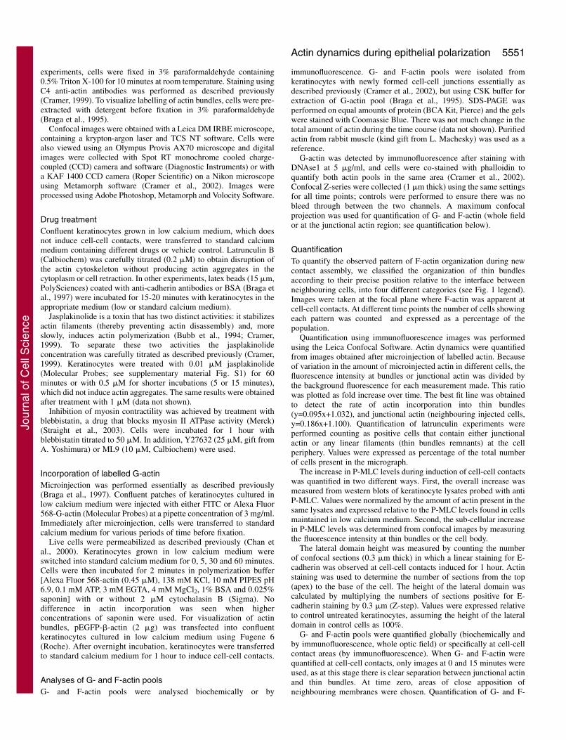
ResultsActin is organized into two spatial populations duringinduction of cell-cell contactsIn keratinocytes grown without cell-cell adhesion, thin actinbundles are already formed in a loose, broad band around thecell periphery (Low Ca2+, Fig. 1A) (Zamansky et al., 1991). Inthe absence of contacts, cadherin receptors are diffuselyexpressed at the cell surface and in the cytoplasm (see also Fig.2A) (Braga et al., 1995). As reported before, at early timepoints after junction initiation, F-actin is localized to cell-celladhesion sites, coincident with cadherin receptors in tworelated staining patterns (Adams et al., 1998; Adams et al.,1996). These patterns are: (1) actin dots associated with initial
cadherin puncta (within 2-4 minutes after contact initiation),and (2) more closely spaced actin dots aligned with cadherinreceptors that tend to appear as a wavy, thin punctate line(Adams et al., 1998; Adams et al., 1996).
We observed both patterns after new cell-cell contactformation: the punctate actin pattern and the wavy, thinpunctate line (Fig. 1A, 15 minutes Std. Ca2+, upper and lowerarrowhead, respectively). We define them together asjunctional actin. We surmise that this temporal transition in thetwo staining patterns for junctional actin represent continuedclustering of cadherin receptors and also actin association withother transmembrane proteins localized at cell-cell contacts.Separate from junctional actin we describe a new, distinctspatial and organizational population of actin, thin actinbundles. These bundles are in the same focal plane asjunctional actin, but clearly flanking junctional actin (arrows,Std. Ca2+, Fig. 1A). Thus, at early time points, two distinctactin populations can be separated by both position and byactin organization (see diagram in Fig. 1A). Moreover, thesetwo populations can also be distinguished at the molecularlevel, according to cytoskeletal proteins that localize to each
population (supplementary material Fig. S2).Thin actin bundles observed after initiation of
contacts seem similar to the looser array of bundlesdetected prior to cell-cell contact formation, except forone key difference, in that after initiation they appearmore tightly packed (Fig. 1A). Furthermore, with
Journal of Cell Science 118 (23)
Fig. 1. Spatial distribution of actin populations duringinduction of cell-cell contacts. (A) Keratinocytes grown inlow calcium medium (Low Ca2+), which does not inducecell-cell contacts, were induced to form cadherin-dependentcell-cell adhesion (standard calcium medium, Std. Ca2+).After junction formation, two actin populations can bedistinguished on the basis of their location: one is present asa wavy, punctate line at junctions (junctional actin,arrowheads); the second population is a tighter array of thinbundles in the cytoplasm, flanking the junctional-actinpopulation (arrow). By 60 minutes, the two populations areless readily distinguished (mature junctional actin). Bar, 50�m. (B) Percentage of keratinocytes classified into fourdifferent categories according to the spatial organization ofthin bundles following junction formation. Categoriesobserved: (1) a wide, loose band of actin filaments, and noF-actin at cell-cell borders; (2) F-actin at junctions is visible(junctional actin) and the band of filaments localizes closerto cell-cell contacts; (3) junctional actin is more intense;filaments are more tightly bundled in a narrower regionproximal to junctions or indistinguishable from F-actin atcell-cell contacts; (4) other phenotypes (less than 5-10% oftotal). Results are the average of two independentexperiments, in which 200 cells were scored per time pointin each experiment. (C) Following induction of cell-cellcontacts in sub-confluent keratinocytes, cytoskeletal changesshow a delayed formation of junctional actin andreorganization of thin bundles. For example, thin bundles arenot coincident with junctional actin after 120 minutes ofjunction formation. Additional actin structures are also seenbefore junctional actin is properly stabilized (i.e. kissingstructures, inset 30 minutes). Presumably these structuresparticipate in the migration of two neighbouring cellstowards each other as they are not readily observed inconfluent cultures.
Jour
nal o
f Cel
l Sci
ence

5553Actin dynamics during epithelial polarization
increased time after cell-cell contact formation (15-60minutes), there seems to be further tighter packing of thin actinbundles such that the array of bundles becomes narrower andmore closely positioned to junctional actin (Fig. 1B). By 60minutes, junctional actin and thin actin bundles could not bedistinguished in about 80% of cells (varies between 60-85%depending on keratinocyte strain and confluence; Fig. 1B). Asthis organization appears similar to that of actin at maturejunctions in keratinocytes (Braga et al., 1995; Hodivala andWatt, 1994), we define it as mature junctional actin (60minutes, Fig. 1A).
In a separate set of experiments, cell-cell junctions wereinduced in sub-confluent keratinocytes to test whether ourresults are applicable to cell-cell adhesion in general (Fig. 1C).In areas of similar sub-confluence, junctional actin formationwas only initiated after 30 minutes of new cell-cell contactformation and progressive reorganization of bundles towardsjunctions was clearly still occurring by 2 hours. In addition, asreported before (Vasioukhin et al., 2000), in sub-confluentkeratinocytes kissing structures (asterisks, Fig. 1C) were seenprior to the formation of junctional actin (arrowhead, Fig. 1C),suggesting that these structures extend to form continuousjunctional actin. Thus, the sequential order of events is similarin confluent and sub-confluent cells, arguing that actinreorganization occurs via similar processes. One exception isthat the formation of coincident junctional actin and thinbundles (seen by 1 hour in confluent keratinocytes) is severelydelayed in sub-confluent cells because of a requirement forkeratinocytes to move towards each other and initiate kissingstructures (data not shown).
The two spatial populations of actin have distinctdynamicsThe dynamics of junctional actin and thin bundles wereevaluated in two ways: actin incorporation, and sensitivity tolatrunculin (Fig. 2). In keratinocytes, within 20 minutes ofinduction of new contacts, incorporation of labelled actinmonomers is detected at cell-cell contacts; this event isinhibited if cadherin adhesion is blocked (Braga et al., 1997).However, the incorporation of labelled actin monomers intothin bundles and their dynamics have not yet been evaluated inepithelial cells.
When keratinocytes were maintained in the absence of cell-cell contacts, there was little incorporation of labelled actin atcell-cell boundaries or into actin bundles, suggesting extremelyslow dynamics (Low Ca2+, Fig. 2B). As reported for otherepithelial cell types, labelled actin recruitment to keratinocytecell-cell contacts occurred rapidly (t1/2=3 minutes) at siteswhere cadherin receptors localized and increased with time(arrowhead Fig. 2A, and data not shown) (Adams et al., 1998;Adams et al., 1996; McNeill et al., 1993). By contrast,incorporation of fluorescent actin monomers into thin bundlesoccurred more slowly (t1/2=12 minutes; arrow Fig. 2B, and datanot shown). Quantification of the above results revealed thatthe rate of actin incorporation at thin bundles (slope of graph0.095) was slower than determined for junctional actin (slopeof graph 0.186, Fig. 1C; see Materials and Methods for details).These data suggest that junctional actin is more dynamic thanthin bundles.
As a second approach, differences in dynamics in the two
actin populations were addressed by sensitivity to latrunculinB (Fig. 2D). Latrunculin sequesters actin monomers,preventing reassembly, and thus more readily causes loss ofactin structures with higher filament turnover (fast dynamics;see Materials and Methods for optimization of the conditions).In untreated cells, the junctional actin and thin bundles wereclearly seen (DMSO, see enlargement, Fig. 2D). Aftertreatment with latrunculin for 5 minutes, junctional actin hadmostly disappeared from sites of adhesion, whereas thinbundles are still present (but qualitatively different fromcontrols). After 15 minutes of new contact formation in thepresence of latrunculin, there was a fourfold reduction in theproportion of cells that contained junctional actin (Latr.enlargement, Fig. 2D,E). However, remnants of thin bundleswere still observed in around 60% of keratinocytes (arrows,Latr., Fig. 2D,E). Thus complete disassembly of thin bundles(>15 minutes) takes much longer than disassembly ofjunctional actin (5 minutes). This result is consistent with theanalysis of labelled actin incorporation (Fig. 2A-C) andsuggests that flanking bundles are more stable and have slowerturnover than junctional actin.
In assessing the effect on junctional actin, it is important todistinguish whether latrunculin prevents actin recruitment topuncta or the process of receptor clustering itself. Todistinguish between these possibilities, as control we clusteredcadherin receptors using antibody-coated beads, a processknown to recruit actin and mimic early signalling events aftercontact assembly (supplementary material Fig. S3) (Betson etal., 2002; Braga et al., 1997). In the presence of latrunculin,actin recruitment is unable to occur, even though cadherinreceptors are clustered around the beads (supplementarymaterial Fig. S3). Thus, latrunculin treatment affectedprimarily new actin polymerization at puncta and not cadherinclustering per se. These results indicate that cadherin clusteringis indirectly perturbed when actin recruitment is inhibited bylatrunculin, leading to the removal of cadherin receptors fromcell-cell contacts (as seen in Fig. 2D). The concentration ofcalcium ions in the medium did not affect actin recruitmentsubstantially, in spite of the reported increase in intracellularcalcium levels (supplementary material Fig. S3) (Sharpe et al.,1993). Taken together, the above results indicate that,following cadherin-dependent adhesion, actin polymerizationoccurs at two distinct intracellular sites with differentdynamics: at cell-cell contacts (faster dynamics) and atflanking bundles (slower dynamics).
What is the contribution of assembly mechanisms tojunctional actin and thin bundles?Actin assembly mechanisms were addressed in terms of the G-to F-actin ratio, filament polarity of actin polymerization andsupply of actin monomers to fuel polymerization (Figs 3, 4).Under our conditions, G- and F-actin can be extracted, and, asexpected, latrunculin treatment (as per Fig. 2D) results in atwofold increase in the levels of G-actin (Fig. 3A) (Cramer etal., 2002). Thus, our conditions are adequate to detect changesin the G- to F-actin ratio.
Initially, we investigated whether junction formationincreased the total levels of polymerized actin in keratinocytes(F-actin) during a time course of junction assembly (Fig.3B,C). Apart from a slight alteration at 15 minutes, the ratio
Jour
nal o
f Cel
l Sci
ence

5554
of monomeric and filamentous actin did not vary afterinduction of cell-cell adhesion (G-actin and F-actin,respectively; Fig. 3C). These results suggested that there is nota major shift towards actin polymerization or depolymerizationduring junction formation. However, it is clear that junction
formation induces new actin polymerization at puncta (Fig. 2)(Adams et al., 1996; Braga et al., 1997). Most probably thislocalized process is beyond the sensitivity of the globalmethods used in this study and reflects the small proportion oftotal actin at junctions. Indeed, when G- and F-actin pools were
Journal of Cell Science 118 (23)
Fig. 2. Junctional actin and thin bundles have distinct dynamics. Keratinocytes grown in low calcium medium (Low Ca2+), which does notinduce cell-cell contacts were microinjected with labelled actin (3 mg/ml pipette concentration), and transferred immediately to standardcalcium medium to induce cell-cell contacts (Std. Ca2+). Control cells were maintained in low calcium medium. Arrowheads point to junctionalactin; arrows show thin bundles. (A) Junctional actin has fast dynamics. After junction formation, images were collected at places where E-cadherin clustering was visible and at the corresponding incorporated actin. (B) Thin bundles have slower kinetics of actin incorporation.Images were collected where filamentous actin (total actin) was present and the corresponding new actin labelling at the bundles (incorporatedactin). In the absence of cell-cell contacts (Low Ca2+), very little actin incorporation was detected into thin bundles. (C) Quantification of actindynamics. Junctional-actin fluorescence intensity was measured only where cadherin recruitment at junctions was observed. Thin bundles weredetected in phalloidin stained images and the fluorescence intensity of labelled actin was measured in the corresponding area. (D) Thin bundlesare less sensitive than junctional actin to latrunculin treatment. Keratinocytes were induced to form contacts (5 and 15 minutes, Std. Ca2+) in thepresence of 0.2 �M latrunculin B (Latr.) or vehicle (DMSO); cells were fixed and stained with phalloidin (total actin) and anti-E-cadherinantibodies. Although the amount and organization of thin bundles are affected by latrunculin, thin bundles are clearly seen after 5 minutesincubation, when the majority of junctional actin was removed. (E) Quantification of the proportion of cells showing thin bundles andjunctional actin in controls (C) or after latrunculin treatment (Latr., at 5 and 15 minutes). Error bars represent error obtained from at least twoindependent experiments. Bar, 50 �m.
Jour
nal o
f Cel
l Sci
ence

5555Actin dynamics during epithelial polarization
quantified specifically at cell-cell contacts, a significantincrease in the F-actin pool was observed when compared toF-actin levels in the absence of cell-cell adhesion (time 0minutes, P<0.05, Fig. 3C).
We next investigated the contribution of the two spatial actinpopulations detected to filament assembly at filament barbedand pointed ends, as both filament ends have been shown to becompetent for assembly in other cell systems (Littlefield et al.,2001). We tested this by briefly permeabilizing keratinocyteswith a buffer containing labelled actin in the presence orabsence of cytochalasin B (Fig. 3D) (Chan et al., 2000). Toassess the assembly at filament-barbed ends (the expectedpreferred site of assembly) we used labelled actin at 0.45 �M,which is well below the critical concentration for assembly atthe pointed end (around 1 �M) and thus only allows assemblyat the barbed end. Cytochalasin B binds to the barbed ends andprevents polymerization at these sites, but allows incorporation
at filament pointed ends (Littlefield et al., 2001). Importantly,the brief treatment with cytochalasin B (2 minutes) is sufficientto block barbed ends but does not disrupt actin structures insidethe cell (total actin, Fig. 3D).
In the absence of cell-cell contacts (Low Ca2+), noincorporation of actin was observed (Fig. 3D). When junctionswere induced in the absence of cytochalasin B, junctional actinwas labelled as early as 5 minutes and the intensity of labellingand proportion of labelled cells increased with time (up to 60minutes, arrows Fig. 3D and data not shown). At all timepoints, punctate labelling was heterogeneous, colocalized withnewly formed cadherin-dependent cell-cell contacts, and thusreflects incorporation into junctional actin (Fig. 3D). Treatmentwith cytochalasin B substantially reduced actin labelling atcell-cell adhesion sites (incorporated actin). The residual actinlabelling can be accounted for by a small proportion of G-actinwe found at junctions (data not shown). Actin incorporation
Fig. 3. Actin assembly mechanisms. (A) G- and F-actinpool were isolated from keratinocytes treated with 0.2�M latrunculin, or left untreated, for 5 minutes. Afterseparation using SDS-PAGE and Coomassie staining,bands were quantified. After latrunculin treatment, therewere increased levels of G-actin, with a concomitantreduction in the amount of F-actin. (B) Global levels ofG-actin and F-actin pools were quantified byimmunofluorescence during a time course afterinduction of cell-cell contacts. Keratinocytes were stained with DNAse1 (G-actin) and phalloidin (F-actin) and the whole optical fieldquantified for each fluorophore. No significant changes were observed in the relative concentration of either actin pool during the time courseexamined by staining or SDS-PAGE (data not shown). Values are expressed as percentage of total actin. (C) Quantification ofimmunofluorescence of G- and F-actin pools at the junctional-actin region. Only junctions where junctional actin was clearly separated fromthin bundles were examined. At time zero, areas of close apposition of neighbouring membranes were quantified. Data in A-C arerepresentative of two independent experiments. Error bars represent standard deviation. *P<0.05. (D) Polymerization of actin at cell-celljunctions occurs preferentially via the barbed end. After formation of new contacts for different amounts of time, cells were permeabilized for 2minutes in cytoskeleton buffer containing labelled Alexa Fluor 568-G-actin (0.45 �M), in the presence or absence of 2 �M cytochalasin B (seetext for details). After fixation, E-cadherin, total actin (phalloidin) and incorporated actin were visualized. Results are representative of at leastthree independent experiments. Arrows indicate labelled actin incorporation at junctions. Bar, 50 �m.
Jour
nal o
f Cel
l Sci
ence
5556
appeared to reach a steady state after 1 hour of cell-celladhesion formation (data not shown). As expected from theshort pulse of labelling (2 minutes) and the known half-life(Fig. 2C), no bundles were labelled under these conditions.Taken together, these results indicated that junctional actin isassembled by new actin polymerization at barbed ends.
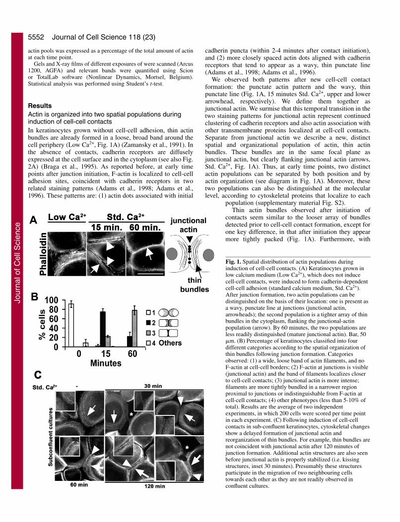
Actin filament disassembly does not play a major role inthe formation of either thin bundles or junctional actinDuring junction assembly, actin monomers from two distinctsources can potentially contribute to the newly polymerizedactin: previously stored monomers, newly disassembled actin,or both. To determine whether recently disassembled actinfilaments might contribute monomers for recycling andreassembly at junctions, actin filaments were stabilized bytreatment with jasplakinolide (see Materials and Methods fortitration; Fig. 4). After induction of contacts in the presence ofjasplakinolide for up to 15 minutes (a time when both actinpopulations are detectable), junctional actin was observed in asimilar pattern to that found for controls (arrowhead, Std. Ca2+,Fig. 4A and data not shown).
To overcome the poor labelling of bundles by anti-actinantibody (C4), GFP-actin was expressed in keratinocytes andcells incubated with jasplakinolide during induction of cell-cellcontacts for 60 minutes (Fig. 4B). At this time, the twospatially distinct populations of actin will be detectable in aproportion of cells whereas in others the two actin populationswill be coincident (Fig. 1). When jasplakinolide-treatedkeratinocytes were compared with controls, there was notmuch difference in the percentage of cells in which bundlesand junctional actin were no longer distinguishable (category
3, Fig. 4C). Thus, our data indicate that bundles are notdisassembled and reassembled at a position closer to junctionalactin during maturation of contacts. Moreover, our resultssuggested that actin depolymerization is not a major supply ofactin monomers for incorporation in either junctional actin orthin bundles.
What is the contribution of myosin contractility tojunctional actin and bundles?When myosin II was inhibited (by blocking myosin light chainkinase), no effect on the clustering of cadherin receptors atpuncta (Vaezi et al., 2002) or maintenance of stable, alreadyassembled junctions was found (Bhowmick et al., 2001;Krendel et al., 1999; Sahai and Marshall, 2002). Here, weaddressed the role of contractility for the formation of thetwo actin populations. We selected blebbistatin, a newlycharacterized inhibitor of myosin II (see Materials andMethods for optimization) (Straight et al., 2003), which isadvantageous over previous myosin II inhibitors as it directlyblocks myosin II ATPase, is rapid, and highly specific (Straightet al., 2003).
Bundles are clearly detected in controls (DMSO, insets, Fig.5), but not in blebbistatin-treated keratinocytes (*, insets, Fig.5). In contrast, junctional actin was able to form at sites of cell-cell contact (arrows, Bleb., inset, Fig. 5). However, junctionsthat formed in the presence of blebbistatin appeared less robust,with weaker and more punctate actin/cadherin staining insteadof a continuous line as seen in controls (arrowheads, Bleb., Fig.5). We also obtained similar results when using other myosinII inhibitors (Y27632 and ML9; data not shown). However,the blebbistatin effects were more potent and observed
Journal of Cell Science 118 (23)
Fig. 4. Actin filament disassembly does not playa major role in the formation of either thinbundles or junctional actin. (A) Recentlydisassembled filaments do not contributemonomers for incorporation at cell-cell junctions.Cell-cell contacts were induced (Std. Ca2+) in thepresence of 0.5 �M jasplakinolide (Jasp.) orvehicle control (methanol, MeOH). Cells werestained with anti-E-cadherin and anti-actinantibodies, because jasplakinolide competes withphalloidin for F-actin interaction. (B) Actindisassembly is not required for bundlereorganization. Keratinocytes expressing GFP-actin were treated with 0.02 �M jasplakinolide(Jasp.) or methanol (MeOH) during junctionassembly. After 60 minutes, thin bundles werevisible either as flanking filaments to junctionalactin (middle images Jasp.) or coincident tojunctions (bottom images, Jasp.). Arrows indicatethin bundles; arrowheads point to junctionalactin. (C) Keratinocytes were classified into fourdifferent categories according to the spatiallocalization of thin bundles as described in Fig. 1legend. The same proportion of cells was foundin category 2 and 3 for both the Jasplakinolide-treated (Jasp.) and untreated (control) cells. Bar,50 �m.
Jour
nal o
f Cel
l Sci
ence

5557Actin dynamics during epithelial polarization
homogeneously throughout the monolayer.Thus, myosin II contractility is not required forjunctional-actin assembly, but is essential forthin bundle stability.
We stained with phosphorylated myosinlight chain (P-MLC) to test which of the actinpopulation was contractile during induction ofcell-cell contacts. In cells without adhesionthere was minimal localization of P-MLC inperipheral bundles (Low Ca2+, Fig. 6),indicating that bundles formed prior to contactinitiation are not contractile. In contrast, oncecontacts were initiated, P-MLC was abundanton peripheral actin bundles (arrows, 30 and 60minutes, Fig. 6). Interestingly, P-MLC wasclearly absent from junctional actin (asterisks,Fig. 6). These results are also consistent withthe ability of cadherin receptors to recruit andassemble actin at cell-cell contacts in theabsence of myosin function (Fig. 5).
We next quantified the increase in P-MLClevels after induction of cell-cell contacts,using western blots and confocal images. After30 minutes of junction formation, the intensityof P-MLC fluorescence at thin bundles was2.7-fold higher than levels seen at bundles inthe absence of cell-cell adhesion (Low Ca2+,Fig. 7A). A smaller increase in P-MLC levelswas also observed in the cell body (1.4 fold,Fig. 7A). Furthermore, overall levels of P-MLC were also increased, as seen by westernblots (Fig. 7B). Thus enhanced P-MLC levelscan be detected both spatially and globallyafter junction assembly, but the increase inphosphorylated MLC is most marked at thinbundles.
What is the role of actin bundles duringepithelial polarization?The recognition of actin bundles flankingjunctional actin and their progressive bundlingduring junction formation is newly describedand thus we began to investigate their function.As thin bundles are not required for the initialactin recruitment to junctions (Fig. 5), afunction during the later stages of epithelialpolarization was investigated. We focused on the formation ofthe cuboidal morphology, the major cell shape change thatoccurs during the maturation step of polarization.
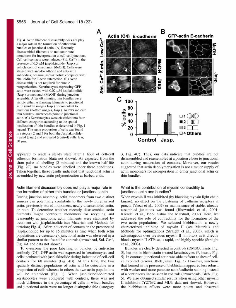
A cuboidal morphology is achieved when cell-cell contactsform a distinct, tall lateral domain perpendicular to the base ofthe cell. We sought to evaluate whether inhibition ofcontractility during junction assembly could affect the heightof lateral domains during polarization. Confocal Z-series ofkeratinocytes incubated in the presence or absence ofblebbistatin during induction of cell-cell contacts werecollected and used for 3D reconstructions or quantification(Fig. 8). Control cells show a uniform linear staining for E-cadherin coincident with junctional actin (control, Fig. 8A).Blebbistatin treatment induced the breakdown of thin bundles
and the formation of small ruffles all over the cell surface,but particularly concentrated at sites of cell-cell contacts(blebbistatin, arrows, Fig. 8A,B). Underneath the small ruffles,cadherin-dependent contacts were present as a continuousthinner line (Fig. 8B),
Z-projections are usually used to determine cell height.However, the presence of small ruffles on top of junctionsprevented quantification of cell height in this manner. Tocircumvent this problem, we measured the height of the lateraldomain, which increases upwards during polarization to forma cuboidal morphology. We quantified the number of confocalsections (0.3 �m) in which linear, continuous cadherin stainingis seen (basal to apical distance, Fig. 8B,C). Cells maintainedin low calcium medium show a small area of contact between
Fig. 5. Contribution of myosin contractility to junctional actin and thin bundles.Confluent keratinocytes grown in low calcium medium (Low Ca2+) or induced to formjunctions (Std. Ca2+) for 1 hour were incubated in the presence or absence ofblebbistatin (Bleb.). Controls were treated with DMSO. Enlargements of the boxedregions in each image are shown on the right. In the absence of cell-cell contacts(Low Ca2+) thin bundles are present in control (DMSO-treated) cells, but not inblebbistatin-treated cells (Bleb.) After cell-cell adhesion, junctional actin is formed inthe absence of thin bundles (Bleb. Std. Ca2+). Asterisks indicate the absence of thinbundles in blebbistatin-treated keratinocytes; arrow indicates junctional actin andarrowheads, wavy junctions. Images are representative of at least three independentexperiments. Bar, 50 �m.
Jour
nal o
f Cel
l Sci
ence

5558
neighbours, presumably mediated by calcium-independentreceptors (around 2 �m high) (Braga et al., 1998). Followinginduction of cell-cell contacts for 1 hour, control keratinocyteswere not yet fully cuboidal (lateral domain was around 4.5 �mhigh as opposed to 6 �m measured at the apex on top of thenucleus of the cell). After inhibition of myosin II-dependentcontractility, the height of the lateral domain was reduced byaround 35% to 3 �m (P<0.001, Fig. 8C). Thus, an increase in
height of the lateral domain during polarization requires thepresence of acto-myosin bundles.
DiscussionWe demonstrate that, after initial cell-cell contact in confluentkeratinocytes, epithelial morphological polarization is inducedrapidly. During keratinocyte polarization, we found two spatialactin populations, junctional actin and thin bundles, which canbe distinguished by their dynamics, molecular components,mechanism of formation and function (Fig. 1, Fig. 9A andsupplementary material Fig. S2). We define junctional actin asa higher order of puncta organization forming a linear array ofactin staining at junctions. Although E-cadherin clustering isthe initial trigger for punctum assembly, additional receptorspresent at cell-cell contacts also contribute to actin recruitmentto form junctional actin. Thin bundles are a pre-formed arrayof loosely associated actin filaments found in the absence ofcell-cell contacts, distinct from stress fibres in their spatialorganization.
Within 1 hour, discrete, stepwise and sequential changes inthe actin cytoskeleton occur (Fig. 9). Similar actin populationsand microfilament reorganization are observed in sub-confluent cultures, but a lag phase is seen as cells have to
Journal of Cell Science 118 (23)
Fig. 6. Following cell-cell junction formation, phosphorylatedmyosin light chain (P-MLC) localizes at thin bundles but notjunctional actin. Keratinocytes grown in low calcium medium (LowCa2+) were induced to form junctions (Std. Ca2+) and stained withphalloidin and anti-P-MLC. Merged confocal images are shown onthe right; Enlargements of the boxed regions are shown below eachimage. After induction of cell-cell contacts, P-MLC is concentratedat peripheral thin bundles, but absent from junctional actin (Std.Ca2+). Arrowheads show junctional actin, arrows point to thinbundles and localization of P-MLC. Asterisks show absence of P-MLC at junctional actin. Bar, 32 �m; 8 �m for enlargements.
Fig. 7. Spatial and global quantification of P-MLC levels duringkeratinocyte polarization. (A) P-MLC levels were quantified at thinbundles and in the cell body from confocal images obtained after 30minutes of induction of cell-cell contacts. Following junctionformation (Std. Ca2+), an increase in fluorescence intensity is seenboth at thin bundles (2.7 fold) and in the cell body (1.4 fold). Valuesare from two independent experiments, using at least 50 differentcells per condition. (B) Western blots showing the overall increase inP-MLC levels in keratinocytes. Quantification reveals that a threefoldincrease in phosphorylation levels is seen after 15 minutes of newjunction formation (P<0.001). Values are the mean of fourindependent experiments.
Jour
nal o
f Cel
l Sci
ence
5559Actin dynamics during epithelial polarization
migrate towards each other prior to initiation of junctions (Fig.1C). Thus the results described here are likely to be importantfor junction formation in general, i.e. following cytokinesis,resealing of epithelial sheets, wound healing andmesenchymal-epithelial conversion during development.
Junctional actin has fast dynamics while thin bundles showslow incorporation of monomers. Actin incorporation at cell-cell junctions (t1/2=3 minutes) is slightly longer thanincorporation of labelled actin in lamellipodia (Adams et al.,1998; Adams et al., 1996; McNeill et al., 1993; Theriot andMitchison, 1991). The slow kinetics of incorporation into thinbundles (t1/2=12 minutes) is similar to that observed into stressfibres (Amato and Taylor, 1986) and circumferential rings in acultured epithelial cell line (L.P.C., unpublished data).
To what extent the actin structures we observe are furtherrelated to other actin structures remains to be directly tested.Flanking actin bundles are more related to circular actin ringsthan stress fibres because of their peripheral spatial distributionand circular orientation within cells. A common theme foractin incorporation at new cell-cell contacts, lamellipodia andfilopodia is that assembly occurs with filament barbed endsfacing the plasma membrane. However, further filamentelongation differs in that junctional actin filament elongationis apparently restricted to within sub-microns of the cell edge,whereas lamellipodia and filopodia can be 2-10 �m deep. Thisinfers differences in precise regulation of actin incorporation,spatial orientation of filament growth and/or length offilaments.
With respect to the mechanism of formation of junctionalactin and thin bundles (Fig. 9A), our findings on actinincorporation and latrunculin argue that actin filamentassembly is a major contributor to junctional actin, but lessimportant for flanking bundles (the latter has reducedsensitivity to latrunculin and thus less reliance on rapidassembly for formation). Actin filament disassembly has noclear role for the formation of either actin population,suggesting that the required monomer for incorporation issupplied from the stored pool of actin monomer (as opposedto recently disassembled actin filaments). Conversely, myosinII function is apparently more important for bundle formation:thin bundles dissolve when myosin II is inhibited, but actinrecruitment at new junctions can occur (this work) (Vaezi etal., 2002). In addition, myosin II-induced contraction is notrelevant to the initial clustering of cadherins at sites of cell-cellcontacts.
How exactly does myosin II contribute to flanking bundlereorganization? Given that actin assembly plays only a minorrole and disassembly of filaments is not required, a mechanismthat relies at least in part on pre-existing filaments seems likely.One idea is a progressive bundling of pre-existing filaments(loose array of filaments observed in cells prior to junctionformation, Fig. 9B). While we can not exclude the participationof bundling proteins in this process, myosin II-inducedcontractility does play a role. Support for this comes fromincreased levels of P-MLC at bundles (2.7-fold, Fig. 7A) anda progressive narrowing of the loose band of filaments towards
Fig. 8. Inhibition of contractility duringkeratinocyte polarization perturbs theacquisition of maximum height atlateral domains. E-cadherin (red) andphalloidin (green) staining wasperformed after 1 hour of cell-cellcontact formation in the absence(control) or presence of blebbistatin(blebbistatin). Confocal sections (0.3�m) were collected at different levelsthrough a keratinocyte monolayer andprocessed as follows. (A) Threedimensional reconstruction. Afterblebbistatin treatment, F-actin isdiffusely localized and cadherinstaining is found as a wavy linebetween neighbouring cells. Smallruffles/projections are seen at the cellsurface and on top of junctions(arrows). (B) Confocal sections takenfrom basal to top of the monolayer at 0,1.5, 3, 4.5 and 6 �m. Arrow points tosmall ruffles/projections sees at the topof junctions. Arrowhead showscadherin staining in controls whereasno similar linear cadherin staining isobserved in blebbistatin-treated cells.(C) Quantification of the maximumheight of the lateral domains (basal toapical distance). Confocal sectionsshowing linear cadherin staining were quantified and values expressed relative to control, untreated cells polarized for 1 hour (see Materials andMethods for details). Maximum height seen in lateral domain of control cells is 4.5 �m compared with 3 �m in blebbistatin-treated cells and 2�m in cells maintained in low calcium (Braga et al., 1998). After blebbistatin treatment, lateral domain height is decreased by 35% (*P<0.001).Results represent the mean of seven different Z-series collected from two independent experiments.
Jour
nal o
f Cel
l Sci
ence

5560
junctional actin shortly after junctions are assembled (Fig. 1,Fig. 9B). Significantly, peripheral actin bundles are onlycontractile once contacts are initiated (Fig. 6, Fig. 7A),inferring signalling to myosin II to activate contraction atbundles
The above mechanism differs from models in the literaturein which disassembly/reassembly of bundles at a locationcloser to junctions is proposed (Krendel et al., 1999; Krendeland Bonder, 1999). This discrepancy may be explained by thefact that in these studies, cells migrate to adhere to their
neighbours. Thus different/additional pathways for actinformation may occur in sub-confluent cultures until junctionalactin is stabilized, whereas we can exclude motility in ourexperimental system (see Fig. 1).
One important issue is that we do not yet know whetherjunctional actin and thin bundles co-exist as separate dynamicpopulations beyond the formation of a mature junction by 1hour. We favour the co-existence of the two populations asactin bundles and mature junctions themselves can be resolvedby electron microscopy (Green et al., 1987; Owaribe et al.,
Journal of Cell Science 118 (23)
Fig. 9. Summary of changes to themicrofilament network triggered byformation of cadherin-dependent cell-cellcontact. (A) Mechanisms of formationand functions of junctional actin and thinbundles. After cadherin receptors cluster,two distinct processes are observed: newactin polymerization and filamentreorganization. Actin assembly is a majorcontributor to junctional actin (thickbrown arrow), but has a minor effect onthin bundles (dashed thin brown arrow).In contrast, myosin function is essentialfor bundle stability, but does not affectinitial clustering of cadherin receptors orjunctional-actin formation. All the aboveevents do not occur in cells maintained inlow calcium medium or in whichcadherin function is blocked. Twoseparate functions are identified (redarrows): junctional actin stabilizescadherin receptors at puncta and thinbundles are essential for development ofmaximum height of the lateral domains.At later time points, these two actinpopulations apparently colocalize at cell-cell contacts to form mature junctionalactin (merged blue and yellow lines; seebelow). *Adams et al., 1996; Nelson,2003. (B) Temporal and spatial events.After initiation of cell-cell adhesion,distinct cellular processes are detected atdifferent time points (horizontal bars).Once initiated, each event increases untila plateau after 60 minutes of cell-celladhesion. E-cadherin clustering at punctais observed within a couple of minutes,and is followed shortly by new actinincorporation as junctional actin (t1/2=3minutes, fast dynamics) and then by thinbundle labelling (approximately t1/2=12minutes; slower dynamics). Increased P-MLC localization at bundles and bundlereorganization occurs later (from 15minutes). The end result is the formationof a cuboidal epithelial morphology, withthe spatial organization of microfilamentsas shown. A possible mechanism,supported by our results, is myosin-dependent contraction of thin bundlesfrom a loose band of filaments at theperiphery to filaments that are coincident with junctional actin. Please see text for functional implications and more details. Arrows indicatebundles; arrowheads indicate junctional actin, open arrow indicates mature junctional actin.
inside
inside
outsideCa2+cadherin
α-cateninβ-catenin
new actin incorporation reorganization pre-existing filaments
junctional actin thin bundles
dynamic via barbed ends stored G-actin pool no effect Ca2+
less dynamic
Height oflateraldomains
Stabilizesclusteredcadherin*
increased P-MLC binding bundling + contractility
mature junctional actin
Mechanisms and functions
Temporal and spatial events
0 5 15 30 60
E-cadherin at cell-cell contacts
G-actin incorporation:junctional actin
thin bundles
bundle reorganization
B
(minutes after cell-cell contacts)
Low Ca2+ Std. Ca2+
2 - 15 min > 15 min > 60 min
new contact assembly stabilization
maturation polarized cell shape
increased P-MLC at bundles
A
Jour
nal o
f Cel
l Sci
ence

5561Actin dynamics during epithelial polarization
1981; Yonemura et al., 1995; Zamansky et al., 1991). Thusin mature junctions, the thick bundles visualized byimmunofluorescence result from the contribution of junctionalactin and compacted, bundled thin filaments.
Presumably, as myosin contraction is required for bundlingand bundles later coincide with junctional actin, myosin II mayhave some indirect role in junctional-actin stability (but notassembly per se). This is perhaps observed as a more irregulardistribution of junctional actin (this work), and junctionmorphology has an immature, wavy shape when myosin II isblocked (Fig. 5) (Krendel et al., 1999; Vaezi et al., 2002;Yonemura et al., 1995). Further experiments are needed tovalidate whether the coalescence of distinct puncta into astraight, continuous line requires acto-myosin-generatedtension.
While the function of actin at puncta has been previouslyidentified, so far no specific role for bundles has been assigned.Here, we suggest a novel role for thin bundle reorganizationand actomyosin contraction in the development of epithelialmorphology: increase in height of the lateral domain to forma cuboidal cell. We argue that this is a specific function offlanking bundles. First, blebbistatin treatment maintains thelateral domain at similar height to cells grown in low calciummedium (around 2-3 �m) as opposed to control cells polarizedfor 1 hour (Fig. 8) (Braga et al., 1998). Second, followingjunction assembly, thin bundles are the major contractile actinpopulation detected, as opposed to junctional actin (Figs 6, 7).Third, the apparent progressive bundling of filaments andincrease in MLC phosphorylation are temporally coincident(Fig. 9B). This argues strongly that bundling and contractionof actin bundles correlate with the increase in height of thelateral domain and cuboidal morphology. However, additionalroles for thin bundles may yet be uncovered, such as thepotential crosstalk with junctional actin mentioned above.
Thus, the cytoskeletal changes reported here occur quickly(within 1 hour), in a precise sequence that reflects theirrequirements and specific functions (Fig. 9B). We concludedthat during epithelial polarization, the formation of actinstructures at junctions is achieved by cooperation ofcontraction and assembly of two spatially distinct actinpopulations (Fig. 9A). Similar cooperation between actinassembly and myosin II contractility for overall cell functionoccurs in other cell systems, such as cell migration (Ridley etal., 2003) and healing of circular wounds in Xenopus oocytes(Mandato and Bement, 2001).
The proposed contractile role of the newly described thinbundles in polarization is consistent with the importance of cellcontraction for epithelial morphogenesis: compaction (Adamset al., 1998; Clayton et al., 1999; Collins and Fleming, 1995),tubulogenesis (Wozniak et al., 2003) and elongation in C.elegans (Wissmann et al., 1999). In addition, during junctionassembly in primary keratinocytes up-regulation of Rho smallGTPase activity occurs (data not shown) (Calautti et al., 2002).Interestingly, in tumorigenic and non-tumorigenic cell linesRho is not activated after cadherin adhesion (Noren et al.,2001), and instead increased contractility may disrupt cell-cellcontacts (Sahai and Marshall, 2002; Zhong et al., 1997). Yet,in some cell lines it has been proposed that increasedcontractility may disrupt cell-cell contacts (Sahai and Marshall,2002; Zhong et al., 1997). These results suggest that a tighttemporal and spatial regulation of Rho activation and
contractility is required for epithelial homeostasis andmorphogenesis.
Our data show that cadherin-mediated adhesion is able tomodulate actin dynamics. After cell-cell contacts are initiated,an increase in actin turnover, new actin incorporation, myosinII phosphorylation, contraction and bundling are observed (Fig.9A). These events are focused on two different actinpopulations, junctional actin and thin bundles that, at early timepoints during keratinocyte polarization, can be separatedmechanistically (Fig. 9A) and spatially (Fig. 9B). Mostimportantly, the unique roles of junctional actin (cadherinclusters stability) and thin bundles (height increase at lateraldomains) cooperate to induce overall morphological change(cuboidal cell shape). Thus, following keratinocyte junctionassembly, two actin populations with two distinct functions arerequired to develop a polarized epithelial cell.
We would like to thank T. Mitchison, M. Takeichi, L. Machesky,M. Matsuda and A. Yoshimura for generous gift of reagents. We thankY. Korchev and J. Gorelik for discussions and suggestion of cell heightexperiments. V.B. is a MRC Senior Research Fellow; M.B. was a PhDstudent from the graduate programme at the MRC Laboratory forMolecular Cell Biology, University College London. L.C. is a RoyalSociety University Research Fellow. This work was generouslysupported by the Medical Research Council to V.B. and by theWellcome Trust, Human Frontiers Science Programme Organizationand the Royal Society (UK) to L.C.
ReferencesAdams, C. L., Nelson, W. J. and Smith, S. J. (1996). Quantitative analysis
of cadherin-catenin-actin reorganization during development of cell-celladhesion. J. Cell Biol. 135, 1899-1911.
Adams, C. L., Chen, Y.-T., Smith, S. J. and Nelson, W. J. (1998).Mechanisms of epithelial cell-cell adhesion and cell compaction revealed byhigh-resolution tracking of E-cadherin-green fluorescent protein. J. CellBiol. 142, 1105-1119.
Amato, P. A. and Taylor, D. L. (1986). Probing the mechanism ofincorporation of fluorescently labeled actin into stress fibers. J. Cell Biol.102, 1074-1084.
Betson, M., Lozano, E., Zhang, J. and Braga, V. M. M. (2002). Racactivation upon cell-cell contact formation is dependent on signaling fromthe epidermal growth factor receptor. J. Biol. Chem. 277, 36962-36969.
Bhowmick, N. A., Ghiassi, M., Bakin, A., Aakre, M., Lundquist, C. A.,Engel, M. E., Arteaga, C. L. and Moses, H. L. (2001). Transforminggrowth factor-�1 mediates epithelial to mesenchymal transdifferentiationthrough a RhoA dependent mechanism. Mol. Biol. Cell 12, 27-36.
Braga, V. (2000). Cadherin adhesion regulation in keratinocytes. In Cell-CellInteractions: a practical approach (ed. T. Fleming), pp. 1-36. Oxford:Oxford University Press.
Braga, V. M. M., Hodivala, K. J. and Watt, F. M. (1995). Calcium-inducedchanges in distribution and solubility of cadherins and their associatedcytoplasmic proteins in human keratinocytes. Cell Adhes. Commun. 3, 201-215.
Braga, V. M. M., Najabagheri, N. and Watt, F.M. (1998). Calcium-inducedintercellular adhesion of keratinocytes does not involve accumulation of �1integrins at cell-cell contact sites and does not involve changes in the levelsor phosphorylation of the catenins. Cell Adhes. Commun. 5, 137-139.
Braga, V. M. M., Machesky, L. M., Hall, A. and Hotchin, N. A. (1997). Thesmall GTPases Rho and Rac are required for the establishment of cadherin-dependent cell-cell contacts. J. Cell Biol. 137, 1421-1431.
Bubb, M. R., Senderowicz, A. M. J., Sausville, E. A., Duncan, K. L. K. andKorn, E. D. (1994). Jasplakinolide, a cytotoxic natural product, inducesactin polymerization and competitively inhibits the binding of phalloidin toF-actin. J. Biol. Chem. 269, 14869-14871.
Calautti, E., Grossi, M., Mammucari, C., Aoyama, Y., Pirro, M., Ono, Y.,Li, J. and Dotto, G. P. (2002). Fyn tyrosine kinase is a downstreammediator of Rho/PRK2 function in keratinocyte cell-cell adhesion. J. CellBiol. 156, 137-148.
Jour
nal o
f Cel
l Sci
ence

5562
Chan, A. Y., Bailly, M., Zebba, N., Segall, J. E. and Condeelis, J. S. (2000).Role of cofilin in epidermal growth factor-stimulated actin polymerizationand lamellipod protusion. J. Cell Biol. 148, 531-542.
Clayton, L., Hall, A. and Johnson, M. H. (1999). A role for the Rho-likeGTPases in the polarisation of mouse eight-cell blastomeres. Dev. Biol. 205,322-331.
Collins, J. and Fleming, T. P. (1995). Epithelial differentiation in the mousepre-implantation embryo: making adhesive cell contacts for the first time.Trends Biochem. Sci. 20, 307-312.
Cramer, L. P. (1999). Role of actin-filament disassembly in lamellipodiumprotrusion in motile cells revealed using the drug jasplakinolide. Curr. Biol.9, 1095-1105.
Cramer, L., Briggs, L. and Dawe, H. (2002). Use of fluorescently labelleddeoxyribonuclease I to spatially measure G-actin levels in migrating andnon-migrating cells. Cell Mot. Cytoskeleton 51, 27-38.
Gloushankova, N. A., Krendel, M. F., Alieva, N. O., Bonder, E. M., Feder,H. H., Vasiliev, J. M. and Gelfand, I. M. (1998). Dynamics of contactsbetween lamellae of fibroblasts: essential role of the actin cytoskeleton.Proc. Natl. Acad. Sci. USA 95, 4362-4367.
Green, K. J., Geiger, B., Jones, J. C. R., Talian, J. C. and Goldman, R. D.(1987). The relationship between intermediate filaments and microfilamentsbefore and during the formation of desmosomes and adherens-type junctionsin mouse epidermal cells. J. Cell Biol. 104, 1389-1402.
Hirai, Y., Nose, A., Kobayashi, S. and Takeichi, M. (1989). Expression androle of E- and P-cadherin adhesion molecules in embryonic histogenesis. I.Lung epithelial morphogenesis. Development 105, 263-270.
Hodivala, K. J. and Watt, F. M. (1994). Evidence that cadherins play a rolein the downregulation of integrin expression that occurs during keratinocyteterminal differentiation. J. Cell Biol. 124, 589-600.
Krendel, M. F. and Bonder, E. M. (1999). Analysis of actin filament bundledynamics during contact formation in live epithelial cells. Cell Motil.Cytoskeleton 43, 296-309.
Krendel, M., Gloushankova, N. A., Bonder, E. M., Feder, H. H., Vasiliev,J. M. and Gelfand, I. M. (1999). Myosin-dependent contractile activity ofthe actin cytoskeleton modulates the spatial organization of cell-cell contactsin cultured epitheliocytes. Proc. Natl. Acad. Sci. USA 96, 9666-9670.
Littlefield, R., Almenar-Queralt, A. and Fowler, V. M. (2001). Actindynamics at pointed ends regulates thin filament length in striated muscle.Nat. Cell Biol. 3, 544-551.
Mandato, C. A. and Bement, W. M. (2001). Contraction and polymerizationcooperate to assemble and close actomyosin rings around Xenopus oocytewounds. J. Cell Biol. 154, 785-798.
McNeill, H., Ryan, T. A., Smith, S. J. and Nelson, J. W. (1993). Spatial andtemporal dissection of immediate and early events following cadherin-mediated epithelial cell adhesion. J. Cell Biol. 120, 1217-1226.
Nelson, W. J. (2003). Adaptation of core mechanisms to generate cell polarity.Nature 422, 766-774.
Noren, N. K., Niessen, C. M., Gumbiner, B. M. and Burridge, K. (2001).Cadherin engagement regulates Rho family GTPases. J. Biol. Chem. 276,33305-33308.
Owaribe, K., Kodama, R. and Eguchi, G. (1981). Demonstration ofcontractility of circumferential actin bundles and its morphogeneticsignificance in pigmented epithelium in vitro and in vivo. J. Cell Biol. 90,507-514.
Philip, N. J. and Nachmias, V. T. (1985). Components of the cytoskeleton inthe retinal pigmented epithelium of the chick. J. Cell Biol. 101, 358-362.
Pollard, T. D., Blanchoin, L. and Mullins, R. D. (2000). Molecular
mechanisms controlling actin filament dynamics in non muscle cells. Annu.Rev. Biophys. Biomol. Struct. 29, 545-576.
Ridley, A. J., Schwartz, M. A., Burridge, K., Firtel, R. A., Ginsberg, M.H., Borisy, G., Parsons, J. T. and Horwitz, A. R. (2003). Cell migration:integrating signals from front to back. Science 302, 1704-1709.
Sahai, E. and Marshall, C. J. (2002). ROCK and Dia have opposing effectson adherens junctions downstream of Rho. Nat. Cell Biol. 4, 408-415.
Sanger, J. W., Sanger, J. M. and Jockusch, B. M. (1983). Differences in thestress fibers between fibroblasts and epithelial cells. J. Cell Biol. 96, 961-969.
Sharpe, G. R., Fisher, C., Gillespie, J. I. and Greenwell, J. R. (1993).Growth and differentiation stimuli induce different and distinct increases inintracellular free calcium in human keratinocytes. Arch. Dermatol. Res. 284,445-450.
Shimoyama, Y., Yoshida, T., Terada, M., Shimosato, Y., Abe, O. andHirohashi, S. (1989). Molecular cloning of a human Ca2+-dependent cell-cell adhesion molecule homologous to mouse placental cadherin: its lowexpression in human placental tissues. J. Cell Biol. 109, 1787-1794.
Stoffler, H. E., Honnert, U., Bauer, C. A., Hofer, D., Schwarz, H., Muller,R. T., Dreckhahn, D. and Bahler, M. (1998). Targeting of the myosin-I tointercellular adherens type junctions induced by dominant active Cdc42 inHela cells. J. Cell Sci. 111, 2779-2788.
Straight, A. F., Cheung, A., Limouze, J., Chen, I., Westwood, N. J., Sellers,J. R. and Mitchison, T. J. (2003). Dissecting temporal and spatial controlof cytokinesis with a myosin II inhibitor. Science 299, 1743-1747.
Theriot, J. A. and Mitchison, T. J. (1991). Actin microfilament dynamics inlocomoting cells. Nature 352, 126-131.
Vaezi, A., Bauer, C., Vasioukhin, V. and Fuchs, E. (2002). Actin cabledynamics and Rho/Rock orchestrate a polarized cytoskeletal architecture inthe early steps of assembling a stratified epithelium. Dev. Cell 3, 367-381.
Vasioukhin, V., Bauer, C., Yin, M. and Fuchs, E. (2000). Directed actinpolymerization is the driving force for epithelial cell-cell adhesion. Cell 100,209-219.
Wissmann, A., Ingles, J. and Mains, P. E. (1999). The Caenorhabditiselegans mel-11 myosin phosphatase regulatory subunit affects tissuecontraction in the somatic gonad and the embryonic epidermis andgenetically interacts with the Rac signalling pathway. Dev. Biol. 209, 111-127.
Wozniak, M. A., Desai, R., Solski, P. A., Der, C. J. and Keely, P. J. (2003).ROCK-generated contractility regulates breast epithelial cell differentiationin response to the physical properties of a three-dimensional collagenmatrix. J. Cell Biol. 163, 583-595.
Yap, A. S., Brieher, W. M. and Gumbiner, B. M. (1997). Molecular andfunctional analysis of cadherin-based adherens junctions. Annu. Rev. CellDev. Biol. 13, 119-146.
Yonemura, S., Itoh, M., Nagafuchi, A. and Tsukita, S. (1995). Cell-to-celladherens junction formation and actin filament organization: similarities anddifferences between non-polarized fibroblasts and polarized epithelial cells.J. Cell Sci. 108, 127-142.
Zamansky, G. B., Nguyen, U. and Chou, I.-N. (1991). Animmunofluorescence study of the calcium-induced coordinatedreorganization of microfilaments, keratin intermediate filaments, andmicrotubules in cultured human epidermal keratinocytes. J. Inv. Dermatol.97, 985-994.
Zhong, C., Kinch, M. S. and Burridge, K. (1997). Rho-stimulated contractilycontributes to the fibroblastic phenotype of Ras-transformed epithelial cells.Mol. Biol. Cell 8, 2329-2344.
Journal of Cell Science 118 (23)
Jour
nal o
f Cel
l Sci
ence