acta protozool. 2004 (suppl.) 43: 3 - 69 · 1943. wenzel got the monograph as a subscriber of the...
TRANSCRIPT

Acta Protozool. 2004 (Suppl.) 43: 1
Preface of Chief Editor
This special issue of Acta Protozoologica is dedicated to Alfred Kahl (1877-1946), one of the most outstandingprotozoologists who ever lived. It was initiated and partially financed by Prof. Wilhelm Foissner, an enthusiasticciliatologist who discovered, together with Prof. Fritz Wenzel, a forgotten monograph of Kahl. I appreciate not onlythe facsimile reproduction of this monograph, but also Kahl’s biography, which is surprisingly short (Corliss 1978).Thanks to the authors for the new data and for translating some important passages of Kahl’s papers. They shownot only Kahl’s scientific thinking, but also that some of our “new” questions and problems are, indeed, not as “new”as we thought.
Jerzy Sikora
Preface of Authors
Bibliographies and general wisdom suggest that Kahl’s last publication appeared in 1935 (Kahl 1935, Corliss1978). Thus, it was a great surprise for the junior author when Prof. Wenzel showed him a Kahl monograph from1943. Wenzel got the monograph as a subscriber of the Mikrokosmos, a widely distributed and still alive journalfor amateur microscopists, presently edited by Prof. Klaus Hausmann (Berlin University). The Mikrokosmospublishers distributed a book addendum to the subscribers biannually. The addendum for the year 1943 was the firstpart of Kahl’s “Infusoria”, the second was planed to be published in volume 38 (1944), but did not appear due tothe second war troubles, that is, the Mikrokosmos ceased for four years, and the first issue of volume 38 appearedonly in October 1948. However, we cannot exclude that Kahl’s manuscript still is in private or public archives.Likewise, we do not know whether further copies exist of part 1. A search in the web, kindly performed by Prof.Helmut Berger, did not reveal any offer for the 1943/44 book supplement of the Mikrokosmos, but it is likely thatit is available in some of the large libraries. Indeed, Prof. Klaus Hausmann, one of the reviewers, recognized thathe has the monograph in his records. However, he could not assign it to a certain author because Kahl’s name wasnot mentioned on the article! Professor Hausmann got the monograph together with a complete Mikrokosmosseries from an old amateur microscopist rather recently.
Kahl is still a very prominent ciliatologist due to his unsurpassed and invaluable monographs, summarizing theexisting knowledge and adding thousands of excellent figures. His keys were even translated into English byPatterson (1978). Thus, Kahl deserves the honour that his last publication becomes distributed more widely,especially because it contains several new species and many improved figures arranged to nice plates.
We and the present Mikrokosmos publisher were unable to locate the copyright holder. Copyright of Kahl’sarticle ends in 2013. Thus, Wilhelm Foissner declares that he will compensate for all financial claims of the presentre-print.
Nine years ago, one of us discovered Dumont’s forgotten monographs, containing hundreds of new protistspecies (Foissner 1995). Now, a forgotten monograph of Kahl is brought to light. How many are waiting to bediscovered, especially in the old Russian and Asian literature?
We warmly thank Prof. Jerzy Sikora for giving us the possibility to distribute Kahl’s last monograph to a wideaudience.
Wilhelm FoissnerFritz Wenzel

Acta Protozool. 2004 (Suppl.) 43: 3 - 69
Life and Legacy of an Outstanding Ciliate Taxonomist, Alfred Kahl(1877-1946), Including a Facsimile of his Forgotten Monograph from 1943
Wilhelm FOISSNER1 and Fritz WENZEL2
1Universität Salzburg, FB Organismische Biologie, Salzburg, Austria; 2Universität Hamburg, Zoologisches Institut und Museum,Hamburg, Germany
Summary. The facsimile presentation of a forgotten ciliate monograph of Alfred Kahl from the year 1943 is a convenient occasion for adetailed biography of this outstanding ciliate researcher. Kahl was born in the village of Warwerort, that is, at the north coast of Germanyon 18th February 1877. Nothing is known about his parents and youth. At the turn of the century, when Kahl was twenty, he became aprimary school master; later, he taught English, French, and natural history in a Gymnasium (high school) in Hamburg, where he marriedand had a daughter, who initiated, as a student of the famous Eduard Reichenow, his microscopic studies.
Kahl published his first paper, a monograph with 241 pages, in the year 1926, when he was nearly fifty. In the following nine years, Kahlproduced 1800 printed pages, containing, inter alia, the descriptions of 17 new ciliate families, 57 new genera, about 700 (!) new species,and thousands of excellent pen- and -ink drawings. Although Kahl had contact with several academic protozoologists, such as E. Reichenowand H. Kirby, he was a self-made man working alone and performing his meticulous live observations with a simple bright field microscopeequipped, however, with a 100:1 oil immersion objective. Kahl did not only excellent original research, but also thorough taxonomic revisions.This culminated in the 1930-35 monographs in Dahl’s Die Tierwelt Deutschlands series. These four reviews, which bring together and freshlycharacterize most ciliates known to that time, soon became “classics” and are Kahl’s most important scientific legacy. Kahl’s meticulousobservations and phylogenetic ideas also influenced the higher classification of the ciliates, though this is less obvious than for speciestaxonomy.
After 10 years of intense work, Kahl abruptly stopped publishing in 1935, possibly because of problems with some academicprotozoologists and zoologists. However, his reviews in the Tierwelt Deutschlands series soon made him famous throughout theprotozoological landscape. This might have stimulated him to commence work again in the early forties, when he produced a revision ofthe 1930-35 monographs. The revision should be a book addendum for the subscribers of the Mikrokosmos, a popular journal for amateurmicroscopists. Unfortunately, only part 1, here reproduced as a facsimile, was published in 1943, while part 2 was likely lost during theSecond World War troubles. This fine piece of work is not only a simple repetition of the previous reviews, but contains 10 new taxa, thefreshwater species described between 1935 and 1940, several nomenclatural novelities, interesting remarks on various genera, and manyimproved figures. Two of the 10 new species were rediscovered recently, and one is redescribed and neotypified here, viz., Phialinidesmuscicola (Kahl, 1943) nov. comb.
Kahl used the morphospecies concept and emphasized that ciliate diversity is much greater than previously recognized. This and othermatters caused conflicts with some academic protozoologists, especially A. Wetzel, who disliked Kahl’s simple drawings and splitting ofseemingly very similar species. However, time confirmed Kahl, whose life and work are an impressive example of how to become anunforgettable taxonomist: excellent original research and revisions, diligence, objectivity, respect for the field’s history and, last but not least,a good deal of talent. Kahl died in November 1946. The reason and his grave are unknown.
Key words: biography, ciliate species recognition, Ciliophora, Kahl Alfred, neotypification, new ciliate species, Phialina binucleata,Phialinides australis, Phialinides muscicola (Kahl, 1943) nov. comb.
Address for correspondence: Wilhelm Foissner, Universität Salzburg, FB Organismische Biologie, Hellbrunnerstrasse 34, A-5020Salzburg, Austria; E-mail (via): [email protected]
4 W. Foissner and F. Wenzel
BIOGRAPHY OF ALFRED KAHL (1877-1946)
Alfred Detlef Fritz Kahl was born on 18th February1877 in the village of Warwerort, that is, in an area calledDithmarschen (E9° N54°) at the north coast of Ger-many, about 80 km NE of the town of Hamburg. Nothingis known about his parents, childhood, and youth. Aconsultation of people from Warwerort in the sixtiesrevealed that nobody could remember the Kahl family.However, the Hamburg city archives show that hepassed the examinations for primary school teachers in1897. Between 1897 and 1901, Kahl was teacher in aprivate elementary school in Hamburg.
Our next official record is from 1934, when Kahltaught natural history, English and French in a public highschool (Gymnasium) in Hamburg. This shows that hepassed further examinations to become a high schoolteacher and his interest in natural history. Further, heobviously had married and a daughter who attended theprotozoological lectures and courses given by Prof.Dr. Eduard Reichenow at the Tropeninstitut (TropicalInstitute) in Hamburg. In the introduction to his firstmonograph, Kahl (1926) informed the readers how hebecame a ciliate researcher: “The very interesting litera-ture and preparations my daughter Lucia brought homefascinated me, as a dedicated biologist, and created thedesire to study this field more deeply….Thus, Ienthusiasticly commenced literature reading and investi-
gation of the small water bodies in my surroundings atthe beginning of the year 1924. Within nine months, I gota rather solid knowledge in drawing and identifying manyspecies….but literature was a problem, until I got Penard’s(1922) ciliate monograph from Prof. Reichenow. InPenard’s fundamental work, I recognized 20-30 newspecies which I had seen previously in my material andalso classified as undescribed”.
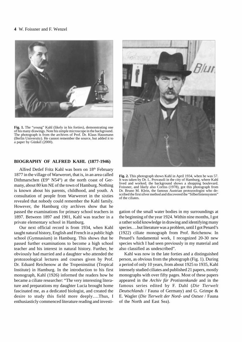
Kahl was now in the late forties and a distinguishedperson, as obvious from the photograph (Fig. 1). Duringa period of only 10 years, from about 1925 to 1935, Kahlintensely studied ciliates and published 21 papers, mostlymonographs with over fifty pages. Most of these papersappeared in the Archiv für Protistenkunde and in thefamous series edited by F. Dahl (Die TierweltDeutschlands / Fauna of Germany) and G. Grimpe &E. Wagler (Die Tierwelt der Nord- und Ostsee / Faunaof the North and East Sea).
Fig. 1. The “young” Kahl (likely in his forties), demonstrating oneof his many drawings. Note his simple microscope in the background.The photograph is from the archives of Prof. Dr. Klaus Hausmann(Berlin University). He cannot remember the source, but added it toa paper by Günkel (2000).
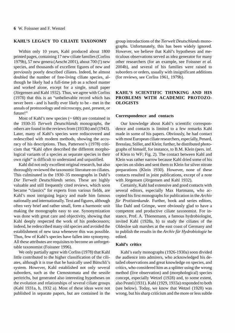
Fig. 2. This photograph shows Kahl in April 1934, when he was 57.It was taken by Dr. L. Provasoli in the city of Hamburg, where Kahllived and worked; the background shows a shopping boulevard.Foissner, and likely also Corliss (1978), got this photograph fromDr. Bruno M. Klein, the famous Austrian protozoologist who de-scribed the first silver method and discovered the “Silberliniensystem”of the ciliates.

Alfred Kahl’s life and legacy 5
In 1934/35, Kahl suddenly ceased his tremendousresearch activities with a supplement to his monographsin the Tierwelt Deutschlands, summarizing the speciesdescribed between 1930 and 1934 and describing sev-eral new species he discovered during this period. Theactual reason(s) why Kahl resigned is not known. On thephotograph from 1934 (Fig. 2), he still appears as adistinguished person, but looks more sceptic than previ-ously (Fig. 1). Likely, Kahl was frustrated by the dispar-aging critic he experienced from several academic pro-tozoologists, especially Prof. A. Wetzel. This is evidentfrom responses Kahl made in several papers (see nextsection). He had problems to publish in acknowledgedjournals, such as the Archiv für Protistenkunde and theZeitschrift für Morphologie und Ökologie der Tiere.
But soon Kahl’s monographs became a bible forciliate taxonomists and other people involved in ciliateidentification. Obviously, this stimulated him to com-mence work again in the forties, when he produced theshortened and revised version of his TierweltDeutschlands monographs reprinted here. Unfortunately,Kahl passed away with 68, that is, in 1946, likely on 20th
November. We did not find any obituaries, except thesimple statement by Dr. Georg Stehli in the 1948Mikrokosmos issue that “Kahl died during the past fouryears the journal could not appear”.
KAHL’S MICROSCOPE AND METHODS FORCILIATE OBSERVATION AND ILLUSTRATION
Ahead, we need to mention that Kahl published onlyin German language, as usual at that time. We decidedto translate literally the most important passages intoEnglish for the sake of wider understanding.
Kahl (1926, p. 203) remarked on the microscope heused and which can be seen in the background of figure1: it is a bright field stative III of the W. & S. Seibertcompany equipped with a compound condenser, a 5 ×eyepiece, a 2.5½ objective (about 3:1), and an oil immer-sion objective 1/12 (100:1). Seen from today’s perspec-tive, this is a simple instrument, providing a highestmagnification of × 500. However, the oil immersionobjectives produced around 1920 had good resolutionand great sharpness in the centre. Thus, the imagesobtained are similar to present-day bright field micro-scopes, especially if combined with a compound con-denser.
Measurements were performed with the “number 4”eyepiece micrometer of the Zeiss Company. Unfortu-nately, Kahl’s measurements are wrong in the firstmonographs because he corrected them upwards byabout 25% in the reviews from 1930-1943! Kahl nevercommented on this mistake, but it is obvious from thesizes provided.
Kahl (1926, 1930b, 1943) gives information on hismethods to observe and illustrate ciliates. Basically, histechniques are simple and the same as used by us(Wenzel 1953, Foissner 1991). Briefly, specimens areisolated with a micropipette and first observed at lowmagnification and without cover glass to note shape,size, movement, and the location of the main organelles.Then, a cover glass with small pillars of vaseline near itsedges is applied; the latter are pressed down with aneedle until the ciliate becomes more or less immobileand can be studied with the oil immersion objective. SeeFoissner (1991) for a detailed description of this method.The nuclear apparatus, Kahl studied mainly in vivo,rarely he used acetic methyl green or borax carmine.Kahl emphasizes the need of detailed live observation;the use of the oil immersion objective; to illustrate onlythose structures which were really seen; and to becritical of his own and literature data.
As concerns the famous illustrations, Kahl stated(1926, p. 204): “From the many drawings of a species,I select those for publication which are most useful fora solid characterization. These figures, I redraw indouble size so that they can be reduced to the originalsize in the paper. This should help to soften the “hard-ness” typical for pen - and - ink drawings. Rarely, I usea camera lucida, mainly for species which remain immo-bile for longer periods. However, the results are verysimilar to my ordinary freehand drawings”. Frequently, itis overlooked that the original monographs of Kahl(1926, 1927a, b, 1928a, b, 1930a, 1931a, b, 1932a) aremuch more detailed, both in text and illustration, than thereviews from 1930-1943, which are extremely con-densed due to space constraints and the inclusion ofliterature data.
Kahl’s simple drawings look not very impressive andwere thus occasionally criticized. However, anyone whoever tried such a “simple drawing” will soon recognizethe talent needed! Kahl was not only a meticulousobserver but, like Fauré-Fremiet (1924), also a talentedillustrator, showing the essence of a species in one or afew seemingly simple drawings.
6 W. Foissner and F. Wenzel
KAHL’S LEGACY TO CILIATE TAXONOMY
Within only 10 years, Kahl produced about 1800printed pages, containing 17 new ciliate families (Corliss1979b), 57 new genera (Aescht 2001), about 700 (!) newspecies, and thousands of excellent figures of new andpreviously poorly described ciliates. Indeed, he almostdoubled the number of free-living ciliate species, al-though he likely had a full-time job as a school masterand worked alone, except for a single, small paper(Jörgensen and Kahl 1932). Thus, we agree with Corliss(1978) that this is an “unbelievable record which hasnever been - and is hardly ever likely to be - met in theannals of protozoology and microscopy, past, present, orfuture!”
Most of Kahl’s new species (~ 680) are contained inthe 1930-35 Tierwelt Deutschlands monographs, theothers are found in the reviews from (1933b) and (1943).Later, many of Kahl’s species were rediscovered andredescribed with modern methods, showing the accu-racy of his descriptions. Thus, Patterson’s (1978) criti-cism that “Kahl often described the different morpho-logical variants of a species as separate species in theirown right” is difficult to understand and unjustified.
Kahl did not only excellent original research, but alsothoroughly reviewed the taxonomic literature on ciliates.This culminated in the 1930–35 monographs in Dahl’sDie Tierwelt Deutschlands series. These are highlyvaluable and still frequently cited reviews, which soonbecame “classics” for experts from various fields, areKahl’s most intriguing legacy and made him famousnationally and internationally. Text and figures, althoughoften very brief and rather small, form a harmonic unitmaking the monographs easy to use. Synonymizationwas done with great care and objectivity, showing thatKahl deeply respected the work of his predecessors;indeed, he redescribed many old species and avoided theestablishment of new taxa whenever this was possible.Thus, few of Kahl’s species have fallen into synonymy.All these attributes are requisites to become an unforget-table taxonomist (Foissner 1996).
We only partially agree with Corliss (1978) that Kahllittle contributed to the higher classification of the cili-ates, although it is true that he basically used Bütschli’ssystem. However, Kahl established not only severalsuborders, such as the Ctenostomata and the sessileperitrichs, but generated also interesting hypotheses onthe evolution and relationships of several ciliate groups(Kahl 1931a, b, 1932 a). Most of these ideas were notpublished in separate papers, but are contained in the
group introductions of the Tierwelt Deutschlands mono-graphs. Unfortunately, this has been widely ignored.However, we believe that Kahl’s hypotheses and me-ticulous observations served as idea generator for manyother researchers (for an example, see Foissner et al.2004b), and several of his families were raised tosuborders or orders, usually with insignificant additions(for reviews, see Corliss 1961, 1979b).
KAHL’S SCIENTIFIC THINKING AND HISPROBLEMS WITH ACADEMIC PROTOZO-OLOGISTS
Correspondence and contacts
Our knowledge about Kahl’s scientific correspon-dence and contacts is limited to a few remarks Kahlmade in some of his papers. Obviously, he had contactwith most European ciliate researchers, especially, Penard,Bresslau, Stiller, and Klein; further, he distributed photo-graphs of himself, for instance, to B.M. Klein (pers. inf.of Klein to WF; Fig. 2). The contact between Kahl andKlein was rather narrow because Kahl dried some of hisspecies on slides and sent them to Klein for silver nitratepreparations (Klein 1930). However, none of thesecontacts resulted in joint publications, except of a notewith Jörgensen (Jörgensen and Kahl 1932).
Certainly, Kahl had extensive and good contacts withseveral editors, especially Max Hartmann, who ac-cepted his first monographs for publication in the Archivfür Protistenkunde. Further, book and series editors,like Dahl and Grimpe, were obviously glad to have acompetent and productive ciliate taxonomist. For in-stance, Prof. A. Thienemann, a famous hydrobiologist,invited Kahl (1928a, b) to study the ciliates of theOldesloe salt marshes at the east coast of Germany andto publish the results in the Archiv für Hydrobiologie heedited.
Kahl’s critics
Kahl’s early monographs (1926-1930a) soon dividedthe audience into admirers, who acknowledged his de-tailed observations and great knowledge on species, andcritics, who considered him as a splitter using the wrongmethod (live observation) and (morphological) speciesconcept, especially Wetzel (1928) and, to some extent,also Pestel (1931). Kahl (1929, 1933a) responded to both(see below). Today, we know that Wetzel (1928) waswrong, but his sharp criticism and the more or less subtle
Alfred Kahl’s life and legacy 7
objection of others caused that Kahl became discreditedand some of his studies were refused by the leadingzoological journals. Kahl (1930a, 1935) complainedabout that several times, for instance, when he describedthe stalk of the peritrichs (Kahl 1935, p. 710): “Unfortu-nately, my detailed research was refused by theeditors of two influential journals, the Zeitschriftfür Wissenschaftliche Zoologie and the Zeitschriftfür Morphologie und Ökologie der Tiere”. What aloss!
Certainly, Kahl made mistakes, as do all diligentpeople, but compared to the countless correct observa-tions, these are negligible. Kahl made some ratherserious mistakes in nomenclature (Corliss 1979b),however we should not forget that the first InternationalCode of Zoological Nomenclature was still in its infancywhen Kahl commenced his research. As a non-academic self-made man, Kahl certainly was very sen-sible to criticism. Likely, the public objections ofWetzel (1928) and the subtle criticism of several journaleditors contributed to Kahl’s stopping work so abruptlyin 1935.
Kahl’s species and scientific concepts
Kahl (1929, 1933a) responded to his actual andpotential critics in two specific papers and, especially, inthe introduction to the monograph on holotrichous ciliates(Kahl 1930a, pp. 313-320). We shall cite mainly from thelast essay, which is timeless and shows Kahl’s scientificconcepts.
Kahl (1930a) commences the essay with a literalcitation of some remarks of Wetzel (1928): “Consideringthe variable shape of many protozoans, it is very easy todescribe new species. Unfortunately, recent taxonomistsare as thoughtless as previous ones in this respect;indeed, their descriptions and figures are even moresuperficial”. Wetzel (1928) does not explicitly refer toKahl, but the further text shows him as the addressee.Kahl (1930a, p. 314) responded to this scathing critiquewith some remarkable statements illuminating also thepresent problems with ciliate diversity (Foissner 1999):“Such views on my publications are not unexpected forme. The huge number of new species I described mustproduce the impression that a “species hunter” is atwork. However, I will keep up my view, without anyrespect to others, that there are much more protozoanspecies than supposed previously. Future research shoulddecide whether or not I was correct and whether theabove cited remarks apply to me”.
As concerns the species concept, Kahl (1930a,pp. 315, 318, 319) defends a strictly morphologicaldefinition: “I consider the species as an abstractionwhich eliminates the individual morphological variabil-ity and summarizes the useful morphological featuresin a concise diagnosis. Species can be recognized onlyempirically, that is, they cannot be proven in a strictsense because empiricism uses only plausibilities”. Wetzel(1928), in contrast, defines ciliate species “as thoseindividuals which are able to conjugate with each other”.Kahl (1930a, pp. 315-318) criticizes this definition forpractical and theoretical reasons, but emphasizes thatconjugation is an excellent additional feature for speciespreviously defined morphologically. Time confirmedWetzel, because his species concept is very similarto that used by most modern biologists. On the otherhand, the practical problems of such a definition remain,for instance, that it may be extremely difficult to excludesex in two similar morphospecies. Thus, the majorityof new protist, plant, and animal species describedthese days, is still defined purely morphologically and/orby gene sequences. Wetzel (1928) also could not solvethe practical problems of his concept because he diag-nosed his new species in the classical (morphological)way!
In the context of species differentiation and recogni-tion, Kahl (1930a, p. 318) also discusses pure culturesand reaches the following conclusion: “If the species isnot extraordinarily variable, a pure culture is less usefulthan observations on ten field populations”. Kahl doesnot deny the advantages of pure cultures, if the users arebeware of the possibility that poorly thriving or degener-ating cultures can produce abnormal specimens seem-ingly bridging the gap between two or more well-definedmorphospecies. Everyone who uses both, pure culturesand field populations will agree!
Kahl (1930a, p. 320) closes the essay with a remark-able, but impracticable suggestion: ”In protozoans, spe-cies recognition is highly subjective. Thus, there shouldbe some standardization, for instance, a species shouldbe considered as valid only if it has been confirmed byat least two specialists”.
Kahl’s essay shows that some of our “new” problemsare, in fact, the old ones! There is still intense discussionabout methods (live observation vs. definition from silverslides only), diversity (few or many species; Foissner1999), species concepts (Foissner et al. 2002), andmaterial (pure cultures vs. field populations). Certainly,discussion will go on and on and each generation of

8 W. Foissner and F. Wenzel
scientists will have its own view in the light of newmethods and concepts.
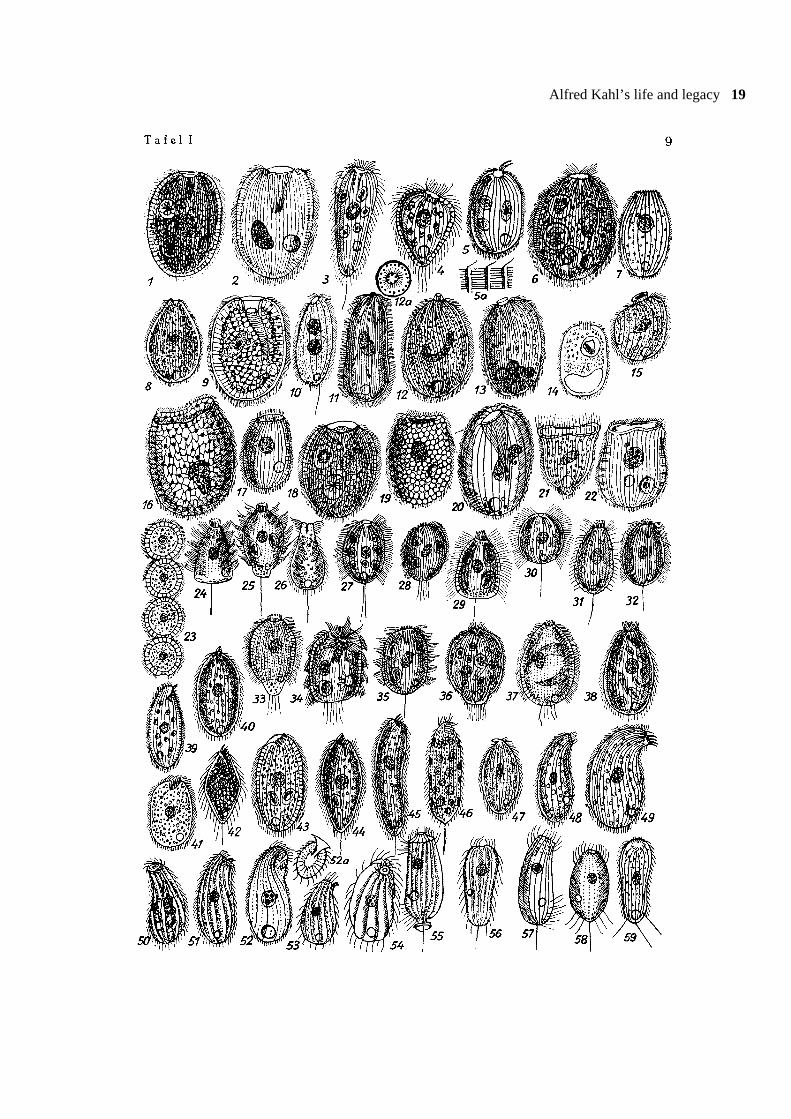
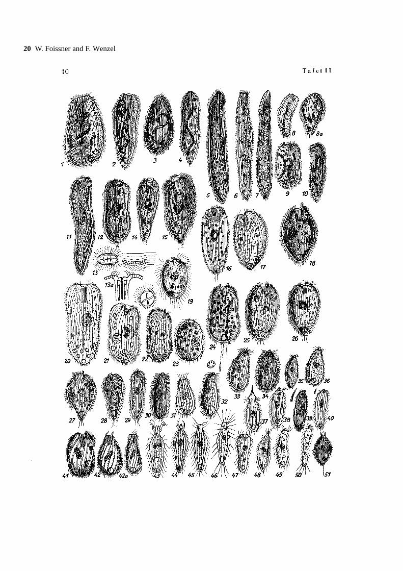
FACSIMILE OF THE “INFUSORIEN” OF KAHL(1943)
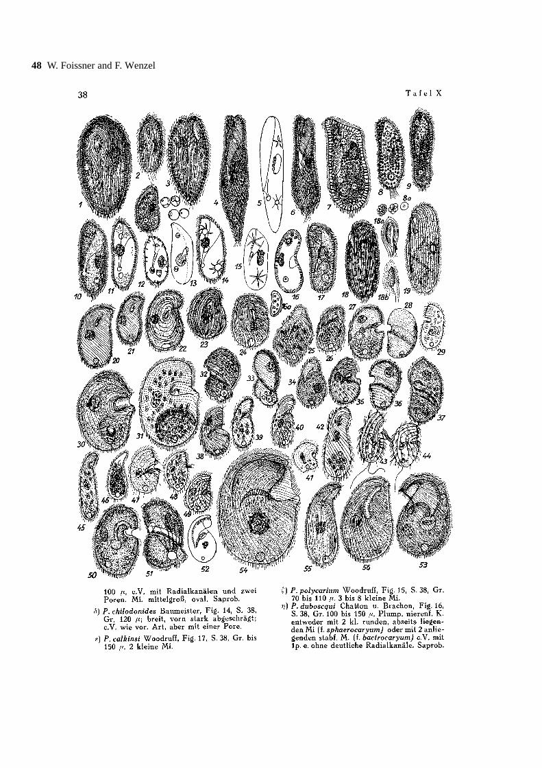
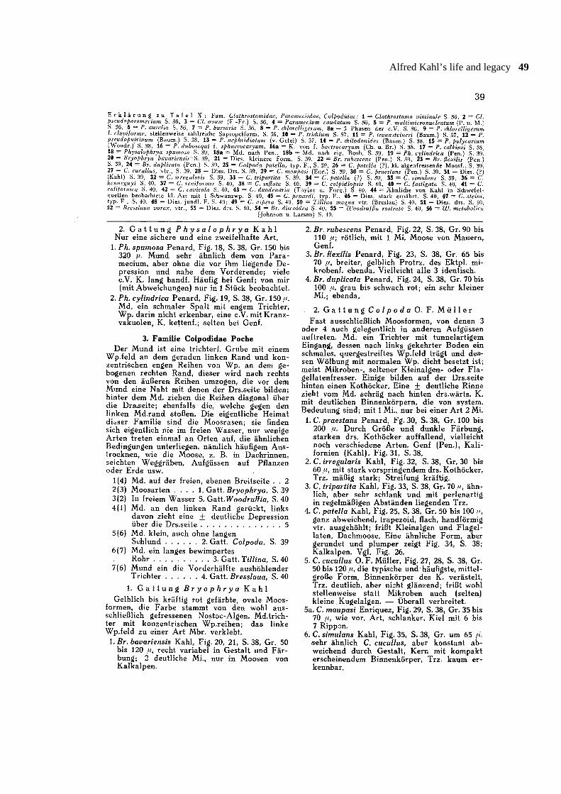
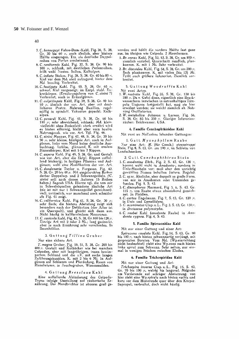
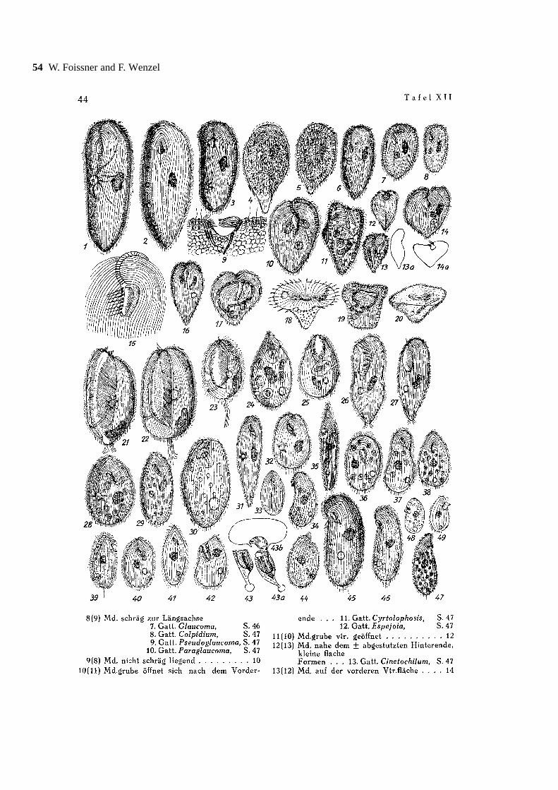
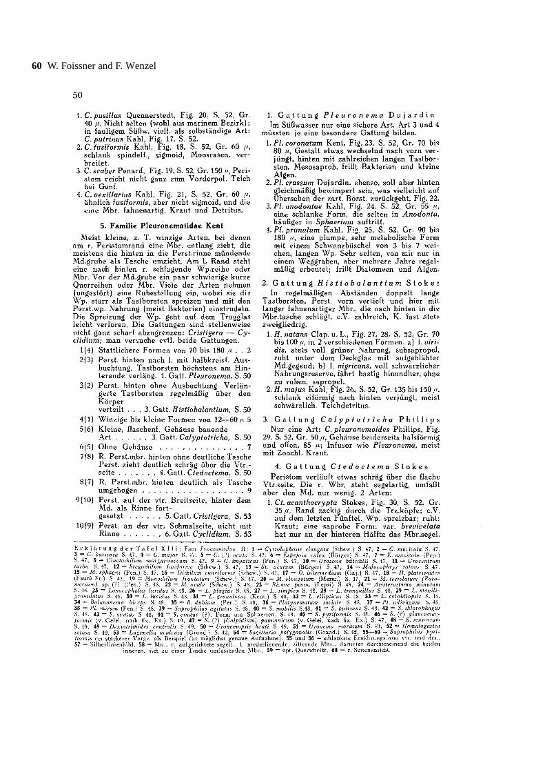
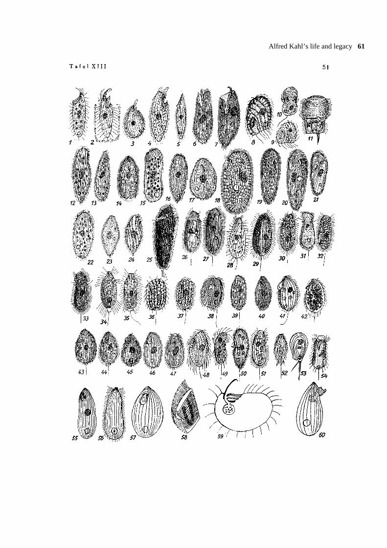
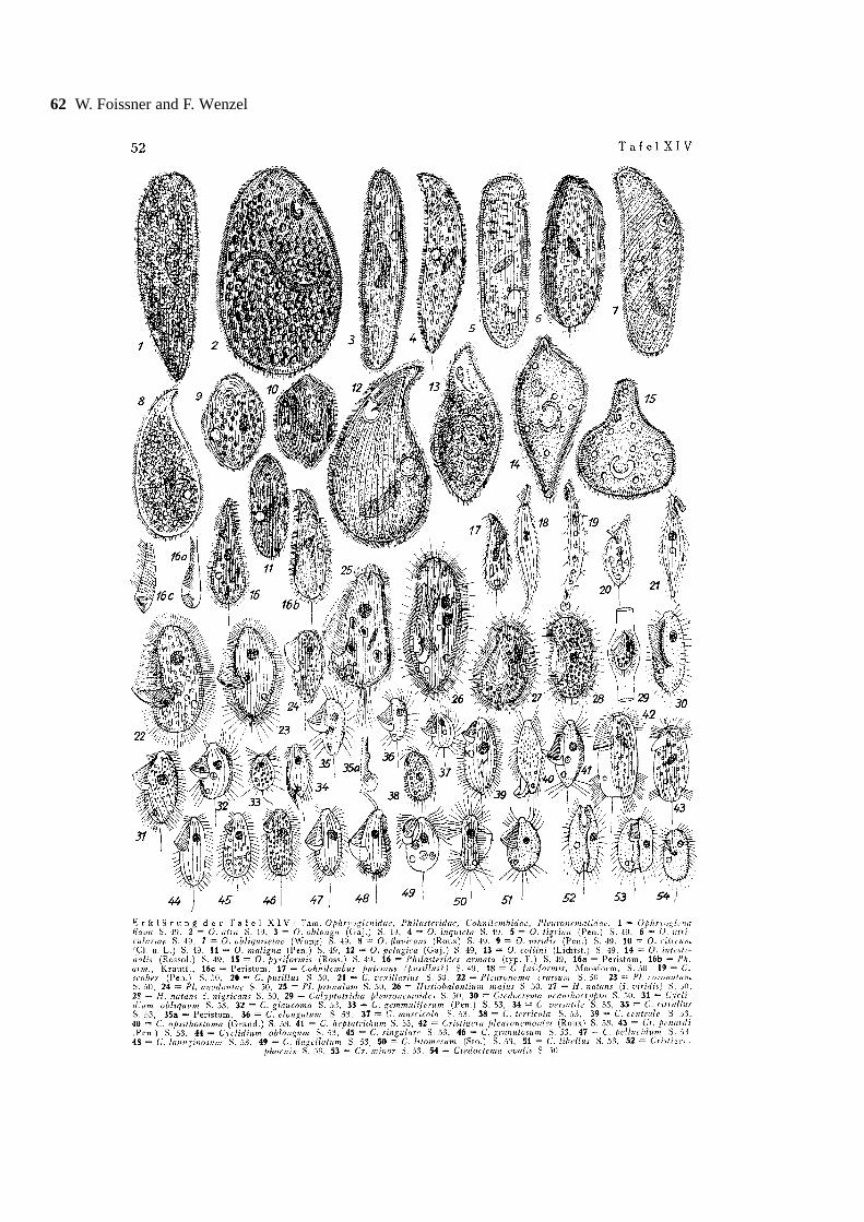
Our and Hausmann’ s exemplar of Kahl’s (1943)“Infusorien” is not bound and lacks a title page. It is notknown whether a title page exists at all. Possibly, itshould have been distributed with part 2, which neverappeared. Thus, the monograph ends abruptly with PlateXIV showing ophryoglenids and scuticociliates; theperitrichs and spirotrichs are lacking.
Complete title in German (obtained fromDr. Rainer Gerstle, editorial assistant to the Mikrokosmosin the seventies)
Kahl A. (1943) Infusorien (1. Teil). Ein Hilfsbuch zumErkennen, Bestimmen, Sammeln und Präparieren derfreilebenden Infusorien des Süßwassers und der Moore.Buchbeilage zum Mikrokosmos Jahrgang 1942/43, d. h.,erschienen in der Reihe “Handbücher für die praktischenaturwissenschaftliche Arbeit”, Band 31/32, 52 pp.Franckh’sche Verlagsbuchhandlung, W. Keller & Co.,Stuttgart.
English translation (by W. Foissner)Kahl A. (1943) Infusoria (part 1). An assistance book
for the knowledge, identification, collection, and prepara-tion of free-living infusoria from freshwaters and bogs.Book supplement to the Mikrokosmos years 1942/43.This supplement series appeared under a distinct name,viz., “Manuals for practical scientific work”, volume31/32, 52 pp. Franckh’sche Publishers, W. Keller & Co.,Stuttgart.
Suggestion for citation in taxonomic studiesKahl A. (1943) Infusorien (1. Teil). Handbücher für
die praktische wissenschaftliche Arbeit 31/32, 52 pp.Franckh’sche Verlagsbuchhandlung, Stuttgart. (Reprintedin Acta Protozoologica 43 (Suppl.): 1-66, 2004)
Taxonomic and nomenclatural innovations in themonograph of Kahl (1943)
The monograph excludes marine species and con-tains several innovations summarized in the followingparagraphs. Further, Kahl has redone all illustrations.Usually, the differences are small, but in some speciesthey are considerable, showing that they are based on
new observations. Thus, the 1943 monograph should beconsulted in all kinds of taxonomic work.
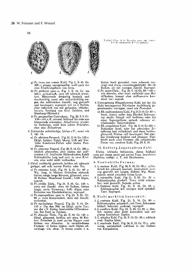
New taxa described (arranged alphabetically):Askenasia armata (p. 22), Askenasia pelagica (p. 22),both not identical with any of the species described in therevision of the genus by Krainer and Foissner (1990);Frontonia angusta (p. 45), raised to species level, asalso suggested by Foissner et al. (1994); Lacrymariamuscicola (p. 17), redescribed at the end of this paper;Loxodes striatus var. fasciformis (p. 33); Parameciumchlorelligerum f. claviforme (p. 36), see also the little-known paper by Baumeister (1969), who describedthree new, likely valid species, viz., P. varionuklei,P. traunsteineri and P. chilodonides; Prorodongeldneri (p. 16), Prorodon sapropelicus (p. 16),Prorodon discolor f. ovalis (p. 16); and Spathidiumcoemeterii (p. 27), redescribed by Foissner et al. (2004a).
As mentioned in this compilation, two out of the tennew taxa described by Kahl (1943) were re-discoveredby Foissner: one is redescribed in Foissner et al. (2004a),the other at the end of this paper. This shows once morethe excellent work Kahl did. Likely, the other speciesalso can be re-discovered on detailed investigations.
Nomenclatural changes: Kahl (1943, p. 21) trans-ferred Chaenea mucicola Wang to Enchelyodon. Inthis genus, however, it is preoccupied by E. mucicolaKahl. Thus, he introduced the new name Enchelyodonwangi nom.n. Kahl (1943, p. 27) recognized preoccupa-tion of Diceras Eberhard and introduced the new nameDiceratophrys nom.n. Accordingly, Diceratula Corliss(1960), who also recognized this homonymy, is a juniorsynonym.
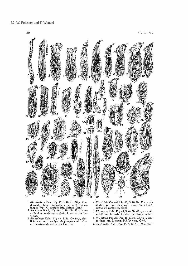
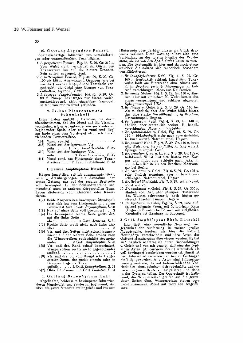
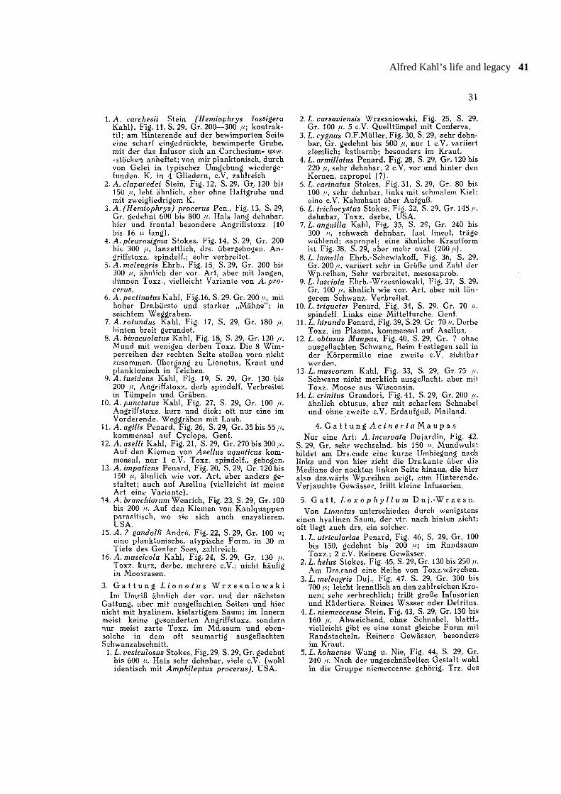
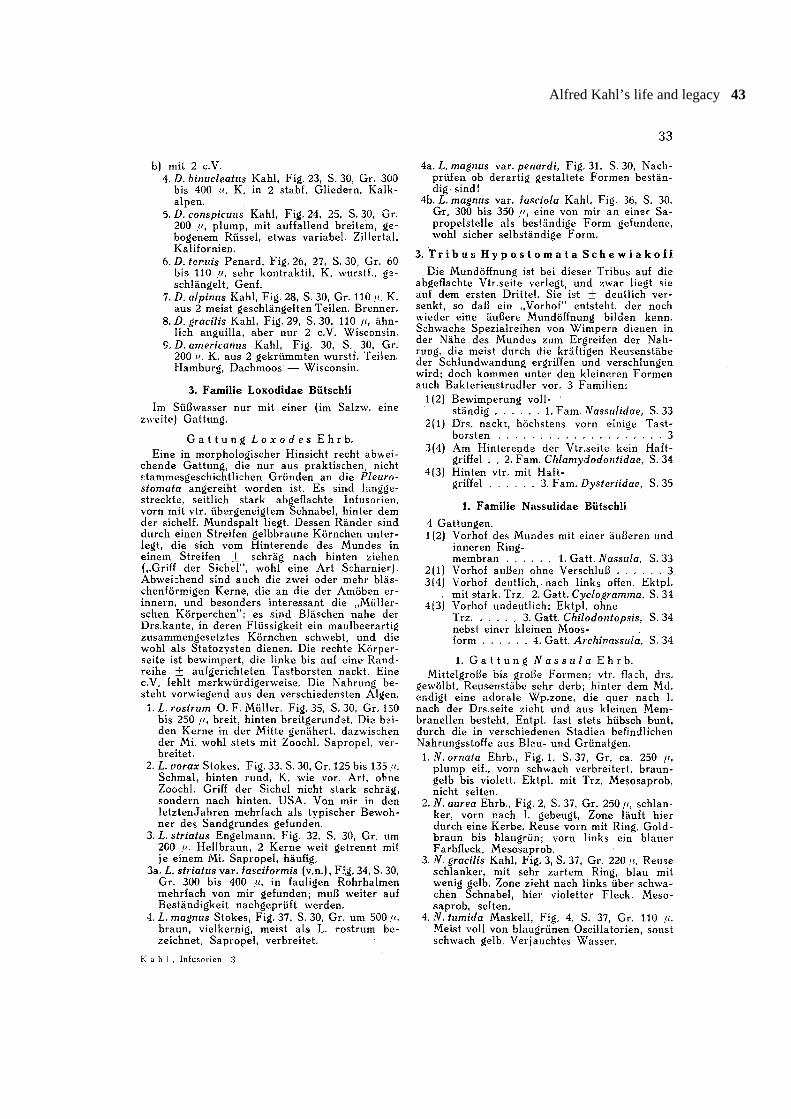
Remarks on generic classifications: The 1943monograph contains, like Kahl’s previous monographs,interesting notes on the morphology and classification ofseveral genera. These remarks are too extensive to befully cited and translated; thus, just a few representativeexamples were selected. Kahl (p. 49) recognized thatIchthyophthirius belongs to the ophryoglenids, whichwas later proven by Lynn et al. (1991); that Hemiophrysis a junior synonym of Amphileptus (p. 28), later provenby Foissner (1984b); and that Dileptus tracheloidesZacharias represents a new genus (p. 32), which wasindependently established by Foissner et al. (1999).Further, interesting notes concern, for instance, thegenera Disematostoma, Stokesia, and Marituja whichKahl considers as synonyms. However, this is not sup-ported by recent data compiled in Foissner et al. (1999).
Supplementary observations: Several descriptionsand many illustrations are obviously based on additional

Alfred Kahl’s life and legacy 9
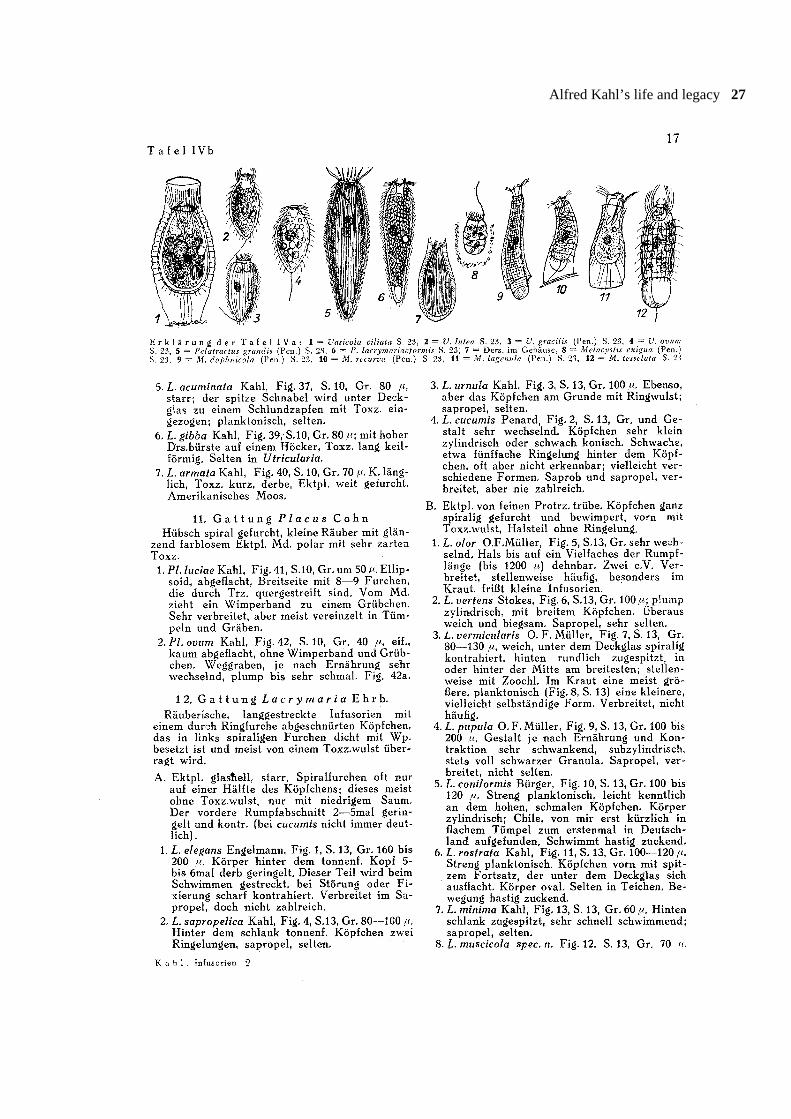
observations. Sometimes, this is obvious, for instance, inLacrymaria vermicularis (p. 17): “Frequently, a largevariety is found among aquatic plants, while a smaller,probably distinct species occurs in the plankton (Fig. 8,p. 13)”; usually, however, the new data are utilized inrefined illustrations without any comment, for instance,
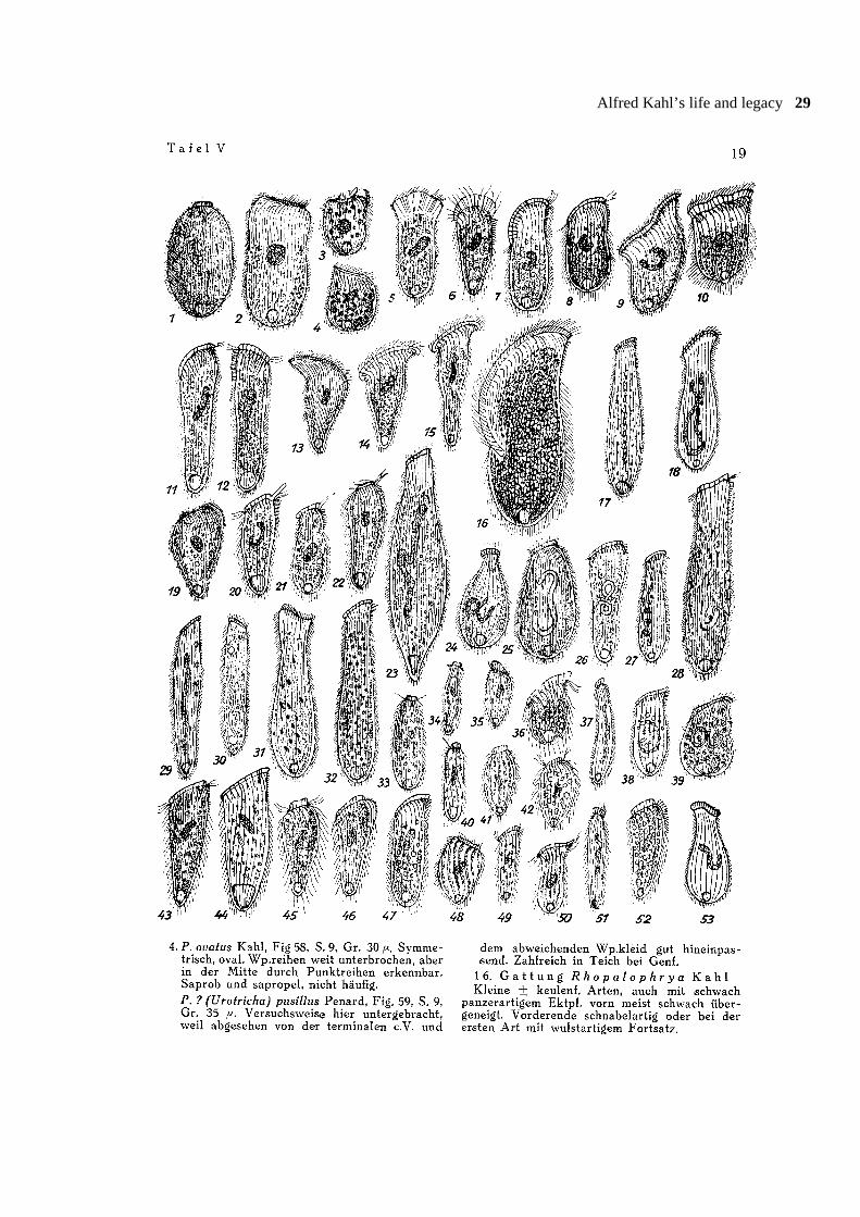
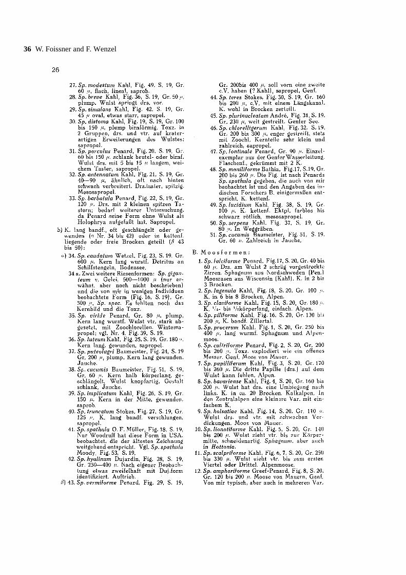
in Phascolodon vorticella, Bresslaua discoidea, andHemicyclium lucidum. Further, Kahl included illustra-tions of several undescribed species, for instance, a500 µm (!) long, sapropelic Spathidium (Fig. 16 onp. 19).
Alfred Kahl’s life and legacy 11
12 W. Foissner and F. Wenzel
Alfred Kahl’s life and legacy 13

14 W. Foissner and F. Wenzel
Alfred Kahl’s life and legacy 15

16 W. Foissner and F. Wenzel
Alfred Kahl’s life and legacy 17

18 W. Foissner and F. Wenzel
Alfred Kahl’s life and legacy 19
20 W. Foissner and F. Wenzel

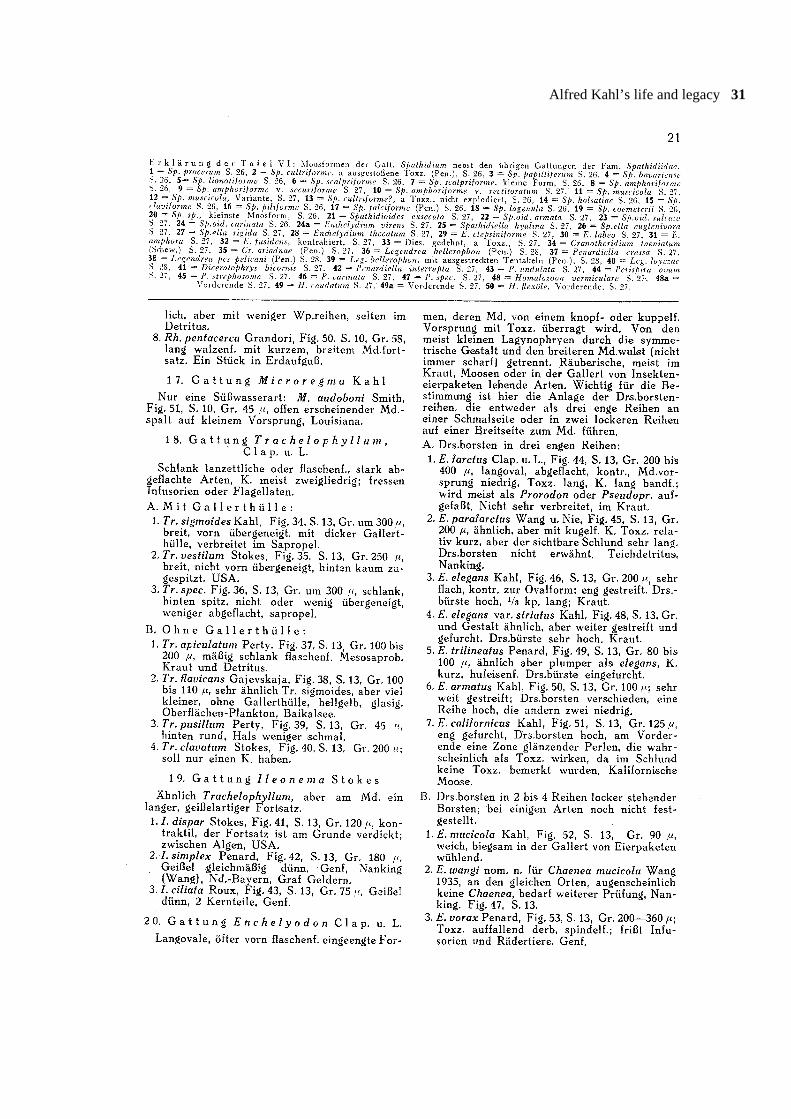
Alfred Kahl’s life and legacy 21
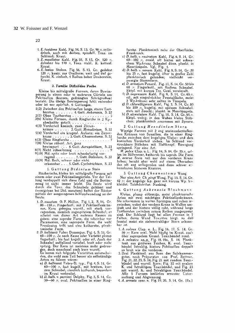
22 W. Foissner and F. Wenzel

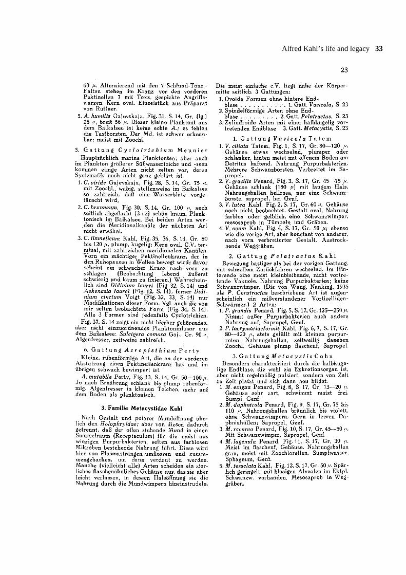
Alfred Kahl’s life and legacy 23

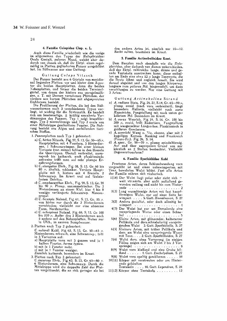
24 W. Foissner and F. Wenzel
Alfred Kahl’s life and legacy 25
26 W. Foissner and F. Wenzel
Alfred Kahl’s life and legacy 27

28 W. Foissner and F. Wenzel
Alfred Kahl’s life and legacy 29
30 W. Foissner and F. Wenzel
Alfred Kahl’s life and legacy 31
32 W. Foissner and F. Wenzel
Alfred Kahl’s life and legacy 33
34 W. Foissner and F. Wenzel

Alfred Kahl’s life and legacy 35
36 W. Foissner and F. Wenzel

Alfred Kahl’s life and legacy 37
38 W. Foissner and F. Wenzel

Alfred Kahl’s life and legacy 39

40 W. Foissner and F. Wenzel
Alfred Kahl’s life and legacy 41

42 W. Foissner and F. Wenzel
Alfred Kahl’s life and legacy 43
44 W. Foissner and F. Wenzel
Alfred Kahl’s life and legacy 45
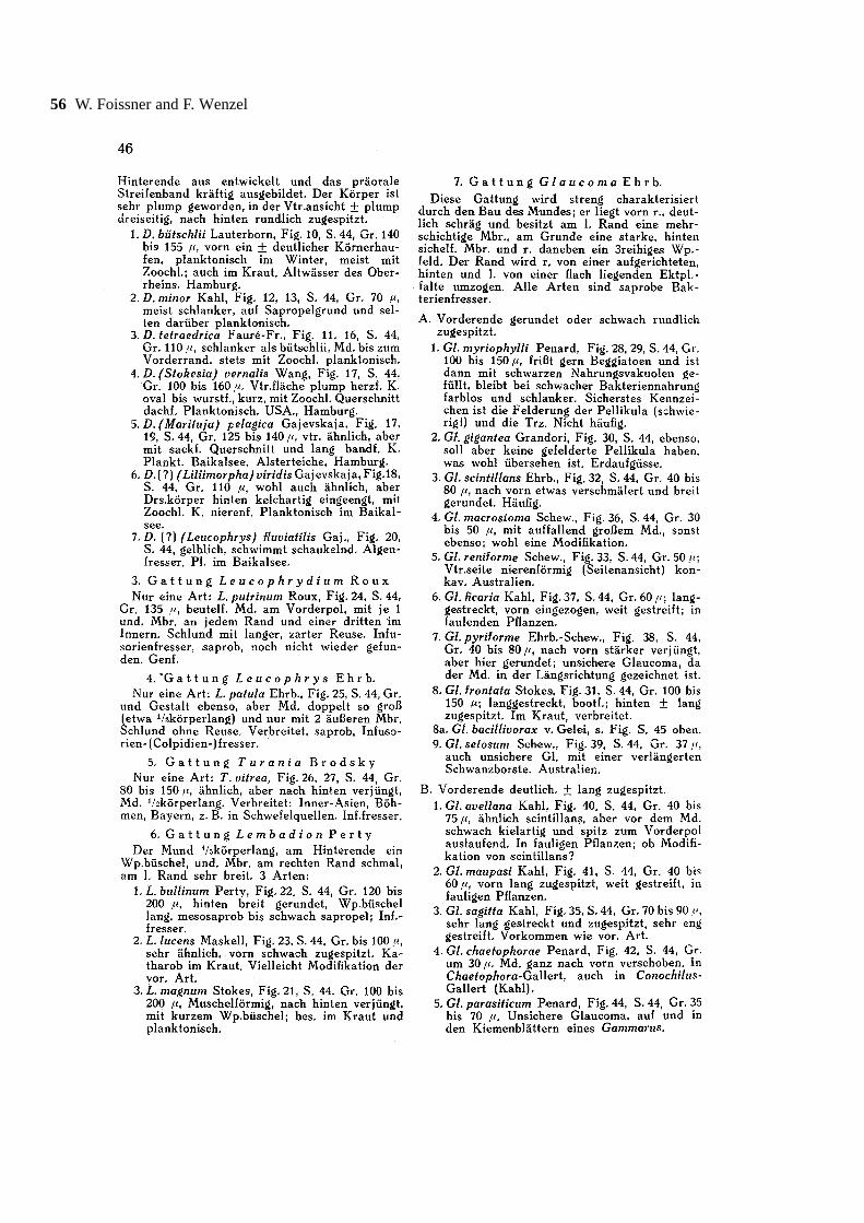
46 W. Foissner and F. Wenzel
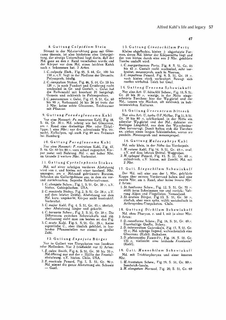
Alfred Kahl’s life and legacy 47
48 W. Foissner and F. Wenzel
Alfred Kahl’s life and legacy 49
50 W. Foissner and F. Wenzel

Alfred Kahl’s life and legacy 51

52 W. Foissner and F. Wenzel

Alfred Kahl’s life and legacy 53
54 W. Foissner and F. Wenzel

Alfred Kahl’s life and legacy 55
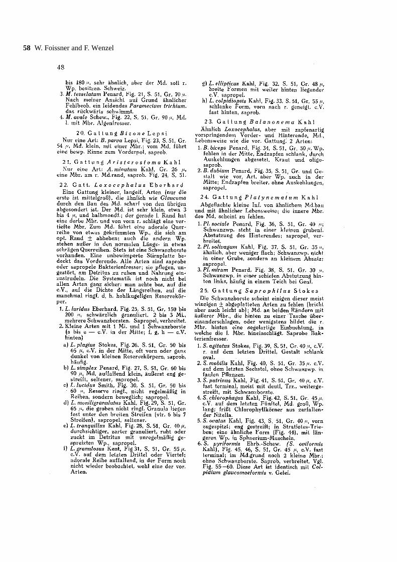
56 W. Foissner and F. Wenzel
Alfred Kahl’s life and legacy 57
58 W. Foissner and F. Wenzel
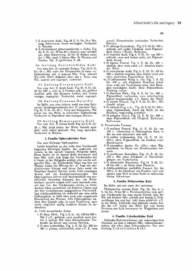
Alfred Kahl’s life and legacy 59
60 W. Foissner and F. Wenzel
Alfred Kahl’s life and legacy 61
62 W. Foissner and F. Wenzel
Alfred Kahl’s life and legacy 63
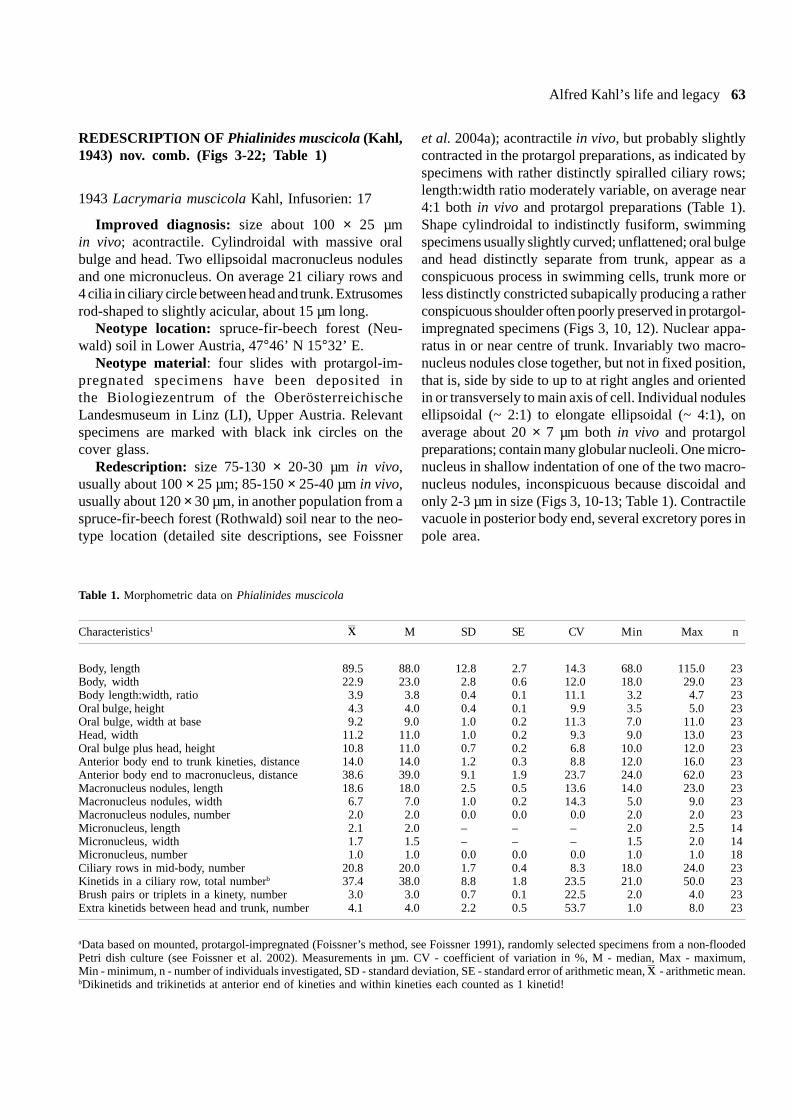
REDESCRIPTION OF Phialinides muscicola (Kahl,1943) nov. comb. (Figs 3-22; Table 1)
1943 Lacrymaria muscicola Kahl, Infusorien: 17
Improved diagnosis: size about 100 × 25 µmin vivo; acontractile. Cylindroidal with massive oralbulge and head. Two ellipsoidal macronucleus nodulesand one micronucleus. On average 21 ciliary rows and4 cilia in ciliary circle between head and trunk. Extrusomesrod-shaped to slightly acicular, about 15 µm long.
Neotype location: spruce-fir-beech forest (Neu-wald) soil in Lower Austria, 47°46’ N 15°32’ E.
Neotype material: four slides with protargol-im-pregnated specimens have been deposited inthe Biologiezentrum of the OberösterreichischeLandesmuseum in Linz (LI), Upper Austria. Relevantspecimens are marked with black ink circles on thecover glass.
Redescription: size 75-130 × 20-30 µm in vivo,usually about 100 × 25 µm; 85-150 × 25-40 µm in vivo,usually about 120 × 30 µm, in another population from aspruce-fir-beech forest (Rothwald) soil near to the neo-type location (detailed site descriptions, see Foissner
et al. 2004a); acontractile in vivo, but probably slightlycontracted in the protargol preparations, as indicated byspecimens with rather distinctly spiralled ciliary rows;length:width ratio moderately variable, on average near4:1 both in vivo and protargol preparations (Table 1).Shape cylindroidal to indistinctly fusiform, swimmingspecimens usually slightly curved; unflattened; oral bulgeand head distinctly separate from trunk, appear as aconspicuous process in swimming cells, trunk more orless distinctly constricted subapically producing a ratherconspicuous shoulder often poorly preserved in protargol-impregnated specimens (Figs 3, 10, 12). Nuclear appa-ratus in or near centre of trunk. Invariably two macro-nucleus nodules close together, but not in fixed position,that is, side by side to up to at right angles and orientedin or transversely to main axis of cell. Individual nodulesellipsoidal (~ 2:1) to elongate ellipsoidal (~ 4:1), onaverage about 20 × 7 µm both in vivo and protargolpreparations; contain many globular nucleoli. One micro-nucleus in shallow indentation of one of the two macro-nucleus nodules, inconspicuous because discoidal andonly 2-3 µm in size (Figs 3, 10-13; Table 1). Contractilevacuole in posterior body end, several excretory pores inpole area.
Table 1. Morphometric data on Phialinides muscicola
Characteristics1 � M SD SE CV Min Max n
Body, length 89.5 88.0 12.8 2.7 14.3 68.0 115.0 23Body, width 22.9 23.0 2.8 0.6 12.0 18.0 29.0 23Body length:width, ratio 3.9 3.8 0.4 0.1 11.1 3.2 4.7 23Oral bulge, height 4.3 4.0 0.4 0.1 9.9 3.5 5.0 23Oral bulge, width at base 9.2 9.0 1.0 0.2 11.3 7.0 11.0 23Head, width 11.2 11.0 1.0 0.2 9.3 9.0 13.0 23Oral bulge plus head, height 10.8 11.0 0.7 0.2 6.8 10.0 12.0 23Anterior body end to trunk kineties, distance 14.0 14.0 1.2 0.3 8.8 12.0 16.0 23Anterior body end to macronucleus, distance 38.6 39.0 9.1 1.9 23.7 24.0 62.0 23Macronucleus nodules, length 18.6 18.0 2.5 0.5 13.6 14.0 23.0 23Macronucleus nodules, width 6.7 7.0 1.0 0.2 14.3 5.0 9.0 23Macronucleus nodules, number 2.0 2.0 0.0 0.0 0.0 2.0 2.0 23Micronucleus, length 2.1 2.0 – – – 2.0 2.5 14Micronucleus, width 1.7 1.5 – – – 1.5 2.0 14Micronucleus, number 1.0 1.0 0.0 0.0 0.0 1.0 1.0 18Ciliary rows in mid-body, number 20.8 20.0 1.7 0.4 8.3 18.0 24.0 23Kinetids in a ciliary row, total numberb 37.4 38.0 8.8 1.8 23.5 21.0 50.0 23Brush pairs or triplets in a kinety, number 3.0 3.0 0.7 0.1 22.5 2.0 4.0 23Extra kinetids between head and trunk, number 4.1 4.0 2.2 0.5 53.7 1.0 8.0 23
aData based on mounted, protargol-impregnated (Foissner’s method, see Foissner 1991), randomly selected specimens from a non-floodedPetri dish culture (see Foissner et al. 2002). Measurements in µm. CV - coefficient of variation in %, M - median, Max - maximum,Min - minimum, n - number of individuals investigated, SD - standard deviation, SE - standard error of arithmetic mean, �- arithmetic mean.bDikinetids and trikinetids at anterior end of kineties and within kineties each counted as 1 kinetid!
64 W. Foissner and F. Wenzel
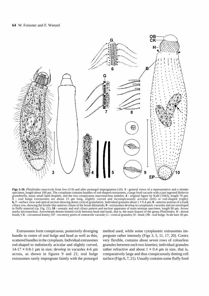
Extrusomes form conspicuous, posteriorly divergingbundle in centre of oral bulge and head as well as thin,scattered bundles in the cytoplasm. Individual extrusomesrod-shaped to indistinctly acicular and slightly curved,14-17 × 0.8-1 µm in size; develop in vacuoles 4-6 µmacross, as shown in figures 9 and 21; oral bulgeextrusomes rarely impregnate faintly with the protargol
method used, while some cytoplasmic extrusomes im-pregnate rather intensely (Figs 3, 5, 11, 17, 20). Cortexvery flexible, contains about seven rows of colourlessgranules between each two kineties; individual granulesrather refractive and about 1 × 0.4 µm in size, that is,comparatively large and thus conspicuously dotting cellsurface (Figs 6, 7, 21). Usually contains some fluffy food
Figs 3-10. Phialinides muscicola from live (3-9) and after protargol impregnation (10). 3 - general views of a representative and a slenderspecimen, length about 100 µm. The cytoplasm contains bundles of rod-shaped extrusomes, a large food vacuole with a just ingested Halteriagrandinella, many small lipid droplets, and the two conspicuous macronucleus nodules; 4 - original figure by Kahl (1943), length 70 µm;5 - oral bulge extrusomes are about 15 µm long, slightly curved and inconspicuously acicular (left) or rod-shaped (right);6, 7 - surface view and optical section showing dense cortical granulation. Individual granules about 1 × 0.4 µm; 8 - anterior portion of a trunkciliary row, showing the bristle-like anterior cilium of the brush dikinetids; 9 - extrusomes develop in cytoplasmic vacuoles and are envelopedin fluffy material (cp. Fig. 21); 10 - somatic and oral ciliary pattern and nuclear apparatus of main neotype specimen, length 90 µm. Arrowmarks micronucleus. Arrowheads denote kinetid circle between head and trunk, that is, the main feature of the genus Phialinides. B - dorsalbrush; CK - circumoral kinety; EP - excretory pores of contractile vacuole; G - cortical granules; H - head; OB - oral bulge. Scale bars 30 µm.
Alfred Kahl’s life and legacy 65
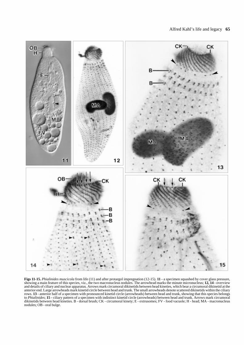
Figs 11-15. Phialinides muscicola from life (11) and after protargol impregnation (12-15). 11 - a specimen squashed by cover glass pressure,showing a main feature of this species, viz., the two macronucleus nodules. The arrowhead marks the minute micronucleus; 12, 14 - overviewand details of ciliary and nuclear apparatus. Arrows mark circumoral dikinetids between head kineties, which bear a circumoral dikinetid at theanterior end. Large arrowheads mark kinetid circle between head and trunk. The small arrowheads denote scattered dikinetids within the ciliaryrows. 13 - anterior half of a specimen with pronounced kinetid circle (arrowheads) between head and trunk, showing that this species belongsto Phialinides; 15 - ciliary pattern of a specimen with indistinct kinetid circle (arrowheads) between head and trunk. Arrows mark circumoraldikinetids between head kineties. B - dorsal brush; CK - circumoral kinety; E - extrusomes; FV - food vacuole; H - head; MA - macronucleusnodules; OB - oral bulge.
66 W. Foissner and F. Wenzel
vacuoles 5-15 µm across and many 0.5-2 µm-sized lipiddroplets. In the non-flooded Petri dish culture, preysmainly on Halteria grandinella, but ingests also thecomparatively large Frontonia depressa without be-coming significantly deformed; prey is ingested wholeand thus integer in young food vacuoles, showing that
oral bulge and head can open very widely. Usuallyassuming a slightly curved shape (Figs 3, 11). Swimsrapidly and jerkily.
Ciliature as typical for the genus, phialinides ciliarycircle, however, often inconspicuous because composedof an average of only four bristles. Cilia needle-like
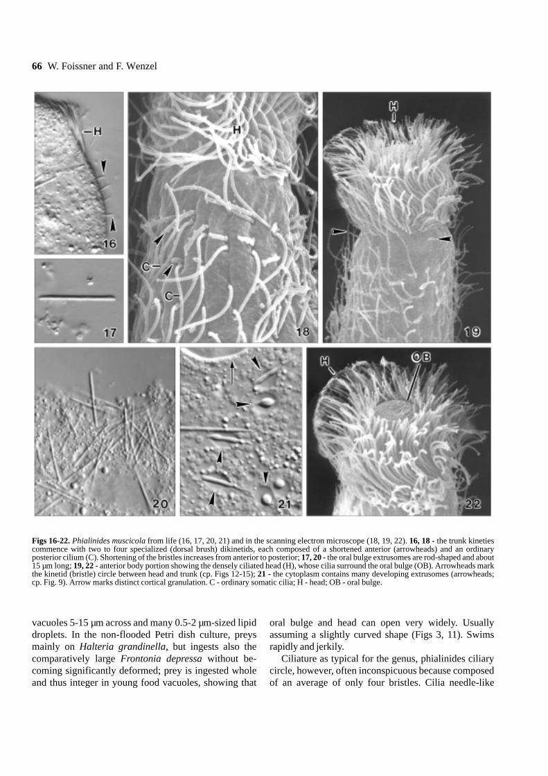
Figs 16-22. Phialinides muscicola from life (16, 17, 20, 21) and in the scanning electron microscope (18, 19, 22). 16, 18 - the trunk kinetiescommence with two to four specialized (dorsal brush) dikinetids, each composed of a shortened anterior (arrowheads) and an ordinaryposterior cilium (C). Shortening of the bristles increases from anterior to posterior; 17, 20 - the oral bulge extrusomes are rod-shaped and about15 µm long; 19, 22 - anterior body portion showing the densely ciliated head (H), whose cilia surround the oral bulge (OB). Arrowheads markthe kinetid (bristle) circle between head and trunk (cp. Figs 12-15); 21 - the cytoplasm contains many developing extrusomes (arrowheads;cp. Fig. 9). Arrow marks distinct cortical granulation. C - ordinary somatic cilia; H - head; OB - oral bulge.
Alfred Kahl’s life and legacy 67
narrowed distally and about 10 µm long in vivo, arrangedin an average of 21 equidistant rows commencing under-neath trunk shoulder and extending longitudinally torather distinctly spirally to rear body end. Cilia ordinarilyspaced, except of some scattered dikinetids within rowsand an average of three dorsal brush dikinetids ortrikinetids at anterior end of rows; posterior cilium ofbrush dikinetids of ordinary shape and length, anteriorslightly fusiform and bristle-like decreasing in lengthfrom 4 µm anteriorly to 2 µm posteriorly (Figs 3, 8, 10,12-16, 18, 19; Table 1).
Oral bulge and head conspicuous because about 12 ×12 µm in vivo and thus half as wide as trunk. Oral bulgedistinct not only in protargol preparations but also in vivobecause almost hemispherically protruding and ratherrefractive due to the extrusome tips contained, separatedfrom head by a rather distinct furrow containing thecircumoral kinety composed of a dikinetid each at ante-rior end of head kineties and an additional dikinetidbetween each two head ciliary rows. Head shaped likea truncated cone or a barrel, distinctly furrowed bynarrowly spaced, obliquely sigmoidal, densely ciliatedhead kineties each commencing with a dikinetid belong-ing to the circumoral kinety, as described above. Oralbasket rods neither recognizable in vivo nor in protargolpreparations (Figs 3, 10-15, 18, 19, 22; Table 1).
Occurrence and ecology: Kahl (1943) discoveredP. muscicola in moss, likely from northern Germany. Wefound P. muscicola in soil from two sub-alpine spruce-fir-beech forests in Lower Austria, about 1000m abovesea-level (Foissner et al. 2004a). Both samples were anacidic (pH 4-5) mixture of leave litter and mineral soilfrom the upper 10 cm. Phialinides muscicola becamerather abundant two weeks after rewetting the air-driedsamples, where it stayed for several weeks although thesoil percolate was collected for preparations two timesand replaced by fresh water (Eau de Volvic).
Identification: Kahl’s description of Lacrymariamuscicola is very brief: “Length 70 µm. Moss-inhabitingspecies with two macronuclei, otherwise similar toL. minima”. He provided a single, rather superficialfigure (Fig. 4). Thus, any identification is more or lessarbitrary. However, when comparing sizes, habitats, andfigures, it is rather obvious that Kahl’s species is thesame as Phialina binucleata Berger et al. (1984) orPhialinides australis Foissner (1988). This would re-quire to synonymize one of these species (but which!)with Lacrymaria muscicola and to establish a newspecies for the population described here. Consideringthe incomplete original description of Lacrymaria
muscicola, this would not make sense, but only increasethe number of names. Thus and because no otherpriorities are violated, we suggest to neotypify Kahl’sGerman species with the Austrian population describedhere.
Generic allocation: Phialinides differs fromPhialina by a circle of rather widely spaced ciliabetween head and trunk (Foissner 1988). This circle isvery distinct in Pelagolacrymaria Foissner et al. (1999),where it consists of ciliated dikinetids. Thus, there is nodoubt that such ciliary circles exist. However, inPhialinides muscicola the ciliary circle is usually incon-spicuous because it consists of only four cilia on average(Table 1). Thus, separation from Phialina appearsindistinct. However, a reinvestigation of several Phialinaspecies suggests that the present population indeedbelongs to Phialinides. A ciliary circle is definitelylacking in Phialina terricola Foissner (1984a);P. jankowskii Foissner (1984a); and a new, stillundescribed Phialina from activated sludge. In Phialinabinucleata Berger et al. (1984), the matter is notentirely clear (see also Foissner 1988): about half of 20specimens checked definitely lack a ciliary circle be-tween head and trunk, while the other half has the lastkinetid of some head kineties more or less distinctlyseparated from the penultimate kinetid, producing aPhialinides-like pattern. However, it is not a truePhialinides pattern because the kinetids are still in linewith the head kineties, whereas the circle kinetids ofPhialinides cannot be allocated to certain head or trunkkineties.
In vivo, Lagynus spp. also resemble Phialinides.However, Lagynus has a prostomatid dorsal brush andsilverline pattern (Foissner et al. 1995), and thus theciliary circles evolved convergently in Lagynus on theone hand and Phialinides and Pelagolacrymaria onthe other.
Comparison with similar species: There are onlytwo terrestrial, binucleate lacrymariids which resemblePhialinides muscicola, viz., Phialinides australisFoissner (1988) and Phialina binucleata Berger et al.(1984). Phialinides australis differs from P. muscicolaby the much more slender body (length:width ratio inprotargol preparations 7:1 vs. 4:1) and the distinctlylower number of ciliary rows (on average 11 vs. 21).Phialina binucleata differs from Phialinides muscicolaby the lack of a ciliary circle between head and trunk(=generic difference) and the much lower number ofciliary rows (on average 10 vs. 21). Phialina terricolaFoissner (1984a) and Phialinides armatus Foissner et

68 W. Foissner and F. Wenzel
al. (2002), two other terrestrial lacrymariids, differ fromPhialinides muscicola in having ≥ eight macronucleusnodules (vs. two) and only nine (vs. 21) ciliary rows.
Acknowledgements. We thank the following colleagues and institu-tions for help: N.G. Günkel (Wartenberg, Germany), G. Göke (Hagen,Germany), Dr. E. Hartwig (Hamburg), Prof. Dr. K. Hausmann(Berlin), Dr. E. Herzog (Salzburg), K. Karb (Franckh-Kosmos Pub-lishers, Stuttgart), Dr. B. Moser (Salzburg), Prof. Dr. J. Schottelius(Hamburg), Prof. E. Steiner (Vienna), and the city archives of thetown of Hamburg. The redescribed species was found during aproject supported by the Austrian Federal Ministry for Agricultureand Forestry, Environment and Watermanagement and was guided byProf. Dr. S. Zechmeister-Boltenstern (Vienna).
KAHL’s BIBLIOGRAPHY AND OTHER REFERENCES
The following list contains all known papers of Kahl and those citedin our text.
Aescht E. (2001) Catalogue of the generic names of ciliates (Protozoa,Ciliophora). Denisia 1: 1-350
Baumeister W. (1969) Drei Paramecien des chrysalis-Typs (Parame-cium varionuklei = P. pseudoputrinum 1931, P. traunsteineri undP. chilodonides) aus Kleingewässern. Mitt. zool. Ges. Braunau1: 43-52
Berger H., Foissner W., Adam H. (1984) Taxonomie, Biometrie undMorphogenese einiger terricoler Ciliaten (Protozoa: Ciliophora).Zool. Jb. Syst. 111: 339-367
Corliss J. O. (1960) The problem of homonyms among generic namesof ciliated protozoa, with proposal of several new names.J. Protozool. 7: 269-278
Corliss J. O. (1961) The Ciliated Protozoa. Characterization, Clas-sification, and Guide to the Literature. Pergamon Press, Oxford,London, New York, Paris
Corliss J. O. (1978) A salute to fifty-four great microscopists ofthe past: a pictorial footnote to the history of protozoology.Part I. Trans. Am. microsc. Soc. 97: 419-458
Corliss J. O. (1979a) Book review: Patterson, D. L. 1978. Kahl’sKeys to the Ciliates. J. Protozool. 26: 234
Corliss J. O. (1979b) The Ciliated Protozoa. Characterization,Classification and Guide to the Literature. 2nd ed. Pergamon Press,Oxford, New York, Toronto, Sydney, Paris, Frankfurt
Fauré-Fremiet E. (1924) Contribution a la connaissance des infusoiresplanktoniques. Bull. biol. Fr. Belg., (Suppl.) 6: 1-171
Foissner W. (1984a) Infraciliatur, Silberliniensystem und Biometrieeiniger neuer und wenig bekannter terrestrischer, limnischer undmariner Ciliaten (Protozoa: Ciliophora) aus den KlassenKinetofragminophora, Colpodea und Polyhymenophora. Stapfia12: 1-165
Foissner W. (1984b) Taxonomie und Ökologie einiger Ciliaten(Protozoa, Ciliophora) des Saprobiensystems. I: Genera Litonotus,Amphileptus, Opisthodon. Hydrobiologia 119: 193-208
Foissner W. (1988) Gemeinsame Arten in der terricolen Ciliatenfauna(Protozoa: Ciliophora) von Australien und Afrika. Stapfia 17:85-133
Foissner W. (1991) Basic light and scanning electron microscopicmethods for taxonomic studies of ciliated protozoa. Europ.J. Protistol. 27: 313-330
Foissner W. (1995) 550 forgotten protist species: the monographs byAbbé E. Dumas. Europ. J. Protistol. 31: 124-126
Foissner W. (1996) How to become an unforgettable taxonomist:Christian Gottfried Ehrenberg (1795-1876) reevaluated. In:Schlegel M. & Hausmann K. (Hrsg.), Christian Gottfried Ehrenberg-Festschrift. Leipziger Universitätsverlag, 47-50
Foissner W. (1999) Protist diversity: estimates of the near-impon-derable. Protist 150: 363-368
Foissner W., Berger H., Kohmann F. (1994) Taxonomische undökologische Revision der Ciliaten des Saprobiensystems -Band III: Hymenostomata, Prostomatida, Nassulida. Informa-tionsberichte des Bayer. Landesamtes für Wasserwirtschaft1/94: 1-548
Foissner W., Berger H., Blatterer H., Kohmann F. (1995) Taxonomischeund ökologische Revision der Ciliaten des Saprobiensystems -Band IV: Gymnostomatea, Loxodes, Suctoria. Informationsberichtedes Bayer. Landesamtes für Wasserwirtschaft 1/95: 1-540
Foissner W., Berger H., Schaumburg J. (1999) Identification andecology of limnetic plankton ciliates. Informationsberichte desBayer. Landesamtes für Wasserwirtschaft 3/99: 1-793
Foissner W., Agatha S., Berger H. (2002) Soil ciliates (Protozoa,Ciliophora) from Namibia (Southwest Africa), with emphasis ontwo contrasting environments, the Etosha Region and the NamibDesert. Denisia 5: 1-1459
Foissner W., Berger H., Xu K., Zechmeister-Boltenstern S. (2004a)A huge, undescribed soil ciliate (Protozoa: Ciliophora) diversityin natural forest stands of Central Europe. Biodiv. Conserv.(in press)
Foissner W., Moon-van der Staay S.-Y., van der Staay G. W. M.,Hackstein J. H. P., Krautgartner W.-D., Berger H. (2004b)Reconciling classical and molecular phylogenies in thestichotrichines (Ciliophora, Spirotrichea), including new sequencesfrom some rare species. Europ. J. Protistol. (in press)
Günkel N. G. (2000) Dilettanten als Könner - Amateure in derMikroskopie. Mikrokosmos 89: 143-150
Jörgensen E., Kahl A. (1932) Tintinnidae (Nachträge). In: DieTierwelt der Nord- und Ostsee 23 (Teil II. c
2), (Eds. G. Grimpe,
E. Wagler), Leipzig, 27-28Kahl A. (1926) Neue und wenig bekannte Formen der holotrichen und
heterotrichen Ciliaten. Arch. Protistenk. 55: 197-438Kahl A. (1927a) Neue und ergänzende Beobachtungen holotricher
Ciliaten. I. Arch. Protistenk. 60: 34-129Kahl A. (1927b) Neue und ergänzende Beobachtungen heterotricher
Ciliaten. Arch. Protistenk. 57: 121-203Kahl A. (1928a) Die Infusorien (Ciliata) der Oldesloer Salz-
wasserstellen. Arch. Hydrobiol. 19: 50-123Kahl A. (1928b) Die Infusorien (Ciliata) der Oldesloer
Salzwasserstellen. Arch. Hydrobiol. 19: 189-246Kahl A. (1929) Persönliche Erwiderung auf Wetzel’s Kritik an meiner
Bearbeitung der Gattung Metopus (Infusoria Heterotricha).Z. Morph. Ökol. Tiere 15: 723-734
Kahl A. (1930a) Neue und ergänzende Beobachtungen holotricherInfusorien. II. Arch. Protistenk. 70: 313-416
Kahl A. (1930b) Urtiere oder Protozoa I: Wimpertiere oder Ciliata(Infusoria) 1. Allgemeiner Teil und Prostomata. In: Die TierweltDeutschlands 18, (Ed. F. Dahl). G. Fischer, Jena, 1-180
Kahl A. (1931a) Über die verwandtschaftlichen Beziehungen derSuctorien zu den prostomen Infusorien. Arch. Protistenk. 73:423-481
Kahl A. (1931b) Familie Plagiopylidae (Plagiopylina) Schew., 1896,Infusoria, Trichostomata. Annls Protist. 3: 111-135
Kahl A. (1931c) Urtiere oder Protozoa I: Wimpertiere oder Ciliata(Infusoria) 2. Holotricha außer den im 1. Teil behandeltenProstomata. In: Die Tierwelt Deutschlands 21, (Ed. F. Dahl).G. Fischer, Jena, 181-398
Kahl A. (1931d) Metopus, eine interessante Infusoriengattung (Infu-soria heterotricha). Mikrokosmos 24 (years 1930/31): 7-12
Kahl A. (1932a) Ctenostomata (Lauterborn) n. subordo. VierteUnterordnung der Heterotricha. Arch. Protistenk. 77: 231-304
Kahl A. (1932b) Urtiere oder Protozoa I: Wimpertiere oder Ciliata(Infusoria) 3. Spirotricha. In: Die Tierwelt Deutschlands 25,(Ed. F. Dahl). G. Fischer, Jena, 399-650
Kahl A. (1933a) Anmerkungen zu der Arbeit von Bruno Pestel:Beiträge zur Morphologie und Biologie des Dendrocometesparadoxus Stein. Arch. Protistenk. 80: 65-71
Kahl A. (1933b) Ciliata libera et ectocommensalia. In: Die Tierweltder Nord - u. Ostsee 23 (Teil II. c
3), (Eds. G. Grimpe, E. Wagler).
Leipzig, 29-146
Alfred Kahl’s life and legacy 69
Kahl A. (1934a) Ciliata entocommensalia et parasitica. In: DieTierwelt der Nord - u. Ostsee 23 (Teil II. c
4), (Eds. G. Grimpe,
E. Wagler). Leipzig, 147-183Kahl A. (1934b) Suctoria. In: Die Tierwelt der Nord - u. Ostsee 23
(Teil II. c5), (Eds. G. Grimpe, E. Wagler). Leipzig, 184-226
Kahl A. (1934c) Ein neuer Bodenheber für Mikrobiologen.Mikrokosmos 27 (years 1933/34): 109-112
Kahl A. (1935) Urtiere oder Protozoa I: Wimpertiere oder Ciliata(Infusoria) 4. Peritricha und Chonotricha; Nachtrag I. In: DieTierwelt Deutschlands 30, (Ed. F. Dahl). G. Fischer, Jena, 651-886
Kahl A. (1943) Infusorien (1. Teil). Handbücher für die praktischewissenschaftliche Arbeit 31/32, 52 pp. Franckh’scheVerlagshandlung, Stuttgart
Klein B. M. (1930) Das Silberliniensystem der Ciliaten. WeitereErgebnisse. IV. Arch. Protistenk. 69: 235-326
Krainer K.-H., Foissner W. (1990) Revision of the genus AskenasiaBlochmann, 1895, with proposal of two new species, anddescription of Rhabdoaskenasia minima n. g., n. sp. (Ciliophora,Cyclotrichida). J. Protozool. 37: 414-427
Lynn D. H., Frombach S., Ewing M. S., Kocan K. M. (1991) Theorganelle of Lieberkühn as a synapomorphy for the Ophryoglenina
(Ciliophora: Hymenostomatida). Trans. Am. microsc. Soc. 110:1-11
Patterson D. J. (1978) Kahl’s Keys to the Ciliates. University ofBristol Printing Unit, Bristol
Penard E. (1922) Études sur les Infusoires d’Eau Douce. Georg & Cie,Genève
Pestel B. (1931) Beiträge zur Morphologie und Biologie desDendrocometes paradoxus Stein. Arch. Protistenk. 75: 403-471
Wetzel A. (1928) Der Faulschlamm und seine ziliaten Leitformen.Z. Morph. Ökol. Tiere 13: 179-328
Wenzel F. (1953) Die Ciliaten der Moosrasen trockner Standorte.Arch. Protistenk. 99: 70-141
Received on 22nd June, 2004; revised version on 15th July, 2004;accepted on July 20th, 2004