acta poloniae pharmaceutica - ptfarm · acta poloniae pharmaceutica ñ drug research volume 71,...
TRANSCRIPT

Drug Research
ACTA POLONIAEPHARMACEUTICAVOL. 71 No. �2 March/April 2014 ISSN 2353-5288

EDITOR
Aleksander P. MazurekNational Medicines Institute, The Medical University of Warsaw
ASSISTANT EDITOR
Jacek BojarskiMedical College, Jagiellonian University, KrakÛw
EXECUTIVE EDITORIAL BOARDThe Medical University of Warsaw
The Medical University of Warsaw
The Medical University of GdaÒsk
The Medical University of Warsaw
K. Marcinkowski University of Medical Sciences, PoznaÒ
The Medical University of Wroc≥aw
Polish Pharmaceutical Society, Warsaw
Czech Pharmaceutical Society
Charles Sturt University, Sydney
Pharmazeutisches Institut der Universit‰t, Bonn
DOV Pharmaceutical, Inc.
Semmelweis University of Medicine, Budapest
Miros≥awa FurmanowaBoøenna GutkowskaRoman KaliszanJan PacheckaJan PawlaczykJanusz PlutaWitold WieniawskiPavel KomarekHenry Ostrowski-MeissnerErhard RˆderPhil SkolnickZolt·n Vincze
This Journal is published bimonthly by the Polish Pharmaceutical Society (Issued since 1937)
Typeset by RADIUS, Warszawa
The electronic version of the journal is a prime and only version.Starting from volume 71, issue no. 2/2014, the journal ActaPoloniae Pharmaceutica - Drug Research is published exclusive-ly in an electronic version. This version can be found in theInternet on page www.actapoloniaepharmaceutica.pl
An access to the journal in its electronic version is free of charge.
Impact factor (2013): 0.665MNiSW score (2013): 15 pointsIndex Copernicus (2012): 13.18
Cited in: Chemical Abstracts, International Pharmaceutical Abstracts, EMBASE/Excerpta Medica, Index Medicus,MEDLINE Science Citation Index Expanded Journal Citation Reports/Sci. Ed., Derwent Drug File

Acta Poloniae Pharmaceutica ñ Drug Research
Volume 71, Number 4 July/August 2014
CONTENTS
REVIEW
525. Mariola åliwiÒska-MossoÒ, Iwona ZieleÒ, New trends in the treatment of nicotine addiction.Halina Milnerowicz
531. Ghulam Murtaza, Amara Mumtaz, Fahran Ahmed Khan, Recent pharmacological advancements in Schiff bases: a review.Saeed Ahmad, Saira Azhar, Muhammad Najam-ul-Haq, Muhammad Atif, Aneela Maalik, Fiaz Alam, Izhar Hussain
537. Jun Qin, Qing Xu Functions and applications of exosomes.
ANALYSIS
545. Dorota Marsza≥ek, Anna Goldnik, Aleksander P. Mazurek, New renin inhibitors ñ stability and activity determination. Part I.Iwona Winiecka, Pawe≥ Jaworskt
555. Dorota Marsza≥ek, Anna Goldnik, Aleksander P. Mazurek, New renin inhibitors ñ stability and activity determination. Part II.Iwona Winiecka, Pawe≥ Jaworski, Sandra SzerszaÒ, Ewa Kozikowska, Tadeusz Pawlik
563. Beata Zabrzewska, Anna Chy≥a, Anna Bogdan Development studies on determination of preservatives decomposition products
575. Bogdan Suchacz, Marek Weso≥owski Identification of similarities in metallic content of herbat infusionsusing non-linear approach.
DRUG BIOCHEMISTRY
583. Miros≥aw Kroúniak, Renata Francik, Joanna Kowalska, Effects of vanadium complexes supplementation on V, Cu, Mn, Ryszard Gryboú, Wojciech M. Kwiatek K, Fe, Zn, and Ca concentration in STZ diabetic rats pancreas.
DRUG SYNTHESIS
593. Omiama M. Abdel Hafez, Mahmoud I. Nassar, Synthesis of some new carbonitriles and pyrazole coumarin Salah M. El-Kousy, Ayman F. Abdel- Razik, derivatives with potent antitumor and antimicrobial activities.Sherien M.M.Atalla, Mai M. El-Ghonemy
603. Mustafa M. Ghorab, Mansour S. Alsaid, Yassin M. Nissan Synthesis and molecular docking of some novel anticancer sulfonamides carrying biologically active pyrrole and pyrrolopyrimidine moieties.
615. Mustafa M. Ghorab, Saleh I. Alqasoumi, Utility of L-norephedrine in the semisynthesis of novel thioureaMaged S. Abdel-Kader, Mansour S. Alsaid and thiazolidine derivatives as a new class of anticancer agents.
NATURAL DRUGS
625. Muhammad H.H.B. Asad, Durr-e-Sabih, Tahir Yaqab, Phospholipases A2: enzymatic assay for snake venom (Naja naja Ghulam Murtaza, Muhammad S. Hussain, karachiensis) with their neutralization by medicinal Muhammad S. Hussain, Muhammad T. Nasir, Saira Azhar, plants of Pakistan. Shujaat A. Khan, Izhar Hussain
631. Rehana Rashid, Ghulam Murtaza, Abida K. Khan, Antioxidant and hypoglycemic effect of Otostegia aucheri Sadullah Mir methanolic extract in streptozotocin-induced diabetic male
Long-Evans rats.
PHARMACEUTICAL TECHNOLOGY
637. Micha≥ J. Nachajski, Micha≥ K. Ko≥odziejczyk, Usefulness of Rosenís postulate for studying the relationship Marek Lukosek, Jacek Kosno, Justyna Ko≥odziejska, between the structure of cholic acid oxyethylation products Marian M. Zgoda and the process of solubilization of lipophilic therapeutic agents
(BCS class II and IV) in aqueous solutions in equilibrium.
647. Mohamed Abbas Ibrahim Tenoxicam-KollicoatÆ IR binary systems: physicochemical and biological evaluation.
APPHAX 71 (4) 523 ñ 698 (2014)

661. Beata J. Stanisz, Sylwia K. Paszun, Anna Zalewska Stability of cilazapril in pediatric oral suspensions prepared from commercially available tablet dosage forms.
PHARMACOLOGY
667. Alamgeer, Muhammad Shoaib Akhtar, Qaiser Jabeen, Possible mechanism of cardiac depressant activity of Berberis Sajid Bashir, Muhammad Nasir Hayak Malik, orthobotrys roots in isolated rabbit heart.Sabeha Karim, Muhammad Naved Mushtaq, Shahid Rasool, Fozia Latif, Nazia Tabbasum, Abdul Qayyum Khan, Haseeb Ahsan, Wasim Khan, Ibrahim Javed
677. Alamgeer, Muhammad Shoaib Akhtar, Qaiser Jabeen, Pharmacological evaluation of antihypertensive effect of aerial Hafeez Ullah Khan, Safirah Maheen, Haroon-ur-Rashid, parts of Thymus linearis Benth.Sabeha Karim, Shahid Rasool, Muhammad Nasir Hayak Malik, Muhammad Naved Mushtaq, Fouzia Latif, Nazia Tabbasum, Abdul Qayyum Khan, Haseeb Ahsan, Wasim Khan
683. Marta Krasny, Kornel Krasny, Piotr Fiedor Safety and efficacy of bone wax in patients on oral anticoagulant therapy.
GENERAL
687. Ernest Kuchar, Stanis≥aw Han, Safety of oral ibuprofen ñ analysis of data from the spontaneous Katarzyna Kar≥owicz-Bodalska, Katarzyna Miúkiewicz, reporting system in Poland.Eløbieta Kutycka
SHORT COMMUNICATION
691. Edyta Sza≥ek, Agnieszka Karbownik, Katarzyna SobaÒska, Penetration of sunitinib through the blood-brain barrier after W≥odzimierz P≥otek, Tomasz Grabowski, the administration of ciprofloxacin.Ma≥gorzata Nowak, Edmund Grzeúkowiak

Acta Poloniae Pharmaceutica ñ Drug Research, Vol. 71 No. 4 pp. 525ñ530, 2014 ISSN 0001-6837Polish Pharmaceutical Society
According to the World Health Organizationstudies, in Poland, percentage of daily smokingwomen over 20 years old is 26%, and percentage ofmen in the same age is 43%. Epidemiological calcu-lations showed, that in the year 2000 smoking wasthe reason of ca. 69 thousands of deaths in Poland(including: 57 thousands of men and 12 thousandsof women) (1).
It has been proven, that smoking tobacco is thebest known factor of many diseases. To the healthconsequences connected with tobacco smokingbelong: diseases of circulation system, chronicobstructive pulmonary disease, hypertension, athero-sclerosis and tumors. The big problem is also pas-sive smoking, result of which is predominantlyincreased risk of lung cancer and ischemic heart dis-
ease (2ñ4). Epidemiological studies, referred to theeffects of smoking on human health, which wereconducted, confirm rightness of tobacco dependencefighting.
In Poland, one of the most frequently usedquestionnaire, which helps to discern tobacco addic-tion is Fagerstˆm test (2, 5). Itís used to measure-ment of pharmacogenic component of nicotineaddiction. Maximal number of points, which can beobtained from this test is 10. Result equal or higherthan 7 indicates probable pharmacological addic-tion. In this case, physician should consider intro-duction of pharmacological treatment (4, 6).
Currently, it its believed that doctor ñ patientconversation and motivating the patient by doctor tostop smoking, and stay in abstinence as long as pos-
REVIEW
NEW TRENDS IN THE TREATMENT OF NICOTINE ADDICTION
MARIOLA åLIWI—SKA-MOSSO—1*, IWONA ZIELE—1 and HALINA MILNEROWICZ1,2
1Katedra i Zak≥ad Biomedycznych Analiz årodowiskowych, Wydzia≥ Farmaceutyczny z Oddzia≥em Analityki Medycznej, Uniwersytet Medyczny im. PiastÛw ålπskich,
ul. Borowska 211, 50-556 Wroc≥aw, Poland2Katedra Podstaw Fizjoterapii, Wydzia≥ Fizjoterapii, Akademia Wychowania Fizycznego,
Al. I.J. Paderewskiego 35, 51-612 Wroc≥aw, Poland
Abstract: The aim of this study was to discuss the therapeutic substances used to treat nicotine addiction, notregistered in Poland. This paper presents the results of the latest clinical trials and the possibility of their use inthe treatment of nicotine addiction. The first two discussed drugs ñ clonidine and nortriptyline are recom-mended by clinical practice guidelines AHRQ (Agency for Healthcare Research and Quality) as the substanceof the second line in the fight against addiction. Nortriptyline belongs to tricyclic antidepressants. Its mecha-nism of action is the inhibition of the reuptake of norepinephrine. It is suggested as the antagonist of activity ofnicotinic receptors. The results confirm its efficacy in the treatment of nicotine addiction, but many side effectslimit its use. Clonidine acts presumably by inhibition of sympathetic hyperactivity characteristic of symptomsassociated with nicotine rehab. The remaining compounds under discussion, such as: venlafaxine, fluoxetine,moclobemide and rimonabant, are not registered in any country with an indication to use in the treatment ofnicotine addiction, however, due to the mechanism in which they act, the possibility of their use in the treat-ment of this disease is considered. The possibility of using anxiolytics such as: buspirone, diazepam, meproba-mate and β-blockers: metoprolol and oxprenolol is also considered in order to treat the anxiety appearing as oneof the symptoms of abstinence. An interesting proposal to combat nicotine addiction are vaccines ñ NicVAX,CYT002-NicQb and TA-NIC. Currently, they are in clinical phase I and II of their development. Their opera-tion would be based on the induction of specific antibodies that bind nicotine in the plasma, thus prevent itreaching the nicotinic receptors. Preliminary results confirm the possible positive effects in the prevention andtreatment of nicotine addiction.
Keywords: nicotine addiction, nortriptyline, clonidine, bupropion, cannabinoid receptor antagonists, anxiolyt-ic drugs, inhibitors of cytochrome CYP2A6, nicotine vaccines
525
* Corresponding author: e-mail: [email protected]; phone: 71-7840174; fax: 71-7840172

526 MARIOLA åLIWI—SKA-MOSSO— et al.
sible has a great importance. But besides Ñconversa-tionî , pharmacological treatment is also used, as ahelp for patient in smoking quitting process, espe-cially to relieve withdrawal symptoms.
In Poland, first choice drugs, which are used innicotine addiction are nicotinic replacement thera-pies and bupropion SR (3, 4). Quite popular drugsalso used in fighting the addiction are vareniclineand cytisine. The choice of drug is usually a result ofdoctors experience in use of certain product, occur-rence of indications and preferences and individualpatient characteristics (3).
Second line drugs in America, but not regis-tered for nicotinism treatment in Poland, are cloni-dine and nortriptyline. Despite the demonstratedeffectiveness of treatment nicotine addiction, the useof them is limited, mainly due to side effects, whichare occurring more often than for the first-linedrugs). This drugs are not approved by FDA (Foodand Drug Administration) as drugs used for nicotin-ism treatment, but are recommended by AHRQ(Agency for Healthcare Research and Quality) insome cases: when using of first-line drugs (individ-ually or in the therapy complexes) is not bringingeffects or they are contraindicated (4, 7ñ10).
Nortriptyline
It is a drug belonging to the tricyclic antide-pressants. Its effect in the treatment of nicotineaddiction results from inhibition of the reuptake ofnorepinephrine. It has a relatively high affinity forboth the serotonin receptors and serotonin convey-ors, as well as dopamine transporters. There is evi-dence that nortriptyline acts as a weak antagonist ofnicotinic receptors, suggesting a potential mecha-nism of action in the fight against nicotine addiction(10ñ12).
The effectiveness of nortriptyline as a medica-ment that helps to stop smoking were evaluated intwo double-blind placebo-controlled studies. In eachof these studies were involved approximately twohundred people. From these studies were excludedthose, who suffered from depression after theirinclusion in the study, in order to eliminate theeffects of non-action of the antidepressant nortripty-line. It was found that the use of nortriptyline statis-tically increases the number of people who stoppedsmoking in comparison to the number of people whostopped smoking using placebo. There was two-foldincrease in the smoking cessation one year after thestart of therapy, resulting from the use of nortripty-line versus placebo. In subsequent studies, a fivefoldincrease was found. These studies involving 413 testpersons, suggest that nortriptyline may be helpful in
quitting smoking (13). Other studies have shownthat nortriptyline combined with transdermal nico-tine system increases the frequency of stop smokingover the average observed when using only nicotinetransdermal patches (14). In the treatment of nico-tine dependence by nortriptyline, dosages appliedwere between 25 mg and 100 mg per day (15).
Nortriptyline, an antidepressant medicationfrom the group of tricyclics, may increase the risk ofsuicide (4). Other side effects in the use of tricyclicantidepressants may be a block of: muscarinicreceptors, (which results in: dry mouth, dim vision,constipation, urinary retention), histamine H1reepors (which results in: sedation, sleepiness,weight gain), and α1-adrenergic receptors (whichresults in orthostatic hypotension) (4, 16, 17).Although nortriptyline may have any of the theseside effects mentioned above, it is considered as oneof the least sedative tricyclic antidepressants, as wellas is rarely associated with orthostatic hypotension(17).
Clonidine
Clonidine is the second of the drugs recom-mended in the treatment of second-line treatment ofnicotine addiction by AHRQ clinical practice guide-lines. Furthermore, a drug is used to treat withdraw-al symptoms occuring during treatment of opioidand alcohol addiction. Its effects are probably relat-ed to the reduction of sympathetic overactivity,characteristic for withdrawal symptoms (11).Clonidine is an agonist of α2-adrenergic receptorsand is usually used as a medicine against hyperten-sion. In connection with the possibility of the emer-gence of withdrawal reactions, characterized by asudden increase in blood pressure, which can lead tohypertensive crisis, the elimination of clonidinetreatment has to be done slowly (11).
Clonidine activity was evaluated in three meta-analyses, which rated the results of research on theimpact of clonidine on smoking cessation. One ofthe meta-analyses based on the conclusions of ninerandomized controlled trials, double-blind, showedthat clonidine is helpful in increasing the percentageof peopleís stop smoking (OR 2.36, 95% CI:1.69ñ32.8) (18). The second meta-analysis has ledto a similar conclusion: OR 2.0 (95% CI: 1.3ñ3.0)(19). Third among the meta-analyzes serving as abasis for AHRQ recommendations also showed thatclonidine increases the percentage of people whostop smoking to a much greater degree than placebo(OR 2.1, 95% CI: 1.4ñ3.2) (16). On the basis ofthese meta-analyzes it was concluded that clonidineis an effective drug to help quit the habit in some
New trends in the treatment of nicotine addiction 527
populations. So far it is not clear which patientsmost effectively react to treatment with clonidine.Studies suggest that clonidine is effective in womenand ineffective in men, while other studies haveshown similar effects of clonidine in both genders(20).
The use of clonidine is contraindicated in preg-nant women, and among people inclined to riskybehavior. The most common side effects of cloni-dine include: dry mouth, drowsiness, dizziness,sedation, above average fatigue or tendency to con-stipation (4, 11). During treatment, however, may bedisclosed much heavier symptoms that cliniciansand patients should be aware of, such as: allergicreactions, slow heartbeat and sometimes an increaseor decrease in blood pressure (11).
In spite of this extensive research, the role ofclonidine as smoking cessation aid is still unclear. Inview of these uncertainties, the occurrence of sideeffects and the possibility of withdrawal reactions,clonidine is considered as a second-line help in quitsmoking.
Antidepressants different than bupropion
and nortriptyline
Recent studies suggest that smoking resultsfrom desire of self-compensation of mood disordersthrough administration of substance, which stimu-lates dopamine release and neurons connected withreward system. There are ongoing researches onpossibility of using substances such as: venlafaxine,fluoxetine and moclobemide in treatment of addic-tion (11, 15). Results of current studies confirm thateffectiveness of this drugs is comparable with place-bo and nicotine replacement therapy (15).
The mechanism of pharmacological action,which is a condition for effectiveness of individualantidepressants is unclear. For example, nortiptylinehas high affinity to norepinephrine and serotonintransporters, but bupropion has relatively low affin-ity. On the other hand, paroxetine, for which effec-tiveness as a drug which may be used in therapy ofaddiction wasnít demonstrated, has similar to bupro-pion and higher than nortriptyline affinity todopamine transporters. According to what staysabove, antidepressants action can not be explainedonly by analyzing of interaction with monoaminer-gic receptors (11).
Cannabinoid receptor antagonists
Endocannabinoids and their receptors CB1 andCB2, which are located on surface of neurons, areforming endocannabinoid system. This structure isresponsible for regulation of synthesis and release of
γ-aminobutyric acid, which controls synthesis ofdopamine (reward system). It has been shown thatfor addicted persons that system is deregulated andthat receptors CB1 are hyperactive. These receptorsplay a role in cerebral system of reward, control offood intake, substance abuse and habitual behavior(11). Rimonabant is a selective antagonist ofcannabinoid receptor CB1 (which is located inbrain, adipose tissue, skeletal muscles and liver(11)). In preclinical studies, drug intake resulted inreducing the amount of ingested nicotine. The effi-cacy of drug was assessed in Cochranes systematicreview, based on reliable, randomized two clinicaltrials of third phase. Higher possibility of stopsmoking and maintenance of abstinence after oneyear was observed in group of patients to whichrimonabant was given in the dose of 20 mg/day,compared with placebo (OR: 1.46, 95% CI:1.16ñ1.85) (21).
In clinical trials, clear evidence about rimona-bant effectivenes was not observed. Usage of rimon-abant probably contribute to significant reduction ofweight gaining, after quit from smoking. The mostcommon side effects of rimonabant are diarrhea andupper respiratory tract infection. The impact ofrimonabant on the cardiovascular system was notnoticed so far. According to that, this drug seems tobe safe and may be used in nicotine addictionaltreatment, with using itís preventing weight gainproperty, which is disruptive side effect in theprocess of quitting smoking for many addicted.However, the introduction of rimonabant as a drugused in nicotine dependence should require morestudies (11).
Anxiolytic drugs
Suggestions about usage of these drugs in thetreatment of nicotine dependence are due to the factthat nicotine has properties to reduce anxiety andtension. Anxiety may also be one of the symptomsthat arise from abstinence. The use of anti-anxietymedication would be designed to reduce withdrawalsymptoms. Suggested anxiolytics include: bus-pirone, diazepam, meprobamate, ondansetron and β-blockers (metoprolol and oxprenolol) (22, 23).
Inhibitors of cytochrome CYP2A6
In human body, ca. 70ñ80% of nicotine ismetabolized to cotinine and this transformation iscatalyzed by CYP2A6 enzymes. It was shown thatpolymorphic differences in formation of theseenzymes are important in pharmacokinetics of nico-tine and formation of dependence. Consideringthese data, we can conclude that inhibitors (specific

528 MARIOLA åLIWI—SKA-MOSSO— et al.
block) of CYP2A6 may be used in nicotine addic-tion treatment. There are suggestions about possibil-ity of using them together with nicotine replacementtherapy (NRT), which may increase the level ofnicotine without changes in its dose (11, 24). It wasobserved that using strong inhibitors of CYP2A6 ñmethoxsalen and tranylcypromine together withnicotine chewing gum, a significant increase of lev-els of nicotine in plasma and reduction of the urge tosmoke (25, 26) occurred.
Opioid receptor antagonists
Nicotine exposition is connected with activa-tion of cholinergic nicotine receptors, resulting in arelease of neurotransmitters (including endorphins).Their presence is associated with a reduction in anx-iety and tension and the feeling of pleasure andrelaxation. There are opinions that using of antago-nists of opioid receptors, can reduce rewarding ofnicotine action. In one study conducted on rats, itwas demonstrated that opioid receptor antagonists,such as naloxone or naltrexone, reduce the numberof cigarettes smoked, lower satisfaction with smok-ing and increase the likelihood of quitting smoking(27). This study suggests that opioid receptors canmodulate the reinforcing effects of nicotine (28).
GABAergic drugs
Theoretically, GABA neurotransmissionaffecting drugs can reduce reinforcing effects ofnicotine that can be helpful in fight against tobaccoaddiction (29). Proposed for this kind of actiondrugs are: vigabatrin, baclofen, gabapentin andtiagabine. Results of studies of these drugs showthat there are neurobiological mechanisms throughwhich GABA neurotransmission affecting drugs canbe helpful in treatment of tobacco dependence.Unfortunately, until now, relatively few studies con-sidering these drugs have been conducted. However,considering results of laboratory and preclinicalstudies, it may be possible that in the future, thesedrugs may be used in such treatment (11).
Mecamylamine and lobeline
Drugs contained in this group previously havealready been assessed earlier in terms of their use-fulness in the treatment of tobacco addiction. Bothdrugs are characterized by a low efficiency, and lowside effect profile (11). Mecamylamine is a non-competitive antagonist of nicotinic cholinergicreceptors. In theory, an antagonist should block thephysiological effects of nicotine, including its rein-forcing effect. Consequently, the use of mecamyl-amine should lead to a reduction in the desire to
smoke a cigarette (30). It was found in some casesthat mecamylamine given smokers instead ofdecreasing, increases nicotine craving and may eventempt to reach for another cigarette (30, 31). Thereis evidence that mecamylamine is useful in treatingnicotine dependence in certain ìresistantî smokers.A limitation to its use are side effects such as:hypotension, dizziness and constipation (31).
Lobeline, along with nicotine, was one of thefirst drugs used in the treatment of nicotine depend-ence (32). Lobeline is the alkaloid and nicotinereceptor agonist, obtained from the leaves of thebloated lobelli ((Lobelia inflata). Starting from thethirties of the twentieth century, it was often used inthe form of different preparations. A recent study onthe effectiveness of lobeline in long-term treatmentof addiction provides evidence proving that lobelinemay be helpful in stopping smoking. Side effects oflobeline include: nausea, dizziness and vomiting.Tablets and pills containing lobeline can cause irri-tation of the throat (32).
Nicotine vaccine
Studies on the development of nicotine vac-cines are now in progress (phase I and II clinical tri-als). The principle of operation is based on the factthat nicotine vaccines induce the production of anti-bodies, which can bind the particles in the plasmanicotine, preventing it to reach the call characteristicof receptors and the effect of smoking. In one of thestudy, rats were given the active vaccine or placebo,and 30 min later they were given nicotine at a doseof 0.03 mg/kg intravenously, corresponding toacceptance by smokers nicotine contained in twocigarettes (33). Compared with the control, the vac-cine reduced the concentration of nicotine in thebrain in dose dependent manner (65% decrease inthe concentration at the highest doses). The use ofvaccine prior to the administration of five doses ofnicotine (corresponding to 10 cigarettes burn) overthe period of 80 min also changed the distribution ofnicotine to the brain (11). Potential mechanisms andclinical usefulness of vaccines is intriguing. On theone hand, thanks to anti-smoking vaccine smokingceases to give pleasure, and it helps to break theaddiction, but on the other hand, as a result of sig-nificant reduction or elimination of nicotine reach-ing the brain, some smokers will increase the dose ofnicotine taken in order to provide commonly used(before treatment) doses.
The results of the studies so far have indicatedthe use of such vaccines in preventing relapse ofaddiction. They may also be used among adoles-cents as a preventative treatment for preventing

New trends in the treatment of nicotine addiction 529
smoking. Undoubtedly, further studies are evaluat-ing the potential benefits and ethical implications ofsuch an intervention (34).
There are several companies conducting clini-cal trials of anti-nicotine vaccines. Among them are:Nabi (NicVAX [Nicotine Conjugate Vaccine]),Cytos (CYT002-NicQb), and Celtic Pharma (TA-NIC) (35). NicVAX vaccine consists of a hapten 3í-aminomethylnicotine, which was connected to pro-tein A obtained from the Pseudomonas aeruginosa(11). Preclinical studies have shown that vaccinationwith NicVAX prevents nicotine to reach the brainand blocks the effects of nicotine, including effectsthat can lead to addiction. Clinical studies haveshown that vaccination with NicVAX of smokingpeople who sincerely want to quit smoking in con-junction with the patientís motivation to quit smok-ing and abstinence as long as possible by a physi-cian, has significant beneficial effects for quittingsmoking. In the second phase of clinical trials, 68smokers not interested in quitting are given threedifferent doses of the vaccine or placebo (36, 37).The vaccination took place on the following days ofthe therapy: 0, 28, 56 and 182. The subjects weremonitored for a period of 38 weeks. The resultsshowed that the vaccine is safe to use and well tol-erated. In addition, although there was no attempt onits effectiveness, it has been observed that the ratioof 30-day abstinence was significantly differentamong the doses, and the highest rate was character-ized by the highest dose of vaccine administration.There was no increase in the number of test personsof cigarettes smoked in order to compensate for thenicotine neutralization effect was observed amongthe patients. In November 2011, the results of phaseIII of clinical trials with NicVAXÆ in which thetreatment not meet the primary endpoint were pub-lished. Further studies of phase II of the trials withNicVAX in combination with varenicline also fail tomeet the primary endpoint. Currently, the clinicaltrials concerning the NicVAX vaccine have beendiscontinued (38).
The vaccine CYT002ñNicQb is based on avirus-like particle obtained by a recombination ofthe bacteriophage Qb mantle protein. In the firstphase of clinical trials, two intramuscular injectionsor a placebo were given to a group of 40 healthy andnon-smoking volunteers in four-week intervals (39).Specific IgM antibodies began to appear after 7 daysand IgG after 14 days. The level of antibodies hasbeen increased after the second injection. It has beenshown that the vaccine is safe and well tolerated. Inphase II of the clinical trials (double-blind sample),340 people addicted to cigarette smoking were vac-
cinated using the said vaccine 5 times in one-monthintervals (40). Among the subjects showed a negli-gible abstinence, lasting 2ñ6 months, slightly highercompared to the abstinence people used a placebo. Asignificant effect was found among a group of peo-ple who have demonstrated high levels of antibod-ies. Moreover, it was not observed that people whore-started smoking compensated for the nicotineísneutralization effect by increasing the number ofcigarettes smoked (40).
Immunotherapeutic vaccine TA-NIC has beenevaluated in two phase study conducted in the UK,studying 120 smokers. During the study there wereno adverse side effects. The vaccineís effectivenessis comparable to the placeboís effect (41, 42).
In summary, it can be stated that the nicotinevaccines may be effective in the treatment of tobac-co addiction, however, the approval of these prod-ucts probably will take several years.
Acknowledgment
The authors thank the Foundation of LowerSilesian Pharmacy (Dolnoúlπska Farmacja) forfinancial support in publishing fee, and specialthanks to Professor Janusz Pluta.
REFERENCES
1. Reduction Program Tobacco Control in Poland.Report on the implementation of the program in2008. The Report: Central Sanitary Inspector-ate. Warsaw (2009).
2. Florek E., Czarnywojtek A., Piekoszewski W.,Warmuz-Stangierska I., Zgorzalewicz-Stachowiak M., Rabska-Pietrzak B., Rucha≥aM. et al.: Alergologia ï Immunologia 4, 35(2007).
3. Florek E., Piekoszewski W.: Przegl. Lek. 10,700 (2008).
4. Patel D.R., Feucht C., Reid L., Patel N.D.: Clin.Pharmacol. 2, 17 (2010).
5. Fagerstrˆm K.O., Schneider N.G.: J. Behav.Med. 12, 159 (1989).
6. Samochowiec J., RogoziÒski D., Hajduk A.,SkrzypiÒska A., Arentowicz G.: Alkohol.Narkom. 14, 323 (2001).
7. Balfour D.J.: Int. J. Clin. Pract. 55, 53 (2001).8. Aubin H.J., Luquiens A., Berlin I.: Br. J. Clin.
Pharmacol. 77, 324 (2014).9. Ahmadi J., Ashkani H., Ahmadi M., Ahmadi
N.: J. Subst. Abuse Treat. 24, 251 (2003).10. Wing V.C., Shoaib M.: Psychopharmacology
(Berl) 219, 847 (2012).

530 MARIOLA åLIWI—SKA-MOSSO— et al.
11. Buchhalter A.R., Fant R.V., Henningfield J.E.Drugs 68, 1067 (2008).
12. Hughes J.R., Stead L.F., Lancaster T.: NicotineTob. Res. 7, 491 (2005).
13. Prochazka A.V., Weaver M.J., Keller R.T.,Fryer G.E., Licari P.A., Lofaso D.: Arch. Intern.Med. 158, 2035 (1998).
14. Prochazka A.V., Kick S., Steinbrunn C.,Miyoshi T., Fryer G.E.: Arch. Intern. Med. 164,2229 (2004).
15. Hughes J.R., Stead L.F., Lancaster T.: CochraneDatabase Syst. Rev. 24, CD000031 (2007).
16. 2008 PHS Guideline Update Panel, Liaisons,and Staff: Respir. Care 53, 1217 (2008).
17. Dhippayom T., Chaiyakunapruk N., Jongchan-sittho T.: Drug Saf. 34, 199 (2011).
18. Covey L.S., Glassman A.H.: Br. J. Addict. 86,991 (1991).
19. Gourlay S.G., Benowitz N.L.: Drugs 50, 197(1995).
20. Hilleman D.E., Mohiuddin S.M., Delcore M.G.,Lucas B.D.: Ann. Pharmacother. 27, 1025(1993).
21. Cahill K., Ussher M.H.: Cochrane DatabaseSyst. Rev. 4, CD005353 (2007).
22. Hughes J.R., Stead L.F., Lancaster T.: CochraneDatabase Syst. Rev. 4, CD002849 (2000).
23. Levin E.D., Bencan Z., Cerutti D.T.: Physiol.Behav. 90, 54 (2007).
24. Strasser A.A., Malaiyandi V., Hoffmann E.,Tyndale R.F., Lerman C.: Nicotine Tob. Res. 9,511 (2007).
25. Damaj M.I., Siu E.C., Sellers E.M., TyndaleR.F., Martin B.R.: J. Pharmacol. Exp. Ther.320, 250 (2007).
26. Alsharari S.D., Siu E.C., Tyndale R.F., DamajM.I.: Nicotine Tob. Res. 16, 18 (2014).
27. Covey L.S., Glassman A.H., Stetner F.: J.Addict. Dis. 18, 31 (1999).
28. Tejeda H.A., Natividad L.A., Orfila J.E., TorresO.V., OíDell L.E.: Psychopharmacology (Berl)224, 289 (2012).
29. Li X., Semenova S., DíSouza M.S., Stoker A.K.,Markou A.: Neuropharmacology 76, 554 (2014).
30. Rose J.E., Behm F.M., Westman E.C., BatesJ.E.: Pharmacol. Biochem. Behav. 76, 307(2003).
31. Rose J.E., Behm F.M., Westman E.C., LevinE.D., Stein R.M., Ripka G.V.: Clin. Pharmacol.Ther. 56, 86 (1994).
32. Stead L.F., Hughes J.R.: Cochrane DatabaseSyst. Rev. 15, CD000124 (2012).
33. Pentel P.R., Malin D.H., Ennifar S., Hieda Y.,Keyler D.E., Lake J.R., Milstein J.R., et al.:Pharmacol. Biochem. Behav. 65, 191 (2000).
34. Hasman A., Holm S.: J. Med. Ethics 30, 344(2004).
35. Cerny T.: Recent Results Cancer Res. 166, 167(2005).
36. Hatsukami D.K, Rennard S., Jorenby D., FioreM., Koopmeiners J., de Vos A., Horwith G.,Pentel P.R.: Clin. Pharmacol. Ther. 78, 456(2005).
37. Hatsukami D.K., Jorenby D.E., Gonzales D.,Rigotti N.A., Glover E.D., Oncken C.A.,Tashkin D.P. et al.: Clin. Pharmacol. Ther. 89,392 (2011).
38. NicVAX. http://www.biotapharma.com/?page=1021001&subpage=1021931[Accesed April22, 2013].
39. Maurer P., Bachmann M.F.: Expert Opin.Investig. Drugs 16, 1775 (2007).
40. Cornuz J., Klinger K., Mueller P.: J. Clin.Oncol., 2005 ASCO Annual Meeting Proceed-ings 2005 Jun 1; 23 (16S Pt I Suppl). ASCO[online]. Available from URL: http://www.asco.org/ASCO/Abstracts+%26+Virtual+Meeting/Abstracts [Accessed March 7, 2013]
41. Trial watch: Xenovaís TA-NIC vaccine showspromise. Expert Rev. Vaccines 3, 386 (2004).
42. Escobar-Ch·vez J.J., DomÌnguez-Delgado C.L.,RodrÌguez-Cruz I.M.: Drug Des. Devel. Ther. 5,211 (2011).
Received: 23. 09. 2013

Acta Poloniae Pharmaceutica ñ Drug Research, Vol. 71 No. 4 pp. 531ñ535, 2014 ISSN 0001-6837Polish Pharmaceutical Society
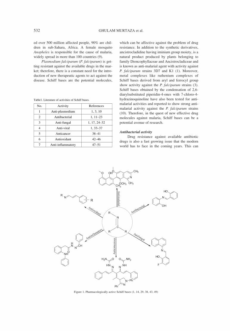
ìSchiff basesî were named after the Germanchemist Hugo Schiff and are produced by reactingthe aldehyde or ketone with primary amines (1),they can be used as reactive intermediates for thesynthesis of many natural products (2). Schiff basesare reported to show a wide range of pharmacologi-cal activities and are used as antimicrobial agentswith the activities including antibacterial, anti-fun-gal, anti-malarial and anti-viral agents as well as theanti-inflammatory, antioxidant and anti-cancerousagents (3, 4). Pharmacological activities attributedby Schiff bases are mainly due to characteristic C=Nfunctionality (Figure 1). Synthetically, condensationof amine with carbonyl compound occurs underreflux conditions with complete removal of watermolecule formed in the system by using molecularsieves; removal of water can also be done by usingwell known dehydrating solvents in situ i.e.,trimethylorthoformate or tetramethylorthosilicate(1).
Other methods has also been reported for syn-thesis of Schiff bases, that involve the use of Lewisor Bronsted-Lowry acids, some common are ZnCl2,TiCl4, MgSO4-PPTS, Ti(OR)4, alumina, H2SO4,NaHCO3, MgSO4, Mg(ClO4)2, H3CCOOH,
Er(OTf)3, P2O5/Al2O3 and HCl, as catalyst (1, 3ñ6).New cost effective and efficient methods including,microwave accelerated, solvent free synthesis, andsolid state synthesis are also being used and report-ed for the synthesis of Schiff bases and their metalcomplexes (1, 7, 8)
This brief review summarizes the pharmaco-logical importance of different synthetic Schiffbases derived from some natural products or fromcommercially available precursors and also suggestthe future perspectives of potential research areas.
PHARMACOLOGICAL SIGNIFICANCE
OF SCHIFF BASES
Biologically active molecules, Schiff bases, areknown to show a variety of pharmacological activi-ties. The literature available and used in this reviewhas been summarized in Table 1.
Antimicrobial activities
Anti-plasmodium activityMalaria, a disease caused by genus
Plasmodium, claims approximately one milliondeath tolls annually and is a serious threat to devel-oping countries. World Health Organization report-
RECENT PHARMACOLOGICAL ADVANCEMENTS IN SCHIFF BASES: A REVIEW
GHULAM MURTAZA1*, AMARA MUMTAZ2, FARHAN AHMED KHAN2, SAEED AHMAD3, SAIRA AZHAR1, MUHAMMAD NAJAM-UL-HAQ4, MUHAMMAD ATIF3, SHUJAAT ALI KHAN1,
ANEELA MAALIK2, FIAZ ALAM1 and IZHAR HUSSAIN1
1 Department of Pharmacy, 2 Department of Chemistry, COMSATS Institute of Information Technology,Abbottabad 22060, Pakistan
3Department of Pharmacy, The Islamia University of Bahawalpur, Bahawalpur, Pakistan4Department of Chemistry, Bahauddin Zakariya University, Multan, Pakistan
Abstract: Schiff bases are the biologically privileged scaffolds in organic chemistry, commonly synthesizedfrom the condensation reaction of carbonyl functional group with amines. Naturally occurring and synthetical-ly prepared Schiff bases are active molecules with many pharmacological activities like antibacterial, anti-can-cer, anti-fungal, anti-malarial, antioxidant and many more. This review article summarizes pharmacologicaldevelopments in the recent few years and gives a brief overview of their therapeutic potential.
Keywords: aldehydes, antibacterial, antifungal, ketones, antiinflammatory, anti-cancer, antioxidant, Schiffbases
531
* Corresponding author: e-mail: [email protected]; mobile: 923142082826; fax: 92992383441
532 GHULAM MURTAZA et al.
ed over 500 million affected people, 90% are chil-dren in sub-Sahara, Africa. A female mosquitoAnopheles is responsible for the cause of malaria,widely spread in more than 100 countries (9).
Plasmodium falciparum (P. falciparum) is get-ting resistant against the available drugs in the mar-ket; therefore, there is a constant need for the intro-duction of new therapeutic agents to act against thedisease. Schiff bases are the potential molecules,
which can be affective against the problem of drugresistance. In addition to the synthetic derivatives,ancistrocladidine having iminium group moiety, is anatural product produced by plants belonging tofamily Dioncophyllaceae and Ancistrocladiceae andis known as anti-malarial agent with activity againstP. falciparum strains 3D7 and K1 (1). Moreover,metal complexes like ruthenium complexes ofSchiff bases derived from aryl and ferrocyl groupshow activity against the P. falciparum strains (3).Schiff bases obtained by the condensation of 2,6-diarylsubstituted piperidin-4-ones with 7-chloro-4-hydrazinoquinoline have also been tested for anti-malarial activities and reported to show strong anti-malarial activity against the P. falciparum strains(10). Therefore, in the quest of new effective drugmolecules against malaria, Schiff bases can be apotential avenue of research.
Antibacterial activityDrug resistance against available antibiotic
drugs is also a fast growing issue that the modernworld has to face in the coming years. This can
Figure 1. Pharmacologically active Schiff bases (1, 14, 29, 38, 43, 49)
Table1. Literature of activities of Schiff bases.
No. Activity References
1 Anti-plasmodium 1, 3, 10
2 Antibacterial 1, 11ñ23
3 Anti-fungal 1, 17, 24ñ32
4 Anti-viral 1, 33ñ37
5 Anticancer 38ñ41
6 Antioxidant 42ñ46
7 Anti-inflammatory 47ñ51
Recent pharmacological advancements in Schiff bases: a review 533
potentially result in a dramatic increase in death rateand infectious diseases especially after accidentaland surgical cases (11). Therefore, in order to copewith these problems, there is an immediate and con-stant need of new synthetic moieties with better andacceptable therapeutic index (12). Schiff bases havebeen considered the agents, which have more effec-tive activity against the infectious bacteria, Schiffbases synthesized from 2-hydroxy-1-naphthalde-hyde and α-amino acids (L-tyrosine, L-arginine, andL-lysine) and their manganese complexes have beenreported to show excellent activity against the Grampositive and Gram negative strains of bacteria (13).Additionally, Schiff bases derived from salicylalde-hyde show potent antibacterial activities, for exam-ple, N-(salicylidene)-2-hydroxyaniline has beenreported to show a prominent activity againstMycobacterium tuberculosis (1), while Schiff basesof 5-chlorosalicylaldehyde show enhanced antibac-terial activity against Escherichia coli (E. coli),Staphylococcus aureus (S. aureus), and Micrococcusluteus (M. luteus) strains of bacteria (14). However,Schiff bases derivatives have also been reported toact as bacteriostatic agents e.g., Schiff bases of 2,4-dichloro-5-fluorophenyl are useful to stop the bacte-rial growth (1, 15). Moreover, Schiff bases bearingnitroimidazole moiety show good antibacterialactivities against various bacterial strains (16). Inaddition, Schiff bases, derivatives of isatin, has beenreported to show antimicrobial activity comparablewith that of the standard drug sulfamethoxazole(17). Schiff bases synthesized from other substrates,including, morpholines, coumarins, o-phthaldehyde,aminothiazolylbromocoumarins, sulfonamides, ace-tophenones, crown ethers, amino acids and 2-aminophenol and 1,2,4-triazoles, were reported toshow very low antibacterial activities (1, 4, 18ñ23).
Anti-fungal activityFungal infections are not limited to tropical
areas but can also lead to increased risk of systemicinfections, which may be life threatening (24).Factors for an increase in systemic fungal infectionsare geriatric patients, surgeries, AIDS, treatment ofvarious tumors, transplantation of hard organs,hematopoietic stem cells and immunosuppressivetreatment (25ñ27). Therefore, it is important todevelop and formulate more effective and safe anti-fungal drugs, which can be effectively used in vari-ous medical conditions (28). Schiff bases have beenreported to show good anti-fungal activity, e.g.,Schiff bases of N-(salicylidene)-2-hydroxyanilineand from 3-fluorosalicylaldehyde are reported toshow antifungal activities (1). However, transition
metal complexes of Schiff bases derived from N,N-ethylene (bis 1-cyclopropyl-6-fluoro-4-oxo-7-(piperazine-1-yl)-quinoline-3-carboxylic acid repor-ted to show higher antifungal activity than their pre-cursor Schiff bases (29). Oxovanadium (IV) com-plexes of Schiff bases show more activity as com-pared to their ligand (30). In addition, chitosanSchiff bases have been reported to stop the growthof many fungal strains including Colletotric humla-genarium and Botrytis cinerea (1, 31). Moreover,isatin based Schiff bases has been reported to showremarkable antifungal activity against various fun-gal strains like Microsporum gypseum andMicrosporum audouinii, furthermore, isatin Schiffbase derivatives also show anti-fungal activityagainst Cryptococcus neoformans (C. neoformans),Epidermophyton floccosum (E. floccosum) andCandida albicans (C. albicans) (1, 17, 32).
Antiviral activityPresently, a large number of viral diseases are
treated either adopting vaccination or by usingantiviral drugs. Drug resistance reported in viral dis-eases is a serious issue for humanity; therefore, newtherapeutic molecules are constantly required (33).Some common viral diseases like, influenza, rubel-la, small pox, chicken pox and polio can be con-trolled by vaccines administration, while viral dis-eases like hepatitis ëCí is still under the process ofvaccine discovery (1, 34). Therefore, Schiff basescan play a vital role due to their reported antiviralnature. Schiff bases derived from isatin and bis-isatin are reported to show activities against diffentstrains of viruses (1, 34, 35). Moreover, Schiff basesderived from prodrug abacavir (Ziagen) are reportedto show good antiviral activity and trials revealedthat they are potent lead molecules for further clini-cal use as anti-HIV therapy (1, 36). Furthermore,Schiff bases of 2-phenylquinazoline-4(3)H-one arereported to show antiviral activity against somestrains of viruses like feline corona virus, influenzaviruses, and herpes simplex virus type 1 and 2 (37).The antiviral potential of these Schiff bases is evi-dent from reported literature and therefore more tar-geted research can help to discover and develop newpotential lead compounds to use them as drug can-didates.
Anticancer activityCancer is a disease which leads to death. More
than 200 cancer types have been reported in thehuman body. Schiff bases obtained from cumarinand pyrazole aldehyde has been tested against can-cerous cell lines and showed mild anti-cancerous

534 GHULAM MURTAZA et al.
activities (38). Moreover, in another study, monoand bis-Schiff bases have been reported effectiveagainst five cancer cell lines (39). Furthermore,Schiff bases can effectively form complexes withtransition metals and these metal complexes arereported to show good anticancer activities; Cu-complexes with vaniline Schiff bases (40) and 5-dimethyl-2-phenyl-4-[(pyridin-2-ylmethylene)-amino]-1,2-dihydro-pyrazol-3-one Schiff bases (41)has been reported for their anti-cancerous activities.Extensive literature is available on the effectivenessof Schiff bases against cancer cell lines, therefore, amore systematic and extensive research, both invitro and in vivo, is suggested to extend their thera-peutic use to alleviate the disease.
Antioxidant activity
Aging is an evident phenomenon that a humanhas to face. Production of reactive oxygen species(ROS) increases with the passage of time, in thehuman body and leads to many physiological disor-ders including cardiovascular diseases. Schiff basesand their metal complexes play an important role inthe production of ROS (42) and therefore, can showantioxidant properties. Recently, Schiff bases of nat-ural phenylpropene derived methoxylated cin-namaldehydes (43), and tin metal complexes havebeen reported for antioxidant activities (44). In arecent study on thymol and carvacrol Schiff basederivatives in 5 µg/mL concentration showed60ñ90% inhibition for antioxidant activity (45).Moreover, Schiff bases of 2-oxoquinoline-3-car-baldehyde have been reported as excellent anti-oxi-dizing agents and their activity was comparable withthe ascorbic acid used as standard (46). Literaturereveals their effectiveness in the antioxidant behav-ior; therefore, more targeted research can possiblylead to their use in the therapy of various ailments.
Anti-inflammatory activity
Non-steroidal anti-inflammatory drugs(NSAIDs) are being used for the treatment of painand perform their function by inhibiting the produc-tion of prostaglandins (PG), which are involved inmany physiological activities (47, 48). Occasion-ally, these NSAIDs are not targeted for the particu-lar enzyme involved in the biosynthesis ofprostaglandins; therefore, for more targeted attackon the particular isozyme new effective moleculesare required. Therefore, Schiff bases derived from 2-(2,6-dichloroanilino) (49) and 4-amino-1,5-dimeth-yl-2-phenylpyrazol-3-one have been reported forexcellent anti-inflammatory activities (50).Moreover, transition metal complexes of Schiff
bases containing aldose group have also beenreported for anti-inflammatory activities (51).Further investigations are suggested for their prefer-ential therapeutic use in sickness and accidental caseof inflammation.
CONCLUSION
Schiff bases and their derivatives are a class ofcompounds with literature evident pharmacologicalimportance and applications. Therapeutic spectrumis also wide and less explored for Schiff bases,therefore, a scientific approach is required to estab-lish the structure activity relationships of these bio-logically and medicinally viable molecules.Concisely, Schiff bases are among the moleculeswhich have therapeutic potential for the treatment ofvarious human diseases.
REFERENCES
1. da Silva C.M., da Silva D.L., Modolo L.V.,Alves R.B., de Resende M.A., Cleide V.B.Martins C.V.B., de F·tima A.: J. Adv. Res. 2, 1(2012).
2. NikoliÊ D., Gˆdecke T., Chen S.-N., White J.,Lankin D.C., Pauli G.F., van Breemen R.B.:Fitoterapia 83, 441 (2012).
3. Adams M., Li Y., Khot H., De Kock C., SmithP.J., Land K., Chibale K., Smith G.S.: DaltonTrans. 42, 4677 (2013).
4. Bhat M.A., Al-Omar M.A., Siddiqui N.: Med.Chem. Res. 9, 4455 (2013).
5. KlemkaitÎ-RamanauskÎ K., éilinskas A.,TaraökeviËius R, Khinsky A., Kareiva A.:Polyhedron 68, 340 (2014).
6. Naeimi H., Salimi F., Rabiei K.: J. Mol. Catal.A 260, 100 (2006).
7. Thaker B.T., Barvalia R.S.: Spectrochim. ActaA 84, 51 (2011).
8. Degirmencioglu I., Bayrak R., Er M., SerbestK.: Dyes Pigments 83, 51 (2009).
9. Alegana V.A., Atkinson P.M.., Wright J.A.,Kamwi R., Uusiku P., Kakotele S., Snow R.W.,Noor A.M.: Spat. Spatiotemporal Epidemiol. 7,25 (2013).
10. Le T.T., Hoang X.T., Vu D.H., Tran K.V.: Lett.Drug Des. Discov. 9, 163 (2012).
11. Theuretzbacher U.: Int. J. Antimicrob. Agents39, 295 (2012).
12. Baquero F.: J. Antimicrob. Chemother. 39,(Suppl. A), 1 (1997).
13. �ak�yan �, ÷zdemir R. ÷�¸tc¸ H.: Synth. React.Inorg. Met. Org. Chem. 44, 417 (2014).

Recent pharmacological advancements in Schiff bases: a review 535
14. Wang Z., Gao J., Wang J., Jin X., Zou M., LiK., Kang P.: Spectrochim. Acta A 83, 511(2011).
15. Karthikeyan M.S., Prasad D.J., Poojary B., BhatK.S., Holla B.S., Kumari N.S.: Bioorg. Med.Chem., 14, 7482 (2006).
16. Makawana J.A., Sun J., Zhu H.-L.: Bioorg.Med. Chem. Lett., 23, 6264 (2013).
17. Aboul-Fadl T., Bin-Jubair F.A.S., Aboul-WafaO.: Eur. J. Med. Chem. 45, 4578 (2010).
18. Raparti V., Chitre T., Botharak., Kumar V.,Dangre S., Khachane C., Gore S., BhavanaDeshmane B.: Eur. J. Med. Chem. 44, 3954(2009).
19. Kulkarni A., Patil S.A., Badami P.S.: Eur. J.Med. Chem. 44, 2904 (2009).
20. Bhat M.A., Al-Omar M.A., Siddiqui N.: Med.Chem. Res. 9, 4455 (2013).
21. Chohan Z.H., Shad H.A. Supuran C.T.: J.Enzyme Inhib. Med. Chem. 27, 58 (2012).
22. Abdallah S.M., Mohamed G.G., Zayed M.A.,El-Ela M.S.A.: Spectrochim. Acta A 73, 833(2009).
23. Adly O.M.I.: Spectrochim. Acta A 95, 483(2012).
24. Rice L.B.: Biochem. Pharmacol. 71, 991(2006).
25. Pawar O., Patekar A., Khan A., Kathawate L,Haram S., Markad G., Puranik V., Salunke-Gawali S. : J. Mol. Struct. 1059, 68 (2014).
26. Liu X., Ling Z, Li L, Ruan B.: Int. J. Infect. Dis.15, e298 (2011).
27. Husain S.: Clinics in Chest Med. 30, 307(2009).
28. Khan F.A., Maalik A., Iqbal Z., Malik I.: Eur. J.Pharmacol. 721, 391 (2013).
29. Shanmugam M., Narayanan K., MahalakshmiM., Kabilan S., Chidambaranathan V.:Spectrochim. Acta A 116, 394 (2013).
30. Sumrra S.H., Chohan Z.H.: J. Enzyme Inhib.Med. Chem. 28, 1291 (2013).
31. Jin X., Wang J., Bai J.: Carbohydrate Res. 344,825 (2009).
32. Prakash C.R., Raja S.: J. Saudi Chem. Soc. 17,337 (2013).
33. Marschall M., Niemann I., Kosulin K., BootzA., Wagner S., Dobner T., Herz T. et al.:Antiviral Res. 100 640 (2013).
34. Abbas S.Y., Farag A.A., Ammar Y.A., AtreesA.A., Mohamed A.F., El-Henawy A.A.:Monatsh. Chem. 144, 1725 (2013).
35. Aliasghar J., Javed S., Ibrahim E.M., Harjeet J.,Taibi B.H.: Med. Chem. Res. 22, 1203 (2013).
36. De Clercq E.: Nat. Rev. Drug Discov. 1, 13(2002).
37. Kumar K.S., Ganguly S., Veerasamy R., DeClercq E.: Eur. J. Med. Chem., 45, 5474 (2010).
38. Ali I., Haque A., Saleem K., Hsieh M.F.:Bioorg. Med. Chem. 21, 3808 (2013).
39. Sondhi S.M, Ayra S., Rani R., Kumar N., RoyP.: Med. Chem. Res. 21, 3620 (2012).
40. Tabassum S., Amir S., Armand F., Pettinari C.,Marchetti F., Masciocchi N., Lupidi G.,Pettinari R.: Eur. J. Med. Chem. 60, 216 (2013).
41. Sathiyaraj S., Sampach K., Butcher R.J.,Pallepogu R., Jayabalakrishna C.: Eur. J. Med.Chem. 64, 81 (2013).
42. Li G., Zhang H.F., Wang R.M., He Y.F., XiongY.B.: Chin. Sci. Bull. 58, 2956 (2013).
43. Sharma U.K., Sood S., Sharma N., Rahi P.,Kumar R., Sinha A.K., Gulati A.: Med. Chem.Res. 22, 5129 (2013).
44. RamÌrez-JimÈnez A., Luna-GarcÌa R., CortÈs-Lozada A., Hern·ndez S., RamÌrez-Apan T.,Nieto-Camacho A., GÛmez E.: J. Organomet.Chem. 738, 10 (2013).
45. Beena D.K., Rawat D.S.: Biorg. Med. Chem.Lett. 23, 641 (2013).
46. Zhang Y., Fang Y., Liang H., Wang H., Hu K.,Liu X., Yi X., Peng Y.: Bioorg. Med. Chem.Lett. 23, 107 (2013).
47. Smith C.J., Zhang Y., Koboldt C.M.,Muhammad J., Zweifel B.S., Shaffer A., TalleyJ.J. et al.: Proc. Natl. Acad. Sci. USA 95, 13313(1998).
48. Warner T.D., Giuliano F., Vaynovie I., BukasaA., Mitchell J.A., Vane J.R.: Proc. Natl. Acad.Sci. USA 96, 7563 (1999).
49. Bhandari S.V., Bothara K.G., Raut M.K., PatilA.A., Sarkate A.P., Vinod J. Mokale V.J.:Bioorg. Med. Chem. 16, 1822 (2008).
50. Alam M.S., Choi J.-H., Dong-Ung Lee D.-U.:Bioorg. Med. Chem. 20, 4103 (2012).
51. Iqbal M.S., Khurshid S.J., Muhammad B.: MedChem. Res. 22, 861 (2013).
Received: 30. 09. 2013

Acta Poloniae Pharmaceutica ñ Drug Research, Vol. 71 No. 4 pp. 537ñ543, 2014 ISSN 0001-6837Polish Pharmaceutical Society
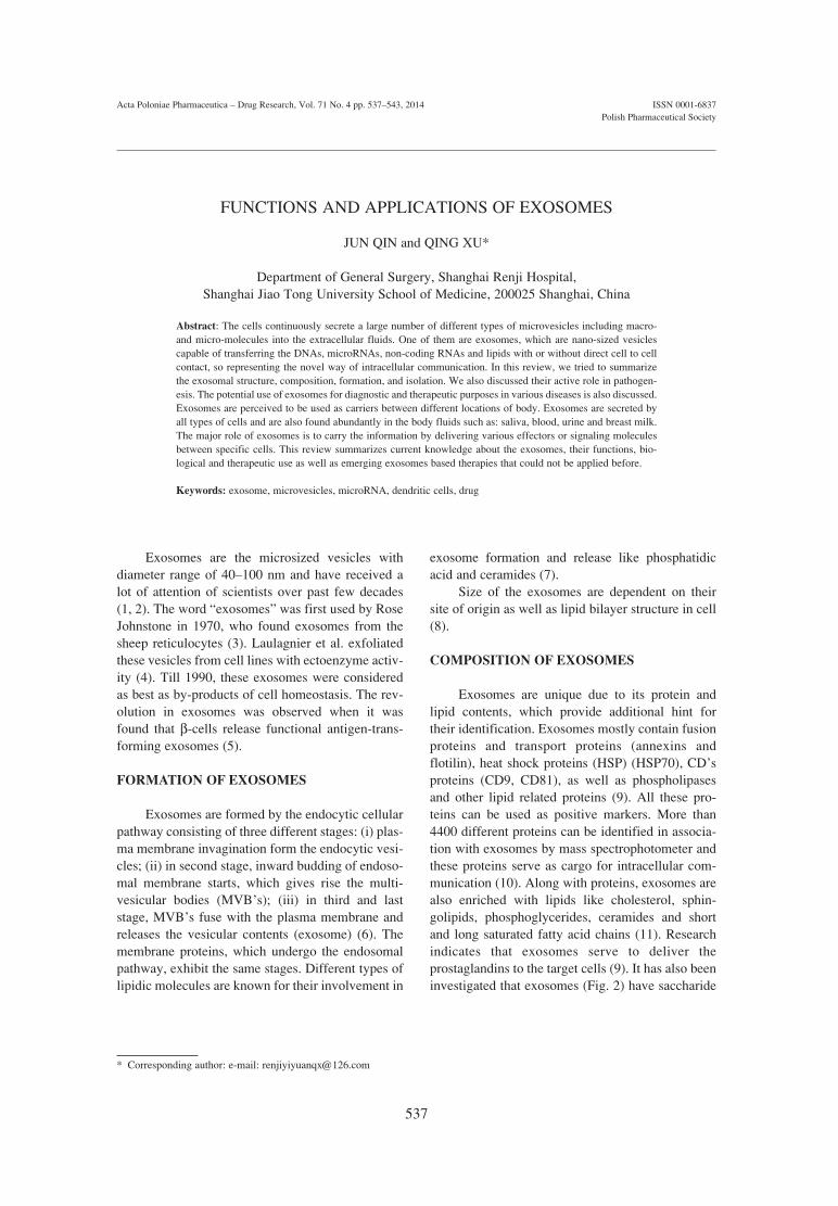
Exosomes are the microsized vesicles withdiameter range of 40ñ100 nm and have received alot of attention of scientists over past few decades(1, 2). The word ìexosomesî was first used by RoseJohnstone in 1970, who found exosomes from thesheep reticulocytes (3). Laulagnier et al. exfoliatedthese vesicles from cell lines with ectoenzyme activ-ity (4). Till 1990, these exosomes were consideredas best as by-products of cell homeostasis. The rev-olution in exosomes was observed when it wasfound that β-cells release functional antigen-trans-forming exosomes (5).
FORMATION OF EXOSOMES
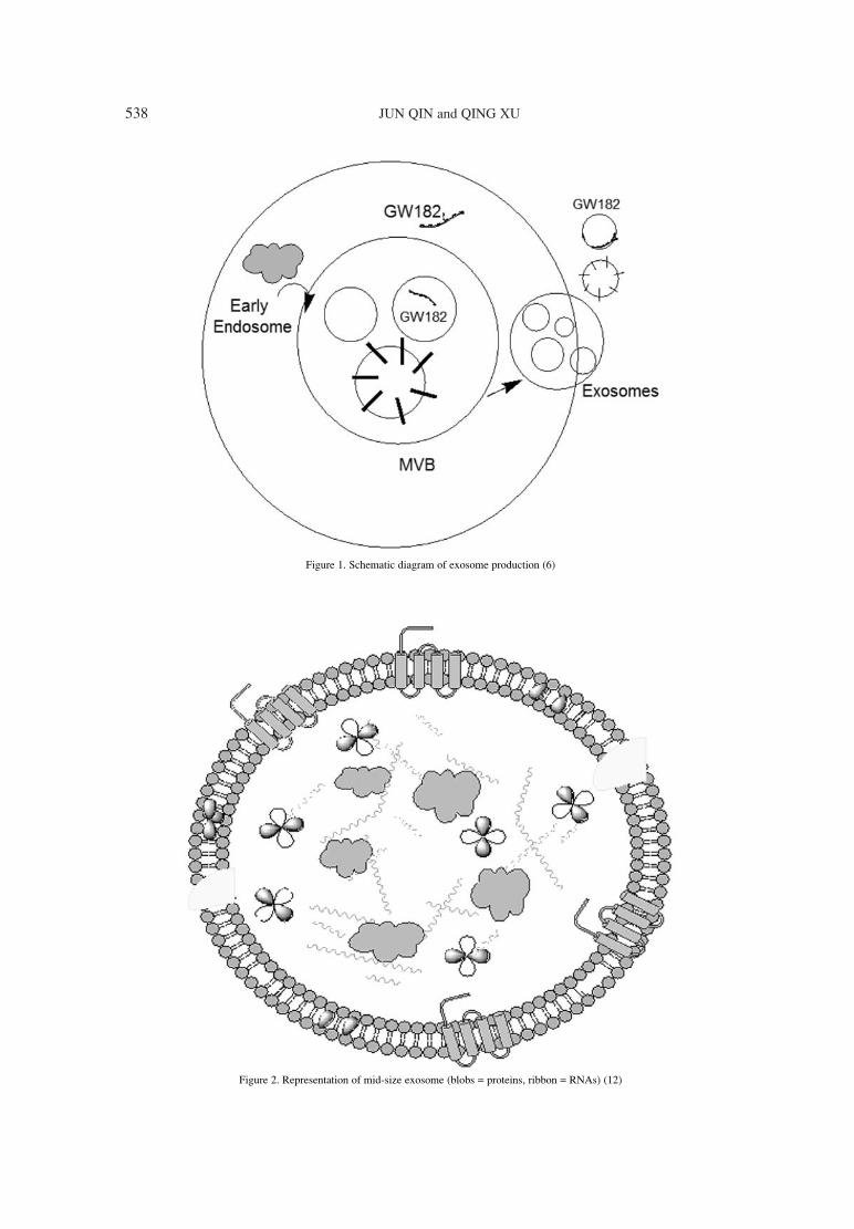
Exosomes are formed by the endocytic cellularpathway consisting of three different stages: (i) plas-ma membrane invagination form the endocytic vesi-cles; (ii) in second stage, inward budding of endoso-mal membrane starts, which gives rise the multi-vesicular bodies (MVBís); (iii) in third and laststage, MVBís fuse with the plasma membrane andreleases the vesicular contents (exosome) (6). Themembrane proteins, which undergo the endosomalpathway, exhibit the same stages. Different types oflipidic molecules are known for their involvement in
exosome formation and release like phosphatidicacid and ceramides (7).
Size of the exosomes are dependent on theirsite of origin as well as lipid bilayer structure in cell(8).
COMPOSITION OF EXOSOMES
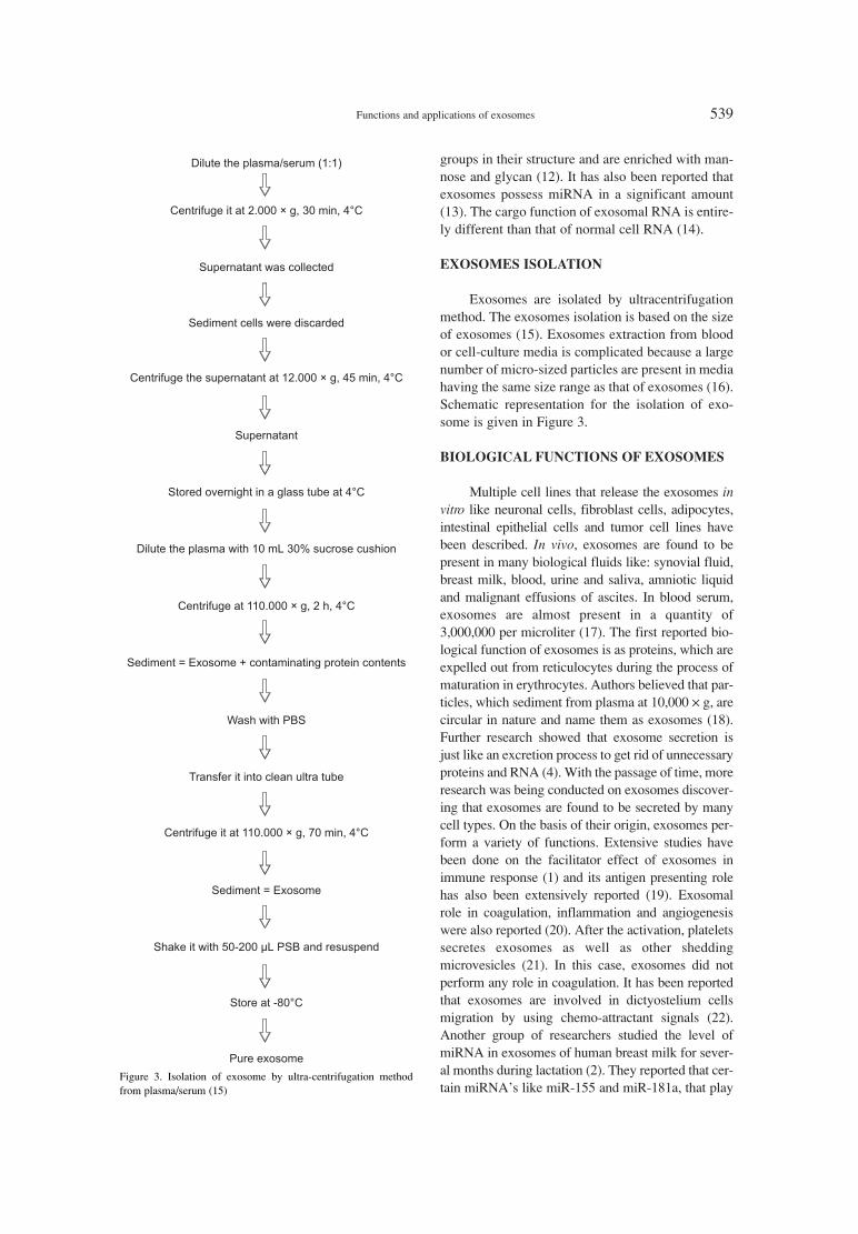
Exosomes are unique due to its protein andlipid contents, which provide additional hint fortheir identification. Exosomes mostly contain fusionproteins and transport proteins (annexins andflotilin), heat shock proteins (HSP) (HSP70), CDísproteins (CD9, CD81), as well as phospholipasesand other lipid related proteins (9). All these pro-teins can be used as positive markers. More than4400 different proteins can be identified in associa-tion with exosomes by mass spectrophotometer andthese proteins serve as cargo for intracellular com-munication (10). Along with proteins, exosomes arealso enriched with lipids like cholesterol, sphin-golipids, phosphoglycerides, ceramides and shortand long saturated fatty acid chains (11). Researchindicates that exosomes serve to deliver theprostaglandins to the target cells (9). It has also beeninvestigated that exosomes (Fig. 2) have saccharide
FUNCTIONS AND APPLICATIONS OF EXOSOMES
JUN QIN and QING XU*
Department of General Surgery, Shanghai Renji Hospital, Shanghai Jiao Tong University School of Medicine, 200025 Shanghai, China
Abstract: The cells continuously secrete a large number of different types of microvesicles including macro-and micro-molecules into the extracellular fluids. One of them are exosomes, which are nano-sized vesiclescapable of transferring the DNAs, microRNAs, non-coding RNAs and lipids with or without direct cell to cellcontact, so representing the novel way of intracellular communication. In this review, we tried to summarizethe exosomal structure, composition, formation, and isolation. We also discussed their active role in pathogen-esis. The potential use of exosomes for diagnostic and therapeutic purposes in various diseases is also discussed.Exosomes are perceived to be used as carriers between different locations of body. Exosomes are secreted byall types of cells and are also found abundantly in the body fluids such as: saliva, blood, urine and breast milk.The major role of exosomes is to carry the information by delivering various effectors or signaling moleculesbetween specific cells. This review summarizes current knowledge about the exosomes, their functions, bio-logical and therapeutic use as well as emerging exosomes based therapies that could not be applied before.
Keywords: exosome, microvesicles, microRNA, dendritic cells, drug
537
* Corresponding author: e-mail: [email protected]
538 JUN QIN and QING XU
Figure 1. Schematic diagram of exosome production (6)
Figure 2. Representation of mid-size exosome (blobs = proteins, ribbon = RNAs) (12)
Functions and applications of exosomes 539
groups in their structure and are enriched with man-nose and glycan (12). It has also been reported thatexosomes possess miRNA in a significant amount(13). The cargo function of exosomal RNA is entire-ly different than that of normal cell RNA (14).
EXOSOMES ISOLATION
Exosomes are isolated by ultracentrifugationmethod. The exosomes isolation is based on the sizeof exosomes (15). Exosomes extraction from bloodor cell-culture media is complicated because a largenumber of micro-sized particles are present in mediahaving the same size range as that of exosomes (16).Schematic representation for the isolation of exo-some is given in Figure 3.
BIOLOGICAL FUNCTIONS OF EXOSOMES
Multiple cell lines that release the exosomes invitro like neuronal cells, fibroblast cells, adipocytes,intestinal epithelial cells and tumor cell lines havebeen described. In vivo, exosomes are found to bepresent in many biological fluids like: synovial fluid,breast milk, blood, urine and saliva, amniotic liquidand malignant effusions of ascites. In blood serum,exosomes are almost present in a quantity of3,000,000 per microliter (17). The first reported bio-logical function of exosomes is as proteins, which areexpelled out from reticulocytes during the process ofmaturation in erythrocytes. Authors believed that par-ticles, which sediment from plasma at 10,000 ◊ g, arecircular in nature and name them as exosomes (18).Further research showed that exosome secretion isjust like an excretion process to get rid of unnecessaryproteins and RNA (4). With the passage of time, moreresearch was being conducted on exosomes discover-ing that exosomes are found to be secreted by manycell types. On the basis of their origin, exosomes per-form a variety of functions. Extensive studies havebeen done on the facilitator effect of exosomes inimmune response (1) and its antigen presenting rolehas also been extensively reported (19). Exosomalrole in coagulation, inflammation and angiogenesiswere also reported (20). After the activation, plateletssecretes exosomes as well as other sheddingmicrovesicles (21). In this case, exosomes did notperform any role in coagulation. It has been reportedthat exosomes are involved in dictyostelium cellsmigration by using chemo-attractant signals (22).Another group of researchers studied the level ofmiRNA in exosomes of human breast milk for sever-al months during lactation (2). They reported that cer-tain miRNAís like miR-155 and miR-181a, that play
Figure 3. Isolation of exosome by ultra-centrifugation methodfrom plasma/serum (15)

540 JUN QIN and QING XU
an important role during immune regulation and werepresent in high concentration during first six monthsof lactation, were significantly reduced afterward (7).Recent studies demonstrate that the exosomes are notonly involved in triggering downstream signaling butthey also specifically target the recipient cells andexchange proteins. Exosomes also deliver the specif-ic nucleic acids and work as cargo (23). The uniquefunction of exosomes is cell to cell communication,especially between the far distance cells in the body.Similarly, exosomes play a unique role in spreadingvarious pathogens like virus and prion from one cellto another (24).
EXOSOMES IN DIAGNOSTICS
For last few years, much research has been doneon diagnostic aspect of exosomes and it was discov-ered that almost all the body fluids (blood, saliva,milk, and urine) contained exosomes. Because ofunique structure of the exosomes, which possess pro-teins, lipids and RNAs, it may be useful for the diag-nostic purposes (25). In late 1970s, microvesicles(MVs) were derived from the cancer cells in personsuffering from Hodgkinís disease (14). Since that dayto-date, considerable efforts have been done to usethe MVs as diagnostic tool (Table 1). It was reportedthat MVs levels were elevated in serum, urine andblood in the cancer patient (26). However,microvesicular components may provide importantinformation regarding a disease. For example, mucinbearing MVs are used as diagnostic marker for thediagnosis of adenocarcinoma (27). A proteomicinvestigation of urine identified eight proteins, whichact as an important diagnostic tool in bladder cancer
(28). Thus it can be said that protein portion of theexosomes are the useful tool for the diagnosis of thediseases. In addition, recent studies have showed thatcancer patients exhibit different patterns of RNA andmiRNA. In cancer patients, RNA and miRNA havebeen found in circulating MVs form (9).
The PCR of miRNA is a sensitive and stablemethod for the diagnosis and detection of miRNA inpatientsí serum, which is a new promising approachto detect disease in early stages. Down-regulation ofmiR-92a in plasma is the biomarker of hepatocellu-lar carcinoma and leukemia (12).
EXOSOMES AS TARGETED DRUG
DELIVERY VEHICLES
Exosomes can be used as targeted drug deliv-ery systems. Alvarez et al. first of all presented andproved this hypothesis (36) by using immature den-dritic cells (DCs). They used DCs derived from thebone marrow of mouse as a source of exosomes andthese exosomes were devoted as stimulatory mole-cules such as MHCII and CD80. They purified theexosomes by ultracentrifugation method and used ascargo for siRNA delivery both in in vitro and in vivostudies. They selected brain as a target tissue inbody, because it is believed that blood brain barrier(BBB) is the major obstacle in drug delivery to cen-tral nervous system. Sealed functions of BBB aredue to the capillary endothelial cells that are tightlysealed by junctions and regulate the barrier func-tions (19). For ensuring targeted delivery of exo-somes, Ratajczak et al. (27) used the novel strategyby utilizing LAMP2B, an exosomal surface protein,that display the targeted peptide on its surface.
Figure 4. Extracellular membrane vesicle therapy (EMVs) A: EMV immunotherapy. Tumor antigen on the membrane surface from dif-ferent sources was introduced in vivo to elicit targeted immune responses. B: EMV drug therapy. Drug packaged into/onto EMVs isolat-ed from donor cells to minimize degradation and increase delivery to intended sites (18)

Functions and applications of exosomes 541
TREATMENT OF BRAIN INFLAMMATORY
DISEASE BY EXOSOMES ENCAPSULATED
WITH DRUGS
Zhuang et al. used the encapsulated curcumin(Exo-cur) or JS1124 (Exo-JS1124) inhibitor of sig-nal transducer and activator of transcription anddelivered it into the microglia cells throughintranasal route. They used lipopolysaccharide(LPS)-induced inflammatory model for the experi-mental mice to induce inflammation. They showedthat mice treated with Exo-cur and Exo-JS1124were protected from LPS-induced inflammation.They believed that exosomes were selectively takenby the microglia cells and subsequently induced theapoptosis of the microglia cells after its intranasaldelivery (30).
EXOSOMES AS AN APPROACH FOR
TREATING ARTHIRITIS
Dendritic cells (DCs) and T-cells have beenused for delivery of immunosuppressive cytokinesfor the treatment of various collagen inducedinflammations in different mouse model (31). DCsare the antigen presenting cells that regulate theimmune activity. Various factors are involved instimulating or suppressing immune responses ofDC. DCs have low level of MHC and other mole-cules such as ICAM-1, so they can suppress T-cellimmune response. The immunosuppressive abilityof DCs enhanced its genetic modification and genet-ically modified DCs showed dramatic control in theprogression of autoimmune diseases like diabetesand arthritis (24). DCs with viral vectors expressingthe immunosuppressive agents exert their effectmore pronouncedly than T-cells or fibroblasts (8).
Due to the ability of genetic modification of DCs,they produce distal therapeutic effects speciallywhen exosomes were delivered along with viral vec-tors (6). Immunosuppressive DCs-exosomes canmodify the endogenous immune cells, such asAPCs, so they may be responsible for anti-inflam-matory effects (1).
EXOSOMES IN IMMUNOTHERAPY
AND NERVOUS SYSTEM
Exosomes/MVs (EMVs) have cell to cellcommunication function for transfer of geneticmaterial (4). The dramatic progress in the researchof MVs for drug delivery is due to its low immuno-genicity and unique delivering properties. With thehelp of genetic engineering, EMVs are used totransport the therapeutic drugs either by directinsertion or by loading onto the targeted gene (38).Exosomes also serve as an excellent therapeuticcargo due to its protection rendered to enclosedcontents. Due to these possible advantages, EMVsmediated therapy is actively studied and is used inthree different fields i.e., immunotherapy, RNA-interference (Fig. 4) and drug delivery (18). Themost wide investigational portfolio of EMVs is inimmunotherapy, which is an efficient way of can-cer treatment through the preparation of vaccinescontaining antigen presenting cells to recognizesthe tumor cells (20). It has been shown that B-lym-phocytes secrete EMVs and these EMVs containMHC-II, which can induces the cell response invitro (19). These EMVs may be used as an emerg-ing therapy in the treatment of various nervous sys-tem diseases. Sun et al. developed drug-loadedexosomes (35). They successfully loaded curcumininto EMVs, which significantly reduced the LPS-induced inflammation.
Table 1. Circulating exosomes as potential diagnostic markers for various diseases.
Sample type Marker Disease Ref.
Quantity Plasma PMPs level Gastric cancer (29)
Serum PMPs level Prostate cancer (30)
Ascites CD24, EpCAM Ovarian cancer (31)
Serum Tissue factor General cancer (32)
Protein expression Plasma Tissue factor Breast cancer (33)
Pleural effusion SNX25, BTG1 Mesothelioma (34)
Urine Fetuin-A Acute kidney injury (9)
miRNA or mRNA Serum Glioblastoma Glioblastoma (10)
expression Serum MAGE-1, HER-2 Gastric cancer (35)

542 JUN QIN and QING XU
ROLE OF EXOSOMES IN SENESCENCE
AND AGING
Senescence is the cellular part of aging of tis-sues due to the irreversible growth arrest and otherphysiological changes occurring in cell morphology,cell behavior and function. The miRNAs are smallnon-coded RNAs, which regulate the gene expres-sion and play an important role in biologicalprocesses. Recent developments have shown thatexosomes contain miRNAs, which are released intovariety of cells and play an important role in cell-to-cell communication and information transfer.Exosomes with miRNAs formed the complex cellu-lar network senescence and contribute to aging (39).
ANTI-TUMORIGENIC ROLE OF
TUMOR-DERIVED EXOSOMES
The protein portion of the exosomes reflectsthe cell type specificity for their cell of origin fromwhich they are secreted. Particularly exosomes,which are derived from tumor may contain tumor-specific antigens on their surface as present in tumorcells (21). Tumor antigens such as carcinoembryon-ic antigen (CEA) (12) and mesothelin (13) areobserved in tumor-derived exosomes. Due to thisobservation, it is suggested that tumor exosome-based cancer vaccines may be developed. Tumor-derived exosomes might be used as a tumor antigensource, which might be able to induce the CD8+ T-cell dependent anti-tumor effects in mice (39).Recently, it was reported that dendritic cells loadedwith tumor exosomes elicited the CD8+ T-cellresponse against the tumor cells in malignantgliomas patient (40). For augmenting anti-tumoractivity/immunity, tumor derived exosomes havebeen investigated for direct application (Table 2).Research showed that tumor-derived exosomes pro-duced specific antitumor activity when its parent
cells were genetically modified. These geneticallymodified exosomes can express pro-inflammatorycytokines such as IL-2 (6). Heat shocked lymphomacells, which releases the exosomes expressing MHCand other co-stimulatory molecules, induce efficientanti-tumor T cell immunity (40).
CONCLUSION
Increasing research efforts are being done onthe exosomes from which we are gaining knowledgeon the mechanism of their formation, secretion, invivo pathways, and biological role of their nucleicacid, protein and lipid. With the emergence of exo-somes/EMVs responsible for cell to cell communi-cation, researchers gathered the information on theirrole, both on physiological and pathological func-tions as well as their use in different therapies. Themost interesting aspect of exosomes is their use asvesicular carriers. They carry the large sized mole-cules such as RNA and proteins that influence geneexpression. These microvesicles are similar to virus-es and are capable of communication from one cellto another and easily pass the contents of cellsacross the cell membrane and deliver the macromol-ecules that are biologically active. Much researchhad been done on therapeutic applications of theexosomes. Further developments are aimed toensure therapeutic functions and clinical potential ofexosomes including their cargo property, targetingfunction and different sources of exosomes thatenable tissue targeted applications of exosomes.
REFERENCES
1. Cocucci E., Racchetti G., Meldolesi J.: TrendsCell Biol. 19, 43 (2009).
2. Raposo G., Nijman H.W., Stoorvogel W.,Liejendekker R., Harding C.V., Melief C.: J.Exp. Med. 183, 1161 (1996).
Table 2. Studies on the immunogenicity of tumor-derived exosomes and their vaccines.
Exosome source Modification Model Results Ref.
Mouse colon Parent cells were Mouse Elevated level of Hsp-70, (1)carcinoma and melanoma heat treated elicit Th 1 response
Mouse lung Parent cells were Mouse Activates DCs and T-cells (32)carcinoma heat treated and produce immune response
Human renal Parent cells were modified to In vitro IL-12 permotes the release (19)cancer release GPI-IL-12 of IFN-α
Ascites from Exosomes were Phase 1 GM-CSF induce beneficial (3)colorectal cancer purified clinical trials tumor specific CTL response

Functions and applications of exosomes 543
3. Trajkovic K., Hsu C., Chiantia S., Rajendran L.,Wenzel D., Wieland F.: Science 319, 1244(2008).
4. Laulagnier K., Grand D., Dujardin A., HamdiS., Vincent-Schneider H., Lankar D.: FEBSLett. 572, 11 (2004).
5. Murtaza G., Ahmad M, Asghar M.W., AamirM.N.: DARU J. Pharm. Sci. 17, 209 (2009).
6. Batista B.S., Eng W.S., Pilobello K.T.,Hendricks-MuÒoz K.D., Mahal L.K.: J.Proteome Res. 10, 4624 (2011).
7. Mittelbrunn M., GutiÈrrez-V·zquez C.,Villarroya-Beltri C., Gonz·lez S., S·nchez-Cabo F., Gonz·lez M.¡.: Nat. Commun. 2, 282(2011).
8. Shahzad M.K., Ubaid M., Murtaza G.: Trop. J.Pharm. Res. 11, 695 (2012).
9. ThÈry C., Ostrowski M., Segura E.: Nat. Rev.Immunol. 9, 581 (2009).
10. Belting M., Wittrup A.: J. Cell Biol. 183, 1187(2008).
11. Vickers K.C., Remaley A.T.: Curr. Opin.Lipidol. 23, 91 (2012).
12. Leblanc P., Alais S., Porto-Carreiro I.,Lehmann S., Grassi J., Raposo G.: EMBO J. 25,2674 (2006).
13. Baran J., Baj-Krzyworzeka M., Weglarczyk K.,Szatanek R., Zembala M., Barbasz J., CzuprynaA. et al.: Cancer Immunol. Immunother. 59, 841(2010).
14. Kim H.K., Song K.S., Park Y.S., Kang Y.H.,Lee Y.J., Lee K.R., Kim H.K. et al.: Eur. J.Cancer 39, 184 (2003).
15. Khan S.A., Ahmad M., Murtaza G., ShoaibH.M., Aamir M.N., Kousar R., Rasool F.,Madni A.: Latin Am. J. Pharm. 29, 1029 (2010).
16. Runz S., Keller S., Rupp C., Stoeck A., Issa Y.,Koensgen D., Mustea A. et al.: Gynecol. Oncol.107, 563 (2007).
17. Zwicker J.I., Liebman H.A., Neuberg D.,Lacroix R., Bauer K.A., Furie B.C., Furie B.:Clin. Cancer Res. 15, 6830 (2009).
18. Hegmans J.P., Bard M.P., Hemmes A., LuiderT.M., Kleijmeer M.J., Prins J.-B., Zitvogel L. etal.: Am. J. Pathol. 164, 1807 (2004).
19. Ahmad M., Iqbal M., Akhtar N., Murtaza G.,Madni M.A.: Pak. J. Zool., 42, 395 (2010).
20. Skog J., W¸rdinger T., van Rijn S., MeijerD.H., Gainche L., Sena-Esteves M., Curry W.T.et al.: Nat. Cell Biol. 10, 1470 (2008).
21. Tanaka M., Oikawa K., Takanashi M., KudoM., Ohyashiki J., Ohyashiki K., Kuroda M.:PLoS One 4, e5532 (2009).
22. Khiljee S., Ahmad M., Murtaza G., MadniM.A., Akhtar N., Akhtar M.: Pak. J. Pharm. Sci.24, 421 (2011).
23. Yang C., Robbins P.D.: Int. J. Rheumatol. 34,2012 (2012).
24. Ahmad M., Ahmad R., Murtaza G.: Adv. Clin.Exp. Med. 20, 599 (2011).
25. Ratajczak J., Miekus K., Kucia M., Zhang J.,Reca R., Dvorak P., Ratajczak M.Z.: Leukemia20, 847 (2006).
26. Ratajczak J., Wysoczynski M., Hayek F.,Janowska-Wieczorek A., Ratajczak M.Z.:Leukemia 20, 1487 (2006).
27. Ahmad M., Murtaza G., Akhtar N., Siddique F.,Khan S.A.: Acta Pol. Pharm. Drug Res. 68, 115(2011).
28. Zhang Y., Liu D., Chen X., Li J., Li L., Bian Z.,Sun F, et al.: Mol. Cell 39, 133 (2010).
29. Dai S., Zhou X., Wang B., Wang Q., Fu Y.,Chen T., Wan T. et al.: J. Mol. Med. 84, 1067(2006).
30. Zhang Y., Luo C.L., He B.C., Zhang J.M.,Cheng G., Wu X.H.: Int. J. Oncol. 36, 133(2010).
31. Aamir M.F., Ahmad M., Murtaza G., KhanS.A.: Latin Am. J. Pharm. 30, 318 (2011).
32. Chen W., Wang J., Shao C., Liu S., Yu Y.,Wang Q., Cao X.: Eur. J. Immunol. 36, 1598(2006).
33. Cho J.A., Lee Y.S., Kim S.H., Ko J.K., KimC.W.: Cancer Lett. 275, 256 (2009).
34. Chen T., Guo J., Yang M., Zhu X., Cao X.: J.Immunol. 186, 2219 (2011).
35. Escudier B., Dorval T., Chaput N., AndrÈ F.,Caby M.P., Novault S., Flament C. et al.: J.Transl. Med. 3, 10 (2005).
36. Morse M.A., Garst J., Osada T., Khan S.,Hobeika A., Clay T.M., Valente N. et al.: J.Transl. Med. 3, 9 (2005).
37. Johnstone R., Bianchini A., Teng K.: Blood 74,1844 (1989).
38. Safaei R., Larson B.J., Cheng T.C., GibsonM.A., Otani S., Naerdemann W., Howell S.B.:Mol. Cancer Ther. 4, 1595 (2005).
39. Waqas M.K., Saqib Q.N.U., Rashid S.U., ShahP.A., Akhtar N., Murtaza G.: Afr. J. Tradit.Complement. Altern. Med. 10, 452 (2013).
40. Shedden K., Xie X.T., Chandaroy P., ChangY.T., Rosania G.R.: Cancer Res. 63, 4331(2003).
Received: 13. 10. 2013

Acta Poloniae Pharmaceutica ñ Drug Research, Vol. 71 No. 4 pp. 545ñ553, 2014 ISSN 0001-6837Polish Pharmaceutical Society
Renin, an aspartic proteinase, catalyzes a spe-cific hydrolysis of the angiotensinogen to give thedecapeptide angiotensin I. Angiotensin convertingenzyme (ACE), converts it to the octapeptideangiotensin II, which is a very strong vasoconstric-tor and it also stimulates aldosterone release andsodium retention. Renin is a specific enzyme thatdisplays specificity for its only one known naturalsubstrate ñ angiotensinogen. Therefore, the inhibi-tion of renin, which action initiates the renin-angiotensin cascade, has been a highly attractivebiological target for new antihypertensive drugs.Drugs that inhibit the renin-angiotensin system, likeACE inhibitors and angiotensin II receptor blockers,are very effective in hypertension treatment butthese drugs are characterized by many side effects(they stimulate compensatory mechanism, whichresults in an increase of angiotensin II level).Therefore, the idea to treat hypertension through therenin inhibition has led to development of manypotent renin inhibitors based on the peptidesequence of natural substrate ñ angiotensinogen.
Many trials to developed effective direct renininhibitors were not successful (synthesized com-pounds, which were peptide substrate analogues,were not stable, they revealed low potency or poor
pharmacological profiles). To avoid such problems,new substrates analogous to non-peptic amino acids,peptide-like inhibitors and fully nonpeptic inhibitorswere developed (1, 2). Aliskiren is the first renininhibitor registered at the FDA (3). The structure ofaliskiren differed in 8ñ13 amino acids fragmentfrom the structure of natural substrate ñangiotensinogen. It shows high effectivnes and goodpharmacokinetic profile.
Searching for new renin inhibitors, a series ofdipeptide analogues of angiotensinogen have beenprepared and they were all derived from renin sub-strate by replacing the scissile amide bond with atransition-state mimic structure and by incorporat-ing bioisosteric replacements for the Val-10 amidebond. These derivatives showed high inhibitingactivity (10-6 ñ 10-9 M) (4). Other transition-staterenin inhibitors containing the dipeptide transitionstate mimic structure: (2S,4S,5S)-5-amino-4-hydroxy-2-isopropyl-7-methyloctanoic acid (LeuOH Val) and (2S,4S,5S)-5-amino-4-hydroxy-2-iso-propyl-6-cyclohexylhexanoic acid (Cha OH Val)were synthesized (5, 6). The goal of such investiga-tion was to lower the molecular weight, to minimizethe number of peptide amide bonds and to enhancein vivo stability. All derivatives showed high activi-
ANALYSIS
NEW RENIN INHIBITORS ñ STABILITY AND ACTIVITY DETERMINATION.PART I
DOROTA MARSZA£EK1*, ANNA GOLDNIK1, ALEKSANDER P. MAZUREK2, IWONA WINIECKA1
and PAWE£ JAWORSKI1
1Department of Drug Chemistry, Medical University of Warsaw, 1 Banacha St., 02-097 Warszawa, Poland2National Medicines Institute, 30/34 Che≥mska St., 00-725 Warszawa, Poland
Abstract: A series of new six potential renin inhibitors containing pseudodipeptides were synthesized. Stabilityfor all compounds (1-6) in homogenates of liver, kidney, lung and in serum, gastric, intestinal juice and in thepresence of α-chymotrypsin was determined. Compound 5 was unstable, compound 6 was stable, other com-pounds were partly unstable, compound 2 was stable except kidney homogenate and compound 4 was stableexcept liver homogenate. Inhibitory activity of the compounds was measured in vitro by HPLC determinationof lowering concentration of substrate (angiotensinogen) in the presence of renin and the potential renininhibitor (compounds 1-6). Compound 2, 4 and 6 showed inhibitory activity (1.4 ◊ 10-6 , 5.2 ◊ 10-6, 1.5 ◊ 10-7
M, respectively). Other compounds (1, 3, 5) showed no inhibitory activity up to 10-5 M.
Keywords: HPLC, activity of potential renin inhibitors, renin inhibitors
545
* Corresponding author: e-mail: [email protected]; phone: 48 22 5720630, fax 48 22 5720697
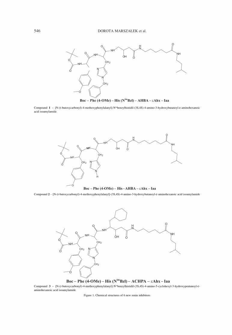
546 DOROTA MARSZA£EK et al.
Compound 1 ñ [N-(t-butoxycarbonyl)-4-methoxyphenylalanyl]-Nimbenzylhistidil-(3S,4S)-4-amino-3-hydroxybutanoyl-ε-aminohexanoicacid isoamylamide
Compound 2 ñ [N-(t-butoxycarbonyl)-4-methoxyphenylalanyl]-(3S,4S)-4-amino-3-hydroxybutanoyl-ε-aminohexanoic acid isoamylamide
Compound 3 ñ [N-(t-butoxycarbonyl)-4-methoxyphenylalanyl]-Nimbenzylhistidil-(3S,4S)-4-amino-5-cyclohexyl-3-hydroxypentanoyl-ε-aminohexanoic acid isoamylamide
Figure 1. Chemical structures of 6 new renin inhibitors
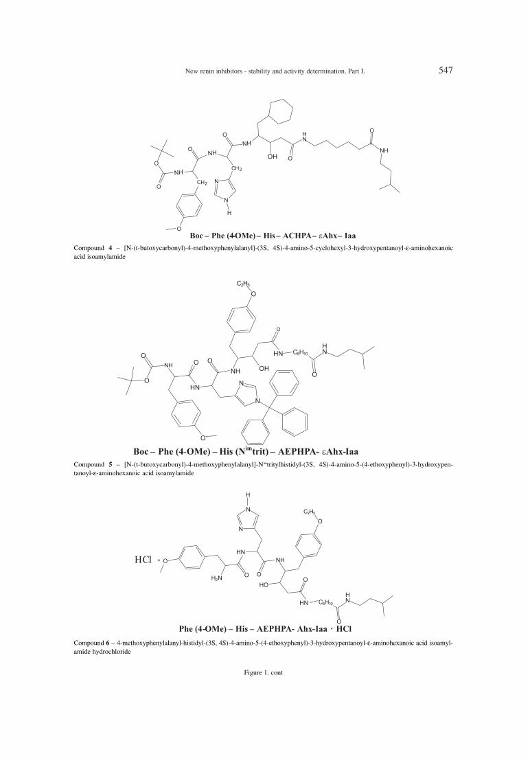
New renin inhibitors - stability and activity determination. Part I. 547
Compound 4 ñ [N-(t-butoxycarbonyl)-4-methoxyphenylalanyl]-(3S, 4S)-4-amino-5-cyclohexyl-3-hydroxypentanoyl-ε-aminohexanoicacid isoamylamide
Compound 5 ñ [N-(t-butoxycarbonyl)-4-methoxyphenylalanyl]-Nimtritylhistidyl-(3S, 4S)-4-amino-5-(4-ethoxyphenyl)-3-hydroxypen-tanoyl-ε-aminohexanoic acid isoamylamide
Compound 6 ñ 4-methoxyphenylalanyl-histidyl-(3S, 4S)-4-amino-5-(4-ethoxyphenyl)-3-hydroxypentanoyl-ε-aminohexanoic acid isoamyl-amide hydrochloride
Figure 1. cont
�

548 DOROTA MARSZA£EK et al.
ty (10-9 M). Other report presented peptide backbonemodifications that lead to greater resistance of theresulting peptides towards enzymatic degradationand some were potent inhibitors of human renin(e.g., Boc-Pro-Phe-Nα-MeHis-Leuψ-(CHOHCH2)-Val-Ile-Amp showed inhibiting activity ñ 2.6 ◊ 10-10
M (7, 8). The synthesis of renin inhibitors (angio-tensinogen analogues) having dehydrostatine,Leuψ[CH2S]Val, or Leuψ[CH2SO]Val at P1-P1ícleavage site was described (9). Activity of thosecompounds was between 10-6 to 10-8 M. Bock et al.obtained a series of statine containing tetrapeptides,modified at the C-terminal with various hydropho-bic aromatic groups (10). The inhibiting activity ofobtained compounds were between 10-6 to 10-8
M, e.g., for compound Boc-Phe-His-Sta-Leu-p-
chlorobenzylamide it was 8.1 ◊ 10-8 M. Other pep-statin analogues of general formula A-X-Y-Sta-Ala-Sta-R were synthesized (11). Various changes of A,X and Y groups were undertaken to improve theinhibitory activity against human plasma renin. Thetert-butyloxycarbonyl group and the isovalerylgroup were the most effective acyl groups (A). Thereplacement of Val with Phe residue (X) and incor-poration of His or other amino acid with an aliphat-ic side chain in the position Y lead to an increase ofinhibition activity against human plasma renin (10-8
M). Changes on C-terminal statine, like estrificationor amidification, had no influence on inhibitorypotency. Jones et al. (12) used solid-phase method tosynthesize a series of peptides (analogues ofangiotensinogen), in which statine or new analogues
Table 1. Characterization of new synthesized renin inhibitors.
No. Compound Formula Mr
1 Boc-Phe(4-OMe)-His(BZL)-AHBA-Ahx-Iaa C43H63O8N7 806.04
2 Boc-Phe(4-OMe)-His-AHBA-Ahx-Iaa C36H57O8N7 715.92
3 Boc-Phe(4-OMe)-His(BZL)-ACHPA-Ahx-Iaa C50H75O8N7 902.20
4 Boc-Phe(4-OMe)-His-ACHPA-Ahx-Iaa C43H69O8N7 812.07
5 Boc-Phe(4-OMe)-His(trit)-AEPHPA-Ahx-Iaa C64H81N7O9 1092.40
6 Phe-(4-OMe)-His-AEPHPA-Ahx-Iaa ◊ HCl C40H59N7O7Cl 784.94
Boc ñ tert-butoxycarbonyl; Iaa ñ isoamylamide; Ahx ñ 6-aminohexanoic acid; AHBA ñ 4-amino-3-hydroxybutanoic acid; ACHPA ñ 4-amino-5-cyclohexyl-3-hydroxypentanoic acid; Phe(4-OMe) ñ 4-methoxyphenylalanine; AEPHPA ñ 4-amino-5-(4-ethoxyphenyl)-3-hydroxypentanoic acid.
Table 2. Chromatographic and validation parameters.
Compd.Column
Mobile phaser
Recovery CVExtractionno. (v/v/v) (%) (%)
Beckman 1 Ultrasphere Octyl
ACNñH2OñH3PO4 0.9976 103.26 ± 8.25 4.93 ± 2.34 alkaline 71%(150 ◊ 4.6 mm)
(42 : 58 : 0.1)
Discovery Wide ACNñH2Oñ2 Pore C8 trichloroacetic acid 0.9988 96.08 ± 10.59 5.13 ± 1.05 acidic 53%
(150 ◊ 4.6 mm) (10 : 90 : 0.1)
Beckman 3 Ultrasphere Octyl MeOHñH2OñH3PO4 0.9993 98.99 ± 3.31 5.66 ± 1.28 acidic 63%
(150 ◊ 4.6 mm) (45 : 55 : 0.1)
4Symmetry C18 ACNñH2OñH3PO4
(150 ◊ 4.6 mm) (30 : 70 : 0.1)0.9972 98.18 ± 6.00 4.48 ± 3.38 acidic 70%
Beckman 5 Ultrasphere Octyl ACNñH2OñH3PO4
(150 ◊ 4.6 mm) (70 : 35 : 0.1) 0.9996 103.21 ± 7.01 10.48 ± 2.85 acidic 81%
Symmetry C18ACNñacetate buffer
6(150 ◊4.6 mm)
pH 4.0 0.9990 100.80 ± 4.92 7.40 ± 4.28 alkaline 20%(40 : 60)

New renin inhibitors - stability and activity determination. Part I. 549
(3S,4S)-3,4-diamino- or (3S,4S)-3,4-diamino-6-methylheptanoic acid and (3S,4S)-4-amino-3-aminomethyl- or (3S,4S)-4-amino-3-aminomethyl-6-methylheptanoic acid replaced either residue 10 orboth residues 10, 11 at the P1-P1í cleavage site.Peptide Boc-His-Pro-Phe-His-Sta-Val-Ile-His-NH2
showed highest inhibiting activity (10-9 M) againsthuman renin among all new synthesized com-pounds. Chen et al. described a novel series of spiro-cyclic renin inhibitors (13). These inhibitors werepotent but the bioavailability was poor in animalmodel.
Our intention was to search for new activeinhibitors with simple structure, high bioavailabilityand easy synthesis. These compounds should beresistant to enzymatic degradation, metabolicallystable and with improved oral absorption. Severalcompounds with unnatural amino acids in positionP3 and P2 and pseudopeptides in position P1-P1 andP2-P3 were synthesized (14). All compounds werestable against chymotrypsin, inhibitory activity wasmoderate but solubility was poor. Other obtainedinhibitors with εAhx ethylamide or εAhx-isoamyl-amide at the P2í- P3í positions showed inhibitoryactivity at the concentration of 10-5 mL-1 (14, 15).The next renin inhibitors obtained in our departmentcontained nonpeptic units: (3S,4S)-4-amino-5-cyclohexyl-3-hydroxypentanoic acid (ACHPA),(3S,4S)-4-amino-3-hydroxy-5-phenylpentanoic acid(AHPPA), (3S,4S)-4-amino-3-hydoxy-6-methylhep-tanoic acid (Sta) and unnatural dipeptide Phe(4-OMe)-MeLeu (16). All compounds were stableagainst chymotrypsin and the inhibitory activity was
measured in vitro and it was < 10-3, 1.0 ◊ 10-6, 4.0 ◊10-4 and 1.0 ◊ 10-6 M, respectively.
To continue the search for the effective renininhibitors, 6 new compounds of potential inhibitingactivity were synthesized. Their chemical structureswere presented in Table 1 and Figure 1.
The purpose of the study was to determine thestability of the newly synthesized compounds inhomogenate of body organs and body fluids (invitro) and to check the inhibiting activity of all syn-thesized potential renin inhibitors (in vitro).
EXPERIMENTAL
Materials and reagents
New renin inhibitors synthesized in theDepartment of Drug Chemistry, MedicalUniversity.of Warsaw are presented in Table 1 andFigure 1.
Renin human and α-chymotrypsin from bovinepancreas were purchased from Sigma. Angioten-sinogen was purchased from Bachem.
Apparatus and methods
A Shimadzu HPLC apparatus that consisted ofan LC-10AT pump and SPD-10A spectrophotome-ter was used with Chroma computer recorder (POL-LAB, Poland) and the Chromax 2001 software(POL-LAB, Poland).
The HPLC method for determination of com-pounds was developed. The separation was carriedout in the reverse phase system, the flow rate was 1mL/min. The concentration range was between
Table 3. Evaluation of stability of compounds 1ñ6
Compd. Gastric Intestinal Kidney Lung Liverno.
α-Chymotrypsin Serum juice juice homogenate homogenate homogenate
first-order first-order1 reaction stable reaction stable stable stable stable
t0.5 = 5 h t0.5 = 78 min
2 stable stable stable stable unstable stable stable
first-order first-order first-order first-order3 stable reaction stable stable reaction reaction reaction
t0.5 = 198 min t0.5 = 73 min t0.5 = 140 min t0.5 = 5 min
first-order4 stable stable stable stable stable stable reaction
t0.5 = 57 min
first-order first-order 5 unstable unstable unstable unstable reaction reaction unstable
t0.5 = 73 min t0.5 = 114 min
6 stable stable stable stable stable stable stable
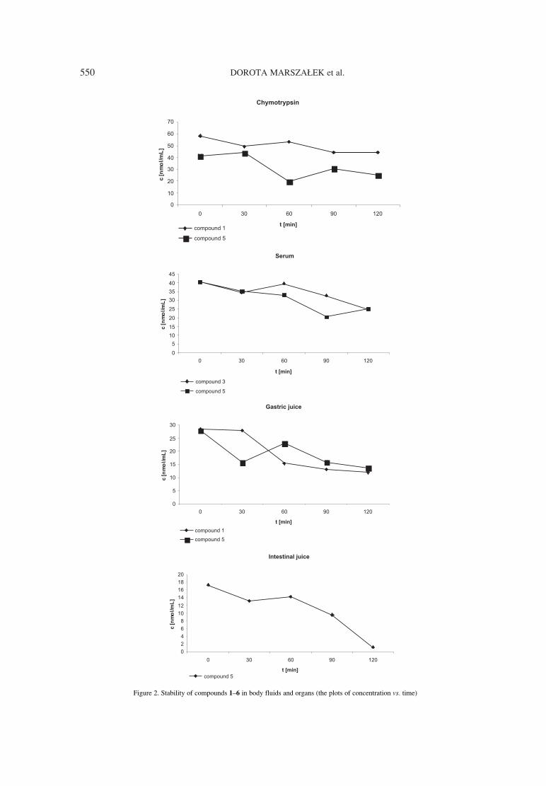
550 DOROTA MARSZA£EK et al.
Figure 2. Stability of compounds 1ñ6 in body fluids and organs (the plots of concentration vs. time)

New renin inhibitors - stability and activity determination. Part I. 551
10ñ50 nmol/mL. The wavelength was 213 nm. Thecolumns, mobile phases and validation parametershave been presented in Table 2.
The method of compounds 1ñ6 determinationwas validated. Recovery, accuracy and linearity ofthe analytical procedure are presented in Table 2.
Stock solutions of determined compoundswere prepared by dissolving each compound inmethanol. The final working concentration for theexamined substances was 100 nmol/mL. The liquid-
Figure 2. cont.
Table 4. Inhibiting activity of compounds 1-6.
Compd. Human renin IC50
no. (M)
1 inactive > 10-5
2 1.4 ◊ 10-6
3 inactive > 10-5
4 5.2 ◊ 10-6
5 inactive > 10-5
6 1.5 ◊ 10-7

552 DOROTA MARSZA£EK et al.
liquid extraction method was used for all biologicalmaterial.
Determination of enzymatic stability
of compounds 1ñ6 in vitroThe stability of all compounds in body fluids
and organ homogenates was examined. The concen-trations of compounds were measured at differenttime points during incubation in biological material.The developed HPLC method was used to determinethe concentration of compounds 1ñ6 isolated frombiological material by liquid-liquid extraction withthe use of diethyl ether.
Stability determination of compounds 1ñ6 in liver,kidney and lung enzymes
The homogenates of body organs (40%) in 0.1mole/L TRIS solution (pH = 8.4) were prepared.Each homogenate was spiked with each of 6 com-pounds and incubated at 37OC. The samples werecollected at the time points 0, 30, 60, 90, 120, 150,180 min and then isolated from homogenates withdiethyl ether, evaporated to dryness, dissolved inmobile phase and determined by HPLC. Results arepresented in Table 3 and Figure 2.
Stability determination of compounds 1ñ6 in serum, gastric and intestinal juice
Each compound was dissolved in certainamount of serum or freshly prepared gastric or intes-tinal juice (according to USP). The solutions wereincubated at 37OC and the samples were collected atthe time points 0, 30, 60, 90, 120 min. The sampleswere isolated from body fluids with diethyl ether,evaporated to dryness, dissolved in mobile phaseand determined by HPLC. Results are presented inTable 3 and Figure 2.
Stability determination of compounds 1ñ6 in the presence of αα-chymotrypsin
α-Chymotrypsin was dissolved in phosphatatebuffor solution pH 7.8. The solution was incubatedat 37OC and the samples were collected at the timepoints 0, 30, 60, 120 min. Then, the samples wereisolated from the solution with diethyl ether, evapo-rated to dryness, dissolved in mobile phase anddetermined by HPLC. Results are presented in Table3 and Figure 2.
Determination of inhibition activity
of compounds 1ñ6 vs. human renin
Renin inhibiting activity of the synthesizedpotential inhibitors was determined in vitro. Themodified HPLC method of determination of
angiotensinogen (substrate) concentration was used(column: Wild Pore C8 15 cm ◊ 4.6 mm, mobilephase MeOH : H2O : H3PO4 40 : 60 : 0.1 (v/v/v),wavelength 213 nm). The HPLC method was modi-fied in comparison with other method (17), used todetermine the renin inhibitor activity. The modifica-tion includes change of chromatographic conditionsand change of the method of inhibiting activitydetermination. Instead of assay of concentration oftetrapeptide (which is the product of reaction), theconcentration of angiotensinogen (substrate) wasmeasured. To check if modified method was reliablein activity determination, we compared results ofactivity measurements obtained by the methoddescribed in this paper with the results obtained withour modified method for renin inhibitor (Boc-Phe-His-Sta-εAhx-OMe) (18). The results of inhibitingactivity for method described in the paper and mod-ified method were IC50 = 5.0 ◊∑10-9 M and IC50 = 5.3◊∑10-9 M, respectively (18). These results confirmedthat the modified method was reliable.
The inhibition of human renin was determinedafter its incubation with angiotensinogen and witheach of compounds 1ñ6. Human renin 9 mU.G. wasincubated with 6 mM angiotensinogen in 30 mM cit-rate-phosphate buffer (pH 7.4) for 2 h at 37OC withlovering amount (10-5, 10-6, 10-7, 10-8, 10-9 M) of eachrenin inhibitor prepared in phosphoric-citric bufferpH 7.4. To stop the reaction after 2 h, the sample wasimmersed in boiling water for 2 min. To control theactivity assay, the sample contained only human reninand angiotensinogen was prepared and the activitywas measured in time 0 and after 2 h of incubation.
The renin inhibitory activity was designed interms of the IC50, which is the molar concentrationof the examined inhibitor causing 50% inhibition ofthe control renin activity.
Results are presented in Table 4.
RESULTS AND DISCUSSION
Determination of enzymatic stability of 6 newcompounds have been performed in test in vitro. Theresults showed that compound 6 was stable, com-pound 5 was totally unstable and all other com-pounds were partly stable in body fluids and organhomogenates. Compound 2 was unstable in kidneyhomogenate, compound 4 in liver homogenate,compound 1 in gastric juice and in the presence ofα-chymotrypsin and compound 3 in serum, kidney,lung and liver homogenates.
Determination of inhibiting activity of com-pounds 1ñ6 against human renin had been per-formed in vitro.

New renin inhibitors - stability and activity determination. Part I. 553
The results showed that the removal of sub-stituents of His was necessary to obtain biologicallyactive compound and made compound more stable.Simultaneous removal of Boc group caused thatcompound was stable in body fluids and organhomogenates and showed the highest inhibitoryactivity (1.5 ׷10-7 M). Compound with AHBAgroup showed activity 10-6 and was rather stable(decomposed only in kidney homogenate).Compound 4 had four times lower activity thancompound 2 and was rather stable (decomposedonly in liver homogenate), the half-time was about60 min. The presence of trityl group caused com-pound unstable in all homogenates and body fluids.The removal of Boc group increased metabolic sta-bility and increased solubility in water.
The search for other compounds with higherpotency is under way.
Acknowledgments
This investigation was supported by MedicalUniversity of Warsaw and grant no. N N405357939.
REFERENCES
1. Maibaum J., Rasetti V., R¸eger H,. Cohen N.C.,Gˆschke R., Mah R., Rahuel J., Gruetter M.G.et al.: in Medicinal Chemistry: Today andTomorrow. Yamazaki M. Ed., BlackwellScience Ltd., Oxford 1997.
2. Wood J.M., Maibaum J., Gruetter M.G., CohenN.C., Rasetti V., R¸eger H., Gˆschke R., StutzS. et al.: Biochem. Biophys. Res. Commun.308, 698 (2003).
3. Waldemeier F., Glaenzel U., Wirtz B., ObererL., Schmid D., Seiberling M., Valencia J.,Riviere G.-J. et al.: Drug Metab. Dispos. 35,1418 (2007).
4. Bolis G., Fung A.K.L., Greer J., Kleinert H.D.,Marcotte P., Perun T.J., Plattner J.J., SteinH.H.: J. Med. Chem. 30, 1729 (1987).
5. Thaisrivongs S., Pals D.T., Kroll L.T., TurnerS.R., Han F.S.: J. Med. Chem. 30, 976 (1987).
6. B¸hlmayer P., Caselli A., Fuhrer W., GˆschkeR., Rasetti V., R¸eger H., Stanton J., CriscioneL., Wood J.M.: J. Med. Chem. 31, 1839 (1988).
7. Thaisrivongs S., Pals D.T., Lawson J.A., TurnerS., Harris D.W.: J. Med. Chem. 30, 536 (1987).
8. Thaisrivongs S., Pals D.T., Harris D.W., KatiW.M., Turner S.R.: J. Med. Chem. 29, 2088(1986).
9. Smith C.W., Saneii H.H., Sawyer T.K., PalsD.T., Scahill T.A., Kamdar B..V, Lawson J.A.:J. Med. Chem. 31, 1377 (1988).
10. Bock M.G., DiPardo R.M., Evans B.E., RittleK.E., Boger J., Poe M., LaMont B.I., Lynch R.J.et al.: J. Med. Chem. 30, 1853 (1987).
11. GuÈgan R., Diaz J., Cazaubon C., BeaumontM., Carlet C., ClÈment J., Demarne H., MelletM. et al.: J. Med. Chem. 29, 1152 (1986).
12. Jones M., Sueiras-Diaz J., Szelke M., LeckieB.J., Beattie S.R., Morton J., Neidle S., KurodaR.: J. Peptide Res. 50, 109 (1997).
13. Chen A., Aspiotis R., Campeau L.C., CauchonE., Chesfon A., Ducharme Y., Falgueyret L.P.,Gagne S. et al.: Bioorg. Med. Chem. Lett. 21,7399 (2011).
14. Paruszewski R., Jaworski P., Winiecka I., TauttJ, Dudkiewicz J.: Chem. Pharm. Bull. 50, 850(2002).
15. Paruszewski R., Jaworski P., Winiecka I., TauttJ., Dudkiewicz J.: Pharmazie 54, 102 (1999).
16. Paruszewski R., Jaworski P., Bodnar M.,Dudkiewicz-WilczyÒska J., Roman I.: Chem.Pharm. Bull. 53, 1305 (2005).
17. Dudkiewicz-WilczyÒska J., Roman I.,Paruszewski R.: Acta Pol. Pharm. Drug Res. 61,171 (2004).
18. Paruszewski R., Tautt J., Dudkiewicz J.: Pol. J.Pharmacol. 45, 75 (1993)
Received: 19. 08. 2013


Acta Poloniae Pharmaceutica ñ Drug Research, Vol. 71 No. 4 pp. 555ñ561, 2014 ISSN 0001-6837Polish Pharmaceutical Society
Hypertension is a major risk for cardiovascularand kidney diseases. Many of patients with hyperten-sion, using antihypertensive drugs, do not have theirblood pressure controlled to recommended target lev-els (< 140/90). Therefore, the renin-angiotensin sys-tem has been a highly attractive biological target fornew drugs, which could regulate the blood pressure.Inhibition of angiotensin converting enzyme (ACE)has led to development of effective antihypertensivedrugs. The other possibility to interrupt of renin-angiotensin system is to inhibit renin. Renin, anaspartyl protease, is involved in the first step of enzy-matic cascade. Numerous laboratories have tried todevelop clinically effective direct renin inhibitors ofhigh potency and stability (1ñ11). The purpose ofthese trials was to search for new active inhibitorswith simple structure, good bioavailability and easysynthesis. The first in a new class of oral, nonpeptidedirect renin inhibitor for treatment of hypertenssion isaliskiren. The structure of aliskiren differs from thestructure of 8ñ13 human angiotensinogen fragment.Searching for a new inhibitors, a series of compoundswith dipeptide replacement [(3S,4S)-4-amino-5-cyclohexyl-3-hydroxypentanoic acid (ACHPA)] intheir molecules were synthesized (12). Some of themcomprised two additional analogs of dipeptide:(3S,4S)-4-amino-3-hydroxy-5-phenylpentanoic acid(AHPPA) and (3S,4S)-4-amino-3-hydroxy-6-methyl-heptanoic acid (statine). The inhibitory activity waslow, so the authors conclude that renin inhibitors
ought to have one or two, but not three dipeptideanalogs in the molecule. Bock et al. synthesized aseries of statine-containing tetrapeptides, modified atthe carboxy terminus with hydrophobic aromaticgroups (13). The Phe amide group, known to be a siteof hepatic metabolism, was replaced with nonpeptidalgroups. It caused the reduction of peptide chainlength but also lowering of activity. Searching formore active renin inhibitors the pentapeptide BOC-Phe-Phe-difluorostatone-Leu-Phe-NH2 was synthe-sized (14). This compound contains a difluorometh-ylene ketone group. It showed an increase in inhibit-ing activity in animal models.
In our previous paper, we presented a series ofnew six pseudodipeptic potential renin inhibitors.Enzymatic stability for all compounds (1ñ6) inhomogenates (liver, kidney, lung) and body fluids(serum, gastric, intestinal juice) and α-chymotrypsinwas determined. Inhibitory activity of the compoundswas measured in vitro by HPLC determination oflowering concentration of substrate (angiotensino-gen) in the presence of renin and the potential renininhibitor (compounds 1ñ6). Compound 2 and 4
showed inhibitory activity 1.4 ◊ 10-6 and 0.9 ◊ 10-6 M,respectively, and compound 6 ñ 1.5 ◊ 10-7 M. Othercompounds showed no inhibitory activity up to 10-5
M. To continue search for the effective renininhibitors, 6 new modified compounds of potentiallyinhibiting activity were synthesized. The chemicalstructures of them are presented in Figure 1.
NEW RENIN INHIBITORS ñ STABILITY AND ACTIVITY DETERMINATION.PART II
DOROTA MARSZA£EK1*, ANNA GOLDNIK1, ALEKSANDER P. MAZUREK2, IWONA WINIECKA1,PAWE£ JAWORSKI1, SANDRA SZERSZA—1, EWA KOZIKOWSKA1 and TADEUSZ PAWLIK1
1Department of Drug Chemistry, Medical University of Warsaw, 1 Banacha St., 02-097 Warszawa, Poland2National Medicines Institute, 30/34 Che≥mska St., 00-725 Warszawa, Poland
Abstract: A series of new six pseudodipeptic potential renin inhibitors were synthesized. Enzymatic stabilityfor all compounds (1ñ6) in homogenates (liver, kidney, lung) and body fluids (serum, gastric, intestinal juice)and α-chymotrypsin was determined. Compounds 4 was stable, compound 5 was unstable and compounds 1,
2, 3, 6 were partly unstable. Inhibitory activity of the compounds was measured in vitro by HPLC determina-tion of lowering concentration of substrate (angiotensinogen) in the presence of renin and the potential renininhibitor (compounds 1ñ6). Compound 4, 5, 6 showed inhibitory activity (0.9 ◊ 10-6 , 1.3 ◊ 10 -8, 2.2 ◊ 10-6 M,respectively). Other compounds showed no inhibitory activity up to 10-5 M.
Keywords: HPLC, inhibition of renin activity, renin inhibitors
555
* Corresponding author: e-mail: [email protected]; phone: 48 22 5720630, fax 48 22 5720697
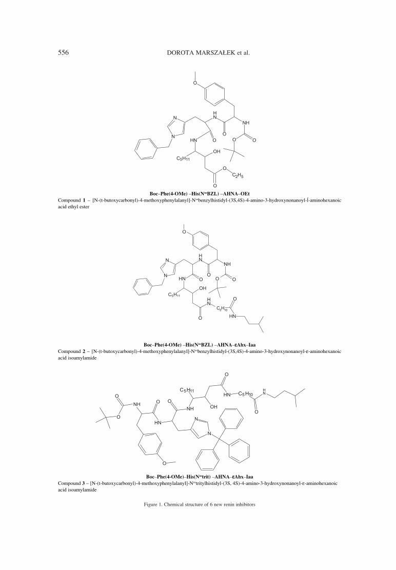
556 DOROTA MARSZA£EK et al.
Figure 1. Chemical structure of 6 new renin inhibitors
BocñPhe(4-OMe) ñHis(NimBZL) ñAHNAñOEt
Compound 1 ñ [N-(t-butoxycarbonyl)-4-methoxyphenylalanyl]-Nimbenzylhistidyl-(3S,4S)-4-amino-3-hydroxynonanoyl-Â-aminohexanoicacid ethyl ester
BocñPhe(4-OMe) ñHis(NimBZL) ñAHNAñεεAhxñIaa
Compound 2 ñ [N-(t-butoxycarbonyl)-4-methoxyphenylalanyl]-Nimbenzylhistidyl-(3S,4S)-4-amino-3-hydroxynonanoyl-ε-aminohexanoicacid isoamylamide
BocñPhe(4-OMe)ñHis(Nimtrit) ñAHNAñεεAhxñIaa
Compound 3 ñ [N-(t-butoxycarbonyl)-4-methoxyphenylalanyl]-Nimtritylhistidyl-(3S, 4S)-4-amino-3-hydroxynonanoyl-ε-aminohexanoicacid isoamylamide
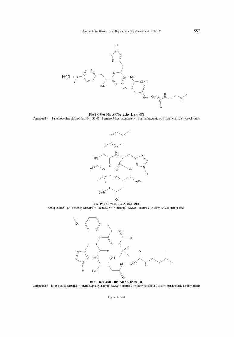
New renin inhibitors - stability and activity determination. Part II 557
Figure 1. cont
Phe(4-OMe)ñHisñAHNAñεεAhxñIaa ◊ HCl
Compound 4 ñ 4-methoxyphenylalanyl-histidyl-(3S,4S)-4-amino-3-hydroxynonanoyl-ε-aminohexanoic acid isoamylamide hydrochloride
BocñPhe(4-OMe)ñHisñAHNAñOEt
Compound 5 ñ [N-(t-butoxycarbonyl)-4-methoxyphenylalanyl]l-(3S,4S)-4-amino-3-hydroxynonanoylethyl ester
BocñPhe(4-OMe)ñHisñAHNAñεεAhxñIaa
Compound 6 ñ [N-(t-butoxycarbonyl)-4-methoxyphenylalanyl]-(3S,4S)-4-amino-3-hydroxynonanoyl-ε-aminohexanoic acid isoamylamide

558 DOROTA MARSZA£EK et al.
Figure 2. Stability of compound 1-6 in body fluids and organs (the plots concentration vs. time)

New renin inhibitors - stability and activity determination. Part II 559
The purpose of this study was to determine thestability of the new synthesized compounds inhomogenates of body organs and body fluids (invitro) and to check the inhibiting activity of sixpotential renin inhibitors (in vitro).
EXPERIMENTAL
Materials and reagents
New renin inhibitors synthesized in the
Figure 2. cont.
Department of Drug Chemistry, MedicalUniversity.of Warsaw are presented in Table 1.Chemical structures are shown in Figure 1.
Renin human and α-chymotrypsin from bovinepancreas were purchased from Sigma. Angioten-sinogen was purchased from Bachem.
Apparatus and methods
The details are the same as in the precedingpaper.

560 DOROTA MARSZA£EK et al.
Table 1. Characterization of new synthesized renin inhibitors.
Compd. no. Compound Formula Mr
1 Boc-Phe(4-OMe)-His(BZL)-AHNA-OEt C38H55N5O7 693.89
2 Boc-Phe(4-OMe)-His(BZL)-AHNA-Ahx-Iaa C47H71N7O8 862.12
3 Boc-Phe(4-OMe)-His(trit)-AHNA-Ahx-Iaa C60H81N7O8 1028.36
4 Phe-(4-OMe)-His-AHNA-Ahx-Iaa ∑ HCl C35H58N7O6Cl 708.36
5 Boc-Phe(4-OMe)-His-AHNA-OEt C31H49O7N5 603.761
6 Boc-Phe(4-OMe)-His-AHNA-Ahx-Iaa C40H65O8N7 772.001
Boc ñ tert-butoxycarbonyl, Iaa ñ isoamylamide, Ahx ñ 6-aminohexanoic acid, AHNA ñ 4-amino-3-hydroxynonanoic acid, Phe(4-OMe) ñ4-methoxyphenylalanine
Table 2. Parameters of chromatographic determination and valdation procedure of determined compounds.
Compd. Mobile phase Recovery CVExtractionno.
Column(v/v/v)
r(%) (%)
Beckman 1 Ultrasphere Octyl
ACNñH2OñH3PO4 0.9988 103.14 ± 8.12 4.96 ± 2.37 alkaline 71%(150 ◊ 4.6 mm)
(42 : 58 : 0.1)
Beckman 2 Ultrasphere Octyl
MeOHñH2OñH3PO4 0.9926 100.36 ± 5.03 6.8 ± 1.64 alkaline 80%(150 ◊ 4.6 mm)
(65 : 35 : 0.1)
Discovery Wide3 Pore C8
MeOHñH2OñH3PO4 0.9999 100.13 ± 1.87 5.11 ± 1.65 acidic 67%(150 ◊ 4.6 mm)
(40 : 60 : 0.1)
Beckman ACNñacetate buffer 4 Ultrasphere Octyl pH 4.0 0.9932 101.99 ± 9.05 5.82 ± 1.65 acidic 67%
(150 ◊ 4.6 mm) (40 : 60)
Symmetry C18 ACNñH2OñH3PO4
5 (150 ◊ 4.6 mm) (80 : 20 : 0.1) 0.9908 100.84 ± 5.95 4.06 ± 2.95 acidic 64%
Beckman6 Ultrasphere Octyl
ACNñH2OñH3PO4 0.9940 98.17 ± 6.00 6.83 ± 2.83 alkaline 47%(150 ◊ 4.6 mm)
(80 : 20 : 0.1)
Table 3. Stability of compounds 1ñ6.
Compd. α-Chymo- Gastric Intestinal Kidney Lung Liver no. trypsin
Serumjuice juice homogenate homogenate homogenate
first-order first-order1 unstable reaction unstable reaction unstable stable unstable
t0.5 = 157 min t0.5 = 239 min
first-order2 stable stable stable unstable unstable reaction unstable
t0.5 = 92 min
first-order first-order 3 stable stable stable stable reaction reaction no results*
t0.5 = 103 min t0.5 = 122 min
4 stable stable stable stable stable stable stable
first-order5 unstable reaction unstable unstable unstable unstable unstable
t0.5 = 25.7 min
first-order first-order6 stable stable unstable unstable reaction reaction stable
t0.5 = 50 min t0.5= 111.8 min
* it was not possible to isolate compound 3 from biological matrix due to very strong hydrophobic bonds.

New renin inhibitors - stability and activity determination. Part II 561
Determination of enzymatic stability
of compounds 1ñ6 in vitroThe stability of compounds 1ñ6 in body fluids
and organ homogenates was examined similarly asdescribed in the preceding paper.
Determination of inhibition activity vs.
human renin of compounds 1ñ6
Renin inhibiting activity of the synthesizedpotential inhibitors 1ñ6 was determined in vitro sim-ilarly as described in the preceding paper. Theresults are presented in Table 4.
RESULTS AND DISCUSSION
Determination of enzymatic stability and deter-mination of inhibiting activity of compounds 1ñ6
against human renin have been performed in test invitro. According to the results of that investigation itwas shown that compound 1, 2 and 3 showed noinhibiting activity. The presence of large trityl sub-stituent of His in the structure of compound 3 causedincreased strength of hydrophobic bonds with bio-logical matrix, which made extraction of that com-pound from liver homogenate impossible. Aremoval of Boc group of Phe and substituent of Hisin compound 4 caused that it was stable in allhomogenates and body fluids. It is possible to con-clude that active renin inhibitors should containamino acid His without any substituents. Compound5, which contained ester bond in C-terminus, wastotally metabolically unstable but showed the high-est activity IC50 = 1.3 ◊ 10-8 M. A removal of Bocgroup and convertion of compound 4 to hydrochlo-ride increased twice the inhibiting activity. It couldbe caused by an increase of solubility. This com-pound was also more stable. The Ahx-Iaa sub-stituent in C-terminus of compounds 2 and 3 pro-tected C-terminus and increased the metabolic sta-bility but reduced inhibiting activity.
The search for other compounds with expectedhigher potency will be continued.
Acknowledgments
This investigation was supported by MedicalUniversity of Warsaw and grant no. N N405357939.
REFERENCES
1. Dellaria J.F., Maki R.G., Bopp B.A., Cohen J.,Kleinert H.D., Luly J.R.,Merits I., Plattner J.J.,Stein H.H.: J. Med. Chem. 30, 2137 (1987).
2. Luly J.R., Yi N., Soderquist J., Stein H., CohenJ., Perun T.J., Plattner J.J.: J. Med. Chem. 30,1609 (1987).
3. TenBrink R.E., Pals D.T., Harris D.W., JohnsonG.A.: J. Med. Chem. 31, 671 (1987).
4. Luly J.R., BaMaung N., Soderquist J., FungA.K.L., Stein H., Kleinert H.D., Marcotte P.A.et al.: J. Med. Chem. 31, 2264 (1988).
5. Rosenberg S.H., Plattner J., Woods K.W., SteinH.H., Marcotte P.A., Cohen J., Perun T.J.: J.Med. Chem. 30, 1224 (1987).
6. Simoneau B., LavallÈe P., Anderson P.C.,Bailey M., Bantle G., Berthiaume S., Chabot C.et al.: Bioorg. Med. Chem. 7, 489 (1999).
7. Jones D.M., Suerias-Diaz J., Szelke M., LeckieB.J., Beattie S.R., Morton J., Neidle S., KurodaR.: J. Peptide Res. 50, 109 (1997).
8. Thaisrivongs S., Pals D.T., Kroll L.T., TurnerS.R., Han F.S.: J. Med. Chem. 30, 976 (1987).
9. B¸hlmayer P., Caselli A., Fuhrer W., GˆscheR., Rasetti V., R¸eger H., Stanton J.L. et al.: J.Med. Chem. 31, 1839 (1987).
10. Paruszewski R., Jaworski P., Bodnar M.,Dudkiewicz-WilczyÒska J., Roman I.: Chem.Pharm. Bull. 53, 1305 (2005).
11. Paruszewski R., Jaworski P., Winiecka I., TauttJ, Dudkiewicz J.: Pharmazie 54, 102 (1999).
12. Winiecka I., Dudkiewicz-WilczyÒska J.,Roman I., Paruszewski R.: Acta Pol. Pharm.Drug Res. 67, 367 (2010).
13. Bock M.G., DiPardo R.M., Evans B.E., RittleK.E., Boger J., Poe M., LaMont B.I. et al.: J.Med. Chem. 30, 1853 (1987).
14. Fearon K., Spaltenstein A., Hopkins P.B., GelbM.H.: J. Med. Chem. 30, 1617 (1987).
15. Dudkiewicz-WilczyÒska J., Roman I.,Paruszewski R.: Acta Pol. Pharm. Drug Res. 61,171 (2004).
Received: 19. 08. 2013
Table 4. Inhibiting activity of compounds 1ñ6.
Compound Human reninno. IC50 (M)
1 inactive > 10-5
2 inactive > 10-5
3 inactive > 10-5
4 0.9 ◊ 10-6
5 1.3 ◊ 10-8
6 2.2 ◊ 10-6

Acta Poloniae Pharmaceutica ñ Drug Research, Vol. 71 No. 4 pp. 563ñ573, 2014 ISSN 0001-6837Polish Pharmaceutical Society
Preservatives are chemical substances whoserole is to protect food products, stimulants, medici-nal products and cosmetics against harmful changescaused by microorganisms (1). When added in prop-er concentrations, preservatives inhibit the growthof microorganisms during manufacturing and use ofmedicinal products (2). They should be tasteless,odorless and colorless. In concentrations used, theyshould be soluble and non-toxic as well as physio-logically and chemically compatible. At present, it isrecommended ñ as more effective ñ to use mixturesof preservatives.
Preservatives are added to sterile medicinalproducts, such as eye drops and multi-dose solutionsfor injections, as well as to non-sterile products,such as water oral solutions, creams, gels, supposi-tories and capsules with liquid content. It is allowedin Poland to use as preservatives the substances list-ed in Appendix no. 1 to the Regulation of HealthMinister dated 16 January 2003, on preservatives,sweeteners, colors and antioxidants that may beincluded in medicinal products (Journal of Laws No.
19, 2003, item 169). These compounds comprisephenols, alcohols, organic acids, organic mercuriccompounds and quaternary ammonium bases. Themost commonly used preservatives include: benzylalcohol, butyl, ethyl, methyl and propyl p-hydroxy-benzoates and their sodium salts. According to theaforementioned Regulation, all preservatives shouldmeet the requirements of the European Pharmaco-poeia and their content in medicinal products shouldbe determined quantitatively.
Benzyl alcohol, as a preservative in medicinalproducts (oral solutions and solutions for injec-tions), is used in concentration up to 2%. Benzylalcohol reveals incompatibilities with oxidizingagents and strong acids. Its antimicrobial activity isreduced in the presence of non-ionic surfactants,such as, for example, polysorbate 80 (1, 3). Inmedicinal products it oxidizes slowly to benzalde-hyde and benzoic acid when exposed to air (4).Three papers have been found on determination ofbenzaldehyde contents in injections with diclofenacwith the use of polarographic method (5) and in
DEVELOPMENT STUDIES ON DETERMINATION OF PRESERVATIVESDECOMPOSITION PRODUCTS
BEATA ZABRZEWSKA1, ANNA CHY£A1* and ANNA BOGDAN1
1 Department of Basic and Applied Pharmacy, National Medicines Institute,30/34 Che≥mska St., 00-725 Warszawa, Poland
Abstract: Preservatives are chemical substances whose role is to protect medicinal products against harmfulchanges caused by microorganisms. They are added to sterile medicinal products, such as eye drops and multi-dose solutions for injections, as well as to non-sterile products, such as water oral solutions, creams, gels, sup-positories and capsules with liquid content. The most commonly used preservatives include: benzyl alcohol,butyl, ethyl, methyl and propyl p-hydroxybenzoates and their sodium salts. In medicinal products benzyl alco-hol slowly oxidizes to benzaldehyde and benzoic acid while esters of p-hydroxybenzoic acid hydrolyze to p-hydroxybenzoic acid. HPLC methods were elaborated for identification and quantitative determination of theparabens, benzyl alcohol, active substances as well as their impurities in pharmaceuticals: oral solutions Amertiland Efforil (contain cetirizine hydrochloride or etilefrine hydrochloride and parabens), eye drops Difadol (con-tains diclofenac sodium and benzyl alcohol) and cream Tenasil (contains terbinafine hydrochloride and benzylalcohol). The HPLC systems consisted of columns: Supelcosil LC-DP, Inertsil ODS-3 or Discovery HS F5 andthree mobile phases ñ mixtures of acetonitrile with buffers of various pH (3, 5 and 7) in proportions 45 : 55(v/v). These systems have been characterized with appropriate selectivity (all the RS values > 2) and sensitivi-ty (LOD approx. 0.01 µg/mL). They also demonstrated satisfactory precision and a linear dependence betweenthe analyte content and the peak area.
Keywords: benzyl alcohol, esters of p-hydroxybenzoic acid, HPLC
563
* Corresponding author: e-mail [email protected]

564 BEATA ZABRZEWSKA et al.
injections with diclofenac, piroxicam and vitamin Bcomplex with the use of GC method (6, 7). Benzylalcohol and diclofenac were determined in variousmedicinal products with spectrophotometric method(8).
Esters of p-hydroxybenzoic acid (parabens)show desirable preserving action within a quite widerange of pH, from weak-acid to weak-alkaline (pH 4ñ 8), though in general, they are the most effectivein acid environment. Above pH 8, the effectivenessof its action decreases due to rapid progress ofhydrolysis. The parabens are more active againstyeast and mould than against bacteria. They are alsomore active against Gram positive than againstGram negative bacteria. Antibacterial action of indi-vidual esters increases together with the increasedchain length, but their solubility decreases. Thisproblem may be omitted by the use of more solublesodium salts of the esters, however, this raises thepH of poorly buffered formulations. Antibacterialactivity may also be increased by the use of a mix ofesters, for example of methyl and propyl p-hydroxy-benzoates.
Antibacterial effect of parabens decreases inthe presence of macromolecular substances, such aspolysorbate 80, as the micelles that are formed closethe esters inside. Addition of propylene glycol(10%) increases the adverse interactions in the pres-ence of non-ionic surfactants. Esters are also incom-patible with other substances, such as: bentonite,magnesium trisilicate, talc, sorbitol and atropine (1,3).
Esters of p-hydroxybenzoic acid are used aspreservatives mainly in oral solutions and prepara-tions administered on skin. In the past, these werealso used in solutions for injections and in the eyedrops, but at now this application is being aban-doned because of their irritating effect. Additionally,in case of medicinal products administered on eyes,a significant disadvantage of parabens is their poorimpact on Gram-negative bacteria, in particularagainst Pseudomonas aeruginosa. Most commonly,a mixture of two esters methyl and propyl is used atconcentrations up to 0.25%. A product of estershydrolysis, p-hydroxybenzoic acid, shows no anti-bacterial effect (2).
In the available literature, several studies werefound describing determination of various esters ofp-hydroxybenzoic acid in medicinal products withmicellar electrophoresis (9, 10) and HPLC methods(11, 12). Only one of these papers takes into consid-eration determining the content of parabens hydro-lysis product ñ p-hydroxybenzoic acid (12).Stability of water solutions of p-hydroxybenzoic
Tab
le 1
. Tim
es o
f re
tent
ion
(tR),
res
olut
ion
betw
een
the
neig
hbor
ing
peak
s (R
S) a
nd p
eak
asym
met
ry (
A10
%)
of t
este
d pr
eser
vativ
es a
nd t
heir
dec
ompo
sitio
n pr
oduc
ts i
n ch
rom
atog
raph
ic s
yste
m a
cc. t
o Ph
. Eur
.m
onog
raph
s fo
r m
ethy
lpar
aben
. Mob
ile p
hase
: 0.0
5 M
sol
utio
n of
KH
2PO
4an
d m
etha
nol 3
5 : 6
5 (v
/v).
Col
umns
use
d: Z
orba
x R
x C
18 (
in a
ccor
danc
e w
ith th
e m
onog
raph
) an
d co
lum
ns s
elec
ted
for
test
s O
DS-
3,D
P, H
S F5
.
Su
bst
an
ce*
Colu
mn
Rx C
18
Colu
mn
OD
S-3
C
olu
mn
DP
Colu
mn
HS
F5
t RR
SA
10%
t RR
SA
10%
t RR
SA
10%
t RR
SA
10%
p-H
BA
c1.
5ñ
1.4
2.8ñ
3.2
ñtr
iple
pea
k3.
2ñ3.
3ñ
doub
le p
eak
3.6-
4.1
ñtr
iple
pea
k
MP
2.2
2.6
1.9
5.1
7.5
1.2
3.9
1.3
1.3
6.3
7.2
1.1
EP
2.7
2.3
1.7
6.6
5.1
1.2
4.0
0.6
1.2
7.7
5.3
1.2
PP
3.7
3.9
1.5
10.0
7.5
1.1
4.3
0.9
1.1
10.0
7.0
1.1
BP
5.7
5.6
1.3
14.8
14.7
1.0
4.6
1.2
1.1
13.7
6.8
1.1
BA
-ol
2.1
1.1
1.1
4.5
2.8
1.2
3.7
1.8
1.1
4.7
ñ1.
4
BA
c1.
9ñ
0.8
3.8
ñ1.
83.
4ñ
1.4
5.6
3.8
1.8
BA
ld2.
492.
01.
55.
74.
20.
94.
01.
31.
86.
01.
81,
1
* ab
brev
iatio
ns s
ee te
xt.

Development studies on determination of preservatives decomposition products 565
Table 2. Times of retention (tR). resolution between the neighboring peaks (RS) and peak asymmetry (A10%) of tested preservatives andtheir decomposition products in elaborated chromatographic systems on the ODS-3 column (Inertsil ODS-3, 5 µm, 250 × 4.6 mm).
Substance*tR RS A10%
pH ~3 pH ~5 pH ~7 pH ~3 pH ~5 pH ~7 pH ~3 pH ~5 pH ~7
p-HBAc 3.6 3.3 2.4 ñ ñ ñ 1.1 1.2 1.2
MP 5.9 5.9 5.9 11.1 12.1 14.5 1.0 1.1 1.1
EP 7.9 8.4 7.9 7.8 7.7 7.5 1.0 1.0 1.1
PP 11.5 11.3 11.4 10.7 10.3 10.1 1.1 1.0 1.0
BP 17.6 17.3 17.3 12.8 12.3 12.1 1.0 1.0 1.1
BAol 5.1 5.1 5.2 ñ 4.4 11.4 1.1 1.1 1.1
BAc 5.6 4.2 2.6 2.2 ñ ñ 1.1 1.6 1.3
BAld 8.4 7.9 8.6 11.8 11.2 13.3 1.1 1.0 1.0
* abbreviations see text.
Table 3. Times of retention (tR), resolution between the neighboring peaks (RS) and peak asymmetry (A10%) of tested preservatives andtheir decomposition products in elaborated chromatographic systems on the DP column (Supelcosil LC- DP, 5 µm, 250 × 4.6 mm).
Substance*tR RS A10%
pH ~3 pH ~5 pH ~7 pH ~3 pH ~5 pH ~7 pH ~3 pH ~5 pH ~7
p-HBAc 3.8 3.5 2.8 ñ ñ ñ 1.0 1.3 1.1
MP 4.6 4.5 4.6 1.7 4.3 16.9 1.0 1.2 1.1
EP 5.1 4.9 5.0 7.8 1.7 2.1 1.0 1.3 1.1
PP 5.8 5.4 5.6 3.1 2.2 2.7 1.0 1.3 1.1
BP 6.7 6.1 6.5 3.9 2.7 3.2 1.0 1.3 1.1
BAol 4.4 4.3 4.4 ñ 2.9 17.9 1.0 1.4 1.1
BAc 4.5 3.7 2.8 1.0 ñ ñ 1.0 1.6 1.1
BAld 5.2 5.0 5.1 3.8 3.1 4.3 1.0 1.2 1.1
* abbreviations see text.
Table 4. Times of retention (tR), resolution between the neighbouring peaks (RS) and peak asymmetry (A10%) of tested preservatives andtheir decomposition products in elaborated chromatographic systems on the HS F5 column (Discovery HS F5, 5 µm, 250 × 4.6 mm).
Substance*tR RS A10%
pH ~3 pH ~5 pH ~7 pH ~3 pH ~5 pH ~7 pH ~3 pH ~5 pH ~7
p-HBAc 4.0 3.9 2.4-2.5 ñ ñ ñ 1.2 1.3 double peak
MP 6.0 5.9 6.1 10.6 9.6 32.3 1.0 1.1 1.2
EP 7.3 7.2 7.4 5.9 5.6 5.6 1.0 1.1 1.3
PP 9.3 9.0 9.4 7.8 7.2 7.4 1.0 1.1 1.3
BP 12.2 11.7 12.3 9.4 8.6 8.6 1.0 1.0 1.4
BAol 5.2 5.1 5.1 ñ ñ 26.2 1.0 1.1 1.1
BAc 5.6 6.8 2.9 2.3 4.8 ñ 1.1 2.7 1.1
BAld 7.8 7.7 7.5 10.6 2.2 9.8 1.0 1.0 1.3
* abbreviations see text.

566 BEATA ZABRZEWSKA et al.
Tab
le 5
. The
line
ar r
elat
ions
hip
betw
een
anal
yte
conc
entr
atio
n (c
) an
d pe
ak a
rea
(x)
for
para
bens
and
ben
zyl a
lcoh
ol in
ela
bora
ted
chro
mat
ogra
phic
sys
tem
s. T
he c
oeff
icie
nts
of c
orre
latio
n r
give
n in
bra
cket
s.
c =
ax +
b
Su
bst
an
ce*
C
olu
mn
OD
S-3
C
olu
mn
DP
Colu
mn
HS
F5
pH ~
3 pH
~5
pH ~
3 pH
~5
pH~3
pH
~5
MP
1.05
Eñ0
5xñ2
.82
8.63
Eñ0
6xñ2
.67
1.15
Eñ0
5xñ4
.16
8.78
Eñ0
6xñ2
.73
9.61
Eñ0
6xñ2
.15
8.55
Eñ0
6xñ2
.45
(r =
0.9
989)
(r
= 0
.999
0)
(r =
0.9
970)
(r
= 0
.999
0)
(r =
0.9
991)
(r
= 0
.999
2)
EP
1.06
Eñ0
5xñ1
.77
8.73
Eñ0
6xñ1
.63
1.19
Eñ0
5xñ3
.54
9.09
Eñ0
6xñ2
.27
1.10
Eñ0
5xñ3
.34
8.77
Eñ0
6xñ1
.74
(r =
0.9
996)
(r
= 0
.999
7)
(r =
0.9
980)
(r
= 0
.999
4)
(r =
0.9
991)
(r
= 0
.999
6)
PP
1.09
Eñ0
5xñ0
.85
9.11
Eñ0
6xñ0
.82
1.13
Eñ0
5xñ2
.27
9.45
Eñ0
6xñ1
.44
1.13
Eñ0
5xñ2
.17
9.19
Eñ0
6xñ0
.99
(r =
0.9
999)
(r
= 0
.999
9)
(r =
0.9
990)
(r
= 0
.999
6)
(r =
0.9
995)
(r
= 0
. 999
8)
BP
1.14
Eñ0
5xñ0
.48
9.60
Eñ0
6xñ0
.58
1.17
Eñ0
5xñ1
.89
9.93
Eñ0
6xñ1
.16
1.17
Eñ0
5xñ1
.59
1.01
Eñ0
5xñ2
.93
(r =
0.9
999)
(r
= 0
.999
9)
(r =
0.9
993)
(r
= 0
.999
9)
(r =
0.9
998)
(r
= 0
.999
7)
BA
ol
6.90
Eñ0
4xñ3
5.7
6.13
Eñ0
4xñ1
0.72
**
5.95
Eñ0
4xñ6
.88
5.77
Eñ0
4ñ3.
405.
93E
ñ04x
ñ8.6
3(r
= 0
.999
4)
(r =
0.9
998)
(r
= 0
.999
9)
(r =
0.9
999)
(r
= 0
.999
9)
* ab
brev
iatio
ns s
ee te
xt. *
*- n
ot c
alib
rate
d du
e to
non
-com
plet
e se
para
tion
of b
enzy
l alc
ohol
with
ben
zoic
aci
d (R
= 1
.0).
Tab
le 6
.The
line
ar r
elat
ions
hip
betw
een
anal
yte
conc
entr
atio
n (c
) an
d pe
ak a
rea
(x)
for
p-hy
drox
yben
zoic
aci
d, b
enzo
ic a
cid
and
benz
alde
hyde
in e
labo
rate
d ch
rom
atog
raph
ic s
yste
ms.
The
coe
ffic
ient
s of
cor
rela
-tio
n r
give
n in
bra
cket
s.
c =
ax +
b
Su
bst
an
ce*
Wavele
ngh
t C
olu
mn
OD
S-3
C
olu
mn
DP
Colu
mn
HS
F5
pH ~
3 pH
~5
pH ~
3 pH
~5
pH ~
3 pH
~5
p-H
BA
c
254
nm
9.13
Eñ0
6xñ0
.24
8.82
Eñ0
6xñ0
.11
8.58
Eñ0
6xñ0
.17
7.83
Eñ0
6xñ0
.08
8.97
Eñ0
6xñ0
.26
7.71
Eñ0
6xñ0
.09
(r =
0.9
999)
(r
= 0
.999
9)
(r =
0.9
999)
(r
= 0
.999
9)
(r =
0.9
999)
(r
= 0
.999
9)
BA
c
220
nm
1.29
Eñ0
5xñ0
.14
1.06
Eñ0
5xñ0
.06
**
1.07
Eñ0
5xñ0
.13
1.36
Eñ0
5xñ0
.24
9.87
Eñ0
6xñ0
.05
(r =
0.9
999)
(r
= 0
.999
9)
(r =
0.9
999)
(r
= 0
.999
9)
(r =
0.9
999)
BA
ld
254
nm
9.59
Eñ0
6xñ0
.24
1.07
Eñ0
5xñ0
.06
**9.
31E
ñ06x
ñ0.2
91.
06E
ñ05x
ñ0.4
08.
71E
ñ06x
ñ0.1
1(r
= 0
.999
9)
(r =
0.9
999)
(r
= 0
.999
9)
(r =
0.9
998)
(r
= 0
.999
9)
* ab
brev
iatio
ns s
ee te
xt.*
* no
t cal
ibra
ted
due
to n
on-c
ompl
ete
sepa
ratio
n of
ben
zyl a
lcoh
ol w
ith b
enzo
ic a
cid
(R =
1.0
).

Development studies on determination of preservatives decomposition products 567
acid n-butyl ester exposed to light (254 nm) was alsoinvestigated (13).
The European Pharmacopoeia specifies thepermissible amount of impurities in parabens andrecommends the HPLC method for testing ofparabens purity. However, there are no guidelineson their levels in medicinal products. They may notbe considered as unknown impurities, as they oftenexceed the level of 0.2% adopted in line with ICHguidelines.
For determination of benzyl alcohol and testingof its purity the European Pharmacopoeia recom-mends a GC method. However, this method may notbe sufficient, therefore a different method for testingof purity and determination of active substanceshould be applied. Simultaneous analysis with useof the two methods increases the costs.
The purpose of this paper was to elaborateHPLC methods for identification and quantitativedetermination of the parabens and benzyl alcohol inpharmaceuticals. It was assumed that the content ofmedicinal substances and preservatives as well astheir purity, taking into consideration the impuritiescoming from the active substance as well as fromthe preservatives, would be determined in one chro-matographic system.
For the testing the following were selected:expired preparations (Amertil, Effortil, Difadol) ñtheir content of preservatives and their impuritieshad been determined earlier, during their period ofvalidity ñ and a multi-dose preparation in its periodof validity (Tenasil cream) ñ the amount of benzylalcohol oxidation products may increase after open-ing of this preparation. This compilation allows esti-mation of the products of preservatives decomposi-tion that are formed when storing under normal con-ditions as well as their rate of formation, and mayfacilitate determination of limits for these impuri-ties.
EXPERIMENTAL
Test materials
LGC standards: methyl (MP), ethyl (EP),propyl (PP) and butyl (BP) p-hydroxybenzoates(parabens); p-hydroxybenzoic acid (p-HBAc); ben-zyl alcohol (BAol); benzaldehyde (BAld); benzoicacid (BAc); certizine dihydrochloride (C), p-chlorobenzophenone (p-CBPon), p-chlorobenzhy-drol (p-CBHol), A impurity acc. to Eu. Ph.;diclofenac sodium (D) and A, B, C and E impuritiesacc. to Eu. Ph.; etilefrine hydrochloride (E) and A
Table 7. Times of retention of selected medicinal substances and their impurities in the elaborated chromatographic systems.
Retention time [min]
Substance* Column ODS-3 Column DP Column HS F5
pH ~3 pH ~5 pH ~3 pH ~5 pH ~3 pH ~5
C 2.7 6.4 40.2 6.2 26.3 7.0
pCB-ol 23.4 25.2 5.9 7.2 10.6 15.4
pCBP-on 49.8 53.17 7.2 9.0 17.7 27.3
Imp. A 2.0 4.9 50.6 9.6 30.1 21.2
D 28.1 18.4 na 5.3 11.1 37.7
Imp. A 23.9 23.8 na 8.0 10.1 14.5
Imp. B 66.1 63.2 na 10.8 19.1 30.0
Imp. C 30.1 29.5 na 8.4 11.7 17.2
Imp. E 3.9 4.3 na 4.1 3.8 4.5
T 2.7 71.1 na 15.8 60.1 62.9
Imp. A 1.7 24.0 na 5.9 11.7 8.2
Imp. B 2.6 68.2 na 15.5 64.0 61.0
Imp. E 1.9 >120 na >80 >120 >120
E 1.7 2.3 15.2 4.3 6.3 4.9
Imp. A 1.7 2.3 14.5 4.5 7.3 5.3
* abbreviations see text. na ñ not analyzed (due to non-complete separation of benzyl alcohol with benzoic acid).

568 BEATA ZABRZEWSKA et al.
impurity acc. to Eu. Ph.; terbinafine hydrochloride(T) and A, B and E impurities acc. to Eu. Ph.
Preparations containing: cetirizine dihy-drochloride and methyl and ethyl p-hydroxyben-zoates ñ Amertil syrup 1 mg/mL s. CP8001, expirydate: 12. 2009 (Biofarm Sp. z o.o.); diclofenac sodi-um and benzyl alcohol ñ Difadol, solution for intra-muscular injections 25 mg/g, s. 01AK1104, expirydate: 11. 2007 (WZF Polfa); etilefrine hydrochlorideand methyl and propyl p-hydroxybenzoates ñEffortil oral drops 7.5 mg/g, s. 433452D, expirydate: 09. 2009 (Boehringer Ingelheim); terbinafinehydrochloride and benzyl alcohol ñ Tenasil cream s.010709, expiry date: 07. 2012 (PharmaceuticalLaboratory HOMEOFARM sp. z o.o.).
The preparations were previously tested inNovember and December 2010.
HPLC reagents of high purity: acetonitrile(Rathburn, Scotland); deionized water (Millipore);85% phosphoric acid; KH2PO4 and K2HPO4
(AppliChem).HPLC columns: used for reconstruction of
pharmacopoeia monograph for parabens: Zorbax RxC18, 5 mm, 150 × 4.6 mm, Agilent; used for elabo-ration of new methods: Supelcosil LC-DP, 5 mm,250 × 4.6 mm, Supelco; Inertsil ODS-3, 5 mm, 250× 4.6 mm, MZ-Analysentechnik GmbH; DiscoveryHS F5, 5 mm, 250 × 4.6 mm, Supelco.
Apparatus: PC controlled liquid chromato-graph with SPD-10AVvp UV detector, LC-10ATvppumps and DGU-14A mobile phase degasser, SCL-10AVvp controller, automatic sample injector SIL-10AVvp ñ manufactured by Shimadzu, Japan.
Solutions used for HPLC: solvent for solu-tions preparation: acetonitrile and water mixture 45: 55 (v/v); standard solutions for determination ofcalibration curves: paraben solutions in a range ofconcentrations from 10 to 200 µg/mL; solutions ofp-hydroxybenzoic acid in a range of concentrations0.03ñ50 µg/mL; solutions of benzyl alcohol in arange of concentrations 10ñ1700 µg/mL; solutionsof benzaldehyde in a range of concentrations0.05ñ75 µg/mL; solutions of benzoic acid in a rangeof concentrations 0.03ñ50 µg/mL; placebo solu-tions: weigh about 50 mg of every excipient and dis-solved in solvent; tested solutions obtained frompharmaceutical preparations: Amertil ñ concentra-tion of medicinal substance ca. 0.1 mg/mL, concen-tration of MP ca. 0.135 mg/mL, concentration of PPca. 0.015 mg/mL; Difadol ñ concentration of medic-inal substance ca 1 mg/mL, concentration of BAolca. 1.4 mg/mL; Effortil ñ concentration of medicinalsubstance ca. 0.75 mg/mL, concentration of MP ca.0.07 mg/mL, concentration of PP ca. 0.03 mg/mL;
Tab
le 8
. Res
ults
of
pres
erva
tives
dec
ompo
sitio
n pr
oduc
ts d
eter
min
atio
n (c
onte
nt e
xpre
ssed
as
perc
enta
ge in
rel
atio
n to
the
para
bens
or
benz
yl a
lcoh
ol c
onte
nt)
in s
elec
ted
med
icin
al p
rodu
cts
with
the
use
of e
labo
-ra
ted
chro
mat
ogra
phic
sys
tem
s.
Eff
ort
il
Am
ert
il
Ass
ay
Col
umn
OD
S-3
Col
umn
DP
Col
umn
HS
F5
Col
umn
OD
S-3
Col
umn
DP
Col
umn
HS
F5
[%]
pH ~
3 pH
~5
pH ~
3 pH
~5
pH ~
3 pH
~5
pH ~
3 pH
~5
pH ~
3 pH
~5
pH ~
3 pH
~5
p-H
BA
c*
9.82
10
.67
9.81
10
.32
9.99
10
.02
0.41
0.
45
ñ**
0.43
ñ*
* 0.
42
Dif
ad
ol
Ten
asi
l
Ass
ay
Col
umn
OD
S-3
Col
umn
DP
Col
umn
HS
F5
Col
umn
OD
S-3
Col
umn
DP
Col
umn
HS
F5[%
]
pH ~
3 pH
~5
pH ~
3 pH
~5
pH ~
3 pH
~5
pH ~
3 pH
~5
pH ~
3 pH
~5
pH ~
3 pH
~5
BA
c*
< L
OQ
<
LO
Q
no
< L
OQ
<
LO
Q
< L
OQ
<
LO
Q
< L
OQ
no
<
LO
Q
< L
OQ
<
LO
Q
BA
ld*
0.03
0.
04
no
0.03
0.
03
0.03
0.
26
0.26
no
0.
25
0.24
0.
28
* ab
brev
iatio
ns s
ee te
xt. n
o ñ
not d
eter
min
ed. L
OQ
ñ li
mit
of q
uant
itatio
n. *
*not
e: p
-HB
Ac
peak
did
not
sep
arat
e co
mpl
etel
y fr
om th
e pl
aceb
o pe
ak.
Development studies on determination of preservatives decomposition products 569
Table 9. Comparison of retention times of active substance, preservatives and products of their decomposition in selected preparationsobtained by elaborated chromatographic systems.
Effortil
Substance* Column ODS-3 Column DP Column HS F5
pH ~3 pH ~5 pH ~3 pH ~5 pH ~3 pH ~5
E 1.7 2.3 15.2 4.3 6.3 4.9
Imp. A 1.7 2.3 14.5 4.5 7.3 5.3
p-HBAc 3.6 3.5 3.8 3.5 4.0 3.9
MP* 5.9 5.9 4.6 4.5 6.0 5.9
PP* 11.5 11.3 5.8 5.4 9.3 9.0
Amertil
Substance* Column ODS-3 Column DP Column HS F5
pH ~3 pH ~5 pH ~3 pH ~5 pH ~3 pH ~5
C* 2.7 6.4 40.2 6.2 26.3 7.0
Imp. A 2.0 4.9 50.6 9.6 30.1 21.2
p-CBol 23.4 25.2 5.9 7.2 10.6 15.4
p-CBPon 49.8 53.17 7.2 9.0 17.7 27.3
p-HBAc 3.6 3.5 3.8 3.5 4.0 3.9
MP 5.9 5.9 4.6 4.5 6.0 5.9
PP 11.5 11.3 5.8 5.4 9.3 9.0
Didafol
Substance* Column ODS-3 Column DP Column HS F5
pH ~3 pH ~5 pH ~3 pH ~5 pH ~3 pH ~5
D 28.1 18.4 na 5.3 11.1 37.7
Imp. A 23.9 23.8 na 8.0 10.1 14.5
Imp. B 66.1 63.2 na 10.8 19.1 30.0
Imp. C 30.1 29.5 na 8.4 11.7 17.2
Imp. E 3.9 4.3 na 4.1 3.8 4.5
BAolc 5.1 5.1 4.4 4.3 5. 5.1
BAc 5.6 4.2 4.5 3.7 5.6 6.8
BAld 8.4 7.9 5.2 5.0 7.8 7.7
Tenasil
Substance* Column ODS-3 Column DP Column HS F5
pH ~3 pH ~5 pH ~3 pH ~5 pH ~3 pH ~5
T 2.7 71.1 na 15.8 60.1 62.9
Imp. A 1.7 24.0 na 5.9 11.7 8.2
Imp. B 2.6 68.2 na 15.5 64.0 61.0
Imp. E 1.9 > 120 na > 80 > 120 > 120
BAolc 5.1 5.1 4.4 4.3 5. 5.1
BAc 5.6 4.2 4.5 3.7 5.6 6.8
BAld 8.4 7.9 5.2 5.0 7.8 7.7
* abbreviations see text. na ñ not analyzed (due to non-complete separation of benzyl alcohol with benzoic acid). Imp. A, B, C, E ñ impu-rity for each substance by Ph. Eur.
570 BEATA ZABRZEWSKA et al.
Figure 1. Chromatograms in chromatographic system acc. to Eur. Ph. monographs for parabens on the columns: Zorbax Rx C18 (recom-mended by Eur. Ph.) and ODS -3, HS F5, DPa) parabens (MP, EP, PP, BP) and p-hydroxybenzoic acid (p-BAc)b) benzyl alcohol (BAol), benzaldehyde (Bald) and benzoic acid (BAc)
Figure 2. Chromatograms in elaborated chromatographic systems on the column ODS-3a) parabens (MP, EP, PP, BP) and p-hydroxybenzoic acid (p-HBAc)b) benzyl alcohol (BAol), benzaldehyde (Bald) and benzoic acid (BAc)

Development studies on determination of preservatives decomposition products 571
Figure 3. Chromatograms in elaborated chromatographic systems on the column DPa) parabens (MP, EP, PP, BP) and p-hydroxybenzoic acid (p-HBAc)b) benzyl alcohol (BAol), benzaldehyde (Bald) and benzoic acid (BAc)
Figure 4. Chromatograms in elaborated chromatographic systems on the column HS F5a) parabens (MP, EP, PP, BP) and p-hydroxybenzoic acid (p-HBAc)b) benzyl alcohol (BAol), benzaldehyde (Bald) and benzoic acid (BAc)

572 BEATA ZABRZEWSKA et al.
Tenasil ñ concentration of medicinal substance ca. 1mg/mL, concentration of BAol ca. 1 mg/mL; solu-tions for identity confirmation of: certizine dihy-drochloride and impurities; diclofenac sodium andimpurities, etilefrine hydrochloride and impurities,terbinafine hydrochloride and impurities.
RESULTS AND DISCUSSION
A solution was prepared of standard sub-stances: methyl, ethyl, propyl and butyl parabensand p-hydroxybenzoic acid in the acetonitrile : watermixture (45 : 55, v/v) with the concentrations ofapprox.100 µg/mL (parabens) and 25 µg/mL (acid).This solution was used to reproduce the HPLC sys-tem acc. to Eu. Ph. for testing of the purity ofparabens. Then, the solution of the following stan-dard substances was prepared: benzyl alcohol, benz-aldehyde and benzoic acid in the mixture of acetoni-trile : water (45 : 55, v/v) in the concentrations of800, 40 and 25 µg/mL, respectively, and it was ver-ified whether the abovementioned HPLC system issuitable for determination of benzyl alcohol and itsdecomposition products (Tab. 1). The retentiontimes for all standard substances are within therange from 1.7 to 5.7 min. Due to the presence ofexcipients in preparations, such retention times arebeing to short. Therefore, the system was modifiedby application of columns with a different fillingmaterial (DP, ODS-3, HS F5 columns) than the onerecommended by Eur. Ph. (Zorbax Rx C18 column)with the same mobile phase, i.e., the mixture of 0.05M KH2PO4 and methanol in the proportion of 35 : 65(v/v). It turned out that the peak of p-hydroxyben-zoic acid was divided and was not suitable for quan-titative determinations. In the case of benzyl alcoholand the products of its decomposition on the ODS-3and HS F5 columns the obtained separation wassuitable, but the peaks were relatively wide andasymmetric (Tab. 1, Fig. 1).
For elaboration of new methods the columnswith variable filling materials were selected: ODS-3ñ alkyl groups bonded to spherical silica, with largespecific surface (450 m2/g); HS F5 ñ pentafluo-rophenyl propyl groups bonded to spherical silica,combining the features of reversed and normalphases, allowing for delaying of polar compoundsretention with no delay in non-polar substancesretention; DP ñ diphenyl groups bonded to sphericalsilica, ensuring higher selectivity for aromaticgroups, with moderate polarity.
Next, three other mobile phases of various pHwere prepared: pH~3 ñ a mixture of acetonitrile withwater in proportions of 45 : 55 (v/v) with addition of
85% phosphoric acid in the amount of 0.2 mL perone litre of the phase; pH~5 ñ a mixture of acetoni-trile with 0.025 M solution of KH2PO4 in a propor-tion of 45 : 55 (v/v); pH~7 ñ a mixture of acetonitrilewith a solution containing 0.0125 M of KH2PO4 and0.0125 M of K2HPO4 in a proportion of 45 : 55 (v/v).
The tests with the use of the mobile phaseswere carried out on each of the columns. Theremaining conditions of analysis have always beenthe same: temperature of column operation: 40OC;flow rate of the mobile phase: 1 mL/min; UV detec-tion ñ 220 nm and 254 nm; sample dose volume ñ 20µL.
The results for parabens and benzyl alcoholand their degradation products recorded for allcolumns and mobile phases are given (Tabs. 2ñ4and Figs. 2ñ4). The mobile phase of pH ~7 waseliminated on all the columns because p-HBAc andBAc acids were eluted too fast and their peaks areoften divided and may overlap the solvent peaks orplacebo peaks in case of preparations analysis.Moreover, no determinations of benzyl alcohol andits decomposition products on the DP column, pH~3 phase could be made because of non-completeseparation of BAol and BAc peaks (Tab. 3, Fig. 3).
For the rest of combinations columnñmobilephase, the calibration curves, relationship betweenthe analyte concentration expressed in µg/mL andthe peak area, analyses were performed. The con-centration ranges were selected as those that are usu-ally applied for determination the compounds inpharmaceutical preparation: parabens (10ñ200µg/mL), benzyl alcohol (10ñ1700 µg/mL), p-hydroxybenzoic acid (0.03ñ50 µg/mL) and ben-zaldehyde (0.05ñ75 µg/mL) and benzoic acid(0.03ñ50 µg/mL) (Tabs. 5, 6). Limits of detectionwere determined at the level of 0.01 µg/mL bydetermining the S/N ratio. Next, the samples, whichwere prepared by dissolving the medicinal productsin the mixture acetonitrile and water (45 : 55, v/v),have been examined in the selected systems.Retention times of medicinal substances and theirimpurities are given in Table 7.
Selection of columns with other filling materi-al (DP, ODS-3, HS F5) and adequate mobile phasesensured proper selectivity (all values RS > 2), shapeof the peaks (Tabs. 2ñ4, Figs. 2ñ4) as well as sensi-tivity (0.01 µg/mL). It was proven that pH of themobile phase influences the time of retention of p-hydroxybenzoic and benzoic acids (Tabs. 2ñ4, Figs.2ñ4) and in the case of products testing ñ the time ofretention of medicinal substances (Tab. 7). The sys-tems that are most suitable for quantitative analysisof decomposition products were selected.

Development studies on determination of preservatives decomposition products 573
The selected systems were used to determinedecomposition products of preservatives in medici-nal products with no impurities indicated in theirspecifications. The results obtained in different sys-tems were generally comparable (Tab. 8). ForAmertil preparation, tested in the phase with a pH~3, the results obtained only with the use of ODS-3column have been presented, because in the case ofDP and HS-F5 columns the p-HBAc peak is notentirely separated from the placebo peaks. Tenasiland Difadol preparations that contain benzyl alcoholwere not tested on DP column with the pH ~3 phasedue to the non-complete separation of BAol andBAc peaks.
Table 9 includes the comparison of retentiontimes of active substance, preservative and theirdecomposition products.
CONCLUSIONS
The abovementioned results show that it is notpossible to simply transfer the pharmacopoeialmethod of parabens purity testing to the columnswith different filling material (Tab. 1, Fig. 1) and toapply these methods in the testing of purity of prepa-rations containing parabens or benzyl alcohol. Thismethod is not applicable in medicinal products dueto too short retention times and a possibility that thepeaks from placebo interfere with the peaks of p-hydroxybenzoic acid.
Therefore, new HPLC systems characterizedwith appropriate selectivity (all the RS values > 2)and sensitivity (LOD approx. 0.01 µg/mL) havebeen elaborated. They allow for testing of parabenspurity in the presence of parabens hydrolysis prod-uct (p-hydroxybenzoic acid) and testing of benzylalcohol purity in the presence of its oxidation prod-ucts (benzaldehyde and benzoic acid). Assessmentof purity of the preservatives may be performed withsatisfactory precision. These systems may also beused for quantitative determination of preservatives,as it was demonstrated that there is a linear depend-ence between the analyte content and the peak areain terms of concentrations usually used for determi-nation of preservatives content in medicinal prod-ucts.
The tests performed on the expired prepara-tions revealed that the amount of preservatives
decomposition products exceeded 0.2% (limit valuefor unknown impurities according to ICH), and con-firmed the need of their determination.
The use of columns with various filling materi-als and mobile phases with a pH ranging from 3 to 7makes it possible to match the system to the compo-sition of an investigated product.
The proposed HPLC systems shall be opti-mized for testing other medicinal products, contain-ing the preservatives.
REFERENCES
1. Rybacki E., Stoøek T.: Excipients in the tech-nology of drug forms (Polish), Biblioteka far-maceuty Vol. 7, PZWL, Warszawa 1980.
2. Martindale: The Complete Drug Reference,Sweetman S.C. Ed., 35th edn., PharmaceuticalPress, London 2007.
3. Handbook of Pharmaceutical Excipients, RoweR.C., Sheskey P.J., Paul J. Weller P.J. Eds., 4th
edn., Pharmaceutical Press, London 2003.4. Sudareva N.N., Chubarova E.V.: . Pharm.
Biomed. Anal. 41, 1380 (2006).5. Kazemifard A.G., Moore D.E., Mohamadi A.:
J. Pharm. Biomed. Anal. 30, 257 (2002).6. Kazemifard A.G., Moore D.E., Mohamadi A.,
Kebriyaeezadeh A.: J. Pharm. Biomed. Anal.31, 685 (2003).
7. Mashyekhi H.A., Rezafee M., Garmaroudi S.S.,Montazeri N., Ahmedi S.J.: Anal. Sci. 27, 865(2011).
8. Ghasemi J., Niazi A., Ghobadi S.: Pharm.Chem. J. 39, 671 (2005).
9. äafra J., PospÌöilov· M.: J. Pharm. Biomed.Anal. 48, 452 (2008).
10. Hamoudov· R., PospÌöilov· M., KavalÌrov· A.,Solich P., äÌcha J.: J. Pharm. Biomed. Anal. 40,215 (2006).
11. äatÌnsk˝ D., Huclov· J., Ferreira R.L.C.,Montenegro M.C.B.S.M., Solich P.: J. Pharm.Biomed. Anal. 40, 287 (2006).
12. Grosa G., Grosso E.D., Russo R., Allegrone G.:J. Pharm. Biomed. Anal. 41, 798 (2006).
13. B≥Ídzka D., Gryglik D., Miller J.S.: J.Photochem. Photobiol. A 203, 131 (2009).
Received: 1. 10. 2013

Acta Poloniae Pharmaceutica ñ Drug Research, Vol. 71 No. 4 pp. 575ñ582, 2014 ISSN 0001-6837Polish Pharmaceutical Society
Herbal remedies have been used for centuriesin order to sustain good health, cure common ill-nesses or treat various health conditions. Nowadays,even though conventional medicine is developing ata high rate with a growing number of drugs obtainedby chemical synthesis, the interest in herbalism andphytopharmaceuticals has not diminished (1). Theincreased use of herbal medicines is based on thebelief that they are natural and therefore safe, sofewer side-effects will be produced. In addition,most of them are freely available (2).
One of the most popular forms of herbal drugsto be taken by patients are the mixtures composed ofseveral medicinal plant raw materials coming fromvarious species of plants. They are usually availablein small sachets and administered by making a tea tobe drunk twice or three times a day in order to curevarious ailments such as: infections, diabetes,rheumatism, inflammatory diseases, influenza etc.(3). However, apart from active organic ingredientsresponsible for therapeutic activities (4), medicinalherbs also contain trace elements. Some of themhave potential synergistic or antagonistic propertiesto pharmacological effects of organic compounds,but others are simply toxic for human health (5).
In recent years, many papers have been pub-lished on analysis of trace elements in various herbalproducts, mostly for quality control reasons (6ñ9)
according to WHO recommendations (10).However, in many cases, along with determinationof metals levels in herbals, different chemometrictechniques were involved (11ñ17). The reason forthat was the fact that samples were described by alarge number of metal concentrations, so it was dif-ficult to identify any intercorrelations between thosemetals straightforward. The most commonly usedmethod was principal component analysis (PCA)(12, 14), most often accompanied by cluster analy-sis (CA) (11, 13, 16) and at times, by linear discrim-inant analysis (LDA) (15, 17). These chemometrictechniques have been applied to investigation of thecontents of heavy metals in herbs or herbal teas inorder to classify and examine any relationshipsbetween these metals (15), identify similaritiesregarding mineral and trace metal contents of spicesand herbs (17), quantify the content of various ele-ments possibly responsible for some therapeuticproperties of Echinacea purpurea (11), recognizepotential anthropogenic contamination sources ofherbal drugs (12, 13), establish standardization andquality control procedures for crude drugs (14), anddifferentiate herbal raw materials belonging to dif-ferent plant families according to metal contents(16).
After reviewing the above literature, it wasconcluded that mostly applied chemometric meth-
IDENTIFICATION OF SIMILARITIES IN METALLIC CONTENT OF HERBALINFUSIONS USING NON-LINEAR APPROACH
BOGDAN SUCHACZ and MAREK WESO£OWSKI*
Department of Analytical Chemistry, Medical University of Gdansk, Al. Gen. J. Hallera 107, 80-416 GdaÒsk, Poland
Abstract: The content of Cd, Cr, Cu, Fe, Pb, Mn, Ni and Zn was determined, using flame atomic absorptionspectrometry in infusions made from commercial herbal mixtures readily available in herbal stores. ANOVAtest revealed that the metal contents did not differ considerably in most of infusions. The scatter plot of princi-pal component analysis (PCA) designated infusions characterized by similar contents of trace elements. It waspossible to indicate distinctive metal concentrations in Pyrosan, Septosan and Urosan infusions as they vastlycontrasted with others. The levels of Mn, Zn, Cd were characteristic for Pyrosan infusions, while Cr, Fe, Cdwere specific to Septosan, and Mn, Zn, Fe to Urosan. Self-organizing maps did not fully correspond with PCAresults because PCA classified samples relying mainly on apparent similarities in metal contents.
Keywords: herbal infusions, principal component analysis, self-organizing maps, trace metals
575
* Corresponding author: e mail: [email protected]

576 BOGDAN SUCHACZ and MAREK WESO£OWSKI
ods used for identification of similarities or correla-tions between metallic content in herbals were basedon linear modeling. Having assumed that interrela-tionships between chemical constituents occurringin nature are rarely of linear character, it was decid-ed to explore relationships in elemental content ofherbal infusions using non-linear modeling. In thispaper, nonlinear approach was proposed using self-organizing maps (SOMs) also known as Kohonenneural networks.
SOMs are intended for unsupervised learningtasks similarly to PCA and CA. Contrary to super-vised learning, the training data set contains onlyinputs and SOM attempts to learn the structure ofthe data on the basis of these variables. SOM iscapable of recognizing clusters of data and relatingclasses similar to each other, which makes under-standing of the data feasible. Once classes of dataare identified, they are tagged and the networkbecomes ready for classification tasks. Kohonennetworks can be also applied for classificationswhere the output classes are known in advance. Insuch circumstances, they are able to highlight simi-larities between groups (18, 19).
In this study, output classes (herbal mixtures)and individual samples were known, therefore, thesubject was to discover the characteristic differencesbetween the classes and similarities within the class-es with regard to the concentrations of some traceelements. Therefore, the heavy metals content (Ni,Cr, Cu, Fe, Mn, Zn, Pb and Cd) was determinedusing flame atomic absorption spectrometry in infu-sions prepared from commonly available herbalmixtures. Furthermore, ANOVA, PCA and SOMswere employed to verify if there are any similaritiesregarding the concentration of the metals. It wasdecided to establish if the presence of certain herbalconstituent in a mixture can be assigned to the lev-els of some trace elements. The results of the map-ping of the infusions performed by SOMs werecompared to widely applied PCA.
EXPERIMENTAL
Herbal material
Herbal mixtures produced by ìHerbapolî com-mercially available in drug or herbal stores wereinvolved in the study. The total number of infusions
Figure 1. The box-whiskers plot of Ni, Cr, Pb, Cd content in herbal infusions

Identification of similarities in metallic content of herbat infusions... 577
was 73, which were prepared from 13 different mix-tures. The analyzed herbal preparations are listedbelow in alphabetical order with two letter abbrevi-ations and samples numbers provided in parenthe-ses: Bobofen (Bo, no. 1ñ4), Bronchial (Br, no.5ñ12), Cholagoga II (Ch, no. 13ñ16), Circulosan(Ci, no. 17ñ20), Diabetosan (Di, no. 21ñ24),Gastrosan (Ga, no. 25ñ32), Infektoten (In, no.33ñ35), Nervinum (Ne, no. 36ñ38), Nervosan (Nv,no. 39ñ44), Normosan (Nm, no. 45ñ53), Pyrosan(Py, no. 54ñ56), Septosan (Se, no. 57ñ64) andUrosan (Ur, no. 65ñ73).
The herbal mixtures consist of various herbalraw materials. Some of them are present in differentmixtures e.g., melissa leaf in 5 mixtures, peppermintleaf and chamomile in 4 and hop in 3. Several mix-tures include one common raw material e.g., birchleaf ñ Urosan and Pyrosan, yarrow herb ñCholagoga II and Nervosan, lime inflorescence ñBronchial and Pyrosan etc. The mixtures are mostlyused as anti-inflammatory, analgesic, diaphoretic,sedative, antipyretic, diuretic, spasmodic, diastolicand cholagogic.
Sample preparation
All batches of herbal mixtures were homoge-nized in a water-cooled grinder Knifetec 1095 (FossTecator, Hˆgan‰s, Sweden) at 20OC for 30 s. Afterthat, an accurately weighed homogenized sample inthe amount of 10 g was put into a beaker and pouredonto with 200 mL of boiling deionized water. Thebeaker was covered with a watch glass and left for15 min to steep. After that time, infusion wascooled, strained using MN 640d ashless filter paper(Macherey-Nagel, Duren, Germany) into a 200 mLvolumetric flask and filled up with deionized water.Each infusion was prepared three times.
Trace elements determination
The content of Cd, Cr, Cu, Fe, Pb, Mn, Ni andZn was determined using spectrometer SpectrAA250 Plus (Varian, Australia) in flame atomic absorp-tion mode. The accuracy and precision of the proce-dure were established on the basis of several mea-surements of the Certified Reference Material CTA-VTL-2 (Virginia Tobacco Leaves), manufactured byThe Institute of Nuclear Chemistry and Technology
Figure 2. The box-whiskers plot of Mn, Fe, Zn, Cu content in herbal infusions

578 BOGDAN SUCHACZ and MAREK WESO£OWSKI
(Warszawa, Poland). The recovery levels were inthe range of 89.1ñ96.0% depending on the element.The lowest recovery was obtained for Cr and thehighest for Zn.
Software
All calculations were made by means of a sta-tistical software ñ STATISTICA 9.0, (Statsoft Inc.,USA).
RESULTS AND DISCUSSION
Results of analysis of variance
Categorized box plots showing the mean contents(small squares) of particular elements in the herbalinfusions are presented in Figures 1 and 2. The meanvalue is surrounded by rectangle, which denotes ±1times the standard deviation (SD), while whiskersrepresent a 95% confidence interval defined as themean ±1.96 times the SD, in view of the fact that thedistribution is normal.
As it is shown in the plots, most of infusionsamples demonstrate similar levels of 8 analyzed
elements, in spite of the fact that commercial herbalmixtures contain different medicinal plant raw mate-rials in varied proportions. However, some waterextracts prepared from Pyrosan, Septosan andUrosan mixtures were presented as exceptions. Theobservations were statistically confirmed byANOVA test (p < 0.05), which pointed to significantdifferences between mean contents of certain met-als. In order to determine which group means differfrom one another, Tukeyís multiple comparison test,for unequal number of cases, was used. The resultsindicated that the fewest statistically significant dif-ferences between mean contents of elements in infu-sions were observed mainly in the case of Ni and Cr.The levels of Pb, Cu, Mn and Zn did not present aconsiderable number of meaningful dissimilaritiesas well. The elements which differentiated betweeninfusions to the largest extent were Cd and Fe.
The analysis of Ni and Cr in infusions showedthat only Septosan infusions statistically differedfrom other extracts (mean content 2.35 mg Ni/kgand 1.08 mg Cr/kg). The levels of Pb distinguishedextracts of Normosan and Cholagoga II with the
Figure 3. The plot of component loadings of trace elements
Identification of similarities in metallic content of herbat infusions... 579
concentration of Pb amounted to 0.472 and 0.682mg/kg, respectively. With regard to Cd, it was notedthat despite numerous statistically significant differ-ences between concentrations of both metals in var-ious infusions, the most noticeable one wasobserved in water extracts prepared from Pyrosan(0.145 mg Cd/kg). Gastrosan, Cholagoga II andInfektoten infusions showed no differences betweeneach other considering Cd content, while being dis-similar to other extracts. The mean contents of Cdwere 0.086, 0.096 and 0.099 mg/kg, respectively.
The mean levels of Mn were found to be char-acteristic for Pyrosan (ca. 42 mg/kg) and Urosaninfusions (88 mg/kg), while Fe for Septosan andCholagoga II, containing on average 39.61 and45.78 mg/kg. The amount of Zn clearly distin-guished Infektoten, Urosan and Pyrosan. The meanconcentrations of Zn were varied between 7.17 and8.29 mg/kg. Cu pointed to the extracts of Nervosan(ca. 7 mg/kg).
Results of principal component analysis
The results of PCA showed that PC1 explained36.94% of overall variance, PC2 ñ 20.01% and PC3ñ 14.12%. Taking into consideration first two PCs,the amount of lost information was equal to 43.05%.The degree of influence on the interpretation of two-dimensional distribution of infusions is determinedby loadings values (Fig. 3), which characterize rela-tions between the levels of metals and principalcomponents. The loadings indicate that extracts withhigher concentration of Cu and Ni are to be locatedin upper right side of the PCA scatter plot, as shown
in Figure 4. The extracts described by higher levelsof Cr, Fe and Pb were shifted towards its lower rightside, while the amount of Mn, Zn and Cd wasresponsible for directing infusions to the lower leftside of the plot. In the distribution of analyzed infu-sions in PCA plot (Fig. 4), seven separated groupswere distinguished, denoted by Roman numeralsfrom I to VII. These groups contain infusions pre-pared from mixtures of the same composition. Thecloser the samples are located to each other, themore similar are the levels of trace metals deter-mined in them.
The most numerous group of extracts, madefrom Urosan mixture, was situated on the left side ofthe plot (group I). The distinctive feature of thesesamples was a high concentration of Mn, above 64mg/kg. The extracts (no. 65) and (no. 67) were sep-arated from other samples as a result of the greatestcontent of Zn, 13.24 and 9.89 mg/kg, respectively.Close to the center of the plot, in group II, there arethree infusions of Infektoten (no. 33ñ35). No strongdominance of any element was noticed. The sampleshad similar levels of Cr (0.470ñ0.497 mg/kg), Zn(above 5.95 mg/kg) and Cd (above 0.076 mg/kg).Group III is composed of Pyrosan extracts (no.54ñ56), with the highest concentration of Cd,exceeding 0.138 mg/kg, and relatively high level ofMn, above 19.44 mg/kg. Although Mn content is thelargest when related to other extracts (excludingUrosan samples), the differences between individualsamples are too great (ca. 1.5ñ3.5-fold). Therefore,it is difficult to regard the concentration of Mn as acharacteristic attribute of Pyrosan infusions.
Figure 4. The scatter plot of PCA scores (PC1 vs. PC2) for herbal infusions

580 BOGDAN SUCHACZ and MAREK WESO£OWSKI
The infusions found in groups IñIII are inmany cases characterized by similar concentrationsof trace elements. It comes to attention that mutualmedicinal plants raw materials are included in theircomposition. Infektoten and Pyrosan contain blackelder flower and willow bark, while the commoningredient for Pyrosan and Urosan is birch leaf.
Group IV represents Normosan infusions (no.45ñ53). The position of the samples was caused bycomparable amounts of Fe (10.56ñ14.45 mg/kg), Cr(0.474ñ0.667 mg/kg) and Pb below 0.49 mg/kg.Unfortunately, three samples representing otherinfusions are characterized by similar concentrationsof those metals. In near vicinity group V was posi-tioned. This group includes Nervosan infusions (no.39ñ44), which are characterized by the highest levelof Cu, in the range of 4.01ñ9.35 mg/kg, and lowcontent of Cd, less than 0.042 mg/kg.
The extracts of Septosan created group VI.Their most distinctive attribute is high concentration
of Fe (28.48ñ50.09 mg/kg). In two samples ofSeptosan (no. 62 and 63), the highest levels of Cr ñ1.240 and 1.721 mg/kg, pushed them to the lowerside of the PCA plot. Cholagoga II infusions, char-acterized by the highest concentration of Fe (41.44to 50.49 mg/kg), formed group VII located in lowerright side of PCA plot. Both Septosan andCholagoga II mixtures contain peppermint leaf. Thisplant raw material is present in Gastrosan andNormosan as well. However, in PCA they wereplaced in the middle of the plot together with infu-sions prepared from other mixtures. Cholagoga IImixture also comprises yarrow herb similarly toNervosan samples, which are on the same side ofPCA plot.
Results of self-organizing maps
In further step of the research, the results ofPCA were juxtaposed with those obtained by appli-cation of self-organizing maps. The mapping of
Figure 5. The distribution of herbal infusions on Kohonen map. (Neurons with no samples are marked dark grey).

Identification of similarities in metallic content of herbat infusions... 581
infusion samples shown in Figure 5 did not distin-guish as many consistent groups as PCA. It turnedout that Nervosan extracts placed together in groupV in PCA plot, were scattered all over Kohonenmap, and its samples can be found in different dis-tant neurons. It proves that despite the same compo-sition of herbal mixture, there are substantial dis-similarities in the concentration of some elements.Furthermore, the level of Cu, quite different in com-parison to other mixtures, was not recognized as aspecial attribute of all Nervosan extracts. A similarcase was observed with regard to Pyrosan andInfektoten infusions, which formed two separategroups II and III in PCA score plot.
Although ANOVA test indicated differencesin Cd levels in the extracts of both mixtures, theconcentration of remaining elements was compara-ble. Two groups of infusions prepared fromCholagoga and Septosan, containing the highestamount of Fe, as opposite to PCA, remained unsep-arated on the map. They were put together in neu-rons located in upper right corner of SOM (greyneurons). The difference in mean concentrations ofCd and Pb caused separation of these two groups ofmixtures in PCA. Kohonen map, however, whiletaking into consideration the levels of all analyzedmetals, did not identify significant distinctions, andplaced them together.
Normosan infusions presented themselves asan interesting case. The analysis of variance did notreveal differences between Normosan and extractsof other mixtures. In spite of this fact, PCA distin-guished these samples as group IV, though situatedclosely to other extracts. In contrast, infusions ofNormosan were gathered in adjacent neurons on theleft side of the Kohonen map, which clearly suggestsa considerable resemblance within the group inregard to metals levels.
The infusions of Urosan, similarly to PCA,were also differentiated from others by SOM. Theycan be found in lower right corner (grey neurons).Undoubtedly, trace elements whose concentrationsgreatly affected such allocation of these extractswere Mn, Zn and, to a lesser extent, Fe.
CONCLUSIONS
The research showed that metals compositionof water extracts, prepared from commercial herbalmixtures, is vastly similar. ANOVA test indicatedthat only in a small number of infusions, the contentof elements considerably differed from each other.PCA indicated that the metals content can be a char-acteristic feature for some infusions. It was demon-
strated by the formation of several consistentgroups. The concentrations of Mn, Zn, Cd inPyrosan, Cr, Fe and Cd in Septosan and Mn, Zn, Fein Urosan extracts, particularly varied from extractsprepared from the rest of herbal mixtures. Infusions,in which the mean content of only one metal deter-mined their distinction from others, were also iden-tified. It was Cu contents in Nervosan, and the lev-els of Fe in Cholagoga II samples.
Non-linear modeling performed by Kohonenmap managed to reveal more subtle similarities inmetal contents in Urosan and Normosan, and tosome extent ñ in Septosan and Chologoga IIextracts. In this way, the results of ANOVA testwere confirmed, pointing out to some simplifica-tions made by PCA method, which considered onlyevident similarities in metal contents.
REFERENCES
1. Heinrich M., Barnes J., Gibbons S., WilliamsonE.M.: Fundamentals of pharmacognosy andphytotherapy. p. 3, Churchil Livingstone,Philadelphia 2004.
2. Gurib-Fakim A.: Mol. Aspects Med. 27, 1(2006).
3. Schulz V., H‰nsel R., Blumenthal M., TylerV.E.: Rational Phytotherapy. A ReferenceGuide for Physicians and Pharmacists, 5th edn.,p. 19, Springer, Heidelberg 2004.
4. Poschenrieder C., AlluÈ J., Tolr‡ R., LluganyM., BarcelÛ J.: in Trace elements as contami-nants and nutrients: Consequences in ecosys-tems and human health, Prasad M.N.V. Ed., p.99, John Wiley & Sons, Hoboken 2008.
5. Singh V., Garg A.N.: Appl. Radiat. Isotopes 48,97 (1997).
6. Jyothi N.V., Mouli P.C., Reddy S.R.: J. TraceElem. Med. Biol. 17, 79 (2003).
7. Suchacz B., Weso≥owski M.: Talanta 69, 37(2006).
8. Nookabkaew S., Rangkadilok N., SatayavivadJ.: J. Agric. Food Chem. 54, 6939 (2006).
9. Karak T., Bhagat R.M.: Food Res. Int. 43, 2234(2010).
10. WHO guidelines for assessing quality of herbalmedicines with reference to contaminants andresidues, World Health Organization Press,Geneva 2007.
11. RaûiÊ S., Onjia A., Potkonjak B.: J. Pharm.Biomed. Anal. 33, 845 (2003).
12. RaûiÊ S., Onjia A., –ogo S., SlavkoviÊ L.,PopoviÊ A.: Talanta 67, 233 (2005).

582 BOGDAN SUCHACZ and MAREK WESO£OWSKI
13. RaûiÊ S.S., –ogo S.M., SlavkoviÊ L.J.:Microchem. J. 84, 93 (2006).
14. Tanaka K., Tamura T., Fukuda S., Batkhuu J.,Sanchir Ch., Komatsu K.: Phytochemistry 69,2081 (2008).
15. Kara D.: Food Chem. 114, 347 (2009).16. Arceusz A., Radecka I., Wesolowski M.: Food
Chem. 120, 52 (2010).
17. Karada∫ C., Kara D.: Food Chem. 130, 196(2012).
18. Hill T., Lewicki P.: Statistics: Methods andapplications. p. 349, StatSoft Inc., Tulsa, OK2007.
19. Kohonen T. Self-organizing maps, (3rd edn.), p.71, Springer, Berlin 2001.
Received: 1. 10. 2013

Acta Poloniae Pharmaceutica ñ Drug Research, Vol. 71 No. 4 pp. 583ñ592, 2014 ISSN 0001-6837Polish Pharmaceutical Society
Diabetes, especially type 2, affects increasingnumbers of people and creates a global threat tohealth and life (1). According to the World HealthOrganization, after the year 2020, the number ofpatients suffering from diabetes will be doubled. Inthis disease the irregular distribution of carbonate,resulting from the insulin deficiency in case of Type1 diabetes and from insulin resistance in case ofType 2 diabetes is a characteristic event. The causefor this injury related to hyperglycemia is the for-mation of glycated proteins, glucose oxidation, andincreased level of free fatty acids (2). This results inoxidative stress in the cells, as well as activation ofoxidative and inflammatory signalling pathways,
which continue to damage the insulin-diarrhoea pro-ducing cells, what leads eventually to various com-plications of diabetes.
At present, the basic aim of the research is toidentify and understand the exact mechanisms of thedevelopment of diabetes. The pancreas is a largegland located behind the stomach and next to thegallbladder. It produces digestive enzymes and hor-mones, including insulin and glucagon that help toregulate bloodís sugar levels. It was also shown thatthe levels of indicators of oxidative stress firstlyincrease and then decrease after antioxidants supple-mentation (2). This suggests that chronic pancreati-tis (CP) must involve a state of heightened free rad-
DRUG BIOCHEMISTRY
EFFECTS OF VANADIUM COMPLEXES SUPPLEMENTATION ON V, Cu, Mn, K, Fe, Zn, AND Ca CONCENTRATION IN STZ DIABETIC
RATS PANCREAS
Miros≥aw Kroúniak1, Joanna Kowalska2.6, Renata Francik3,4, Ryszard Gryboú5 and Wojciech M. Kwiatek6
1 Jagiellonian University Medical College, Department of Food Chemistry and Nutrition, 9 Medyczna St., 30-688 KrakÛw, Poland
2 Current address: Max Planck Institute for Chemical Energy Conversion, Stifstrasse 34-36, 45-470 M¸lheim an der Ruhr, Germany
3Jagiellonian University Medical College, Department of Bioorganic Chemistry, 9 Medyczna St., 30-688 KrakÛw, Poland
4 State Higher Vocational School, Institute of Health, Staszica 1 Str. 33-300 Nowy Sπcz5 Jagiellonian University, Faculty of Chemistry, 3 Ingardena St., 30-060 KrakÛw, Poland
6 Institute of Nuclear Physics, Polish Academy of Sciences, Department of Experimental Physics ofComplex Systems, 152 Radzikowskiego St. 31-342 KrakÛw, Poland
Abstract: The objective of the study was to assess the effects of Na[VVO(O2)2(2,2í-bpy)] ◊ 8 H2O (complex 1),Na[VVO(O2)2(1,10í-phen)] ◊ 5 H2O (complex 2), Na[VVO(O2)2(4,4í-Me-2,2í-bpy)] ◊ 8 H2O (complex 3),
[VIVO(SO4)(1,10í-phen)] ◊ 2 H2O, (complex 4), [VIVO(SO4)(2,2í-bpy)] ◊ H2O (complex 5), where: 2,2í-bpy =2,2í-bipyridine, 1,10í-phen = 1,10í-phenanthroline, 4,4í-Me-2,2í-bpy = 4,4í-dimethyl-2,2í-bipyridine and asmall insulin injection on V, Cu, Mn, K, Fe, Zn, and Ca concentration in the STZ (streptozotocin) diabetic ratspancreas during a 5-week treatment with the tested complexes. In all groups of animals metal concentration inthe pancreas was investigated by means of Proton Induced X-ray Emission (PIXE) method. Maximum con-centration of vanadium was observed in the pancreas for complex 5 (1.69 ± 0.09 mg/kg dry weight), lower forcomplex 3 (1.51 ± 0.10 mg/kg dry weight), and the lowest for complex 1 (1.21 ± 0.27 mg/kg dry weight) sup-plementation. The influence of vanadium administration on other metalsí concentration in the ratsí pancreaswas also investigated. All vanadium-tested complexes showed an increase of zinc concentration in the exam-ined pancreas in comparison to the diabetic animals not treated with vanadium. The results were the highest forcomplex 1 and the lowest for complex 5. The concentration of Fe, Cu, Mn, K and Ca in the pancreas is not evi-dently influenced by administration of the vanadium complexes.
Keywords: pancreas, rats, PIXE, diabetes, vanadium, copper, manganese, potassium, iron, zinc, calcium
583
* Corresponding author: e-mail: [email protected]
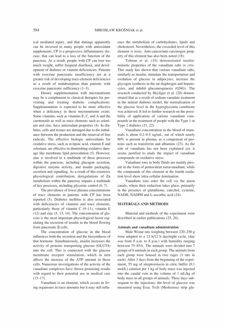
584 MIROS£AW KROåNIAK et al.
ical mediated injury, and that damage apparentlycan be inversed in many people with antioxidantsupplements. CP is a progressive inflammatory dis-ease, that can lead to a loss of the function of thepancreas. As a result, people with CP can lose toomuch weight, suffer frequent diarrhoea, and devel-opment of diabetes or vitamin deficiencies. Patientswith exocrine pancreatic insufficiency are at agreater risk of developing trace-element deficienciesas a result of malabsorption than patients withexocrine pancreatic sufficiency (1ñ3).
Dietary supplementation with micronutrientsmay be a complement to classical therapies for pre-venting and treating diabetic complications.Supplementation is expected to be more effectivewhen a deficiency in these micronutrients exists.Some vitamins, such as vitamins E, C, and A and thecarotenoids as well as trace elements such as seleni-um and zinc, have antioxidant properties (4). In dia-betes, cells and tissues are damaged due to the imbal-ance between the production and the removal of freeradicals. The effective biologic antioxidants foroxidative stress, such as α-lipoic acid, vitamin E andselenium, are effective in diminishing oxidative dam-age like membrane lipid peroxidation (5). However,zinc is involved in a multitude of these processeswithin the pancreas, including glucagon secretion,digestive enzyme activity, and insulin packaging,secretion and signalling. As a result of this extensivephysiological contribution, deregulations of Znmetabolism within the pancreas impairs a multitudeof key processes, including glycemic control (6, 7).
The prevalence of lower plasma concentrationsof trace elements in patients with CP has beenreported (5). Diabetes mellitus is also associatedwith deficiencies of vitamins and trace elements,particularly those of vitamin C (9ñ11), vitamin E(12) and zinc (5, 13, 14). The concentration of glu-cose is the most important physiological factor reg-ulating the secretion of insulin in the blood flowingfrom pancreatic β cells.
The concentration of glucose in the bloodinfluences both the secretion and the biosynthesis ofthat hormone. Simultaneously, insulin increases theactivity of proteins transporting glucose (GLUT4)into the cell. This is connected with the glucosemembrane receptor stimulation, which in turnaffects the increase of the ATP amount in thosecells. Numerous investigations of the activity of thevanadium complexes have shown promising resultswith regard to their potential use in medical care(15ñ17).
Vanadium is an element, which occurs in liv-ing organisms in trace amounts but it may still influ-
ence the metabolism of carbohydrates, lipids andcholesterol. Nevertheless, the exceeded level of thiselement is toxic. Anti-cancer/anti-carcinogen prop-erty of this element has also been noted (18).
Tolman et al. (19) demonstrated insulin-mimetic properties of the vanadium salts in vitro.This study has shown that various vanadium salts,similarly as insulin, stimulate the transportation andoxidation of glucose in adipocytes, increase theglycogen synthesis in the rat diaphragm and hepato-cytes, and inhibit gluconeogenesis (GNG). Theresearch conducted by Heyliger et al. (20) demon-strated that as a result of sodium vanadate treatmentin the animal diabetes model, the normalization ofthe glucose level in the hyperglycemia conditionswas achieved. It led to further research on the possi-bility of application of various vanadium com-pounds in the treatment of people with the Type 1 orType 2 diabetes (21, 22).
Vanadium concentration in the blood of mam-mals is about 0.2ñ0.5 ng/mL, out of which nearly80% is present in plasma, as a component of pro-teins such as transferrin and albumins (23). As therole of vanadium has not been explained yet, itseems justified to study the impact of vanadiumcompounds on oxidative stress.
Vanadium ions in body fluids are mainly pres-ent in the form of pentavalent metavanadium, whilethe compounds of this element at the fourth oxida-tion level show intra-cellular domination.
Vanadium ions enter the cell via the anioncanals, where their reduction takes place, primarilyin the presence of glutathione, catechol, cysteine,NADH, NADPH and L-ascorbic acid (24).
MATERIALS AND METHODS
Material and methods of the experiment weredescribed in earlier publications (25, 26).
Animals and vanadium administration
Male Wistar rats weighing between 220ñ250 gwere adapted to a 12 h/12 h day/night cycle, (daywas from 8 a.m. to 8 p.m.) with humidity rangingbetween 75ñ85%. The animals were divided into 7groups of 6 animals in each group. The animals fromeach group were housed in two cages (3 rats ineach). After 3 days from the beginning of the exper-iment, 55 mg of streptozotocin in citric buffer (0.1mol/L) solution per 1 kg of body mass was injectedinto the caudal vein in the volume of 1 mL/kg ofbody mass in all groups of animals. Three days sub-sequent to the injections, the level of glucose wasmeasured using Exac Tech (Medisense) strip glu-
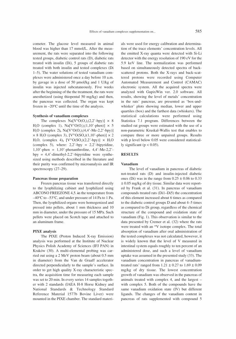
Effects of vanadium complexes supplementation on... 585
cometer. The glucose level measured in animalblood was higher than 17 mmol/L. After the meas-urement, the rats were separated into the followingtested groups, diabetic control rats (D), diabetic ratstreated with insulin (Di), 5 groups of diabetic ratstreated with both insulin and tested complexes (Di1ñ5). The water solutions of tested vanadium com-plexes were administered once a day before 10 a.m.by gavage in a dose of 50 µmol/kg and 1 U/kg ofinsulin was injected subcutaneously. Five weeksafter the beginning of the the treatment, the rats wereanesthetized (using thiopental 50 mg/kg) and then,the pancreas was collected. The organ was keptfrozen in ñ20OC until the time of the analysis.
Synthesis of vanadium complexes
The complexes Na[VVO(O2)2(2,2í-bpy)] ◊ 8H2O (complex 1), Na[VVO(O2)2(1,10í-phen)] ◊ 5H2O (complex 2), Na[VVO(O2)2(4,4í-Me-2,2í-bpy)]◊ 8 H2O (complex 3), [VIVO(SO4)(1,10í-phen)] ◊ 2H2O, (complex 4), [VIVO(SO4)(2,2í-bpy)] ◊ H2O(complex 5), where: 2,2í-bpy = 2,2í-bipyridine,1,10í-phen = 1,10í-phenanthroline, 4,4í-Me-2,2í-bpy = 4,4í-dimethyl-2,2í-bipyridine were synthe-sized using methods described in the literature andtheir purity was confirmed by microanalysis and IRspectroscopy (27ñ29).
Pancreas tissue preparation
Frozen pancreas tissue was transferred directlyto the lyophilizing cabinet and lyophilized usingABCONO FREEZONE 4,5. in the temperature fromñ40OC to ñ53OC, and under pressure of 14 Pa to 1 Pa.Then, the lyophilized organs were homogenized andpressed into pellets, about 1 mm thickness and 10mm in diameter, under the pressure of 15 MPa. Suchpellets were placed on Scotch tape and attached toan aluminum frame.
PIXE analysis
The PIXE (Proton Induced X-ray Emission)analysis was performed at the Institute of NuclearPhysics Polish Academy of Sciences (IFJ PAN) inKrakÛw (30). A multi-elemental probing was car-ried out using a 2 MeV proton beam (about 0.5 mmin diameter) from the Van de Graaff acceleratordirected perpendicularly to the sampleís surface. Inorder to get high quality X-ray characteristic spec-tra, the acquisition time for measuring each samplewas set to 20 min. In every series 14 samples togeth-er with 2 standards (IAEA H-8 Horse Kidney andNational Standards & Technology StandardReference Material 1577b Bovine Liver) weremounted in the PIXE chamber. The standard materi-
als were used for energy calibration and determina-tion of the trace elementsí concentration levels. Allthe emitted X-ray quanta were detected with Si-Lidetector with the energy resolution of 190 eV for the5.9 keV line. The normalization was performedbased on simultaneously detected spectra of back-scattered protons. Both the X-rays and back-scat-tered protons were recorded using ComputerAutomated Measurement and Control (CAMAC)electronic system. All the acquired spectra wereanalyzed with GupixWin ver. 2.0 software. Allresults, showing the level of metalsí concentrationin the ratsí pancreas, are presented as ëbox-and-whiskerí plots showing median, lower and upperquartiles (box) and the farthest data (whiskers). Thestatistical calculations were performed usingStatistica 7.1 program. Differences between thestudied rat groups were estimated with the use of anon-parametric Kruskal-Wallis test that enables tocompare three or more unpaired groups. Resultswith p level below 0.05 were considered statistical-ly significant (p < 0.05).
RESULTS
Vanadium
The level of vanadium in pancreas of diabeticnot-treated rats (D) and insulin-injected diabeticones (Di) was in the range from 0.25 ± 0.06 to 0.33± 0.05 mg/kg of dry tissue. Similar data were report-ed by Frank et al. (31). In pancreas of vanadiumcompounds treated rats (Di1ñDi5) the concentrationof this element increased about 6 times as comparedto the diabetic control groups D and about 4ñ5 timesas compared to Di group, regardless of the chemicalstructure of the compound and oxidation state ofvanadium (Fig. 1). This observation is similar to thedata presented by Cremer et al. (32) where the ratswere treated with an 48V isotope complex. The totalabsorption of vanadium after oral administration ofthe tested complexes was not calculated, however, itis widely known that the level of V measured inintestinal system equals roughly to ten percent of anadministered dose, and such a level of vanadiumuptake was assumed in the presented study (33). Thevanadium concentration in pancreas of vanadium-treated ratsí ranged from 1.21 ± 0.27 to 1.69 ± 0.09mg/kg of dry tissue. The lowest concentrationgrowth of vanadium was observed in the pancreas ofanimals treated with complex 4, and the largest ñwith complex 5. Both of the compounds have thesame vanadium oxidation state (IV) but differentligands. The changes of the vanadium content inpancreas of rats supplemented with compound 5

586 MIROS£AW KROåNIAK et al.
were statistically significantly different in compari-son to group D and Di (p < 0.05).
It is known that the presence of metal adminis-tration can influence the level of other metals suchas iron, copper, zinc, manganese, calcium or potas-sium (34). Therefore, the concentrations of thesemetals in the pancreas have been determined in ouranalysis.
Copper
The contents of copper determined in pancreasfor all the tested groups are presented in Fig. 2. Theapplication of insulin (group Di) caused the largestdecrease in the level of copper (50.72 ± 12.63 mg/kg of dry tissue). Applying vanadium complexes
increased the Cu level in comparison to the Digroup. Statistically significant growth of the con-centration of this element occurred for the complex1, including the vanadium (V) and ligands bipyri-dine (Di 1), and the complex 4 (Di 4) with vanadi-um (IV) and ligands phenyls (82.50 ± 11.03 and81.34 ± 26.95 mg/ kg of dry tissue, respectively).
Manganese
In case of the animals fed with the addition ofvanadium complexes 1, 4 and 5, the concentration ofmanganese in the pancreas was reduced in compari-son to the quantity of this microelement in thegroups D and Di (Fig. 3). The twofold decrease ofthe manganese concentration was observed only in
Figure 1. Vanadium level in the study rat groups: (group abbreviations: diabetic control rats (D), diabetic rats with insulin (Di), diabeticrats with insulin and tested complexes (Di 1 ñ Na[VO(O2)2(2,2í-bpy)] ◊ 8 H2O, Di2 ñ Na[VO(O2)2(1,10í-phen)] ◊ 5 H2O , Di3 ñNa[VO(O2)2(4,4í-Me-2,2í-bpy)] ◊ 8 H2O, Di4 ñ [VO(SO4)(1,10í-phen)] ◊ 2 H2O, Di5 ñ [VO(SO4)(2,2í-bpy)] ◊ H2O)
Figure 2. Copper level in animal groups: (group abbreviations see Figure 1)

Effects of vanadium complexes supplementation on... 587
case of the complex 1 (p < 0.05), while complexes 4and 5 reduced the Mn quantity imperceptibly. Theinfluence of complexes 2 and 3 on the Mn concen-tration in the pancreas was not observed.
Potassium
The concentration of potassium in the pancreasof the rats (Fig. 4), which were treated with vanadiumcomplex 3 (16.32 ± 5.06 g/kg) increased in compari-son to the animals from the group D (10.16 ± 3.27g/kg) and Di (11.19 ± 2.17 g/kg), and the increasewas statistically significant (p < 0.05). Organic com-plex of vanadium including methylbipyridine deriva-tive (complex 3) raised the level of potassium in thepancreas in comparison to the complex includingonly bipyridine as a ligand (complex 1), and the resultwas statistically significant (p < 0.05).
The degree of the vanadium oxidation stateessentially influenced the concentration of potassi-um in the pancreas. In case of vanadium complexesat the IV oxidation level (complex 4 and 5), adecrease in the potassium concentration in the pan-creas was observed. The lowest potassium concen-tration occurred in group Di 4 (7.16 ± 1.88 g/kg). Adecrease in potassium concentration in the pancreasin case of the Di 4 and Di5 groups in comparison toD and Di group, was observed but it was not statis-tically significant.
Iron
The highest concentration of the iron in thepancreas was observed in the diabetic control group(317.0 ± 112.9 mg/kg of dry tissue) (Fig. 5). Afterthe injection of insulin in the Di group, the concen-
Figure 3. Manganese level in animal groups: (group abbreviations see Figure 1)
Figure 4. Potassium level in animal groups: (group abbreviations see Figure 1)

588 MIROS£AW KROåNIAK et al.
tration of this element dropped twice as compared tothe D group. Vanadium complexes 1, 3, 4 and 5reduced the concentration of Fe to a statistically sig-nificant degree in comparison to the groups Di (p <0.05) and D (p < 0.05). It was also observed thatcomplexes 2 and 3 had not influenced the change ofthe concentration of Fe in comparison to the Digroup.
Zinc
As shown in Fig. 6, zinc concentration was thelowest in the D control group (19.83 ± 3.28 mg/kg),not treated with vanadium. In the diabetic groupwith insulin administration (Di), Zn concentrationwas also very low (22.06 ± 33.47 mg/kg). In all thevanadium-treated animal groups, the Zn concentra-tion increased and the level was dependent on thetype of vanadium complexes used. It was statistical-
ly significant and 2 up to 6 times higher in compar-ison to the diabetic group D and Di. Zn concentra-tion was the highest in the pancreas of Di 1 and Di2 animal groups (110.77 ± 13.74 mg/kg and 84.67 ±16.34 mg/kg, respectively). In the Di 5 group, theincrease of Zn concentration was the lowest forvanadium-treated rats (41.08 ± 17.28 mg/kg) butstill about two times higher in comparison to thediabetic groups (D and Di), not treated with vanadi-um. After both vanadium compounds 3 and 4administration similar zinc concentration in the pan-creas (64.59 ± 8.66 mg/kg and 63.36 ± 8.42 mg/kg,respectively) was observed.
Calcium
As shown in Fig 7, calcium concentration inthe pancreas was higher in the animals treated by thevanadium complexes 3 and 5 (Di 3 and Di 5, respec-
Figure 5. Iron level in the study animal groups: (group abbreviations see Figure 1)
Figure 6. Zinc level in animal groups: (group abbreviations see Figure 1)

Effects of vanadium complexes supplementation on... 589
tively, 204.3 ± 177.8 mg/kg, 219.2 ± 39/6 mg/kg,210.5 mg/kg) than in the D group of diabetic rats(172.3 ± 74.8 mg/kg), not treated with vanadium,and the Di group (168.3 ± 57.7 mg/kg). Vanadium(V) with bipyridine ligand (Di 1) treatment reducedcalcium concentration in the pancreas in a statisti-cally significant way in comparison to the effect ofcomplex 3 and 5 and without statistical significancein comparison to the pancreas of not diabetic rats nottreated with vanadium. Changes in the group Di 4with vanadium (IV) with phenyl ligand in compari-son to the diabetic not treated animals were notobserved.
Complex 1 in comparison to the others vanadi-um complexes had the strongest influence on the Cu,Mn and Zn concentration and also an essential effecton Fe concentration in the pancreas and at the sametime it had the weakest influence on the calcium, andpotassium concentration. Complex 2 showed astrong influence on zinc concentration and mostlynon influence on other elementsí concentration ascompared to the Di group, however a decrease iniron concentration as compared to the D group wasvisible. Complex 3 had a substantial influence onzinc and potassium concentration in the pancreas. Allthe three tested complexes (groups Di1ñDi3) con-tained vanadium (V) and different ligands. The influ-ence on the measured metal concentration in pan-creas is associated not only with the vanadium oxi-dation state but also with the type of used ligands.Complexes 4 and 5 contained both vanadium (IV).Complex 4 caused more significant decrease on thepotassium and iron concentrations while complex 5on zinc concentration in the pancreas.
DISCUSSION
It has been shown that vanadium possessesanti-diabetic activity and that it can be used as apotential therapeutic agent in diabetes treatment inseveral diabetic models (23). Simultaneously, sup-plementation of this metal can influence the levelsof other metals in tissues and also indirectly, meta-bolic parametersí such as enzymatic activity (35).Scientific investigations showed that vanadium hasinsulin mimetic properties and the ability of enhancethe effect of insulin (22, 35). Moreover, vanadiumcan inhibit degradation of ligand-receptor complexin lysosomes in different types of cells (36).
Manganese is necessary for the metabolism ofvitamin B1 and vitamin E. It activates someenzymes, takes part in the process of energy produc-tion, synthesis of glycogen as well as urea. Duringvanadium treatment, the changes of manganese con-centration in ratsí pancreas were not significant.This may suggest a minor influence of the testedvanadium complexes on the functions regulated bymanganese in the pancreas. On the other hand, theratio of manganese to other elements concentrationscan affect the activity of some enzymes in pancreas.Therefore, the influence of vanadium on manganeselevel in pancreas and its possible effects should befurther studied. Enzymes activated by manganese inpancreas and also possibly in other organs can beuseful in the elimination of oxidative stress inpatients with diabetes.
The increased potassium concentration maysuggest hyperkalemia, which is usually the result ofhandicapped dismissing of potassium with urine and
Figure 7. Calcium level in animal groups: (group abbreviations see Figure 1)

590 MIROS£AW KROåNIAK et al.
excessive freeing of potassium from cells. Thisobservation is associated only with the vanadiumcomplex 3 with vanadium (V) and methylbipyridineligand.
Iron is a component of organic biomoleculeslike: porphyrin, hemoglobin and myoglobin. It alsooccurs in active sites of some enzymes. The concen-tration of iron in the serum depends on absorption inthe alimentary line. The highest concentration ofiron is found in the intestines, the spleen (23) and thebone marrow. The decrease of the iron concentra-tion causes, among others, a chronic failure of kid-neys and/or the shortage of C vitamin absorption. Indiabetic rats, not treated with insulin, the iron con-centration was the highest. The administration ofinsulin decreases the concentration of that elementin pancreas. This also confirms the insulin-mimeticaction of vanadium in diabetes. The untreated dia-betes and biochemical changes associated with thisillness probably increase the iron concentration inpancreas tissue. This observation needs to be con-firmed by additional investigation. Administrationof insulin and/or vanadium caused the decrease ofthe copper level in pancreas. The tested vanadiumcomplexes showed a similar influence on the levelof iron in pancreas. However, the influence differeddepending on the type of the vanadium complexused. In groups Di 1, Di 4 and Di 5 in comparison tothe Di 2 and Di 3 the decrease of iron level wasgreater. Surprisingly, thereís no clear trend of suchinfluence. Complex 1 as compared to 4 and 5 dif-fered in oxidation state (V vs. IV) while complex 4differed in ligand environment as compared to com-plex 1 and 5. Additionally, the less significantdecrease in Fe concentration in group Di2 and Di3could be related to vanadium oxidation state (V) butthis observation is in opposite to the influence ofcomplex 1, which was also vanadium (V) compo-nent.
At the molecular and cellular level, zinc (Zn) isintimately involved in the insulin synthesis, secre-tion and signalling, and thus, in the subsequenteffect of insulin on metabolism. Various clinical andepidemiological studies suggest that reduced Zn sta-tus is associated with diabetes (37, 38). The resultsof presented study support this observation andstatement. The induced diabetes reduced zinc con-centration in the pancreas of not treated animals.
Pancreas is the site of high Zn turnover and oneof the few organs that show reduced Zn concentrationduring Zn deficiency (39). Reduced pancreatic Znconcentrations have been reported in genetic mousemodels of Type 2 diabetes, ob/ob (mutation in ob(leptin) gene and db/db (mutation in leptin receptor)
mice (40ñ42) and GK rats (Type 2 model producedby selective breeding of rats with glucose intolerance)(43) as compared to the non-diabetic ones.
Although a lot of effort has been placed onstudying Zn and pancreas, the consequences ofreduced pancreatic concentrations of other minerals(e.g., Fe, Mn and Mg in GK rats) in co-action withZn have not been explored (42). Adequate levels ofpancreatic Zn may also be crucial to provide antiox-idant protection, given that oxidative stress is a fac-tor of tissue damage in Type 1 and Type 2 diabetesand it is associated with complications occurring inthis disease (44, 45). Compared to several other tis-sues, β-cells have lower levels of antioxidantdefense components and are susceptible to oxidativedamage (46ñ48). Zinc contributes to antioxidantdefense as a component of CuZn superoxide dismu-tase (CuZnSOD) and metallothionein (MT).
Calcium level in different investigated groupshas not changed statistically and it is difficult to dis-cuss the mutual relationship between the diabetesinsulin and the tested vanadium complexes.
This investigation showed that diabetes and itsítreatment has an influence on the elemental levelmeasured in pancreas. Pancreas, being very impor-tant organ in the diabetes control, is responsible forthe glucose level changes by insulin secretionresponse. The investigated changes in the trace ele-mentsí level were mostly perceptible for zinc andiron. Zinc plays an important role in the physiologi-cal function of pancreas. The animal group with dia-betes induced by STZ had significantly lowered zinclevel in comparison to the vanadium-treated diabet-ic groups. Vanadium stimulates the activity ofadenylate cyclase (CA), phospholipase C (PLC) andphospholipase A2 (PLA2) (47). This action results inthe increased concentration of the secondary trans-mitters, such as cAMP, IP3, DAG and also arachidicacid. The increase of IP3 and arachidic acid levelcaused an increase of the level of Ca2+ ions. It is pos-sible that all these mechanisms participate also inthe zinc level elevation in pancreas. All the effectsassociated with an anti-diabetic activity of vanadiumare not only the results of the insulin mimetic sig-nalling pathway but also of the zinc concentrationprotection, especially in pancreas. Zinc is an ele-ment, which is known to have a protective activity inthe Type 2 diabetes. Such an observation is reportedfor the first time in the literature. Therefore, it is notpossible to discuss it with the results of otherresearchers.
Currently, the mutual interaction betweenvanadium and zinc in pancreas remains unknown.Better understanding of their interaction may offer

Effects of vanadium complexes supplementation on... 591
another possibility of increasing the understandingof diabetes mechanisms and possible procedures fortreatment of that disease.
Acknowledgments
We would like to express our gratitude to prof.Janine Br�s for her advice and to Krzysztof Ryt forlyophilization of samples. This work has been par-tially supported by an individual grant WL/403/P/F.
REFERENCES
1. Leahy J.L.: Arch. Med. Res. 36, 197 (2005). 2. Davi G., Falco A., Patrono C.: Antioxid. Redox.
Signal. 7, 256 (2005).3. Yokota T., Tsuchiya K., Furukawa T.,
Tsukagoshi H., Miyakawa H., Hasumura Y.: J.Neurol. 237, 103 (1990).
4. Bonnefont-Rousselot D.: Treat. Endocrinol. 3,41 (2004).
5. Quilliot D., Dousset B., Guerci B., Dubois F.,Drouin P., Ziegler O.: Pancreas 22, 299 (2001).
6. Karatug A., Bolkent S.: Exp. Toxicol. Pathol.65, 255 (2013).
7. Jou M.Y., Philipps A.F., Lonnerdal B.: J. Nutr.140, 1621 (2010).
8. Hall A.G., Kelleher S.L., Lonnerdal B., PhilippsA.F.: J. Pediatr. Gastroenterol. Nutr. 41, 72(2005).
9. Yadav S., Day J.P., Mohan V., Snehalatha C.,Braganza J.M.: Pancreas 6, 528 (1991).
10. Som S., Basu S., Mukherjee D., Deb S.,Choudhury P.R., Mukherjee S., Chatterjee S.N.,Chatterjee I.B.: Metabolism 30, 572 (1981).
11. Cunningham J.J., Ellis S.L., McVeigh K.L.,Levine R.E., Calles-Escandon J.: Metabolism40, 146 (1991).
12. Karpen C.W., Cataland S., OíDorisio T.M.,Panganamala R.V.: Diabetes 34, 526 (1985).
13. McNair P., Kiilerich S., Christiansen C.,Christensen M.S., Madsbad S. Transbol I.: Clin.Chim. Acta 112, 343 (1981).
14. Mateo M.C., Bustamante J.B., CantalapiedraM.A.: Biomedecine 29, 56 (1978).
15. Soares S.S., Martins H., Aureliano M.: Arch.Environ. Contam. Toxicol. 50, 60 (2006).
16. Urban J., Antonowicz-Juchniewicz J., Andrze-jak R.: Med. Pr. 52, 125 (2001).
17. Mukherjee B., Patra B., Mahapatra S., BanerjeeP., Tiwari A., Chatterjee M.: Toxicol. Lett. 150,135 (2004).
18. Evangelou A.M.: Crit. Rev. Oncol. Hematol.42, 249 (2002).
19. Tolman E.L., Barris E., Burns M., Pansini A.,Partridge R.: Life Sci. 25, 1159 (1979).
20. Heyliger C.E., Tahiliani A.G., McNeill J.H.:Science 227,1474 (1985).
21. Srivastava A.K.: Mol. Cell. Biochem. 206, 177(2000).
22. Srivastava A.K., Mehdi M.Z.: Diabet. Med. 22,2 (2004).
23. Thompson K.H., Orvig C.H.: J. Inorg.Biochem. 100, 1925 (2006).
24. Scibior A., Zaporowska H.: Zeszyty NaukoweKomitetu ÑCz≥owiek i årodowiskoî 33, 511(2002).
25. Kroúniak M., Francik R., Kowalska J., GryboúR., Blusz M., Kwiatek W.M.: Acta Pol. Pharm.Drug Res. 70, 71 (2013).
26. Kroúniak M., Kowalska J., Francik R., GryboúR., Blusz M., Kwiatek W.M.: JTEMB 53, 319(2013).
27. Kroúniak M., Zachwieja Z., Filipek B.,Zygmunt M., Gryboú R.: Arch. Pharm. Pharm.Med. Chem. 334, 373 (2001).
28. Przybylski W., Gryboú R., Rehder D., Ebel M.,Grzywa M, £asocha W., LewiÒski K.,Szklarzewicz JT.: Polyhedron 28, 1429 (2009).
29. Kroúniak M, Gawlik M, Gryboú R.: Bull. Vet.Inst. Pulawy 53, 535 (2009).
30. Kowalska J., Kroúniak M., Gryboú R., KwiatekW.M.: Acta Phys. Pol. A 2, 556 (2009).
31. Frank A., Madej A., Galgan V., Petersson L.R.:Sci. Total Environ. 181, 73 (1996).
32. De Cremer K., Van Hulle M., ChÈry C., Corne-lis R., Strijckmans K., Dams R., Lameire N., Van-holder R.: J. Biol. Inorg. Chem. 7-8, 884 (2002).
33. Azay J., Br�s J., Krosniak M., Teissedre P.L.,Cabanis J.C., Serrano J.J., Cros G.: Fundam.Clin. Pharmacol. 15, 313 (2001).
34. Qureshi M.A., Hill C.H., Heggen C.L.: Vet.Immunol. Immunopathol. 68, 61 (1999).
35. Dziga D.: Post. Biol. Kom. 9, 579 (2002).36. Goldfine A.B., Patti M.E., Zuberi L., Goldstein
B.J., LeBlanc R., Landaker E.J., Jiang Z.Y.,Willsky G.R., Kahn C.R.: Metabolism 49, 400(2000).
37. Chausmer A.B.: J. Am. Coll. Nutr. 17, 109(1998).
38. Tallman D.L., Taylor C.G.: Can. J. Physiol.Pharmacol. 7, 919 (1999).
39. Roth H.P., Kirchgessner M.: World Rev. Nutr.Diet. 34, 144 (1980).
40. Begin-Heick N., Dalpe-Scott M., Rowe J.,Heick H.M.: Diabetes 34, 179 (1985).
41. Kennedy M.L., Failla M.L.: J. Nutr. 117, 886(1987).

592 MIROS£AW KROåNIAK et al.
42. Simon S.F., Taylor C.G.: Exp. Biol. Med. 226,43 (2001).
43. Takita S., Wakamoto Y., Kunitsugu I.,Sugiyama S., Okuda M., Houbara T.: J.Toxicol. Sci. 29, 195 (2004).
44. Ho L.H., Ruffin R.E., Murgia C., Li L., KrilisS.A., Zalewski P.D.: J. Immunol. 172, 7750(2004).
45. Robertson R.P., Harmon J., Tran P.O., PoitoutV.: Diabetes 53 (Suppl. 1), 119 (2004).
46. Lenzen S., Drinkgern J., Tiedge M.: Free Radic.Biol. Med. 20, 463 (1996).
47. Tiedge M., Lortz S., Drinkgern J., Lenzen S.:Diabetes 46, 1733 (1997).
48. Zaporowska H, åcibior A.: Pol. J. Environ.Stud. 6 (Suppl.), 206 (1997).
Received: 28. 10. 2013

Acta Poloniae Pharmaceutica ñ Drug Research, Vol. 71 No. 4 pp. 593ñ601, 2014 ISSN 0001-6837Polish Pharmaceutical Society
Cancer represents one of the most severehealth problems worldwide and the development ofnew anticancer drugs and more effective strategiesare the areas of the utmost importance in drug dis-coveries and clinical therapy. Much of the researchin these areas is currently focused in cancer specif-ic mechanism and the corresponding molecular tar-gets (1).
Coumarins are classified as a members of ben-zopyrone family. All of them consist of a benzenering joined to a pyrone ring. Coumarins are of greatinterest due to their pharmacological properties, inparticular, their antitumor and antimicrobial activitymade these compounds attractive for further deriva-tization and screening as novel therapeutic agents.The literature investigation revealed cytotoxic activ-ity of coumarin against several human tumor celllines (2). From the literature, we can conclude thatthe chalcones have been reported to possess biolog-
ical property as antitumor activity (3ñ6) and antimi-crobial activity (7).
It was found that pyrazoles present an interestgroup of compounds, many of which possess wide-spread pharmacological properties as antitumoractivities (8ñ11) and antimicrobial activity (12). Inaddition, we can say that the anticancer activity ofmany compounds is due to the presence of nitrogenheterocyclic ring (13).
Our goal in this work was to prepare some car-bonitriles and pyrazolyl 4-hydroxycoumarins withpotent antitumor and antimicrobial activities.
EXPERIMENTAL
Materials and methods
Melting points were determined onElectrothermal IA 9000 apparatus and were uncor-rected. Elemental microanalyses were performed on
DRUG SYNTHESIS
SYNTHESIS OF SOME NEW CARBONITRILES AND PYRAZOLE COUMARINDERIVATIVES WITH POTENT ANTITUMOR AND ANTIMICROBIAL
ACTIVITIES
OMIAMA M. ABDEL HAFEZ1*, MAHMOUD I. NASSAR1, SALAH M. EL-KOUSY2, AYMAN F. ABDEL-RAZIK1, SHERIEN M. M. ATALLA3 and MAI M. EL-GHONEMY1
1Department of Chemistry of Natural Products, National Research Center, Dokki, Cairo, Egypt2Department of Chemistry, Faculty of Science, Menoufia University, Shibin-EL-Kom, Menoufia, Egypt
3Chemistry of Natural and Microbial Products Department, National Research Centre, Dokki, Cairo, Egypt
Abstract: 3-Acetyl-4-hydroxycoumarin (2) was reacted with some aldehydes (4-chlorobenzaldehyde, 4-bro-mobenzaldehyde, 5-methylfurfural) to afford the chalcones (3añc). Cyclization of these chalcones with mal-ononitrile in the presence of ammonium acetate afforded pyridine carbonitriles (4añc), while the cyclizationreaction of chalcones (3añc) with ethyl cyanoacetate afforded the oxopyridine carbonitriles (5añc). On the otherhand, the chalcones (3añc) reacted with hydrazine hydrate in alcohol to yield pyrazoles (6añc), but when thesame reaction is carried out in the presence of acetic acid, the acetyl pyrazole derivatives (7añc) were obtained.Finally, the reaction of the chalcones (3añc) with phenylhydrazine afforded phenylpyrazole derivatives (8añc).The structures of synthesized compounds were confirmed by their micro analysis and spectral data (IR, NMRand MS). Twelve samples were evaluated for the human breast adenocarcinoma cytotoxicity, three of themshowed moderate activity, the rest of the samples showed weak cytotoxic activity (very high IC50), but for thehepatocarcinoma cell lines four samples showed weak cytotoxic effect, while the rest of the compounds showedvery weak effect. For antimicrobial study, three compounds proved to be the most promising against tested bac-terial organisms.
Keywords: coumarin, chalcones, carbonitriles, pyrazoles, antitumor, antimicrobial
593
* Corresponding author: e-mail: [email protected]; mobile: +20 1227817017; fax: +20 202 33370931
594 OMIAMA M. ABDEL HAFEZ et al.
Elementar, Vario EL, at the microanalytical center.The infrared (IR) spectra were recorded on Nexus670 FT-IR FT-Raman spectrometer in potassiumbromide discs, the proton nuclear magnetic reso-nance (1H NMR) spectra were determined on VarianMercury 500 MHz spectrometer, using tetramethyl-silane (TMS) as an internal standard. The mass spec-tra (MS) were performed on Jeol JMS-AX500 massspectrometer. All spectral data were carried out atthe National Research Center, Cairo, Egypt. Thereactions were followed by TLC (silica gel, alu-minum sheets 60 F 254, Merck) using benzene :ethyl acetate (8 : 2, v/v) as eluent and visualizedwith iodine-potassium iodide reagent.
Synthesis of 3-acetyl-4-hydroxy-2H-chromen-2-
one (2)
3-Acetyl-4-hydroxycoumarin has been synthe-sized by boiling 4-hydroxycoumarin (1) (1 g, 6mmol) with phosphorus oxychloride (2 mL) in gla-cial acetic acid (5 mL). The solution was cooled andwater was added to precipitate the desired yellowishbrown solid of 3-acetyl-4-hydroxycoumarin (0.90 g,73% yield) that was recrystallized from ethyl alco-hol ñ water to yield the desired compound with m.p.132ñ134OC (as reported in (14)).
General procedure for the synthesis of 1-(4-
hydroxy-2-oxo-2H-chromen-3-yl)-3-aryl-2-
propen-1-ones (3añc)
A solution of 3-acetyl-4-hydroxycoumarin (2)(1 g, 5 mmol) in ethyl alcohol (10 mL) and theselected aldehyde, namely: 4-bromobenzaldehyde,4-chlorobenzaldehyde, and 5-methylfurfural (5mmol) in the presence of piperidine (1 mL) wasrefluxed for 5ñ7 h. The solution was cooled andwater was added to precipitate the desired chalconecompound.
Compounds 3a and 3b were prepared accord-ing to Zavrsink et al. (15).
4-Hydroxy-3-[(E)-3-(5-methylfuran-2-yl)acry-
loyl]-2H-chromen-2-one (3c)
M.p. 218ñ221OC; yield 84%. IR (ν, cm-1): 3434(OH, brs), 2974 (-CH aliphatic stretching), 1725(C=O, α-pyrone), 1611 (C=O), 1527 (C=C). 1H NMR(CDCl3, δ, ppm): 2.2 (3H, s, CH3), 6.2ñ6.8 (2H, d,acryl-H), 7.2ñ8.1 (6H, m, Ar-H), 9.1 (1H, s, OH, D2Oexchangeable). MS: m/z (R.A. %): (M+) 296 (15%),279 (32%), 254 (20%), 135 (3%), 64 (100%).
General procedure for the synthesis of 4-aryl-2-
amino-6-(4-hydroxy-2-oxo-2H-chromen-3-yl)-
pyridine-3-carbonitriles (4añc)
Method A: To a mixture of 3-acetyl-4-hydroxy-coumarin (1 g, 5 mmol) and the appropriate alde-hyde, namely: 4-bromobenzaldehyde, 4-chloroben-zaldehyde or 5-methylfurfural (5 mmol) in ethylalcohol (20 mL), malononitrile (0.33 g, 5 mmol) andammonium acetate (0.75 g, 10 mmol) were added.The reaction mixture was refluxed for 5ñ6 h. Theobtained solid was filtered off, washed with absoluteethyl alcohol and recrystallized from methyl alcoholto give the desired compounds.
Method B: An ethanolic mixture of the select-ed chalcone (3añc) (1.25 mmol), and malononitrile(0.06 g, 1.25 mmol) in the presence of ammoniumacetate (0.2 g, 2.5 mmol) was refluxed for 4ñ5 h.After cooling, the obtained solid was filtered off,washed with ethyl alcohol and recrystallized frommethyl alcohol to give the title compounds.
Compounds 4b and 4c were prepared accord-ing to Mohamed et al. (16).
2-Amino-4-(4-bromophenyl)-6-(4-hydroxy-2-oxo-
2H-chromen-3-yl)pyridine-3-carbonitrile (4a)
M.p. 295ñ295OC; yield: 89%. IR (ν, cm-1):3472 (OH, brs), 3372 (NH2), 2213 (C=N), 1698(C=O, α-pyrone), 1595 (C=N). 1H NMR (DMSO-d6,δ, ppm): 4.33 (2H, s, NH2, D2O exchangeable),7.55ñ8.5 (8H, m, Ar-H) and 7.33 (1H, s, pyridineproton), 8.92 (1H, s, OH, D2O exchangeable). MS:m/z (R.A. %): (M+ + 2) 436 (35%), 434 (100%), 408(13%), 313 (22%), 179 (13%), 121 (93%).
General procedure for the synthesis of 4-aryl-1,2-
dihydro-6-(4-hydroxy-2-oxo-2H-chromen-3-yl)-2-
oxopyridine-3-carbonitriles (5añc)
An ethanolic mixture of chalcone (3añc) (1.6mmol), and ethyl cyanoacetate (0.19 mL, 1.6 mmol)in the presence of ammonium acetate (0.23 g, 3.33mmol) was refluxed for 12 h. After cooling, theobtained solid was filtered off, washed with ethylalcohol and recrystallized from methyl alcohol togive the title compounds.
4-(4-Chlorophenyl)-1,2-dihydro-6-(4-hydroxy-2-
oxo-2H-chromen-3-yl)-2-oxopyridine-3-carboni-
trile (5b)M.p. 171ñ173OC; yield 80%. IR (ν, cm-1): 3420
(OH, brs), 3100 (NH), 2700 (C=N), 1680 (C=O, α-pyrone), 1612 (C=O). 1H NMR (DMSO-d6, δ, ppm):7.9 (1H, s, NH, D2O exchangeable), 7.2ñ7.8 (8H, m,Ar-H) and 6.8 (1H, s, pyridine carbonitrile proton),14.6 (1H, s, OH, D2O exchangeable). MS: m/z (R.A.%): (M+) 390 (22%), 364 (36%), 312 (65%), 229(8%), 163 (13%).

Synthesis of some new carbonitriles and pyrazole coumarin derivatives... 595
1,2-Dihydro-6-(4-hydroxy-2-oxo-2H-chromen-3-
yl)-4-(5-methylfuran-2-yl)-2-oxopyridine-3-car-
bonitrile (5c)
M.p. 100ñ102OC; yield 70%. IR (ν, cm-1): 3228(OH, brs) 3124 (NH), 2272 (C=N), 1746 (C=O, α-pyrone), 1667 (C=O). 1H NMR (DMSO-d6, δ, ppm):2.4 (3H, s, CH3), 7.25 (1H, s, NH, D2O exchange-able), 7.3ñ8.1 (6H, m, Ar-H) and 6.5 (1H, s, pyri-dine carbonitrile proton), 14.5 (1H, s, OH, D2Oexchangeable). MS: m/z (R.A. %): (M+) 360(0.56%), 337 (0.45%), 257 (0.71%), 151 (8%), 109(21%), 63 (22%).
General procedure for the synthesis of 3-(5-aryl-
4,5-dihydro-1H-pyrazol-3-yl)-4-hydroxy-2H-
chromen-2-one derivatives (6añc)
A mixture of the appropriate chalcone (3añc)(1 mmol) and hydrazine hydrate 99% (2 mmol) inethyl alcohol (30 mL) was refluxed for one hour.The reaction mixture was cooled and the formedprecipitate was filtered off, washed and recrystal-lized from methyl alcohol to give compounds(6añc).
3-[5-(4-Bromophenyl)-4,5-dihydro-1H-pyrazol-3-
yl]-4-hydroxy-2H-chromen-2-one (6a)
M.p. 229ñ231OC; yield 84%. IR (ν, cm-1): 3289(OH, brs), 3167 (NH), 1675 (C=O, α-pyrone), 1608(C=N). 1H NMR (CDCl3, δ, ppm): 3.5 (1H, dd, Ha),4.05 (1H, Hc), 4.8 (1H, dd, Hb), 7.0 (1H, s, NH, D2Oexchangeable), 7.2ñ8.0 (8H, m, Ar-H), 13.37 (1H, s,OH, D2O exchangeable). MS: m/z (R.A. %): (M+ +2) 386 (2%), 384 (7%), 303 (15%), 226 (41%), 78(9%).
3-[4,5-Dihydro-5-(5-methylfuran-2-yl)-1H-pyra-
zol-3-yl]-4-hydroxy-2H-chromen-2-one (6c)
M.p. 228ñ230OC; yield 90%. IR (ν, cm-1): 3746(OH, brs), 3220 (NH), 1673 (C=O, α-pyrone), 1614(C=N). 1H NMR (CDCl3, δ, ppm): 2.3 (3H, s, CH3),3.9 (1H, dd, Ha), 5.8 (1H, Hc), 6.3 (1H, dd, Hb),7.2ñ8.0 (6H, m, Ar-H), 7.0 (1H, s, NH, D2Oexchangeable), 13.37 (1H, s, OH, D2O exchange-able). MS: m/z (R.A.%): (M+) 310 (9%), 295 (12%),229 (5%), 201 (20%), 81 (5%).
General procedure for the synthesis of 3-(5-aryl-
1-acetyl-4,5-dihydro-1H-pyrazol-3-yl)-4-
hydroxy-2H-chromen-2-ones (7añc)
A mixture of chalcone (3añc) (1 mmol) andhydrazine hydrate 99% (2 mmol) in glacial aceticacid (30 mL) was refluxed for 4ñ8 h. The reactionmixture was cooled and diluted with water, theformed precipitate was filtered off, washed and
recrystallized from ethyl alcohol to give compounds(7añc).
3-[1-Acetyl-5-(4-bromophenyl)-4,5-dihydro-1H-
pyrazol-3-yl]-4-hydroxy-2H-chromen-2-one (7a)
M.p. 261ñ263OC; yield 78%. IR (ν, cm-1): 3424(OH, brs), 2950 (-CH aliphatic stretching), 1723(C=O, α-pyrone), 1667 (C=O), 1615 (C=N). 1HNMR (CDCl3, δ, ppm): 2.4 (3H, s, COCH3), 3.6(1H, dd, Ha), 4.1 (1H, Hc), 5.4 (1H, dd, Hb), 7.1ñ8.02(8H, m, Ar-H), 13.37 (1H, s, OH, D2O exchange-able). MS: m/z (R.A. %): (M+ + 2) 429 (2%), 427(5%), 337 (16%), 287 (32%), 234 (51%), 220(37%), 116 (30%).
3-[1-Acetyl-5-(4-chlorophenyl)-4,5-dihydro-1H-
pyrazol-3-yl]-4-hydroxy-2H-chromen-2-one (7b)
M.p. 251ñ254OC; yield 85%. IR (ν, cm-1): 3425(OH, brs), 2952 (-CH aliphatic stretching), 1718(C=O, α- pyrone), 1668 (C=O), 1616 (C=N). 1HNMR (CDCl3, δ, ppm): 2.39 (3H, s, COCH3), 3.6(1H, dd, Ha), 4.09 (1H, Hc), 5.51 (1H, dd, Hb),7.1ñ8.02 (8H, m, Ar-H), 13.37 (1H, s, OH, D2Oexchangeable). MS: m/z (R.A. %): (M+) 382 (1%),360 (23%), 277 (27%), 245 (6%), 230 (100%), 171(8%), 111 (20%).
General procedure for the synthesis of 3-(5-aryl-
4,5-dihydro-1-phenyl-1H-pyrazol-3-yl)-4-
hydroxy-2H-chromen-2-one derivatives (8añc)
A mixture of the selected chalcone (3añc) (1mmol) and phenylhydrazine (0.1 mL, 1 mmol) inethyl alcohol (30 mL) was refluxed for 1ñ2 h. Thereaction mixture was cooled and the formed precip-itate was filtered off, washed and recrystallized frommethyl alcohol to give the desired compounds(8añc).
3-(5-(4-Bromophenyl)-4,5-dihydro-1-phenyl-1H-
pyrazol-3-yl)-4-hydroxy-2H-chromen-2-one (8a)
M.p. 227ñ229OC; yield 87%. IR (ν, cm-1): 3444(OH, brs), 1715 (C=O, α-pyrone), 1616 (C=N). 1HNMR (CDCl3, δ, ppm): 3.5 (1H, dd, Ha), 4.2 (1H,Hc), 5.17 (1H, dd, Hb), 6.8ñ8.04 (13H, m, Ar-H),13.9 (1H, s, OH, D2O exchangeable). MS: m/z (R.A.%): (M+ + 2) 463 (5%), 461 (3%), 443 (3%), 383(5%), 305 (8%), 213 (10%), 168 (64%).
3-[4,5-Dihydro-5-(5-methylfuran-2-yl)-1-phenyl-
1H-pyrazol-3-yl]-4-hydroxy-2H-chromen-2-one
(8c)
M.p. 201ñ203OC; yield 92%. IR (ν, cm-1): 3421(OH, brs), 1710 (C=O, α- pyrone), 1593 (C=N). 1HNMR (CDCl3, δ, ppm): 2.2 (3H, s, CH3), 3.83 (1H,

596 OMIAMA M. ABDEL HAFEZ et al.
dd, Ha), 4.1 (1H, Hc), 5.1 (1H, dd, Hb), 5.8ñ8.03(11H, m, Ar-H), 13.9 (1H, s, OH, D2O exchange-able). MS: m/z (R.A. %): (M+) 386 (3%), 369 (10%),309 (5%), 285 (7%), 161 (13%), 77 (81%).
Antitumor activity
Cell culture
Human hepatocarcinoma cell line (HepG2) andhuman breast adenocarcinoma cell line (MCF-7) pur-chased from ATCC (American Type CultureCollection), were used to evaluate the cytotoxic effectof the tested samples. Cells were routinely cultured inDMEM (Dulbeccoís modified Eagleís medium),which was supplemented with 10% fetal bovineserum (FBS), 2 mM L-glutamine, containing 100units/mL of penicillin G sodium, 100 units/mL ofstreptomycin sulfate, and 250 ng/mL of amphotericinB. Cells were maintained at sub-confluency at 37OCin humidified air containing 5% CO2. For sub-cultur-ing, monolayer cells were harvested aftertrypsin/EDTA treatment at 37OC. Cells were usedwhen confluence had reached 75%. Tested samples
(20 µL) were dissolved in dimethyl sulfoxide(DMSO), and then diluted serially in the assay tobegin with the mentioned concentration. All cell cul-ture material was obtained from Cambrex BioScience(Copenhagen, Denmark). All chemicals were fromSigma/Aldrich, USA, except mentioned otherwise.All experiments were repeated three times, unlessmentioned otherwise. Cytotoxicity of tested sampleswas measured against HepG2 cells using the MTTCell Viability Assay. MTT (3-[4,5-dimethylthiazol-2-yl]-2,5-diphenyltetrazolium bromide) assay isbased on the ability of active mitochondrial dehydro-genase enzyme of living cells to cleave the tetrazoli-um rings of the yellow MTT and form a dark blueinsoluble formazan crystals, which are largely imper-meable to cell membranes, resulting in its accumula-tion within healthy cells. Solubilization of the cellsresults in the liberation of crystals, which are then sol-ubilized. The number of viable cells is directly pro-portional to the level of soluble formazan dark bluecolor. The extent of the reduction of MTT was quan-tified by measuring the absorbance at 570 nm (17).
Scheme 1. Synthesis of 4-hydroxycoumarin-3-yl chalcones (3añc) and 4-hydroxycoumarin-3-ylpyridine carbonitrile derivatives (4añc) and(5añc)

Synthesis of some new carbonitriles and pyrazole coumarin derivatives... 597
Reagents preparation
MTT solution: 5 mg/mL of MTT in 0.9%NaCl. Acidified isopropanol: 0.04 M HCl inabsolute isopropanol.
Procedure
Cells (0.5 ◊ 105 cells/well), in serum-freemedia, were plated in a flat bottom 96-wellmicroplate, and treated with 20 µL of different con-
centrations of the tested sample for 48 h at 37OC, ina humidified 5% CO2 atmosphere. After incubation,media were removed and 40 µL MTT solution/wellwere added and incubated for additional 4 h. MTTcrystals were solubilized by adding 180 µL of acidi-fied isopropanol/well and plate was shaken at roomtemperature, followed by photometric determinationof the absorbance at 570 nm using microplateELISA reader. Triplicate repeats were performed for
Table 1. Microanalysis of the synthesized derivatives.
Comp. Molecular Molecular Microanalysis %no. formula weight Calcd. Found
3c C17H12O5 296 C 68.92 C 68.89H 4.08 H 4.1
C 58.08 C 58.004a C21H12BrN3O3 434.24 H 2.79 H 2.75
N 9.68 N 9.63
C 57.59 C 57.565a C21H11BrN2O4 435.2 H 2.55 H 2.57
N 6.44 N 6.33
C 64.54 C 64.495b C20H12ClN2O4 390.7 H 2.84 H 2.80
N 7.17 N 7.09
C 66.67 C 66.695c C20H12N2O5 360.32 H 3.36 H 3.29
N 7.77 N 7.70
C,57.59 C 57.616a C18H13Br N2O3 384 H,2.55 H 2.49
N,6.44 N 6.41
C 63.44 C 63.406b C18H13Cl N2O3 340.7 H 3.85 H 3.81
N 8.22 N 8.25
C 65.80 C 65.786c C17H14N2O4 310.3 H 4.55 H 4.51
N 9.03 N 8.97
C 56.22 C 56.197a C20H15Br N2O4 427 H 3.54 H 3.57
N 6.56 N 6.51
C 62.75 C 62.717b C20H15Cl N2O4 382.8 H 3.95 H 3.97
N 7.32 N 7.29
C 64.77 C 64.727c C19H16N2O5 352.3 H 4.58 H 4.62
N 7.95 N 7.90
C 62.49 C 62.518a C24H17Br N2O3 461.3 H 3.71 H 3.69
N 6.07 N 6.00
C 69.15 C 69.118b C24H17Cl N2O3 416 H 4.11 H 4.09
N 6.72 N 6.69
C 71.49 C 71.458c C23H18N2O4 386.4 H 4.70 H 4.68
N 7.25 N 7.27

598 OMIAMA M. ABDEL HAFEZ et al.
each concentration and the average was calculated.Data were expressed as the percentage of relativeviability compared with the untreated cells com-pared with the vehicle control, with cytotoxicityindicated by < 100% relative viability.
Calculation
Percentage of relative viability was calculatedusing the following equation: [Absorbance of treat-
ed cells/Absorbance of control cells)] ◊ 100, thenthe half maximal inhibitory concentration (IC50) wascalculated from the equation of the dose responsecurve.
Antimicrobial testing
Media
Czapek-Dox agar (CDA) (20): NaNO3 2.0 g;K2HPO4 1.0 g; KCl 0.5 g; MgSO4 ◊ 7 H2O 0.5 g;
Scheme 2. Synthesis of 4-hydroxycoumarin-3-yl-pyrazoline derivatives
Table 2. Antimicrobial activity of the synthesized compounds.
Test Diameter of inhibition zone (mm)
organisms → Bacterial strain Yeast Fungal strain
↓ Comp. no. B. subtilis E. coli S. aureus C. albicans F. oxysporum F. solani F. verticillioides
2 15 20 25 18 12 18 12
3a 16 14 15 11 ñ 12 ñ
3b 16 17 15 12 ñ ñ ñ
4b 16 ñ 12 ñ ñ ñ ñ
4c 11 13 17 10 16 12 14
5a 10 ñ 14 ñ ñ ñ ñ
5c 30 26 24 25 12 14 11
8b 17 ñ ñ ñ 15 ñ ñ
ñ = no inhibition for a microbe by the tested compound

Synthesis of some new carbonitriles and pyrazole coumarin derivatives... 599
FeSO4 ◊ 7 H2O 0.001 g; sucrose 30 g; agar 20 g;H2O 1 L.
Nutrient agar (NA): mass/volume): 0.5% pep-tone, 0.3% beef extract/yeast extract, 1.5% agar,0.5% NaCl and 1000 mL distilled water, pH adjust-ed to neutral (6.8) at 25OC.
Antagonistic effect between compounds and test
organisms
The aim of these experiments was to determinethe antimicrobial activities of the selected com-pound against pathogenic fungi (Fusarium oxyspo-rum, Fusarium solani and Fusarium verticillioides),bacteria (Bacillus subtilis; Escherichia coli andStaphylococcus aureus) and yeast (Candida albi-cans).
All the synthesized compounds were screenedfor their toxicity against the pathogenic fungi, bac-teria and yeast. Two mL spore suspension of 7 daysold culture of fungi, bacteria and yeast were inocu-lated on surface of Petri dish containing CDA medi-um for fungi and NA medium for bacteria and yeast.Filter paper disk method was applied (18, 19). Asample of 20 µg of the pure toxin was dissolved inthe proper solvent (CHCl3, DMSO) and applied tothe filter paper disk (5 mm in diameter). The pre-pared disks were dried and firmly applied to the sur-face of the inoculated agar plates, then the plateswere incubated at 28ñ30OC for 48ñ72 h for fungi andfor 24 h for bacteria. Diameter of inhibition zonearound each disk was measured in mm.
RESULTS AND DISCUSSION
Chemistry
The present work deals with the synthesis ofsome pyridine carbonitriles and pyrazolyl deriva-tives derived from the 4-hydroxycoumarin ofexpected antitumor and antimicrobial activities.
The reflux of 4-hydroxycoumarin (1) withacetic acid in the presence of phosphorus oxychlo-ride leads to the formation of 3-acetyl-4-hydroxy-coumarin (2), which reacted with some aldehydes(4-chlorobenzaldehyde, 4-bromobenzaldehyde, 5-methylfurfural) to yield the chalcones (3añc)(Scheme 1).
The cyclization of the chalcones (3añc) withmalononitrile in ethyl alcohol in the presence ofammonium acetate leads to the formation of pyri-dine carbonitrile, (4añc) (Scheme 1), while the reac-tion of (3añc) with ethyl cyanoacetate afforded theoxopyridine carbonitrile (5añc) (Scheme 1).
The pyrazolyl derivatives (6añc) (Scheme 2)were formed by the reaction of the chalcones (3añc)with hydrazine hydrate in alcohol, while theacetylpyrazole derivatives (7añc) (Scheme 2) wereformed when the same reaction is carried out in the
Figure 1. A general model for proton positions in the pyrazole ringof compounds (6añc) ñ (8añc)
Figure 2. Cytotoxic effect of different samples against MCF-7cellsusing MTT assay (n = 4); data expressed as the mean value of cellviability (% of control) ± SE
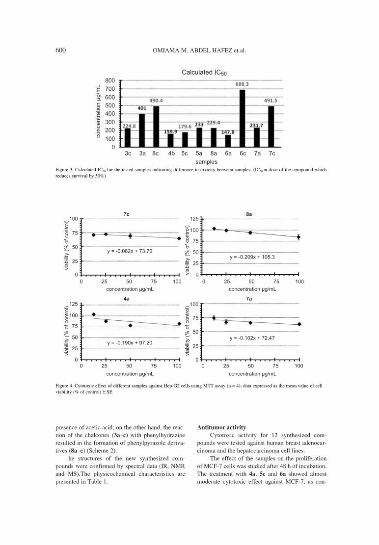
600 OMIAMA M. ABDEL HAFEZ et al.
presence of acetic acid; on the other hand, the reac-tion of the chalcones (3añc) with phenylhydrazineresulted in the formation of phenylpyrazole deriva-tives (8añc) (Scheme 2).
he structures of the new synthesized com-pounds were confirmed by spectral data (IR, NMRand MS).The physicochemical characteristics arepresented in Table 1.
Antitumor activity
Cytotoxic activity for 12 synthesized com-pounds were tested against human breast adenocar-cinoma and the hepatocarcinoma cell lines.
The effect of the samples on the proliferationof MCF-7 cells was studied after 48 h of incubation.The treatment with 4a, 5c and 6a showed almostmoderate cytotoxic effect against MCF-7, as con-
Figure 3. Calculated IC50 for the tested samples indicating difference in toxicity between samples. (IC50 = dose of the compound whichreduces survival by 50%)
Figure 4. Cytotoxic effect of different samples against Hep-G2 cells using MTT assay (n = 4); data expressed as the mean value of cellviability (% of control) ± SE

Synthesis of some new carbonitriles and pyrazole coumarin derivatives... 601
cluded from their close IC50 values 159.9, 179.6 and147.8 µg/mL, respectively, as shown in Figure 2,treatment with samples 3c, 5a, 8a and 7a showedweak cytotoxic effect as they had higher IC50 calcu-lated to be 224.8, 233, 229.4 and 231.7 µg/mL,respectively. Samples 3a, 8c, 6c and 7c showed veryweak cytotoxic effect concluded from their veryhigh IC50 401, 490.4, 688.3 and 491.5 µg/mL,respectively. Finally, sample 4c did not show anycytotoxic effect as it has very high IC50 (2654µg/mL) as shown in Figure 3. For human hepatocar-cinoma cell line (HepG2), samples 3c, 3a, 8c, 4c,5a, 6a, 6c and 5c did not show any cytotoxic effectas they increased proliferation of cells and samples4a, 8a, 7a and 7c showed weak cytotoxic effect asshown in Figure 4.
Antimicrobial activity
The data in Table 2 show various degrees ofantagonism against pathogenic bacteria, yeast andfungi. Three compounds proved to be most promis-ing against tested bacterial organisms i.e., com-pounds 2, 4c and 5c, which were the most activeagainst both Gram negative (Escherichia coli) andGram positive bacteria (Bacillus subtilis, andStaphylococcus aureus). Compounds 3b and 3a
proved to be active against bacteria and yeast whileno activity was determined with fungi. Compounds2 and 5c proved to be most active against Candidaalbicans and all tested fungal organisms.Compounds 4b, 5a and 8b showed moderate activi-ty and the rest of the compounds showed low effectagainst the tested bacterial organisms.
REFERENCES
1. Vijay K., Ganapaty S., Srinivas R.A.: Indian J.Chem. 49B, 11099 (2010).
2. Trykowska Konc J., Hejchman E., KruszewskaH., Wolska I., Maciejewska D.: Eur. J. Med.Chem. 46, 2252 (2011).
3. Zheng Y.X., Yang Y., Peng X., Bastow K.F.,Lee K.H.: Bioorg. Med. Chem. Lett. 10, 699(2000).
4. Lawrence N.J., Patterson R.P., Ooi L.L.:Bioorg. Med. Chem. 16, 5844 (2004).
5. Modzelewska A., Pettit C., Achanta G.,Davidson N.E., Huang P., Khan S.R.: Bioorg.Med. Chem. 14, 3491 (2006).
6. Davis F.A., Chen B.C.: J. Org. Chem. 58, 1751(1993).
7. Alkane C., Vijay J.: Int. J. Pharm. Pharm. Sci. 3,125 (2011).
8. Varmus H.: Science 312, 1162 (2006).9. Hoult J.R.S., Paya M.: Gen. Pharmacol. 27, 713
(1996).10. Kidane A.G., Salacinski H.A., Tiwari K.R.,
Bruckdorfer A.M.: Biomacromolecules 5, 798(2004).
11. Ma T., Liu L., Xue H., Li L., Han C., Wang L.,Chen Z., Liu G.: J. Med. Chem. 51, 1432(2008).
12. Ravindra B.P., Chovatia P.T., Parekh H.H.:Bioorg. Med. Chem. Lett. 22, 5129 (2012).
13. Nassar E.: J. Am. Sci. 6, 463 (2010).14. Klosa J.: Arch. Pharm. (Weinheim) 286, 37
(1953).15. Zavrsink D., Halivoic S.S., Softic D.: Periodic
Biologorum 113, 93 (2011).16. Mohamed M.S., Zaghary W.A., Hafez T.S.,
Ibrahim N.M., Abo El-Alamin M. M., MahranM.R.H.: Bull. Faculty of Pharm. (Cairo Univ.)40, 175 (2002).
17. Hansen M.B., Nielsen S.E., Berg K.: J.Immunol. Methods 10, 119 (1989).
18. Clement N.L.: J. AOAC 51, 611 (1968). 19. Atalla M.M., Nour El-Din K.: J. Microbiol. 28,
193 (1993).
Received: 17. 06. 2013

Acta Poloniae Pharmaceutica ñ Drug Research, Vol. 71 No. 4 pp. 603ñ614, 2014 ISSN 0001-6837Polish Pharmaceutical Society
Nitrogen heterocyclic compounds are of spe-cial interest because they constitute an importantclass of natural and synthetic products, many ofwhich exhibit useful biological activities. Pyrroleand fused pyrrole compounds such as pyrrolopyrim-idine derivatives exhibit a broad spectrum of biolog-ical activities such as antimicrobial (1), analgesic(2), anti-inflammatory (3), antiviral (4) and anti-cancer activity (5ñ7). The potential use of aromat-ic/heterocylic sulfonamides as carbonic anhydrase(CA) inhibitors has been little explored to date in thetreatment of cancer. Acetazolamide (CAS 59- 66- 5)I was reported as a strong inhibitor of several car-bonic anhydrase isozymes (8). It acts as a potentialmodulator of anticancer therapies in combinationwith different cytotoxic agents (alkylating agents,nuclueoside analogs, etc.). Also, 5,7-diphenylpyrro-lo[2,3-d]pyrimidines II were described as potentinhibitors of the tyrosine kinase C-Src, which is nowconsidered as an attractive target of cancer therapy(9). In addition, it was reported that pyrrolopyrimi-dine U101033 (phase III clinical trials for ischemicbrain injuries; have not yet CAS number) [(9-(2-morpholinyl)indole monohydrochloride hydrate]efficiently protected against hydroxy radical-induced lipid peroxidation that occurs deeply withinthe membrane bilayer (10). Due to our interest in the
development of novel anticancer agents, in thisstudy, we report the synthesis of some novel pyrroleand pyrrolopyrimidine derivatives containing a bio-logically active sulfonamide moiety as analogs to Iand II, respectively, hoping that these new com-pounds might show significant anticancer activity.
I II
EXPERIMENTAL
Chemistry
Melting points were determined in open capil-laries on a Gallenkamp melting point apparatus(Sanyo Gallenkamp, Southborough, UK) and areuncorrected. Pre-coated silica gel plates (silica gel,60 F254; Merck, Germany) were used for thin layerchromatography, dichloromethane/methanol (9.5 :0.5, v/v) mixture was used as a developing solventsystem and the spots were visualized by ultravioletlight and/or iodine. Infra-red spectra were recorded
SYNTHESIS AND MOLECULAR DOCKING OF SOME NOVEL ANTICANCERSULFONAMIDES CARRYING A BIOLOGICALLY ACTIVE PYRROLE
AND PYRROLOPYRIMIDINE MOIETIES
MOSTAFA M. GHORAB1*, MANSOUR S. ALSAID1 and YASSIN M. NISSAN2
1Pharmacognosy Department, College of Pharmacy, King Saud University, P.O. Box 2457, Riyadh 11451, Saudi Arabia
2Pharmaceutical Chemistry Department, Faculty of Pharmacy, Cairo University, Kasr Elini St., Cairo 11562, Egypt
Abstract: A novel series of pyrroles and pyrrolopyrimdines carrying a biologically active sulfonamide moietyhave been synthesized. The structures were confirmed by elemental analyses and spectral data. All the targetcompounds were subjected to in vitro cytotoxic screening on breast cancer cell line (MCF-7). Most of the syn-thesized compounds showed good activity as cytotoxic agents with better IC50 than doxorubicin as a referencedrug. In order to suggest a mechanism of action for their activity, molecular docking on the active site of humanc-Src was performed for all synthesized compounds.
Keywords: sulfonamide derivatives, anticancer activity
603
* Corresponding author: e-mail: [email protected]; phone: +966 534292860; fax: +966 014670560

604 MOSTAFA M. GHORAB et al.
in KBr discs using IR-470 Shimadzu spectrometer(Shimadzu, Tokyo, Japan). NMR spectra in DMSO-d6 were recorded on Bruker Ac-500 ultrashield NMR spectrometer (Bruker, Flawil,Switzerland) at 500 MHz, using TMS as internalstandard and peak multiplicities are designed as fol-lows: s, singlet; d, doublet; t, triplet; m, multiplet.Elemental analyses were performed on Carlo Erba1108 Elemental Analyzer (Heraeus, Hanau,Germany). All compounds were within ± 0.4% ofthe theoretical values.
4-[2-(4-Bromophenyl)-2-oxoethylamino]benzene-
sulfonamide (3)
A mixture of sulfanilamide 1 (1.72 g, 0.01 mol)and 4-bromophenacylbromide 2 (2.77 g, 0.01 mol)was refluxed in N,Ní-dimethylformamide (20 mL)in the presence of catalytic amount of triethylaminefor 6 h. The solid obtained was filtered and recrys-tallized from ethanol to give 3. Yield 89%, m.p.232.6OC, IR: (νmax, cm-1): 3358, 3255 (NH, NH2),3100 (CH arom.), 2970, 2863 (CH aliph.), 1685(C=O), 1381, 1157 (SO2).1H-NMR (DMSO-d6, D2O,δ, ppm): 4.7 (s, 2H, CH2), 6.6 (s, 1H, NH, D2Oexchangeable), 6.7, 7.5 (2d, 4H, Ar-H, AB system, J= 7.1 Hz), 7.8, 8.0 (2d, 4H, Ar-H, AB system, J =6.9 Hz). 13C-NMR (DMSO-d6, δ, ppm): 49.4, 111.4(2), 127.1, 127.7, 129.9 (2), 130.7 (2), 131.8 (2),133.9, 150.8, 195.3. Analysis: calcd. forC14H13BrN2O3S (369.23): C, 45.54; H, 3.55; N,7.59%; found: C, 45.54; H, 3.31; N, 7.24%.
4-[2-Amino-4-(4-bromophenyl)-3-cyano-1H-
pyrrol-1-yl]benzenesulfonamide (5)
A mixture of compound 3 (3.69 g, 0.01 mol)and malononitrile (0.66 g, 0.01 mol) in ethanol (20mL) containing sodium ethoxide (0.5 g) wasrefluxed for 8 h. The reaction mixture was cooledand acidified with dil. HCl. The solid obtained wasfiltered and recrystallized from dioxane to give 5.Yield 78%, m.p. 221.2OC, IR: (νmax, cm-1): 3419,3344, 3238 (NH2), 3095 (CH arom.), 2187 (C=N),1635 (C=N), 1342, 1176 (SO2). 1H-NMR (DMSO-d6, D2O, δ, ppm): 6.1 (s, 2H, NH2, D2O exchange-able), 7.0 (s, 1H, CH pyrrole), 7.5ñ7.9 (m, 10H, Ar-H + SO2NH2).13C-NMR (DMSO-d6, δ, ppm): 70.5,113.5, 117.5, 119.5, 121.3 (2), 125.2, 127.3, 131.2(2), 131.6 (2), 132.3 (2), 139.5, 142.9, 148.5.Analysis: calcd. for C17H13BrN4O2S (417.28): C,48.93; H, 3.14; N, 13.43%; found: C, 48.71; H, 3.50;N, 13.16.%
4-[4-Amino-5-(4-bromophenyl)-7H-pyrrolo[2,3-
d]pyrimidin-7-yl]benzenesulfonamide (6)
A solution of compound 5 (4.17 g, 0.01 mol) informamide (20 mL) was refluxed for 8 h. The reac-tion mixture was cooled and then poured ontoice/cold water. The formed residue was recrystal-lized from dioxane to give 6. Yield 75%, m.p. >350OC, IR: (νmax, cm-1): 3470, 3380, 3217 (NH2),3100 (CH arom.), 1620 (C=N), 1381, 1159 (SO2).1H-NMR (DMSO-d6, D2O, δ, ppm): 6.5 (s, 2H, NH2,D2O exchangeable), 7.2 (s, 1H, CH pyrrole), 7.4ñ8.0 (m, 10H, Ar-H + SO2NH2), 8.6 (s, 1H, CHpyrimidine).13C-NMR (DMSO-d6, δ, ppm): 100.9,116.8, 120.4, 122.5, 123.2, 124.3, 126.7, 127.0,128.5, 130.5, 131.7, 132.9, 139.9, 141.3, 152.6,157.6, 160.2, 162.9. Analysis: calcd. forC18H14BrN5O2S (444.31): C, 48.66; H, 3.18; N,15.76%; found: C, 48.41; H, 3.46; N, 15.98%.
General procedure for synthesis of compounds
7añm
A mixture of 5 (4.17 g, 0.01 mol) and aromat-ic aldehydes (0.01 mol.) in glacial acetic acid (20mL) was refluxed for 4 h. The reaction mixture wascooled, filtered and the obtained solid was recrystal-lized from dioxane to give 7añm, respectively.
4-[4-(4-Bromophenyl)-3-cyano-2-(4-methylben-
zylideneamino)-1H-pyrrol-1-yl]benzenesulfon-
amide (7a)
Yield 68%, m.p. 277.3OC, IR: (νmax, cm-1):3363, 3269 (NH2), 3100 (CH arom.), 2970, 2816(CH aliph.), 2202 (C=N), 1600 (C=N), 1328, 1166(SO2). 1H-NMR (DMSO-d6, D2O, δ, ppm): 2.4 (s,3H, CH3), 7.3 (s, 1H, CH pyrrole), 7.4ñ7.9 (m, 14H,Ar-H + SO2NH2), 9.0 (s, 1H, N=CH). 13C-NMR(DMSO-d6, δ, ppm): 21.2, 78.7, 116.8, 119.4, 120.6,124.0, 125.6, 126.5, 127.9, 129.2 (2), 129.6 (2),129.7 (2), 131.3, 131.8, 132.4, 133.9 (2), 139.4,143.0, 145.2, 147.0, 163.4. Analysis: calcd. forC25H19BrN4O2S (518.04): C, 57.81; H, 3.69; N,10.79%; found: C, 57.50; H, 3.88; N, 10.49%.
4-[4-(4-Bromophenyl)-3-cyano-2-(4-hydroxyben-
zylideneamino)-1H-pyrrol-1-yl]benzenesulfon-
amide (7b)
Yield 82%, m.p. 194.3OC, IR: νmax, cm-1): 3410(OH), 3356, 3270 (NH2), 3095 (CH arom.), 2966,2846 (CH aliph.), 2208 (C=N), 1595 (C=N), 1346,1161 (SO2). 1H-NMR (DMSO-d6, D2O, δ, ppm): 6.9(s, 1H, CH pyrrole), 7.0ñ8.0 (m, 14H, Ar-H +SO2NH2), 8.9 (s, 1H, N=CH), 10.5 (s, 1H, OH, D2Oexchangeable). 13C-NMR (DMSO-d6, δ, ppm): 78.2,117.0, 117.6, 118.8, 119..5, 120.5, 121.3 (2), 125.5,126.5, 127.3, 128.3 (2), 131.4 (2), 131.8 (2), 132.3(2), 139.5, 142.8, 148.5, 162.1, 163.1. Analysis:

Synthesis and molecular docking of some novel anticancer sulfonamides... 605
calcd. for C24H17BrN4O3S (521.38): C, 55.29; H,3.29; N, 10.75%. found: C, 55.50; H, 3.10; N,10.66%.
4-[4-(4-Bromophenyl)-3-cyano-2-(4-fluoroben-
zylideneamino)-1H-pyrrol-1-yl]benzenesulfon-
amide (7c)
Yield 80%, m.p. 142.9OC, IR: (νmax, cm-1 3300,3270 (NH2), 3055 (CH arom.), 2971, 2836 (CHaliph.), 2210 (C=N), 1598 (C=N), 1372, 1165(SO2).1H-NMR (DMSO-d6, D2O, δ, ppm): 7.3(s, 1H,CH pyrrole), 7.4ñ7.9 (m, 14H, Ar-H + SO2NH2), 9.1(s, 1H, N=CH).13C-NMR (DMSO-d6, δ, ppm): 78.9,116.4 (2), 116.7, 119.7, 124.1, 125.2 (2), 126.5, 127.1,127.3 (2), 127.8, 128.0 (2), 131.9 (2), 132.3 (2), 139.5,143.1, 146.6, 162.2, 165.8. Analysis: calcd. forC24H16BrFN4O2S (522.01): C, 55.08; H, 3.08; N,10.70%; found: C, 55.25; H, 3.31; N, 10.59%.
4-[4-(4-Bromophenyl)-3-cyano-2-(2-methoxyben-
zylideneamino)-1H-pyrrol-1-yl]benzenesulfon-
amide (7d)
Yield 56%, m.p. 137.6OC, IR: (νmax, cm-1):3280, 3263 (NH2), 3074 (CH arom.), 2941, 2839(CH aliph.), 2212 (C=N), 1597 (C=N), 1340, 1163(SO2). 1H-NMR (DMSO-d6, D2O, δ, ppm): 3.9 (s,3H, OCH3), 7.0 (s, 1H, CH pyrrole), 7.2ñ7.9 (m,14H, Ar-H + SO2NH2), 9.2 (s, 1H, N=CH). 13C-NMR (DMSO-d6, δ, ppm): 56.0, 110.2, 112.6, 116.8(2), 119.8, 120.9, 122.7, 123.0 (2), 124.4, 128.0,129.6 (2), 131.8 (2), 132.3, 134.7, 136.6, 136.7,139.4, 143.0, 147.3, 160.0, 161.4. Analysis: calcd.for C25H19BrN4O3S (535.41): C, 56.08; H, 3.58; N,10.46%; found: C, 56.26; H, 3.31; N, 10.71%.
4-[4-(4-Bromophenyl)-3-cyano-2-(4-methoxyben-
zylideneamino)-1H-pyrrol-1-yl]benzenesulfon-
amide (7e)
Yield 66%, m.p. 141.2OC, IR: (νmax, cm-1):3338, 3259 (NH2), 3100 (CH arom.), 2970, 2860(CH aliph.), 2212 (C=N), 1593 (C=N), 1370, 1166(SO2). 1H-NMR (DMSO-d6, D2O, δ, ppm): 3.8 (s,3H, OCH3), 7.1 (s, 1H, CH pyrrole), 7.2ñ7.9 (m,14H, Ar-H + SO2NH2), 9.0 (s, 1H, N=CH). 13C-NMR (DMSO-d6, δ, ppm): 56.0, 78.3, 114.6 (2),116.9, 119.1, 120.5, 123.9 (2), 125.6, 126.5, 127.7,127.9 (2), 129.6 (2), 131.2 (2), 131.8 (2), 139.5,142.9, 147.5, 162.9, 164.1. Analysis: calcd. forC25H19BrN4O3S (535.41): C, 56.08; H, 3.58; N,10.46%; found: C, 56.30; H, 3.36; N, 10.68%.
4-[2-(Benzo[d][1,3]dioxol-5-ylmethyleneamino)-
4-(4-bromophenyl)-3-cyano-1H-pyrrol-1-yl]ben-
zenesulfonamide (7f)
Yield 66%, m.p. 111.9OC, IR: (νmax, cm-1):3346, 3261 (NH2), 3100 (CH arom.), 2920, 2860(CH aliph.), 2210 (C=N), 1587 (C=N), 1340, 1165(SO2). 1H-NMR (DMSO-d6, D2O, δ, ppm): 6.1 (s,2H, CH2), 7.0 (s, 1H, CH pyrrole), 7.1ñ7.9 (m, 13H,Ar-H + SO2NH2), 8.9 (s, 1H, N=CH). 13C-NMR(DMSO-d6, δ, ppm): 102.2, 106.2, 108.6, 116.8,119.2, 120.5, 123.9, 124.4 (2), 124.8, 126.5, 127.9,128.5, 129.6 (2), 131.4 (2), 131.8 (2), 139.4, 143.0,148.3, 151.6, 152.7, 162.5. Analysis: calcd. forC25H17BrN4O4S (549.39): C, 54.65; H, 3.12; N,10.20%; found: C, 54.91; H, 3.39; N, 10.46%.
4-[4-(4-Bromophenyl)-3-cyano-2-(4-(dimethy-
lamino)benzylideneamino)-1H-pyrrol-1-yl]ben-
zenesulfonamide (7g)
Yield 76%, m.p. 99.7OC, IR: (νmax, cm-1): 3300,3246 (NH2), 3100 (CH arom.), 2920, 2870 (CHaliph.), 2206 (C=N), 1593 (C=N), 1373, 1165 (SO2).1H-NMR (DMSO-d6, D2O, δ, ppm): 3.0 (s, 6H,2CH3), 6.7 (s, 1H, CH pyrrole), 7.0ñ7.9 (m, 14H,Ar-H + SO2NH2), 9.6 (s, 1H, N=CH). 13C-NMR(DMSO-d6, δ, ppm): 41.1 (2), 111.5, 115.1 (2),117.3, 119.4, 120.4, 124.4 (2), 125.5, 126.6, 127.3,127.9 (2), 131.5 (2), 132.3 (2), 134.3 (2), 139.7,142.1, 148.7, 153.3, 162.7. Analysis: calcd. forC26H22BrN5O2S (548.45): C, 56.94; H, 4.04; N,12.77%; found: C, 56.68; H, 4,36; N, 12.55%.
4-[4-(4-Bromophenyl)-3-cyano-2-(4-nitrobenzyli-
deneamino)-1H-pyrrol-1-yl]benzenesulfonamide
(7h)
Yield 85%, m.p. 318.5OC, IR: (νmax, cm-1):3373, 3269 (NH2), 3100 (CH arom.), 2910, 2860(CH aliph.), 2214 (C=N), 1593 (C=N), 1398, 1165(SO2). 1H-NMR (DMSO-d6, D2O, δ, ppm): 7.5 (s,1H, CH pyrrole), 7.7ñ8.0 (m, 14H, Ar-H +SO2NH2), 9.2 (s, 1H, N=CH). 13C-NMR (DMSO-d6,δ, ppm): 79.7, 116.4, 120.8 (2), 124.2 (2), 124.7,125.8 (2), 126.6, 128.0 (2), 130.6 (2), 131.0 (2),131.9 (2), 139.2, 140.4, 143.3, 145.3, 149.4, 160.6.Analysis: calcd. for C24H16BrN5O2S (549.01): C,52.37; H, 2.93; N, 12.72%; found: C, 52.10; H, 2.66;N, 12.44%.
4-[4-(4-Bromophenyl)-3-cyano-2-(2,4-dinitroben-
zylideneamino)-1H-pyrrol-1-yl]benzenesulfon-
amide (7i)
Yield 77%, m.p. 210.2OC, IR: (νmax, cm-1): 3300,3211 (NH2), 3095 (CH arom.), 2930, 2860 (CHaliph.), 2218 (C=N), 1583 (C=N), 1346, 1165 (SO2).1H-NMR (DMSO-d6, D2O, δ, ppm): 6.9 (s, 1H, CHpyrrole), 7.1ñ7.9 (m, 13H, Ar-H + SO2NH2), 10.3 (s,1H, N=CH). 13C-NMR (DMSO-d6, δ, ppm): 104.4,

606 MOSTAFA M. GHORAB et al.
119.7, 120.6, 123.6, 126.6, 127.6 (2), 128.5, 129.0,129.5 (2), 131.5 (2), 131.9, 132.2, 132.4 (2), 134.4,134.8, 146.0, 148.9, 149.5, 151.5, 156.0. Analysis:calcd. for C24H15BrN6O6S (595.38): C, 48.42; H, 2.54;N, 14.12%; found: C, 48.76; H, 2.26; N, 14.51%.
4-[4-(4-Bromophenyl)-2-(4-chlorobenzylide-
neamino)-3-cyano-1H-pyrrol-1-yl]benzenesulfon-
amide (7j)
Yield 79%, m.p. 164.6OC, IR: (νmax, cm-1):3305, 3255 (NH2), 3095 (CH arom.), 2910, 2861(CH aliph.), 2212 (C=N), 1593 (C=N), 1338, 1165(SO2), 719 (C-Cl). 1H-NMR (DMSO-d6, D2O, δ,ppm): 7.0 (s, 1H, CH pyrrole), 7.4ñ7.9 (m, 14H, Ar-H + SO2NH2), 9.1 (s, 1H, N=CH). 13C-NMR(DMSO-d6, δ, ppm): 79.1, 116.6, 119.9, 120.7,124.3 (2), 125.7, 126.6, 127.8 (2), 129.3 (2), 131.2(2), 131.9 (2), 133.8, 134.7 (2), 137.5, 139.3, 143.1,146.3, 162.0. Analysis: calcd. for C24H16BrClN4O2S(537.98): C, 53.40; H, 2.99; N, 10.38%; found: C,53.67; H, 2.68; N, 10.76%.
4-[4-(4-Bromophenyl)-3-cyano-2-(2,4-dichloro-
benzylideneamino)-1H-pyrrol-1-yl]benzenesul-
fonamide (7k)
Yield 86%, m.p. 115.6OC, IR: (νmax, cm-1):3346, 3265 (NH2), 3088 (CH arom.), 2926, 2836(CH aliph.), 2216 (C=N), 1581 (C=N), 1379, 1166(SO2), 719, 825 (2 C-Cl). 1H-NMR (DMSO-d6, D2O,δ, ppm): 7.1 (s, 1H, CH pyrrole), 7.2ñ7.9 (m, 13H,Ar-H + SO2NH2), 10.2 (s, 1H, N=CH). 13C-NMR(DMSO-d6, δ, ppm): 79.6, 120.6, 125.7, 125.9,126.5 (2), 127.3, 128.2, 129.2, 130.6 (2), 131.0 (2),132.4, 133.6, 136.3, 137.1 (2), 139.7 (2), 144.9,145.3, 145.5, 156.8. Analysis: calcd. forC24H15BrCl2N4O2S (574.27): C, 50.19; H, 2.63; N,9.76%; found: C, 50.47; H, 2.37; N, 9.50%.
4-[4-(4-Bromophenyl)-3-cyano-2-[(2-methoxy-
naphthalen-1-yl)methylene-amino]-1H-pyrrol-1-
yl]benzenesulfonamide (7l)
Yield 65%, m.p. 278.2OC, IR: (νmax, cm-1):3346, 3255 (NH2), 3074 (CH arom.), 2971, 2842(CH aliph.), 2210 (C=N), 1624 (C=N), 1371, 1151(SO2). 1H-NMR (DMSO-d6, D2O, δ, ppm): 4.0 (s,3H, OCH3), 7.4 (s, 1H, CH pyrrole), 7.5ñ8.0 (m,16H, Ar-H + SO2NH2), 9.9 (s, 1H, N=CH). 13C-NMR (DMSO-d6, δ, ppm): 57.1, 104.3, 113.5,114.5, 117.0, 117.6, 119.6, 120.0 (2), 120.5, 124.3,125.1, 126.4, 126.5, 128.1 (2), 128.5, 128.7, 130.8(2), 131.5, 131.8 (2), 136.2, 139.7, 143.5, 148. 4,160.1, 161.5. Analysis: calcd. for C29H21BrN4O3S(584.05): C, 59.49; H, 3.62; N, 9.57%; found: C,59.21; H, 3.91.37; N, 9.81%.
4-[4-(4-Bromophenyl)-3-cyano-2-[(4-methoxy-
naphthalen-1-yl)methylene-amino]-1H-pyrrol-1-
yl]benzenesulfonamide (7m)
Yield 73%, m.p. 181.2OC, IR: (νmax, cm-1):3327, 3310 (NH2), 3100 (CH arom.), 2955, 2846(CH aliph.), 2208 (C=N), 1590 (C=N), 1370, 1157(SO2). 1H-NMR (DMSO-d6, D2O, δ, ppm): 4.0 (s,3H, OCH3), 7.1ñ8.0 (m, 16H, Ar-H + SO2NH2), 8.9(s, 1H, CH pyrrole), 9.5 (s, 1H, N=CH). 13C-NMR(DMSO-d6, δ, ppm): 56.2, 104.0, 104.7, 117.2,119.2, 120.5, 122.0, 122.3 (2), 122.5, 124.0, 124.2,124.6, 124.8, 125.9, 126.1, 126.5, 127.9 (2), 131.5(2), 134.5 (2), 139.6, 140.3, 143.2, 148.1, 159.1,160.1. Analysis: calcd. for C29H21BrN4O3S (585.47):C, 59.49; H, 3.62; N, 9.57%; found: C, 59.70; H,3.41.37; N, 9.19%.
4-[5-(4-Bromophenyl)-4-oxo-3,4-dihydropyrro-
lo[2,3-d]pyrimidin-7-yl]benzenesulfonamide (8)
A solution of compound 5 (4.17 g, 0.01 mol.)in formic acid (20 mL) was refluxed for 6 h. Thereaction mixture was poured onto ice/water and thesolid obtained was recrystallized from dioxane togive 8. Yield 81%, m.p. 313.7OC, IR: (νmax, cm-1):3320, 3236, 3156 (NH, NH2), 3094 (CH arom.),1683 (C=O), 1591 (C=N), 1386, 1161 (SO2). 1H-NMR (DMSO-d6, D2O, δ, ppm): 7.5 (s, 1H, CH pyr-role), 7.6ñ8.0 (m, 10H, Ar-H + SO2NH2), 8.1 (s, 1H,CH pyrimidine), 12.3 (s, 1H, NH, D2O exchange-able). 13C-NMR (DMSO-d6, δ, ppm): 106.3, 119.7,120.3, 121.5 (2), 124.4, 126.7, 127.9 (2), 130.8 (2),132.3 (2), 141.1, 142.2, 145.0, 148.2, 158.4.Analysis: calcd. for C18H13BrN4O3S (445.29): C,48.55; H, 2.94; N, 12.58%; found: C, 48.30; H, 2.65;N, 12.84%.
N - [4- (4-bromophenyl) -3-cyano-1-(4-sul -
famoylphenyl)-1H-pyrrol-2-yl]-2,2,2-trifluoroac-
etamide (9)
A solution of compound 5 (4.17 g, 0.01 mol.)in trifluoroacetic anhydride (15 mL) was refluxedfor 20 h. The solid obtained was recrystallized fromethanol to give 9. Yield 69%, m.p. 206.5OC, IR:(νmax, cm-1): 3309, 3291 (NH2), 3100 (CH arom.),2202 (C=N), 1681 (C=O), 1610 (C=N), 1336, 1151(SO2). 1H-NMR (DMSO-d6, D2O, δ, ppm): 7.1 (s,1H, CH pyrrole), 7.2ñ8.4 (m, 10H, Ar-H +SO2NH2), 13.8 (s, 1H, NH, D2O exchangeable). 13C-NMR (DMSO-d6, δ, ppm): 118.6, 119.4, 120.2,123.8, 124.1, 124.5, 124.9, 126.5, 126.8, 127.1 (2),130.3, 130.5, 131.0, 131.5, 131.7, 138.9, 147.7,159.9. Analysis: calcd. for C19H12BrF3N4O3S(513.29): C, 44.46; H, 2.36; N, 10.92%; found: C,44.12; H, 2.71; N, 10.66%.

Synthesis and molecular docking of some novel anticancer sulfonamides... 607
N-[4-(4-bromophenyl)-3-cyano-1-(4-sulfamoyl-
phenyl)-1H-pyrrol-2-yl]acetamide (10)
A solution of compound 5 (4.17 g, 0.01 mol.)in acetic anhydride (20 mL) was refluxed for 5 min.The reaction mixture was concentrated and the solidseparated was recrystallized from ethanol to give 10.Yield 81%, m.p. 113.0OC, IR: (νmax, cm-1): 3389,3344, 3238 (NH, NH2), 3095 (CH arom.), 2981,2862 (CH aliph.), 2189 (C=N), 1685 (C=O), 1635(C=N), 1340, 1163 (SO2). 1H-NMR (DMSO-d6,D2O, δ, ppm): 2.0 (s, 3H, COCH3), 6.1 (s, 1H, CHpyrrole), 7.0ñ8.0 (m, 10H, Ar-H + SO2NH2), 10.2 (s,1H, NH, D2O exchangeable) . 13C-NMR (DMSO-d6,δ, ppm): 22.3, 113.5, 117.5, 119.5, 121.3, 123.0,124.8, 125.8, 127.8, 131.3 (2), 132.3 (2), 134.3 (2),139.2, 142.9, 148.5, 170.0. Analysis: calcd. forC19H15BrN4O3S (459.32): C, 49.68; H, 3.29; N,12.20%; found: C, 49.36; H, 3.51; N, 12.56%.
N-acetyl-N-[4-(4-bromophenyl)-3-cyano-1-(4-sul-
famoylphenyl)-1H-pyrrol-2-yl]acet-amide (11)
A solution of compound 5 (4.17 g, 0.01 mol.)in acetic anhydride (20 mL) was refluxed for 24 h.The reaction mixture was concentrated and the solidobtained was recrystallized from dioxane to give 11.Yield 59%, m.p. 142.8OC, IR: (νmax, cm-1): 3495,3122 (NH2), 3055 (CH arom.), 2920, 2862 (CHaliph.), 2227 (C=N), 1734, 1718 (2 C=O), 1593(C=N), 1369, 1161 (SO2). 1H-NMR (DMSO-d6,D2O, δ, ppm): 2.4 (s, 6H, 2COCH3), 7.4 (s, 1H, CHpyrrole), 7.5ñ8.1 (m, 10H, Ar-H + SO2NH2). 13C-NMR (DMSO-d6, δ, ppm): 21.0 (2), 113.9, 120.4,120.8, 120.9, 121.8 (2), 124.9, 125.3, 128.8 (2),129.5 (2), 133.5 (2), 139.5, 140.1, 146.5, 171.8 (2).Analysis: calcd. for C21H17BrN4O4S (501.35): C,50.31; H, 3.42; N, 11.18%; found: C, 50.63; H, 3.12;N, 11.41%.
N-[4-(4-bromophenyl)-3-cyano-1-(4-sulfamoyl-
phenyl)-1H-pyrrol-2-yl]-2-chloroacetamide (13)
A solution of compound 5 (4.17 g, 0.01 mol.)in chloroacetylchloride (20 mL) was refluxed for 1h. The reaction mixture was poured onto ice/waterand the solid obtained was recrystallized from diox-ane to give 13. Yield 80%, m.p. 154.4OC, IR: (νmax,cm-1): 3470, 3380, 3210 (NH, NH2), 3100 (CHarom.), 2956, 2817 (CH aliph.), 2223 (C=N), 1707(C=O), 1627 (C=N), 1388, 1149 (SO2), 725 (C-Cl).1H-NMR (DMSO-d6, D2O, δ, ppm): 4.2 (s, 2H, CH2),7.4 (s, 1H, CH pyrrole), 7.5ñ7.9 (m, 10H, Ar-H +SO2NH2), 8.2 (s, 1H, NH, D2O exchangeable). 13C-NMR (DMSO-d6, δ, ppm): 42.4, 89.5, 114.7, 119.4,121.3, 124.9 (2), 125.1, 128.5, 129.3, 130.5, 131.1,131.4, 132.3, 133.1, 138.8, 142.6, 149.7, 166.8.
Analysis: calcd. for C19H14BrClN4O3S (493.76): C,46.22; H, 2.86; N, 11.35%; found: C, 46.52; H, 2.67;N, 11.14%.
N-[4-(4-bromophenyl)-3-cyano-1-(4-sulfamoyl-
phenyl)-1H-pyrrol-2-yl]-2-chloro-N-(2-chloro-
acetyl)acetamide (14)
A solution of compound 5 (4.17 g, 0.01 mol) inchloroacetylchloride (20 mL) was refluxed for 16 h.The solid obtained was recrystallized from aceticacid to give 14. Yield 64%, m.p. 359.2OC, IR: (νmax,cm-1): 3253, 3190 (NH2), 3088 (CH arom.), 2944,2865 (CH aliph.), 2223 (C=N), 1720, 1705 (2 C=O),1591 (C=N), 1388, 1174 (SO2), 727 (C-Cl). 1H-NMR (DMSO-d6, D2O, δ, ppm): 4.2 (s, 4H, 2 CH2),7.3 (s, 1H, CH pyrrole), 7.4ñ8.0 (m, 10H, Ar-H +SO2NH2). 13C-NMR (DMSO-d6, δ, ppm): 42.1, 42.8,89.7, 114.6, 119.4, 120.6, 123.3 (2), 124.8, 125.1,125.8, 127.8 (2), 129.1 (2), 131.0, 131.9, 133.0,138.2, 140.5, 165.2, 166.8. Analysis: calcd. forC21H15BrCl2N4O4S (570.24): C, 44.23; H, 2.65; N,9.83%; found: C, 44.51; H, 2.36; N, 10.19%.
Molecular docking
All the molecular modeling studies were car-ried out on an Intel Pentium 1.6 GHz processor, 512MB memory with Windows XP operating systemusing Molecular Operating Environment (MOE,10.2008) software. All the minimizations were per-formed with MOE until a RMSD gradient of 0.05kcal mol-1�-1 with MMFF94X force field and thepartial charges were automatically calculated. TheX-ray crystallographic structure of c-Src complexwith its ligand (PDB ID: 1YOL) was obtained fromthe protein data bank. The enzyme was prepared fordocking studies where: (i) Ligand molecule wasremoved from the enzyme active site. (ii) Hydrogenatoms were added to the structure with their stan-dard geometry. (iii) MOE Alpha Site Finder wasused for the active sites search in the enzyme struc-ture and dummy atoms were created from theobtained alpha spheres. (iv) The obtained model wasthen used in predicting the ligandñenzymes interac-tions at the active site (Table 1).
In vitro antitumor activity
Human tumor breast cell line (MCF7) wasused in this study. The cytotoxic activity was meas-ured in vitro for the newly synthesized compoundsusing the sulfo-rhodamine-B stain (SRB) assayusing the method of Skehan et al. (11). The in vitroanticancer screening was done by the pharmacologyunit at the National Cancer Institute, CairoUniversity. Cells were plated in 96-multiwell plate

608 MOSTAFA M. GHORAB et al.
(104 cells/well) for 24 h before treatment with thecompound(s) to allow attachment of cell to the wallof the plate. The tested compounds were dissolvedin dimethyl sulfoxide. Different concentrations ofthe compound under test (10, 25, 50, and 100 µM)were added to the cell monolayer. Triplicate wellswere prepared for each individual concentration.Monolayer cells were incubated with the com-pound(s) for 48 h at 37OC and in atmosphere of 5%CO2. After 48 h, cells were fixed, washed andstained for 30 min with 0.4% (w/v) SRB dissolvedin 1% acetic acid. An excess of unbound dye wasremoved by four washes with 1% acetic acid andattached stain was recovered with Tris EDTAbuffer. Color intensity was measured in an ELISAreader. The relation between surviving fraction anddrug concentration is plotted to get the survivalcurve for breast tumor cell line after the specified
time. The molar concentration required for 50%inhibition of cell viability (IC50) was calculated andcompared to the reference drug doxorubicin (CAS,25316-40-9). The surviving fractions wereexpressed as the means ± standard error and theresults are given in Table 2.
RESULTS AND DISCUSSION
Chemistry
The synthesized compounds were designedwith the aim of exploring their anticancer activity. Inthis investigation, a novel series of bromopyrroles 5,7añm, 9ñ11, 13, 14 and bromopyrrolopyrimidines 6,8 having a biologically active sulfonamide moietieswere synthesized to evaluate their in vitro anticanceractivity. Thus, interaction of sulfanilamide 1 with 4-
Table 1. Binding scores and amino acid interactions of the docked compounds on the active site of c-Src.
Compd. S Amino acid Interacting H bondNo. Kcal/mol interactions groups length �
5 ñ13.6201 Asp 350, Met 343 NH2 3.22, 2.22Ser 347 N pyrimidine 3.09
6 ñ16.1961 Glu 341, Met 343 NH2, N pyrimidine 1.94, 2.89Asp 350, Ser 347 SO2NH2, SO2NH2 1.42, 3.02
7a ñ17.7147 Glu 312, Asp 436 SO2NH2, SO2NH2 2.43, 201Asp 350 CN 3.74
7b ñ9.3848 Met 343, Asp 350 OH, SO2NH2 2.89, 1.98Ser 347 SO2 3.47
7c ñ10.4441 Asp 350 SO2 3.04
7d ñ14.8247 Glu 281, Asp 350 OCH3, SO2NH2 3.49, 1.67Asp 350 3.32
7e ñ11.6741 Asp 350 SO2NH2 2.34ñ1.80
7f ñ12.3196 Asp 350 SO2NH2 3.01
7g ñ20.4112 Asp 350 N=C 3.11
7h ñ17.8049 Glu 312, Asp 406 SO2NH2, SO2NH2 2.14, 2.15
7i ñ15.5585 Asp 350 CN 3.74
7j ñ13.7170 Met 343 SO2, SO2NH2 3.25, 2.01
7k ñ19.0188 Asp 350 SO2NH2 2.40
7l ñ22.8663 Gly 281 SO2NH2 3.28, 3.15
7m ñ10.4936 Asp 350 SO2NH2 1.78
8 ñ3.1180 Asp 350, Ser 347 SO2NH2, SO2NH2 1.40, 1.97
9 ñ11.6900 Asp 350, Asp 406 SO2NH2, SO2NH2 3.48, 3.05Glu 312 SO2NH2 2.16
10 ñ16.0731 Asp 350 SO2NH2 1.52
11 ñ10.0602 Asp 350 SO2NH2 2.51
13 ñ13.0866 Ala 382 C=O 2.43
14 ñ12.9995 Asp 350 SO2NH2 1.57
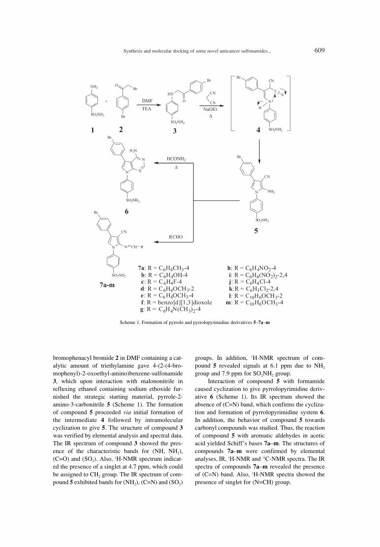
Synthesis and molecular docking of some novel anticancer sulfonamides... 609
Scheme 1. Formation of pyrrolo and pyrrolopyrimidine derivatives 5ñ7añm
bromophenacyl bromide 2 in DMF containing a cat-alytic amount of triethylamine gave 4-(2-(4-bro-mophenyl)-2-oxoethyl-amino)benzene-sulfonamide3, which upon interaction with malononitrile inrefluxing ethanol containing sodium ethoxide fur-nished the strategic starting material, pyrrole-2-amino-3-carbonitrile 5 (Scheme 1). The formationof compound 5 proceeded via initial formation ofthe intermediate 4 followed by intramolecularcyclization to give 5. The structure of compound 3was verified by elemental analysis and spectral data.The IR spectrum of compound 3 showed the pres-ence of the characteristic bands for (NH, NH2),(C=O) and (SO2). Also, 1H-NMR spectrum indicat-ed the presence of a singlet at 4.7 ppm, which couldbe assigned to CH2 group. The IR spectrum of com-pound 5 exhibited bands for (NH2), (C=N) and (SO2)
groups. In addition, 1H-NMR spectrum of com-pound 5 revealed signals at 6.1 ppm due to NH2
group and 7.9 ppm for SO2NH2 group.Interaction of compound 5 with formamide
caused cyclization to give pyrrolopyrimidine deriv-ative 6 (Scheme 1). Its IR spectrum showed theabsence of (C=N) band, which confirms the cycliza-tion and formation of pyrrolopyrimidine system 6.In addition, the behavior of compound 5 towardscarbonyl compounds was studied. Thus, the reactionof compound 5 with aromatic aldehydes in aceticacid yielded Schiffís bases 7añm. The structures ofcompounds 7añm were confirmed by elementalanalyses, IR, 1H-NMR and 13C-NMR spectra. The IRspectra of compounds 7añm revealed the presenceof (C=N) band. Also, 1H-NMR spectra showed thepresence of singlet for (N=CH) group.

610 MOSTAFA M. GHORAB et al.
Refluxing compound 5 in formic acid causedcyclization via elimination of water to give thepyrrolopyrimidine derivative 8 (Scheme 2). Its IRspectrum exhibited the absence of (C=N) band,which confirms the cyclization and formation ofpyrrolopyrimidine system 8. The reactivity of com-pound 5 towards acid anhydride in different time ofreflux was observed. Thus, reaction of compound 5with acetic anhydride for 5 min furnished themonoacetyl derivative 10. On the other hand, when
compound 5 was reacted with acetic anhydride forlong time (24 h), the corresponding diacetyl deriva-tive 11 was obtained instead of the cyclic systempyrrolopyrimidine derivative 12 on the basis of ele-mental analysis and IR spectrum, which showed thepresence of (C=N) band. When compound 5 wasreacted with trifluoroacetic anhydride for long time(20 h), the corresponding monoacetyl derivative 9
was obtained, on the basis of elemental analysis andIR spectrum, which showed the presence of (C=N)
Scheme 2. Formation of new pyrrolo and pyrrolopyrimidine derivatives 8ñ11

Synthesis and molecular docking of some novel anticancer sulfonamides... 611
for long time (16 h), the corresponding diacetylchlo-ride derivative 14 was obtained in a good yieldrather than the cyclic pyrrolopyrimidine derivative15, based on the elemental analyses and spectraldata. The IR spectrum of compound 13 showed thepresence of (C=N), (C=O) and (C-Cl) bands. 1H-
Scheme 3. Formation of some novel pyrrole derivatives 13 and 14
band. 1H-NMR spectrum of 11 showed singlet at 2.4ppm for two acetyl groups.
On the other hand, when compound 5 wasreacted with chloroacetylchloride for short time (1h), the corresponding monoacetylchloride derivative13 was obtained while, applying the same reaction

612 MOSTAFA M. GHORAB et al.
NMR spectrum of 13 revealed the presence of a sin-glet at 4.2 ppm assigned for CH2 group. The IRspectrum of 14 revealed the presence of (C=N) at2223 cm-1 and 2C=O at 1720 and 1705 cm-1. The 1H-NMR spectrum of 14 exhibited singlet at 4.2 ppmdue to two CH2 groups (Scheme 3).
Molecular docking
Several classes of inhibitors are currently usedto inhibit the activity of c-Src in a number of celltypes. However, they often show poor selectivitywithin the c-Src family, which in mammals com-prises at least eight members involved in many keyfunctions of the cell (12). Recently, it has beenshown that c-Src inhibitors of the pyrrolopyrimidineclass exhibit a powerful inhibitory activity and aseveral-fold greater selectivity for c-Src againstmost tyrosine kinases (13ñ15). suggesting that c-Src
can be activated downstream of receptor activator ofNF-κB (RANK) (16), a member of the tumor necro-sis factor (TNF) receptor superfamily that isinvolved in cell differentiation, function, and sur-vival (17ñ19).
In order to realize the aim of the present investi-gations, the authors have performed molecular dockingof the synthesized compounds on the active sites of c-Src, which may provide an understanding of their effectas antitumor agents. The protein data bank file (PDBID:1YOL) was selected for this purpose. The file con-tains c-Src enzyme co-crystallized with a pyrrolopyrim-idine ligand. All docking procedures were achieved byMOE (Molecular Operating Environment) software10.2008 provided by chemical computing group,Canada. Docking on the active site of c-Src enzyme wasperformed for all synthesized compounds. Dockingprotocol was verified by redocking of the co-crystal-
Table 2. In vitro anticancer screening of the synthesized compounds against human breast cancer cell line (MCF7).
Compound concentration (µM)
Compound 10 µM 25 µM 50 µM 100 µM IC50 (µM)
Surviving fraction (mean ± SE)*
Doxorubicin 0.314 ± 0.032 0.309 ± 0.016 0.251 ± 0.023 0.266 ± 0.032 8.02
5 0.327 ± 0.121 0.273 ± 0.043 0.233 ± 0.011 0.255 ± 0.020 7.56
6 0.340 ± 0.090 0.294 ± 0.021 0.246 ± 0.110 0.256 ± 0.002 7.56
7a 0.275 ± 0.113 0.265 ± 0.005 0.323 ± 0.010 0.349 ± 0.032 6.74
7b 0.301 ± 0.121 0.244 ± 0.055 0.236 ± 0.001 0.238 ± 0.012 7.01
7c 0.300 ± 0.090 0.247 ± 0.035 0.238 ± 0.023 0.311 ± 0.066 7.34
7d 0.345 ± 0.133 0.262 ± 0.100 0.313 ± 0.043 0.319 ± 0.065 7.84
7e 0.234 ± 0.033 0.225 ± 0.111 0.248 ± 0.015 0.279 ± 0.076 6.47
7f 0.256 ± 0.067 0.362 ± 0.021 0.345 ± 0.034 0.235 ± 0.041 7.90
7g 0.252 ± 0.084 0.265 ± 0.142 0.231 ± 0.101 0.264 ± 0.102 6.46
7h 0.338 ± 0.066 0.261 ± 0.031 0.234 ± 0.116 0.263 ± 0.131 7.56
7i 0.235 ± 0.019 0.228 ± 0.008 0.261 ± 0.032 0.256 ± 0.056 6.46
7j 0.273 ± 0.191 0.205 ± 0.018 0.237 ± 0.011 0.251 ± 0.007 6.74
7k 0.331 ± 0.012 0.310 ± 0.110 0.295 ± 0.044 0.299 ± 0.033 8.15
7l 0.334 ± 0.061 0.319 ± 0.029 0.287 ± 0.017 0.289 ± 0.049 6.70
7m 0.391 ± 0.028 0.300 ± 0.015 0.299 ± 0.006 0.311 ± 0.112 8.45
8 0.273 ± 0.004 0.315 ± 0.017 0.276 ± 0.003 0.287 ± 0.001 6.74
9 0.361 ± 0.011 0.379 ± 0.089 0.298 ± 0.042 0.321 ± 0.058 7.84
10 0.353 ± 0.071 0.323 ± 0.006 0.273 ± 0.027 0.222 ± 0.010 7.56
11 0.288 ± 0.055 0.229 ± 0.091 0.240 ± 0.042 0.242 ± 0.080 6.74
13 0.324 ± 0.010 0.340 ± 0.021 0.294 ± 0.069 0.316 ± 0.049 7.29
14 0.332 ± 0.023 0.273 ± 0.011 0.285 ± 0.048 0.234 ± 0.052 7.29
* Each value is the mean of three values ± standard error (SE)

Synthesis and molecular docking of some novel anticancer sulfonamides... 613
lized ligand in the vicinity of the active site of theenzyme with energy score (S) = ñ22.6799 Kcal/ moland root mean standard deviation (RMSD) = 0.8205(Fig. 1). The ligand interacts with the active site aminoacids by three interactions: with Met 343 with hydrogenbond of 3.09 �, with Glu 341 with hydrogen bond of2.22 � and with Thr 340 with hydrogen bond of 3.22 �.
All the synthesized compounds were docked onthe active site of the enzyme showing good fitting. Theenergy score (S) as well as amino acid interactions ofthe synthesized compounds are listed in Table 1.
The best energy scores were exhibited by com-pounds 7g and 7l with S = ñ20.4112 and ñ22.8663Kcal/ mol, respectively. Figures 2 and 3 describe theamino acid interactions with compounds 7g and 7l,respectively.
In vitro antitumor activity
The newly synthesized compounds were eval-uated for their in vitro cytotoxic activity against
human breast cancer cell line (MCF7). Doxorubicin,which is one of the most effective anticancer agents,was used as the reference drug in this study. Therelationship between surviving fraction and drugconcentration was plotted to obtain the survivalcurve of breast cancer cell line (MCF7). Theresponse parameter calculated was the IC50 value,which corresponds to the concentration required for50% inhibition of cell viability. In vitro cytotoxicactivity of the synthesized compounds compared tothe reference drug is presented in Table 2.
The strategic starting material ñ pyrrole deriv-ative 5 ñ showed IC50 value 7.65 µM which did notchange upon cyclization to the amino pyrrolopyrim-idine derivative 6. However, the formation of sever-al Schiffís bases 7añm dramatically influenced theactivity, especially for compounds 7a, 7e, 7g, 7i, 7j,and 7l, with IC50 values of 6.74, 6.47, 6.46, 6.46,6.74 and 6.70 µM, respectively. On the other hand,the pyrrolopyrimidine derivative 8 showed goodIC50 value of 6.74 µM. In case of the trifluoroacetylderivative 9, the IC50 value was 7.84 µM while themonoacetyl pyrrolo derivative 10 showed IC50 valueof 7.56 µM, which was better for the diacetyl ana-logue 11 with IC50 value of 6.74 µM. The IC50 valuesof 7.29 µM were observed for the mono anddichloroacetyl derivatives 13 and 14, respectively.Finally, all the synthesized compounds showed bet-ter IC50 than doxorubicin except compounds 7k and7m with IC50 values of 8.15 and 8.45 µM, respec-tively. Compounds 7g and 7l showed both good IC50
of 6.46 and 6.70 µM and also good docking score ofñ20.4112 and ñ22.8663 Kcal/mol, suggesting goodcandidates for c-Src inhibitors.
CONCLUSION
The objective of the present study was to syn-thesize and investigate the anticancer activity ofsome novel pyrrolo and pyrrolopyrimidine deriva-
Figure 1. Pyrrolopyrimidine ligand on the active site of c-Srcenzyme
Figure 2. Compound 7g on the active site of c-Src enzyme
Figure 3. Compound 7l on the active site of c-Src enzyme

614 MOSTAFA M. GHORAB et al.
tives carrying the biologically active sulfonamidemoieties. Most of the synthesized compoundsshowed good activity with better IC50 than doxoru-bicin as reference drug, especially compounds 7a,7e, 7g, 7i, 7j, 8 and 11. Also, compounds 5, 6, 7bñd,7f, 7h, 7l, 9, 10, 13, 14 are nearly as active as doxo-rubicin as positive control, while compounds 7k and7m showed activity lower than doxorubicin.
Acknowledgment
The authors would like to extend their sincereappreciation to the Deanship of Scientific Researchat King Saud University for its funding of thisresearch through the Research Group Project no.RGP-VPP-302.
REFERENCES
1. Dang Q., Gomez-Galeno J.E.: J. Org. Chem. 67,8703 (2002).
2. Danchev N., Bijev A.,Yaneva D., VladimirovaS., Nikolova I.: Arch. Pharm. (Weinheim) 339,670 (2006).
3. Jarvis M.F., Yu H., McGaraughty S., WismerC.T., Mikusa J., Zhu C., Chu K. et al.: Pain 96,107 (2002).
4. Gangjee A., Jain H. D., Kisliuk R.L.: Bioorg.Med. Chem. Lett. 15, 2225 (2005).
5. De Clercq E., Balzarini J., Madej D., HansskeF., Robins M.J.: J. Med. Chem. 30, 481 (1987).
6. Finch R.A., Revankar G.R., Chan P.K.:Anticancer Drug Des. 12, 205 (1997).
7. Krawczyk S.H., Nassiri M.R., Kucera L.S.,Kern E.R., Ptak R.G., Wotring L.L., Drach J.C.
Townsend L.B.: J. Med .Chem. 38, 4106(1995).
8. Leaf D.E., Goldfarb D.S.: J. Appl. Physiol. 102,1313 (2007).
9. Sundaramoorthi R., Shakespeare W.C., KeenanT.P., Metcalf C.A., Wang Y., Mani U., TaylorM. et al.: Bioorg. Med. Chem. Lett. 13, 3063(2003).
10. Hall E.D., Andrus P.K., Smith S.L., Fleck T.J.,Scherch H.M., Lutzke B.S., Sawada G.A. et al.:J. Pharmacol. Exp. Ther. 281, 895 (1997).
11. Skehan P., Storeng R., Scudiero D., Monks A.,McMahon J., Vistica D., Warren J. et al.: J.Natl. Cancer Inst. 82, 1107 (1990).
12. Thomas S.M., Brugge J.S.: Annu. Rev. CellDev. Biol. 13, 513 (1997).
13. Missbach M., Jeschke M., Feyen J., Muller K.,Glatt M., Green J., Susa M.: Bone 24, 437(1999).
14. Missbach M., Altmann E., Widler L., Susa M.,Buchdunger E., Mett H., Meyer T., Green J.:Bioorg. Med. Chem. Lett. 10, 945 (2000).
15. Susa M., Luong-Nguyen N.H., Crespo J., MaierR., Missbach M., McMaster G.: Protein Expr.Purif. 19, 99 (2000).
16. Wong B.R., Josien R., Choi Y.: J. Leukoc. Biol.65, 715 (1999).
17. Dougall W.C., Glaccum M., Charrier K.,Rohrbach K., Brasel K., De Smedt T., Daro E.et al.: Genes Dev. 13, 2412 (1999).
18. Fuller K., Wong B., Fox S., Choi Y., ChambersT.J.: J. Exp. Med. 188, 997 (1998).
19. Hofbauer L.C., Khosla S., Dunstan C.R., LaceyD. L., Boyle W.J., Riggs B.L.: J. Bone Miner.Res. 15, 2 (2000).
Received: 11. 09. 2013

Acta Poloniae Pharmaceutica ñ Drug Research, Vol. 71 No. 4 pp. 615ñ623, 2014 ISSN 0001-6837Polish Pharmaceutical Society
Most cancer patients are subjected tochemotherapy for the treatment of advanced can-cers. However, most metastatic solid tumors eventu-ally remain incurable even by treatment with recentanticancer drugs. Also, cancer is a disease of strik-ing significance in the world today. It is the secondleading cause of death in the world after cardiovas-cular diseases and it is projected to beginning theprimary cause of death there within the comingyears (1, 2). The identification of novel structuresthat can be potentially useful in designing new,potent selective and less toxic anticancer agents isstill a major challenge to medicinal chemistryresearchers (3). Despite of the important advancesachieved over recent decades in the research anddevelopment of various cancerostatic drugs, currentantitumor chemotherapy still suffers from two majorlimitations - the first is the lack of selectivity of con-ventional chemotherapeutic agents for cancer tis-sues, bringing about unwanted side effects. The sec-
ond is the acquisition by cancer cells of multiple-drug resistance. Unwanted side effects of antitumordrugs could be overcome with agents capable of dis-criminating tumor cells from normal proliferativecells and the resistance is minimized using com-bined modality approach with different complemen-tary mechanism of action (4). The current scenariohighlights the need for the discovery and develop-ment of new lead compounds of simple structure,exhibiting optimal in vivo antitumor potency andnew mechanisms of action. Recent advances in clin-ical techniques, including large cooperative studies,are allowing more rapid and reliable evaluation ofnew drugs. The combination of these advantageswith improved preliminary screening systems isenhancing the emergence of newer and more potentcompounds. In this regard, it should be emphasizedthat National Cancer Institute (NCI) in vitro primaryanticancer drug screen represents a valuableresearch tool to facilitate the drug discovery of new
UTILITY OF L-NOREPHEDRINE IN THE SEMISYNTHESIS OF NOVELTHIOUREA AND THIAZOLIDINE DERIVATIVES AS A NEW CLASS
OF ANTICANCER AGENTS
MOSTAFA M. GHORAB1,2*, SALEH I. ALQASOUMI1, MAGED S. ABDEL-KADER3,4
and MANSOUR S. ALSAID1
1Department of Pharmacognosy, College of Pharmacy, King Saud University, P.O. Box 2457, Riyadh 11451, Kingdom of Saudi Arabia
2Department of Drug Radiation Research, National Center for Radiation Research and Technology, Atomic Energy Authority, P.O.Box 29 Nasr City, Cairo, Egypt
3Department of Pharmacognosy, College of Pharmacy, Salman Bin AbdulAziz University, P.O. Box 173, Al-Kharj 11942, Kingdom of Saudi Arabia
4Department of Pharmacognosy, College of Pharmacy, Alexandria University, Alexandria 21215, Egypt
Abstract: The natural alkaloid l-norephedrine 1 was utlized in the synthesis of some novel thiourea derivatives2, 5 and thiazolidinones 4a,b and 6, 7. Structures of the synthesized compounds were confirmed by analyticaland spectral data. The synthesized compounds were evaluated in vitro for anticancer activity against the humanbreast (MCF-7), human liver (HEPG2) and human colon (HCT116) cancer cell lines. Thiazolidinone derivative7 was the most active against all the cell lines with values IC50 = 2.60, 2.80 and 2.60 µg/mL compared with doxo-rubicin (IC50 = 5.40, 2.97 and 5.26 µg/mL). Thiazolidinone derivative 6 exhibited higher activity with IC50 value(3.20 µg/mL) against HCT116 when compared with doxorubicin with IC50 value (5.26 µg/ mL) as positive con-trol.
Keywords: l-norephedrine, thiourea, oxazolidine, thiazolidinones, anticancer activity
615
* Corresponding author: e-mail: [email protected]

616 MOSTAFA M. GHORAB et al.
structural/mechanistic types of antitumor agents (5).The main sources of lead compounds for drug devel-opment are natural products, new synthetic com-pounds and analogs of new agents (6). The thioureaderivatives (I) (7ñ9) represent one of the generallymost useful classes of anticancer agents (Fig. 1). Inaddition, thiazolidinone template is a privilegedstructure fragments in modern medicinal chemistryconsidering its broad pharmacological spectrum andaffinity for various biotargets of these class hetero-cyclic compounds. It is among the usually occurredheterocyclic nuclei in many marine as well as natu-ral plant products possessing wide range of biologi-cal applications (10ñ12). On the other hand, thiazo-lidinone derivatives are well known class of biolog-ical active substances (13ñ15) that became basic forthe whole number of innovative medicinal agents,such as hypoglycemic thiazolidinediones (pioglita-zone and its analogs) (16), aldose reductaseinhibitors (epalrestat) (17), dual inhibitors of COX-2/5-LOX (darbufelon) (18), modern diuretics. (eto-zoline) (19), Mur family inhibitors (UDP-MurNAc/L-Ala ligases) (20). Recently, thiazolidi-nones research area unexpectedly became interest-ing and promising for oncology. In depth study ofPPARs allowed to put forward and validate the con-cept of anticancer potential existence of PPAR ago-nists including thiazolidinediones (21, 22). In addi-tion, inhibitors of antiapoptotic proteins Bcl-XL andBH3 (23), which contribute to modulation of pro-grammed cell death (apoptosis), as well as inhibitors
of tumor necrosis factor TNFα (24), necroptosisinhibitors (25), integrin antagonists (26), inhibitorsof JSP-1 (27), Pim-2 and Pim-1 protein kinases (28),COX-2 (29) were identified among 4-thiazolidi-nones. Figure 1 presents ´hit-compoundsª (I ñ IV)from different groups (30, 31) that possess highantimitotic effect in vitro in submicromolar concen-trations (10, 5, 10 and 7 M, respectively) and arecharacterized by the low in vivo toxicity level.
Since 1990, we have been working on the syn-thesis of polycyclic systems containing quinoline,quinazoline and thienopyrimidine nucleus with abiologically active sulfonamide moiety in order totest their anticancer and radiosensitizing activities(32ñ38). In the light of these facts, and as a continu-ation of reported work (39, 40), we planned to syn-thesize novel thiourea and thiazolidinone deriva-tives by using l-norephedrine (phenylpropanol-amine) 1 as starting material to evaluate their anti-cancer activities hoping to obtain compounds withsignificant anticancer potential.
EXPERIMENTAL
Chemistry
Reagents were obtained from commercial sup-pliers and were used without purification. Meltingpoints were determined in open capillary tubes usingThermosystem FP800 Mettler FP80 central proces-sor supplied with FP81 MBC cell apparatus, and areuncorrected. Elemental analyses (C, H, N) were per-
Figure 1. Structures of some anticancer lead compounds from the literature

Utility of L-norephedrine in the semisynthesis of novel thiourea... 617
formed on a Perkin- Elmer 2400 Instrument (USA).All results were within ±0.4% of the theoretical val-ues. Infrared (IR) spectra (KBr disc) were recordedon FT-IR spectrophotometer (Perkin Elmer) at theResearch Center, College of Pharmacy, King SaudUniversity, Saudi Arabia.1H and 13C NMR spectrawere recorded on a UltraShield Plus 500 MHz(Bruker) (NMR Unite, College of Pharmacy,Salman Bin Abdulaziz University) spectrometeroperating at 500 MHz for proton and 125 MHz forcarbon, respectively. The chemical shift values arereported in δ (ppm) relative to the residual solventpeak, the coupling constants (J) are reported inHertz (Hz). 2D-NMR experiments (COSY,NOESY, HSQC and HMBC) were obtained usingstandard Bruker programs. Mass spectra were runusing a HP Model MS-5988 (Hewlett Packard).
1-Ethyl-3-(1-hydroxy-1-phenylpropan-2-yl)thio-
urea (2) and 4-methyl-5-phenyloxazolidine-2-
thione (3b)
l-Norephedrine (151 mg, 1 mmol) was addedto a solution of ethyl isothiocyanate (87 mg, 1mmol) dissolved in chloroform (20 mL) containingtriethylamine (101 mg, 1 mmol). The reaction mix-ture was stirred for 5 min at room temperature. Thesolution was evaporated to dryness under reducedpressure and chromatographed on silica gel column(2 mm i.d., 30 g) eluted with dichloromethane.Polarity was increased with methanol in a gradientsystem. Fractions of 50 mL were collected, screenedby TLC and similar fractions were pooled. Fractions3ñ5 afforded 26 mg of 3b. Fractions 8ñ10 were fur-ther purified on silica gel PTLC usingCH2Cl2/MeOH 95 : 5 (v/v) as developing system toyield 135 mg of 2.
2: Yield 68%; semisolid; IR (KBr, cm-1): 3483(OH), 3387, 3219 (2NH), 3087 (CH arom.), 2977,2836 (CH aliph.), 1287 (C=S). 1H-NMR (500 MHz,DMSO-d6, δ, ppm): 0.86 (d, J = 6.7 Hz, 3H, CH3 ofl-norephedrine), 1.08 (t, J = 7.2 Hz, 3H, NH-CH2-CH3), 3.41 (NH-CH2-CH3, signal partially obscuredby H2O signal), 4.47 (bs, 1H, NH-CH- of l-norephedrine), 4.87 (bs, 1H, CH-OH), 5.47 (bs, 1H,CH-OH), 7.22ñ7.52 (m, 6H, aromatic and NH of l-norephedrine), 7.46 (t, J = 4.5 Hz, 1H, NH-CH2-CH3). 13C-NMR (125 MHz, DMSO-d6, δ, ppm):12.68, 14.42, 38.77, 54.72, 73.43, 125.69ñ127.81(5), 143.49, 181.31. MS m/z (%): 238 (12.8, M+).Analysis: calcd. for C12H18N2OS: C, 60.47; H, 7.61;N, 11.75%; found: C, 60.12; H, 7.39; N, 11.98%.
3b: Yield 29%, m.p. 90ñ92OC, IR (KBr; cm-1 ):3280 (NH), 3099 (CH arom.), 2956, 2871 (CHaliph.), 1280 (C=S). 1H-NMR (500 MHz, DMSO-d6,
δ, ppm): 0.65 (d, J = 5.8 Hz, 3H, CH3), 4.42 (bs, 1H,NH-CH-CH3), 6.00 (d, J = 8.5 Hz, 1H, -CH-O),7.25ñ7.42 (m, 5H, arom.), 10.20 (bs, 1H, NH) . 13C-NMR (125 MHz, DMSO-d6, δ, ppm): 16.04, 54.89,84.87, 126.10ñ128.41 (5), 134.82, 187.44. MS m/z(%): 193 (96.0, M+). Analysis: calcd. forC10H11NOS: C, 62.15; H, 5.74; N, 7.25%; found: C,62.44; H, 5.51; N, 7.61%.
2-(Ethylimino)-3-(1-hydroxy-1-phenylpropan-2-
yl)thiazolidin-4-one (4a) and 3-ethyl-2-(1-
hydroxy-1-phenylpropan-2-ylimino)thiazolidin-
4-one ( 4b)
A solution of chloroacetyl chloride (70 mg, 0.6mmol) in 15 mL of chloroform was added dropwiseto a stirred solution of 2 (119 mg, 0.5 mmol) and tri-ethylamine (60 mg, 0.6 mmol). The reaction wasstirred overnight and then the mixture was evaporat-ed to dryness under reduced pressure. The residuewas chromatographed on a silica gel column (2 mmi.d., 20 g) eluted with dichloromethane. Polarity wasincreased with methanol in a gradient system.Fractions 4ñ7 afforded 7 mg of 4a, while fractions9ñ15 afforded 54 mg of 4b.
4a: Yield 19%; semisolid, IR (KBr, cm-1): 3455(OH), 3100 (CH arom.), 2960, 2848 (CH aliph.),1699 (C=O), 1612 (C=N). 1H-NMR (500 MHz,CDCl3, δ, ppm): 1.21 (m, 6H, 2◊CH3), 3.34 (bs, 2H,S-CH2-C=O), 3.88 (q, J = 8.4 Hz, 2H, N-CH2-CH3),4.80 (bs, 1H, N-CH- of l-norephedrine), 5.02 (bs,1H, CH-OH), 7.26ñ7.46 (m, 5H, arom.). 13C-NMR(125 MHz, CDCl3, δ, ppm): 12.26, 15.49, 32.66,46.62, 58.65, 75.05, 126.05ñ128.32 (5), 142.06,171.51. MS m/z (%): 278 (26.2, M+). Analysis:calcd. for C14H18N2O2S: C, 60.41; H, 6.52; N,10.06%; found: C, 60.18; H, 6.88; N, 10.27%.
4b: Yield 61%; semisolid, IR (KBr, cm-1):3412 (OH), 3068 (CH arom.), 2969, 2855 (CHaliph.), 1702 (C=O), 1608 (C=N). 1H-NMR (500MHz, CDCl3, δ, ppm): 0.98 (m, 6H, 2◊CH3), 3.45(bs, 1H, NH-CH-CH3), 3.60 (m, 2H, 2◊CH2), 4.47(bs, 1H, N-CH- of l-norephedrine), 4.61 (bs, 1H,CH-OH), 4.87 (bs, 1H, CH-OH), 7.17ñ7.27 (m, 5H,arom.). 13C-NMR (125 MHz, CDCl3, δ, ppm): 12.28,15.15, 32.64, 37.98, 63.16, 77.19, 126.61ñ128.14(5), 141.21, 151.65, 171.44. MS m/z (%): 278 (18.3,M+). Analysis: calcd. for C14H18N2O2S: C, 60.41; H,6.52; N, 10.06%; found: C, 60.67; H, 6.28; N,9.71%.
1-Cyclohexyl-3-(1-hydroxy-1-phenylpropan -2-
yl) thiourea (5)
l-Norephedrine 1 (151 mg, 1 mmol) was addedto a solution of cyclohexyl isothiocyanate (141 mg,

618 MOSTAFA M. GHORAB et al.
1 mmol) dissolved in chloroform (20 mL) contain-ing triethylamine (101 mg, 1 mmol). The reactionmixture was stirred for 10 min at room temperature.The solution was evaporated to dryness underreduced pressure and loaded on the top of silica gelcolumn (2 mm i.d., 30 g) eluted withdichloromethane. Fractions of 50 mL were collect-ed, screened by TLC and similar fractions werepooled. Fractions 3ñ6 afforded 170 mg of 5.
5: Yield 93%; m.p. 124ñ125OC, IR (KBr, cm-1):3446 (OH), 3327, 3276 (2NH), 3088 (CH arom.),2949, 2868 (CH aliph.), 1288 (C=S). 1H-NMR (500MHz, DMSO-d6, δ, ppm): 0.84 (d, J = 6.7 Hz, 3H),1.16ñ1.86 (m, 10H), 4.01 (bs, 1H, CH- cyclohexyl),7.21ñ7.42 (m, 7H, arom., 2NH), 4.47 (bs, 1H, CH-NH l-norephedrine); 4.87 (bs, 1H, CH-OH), 5.50(bs, 1H, CH-OH). 13C-NMR (125 MHz, DMSO-d6,δ, ppm): 12.51, 24.35 (2), 25.19, 32.34 (2), 51.48,54.57, 73.36, 125.63ñ127.83 (5), 143,54,181.57.MS m/z (%): 292 (6.4, M+). Analysis: calcd. forC16H24N2OS: C, 65.71; H, 8.27; N, 9.58%; found: C,65.48; H, 8.52; N, 9.26%.
2-(Cyclohexylimino)-3-(1-hydroxy-1-phenyl-
propan-2-yl)-thiazolidin-4-one (6) and 2-(3-cyclo-
hexyl-4-oxothiazolidin-2-ylideneamino)-1-phen-
ylpropyl-2-chloroacetate (7)
A solution of chloroacetyl chloride (70 mg, 0.6mmol) in chloroform (15 mL) was added dropwiseto a stirred solution of 5 (146 mg, 0.5 mmol) and tri-ethylamine (60 mg, 0.6 mmol). The reaction wasstirred overnight and then the reaction mixture wasevaporated to dryness under reduced pressure, Theresidue was chromatographed on a silica gel column(2 mm i.d., 30 g) eluted with dichloromethane.Polarity was increased with methanol in a gradient
system. Fractions 3ñ5 afforded 94 mg of 7, whilefractions 7ñ11 afforded 35 mg of 6.
6: Yield 23%; semisolid, IR (KBr, cm-1): 3493(OH), 3094 (CH arom.), 2944, 2863 (CH aliph.),1689 (C=O), 1598 (C=N). 1H-NMR (500 MHz,DMSO-d6, δ, ppm): 1.16 (d, J = 6.5 Hz, 3H),1.17ñ1.72 (m, 10H), 3.08 (bs, 1H, CH- cyclohexyl),3.79 (q, J = 10.5 Hz, 2H, O=C-CH2-S), 4.71 (d, J =5.5 Hz, 1H, CH-NH l-norephedrine); 4.94 (bs, 1H,CH-OH), 7.17ñ7.37 (m, 5H, arom.). 13C-NMR (125MHz, DMSO-d6, δ, ppm): 9.16, 24.37 (2), 25.49,33.13 (2), 33.37, 58.60, 61.29, 75.11,126.06ñ128.20 (5), 142.01, 169.98. MS m/z (%):332 (18.5, M+). Analysis: calcd. for C18H24N2O2S: C,65.03; H, 7.28; N, 8.43%; found: C, 64.76; H, 7.54;N, 8.77%.
7: Yield 74%; semisolid, IR (KBr, cm-1): 3079(CH arom.), 2981, 2836 (CH aliph.), 1710, 1688(2C=O), 1618 (C=N), 786 (C-Cl).1H-NMR (500MHz, DMSO-d6, δ, ppm): 1.08 (d, J = 6.2 Hz, 3H),1.17ñ1.71 (m, 10H), 3.57 (d, J = 5.5 Hz, 1H, CH-NH l-norephedrine), 4.06 (m, 4H, O=C-CH2-S andCl-CH2-C=O), 4.94 (bs, 1H, CH- cyclohexyl), 5.76(bs, 1H, CH-OH), 7.17ñ7.30 (m, 5H, arom.). 13C-NMR (125 MHz, DMSO-d6, δ, ppm): 17.13, 24.35(2), 25.70, 32.37 (2), 33.47, 41.05, 53.10, 55.65,61.33, 80.77, 127.62ñ128.81 (5), 136.44, 166.35,171.61. MS m/z (%): 408 (15.2, M+). Analysis:calcd. for C20H25ClN2O3S: C, 58.74; H, 6.16; N,6.85%; found: C, 58.64; H, 6.51; N, 6.49%.
In vitro antitumor activity
The cytotoxic activity was measured in vitrofor the newly synthesized compounds using the sul-forhodamine B stain (SRB) assay using the methodof Skehan et al. (41). The in vitro anticancer screen-
Table 1. In vitro anticancer screening of the newly synthesized compounds against human breast (MCF-7), liver(HEPG2), and colon (HCT 116) cancer cell lines.
Compd. No.IC50 (µg/mL)a
MCF-7 HEPG2 HCT116
Doxorubicin 5.40 2.97 5.26
2 24.00 36.20 40.80
3b 43.00 37.40 41.80
4a NA NA NA
4b 34.80 32.80 27.00
5 41.40 28.60 25.00
6 6.80 NA 3.20
7 2.60 2.80 2.60
a: IC50 value: Concentration causing 50% inhibition of cell viability. NA = No activity.

Utility of L-norephedrine in the semisynthesis of novel thiourea... 619
ing was done at the Pharmacology Unit, theNational Cancer Institute, Cairo University. Cellswere plated in 96 multiwall microtiter plate (104cells/well) for 24 h before treatment with the com-pound(s) to allow attachment of cell to the wall ofthe plate. Test compounds were dissolved in DMSOand diluted with saline to the appropriate volume.Different concentrations of the compound under test(5, 12.5, 25 and 50 µg/mL) were added to the cellmonolayer. Triplicate wells were prepared for eachindividual dose. Monolayer cells were incubatedwith the compound(s) for 48 h at 37OC and in atmos-phere of 5% CO2. After 48 h, cells were fixed,washed, and stained for 30 min with 0.4% (w/v)SRB dissolved in 1% acetic acid. Excess unbound
dye was removed by four washes with 1% aceticacid and attached stain was recovered with Tris-EDTA buffer. Color intensity was measured in anenzyme-linked immunosrbent assay ELISA reader.The relation between surviving fraction and drugconcentration is plotted to get the survival curve forbreast tumor cell line after the specified time (41).The molar concentration required for 50% inhibitionof cell viability (IC50) was calculated and the resultsare given in Table 1.
The relationship between surviving fractionand drug concentration was plotted to obtain the sur-vival curve of breast cancer cell line (MCF-7),(HepG2) and (HCT 116). The response parametercalculated was IC50 value, which corresponds to the
Scheme 1. Synthesis of oxazolidine and thiazolidinone derivatives
Scheme 2. Postulated mechanism for the formation of compound 3b
( 3b)

620 MOSTAFA M. GHORAB et al.
concentration required for 50% inhibition of cellviability.
RESULTS AND DISCUSSION
Chemistry
The compounds were designed in order toexplore their anticancer activity. The behavior of thenatural alkaloid l-norephedrine 1 with both nitrogenand oxygen nucleophiles toward isothiocyanate wasstudied. Thus, when 1 (2-amino-1-phenylpropan-1-ol) was allowed to react with ethyl isothiocyanate inchloroform in the presence of triethylamine as cata-lyst at room temperature for 5 min, the correspon-ding 1-ethyl-3-(1-hydroxy-1-phenylpropan-2-yl)-thiourea (2) and 4-methyl-5-phenyloxazolidine-2-thione 3b (42) were obtained (Scheme 1). Com-pound 3b was produced via initial formation ofintermediate 3a followed by intermolecular cycliza-tion with elimination of ethylamine (Scheme 1, 2).Also, 3b was obtained in one step under differentconditions via reaction of 1 with carbon disulfide inpyridine (m.p. = 90ñ92OC as reported) (Scheme 1).The structures of 2 and 3b were elucidated from ele-mental analysis and spectral data. The IR spectra of2 showed the absence of NH2 bands and the presenceof characteristic bands for OH, NH and C=S. Its 1H-NMR spectrum showed CH3-CH2-NH of ethyl isoth-iocyanate, which appeared at δH 1.07 (t) ppm, δC
14.42 ppm, CH3-CH2-NH δH 3.40 (m) ppm, δC
38.18, CH3-CH2-NH δH 7.46 (t) ppm. These assign-ments were based on COSY, HSQC and HMBCexperiments. Further support for the structure of 2was the carbon signal at δC 181.49 ppm assigned forthe C=S carbon. The secondary alcohol group of 2-amino-1-phenylpropan-1-ol showed signals at δH
4.87 (bs) ppm and δC 73.44 ppm in 1H- and 13C NMR,respectively. The hydroxyl proton was assigned tothe signal at δH 5.47 (bs) ppm based on the COSYcorrelation with the CH-O proton at δH 4.87 ppm.The oxazolidine-2-thione moiety in 3b was evidentfrom signals at δH 6.01 (d); δC 84.87 (CH-O-), δH
4.42 (bs); δC 54.89 (CH-NH) and δH 10.20 (bs) ppm(CH-NH) and the disappearance of the OH signal.
Subsequent synthesis of 2-imino-4-thiazolidi-nones 4a,b was performed by condensation ofthiourea 2 with chloroacetyl chloride in the presenceof triethylamine in CHCl3 at room temperature. Thereaction provided a mixture of two isomers: ethyl-imino 4a and hydroxyphenylpropaneimino 4b inabout 1 : 3 ratio after 15 min. The ratio was main-tained when the reaction was left overnight andmonitored by TLC. They conceivably originatedfrom the condensation of chloroacetyl chloride withthe sulfur atom of two different intermediate thiolsgenerated from 2 by delocalization of the lone pairsof the two different nitrogen atoms on the adjacentthiocarbonyl group. The synthesis of the isomer 4b
was favored by the intermediate thiol involving theNH group adjacent to the electron releasing group.
Scheme 3. Rearrangement of 4b into 4a

Utility of L-norephedrine in the semisynthesis of novel thiourea... 621
On the other hand, when the reaction was performedin MeOH at reflux, both isomers were present withnotable decrease of 4b by time based on TLC study.After 24 h, the most stable isomer 4a was the onlyproduct indicating that 4b converted to 4a. Thesuggested mechanism of the conversion of 4b into4a is depicted in Scheme 3. The extended electron-ic delocalization of amidine system gave rise to thecleavage of the cyclic amide bond. A possible lowbarrier around the C-S σ bond and the subsequentcyclization accounted for the intermolecularrearrangement providing the most stable com-pound 4a.
The structures of 4a,b were supported basedon elemental analysis, IR, 1H-, 13C-NMR and massspectral data. The IR spectrum of 4a exhibited theabsence of NH band and the presence of character-istic bands for OH and C=O. In both compounds,the groups adjacent to the newly formed C=N res-onate at different chemical shifts due to theincreased deshielding effect generated by theextended electronic delocalization of the N lonepair. The 1H- and 13C-NMR spectra of 4a indicatedthat the resonances of the ñCH2-N- group of ethylisothiocyanate was shifted to δH 3.88 (q) and δC
46.62 ppm. In 4b, the shift was more evident at the2-amino-1-phenylpropan-1-ol moiety. The =N-CH(CH3)-CH-OH signals appeared at δH 3.45; δC
63.16 (=N-CH), δH 0.98 (d); δC 12.28 (CH3), δH 4.61(bs); δC 77.13 (CH-OH) ppm. On the other hand,when 1 was allowed to react with cyclohexyl iso-thiocyanate in CHCl3 in the presence of triethyl-amine as catalyst at room temperature for 15 min,the corresponding 1-cyclohexyl-3-(1-1-hydroxy-1-phenylpropan-2-yl) thiourea 5 was obtained in goodyield (Scheme 4). Structure of compound 5 wassupported on the basis of elemental analysis, IR, 1H-,13C-NMR and mass spectral data. Its IR spectrumexhibited the absence of NH2 band, and the pres-ence of a characteristic bands for NH, OH and C=S.1H-NMR showed signals at dH 7.21ñ7.42 ppm cor-responding for five aromatic protons and δH
1.16ñ1.86 ppm assigned to ten cyclohexyl protons.The CH- of cyclohexyl appeared at δH 4.01 (bs); δC
51.48 ppm, while that of 2-amino-1-phenylpropan-1-ol appeared at δH 4.47 (bs); δC 54.57 ppm in both1H- and 13C NMR. The CH- of cyclohexyl appearedat δH 7.34, while that of 2-amino-1-phenylpropan-1-olappeared at δH 7.23 ppm. These assignments werebased on COSY, HSQC and HMBC experiments.
Scheme 4. Formation of thiourea and thiazolidinone derivatives
( 5 )
622 MOSTAFA M. GHORAB et al.
Further support for the thiourea derivative 5 was thecarbon signal at δC 181.57 ppm assigned for theC=S carbon. One of the characteristic groups of thestructure is the secondary alcohol group of 2-amino-1-phenylpropan-1-ol showed signals at dH
4.87 (bs); δC 73.36 ppm in both 1H- and 13C-NMRspectra. The hydroxyl proton was assigned to thesignal at δH 5.50 (bs) ppm based on the COSY cor-relation with the CH-O proton at δH 4.87 ppm.Subsequent synthesis of 2-imino-4-thiazolidinones6, 7 was performed by condensation of thiourea 5with chloroacetyl chloride in chloroform in thepresence of triethylamine at room temperature(Scheme 4). The structure of isomers 6 and 7 wasassessed by elemental analysis, IR, 1H- and 13C-NMR. In compound 6, the resonances of the -CH-NH of cyclohexyl was shifted to δH 3.08 (bs); δC
60.23 ppm. The hydroxyl proton appears at δH 4.94(bs) based on its COSY correlation with the CH-Oproton at δH 4.71 ppm. In compound 7, the shiftswere more evident at the 2-amino-1-phenylpropan-1-ol moiety. The =N-CH(CH3)-CH-OH signalsappeared at δH 3.57; δC 61.33 (=N-CH), δH 1.08 (d);δC 17.13 (CH3), δH 5.57 (bs); δC 80.77 (CH-OH)ppm. In both compounds the groups adjacent to thenewly formed C=N resonate at different chemicalshift due to the increased deshielding effect gener-ated by the extended electronic delocalization of theN lone pair. In the spectrum of 7 the disappearanceof the OH proton, the down field shift of the CH-OH, the additional CH2 signal at δH 4.06; δC 41.05ppm and the carbonyl signal at δC 166.31 ppm werediagnostic for further acylation of the OH group.The inability of the hydroxyl group in 6 to reactwith chloroacetyl chloride under the same condi-tions as compound 7 is most likely due to the for-mation of hydrogen boding with the carbonyl ofthiazolidinone ring (Scheme 4).
In vitro antitumor activity
The newly synthesized compounds were eval-uated for their in vitro cytotoxic activity againsthuman breast cancer cell line (MCF-7), human livercancer cell line (HEPG2) and human colon cancercell line cell line (HCT 116). Doxorubicin, which isone of the most effective anticancer agents, wasused as the reference drug in this study. The rela-tionship between surviving fraction and drug con-centration was plotted to obtain the survival curve ofcancer cell lines. The response parameter calculatedwas the IC50 value, which corresponds to the con-centration required for 50% inhibition of cell viabil-ity. Table 1 shows the in vitro cytotoxic activity ofthe synthesized compounds, where the thiazolidi-
none derivative 7 having the cyclohexyl moiety at 3-position with 1-hydroxyl-1-phenylpropane-iminomoiety at 2-position was the most active compoundagainst all cancer cell lines with IC50 values (2.60,2.80, 2.60 µg/mL) compared with the doxorubicinwith IC50 value (5.40, 2.97, 5.26 µg/mL). On theother hand, thiazolidinone derivative 6 carrying thecyclohexylimino moiety at 2-position with 1-hydroxy-1-phenylpropane at 3-position exhibitedhigher activity against the HCT 116 with IC50 value(3.20 µg/mL) compared with doxorubicin with IC50
value (5.26 µg/mL) and moderate activity less thandoxorubicin with IC50 value (6.80 µg/mL) againstMCF-7.
CONCLUSION
The objective of the present study was to semi-synthesize and investigate the anticancer activity ofsome novel thiourea and thiazolidinone derivativescarrying the biologically active cyclohexyl andcyclohexylimino moieties. Compounds 7 showedpromising anticancer activity higher than that ofdoxorubicin as reference drug against all the testedcancer cell lines, while compound 6 exhibited high-er activity against colon cancer cell line and moreactive than that of doxorubicin. Also, compound 6 isnearly as active as doxorubicin as positive controlagainst breast cancer cell line.
Acknowledgment
The authors would like to extend their sincereappreciation to the Deanship of Scientific Researchat King Saud University for its funding of thisresearch through the Research Group Project no.RGP-VPP-302.
REFERENCES
1. Gibbs J.B.: Science. 287, 1969 (2000). 2. Unger C.: Drug Future 22, 1337 (1997).3. Heffeter P., Jakupec M.A., Orner W., Wild S.,
Keyserlingk NG., Elbling L., Zorbas H. et al.:Biochem. Pharmacol. 71, 426 (2006).
4. Menta E., Palumbo M.: Exp. Opin. Ther. Pat. 7,1401 (1997).
5. Boyd M.R., Paull K.D.: Drug Dev. Res. 34, 91(1995).
6. Harvey A.L.: Drug Discov. Today 13, 894(2008).
7. Liu S., Crider A.M., Tang C., Ho B., ShenquanL., Cheng T., Bin H. et al.: J. Med. Chem. 41,4693 (1998).

Utility of L-norephedrine in the semisynthesis of novel thiourea... 623
8. Furuta T., Sakai T., Senga T., Osawa T., KuboK., Shimizu T., Suzuki R. et al.: J. Med. Chem.49, 2186 (2006).
9. Huhtinimi T., Suuronen T., Rinne V.M.,Wittekindt C., Lahtela-Kakkonen M., Jarho E.,WallÈn E.A.A. et al.: J. Med. Chem. 51, 4377(2008).
10. Burdock G.A.: Encyclopedia of Food and ColorAdditives, Technology and Engineering, CRCPress, Boca Raton, FL, 255 (1996).
11. Carroll A.R., Scheuer P.J.: J. Org. Chem. 55,4426 (1990).
12. Gunawardana G P., Kohmoto S., Burres N.S.:Tetrahedron Lett. 30, 4359 (1989).
13. Lesyk R.B., Zimenkovsky B.S.: Curr. Org.Chem. 16, 1547 (2004).
14. Prabhakar Y.S., Solomon V.R., Gupta M.K.,Katti S.B.: Top. Heterocycl. Chem. 4, 161(2006).
15. Tomasic T., Masic L.P.: Curr. Med. Chem. 13,1596 (2009).
16. Reginato M.J., Bailey S.T., Krakow S.L.,Minami C., Ishii S., Tanaka H., Lazar M.A.: J.Biol. Chem. 273, 32679 (1998).
17. Kador P.F., Kinoshita J.H., Sharpless N.E.: J.Med. Chem. 28, 841 (1985).
18. Charlier C., Mishaux C.: Eur. J. Med. Chem.38, 645 (2003).
19. OíNeil M.J.: The Merck Index, 13th edn., p.1818, New Jersey 2001.
20. Sim M.M., Ng S.B., Buss A.D., Crasta S.C.,Goh K.L., Lee S.K.: Bioorg. Med. Chem. Lett.12, 697 (2002).
21. Theocharisa S., Margeli A., Kouraklis G.: Curr.Med. Chem. Anticancer Agents 3, 239 (2003).
22. Murphy G.J., Holder J.C.: Trends Pharmacol.Sci. 21, 469 (2000).
23. Desai S.B., Desai P.B., Desai K.R.: Heterocycl.Commun. 7, 83 (2011).
24. Havryluk D., Mosula L., Zimenkovsky B.,Vasylenko O., Gzella A., Lesyk R.: Eur. J. Med.Chem. 45, 5012 (2010).
25. Crus A., Contreras R., Padilla-Martinez I.I.,Juarez-Juarez M.: Tetrahedron Asymm. 17,1499 (2006).
26. Dayam R., Aiello F., Deng J., Wu Y., GarofaloA., Chen X., Neamati N.: J. Med. Chem. 49,4526 (2006).
27. Cutshall N.S., OíDay C., Prezhdo M.: Bioorg.Med. Chem. Lett. 15, 3374 (2005).
28. Xia Z., Knaak C., Ma J., Beharry Z.M.,McInnes C., Wang W., Kraft A.S., Smith C.D.:J. Med. Chem. 52, 74 (2009).
29. Ottana R., Maccari R., Barreca M.L., Bruno G.,Rotondo A., Rossi A., Chiricosta G. et al.:Bioorg. Med. Chem. 13, 4243 (2005).
30. Zaharevitz D.W., Holbeck S.L., Bowerman C.,Svetlik P.A.: J. Mol. Graph. Model. 20, 297(2002).
31. Paull K.D., Shoemaker R.H., Hodes L., MonksA., Scudiero D.A., Rubinstein L., Plowman J.Boyd M.R.: J. Natl. Cancer Inst. 81, 1088(1989).
32. Ghorab M.M., Ismail Z.H., Radwan A., AbdallaM.: Acta Pharm. 63, 1 (2013)
33. Al-Dosari M.S., Ghorab M.M., AlSaid M.S.,Yasin N.: Chem. Pharm. Bull. 61,50 (2013 ).
34. Ghorab M.M., AlSaid M.S., Yasin N.:Arzneimittelforschung 62, 497 (2012).
35. Abou El Ella D.A., Ghorab M.M., Heiba H.I.,Iten M.S.: Med. Chem. Res. 21, 2395 (2012).
36. Ghorab M.M., Ragab F.A., Heiba H.I., GhorabW.M.: Arch. Pharmacal Res. 35, 1335 (2012) .
37. Ghorab M.M., AlSaid M.S., Yasin N.: Chem.Pharm. Bull. 60, 1019 (2012).
38. AlSaid M.S., Ghorab M.M., Yasin N.: Chem.Pharm. Bull. 60, 2 (2012).
39. Ghorab M.M., Shaaban M.A., Refaat H.M.,Sara S.A.: Eur. J. Med. Chem. 53, 403 (2012).
40. Shaaban M.A., Ghorab M. M., Heiba H.I.,Kamel M.M., Zaher N.H., Mostafa M.I.: Arch.Pharm. (Weinheim) 343, 404 (2010).
41. Skehan P., Storeng R., Scudiero D., Monks A.,McMahon J., Vistica D., Warren J T. et al.: J.Natl. Cancer Inst. 82, 1107 (1990).
42. Delaunay D., Toupet L., Le Corre M.: J. Org.Chem. 60, 6604 (1995).
Received: 31. 10. 2013


Acta Poloniae Pharmaceutica ñ Drug Research, Vol. 71 No. 4 pp. 625ñ630, 2014 ISSN 0001-6837Polish Pharmaceutical Society
Fright from snakes is as primitive as humanhistory. Perhaps snake biting is the most neglectedtropical disease, which affects 2.5 million peopleand results in 100 000 deaths annually (1). There aremore than two hundred species of the snakes, whichare venomous on the earth. They belong to poly-phyletic group of Colubroidea, however, can beclassified into Cortalidae, Elapidae, Hydrophidaeand Viperidae families (2, 3). Elapidae belonging togenus Naja are quite ubiquitous and constitute tenfull species of Asiatic Naja. Among them, black
Pakistan cobra (Naja naja karachiensis) is deadlypoisonous and considered a sign of threat particular-ly in southern Punjab province of Pakistan (4, 5).
In fact, snake venoms are complex mixture ofvarious enzymatic (hydrolytic), non-enzymatic, inor-ganic and organic molecules. Among hydrolyticenzymes, phospholipases A2 are present in copiousquantity, therefore their contribution towards detrimen-tal effects cannot be waived (6). Owing to their diverseactions (mostly on the site of action) they were classi-fied into phospholipases A1 (hydrolyze 1-acyl group),
NATURAL DRUGS
PHOSPHOLIPASES A2: ENZYMATIC ASSAY FOR SNAKE VENOM (NAJA NAJA KARACHIENSIS) WITH THEIR NEUTRALIZATION
BY MEDICINAL PLANTS OF PAKISTAN
MUHAMMAD H.H.B. ASAD1, DURR-E-SABIH2, TAHIR YAQAB3, GHULAM MURTAZA1*,MUHAMMAD S. HUSSAIN4, MUHAMMAD S. HUSSAIN5, MUHAMMAD T. NASIR6, SAIRA
AZHAR1, SHUJAAT A. KHAN1 and IZHAR HUSSAIN1
1Department of Pharmaceutical Sciences, COMSATS Institute of Information Technology, Abbottabad, 22060 Pakistan
2Multan Institute of Nuclear Medicine and Radiotherapy (MINAR), Nishtar Hospital Multan, Pakistan3Quality Operation Laboratory, 4Department of Physiology,
University of Veterinary and Animal Sciences, Lahore, Pakistan5Government Degree College Dultala, Rawalpindi, Punjab, Pakistan
6Faculty of Pharmacy, Bahauddin-Zakariya University, Multan, Pakistan
Abstract: Phospholipases A2 (PLA2) are the most lethal and noxious component of Naja naja karachiensisvenom. They are engaged to induce severe toxicities after their penetration in victims. Present study wasdesigned to highlight hydrolytic actions of PLA2 in an egg yolk mixture and to encounter their deleteriouseffects via medicinal plants of Pakistan. PLA2 were found to produce free fatty acids in a dose dependent man-ner. Venom at concentration of 0.1 mg was found to liberate 26.6 µmoles of fatty acids with a decline in pH of0.2 owing to the presence of PLA2 (133 Unit/mg). When quantity of venom was increased up to 8 mg, it causedto release 133 µmoles of free fatty acids with a decrease in 1.0 pH due to abundance in PLA2 (665 Unit/mg).The rest of other doses of venom (0.3ñ4.0 mg) was found to liberate fatty acids between these two upper andlower limits. Twenty eight medicinal plants (0.1ñ0.6 mg) were tried to abort PLA2 hydrolytic action, however,all were found useful (50ñ100%) against PLA2. Bauhinia variegate L., Citrus limon (L.). Burm. f, Enicostemmahyssopifolium (Willd.) Verdoorn, Ocimum sanctum, Psoralea corylifolia L. and Stenolobium stans (L.) D. Donwere found excellent in switching off 100% phospholipases A2 at their lowest concentration (0.1 mg). Threeplants extract were found useful only at lower concentration (0.1 mg), however, their higher doses were seemedto aggravate venom response. Eight medicinal plants failed to neutralize PLA2 rather their higher doses werefound effective. Standard antidote and rest of other plants extract were able to show maximum of 50% effi-ciencies. Therefore, it is necessary to identify and isolate bioactive constituent(s) from above cited six medici-nal plants to eradicate the problem of snake bite in the future.
Keywords: phospholipases A2, Pakistan cobra, medicinal plants, acidimetric assay, antidote
625
* Corresponding author: e-mail: [email protected]; mobile: +92-314-2082826, fax: +92-992-383441

626 MUHAMMAD H.H.B. ASAD et al.
phospholipases A2 (hydrolyze central and 2-acylgroup) phospholipases C and phospholipases D thathydrolyze phosphodiester linkages (7ñ9).Phospholipases A2 have been responsible for manypathophysiological disorders like cardiotoxicity, neu-rotoxicity, edema, necrosis, hemolysis, amputation andanti-coagulation. Moreover, generation of free radicalsalong with reactive oxygen produced toxicities ofphospholipases in the victims of snake bite (10, 11).
To neutralize deleterious effects of these phos-pholipases (PLA2) numerous enzyme inhibitorshave been tested previously to abstain from their
undesired effects. Among them, natural antidotes(medicinal plants) have been considered the mostreliable source to neutralize snakes venom. Pakistanis the hub of medicinal flora where people relay onmedicinal plants for their health-related problemsparticularly to treat snakebite (12). It was therefore,inevitable to prove scientifically folklore claimsabout these medicinal plants as anti-snake venom.
To bridge this gap, present research work wasdesigned to rationalize scientifically medicinalplants of Pakistan against Naja naja karachiensisphospholipases induced toxicities. It includes plants
Table1. Complete description about Pakistani medicinal plants collected for evaluation of their potentials asanti-snake venom.
No. Tested sample Part References(medicinal plants) collected (anti-venom)
1 Albizia lebbeck (L.) Benth. Seeds 18
2 Allium cepa L. Bulb 19
3 Allium sativum L. Bulb 20
4 Althaea officinalis L. Roots 12
5 Bauhinia variegate L. Roots 21
6 Brassica nigra (L. Koch) Seeds 18
7 Calotropis procera (Wild.) R.Br. Exudates 12Flowers
8 Cedrus deodara G. Don Bark 18
9 Citrus limon (L.) Burm. f Fruit 22
10 Citrullus colocynthis Schard. Fruits 18
11 Cuminum cyminum L Seeds 18
12 Enicostemma hyssopifolium (Willd.) Full plant 23Verdoorn
13 Fagonia cretica L. Leaves 13
14 Leucas capitata Desf. Full plant 21
15 Matthiloa incana (L) R.Br. Seeds 18
16 Momordica charantia L. Fruit 18
17 erium indicum Mill. Whole plant 12
18 Ocimum sanctum Full plant 24
19 Pinus roxburghii Sargent Oleoresin 18
20 Pistacia integerrima Galls 18
21 Psoralea corylifolia L. Seeds 18
22 Rhazya stricta Dcne Leaves 12
23 Rubia cordifolia Stems 8
24 Sapindus mukorossi Gaertn. Fruits 24
25 Stenolobium stans (L) D. Don Roots 18
26 Terminalia arjuma Wight and Arn Bark 18, 24
27 Trichodesma indicum (L.) R.Br. Whole plant 18
28 Zingiber officinalis Roscoe Rhizome 25
Phospholipases A2: enzymatic assay for snake venom (Naja naja karachiensis)... 627
listed in Table 1 in comparison with reference stan-dard (anti-sera) used in hospitals to treat snake bite.
MATERIALS AND METHODS
Collection of snakes
Naja naja karachiensis (Naja. N. karachiensis)were collected with the help of local charmers fromCholistan desert located in southern Punjab provinceof Pakistan. After collection they were dully identi-fied by zoologist.
Snakes venom extraction
Venom from Naja N. karachiensis was extract-ed by compressing the glands below their eyes in lowlight atmosphere. After collection, it was lyophilizedand preserved for further use in light resistant bottlein a refrigerator. Before use it was reconstituted in0.9% saline in terms of its dry weight (13).
Plants collection
Folklore claimed twenty eight medicinal plantsof Pakistan as anti snake venom (Table 1) were col-lected from various locations in Pakistan. After theircollection, they were authenticated by renownedbotanist Prof. Dr. Altaf Ahmad Dasti, Bhauddin-Zakariya University, Multan, Pakistan. Voucherspecimens were deposited in the herbarium of theBotany department.
Preparation of plants extract
After washing and shade drying differentplants material (1 kg) was crushed and subjected tosimple maceration process. Methanol (5 L) was usedas solvent for extraction and kept in extraction bot-tles for a period of 4 weeks. They were filtered ini-
tially by muslin cloth followed by Whatman filterpaper number 41. Various plants extracts wereweighed and preserved for further use after evapora-tion of methanol in a water bath (14).
Reference standard antidote (anti-venom)
Reference standard antidote (immunoglobu-lins) was purchased from local pharmacy of NishtarHospital, Multan, Pakistan. It was used to compareresults of various plants extracts with standard anti-venom. It was manufactured by Bharat Serums andVaccines Ltd., Ambernath (E) ñ 421 501, India (13).
Acidimetric enzymatic assay for snake venom
phospholipases A2
Acidimetric assay for PLA2 enzymes was fol-lowed as described by Tan and Tan (15). Constantvolumes of substrate comprising calcium chloride(18 mM), sodium deoxycholate (8.1 mM) and eggyolk were mixed and stirred for 10 min to gethomogenous egg yolk suspension. By addition ofsodium hydroxide (1 M) pH of the suspension wasadjusted to 8.0. Snake venom (0.1ñ8 mg/0.1 mL)was added to the above mixture (15 mL) to initiatethe process of hydrolysis and saline was used ascontrol. A decrease in pH of the suspension wasnoted after two minutes with the help of a pH meter.A decline in 1.0 pH unit corresponds to the 133µmoles of fatty acid released in the egg yolk mix-ture. Furthermore, enzymatic activity of phospholi-pases A2 was calculated from the data obtained asmicromoles of fatty acid released / minute (15).
To test anti-venom potentials of medicinalplants snake venom (0.1 mg) was pre-incubatedwith their extracts (0.1ñ0.6 mg/mL) to neutralizePLA2 hydrolytic action. Protection offered by vari-
Table 2. Effect of various concentration of venom on the amount of free fatty acids released in terms of changein pH of egg yolk suspension.
Concentration of Change Fatty acid Enzyme activityNo. venom in pH released/min (units/mg of
(mg/0.1 mL) (mean ± SEM) (µmole) crude venom)
1. Control (saline) (8 ± 0) 0 0
2. 0.1 (7.8 ± 0.028) 26.6 133
3. 0.3 (7.6 ± 0.028) 53.2 266
4. 0.5 (7.5 ± 0.028) 66.5 332
5. 1.0 (7.4 ± 0.028) 79.8 399
6. 2.0 (7.2 ± 0) 106.4 532
7. 4.0 (7.1 ± 0.028) 119.7 598
8. 8.0 (7.0 ± 0.028) 133 665

628 MUHAMMAD H.H.B. ASAD et al.
ous plants against phospholipases was measured andrepresented in terms of percentage.
RESULTS AND DISCUSSION
PLA2 are the most significant component ofcobra venom. In this acidimetric assay, they werehydrolyzed and released free fatty acids in the pres-ence of sodium deoxycholate.
Snake venom was found to liberate free fattyacids in dose dependent manner. This was measuredin terms of a decrease in pH of the egg yolk mixture.Snake venom at concentration of 0.1 mg was foundto liberate 26.6 µmol of free fatty acids. However,on increasing the quantity of venom (0.3, 0.5, 1.0,2.0 and 4.0 mg) large amount of fatty acids (53.2,66.5, 79.8, 106.4 and 119.7 µmol/min) were liberat-ed, respectively. Complete drop of 1.0 pH from 8 to
Table 3. Medicinal plants of Pakistan having antidotal properties against Naja naja karachiensis phospholipases A2 in terms of an increasein pH of an egg yolk suspension.
pH at various concentrations MaximumScientific names of plants of plants extract (mg/mL) protection
0.1 0.3 0.6 (%)
Albizia lebbeck (L.) Benth. 7.9 7.9 7.9 50
Allium cepa L. 7.9 7.9 7.9 50
Allium sativum L. 7.9 7.9 7.9 50
Althaea officinalis L. 7.9 8.0 8.0 100
Bauhinia variegate L. 8.0 8.0 8.0 100
Brassica nigra (L. Koch) 7.9 7.9 7.9 50
Calotropis procera (Wild.) R.Br. (exudates) 7.9 7.9 7.9 50
Calotropis procera (Wild.) R.Br. (flower) 7.9 7.9 7.9 50
Cedrus deodara G. Don 7.9 7.9 7.9 50
Citrullus colocynthis 7.9 7.9 8.0 100
Citrus limon (L). Burm. f 8.0 8.0 8.0 100
Cuminum cyminum L. 7.9 7.9 8.0 100
Enicostemma hyssopifolium (Willd.) Verdoorn 8.0 8.0 8.0 100
Fogonia cretica L. 7.9 7.9 7.9 50
Leucas capitata Desf. 7.9 7.9 7.9 50
Matthiloa incana (L) R.Br. 8.0 7.9 7.9 100
Momordica charantia L. 7.8 7.8 8 100
Nerium indicum Mill. 7.9 8.0 8.0 100
Ocimum sanctum 8.0 8.0 8.0 100
Pinus roxburghii Sargent 7.9 8.0 8.0 100
Pistacia integerrima 8.0 7.9 7.9 100
Psoralea corylifolia L. 8.0 8.0 8.0 100
Rhazya stricta Dcne 7.9 7.9 7.9 50
Rubia cordifolia 7.9 7.9 7.9 50
Sapindus mukorossi Gaertn. 7.9 7.9 8.0 100
Stenolobium stans (L) D. Don 8.0 8.0 8.0 100
Terminalia arjuma Wight & Arn 7.9 7.9 8.0 100
Trichodesma indicum (L.) R.Br. 7.9 7.9 7.9 50
Zingiber officinalis Roscoe (A-rak) 8.0 7.9 7.9 100
Standard anti-sera (reference standard) 7.9 7.9 7.9 50
Saline 8.0 8.0 8.0 Control
Phospholipases A2: enzymatic assay for snake venom (Naja naja karachiensis)... 629
7 (corresponds to 133 µmol of fatty acids) wasobserved at concentration of 8.0 mg of Naja najakarachiensis venom. Enzymatic activity of PLA2
(units/mg) was found to be elevated as the quantityof venom was improved. Values of 133, 266, 332,399, 532, 598 and 665 units/mg of PLA2 were foundin 0.1, 0.3, 0.5, 1.0, 2.0, 4.0 and 8.0 mg of crudevenom, respectively. Complete data about enzymat-ic activity of PLA2 are shown in Table 2.
Twenty eight medicinal plants having concen-tration ranging from 0.1 to 0.6 mg/mL were evaluat-ed to neutralize venomís phospholipases activity.All medicinal plants were found to have potentialsto inhibit phospholipases hydrolytic action but varyin their potency. Among them Bauhinia variegateL., Citrus limon (L.). Burm. f, Enicostemma hyssopi-folium (Willd.) Verdoorn, Ocimum sanctum,Psoralea corylifolia L. and Stenolobium stans (L.)D. Don were effective at 0.1 mg/mL to inhibit com-plete PLA2 hydrolytic action(s). Their lower doseswere equally valuable as their higher doses tobestow 100% protection. Althaea officinalis L.,Citrullus colocynthis, Cuminum cyminum L.,Momordica charantia L., Nerium indicum Mill.,Pinus roxburghii Sargent, Sapindus mukorossiGaertn., Terminalia arjuma Wight & Arn showedtheir potentials at higher concentration (0.3 or 0.6mg/mL) to inhibit completely (100%) PLA2 enzy-matic actions. Three plants (Matthiloa incana (L.)R.Br., Pistacia integerrima and Zingiber officinalisRoscoe) were recorded to halt 100% PLA2 activityonly at lower dose (0.1 mg/mL). Their higher con-centrations were proved to aggravate venomresponse. Rest of all plants was found to neutralize50% PLA2 hydrolytic actions at all concentrations(0.1ñ0.6 mg/mL). Among them Albizia lebbeck (L.)Benth., Allium cepa L., Allium sativum L., Brassicanigra (L. Koch), Calotropis procera Wild. (flowersand exudates), Cedrus deodara G. Don, Fogoniacretica L., Leucas capitata Desf., Rhazya strictaDcne, Rubia cordifolia and Trichodesma indicum(L.) R.Br. are included. Standard antidote was found50% effective to combat poisonous effects of PLA2.Complete detail about working efficiencies of vari-ous antidotes have been summarized in Table 3.
People living in tropical and subtropical areasof the world are awfully more prone to snake biteenvenomation. Among various noxious proteins ofcobra, venom PLA2 are in abundance and play a piv-otal role in emergence of toxicities. They are tinyrichest molecules that cause havoc by alteration inphysiological processes within victims (16).
PLA2 were found to interact with other compo-nents particularly lipids of the cells. Both specific
and non specific PLA2 developed protein ñ phos-pholipids interaction with covalent, non covalent ordisulfide interactions. PLA2 (Asp49 variants) causedto hydrolyze phospholipids or released free fattyacids (16, 17). This was the reason when quantity ofsnake venom was increased it liberated greateramount of free fatty acids (in the presence of deoxy-cholic acid) due to abundance of PLA2.
There are numerous protein (enzyme) bindingconstituents that have been reported previously to neu-tralize snake venom peptides. Medicinal plants arestuffed with these compounds, therefore haveengrossed therapeutic priorities in application of natu-ral inhibitors. Medicinal plants of Pakistan haveprimeval record to neutralize snake venom proteinsdue to abundance of miscellaneous secondary metabo-lites. Among them quinonoids, terpenoids, polyphe-nols, xanthenes and flavonoids were documented pre-viously to minimize snake venom toxins (12).
CONCLUSION
On above grounds, present research work hasconfirmed folklore claims (as anti-snake venom)about medicinal plants to rationalize them scientifi-cally in traditional system of medicine. Among 28medicinal plants extracts only six were found to pro-vide 100% protection against PLA2 hydrolyticactions at minimum concentration of 0.1 µg/mL.They were proved more potent and effective whencompared with standard antidote (reference stan-dard). However, further study is inevitable for dis-covery and isolation of bioactive constituent(s) fromsix the most potent and effective medicinal plants tohandle adroitly the problem of snake poisoning.
REFERENCES
1. Chippaux J.P.: Bull. World Health Org. 76, 515(1998).
2. Matsui T., Fujimura Y., Titani K.: Biochim.Biophys. Acta 1477, 146 (2000).
3. Marshall D.M.C.: Appl. Herpetol. 2, 109(2005).
4. Wuster W.: Toxicon 34, 399 (1996).5. Feroze A., Malik S.A., Kilpatrick W.C.: J.
Anim. Plant Sci. 20(3), 147 (2010).6. Dhananjaya B.L., Nataraju A., Rajesh R.,
Raghavendra G., Sharath B.K., VishwanathB.S., DíSouza C.J.M.: Toxicon 48, 411 (2006).
7. Waite M.: Phospholipases in biochemistry oflipids, lipoproteins and membranes. Edited byD.E. Vance & J. Vance., pp. 211, Elsevier,Amsterdam 1996.

630 MUHAMMAD H.H.B. ASAD et al.
8. Dennis E.A.: J. Biol. Chem. 269, 13057 (1994).9. Dennis E.A.: Trends Biochem. Sci. 22, 1
(1997).10. Chethankumar M.: J. Curr. Pharm. Res. 3, 29
(2010).11. Wadood A., Ali S.A., Sattar R., Lodhi M.A.,
Zaheer H.: Chem. Biol. Drug Des. 79, 431(2012).
12. Asad, M.H.H.B., Murtaza G., Siraj S., KhanS.A., Azhar S., Hussain M.S., Ismail T.,Hussain M.S., Hussain I.: Afr. J. Pharm.Pharmacol. 5, 2292 (2011).
13. Razi M.T., Asad M.H.H.B., Khan T.,Chaudhary M.Z., Ansari M.T., Arshad M.A.,Saqib Q.N.U.: Nat. Prod. Res. 25, 1902 (2011).
14. Hussain A., Zia M., Mirza B.: Turk. J. Biol. 31,19 (2007).
15. Tan N.H., Tan C.S.: Anal. Biochem. 170, 282(1988).
16. Kini R.M.: Toxicon 42, 827 (2003).17. Condrea E., DeVries A., Mager J.: Biochim.
Biophys. Acta 84, 60 (1964).18. Baquar S.R.: Medicinal and poisonous plants of
Pakistan. 1st edn., p. 21, Printas, Karachi,Pakistan 1989.
19. Makhija I.K., Khamar D.: Der Pharmacia Lettre2, 399 (2010).
20. Ugulu I.: Int. J. Med. Arom. Plants 1, 101(2011).
21. Shinwari M.I., Shinwari M.I., Shah M.:Medicinal plants of Margalla hills National parkIslamabad. 1st edn., p. 35, D.G. Administration,Higher Education Commission, Islamabad2007.
22. Rita P., Animesh D.K., Aninda M., BenoyG.K., Sandip H.: Int. J. Res. Ayurv. Pharm. 2,1060 (2011).
23. Daniel M.: Medicinal plants: chemistry andproperties. Part A; secondary metabolites (alka-loids). p. 37, Science Publishers, Enfield, USA2006.
24. Prajapati N.D., Purohit S.S., Sharma A.K.,Kumar T.: A handbook of medicinal plants: Acomplete source book. pp. 367, Agrobios,Jodhpur, India 2010.
25. Duke J.A., Ayensu E.S.: Medicinal plants ofChina. Medicinal plants of the world. vol. 1. pp.362 Reference publications Inc., Algonac, MI1985.
Received: 29. 08. 2013

Acta Poloniae Pharmaceutica ñ Drug Research, Vol. 71 No. 4 pp. 631ñ635, 2014 ISSN 0001-6837Polish Pharmaceutical Society
The medicinal plants contribute a lot in supportof human communities all over the world (1). Inaddition to other uses, plants remained the mainsource of medicines. The traditional plant remedieshave always been pragmatically practiced for vari-ous ailments like diabetes, hypertension, jaundice,cardiovascular diseases and cancer in traditionalways (2). An endocrine disorderness is the majorcause of diabetes mellitus (DM) and oral hypo-glycemic drugs and/or insulin is used generally forthe treatment (3). As the immune system of the bodydiminishes, the pancreatic β cells that produceinsulin for the regulation of blood glucose level, getaffected, hence, develop type I (IDDM) diabetes. Intype II (NIDDM) diabetes, resistance is developedagainst the use of insulin by cells. As a result, theneed for insulin increases and pancreas graduallyloses its ability to produce insulin. Although patho-genesis of both IDDM and NIDDM diabetes are var-ied, hyperglycemia and its related complications arecommon to some extent in both types (4).Cardiovascular complications are due to the
increased level of lipid profile in the diabetes (5).The production of free radicals is usually increasedin diabetes, hence, simultaneous decline of antioxi-dants defense mechanisms (6) causes oxidativestress (7). The elevation of oxidative stress conse-quently prop up the complications (8).
In fact, diabetes is manageable using pharma-cologic products and changing life style, like con-trolled diet and light exercise (9, 10) along with tak-ing effective antidiabetes phyto-medicines. Since allof the pharmacologic agents are exceptionally notwithout severe side effects, so the researchers aremotivated to seek for remedies in traditional medi-cines that have milder toxicity than available allo-pathic drugs (11). Diabetes complications havelethal effects due to multiple defects in its patho-physiology (12). Current research trend is divertedtowards traditional medicinal herbs and plants aspotential alternative source to manage diabetes withits multiple pharmacologic actions (13). A largenumber of antidiabetic phytoconstituents were iso-lated and characterized from natural sources and still
ANTIOXIDANT AND HYPOGLYCEMIC EFFECT OF OTOSTEGIA AUCHERIMETHANOLIC EXTRACT IN STREPTOZOTOCIN-INDUCED DIABETIC
MALE LONG-EVANS RATS
REHANA RASHID1*, GHULAM MURTAZA2, ABIDA K. KHAN1 and SADULLAH MIR1
1Department of Chemistry, 2Department of Pharmacy, COMSATS Institute of Information Technology,2260 Abbottabad, Pakistan
Abstract: Present study is based on the investigation of antioxidant and antihyperglycemic effect of methano-lic extract from areal parts of Otostigea aucheri (OA). 2,2-Diphenyl-1-picrylhydrazyl (DPPH) method was usedto measure the antioxidant activity of extract of the species Otostigea aucheri. The observed scavenging activ-ity for the free radicals was significant and it was compared with the standard BHT inhibition method. The IC50
value obtained of methanolic extract was 2.23 µg/mL. The methanolic extract of OA on the blood glucose levelwas further studied in normal (non-diabetic), streptozotocin (STZ)-induced type I and type II diabetic maleLong-Evans rats at postprandial glucose load state. The results revealed that the oral administration of methano-lic extract (1.25 g/kg) of OA showed no remarkable hypoglycemic effect in normal and type I (IDDM) diabet-ic rats. However, the methanolic extract significantly lowered (p < 0.005) serum glucose level in type II dia-betic (NIDDM) models when simultaneous glucose was administered. This screening for antioxidant activityinterprets the pernicious effects of diabetes that have been associated with mediation through the oxidationstress. The study also suggests to introduce natural source of the potential orally active antioxidant and activeantihyperglycemic phytochemicals for the future. It may also improve the impaired antioxidant defense system.
Keywords: Otostigea aucheri L., antioxidant activity, hypoglycemic effect, streptozotocin, diabetes
631
* Corresponding author: e-mail: [email protected]; phone: +92-0992-383591-6; fax: + 92-0992-383441

632 REHANA RASHID et al.
research is continuously focused to explore newantidiabetic better natural lead molecules for thebenefit of humanity (14).
Although the complementary and alternativemedicine consists of phytomedicine and nutraceu-tics as alternatives, to mainstream allopathic treat-ment is another better option. According to therecent estimation, up to 30% diabetics take dietarysupplements as alternative medicine (15). In thethird world countries hypoglycemic plants con-tribute a lot in controlling and management of dia-betes (16). Natural products from various sourceslike plants, animals and microorganisms are consid-ered to be strong candidate of pharmaceutical drugs.It has been clearly described in the ethnobotanicalliterature that a large number of plants species withantidiabetic potential are used worldwide (17).
In Balochistan, there is a wide arsenal ofmedicinal plants because of the high altitude. Ahuge number of medicinal plants with antidiabeticefficacy are yet to be scientifically studied and com-mercially formulated as modern drugs, since theyhave been commended for their potential hypo-glycemic therapeutic in traditional medicine (18).
The genus Otostegia belongs to familyLamiaceae and consists of more than 31 speciesgrowing in Asia and Mediterranean region (19).
Only three species are known in Pakistan,Otostegia limbata, Otostegia persica and Otostegiaaucheri (20). They are all biologically active andwere subjected to the phytochemical isolation andcharacterization studies. Alcoholic extract of O.persica showed hypoglycemic and hypolipidemiceffect in normoglycemic and streptozotocin-induced diabetic rats with significant antioxidantefficacy (21).
In recent research, indigenous plant O. aucheriwas subjected to evaluation of the antioxidant andanti-diabetes efficacy on account of the reported lit-erature (22).
The traditional medicinal plants should beinvestigated to get better understanding of theirchemistry to elucidate the structure of the bioactiveconstituents, their efficacy, selectivity, specificityand safety. Thus, the provision of new aspects maysolve the health and economical challenges in theregion (23).
In the presented studies, methanolic extract ofindigenous plant species Otostigea aucheri (OA)have been taken into consideration to investigate theantioxidant potential using the 1,1-diphenyl-2-picrylhydrazyl (DPPH) radical scavenging activityassay. The pharmacological screening of the plantextract revealed potent antioxidant activity. Further
in vivo antidiabetic activity for the crude plantextract was also determined by streptozotocin(STZ)-induced diabetic model in rats. These resultsadd new imminence for exploitation of source ofnatural antioxidant and antidiabetic drugs as thescreened plant exemplified potential. Furtherresearch is awaited to explore and identify the bioac-tive moieties from the fractions present in the crudeextract and also to determine their full spectrum ofefficacy, selectivity and specificity.
EXPERIMENTAL
Plant material
The plant Otostegia aucheri (Lamiaceae) localname Gul Gaider was collected from Kohlu,Balochistan province, Pakistan. The species wasconfirmed by Prof. G. Rasool Tareen (Departmentof Botany, University of Balochistan, Quetta,Pakistan) and specimens (Voucher no. 335) weredeposited in the herbarium of Department ofBotany, University of Balochistan, Quetta, Pakistan.
Extraction
The air dried aerial parts of O. aucheri werefinely powdered, and 10 kg sample was soaked in80% methanol for a week. It was exhaustivelyextracted with methanol (3 ◊ 10 L). The extract wasevaporated under reduced pressure to yield theresidue, 1.52 kg.
In vitro antioxidant assay
In this experiment, methanolic extract of O.aucheri and standard BHT ((butylated hydroxy-toluene) were subjected to evaluation of antioxidantactivity. 1,1-Diphenyl-2-picrylhydrazyl (DPPH)was used for measuring free radical scavengingactivity (RSA) IC50 (24). O. aucheri extract (90, 60,30, 3.0 µg/mL) in methanol were added to 2 mL ofmethanolic DPPH solution (4 ◊ 10ñ5 g/mL MeOH).The BHT standard was used as control. The absorp-tion of sample was measured at 517 nm after 30 min.The percentage of inhibition of activity was calcu-lated as follows:
Inhibition (%) =100 ñ (Sample Absorption ñControl Absorption)/Blank Absorption) ◊ 100
Analyses of at least 3 samples were carried outin triplicate.
Antidiabetic bioassay
Induction of diabetes Male Long-Evans rats (150ñ210 g) were used
to screen the efficacy of the crude extract. Only sin-gle dose administration of the crude extract with

Antioxidant and hypoglycemic effect of Otostegia aucheri methanolic extract... 633
simultaneous glucose was fed to normal rats, insulindependent diabetes mellitus (IDDM) models andnoninsulin dependent diabetes mellitus (NIDDM)rats.
The standard conditions of temperature andrelative humidity, with a 12 h light/dark cycle weremaintained in animal house. Water and commercialrat feed were provided ad libitum. The currentexperiments were carried out with a prior permis-sion from the institutional ethical committee regard-ing animals used in experiments. The IDDM dia-betes was induced in rats by single interperitonealinjection of freshly prepared streptozotocin (STZ)solution in citrate buffer (pH 4.5) to 12 h fasted adultrats at a dose of 65 mg/kg body weight. After 84 hof STZ administration, the serum glucose level ofeach rat was determined for confirmation of dia-betes.
Type II diabetes (NIDDM) was produced bysingle intraperitoneal injection of freshly preparedstreptozotocin (STZ) solution to 48 h old pups at adose of 90 mg/kg body weight (25). The diabeticstatus of the models was checked prior to experi-mentation by blood glucose estimation. Rats withserum glucose level above 250 mg/dL were consid-ered as diabetic and were used in the experiments.
Plant extract administration with simultaneous glucose load
The rats were fasting for 12 h, but they had freeaccess to water. The alcoholic extract of O. aucheri(1.25 g/kg body weight in 2 mL distilled water) sus-pension was orally administered simultaneouslywith glucose (2.5 g/kg body weight) by gastric tubeattached with 5 mL syringe. The experimental con-trol groups received only glucose at the same dose.
Blood sample collection Keeping experimental rats under mild ether
anesthesia, blood samples were collected by cuttingthe tail tip, after immersing tail in luke-warm waterfor 30 s for vascular dilation. The samples were col-lected at zero minute followed by 30 and 75 minintervals. The serum was separated by centrifuga-
tion at the rate of 5700 rpm for 5 min. The glucoselevel was measured immediately and rest of serumwas stored at ñ20OC until further analyzed.
Estimation of blood glucose levelThe glucose level of serum was immediately
estimated by enzymatic-colorimetric method usingglucose-oxidase method GOD-PAP (26). Theabsorbance was measured by a microplate reader(Bio-Tek, ELISA). The 10 mL of 20 mmol glucosesolution was prepared in different dilutions 8, 6, 4, 2and 1 mmol/L and distilled water was used as blankwith zero mmol/L glucose. Serum sample (2.5 µL)with 5 µL of each concentration of the glucose solu-tion were dispensed into the wells of the micro titerplate and on top of that 200 µL glucose oxidaseGOD-PAP was added simultaneously by multi-channel pipette, incubated for 15 min at 37OC andthe absorbance was measured at 490 nm usingmicroplate ELISA reader (Bio-Tek EL.). Each valuerepresents the mean of duplicate measurements.
Statistical analysis
All results are tabulated as the mean ± SEM.Data analysis was done by applying unpairedStudentís t-test and results were compared to meanvalues between control and treated groups. Valuesof p less than 0.05 and 0.005 in different experi-ments were considered significant.
RESULTS AND DISCUSSION
The antioxidant and antidiabetic activities ofO. aucheri (AO) are presented in the tabular form.The statistical analysis of the antioxidant activityshowed significant (p < 0.05) differences betweenresults of extract and BHT, the results indicated thatthe methanolic extract has remarkable free radicalscavenging activity compared to standard (Table 1).
IC50 of samples from O. aucheri with
DPPH method
In male rats, it has been reported that oral sin-gle administration of extract significantly lower the
Table 1. IC50 of samples from O. aucheri with DPPH method.
Samples/Standard DPPH/30 min
(µg/mL)
Otostigea aucheri extract 2.23 ± 0.97 *
BHT (Butylated hydroxytoluene) 110.97 ± 8.25
* Values are the mean ± SD (n = 3), p < 0.05 considered significant

634 REHANA RASHID et al.
serum glucose level and increase insulin level. Evenbiologically active phytochemicals are potentialsource of several modern drugs, which producehypoglycemia in the diabetic animals and non-sig-nificant in normal animals (27).
The oxidative stress mediated deadly effects ofdiabetes, since it is associated with increased releaseof reactive oxygen species and impaired antioxidantimmune system .Due to this lipid peroxidation,change in antioxidants enzymes, impaired glu-tathione metabolism and a decrease in ascorbic acidoccurred mainly on account of ineffective scaveng-ing of reactive oxygen species that play a pro-nounced role in diabetes mellitus complications.Hence, disturbance of antioxidant defense system indiabetes mellitus (28, 29). The alcoholic extract wasfurther analyzed for its hypoglycemic activity.
As shown by the results, there was not remark-able change in the normal rats and IDDM modelupon feeding the methanolic extract of Otostigeaaucheri with simultaneous glucose load (Table 2).However, the oral administration of methanolicextract of O. aucheri significantly lowered the bloodglucose level in NIDDM rats. The extract revealedto be proficient in lowering sugar levels within 30min (*p < 0.005), thus indicating that the extract ofO. aucheri might not affect insulin secretion by pan-creatic cells in NIDDM rats. It may delay the devel-opment of the diabetic complications. The plantextract of O. aucheri may be adapting the extra-pan-creatic mechanism as antidiabetes agent, whichresembles the mode of action of biguanides drugs(30).
As the isolated phytoconstituents of this planthave not been studied and characterized as antidia-betic agents, therefore, the mechanism of mode of
action of the extract as antidiabetic and antioxidantis focused. This study is in sequence to an ethno-botanical assessment of medicinal plant alleged forthe cure of diabetes mellitus. Comparative availableliterature study with other ethnobotanical analysis ofplants used conventionally for the management ofdiabetes mellitus recommends that plant genus buildclaim of new information on antidiabetic usefulness.According to literature, antidiabetic drugs increasethe blood insulin levels of diabetic rats by improv-ing the antioxidant status and decreased lipid perox-idation (31).
Present results support the use of O. aucheri asan antidiabetic agent by conventional healers inurban environment. Hence, it is necessary to opennew path for developing broad-spectrum antidiabet-ic drugs. The administration of new plant used in thepresent research showed the significance of the widevariety of such ethnobotanical understanding. Theexecutive use of O. aucheri results in considerablerestoration of the blood glucose levels.
CONCLUSION
O. aucheri exhibited powerful antioxidant andhypoglycemic effect in type II diabetic model rats. Itis known that oxidative stress is produced under dia-betic conditions and O. aucheri extract is consideredfor significant hypoglycemic activity as it is also agood antioxidant. It can be utilized as natural antiox-idant and a preventing agent for diseases caused byfree radicals and as antidiabetic in terms of associat-ed complications. However, further investigationsmust be conducted to evaluate the mode of actionwith antidiabetic and antioxidant effect of the plantextract and identification of phytochemicals respon-
Table 2. Effect of O. aucheri extract on the serum glucose level (mmol/L) in normal (non diabetic), IDDM and NIDDM model rats.
Treatment groups 0 min 30 min 75 min ∆∆
Normal (non-diabetic) rats
Control (n = 12) 8.28 ± 0.22 11.94 ± 0.42 9.5 ± 0.30 4.92 ± 0.87
O. aucheri (n = 9) 7.74 ± 0.23 11.9 ± 0.51 10.0 ± 0.45 6.50 ± 0.58
IDDM model rats
Control (n = 8) 24.3 ± 1.6 31.60 ± 1.89 27.4 ± 1.7 10.3 ± 2.9
O. aucheri (n = 8) 22.9 ± 1.32 29.9 ± 1.2 24.3 ± 1.03 8.3 ± 2.8
NIDDM model rats
Control (n = 10) 8.8 ± 0.39 20.1 ± 1.76 15.7 ± 2.1 18.2 ± 3.2
O. aucheri (n = 7) 9.4 ± 0.50 11.3 ± 1.3* 10.9 ± 1.6 3.23 ± 2.5
Data are expressed in terms of means ± SEM; ëní indicates the number of rats in each groups. ∆, cumulative increment over basal value,* significant compared to baseline value (0 minute)

Antioxidant and hypoglycemic effect of Otostegia aucheri methanolic extract... 635
sible for this action. These activities may not only beattributed to the presence of potential antioxidantand antidiabetic molecule of the future but also pro-vide appropriate mechanism to explore the intercon-nected management of diabetes complicationsthrough natural antioxidant.
Acknowledgment
The authors are grateful to Prof. Dr. RasoolBuksh Tareen for the identification of plant andProf. Dr. Liaquat Ali and Prof. Begum Rokeya,Research Division BIRDEM. Dhaka-1000,Bangladesh for sponsorship and provision ofresearch facilities. The author offers special thanksto all staff members and fellows for their coopera-tion.
REFERENCES
1. Shinwaria Z.K., Gilani S.S.: J. Ethnopharmacol.84, 289 (2003).
2. Hossain M.M.: Fitoterapia 82, 102 (2011).3. Arayne M.S., Sultana N., Mirza A.Z., Zuberi
M.H., Siddiqui F.A.: Pak. J. Pharm. Sci. 20, 268(2007).
4. Chen J., Li W.L., Wu J.L., Ren B.R., ZhangH.Q.: Phytomedicine 15, 98 (2008).
5. Yang K., Jeong S.C., Lee H.J., Sohn D.H., SongC.H.: Arch. Pharm. Res. 29, 73 (2006).
6. Dandu A.M., Inamdar N.M.: Pak. J. Pharm. Sci.22, 49 (2009).
7. Zhang X.F., Tan B.K.: Clin. Exp. Pharmacol.Physiol. 27, 358 (2000).
8. Maritim A.C., Sanders R.A., Watkins J.B. 3rd.:J. Biochem. Mol. Toxicol. 17, 24 (2003).
9. Kasabri V., Afifi F.U., Hamdan I.: J.Ethnopharmacol. 133, 888 (2011).
10. Jin J., Huang W.L., Rao G.L., Qian H., ZhandH.B., Zhou J.P., Wand J.J., Chen W., Du K.:Chin. J. Nat. Med. 9, 58 (2011) .
11. Palsamy P., Subramanian S.: Biomed.Pharmacother. 62, 598 (2008).
12. Gispen W.H., Biessels G.J.: Trends Neurosci.23, 542 (2000).
13. Chandramohan G., Ignacimuthu S., PugalendiK.V.: Eur. J. Pharmacol. 590, 437 (2008).
14. Anwar K.J., Mosihuzaman M., Nahar N., KhanS.H., Rashid R., Sultana N.: J. BangladeshChem. Soc. 17, 18 (2004).
15. Ryan E.A., Pick M.E., Marceau C.: Diabet.Med. 18, 242 (2001).
16. Bnouham M., Ziyyat A., Mekhfi H., Tahri A.,Legssyer A.: Int. J. Diabetes Metabolism 14, 1(2006).
17. Sokeng S.D., Rokeya B., Mostafa M., Nahar N.,Mosihuzzaman M., Ali L., Kamtchouing P.:Afr. J. Trad. Comp. Alt. Med. 2, 94 (2005).
18. Wadkar K.A., Magdum C.S., Patil S.S.,Naikwade N.S.: J. Herbal Med. Toxicol. 2, 45(2008).
19. Ayatollahi S.A., Kobarfard F., Asgarpanah J.,Rahmati R.M., Fanai G.H., Iqbal M.C.: DARUJ. Pharm. Sci. 17, 290 (2009).
20. Nasir E., Ali S.I. Eds.: Flora of Pakistan.1980ñ2005. University of Karachi, Pakistan.
21. Sharififar F., Yassa N., Shafiei A.: Iran. J.Pharm. Res. 2, 235 (2003).
22. Yassa N., Sharififar F., Shafiee A.: Pharm. Biol.43, 33 (2005).
23. Shabbir A., Javaik A.: J. Agric. Res., 48, 483(2010).
24. Larrauri J.A., Moreno C.S., Rupe¥rez P.,Calixto F.S.: J. Agric. Food Chem. 47, 1603(1999).
25. Bonner W.S., Trent D.F., Honey R.N., WeirG.C.: Diabetes 30, 64 (1981).
26. Goth A.: Medical Pharmacology. 9th edn., p.471, CV Mosby, St Louis 1985.
27. Sokeng S.D., Rokeya B., Hannan J.M.A., AliL., Kamtchouing P.: J. Diabetes Mellitus. 3, 111(2013).
28. Bagri P., Ali M., Aeri V., Bhowmik M., SultanaS.: Food Chem. Toxicol. 47, 50 (2009).
29. Dewanjee S., Das A.K., Sahu R.,Gangopadhyay M.: Food Chem. Toxicol. 47,2679 (2009).
30. Hundal R.S., Krssak M., Dufour S., Laurent D.,Lebon V., Chandramouli V., Inzucchi S.E. etal.: Diabetes 49, 2063 (2000).
31. Maroo J., Ghosh A., Mathur R., Vasu V.T.,Gupta S.: Pharm. Biol. 41, 388 (2003).
Received: 12. 02. 2014


Acta Poloniae Pharmaceutica ñ Drug Research, Vol. 71 No. 4 pp. 637ñ645, 2014 ISSN 0001-6837Polish Pharmaceutical Society
Rosen is known for implementing p value tothe analysis of the surface activity of surfactants.This value is the logarithm of reciprocal concentra-tion by which the surface tension of water can bedecreased by 20 mN/m (1). Revealing the linearrelationship between log(1/cπ=20) and the number ofcarbon atoms in the lipophilic radical [(nc)] of thehomological structures of ionic and non-ionic sur-factants constitutes the basis for using the direction-al coefficients of correlation equation to calculatethe energy of transport to the phase boundary oflipophilic radical ñ ∆Gtr(1)[Fw↑], as well as to the hydrophilic part of surfactant molecule ñ∆Gtr(h)[Fc↓].
Previous application studies of the surfaceactivity and solubilization properties of the aque-ous solutions of Pluronics and Rokopols (2), M-PEG (3, 4), and Rokanols and Rokacets (5, 6) con-stituted the basis for calculating log(1/cπ=20) valueand analyzing its relationship with the number ofoxyethylated segments (CH2CH2O) in surfactantstructure (7). The calculated values of transport
energy, ∆Gtr(h) for hydrophilic segments(CH2CH2O)n = 1 and ∆Gtr(1) for lipophilic radicalsallowed, in response to Laughlinís postulate (8), topropose H/L ∆Gtr(h/l) ratio as the thermodynamicversion of hydrophilic-lipophilic balance (H/L,HLB). The previously published (9) thermodynam-ic approach to the hydrophilic-lipophilic balanceH/L (∆Gtr) of surfactants can explain the process ofthe micellar solubilization of lipophilic therapeuticagent (solid body) in equilibrium by the micellarsolution of surfactant (cexp ≥ cmc) at phase bound-ary. These findings constituted the basis for verify-ing the possibility of using ìRosenís postulateî notonly to determine the surface activity of the aque-ous solutions of cholic acid oxyethylation productswith nTE = 20ñ70, but also to explain the so-calledRebinderís effect, which in thermodynamicapproach accompanies the process of the solubi-lization of lipophilic therapeutic agents (BCS classII and IV) in equilibrium in the aqueous solutionsof surfactant with cexp ≥ cmc (critical micellar con-centration) (10ñ14).
PHARMACEUTICAL TECHNOLOGY
USEFULNESS OF ROSENíS POSTULATE FOR STUDYING THE RELATIONSHIP BETWEEN THE STRUCTURE OF CHOLIC ACID
OXYETHYLATION PRODUCTS AND THE PROCESS OF SOLUBILIZATIONOF LIPOPHILIC THERAPEUTIC AGENTS (BCS CLASS II AND IV)
IN AQUEOUS SOLUTIONS IN EQUILIBRIUM
MICHA£ JAKUB NACHAJSKI1*, MICHA£ KRZYSZTOF KO£ODZIEJCZYK1, MAREK LUKOSEK2,JACEK KOSNO2, JUSTYNA KO£ODZIEJSKA1 and MARIAN MIKO£AJ ZGODA1
1Chair of Applied Pharmacy, Medical University of Lodz, MuszyÒskiego 1, 90-151 £Ûdü, Poland2Surface-Active Agent Plant ìICSO Blachowniaî, EnergetykÛw 9, 47-225 KÍdzierzyn Koüle, Poland
Abstract: We verified the usefulness of ìRosenís postulateî, i.e., the logarithm of reciprocal concentration ofsurfactant ñlog(1/cπ=20) by which the surface tension of a solution can be decreased by 20 mJ/m2 in relation towater (physiological value γ25 = 48ñ52 mJ/m2) in the evaluation of the applicatory properties of cholic acidoxyethylation products. Moreover, the values of ∆G0
m for solubilizers and their micellar adducts with diclofenac,naproxen, and loratadine constituted the basis for estimating the thermodynamic value of ìRebinderís effectî,associated with change in the state of matter of therapeutic agent. We determined critical micellar concentra-tion for the aqueous products of oxyethylation and for micellar adducts with diclofenac, naproxen, and lorata-dine, and used these values to calculate (thermodynamic potential of micelle formation).
Keywords: oxyethylation, cholic acid, oxyethylates of cholic acid, surfactants, Rosenís postulate
637
* Corresponding author: e-mail: [email protected], phone: +48 426779240; fax: +48 426779240
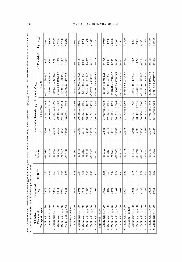
638 MICHA£ JAKUB NACHAJSKI et al.T
able
1. C
orre
latio
n fo
rmul
as d
escr
ibin
g re
latio
nshi
p: γ
25 roz
= f(
c, m
ol/d
m3 )
, con
stitu
ting
the
basi
s fo
r ca
lcul
atin
g ìR
osen
ís p
ostu
late
î ñ
log(
1/c π
=20)
, alo
ng w
ith th
e de
term
ined
num
ber
of n
TE
and
HL
B1H
NM
Rfo
r so
lu-
biliz
er a
nd it
s m
icel
lar
addu
cts
with
dic
lofe
nac,
nap
roxe
n, a
nd lo
rata
dine
.
Solu
bil
izer
Dete
rmin
ed
∆∆ G0 m
Corr
ela
tion
form
ula
: γγ2
5 roz. =
f(c
, m
ol/
dm
3)
cππ =
20
Ch
oli
c a
cid
n
TE
HL
B1H
NM
Rk
J/m
ol
r2a
± da
[γγ25 gra
m.]
b ±
db
◊ 1
04
mol/
dm
3lo
g[1
/cππ =
20]
Th
era
peu
tic a
gen
t
1. C
holic
aci
d n T
E=
2023
.79
12.8
5ñ1
9.76
470.
9708
69.7
142
± 3.
6484
ñ175
235.
2 ±
7949
1.3
1.01
253.
9945
2. C
holic
aci
dn T
E=
3029
.88
13.1
4ñ1
9.63
450.
9901
70.3
569
± 1.
2116
ñ179
686.
4 ±
4705
9.3
1.02
323.
9900
3. C
holic
aci
dn T
E=
4042
.66
15.0
1ñ1
9.88
230.
9918
71.2
818
± 0.
9700
ñ180
235.
1 ±
4248
8.9
1.07
143.
9700
4. C
holic
aci
dn T
E=
5050
.11
16.5
9ñ2
0.24
970.
9755
70.7
455
± 1.
8763
ñ242
231.
4 ±
1002
58.7
1.13
423.
9453
5. C
holic
aci
dn T
E=
6057
.28
17.7
9ñ2
0.54
860.
9761
69.5
929
± 2.
1544
ñ149
315.
4 ±
6109
6.9
1.18
023.
9280
6. C
holic
aci
dn T
E=
7073
.57
19.2
2ñ2
1.82
110.
9803
66.4
608
± 1.
0527
ñ110
624.
0 ±
4092
0.2
1.30
993.
8828
Dic
lofe
nac
ñ ad
duct
1. C
holic
aci
dn T
E=
2020
.37
15.5
9ñ1
9.49
250.
9921
69.8
262
± 1.
1407
ñ194
765.
3 ±
4520
2.7
0.91
684.
0377
2. C
holic
aci
dn T
E=
3028
.01
16.5
4ñ1
9.51
310.
9901
70.5
182
± 1.
4522
ñ227
375.
0 ±
5923
9.5
0.81
574.
0884
3. C
holic
aci
dn T
E=
4039
.11
17.3
8ñ2
0.28
990.
9919
69.8
224
± 1.
1555
ñ223
134.
0 ±
5242
5.8
0.80
014.
0968
4. C
holic
aci
dn T
E=
5051
.85
17.9
5ñ2
0.71
470.
9808
68.7
459
± 1.
4252
ñ244
641.
0 ±
8915
4.6
0.68
574.
1638
5. C
holic
aci
dn T
E=
6057
.22
18.1
2ñ2
1.09
690.
9523
69.1
569
± 2.
4991
ñ332
979.
1 ±
1959
52.1
0.51
614.
2872
6. C
holic
aci
dn T
E=
7067
.09
18.3
7ñ2
1.74
670.
9779
70.7
762
± 1.
9292
ñ395
400.
7 ±
1552
89.6
0.47
564.
3272
Nap
roxe
n ñ
addu
ct
1. C
holic
aci
dn T
E=
2019
.22
16.6
6ñ1
9.73
220.
9842
71.0
299
± 1.
7985
ñ238
866.
8 ±
7882
9.1
0.79
794.
0980
2. C
holic
aci
dn T
E=
3030
.09
16.7
4ñ1
9.78
980.
9919
69.8
225
± 1.
1555
ñ223
134.
0 ±
5242
5.8
0.80
014.
0968
3. C
holic
aci
dn T
E=
4040
.77
17.4
4ñ2
0.16
980.
9811
69.3
128
± 1.
4891
ñ221
7841
.4 ±
787
42.7
0.79
614.
0990
4. C
holic
aci
dn T
E=
5051
.01
17.6
4ñ2
0.78
380.
9565
70.3
319
± 2.
6044
ñ314
868.
7 ±
1764
13.8
0.58
314.
2342
5. C
holic
aci
dn T
E=
6056
.61
18.1
1ñ2
0.97
580.
9921
69.5
545
± 1.
1025
ñ347
707.
3 ±
8069
5.2
0.50
574.
2961
6. C
holic
aci
dn T
E=
7065
.29
18.3
3ñ2
1.57
480.
9374
67.2
716
± 2.
6267
ñ347
344.
5 ±
2370
46.9
0.44
054.
3560
Lor
atad
ine
ñ ad
duct
1. C
holic
aci
dn T
E=
2022
.21
15.8
5ñ1
9.61
270.
9803
66.4
607
± 1.
0528
ñ110
624.
0 ±
4092
0.2
1.30
993.
8227
2. C
holic
aci
dn T
E=
3031
.39
16.8
5ñ1
9.22
410.
9991
70.1
485
± 0.
3937
ñ176
915.
3 ±
1311
7.5
1.02
753.
9882
3. C
holic
aci
dn T
E=
4042
.98
17.5
8ñ2
0.69
480.
9792
70.7
474
± 2.
3500
ñ261
099.
0 ±
9925
1.3
0.71
914.
1431
4. C
holic
aci
dn T
E=
5051
.99
17.6
4ñ2
0.98
360.
9565
70.3
319
± 2.
6044
ñ314
868.
0 ±
8069
5.2
0.58
314.
2348
5. C
holic
aci
dn T
E=
6056
.28
18.0
9ñ2
0.51
570.
9547
64.9
535
± 1.
9643
ñ336
857.
9 ±
1927
23.6
0.38
544.
4140
6. C
holic
aci
dn T
E=
7069
.81
18.4
2ñ2
1.96
920.
9927
65.4
909
± 0.
7084
ñ943
116.
9 ±
2091
07.7
0.14
334.
3843
5
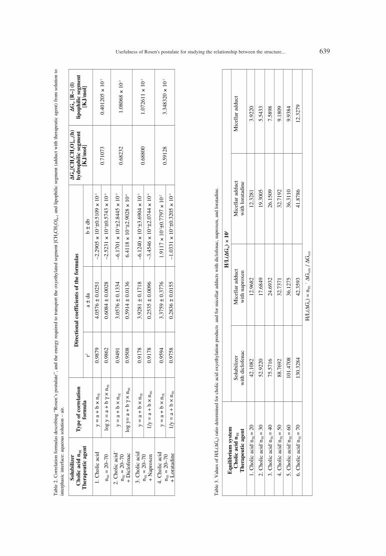
Usefulness of Rosen's postulate for studying the relationship between the structure... 639T
able
2. C
orre
latio
n fo
rmul
as d
escr
ibin
g ìR
osen
ís p
ostu
late
î, a
nd th
e en
ergy
req
uire
d to
tran
spor
t the
oxy
ethy
late
d se
gmen
t [C
H2C
H2O
] n=1
and
lipop
hilic
seg
men
t (ad
duct
with
ther
apeu
tic a
gent
) fr
om s
olut
ion
toin
terp
hasi
c in
terf
ace:
aqu
eous
sol
utio
n ñ
air.
Solu
bil
izer
∆∆ Gtr[C
H2C
H2O
] n=
1(h
)∆∆ G
tr[R
ñ]
(l)
Ch
oli
c a
cid
nT
ET
yp
e o
f corr
ela
tion
Dir
ecti
on
al
coeff
icie
nts
of
the f
orm
ula
sh
yd
rop
hil
ic s
egm
en
tli
pop
hil
ic s
egm
en
t
Th
era
peu
tic a
gen
t fo
rmu
la
r2a
± da
b
± db
[KJ/m
ol]
[K
J/m
ol]
1. C
holic
aci
dy
= a
+ b
◊n T
E
0.98
79
4.05
76 ±
0.0
251
ñ2.2
905
◊ 1
0-3±0
.510
9 ◊
10-3
0.71
073
0.40
1205
◊ 1
0-3
n TE
= 20
ñ70
log
y =
a +
b γ
◊ n T
E
0.98
62
0.60
84 ±
0.0
028
ñ2.5
231
◊ 1
0-4±0
.574
3 ◊
10-4
2. C
holic
aci
d∑y
= a
+ b
◊ n T
E
0.94
91
3.05
76 ±
0.1
334
ñ6.1
701
◊ 1
0-3±2
.844
5 ◊
10-3
0.68
232
1.08
068
◊ 1
0-3n T
E=
20ñ7
0 lo
g y=
a +
b γ
◊ n T
E
0.95
08
0.59
14 ±
0.0
136
6.41
18 ◊
10-4
±2.9
028
◊ 1
0-4+
Dic
lofe
nac
3. C
holic
aci
dy
= a
+ b
◊ n T
E
0.91
78
3.92
81 ±
0.1
718
ñ6.1
240
◊ 1
0-3±3
.690
4 ◊
10-3
0.68
800
1.07
2611
◊ 1
0-3n T
E=
20ñ7
0 1/
y =
a +
b ◊
n TE
0.
9178
0.
2535
± 0
.009
6 ñ3
.454
6 ◊
10-4
±2.0
744
◊ 1
0-4+
Nap
roxe
n
4. C
holic
aci
dy
= a
+ b
◊ n T
E
0.95
94
3.37
59 ±
0.3
776
1.91
17 ◊
10-2
±0.7
797
◊ 1
0-2
0.59
128
3.34
8320
◊ 1
0-3n T
E=
20ñ7
0 1/
y =
a +
b ◊
n TE
0.
9758
0.
2836
± 0
.015
5 ñ1
.033
1 ◊
10-3
±0.3
205
◊ 1
0-3+
Lor
atad
ine
Tab
le 3
.Val
ues
of H
/L(∆
Gtr)
ratio
det
erm
ined
for
cho
lic a
cid
oxye
thyl
atio
n pr
oduc
ts a
nd f
or m
icel
lar
addu
cts
with
dic
lofe
nac,
nap
roxe
n, a
nd lo
rata
dine
.
Eq
uil
ibri
um
syst
em
H/L
(∆∆G
tr)
◊ 1
03
Ch
oli
c a
cid
nT
ESo
lubi
lizer
M
icel
lar
addu
ct
Mic
ella
r ad
duct
Mic
ella
r ad
duct
Th
era
peu
tic a
gen
t w
ith d
iclo
fena
c
with
nap
roxe
n w
ith lo
rata
dine
1. C
holic
aci
d∑n T
E =
20
42.1
082
12.9
682
12.3
281
3.92
20
2. C
holic
aci
d∑n T
E =
30
52.9
220
17.6
849
19.3
005
5.54
33
3. C
holic
aci
d∑n T
E =
40
75.5
716
24.6
932
26.1
509
7.58
98
4. C
holic
aci
d∑n T
E =
50
88.7
692
32.7
371
32.7
192
9.18
09
5. C
holic
aci
d∑n T
E =
60
101.
4708
36.1
275
36.3
110
9.93
84
6. C
holic
aci
d∑n T
E =
70
130.
3284
42.3
593
41.8
786
12.3
279
H/L
(∆G
tr)
= n T
E∑
∆Gtr
(h)/ ∆
Gtr
(l)

640 MICHA£ JAKUB NACHAJSKI et al.
Consequently, the values of cmc determinedfor the aqueous solutions of the oxyethylation prod-ucts and micellar adducts with diclofenac, naproxen,and loratadine were used to calculate ∆G0
m (thermo-dynamic potential of micelle formation). Moreover,the total solids obtained after the evaporation ofwater (~ 37OC) from saturated micellar solutionsformed in the process of solubilization in equilibri-um constituted the basis for determining the contentof oxyethylated segments, nTE and HLB1HNMR, using1HNMR method. The results of these studies, alongwith the aggregates formed as a result of evaporat-ing water from saturated micellar solutions formedduring the micellar solubilization of diclofenac,naproxen, and loratadine in equilibrium, constitutedsignificant material deliverables of the verificationof the usefulness of ìRosenís postulateî for the ther-modynamic estimation of energy required to changethe state of matter at phase boundary: therapeuticagent (solid body) ñ micellar solution of surfactantwith cexp ≥ cmc. The aim of the research reportedhere was to confirm the usefulness of Rosenís pro-posal for estimating the solubilizing properties andthe thermodynamic stability of the micellar adduct.
EXPERIMENTAL
Materials
Product of the catalytic oxyethylation of cholicacid with declared molar content of ethylene oxide ñnTE = 20ñ70. Structural characteristics and aqueoussolutions of the novel class of surfactants weredescribed in our previous publication (10).Diclofenac: 2-{2-[(2,6-dichlorophenyl)amino]phen-yl}acetic acid, pure for analysis (Sigma, Germany).Naproxen: 2-(6-methoxy-2-naphthyl)propionic acid,pure for analysis (Zydus Cadila ñ Cadila HealthcareLtd., India). Loratadine: 4-(8-chloro-5,6-dihydro-11H-benzo[5,6]cyclohepta[1,2b]pyridin-11-yli-dene)-1-piperidine-1-carboxylic acid ethyl ester,pure for analysis (Zydus Cadila ñ Cadila HealthcareLtd., India). All the substances used in the course ofresearch have a low level of water solubility andbelong to BCS Class II.
Equipment
Stalagmometers with V = 28.20 cm3 and V =45.0 cm3 (Tropfen Wasser type) with MLW-U2Ctype thermostatic system of measurement area(Medingen Sitz Freital, Germany).
Research methodology
The surface activity of aqueous solutions of theproducts of cholic acid oxyethylation and their
Tab
le 4
.Cor
rela
tion
form
ulas
des
crib
ing
rela
tions
hip
betw
een
G0 m
and
HL
B1H
NM
Rfo
r so
lubi
lizer
and
its
mic
ella
r ad
duct
s w
ith th
erap
eutic
age
nts
as a
fun
ctio
n of
nT
E.
Solu
bil
izer
Typ
e o
f corr
ela
tion
Dir
ecti
on
al
coeff
icie
nts
of
the f
orm
ula
s
Ch
oli
c a
cid
∑nT
Efo
rmu
la f
or
fun
cti
on
r2a ±
da
a ±
da
Th
era
peu
tic a
gen
t
∆G0 m
= f(
n TE)
1. C
holic
aci
d∑n T
E =
20ñ
70y
= a
+ b
◊x
0.92
62
ñ18.
4190
± 1
.139
4 ñ4
.106
5 ◊
10-2
± 2.
3193
◊10
-2
2. C
holic
aci
d∑n T
E =
20ñ
70 +
Dic
lofe
nac
y =
a +
b ◊
x 0.
9768
ñ1
8.59
66 ±
0.5
766
ñ4.0
485
◊10
-2±
1.22
97 ◊
10-2
3. C
holic
aci
d∑n T
E =
20ñ
70 +
Nap
roxe
n y
= a
+ b
◊x
0.96
91
ñ18.
7013
± 0
.676
3 ñ4
.113
5 ◊
10-2
± 1.
4526
◊10
-2
4. C
holic
aci
d∑n T
E =
20ñ
70 +
Lor
atad
ine
y =
a +
b ◊
x 0.
9567
ñ1
7.61
87 ±
2.2
685
ñ5.6
061
◊10
-2±
4.68
46 ◊
10-2
HL
B1H
NM
R=
f(n T
E)
1. C
holic
aci
d∑n T
E =
20ñ
70
y =
a +
b ◊
x 0.
9896
9.
3678
± 1
.369
8 01
384
± 0.
0278
2. C
holic
aci
d∑n T
E =
20ñ
70 +
Dic
lofe
nac
y =
a +
b ◊
x0.
9644
14
.774
3 ±
1.03
420.
0581
± 0
.022
1
3. C
holic
aci
d∑n T
E =
20ñ
70 +
Nap
roxe
n y
= a
+ b
◊x
0.97
88
15.7
706
± 0.
5291
0.03
91 ±
0.0
113
4. C
holic
aci
d∑n T
E =
20ñ
70 +
Lor
atad
ine
y =
a +
b ◊
x 0.
9609
15
.044
1 ±
0.99
88
0.05
15 ±
0.0
206
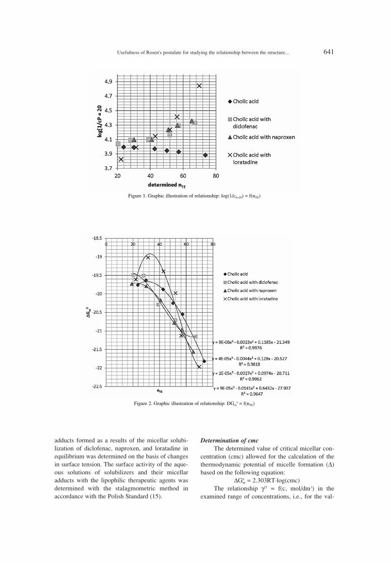
Usefulness of Rosen's postulate for studying the relationship between the structure... 641
adducts formed as a results of the micellar solubi-lization of diclofenac, naproxen, and loratadine inequilibrium was determined on the basis of changesin surface tension. The surface activity of the aque-ous solutions of solubilizers and their micellaradducts with the lipophilic therapeutic agents wasdetermined with the stalagmometric method inaccordance with the Polish Standard (15).
Determination of cmcThe determined value of critical micellar con-
centration (cmc) allowed for the calculation of thethermodynamic potential of micelle formation (∆)based on the following equation:
∆G0m = 2.303RT∑log(cmc)
The relationship γ25 = f(c, mol/dm3) in theexamined range of concentrations, i.e., for the val-
Figure 1. Graphic illustration of relationship: log(1/cπ=20) = f(nTE)
Figure 2. Graphic illustration of relationship: DGm0 = f(nTE)
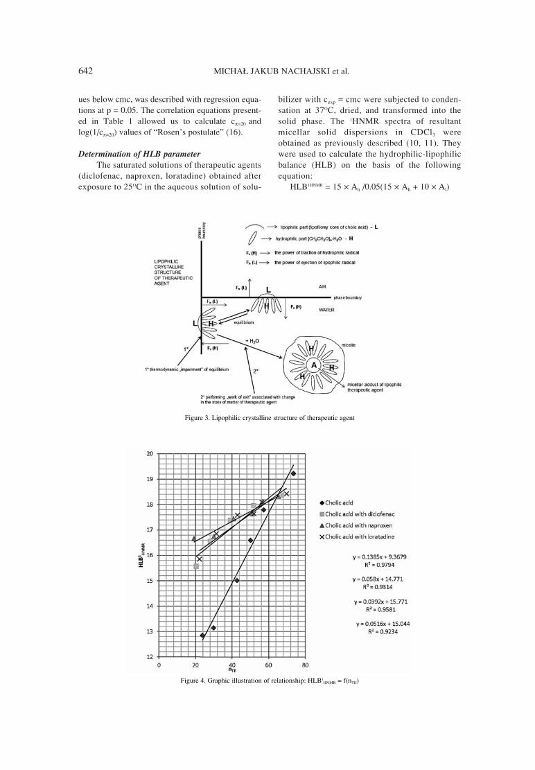
642 MICHA£ JAKUB NACHAJSKI et al.
ues below cmc, was described with regression equa-tions at p = 0.05. The correlation equations present-ed in Table 1 allowed us to calculate cπ=20 andlog(1/cπ=20) values of ìRosenís postulateî (16).
Determination of HLB parameterThe saturated solutions of therapeutic agents
(diclofenac, naproxen, loratadine) obtained afterexposure to 25OC in the aqueous solution of solu-
bilizer with cexp = cmc were subjected to conden-sation at 37OC, dried, and transformed into thesolid phase. The 1HNMR spectra of resultantmicellar solid dispersions in CDCl3 wereobtained as previously described (10, 11). Theywere used to calculate the hydrophilic-lipophilicbalance (HLB) on the basis of the followingequation:
HLB1HNMR = 15 ◊ Ah /0.05(15 ◊ Ah + 10 ◊ Al)
Figure 3. Lipophilic crystalline structure of therapeutic agent
Figure 4. Graphic illustration of relationship: HLB1HNMR = f(nTE)

Usefulness of Rosen's postulate for studying the relationship between the structure... 643
Determination of the overall number oflipophilic protons ΣH1 = 36 in the structure of themolecule of cholic acid made it possible to calculatethe content of oxyethylated segments (CH2CH2O) indry micellar adduct after solubilization in equilibri-um on the basis of the following equation:
nTE = (36 ◊ Ah /Al ñ 3) / 4The determined values of nTE and HLB1HNMR are
presented in Table 1.
RESULTS AND DISCUSSION
The values of 1/cπ=20, HLB1HNMR, and nTE pre-sented in Table 1 point to significant variability inthe surface activity of the micellar adduct of solubi-lizer with diclofenac, naproxen, and loratadine, aswell as to the variability of ∆G0
m, the thermodynam-ic potential of micelle formation, in relation to theexposure solution of derivative of the oxyethylationof cholic acid with nTE = 20ñ70 (Table 1). The nTE
and HLB1HNMR of the solubilizer and its micellaradduct also suggest individual variability in thevalue of ìRosenís postulateî, log1/cπ=20).
We analyzed the relationship betweenlog1/cπ=20) and the determined number of oxyethy-lated segments, nTE (Table 1, Fig. 1), to estimate thecharacter of thermodynamic interactions occurringat phase boundary. Correlation equations describingthe abovementioned relationship at p = 0.05 are pre-sented in Table 2. The directional coefficients of thecorrelation equations (Table 2) allowed us to calcu-late the value of transport energy to the phase
boundary of the lipophilic part of solubilizer, [F↑](∆Gtr(l)), as well as the value of the transport energyof hydrophilic oxyethylated segment, [CH2CH2O]n =
1 [F↓] (∆Gtr(h)), using the following formulas:∆Gtr(l) = a/2.303 ◊ R ◊ T∆Gtr(h) = b/2.303 ◊ R ◊ T
Presented in Table 2 values of the energy oftransport ∆Gtr(l) and ∆Gtr(h), in all conditionsdescribed by Loughlin allowed us to calculate theratio:
H/L(∆Gtr) = nTE ∑ ∆Gtr(h) / ∆Gtr(1)
which can constitute our thermodynamic interpreta-tion of hydrophilic-lipophilic balance at phaseboundary. The results are presented in Table 3.
The calculated values of H/L(∆Gtr) ratio (Table3) suggest that individual progression of this param-eter is determined by the method and place of thesolubilization of therapeutic agent and significantthermodynamic stability (∆G0
m) of micellar adduct.The values of H/L(∆Gtr) presented in Table 3inspired us to analyze the relationship ∆G0
m = f(nTE)and to estimate ìRebinderís effectî, i.e., the value ofenergy required to transform the molecule of thera-peutic agent from solid body to micelle (molecularstate of dispersion). The correlation equations for∆G0
m = f(nTE) formula, obtained at p = 0.05, are pre-sented in Table 4. The directional coefficient ìaî ofthe correlation equation y = a + b∑x by nTE = 0 canrefer to ∆G0
m(gr) of the lipophilic core of cholic acidand its micellar adducts with lipophilic therapeuticagents, i.e., diclofenac (∆G0
m(gr)D), naproxen (∆G0m(gr)N),
and loratadine (∆G0m (gr)L).
Figure 5. Microscopic structure of the solutions of solubilizers with nTE = 70 and the solutions of adducts after the loss of volatile compo-nents (evaporation of solvent): A ñ solution of surfactant, B ñ adduct with diclofenac, C ñ adduct with loratadine
Figure 6. Microscopic structure of the solutions of solubilizers with nTE = 50 and the solutions of adducts after the loss of volatile compo-nents (evaporation of solvent): A ñ solution of surfactant, B ñ adduct with diclofenac, C ñ adduct with loratadine
644 MICHA£ JAKUB NACHAJSKI et al.
In such a state, due to the proposed experimen-tal model, one can estimate the level of energy (orwork) of ìRebinderís effectî required to transportthe molecules of therapeutic agent (following com-plete moistening) from the surface of solid body tomicellar structure, and to form the thermodynami-cally stable adduct. The values of ∆G0
m(gr) coeffi-cients presented in Table 4 allowed us to estimatethe energy of transport required to change the stateof matter on the basis of the following formula:∆(ìRebinderís effectî eR) = ∆G0
m (gr) D,N,L ñ ∆G0m(gr)
of cholic acidThe values of eR calculated on the basis of the
abovementioned formula equaled to:for diclofenac: ∆îeRî = ñ0.1776 kJ/molfor naproxen: ∆îeRî = ñ0.2823 kJ/molfor loratadine: ∆îeRî = ñ0.8003 kJ/mol
The thermodynamic stability of solubilizerísmicelle and the adduct with lipophilic therapeuticagents at phase boundary (air/water) was confirmedby the values of H/L(∆Gtr) ratio (Table 3) and therelationship HLB1HNMR = f(nTE) presented in Figure 4.
On the basis of the abovementioned relation-ships, we observed that the solubilization oflipophilic therapeutic agent is associated with anincrease in the hydrophilicity of the adduct(obtained in solid phase). This was suggested by thevalues of the directional coefficients of correlationequations presented in Table 4, which varied signif-icantly depending on solubilizers, i.e., the oxyethy-lation products of cholic acid with nTE = 20ñ70. Werevealed that the products (solid dispersions)obtained upon the micellar solubilization ofdiclofenac, naproxen, and loratadine in equilibrium,and subsequent dehydration (drying at 37OC) haveHLB1HNMR, which suggests their significanthydrophilicity, irrespective of being the adducts ofthe lipophilic therapeutic agents.
Analyzing the microscopic structure of com-pounds obtained upon the loss of the volatile com-ponents of the solutions of solubilizer with cexp ≥cmc and the saturated solutions of adducts, wefound the important material explanation for theabovementioned statement (Figs. 5, 6) as the thera-peutic agents were located in the core, that is to say,in the central part of solubilization space (micelle).
CONCLUSIONS
1. Analyzing an array of homologous structures ofcholic acid oxyethylation products with nTE =20ñ70, as well as their micellar adducts withlipophilic therapeutic agents: diclofenac, naprox-en, and loratadine, we confirmed the relationship
between ìRosenís postulateî [log(1/cπ=20)] andthe content of oxyethylated segments (nTE). Thecalculated values of transport energy, Gtr(h) andGtr(1), constituted the base for determiningH/L(Gtr) ratio, being the thermodynamic charac-teristic of hydrophilic-lipophilic balance at thephase boundary.
2. The values of ∆G0m(gr) for solubilizer and its
micellar adducts with diclofenac, naproxen, andloratadine, which were calculated with approxi-mation formulas, constituted the basis for esti-mating ìRebinderís effectî associated with theenergy required to transform the molecule oftherapeutic agent into the state of molecular dis-persion. The value of ∆G0
m(gr) points to thermody-namic stability and to the possibility of estimat-ing the energy of transport associated withchanging the state of matter.
3. The structural character of interaction occurringat phase boundary between the micellar solutionof solubilizer and solid body (dispersed thera-peutic agent) was confirmed by the values oflog(1/cπ=20), ∆G0
m(gr), HLB1HNMR, and H/L(Gtr), aswell as by the documentation obtained afterevaporating water from saturated solutionformed as a result of the solubilization oflipophilic therapeutic agent in equilibrium.
4. The results of this study confirm the possibilityof comprehensive utilization of ìRosenís postu-lateî for the evaluation of solubilization proper-ties of cholic acid oxyethylation products.Moreover, they allow for the thermodynamicestimation of ÑRebinderís effectî associated withthe energy of transport required for changing thestate of matter of lipophilic therapeutic agent.The results of the research facilitate the applica-tion of Rosenís proposal for the assessment ofthe usefulness of solubilizers and the stability ofthe systems obtained.
REFERENCES
1. Rosen M.J.: J. Am. Oil Chem. Soc. 51, 461(1974).
2. Zgoda M.M.: Acta Pol. Pharm. Drug Res. 47,78 (1990).
3. Woskowicz M., Zgoda M.M.: Farm. Pol. 59,363 (2003).
4. Woskowicz M., Zgoda M.M.: Pol. J. Cosmetol.2, 132 (2003).
5. Ko≥odziejczyk M., RedliÒski A., Zgoda M.M.:Polimery 43, 307 (1998).
6. Zgoda M.M., Ko≥odziejczyk M., RedliÒski A.:Polimery 43, 612 (1998).

Usefulness of Rosen's postulate for studying the relationship between the structure... 645
7. Zgoda M.M., Ko≥odziejczyk M., RedliÒski A.:Pol. J. Cosmetol. 4, 280 (1999).
8. Laughlin G.R.: J. Soc. Cosmet. Chem. 32, 371(1981).
9. PodgÛrski R.K., PluciÒski J.: Wiad. Chem. 43,191 (1989).
10. Zgoda M.M., Nachajski M.J., Ko≥odziejczykM.K., Woskowicz M.H., Lukosek M.: Polim.Med. 37, 21 (2007).
11. Zgoda M.M., Nachajski M.J., Ko≥odziejczykM.K., Woskowicz M.H., Lukosek M.: Polim.Med. 37, 37 (2007).
12. Deng Z., Xu S., Li S.: Int. J. Pharm. 351, 236(2008).
13. Makarewicz E., Zalewska A.: Prog. Org. Coat.54, 43 (2005).
14. Bhatnagar A., Khandewal M., Roo K.V.N.:Mining Sci. Technol. 20, 400 (2010).
15. PN-90/C-04809 ìSurface active agents.Determination of surface tension (Polish), Dz.Norm i Miar nr 2/1991, poz. 4.
16. Ko≥odziejska J.: Mag. Stom. 3, 56 (2000).
Received: 24. 04. 2013

Acta Poloniae Pharmaceutica ñ Drug Research, Vol. 71 No. 4 pp. 647ñ659, 2014 ISSN 0001-6837Polish Pharmaceutical Society
Tenoxicam (TNX) is a member of the non-steroidal anti-inflammatory drugs. It inhibits thebiosynthesis of prostaglandins by inhibiting thecyclooxygenase pathway. It is also considered as aneffective anti-inflammatory agent and has been usedin the management of rheumatic and inflammatorydiseases, including osteoarthritis (1). However,TNX is very slightly water soluble drug and, as withall poorly soluble drugs, its dissolution may be therate determining step in the absorption process.
The enhancement of the dissolution rate andsolubility of poorly soluble drugs is connected withthe application of auxiliary substances or with newtechnological possibilities (2). Solid dispersions ofdrugs in water-soluble carriers have attracted con-siderable interest as a means of improving the disso-lution rate, and hence possibly bioavailability, of arange of hydrophobic drugs (3). In such systems,drug undergoes particle size reduction and the con-sequent increase in the surface area results in theimproved dissolution (4), break up the crystal lattice(5) or increasing drug wettability by surroundinghydrophilic carriers (4).
Among the techniques to prepare a solid dis-persion, spray-drying has the ability to produce
spherical and size controlled particles and simulta-neously to improve the dissolution properties (6).
The dissolution rates of several drugs havebeen improved by spray-drying with hydrophilicpolymers, including indomethacin (7), tolbutamide(8), carbamazepine (9), and ketoprofen (10).
Kollicoat IR is a poly(vinyl alcohol)-poly(eth-ylene glycol) graft copolymer (PVA-PEG) (11). El-Badry et al. (12) showed that Kollicoat IR-omepra-zole microparticles prepared using spray- andfreeze-drying techniques revealed the transforma-tion of omeprazole from crystalline to amorphousstate with increasing its dissolution rate nine timesin comparison to the rate of the physical mixture.Also, Janssens et al. (13) showed that itraconazolewas dispersed on a molecular level in the KollicoatIR microparticles prepared by spray-drying, andshowed enhanced dissolution.
Due to the high melting point of Kollicoat IR(210OC) and its poor solubility in the organic sol-vents, it will be difficult to prepare solid drug-Kollicoat dispersions in its matrix either by meltingor solvent evaporation methods.
The present study aims at the preparation ofTNX-KL binary systems either by spray-drying or
TENOXICAM-KOLLICOAT IRÆ BINARY SYSTEMS: PHYSICOCHEMICALAND BIOLOGICAL EVALUATION
MOHAMED ABBAS IBRAHIM*
Department of Pharmaceutics and Industrial Pharmacy, Faculty of Pharmacy, Al-Azhar University, Assiut, Egypt
Abstract: Tenoxicam (TNX) binary systems in Kollicoat IR (KL) matrix were prepared in different drug : poly-mer ratios using kneading and spray-drying method. The prepared binary systems were characterized for drugdissolution rate, differential scanning calorimetry (DSC), IR spectroscopy and x-ray diffractometry. The resultsshowed that the drug dissolution rate was remarkably enhanced by incorporating it in the KL matrix either bykneading or spray-drying, and the dissolution rate was increased by decreasing the drug weight ratio. The DScand x-ray studies revealed the presence of TNX in less crystalline or amorphous state in its-KL binary systems.Moreover, the spray-dried TNX-KL system in 1 : 4 ratio, that exhibited the faster dissolution rate, was formu-lated in oral disintegrating tablets (ODTs). The data indicated that a fast disintegration and higher drug disso-lution rate was achieved in case of the ODTs containing the spray-dried form compared to the ODTS contain-ing untreated drug or the commercial tablet (EpicotilÆ). Also, the drug exhibited significantly (p < 0.01) fasteronset of the anti-inflammatory analgesic activities in case of the ODTs containing the spray-dried form, thatwas superior to that observed with both the commercial tablet product and the ODTS containing untreated drug.
Keywords: tenoxicam, Kollicoat IR, spray-drying, kneading, oral disintegrating tablets
647
* Corresponding author: Current address: Kayyali Chair for Pharmaceutical Industries, Department of Pharmaceutics, College ofPharmacy, King Saud University, Riyadh, KSA; e-mail: [email protected]; phone: +(966)-1-4676228, fax: +(966)-1-4676295
648 MOHAMED ABBAS IBRAHIM
kneading and the physicochemical characterizationof such systems. In addition, oral disintegratingtablets of an improved TNX-KL binary system willbe prepared and characterized in comparison withthe untreated drug and the commercial tablet prod-uct.
EXPERIMENTAL
Materials
Tenoxicam was kindly supplied by EgyptianInternational Pharmaceutical Industries Co., EIPICo(Cairo, Egypt). Kollicoat IR (KL) was obtainedfrom BASF (Ludwigshafen, Germany). Micro-crystalline cellulose (Avicel PH101) was purchasedfrom Serva Feinbiochemica (Heidelberg, Germany).Spray-dried mannitol (MannogemTM EZ), used as afiller for the orally disintegrating tablets, was kindlysupplied by SPI (Grand Haven, USA). Cros-povidone (CPV) was kindely supplied by RiyadhPharma (Riyadh, KSA). Magnesium stearate waspurchased from Riedel-de HaÎn (Seelze, Germany).EpicotilÆ tablets (immediate release oblong scoredfilm coated tablets, weighing 200 mg and containing20 mg TNX), batch number 1108632, was producedby EIPICo (Cairo, Egypt). Carrageenin was pur-chased from Sigma Chemical Co. (USA). Othermaterials and solvents are of reagent or analyticalgrade, and they were used without further purifica-tion.
Methodology
Preparation of tenoxicam-Kollicoat IR binary sys-tems
Since Kollicoat is insoluble in most organicsolvents, it was not possible to prepared TNX soliddispersions with it using coevaporation method.Therefore, TNX-KL binary systems in differentdrug : polymer ratios (1 : 1, 1 : 2 and 1 : 4) were pre-pared either by spray-drying from aqueous solutionor kneading methods.
Kneading methodTNX-KL physical mixture was kneaded with
appropriate amounts of water (0.1 mL of distilledwater for each gram of physical mixture) using amortar and pestle for 10 min. The mass was dried(room temperature overnight), crushed, sieved anddried again in an oven (Heraeus, Germany) (40OCfor 24 h).
Spray-dryingKollicoat solution was prepared by dissolving
the polymer in distilled water in different concentra-
tions (0.5, 1 and 2%) according to the selected TNX: Kollicoat ratios. To the polymeric solution, theweighed amount of TNX was added and dispersed.Thereafter, 1ñ2 mL of 30% ammonium hydroxidesolution was added to raise the solution pH to about8ñ9. The clear polymeric solution of TNX was thenspray-dried using B¸chi 190 mini spray drier (B¸chiLabortechnik AG, Germany) with 0.5 mm nozzle.The drug-polymer solution was fed to the nozzlethrough a peristaltic pump in a spray flow rate of 5mL/min. The solution was sprayed under the effectof compressed air force (air flow rate of 4 pound persquare inch) with an aspiration rate of 100%. Thesprayed droplets were dried to remove the solvent indrying chamber by the blown hot air (inlet air tem-perature of 150OC and outlet air temperature of80OC). Finally, the resulting dried product was col-lected from vessel, weighed and stored in tightlyclosed amber glass containers pending further inves-tigations.
Physical mixtureTNX-KL physical mixtures in different drug
weight ratios were prepared by gentle mixing of theweighed amounts the drug and carrier in porcelainmortar.
Characterization of tenoxicam-Kollicoat IR binarysystemsIn vitro dissolution studies
The in vitro dissolution experiments from itsKL binary systems and ODTs were performed usingUSP dissolution apparatus 2, paddle method,(Caleva Ltd., Model 85T), at 100 rpm using a con-tinuous automated monitoring system. This systemconsists of an IBM computer PK8620 series and PU8605/60 dissolution test software, PhilipsVIS/UV/NIR single beam eight cell spectropho-tometer Model PU 8620, Epson FX 850 printer, andWatson-Marlow peristaltic pump using in each flaska 900 mL phosphate buffer, pH 6.8. The temperaturewas maintained at 37 ± 0.5OC. Twenty milligramsTNX or equivalent amount was spread over the dis-solution medium. At predetermined time intervals,absorbance was recorded automatically at 362 nmand the percentage of TNX dissolved was deter-mined as a function of time in triplicates.
Dissolution efficiency (DE%) was calculatedfrom the area under the dissolution curve at time t(measured using the trapezoidal rule) and expressedas percentage of the area of the rectangle describedby 100% dissolution in the same time (14). Also, therelative dissolution rate (RDR15) data of the differentsamples were calculated by determining the amount

Tenoxicam-KollicoatÆ IR binary systems: physicochemical and ... 649
of TNX dissolved from a particular sample and nor-malizing for the amount of drug dissolved from puredrug sample over the same time interval (15 min).
In case of oral disintegrating tablets, the sameprevious conditions were adopted, but the dissolu-tion experiments were carried out on six tablets.
Solubility studiesThe aqueous solubility of TNX in its KL bina-
ry systems was investigated as follows: an excessamount of TNX or TNX-KL binary system wasadded to 10 mL of distilled water in a 50 mL glassstoppered bottle. The bottles were firmly closed andplaced into the mechanical shaking water bath pre-viously adjusted at 37 ± 0.1OC. After equilibrationhas been attained (24 h), one mL aliquot sample waswithdrawn from each tested solution and diluted toan appropriate volume with distilled water. Theabsorbance was measured at 362 nm against a suit-able blank similarly treated and the drug concentra-tion was calculated
Scanning electron microscopy (SEM)Morphological characteristics of certain TNX-
KL spray-dried and kneaded systems compared tothe individual components were observed by scan-ning electron microscopy (SEM). The samples weresputter-coated with a thin gold palladium layerunder an argon atmosphere using a gold sputtermodule in a high-vacuum evaporator. Coated sam-ples were then scanned and photomicrographs weretaken with an SEM (Jeol JSM-1600, Japan).
Differential scanning calorimetry (DSC) DSC scans were recorded for TNX-KL binary
systems compared to that of the individual com-ponenets in order to determine the extent of crys-tallinity of the drug in the presence of the studiedpolymers.
The samples (3ñ5 mg) were hermeticallysealed in aluminum pans and heated at a constantrate of 10OC/min, over a temperature range of 25
to 250OC. Thermograms of the samples wereobtained using differential scanning calorimetry(DSC-60, Shimadzu, Japan). Thermal analysisdata were recorded using a TA 50I PC system withShimadzu software programs. Indium standardwas used to calibrate the DSC temperature andenthalpy scale. N2 was used as purging gas at rateof 30 mL/min.
X-ray diffraction analysisThe x-ray diffraction patterns of the powder
samples were obtained using RIGAKU diffractome-ter (Japan), which was equipped with curvedgraphite crystal monochromator, automatic diver-gence slit and automatic controller PW/1710. Thetarget used was CuKa radiation operating at 40 kVand 40 mA (λka = 1.5418 �). The diffraction patternswere achieved using continuous scan mode with 2θO
ranging from 4 to 60O.
Fourier transform infrared spectroscopy (FTIR)The FTIR spectra of TNX, KL and their bina-
ry systems were recorded using FTIR Perkin Elmerspectrophotometer (Spectrum BX). Samples weremixed with potassium bromide (spectroscopicgrade) and compressed into disks using hydraulicpress before scanning from 4000 to 600 cm-1. Thedata were analyzed using Perkin Elmer software(Spectrum V5.3.1).
Preparation of TNX oral disintegrating tablets(ODTs) by direct compression method
TNX ODTs were prepared by direct compres-sion method and the composition of the preparedODTS containing TNX is listed in Table 1.
The corresponding amounts of TNX, avicel pH101 and superdisintegrant (CPV) were accuratelyweighed and mixed using Turbula mixer (Erweka,S2Y, Heusenstamm, Germany) for 5 min.Thereafter, the corresponding amount of mannitolwas accurately weighed, added to the mixture andmixed for further 5 min. Finally, the amount of mag-nesium stearate was mixed with the powder in theTurbula mixer for further 2 min. The powder wascompressed into tablets weighing 200 mg usingKorsh single punch machine with 9 mm flat punch-es (Erweka, EKO, Germany).
Evaluation of ODTs containing TNXWeight variation
Twenty tablets were weighed individually(analytical balance, Shimadzu, EB-3200D, Kyoto,Japan) and the average tablet weight and standarddeviation were calculated.
Table 1. Composition of TNX ODTs formulation.
Ingredient Weight (mg)
TNX (or spray-dried TNX-KL equivalent to 20 mg TNX)
20
Crosspovidone (CPV) 10
Spray-dried mannitol 50
Magnesium stearate 2
Avicel PH101 To 200 mg

650 MOHAMED ABBAS IBRAHIM
Thickness
Pre-weighed 10 tablets were tested for thick-ness using a micrometer (Mitutoyo M 110-25,Japan), the average thickness and standard deviationwere calculated.
Hardness
Tablet hardness of 10 tablets of known weightand thickness was measured using a hardness tester(Pharma test GmbH, Hainburg, Germany). Theaverage hardness and standard deviation werereported.
Friability
Tablet friability was determined according toUSP30-NF25. In brief, 20 tablets were weighed(W1) and placed into the friabilator (Erweka, TA3R,Heusenstamm, Germany), which was rotated at 25rpm for 4 min. The tablets were then reweighed afterremoval of fines (W2), and the loss % was calculat-ed by:
100 ◊ (W1 - W2)/W1
In vitro disintegration
In vitro disintegration test was assessed accord-ing to the USP30-NF25 requirements. One dosageunit was put in each of the six tubes of the basket(Electrolab, ED-21, Mumbai, India). The apparatuswas operated, using phosphate buffer, pH 6.8, as theimmersion fluid, which was maintained at 37 ±0.5OC. Time for complete disintegration of eachtable, standard deviation and relative standard devi-ation were calculated.
In vivo studies Anti-inflammatory activity
The anti-inflammatory activity of TNX ODTscompared to the commercial product (EpicotilÆ) wasevaluated using carrageenin-induced paw edemamodel (15). All studies were in accordance with theGuidelines of Animal Ethical Committee of KingSaud University and had its approval.
The experiment was conducted on 25 albinorats of both sex weighing 90ñ120 g fasted for 18 hwith water available ad libitum. They were equallyand randomly allocated in 4 groups (6 rats pergroup). The first group was the rats received ODTscontaining untreated TNX; the second groupreceived ODTs containing spray dried TNX-KL sys-tem (1 : 4); the third group received the commercialtablet product (EpicotilÆ) and the fourth group wasconsidered as control.
The rats were anesthetized with urethane (0.5mL, intraperitoneal) and 100 µL of 1% w/v car-rageenin physiologic solution was injected subcuta-neously into the treated area. One hour later, a defi-nite weight of the tested tablet containing TNX dose(20 mg/kg) was dispersed in 5 mL of distilled waterand immediately given to the rat by an esophagealtube.
Edema volume was measured using a plathys-mometer, at suitable time intervals (0.5, 1, 2, 3, 4and 5 h).
The anti-inflammatory activity (% response)was calculated according to the following equation(16):
Figure 1. Dissolution profiles of TNX-KL spray-dried (spr dr) systems compared to the corresponding physical mixtures (PM)

Tenoxicam-KollicoatÆ IR binary systems: physicochemical and ... 651
C ñ T% Response = óóóó ◊ 100C
where C = the volume of right paw minus volume ofleft paw for control rat and T = volume of right pawminus volume of left paw for treated rat.
Hot-plate analgesic testThe analgesic test of the drug ODTs was evaluat-
ed using hot-plate method in mice. Mice were dividedinto 4 groups (6 mice/group). The first group was themice received ODTs containing untreated tenoxicam;the second group received ODTs containing spraydried TNX-KL system (1 : 4); the third group receivedthe commercial tablet product (EpicotilÆ) and thefourth group was considered as control. The tempera-ture of the hot-plate metal surface was kept constant at54 ± 1.0OC. A specific weight of each tablet formula-tion containing the required drug dose for mice (20mg/kg) was dispersed in 2 mL of distilled water andadministered immediately via an esophageal tube. The
time taken by the animals to lick the fore or hind pawor jump out of the place was taken as the reaction time.Latency to the licking paws or jumping from plate wasrecorded by a stop watch before and after treatment. Alatency period of 30 s was defined as complete analge-sia cut off time to prevent damage to mice (17).
Statistical analysis
The data from each treatment group were ana-lyzed using an analysis of variance test to determinethe p-value for different variables. The Fisherís leastsignificant difference test was used to determine sig-nificant differences between two variables.
RESULTS AND DISCUSSION
Characterization of TNX-KL binary systems
In vitro dissolutionFigure 1 shows the dissolution profiles of
TNX-KL spray-dried systems in different drug :
Table 2. Dissolution efficiency percentages after 15 min (DE%15), relative dissolution rate after 15 min (RDR15)and aqueous solubility of TNX from its-KL binary systems.
System DE%15 RDR15
Solubility*(mg/mL)
TNX 31.97 - 0.087
TNX-KL spray dried 66.82 2.06 0.733
(1 : 1)
TNX-KL spray dried 73.65 2.18 0.754
(1 : 2)
TNX-KL spray dried 79.67 2.30 1.109
(1 : 4)
TNX-KL Kneaded mixture 52.51 1.94 0.717
(1 : 1)
TNX-KL Kneaded mixture 63.54 1.96 0.88
(1 : 2)
TNX-KL Kneaded mixture 66.54 2.20 0.950
(1 : 4)
TNX-KL Physical mixture 35.84 1.25 0.102
(1 : 1)
TNX-KL Physical mixture 38.60 1.30 0.109
(1 : 2)
TNX-KL Physical mixture 40.27 1.41 0.145
(1 : 4)
*Determined at 37OC
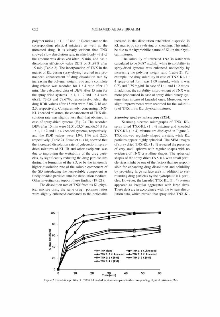
652 MOHAMED ABBAS IBRAHIM
polymer ratios (1 : 1, 1 : 2 and 1 : 4) compared to thecorresponding physical mixtures as well as theuntreated drug. It is clearly evident that TNXshowed slow dissolution rate, in which only 47% ofthe amount was dissolved after 15 min, and has adissolution efficiency value DE% of 31.97% after15 min (Table 2). The incorporation of TNX in thematrix of KL during spray-drying resulted in a pro-nounced enhancement of drug dissolution rate byincreasing the polymer weight ratio and a completedrug release was recorded for 1 : 4 ratio after 10min. The calculated data of DE% after 15 min forthe spray-dried systems 1 : 1, 1 : 2 and 1 : 4 were66.82, 73.65 and 79.67%, respectively. Also, thedrug RDR values after 15 min were 2.06, 2.18 and2.3, respectively. Comparatively, concerning TNX-KL kneaded mixtures, the enhancement of TNX dis-solution rate was slightly less than that obtained incase of spray-dried systems (Fig. 2). The recordedDE% after 15 min were 52.51, 63.54 and 66.54% for1 : 1, 1 : 2 and 1 : 4 kneaded systems, respectively,and the RDR values were 1.94, 1.96 and 2.20,respectively (Table 2). Fouad et al. (18) showed thatthe increased dissolution rate of celecoxib in spray-dried mixtures of KL IR and other excipients wasdue to improving the wettability of the drug parti-cles, by significantly reducing the drug particle sizeduring the formation of the SD, or by the inherentlyhigher dissolution rate of the soluble component ofthe SD introducing the less-soluble component asfinely divided particles into the dissolution medium.Other investigators support these finding (19ñ21).
The dissolution rate of TNX from its KL phys-ical mixture using the same drug : polymer ratioswere slightly enhanced compared to the noticeable
increase in the dissolution rate when dispersed inKL matrix by spray-drying or kneading. This mightbe due to the hydrophilic nature of KL in the physi-cal mixtures.
The solubility of untreated TNX in water wascalculated to be 0.087 mg/mL, while its solubility inspray-dried systems was enhanced noticeably byincreasing the polymer weight ratio (Table 2). Forexample, the drug solubility in case of TNX-KL 1 :4 spray-dried form was 1.09 mg/mL, while it was0.73 and 0.75 mg/mL in case of 1 : 1 and 1 : 2 ratios.In addition, the solubility improvement of TNX wasmore pronounced in case of spray-dried binary sys-tems than in case of kneaded ones. Moreover, veryslight improvements were recorded for the solubili-ty of TNX in its KL physical mixtures.
Scanning electron microscopy (SEM)Scanning electron micrographs of TNX, KL,
spray dried TNX-KL (1 : 4) mixture and kneadedTNX-KL (1 : 4) mixture are displayed in Figure 3.TNX showed regularly shaped crystals, while KLparticles appear highly spherical. The SEM imagesof spray dried TNX-KL (1 : 4) revealed the presenceof very small spheres with regular shapes with noevidence of TNX crystalline shapes. The sphericalshapes of the spray-dried TNX-KL with small parti-cle sizes might be one of the factors that are respon-sible for enhancing drug dissolution and solubilityby providing large surface area in addition to sur-rounding drug particles by the hydrophilic KL parti-cles. However, the kneaded TNX-KL (1 : 4) systemappeared as irregular aggregates with large sizes.These data are in accordance with the in vitro disso-lution data, which proved that spray-dried TNX-KL
Figure 2. Dissolution profiles of TNX-KL kneaded mixtures compared to the corresponding physical mixtures (PM)

Tenoxicam-KollicoatÆ IR binary systems: physicochemical and ... 653
improved both drug dissolution rate and aqueoussolubility in comparison to the correspondingkneaded form.
The pronounced change of the particles shapein spray-dried mixture may indicate the presence ofa new solid phase (22, 23).
Differential scanning calorimetry Figures 4A and B show the DSC scans of
TNX-KL kneaded systems and TNX-KL spray-dried systems in different drug weight ratios com-pared to the individual components. The DSCcurves of TNX show an endothermic peak at215.9OC with a thaw point at 214OC and heat offusion, DH, of ñ47.88 joule/g at a scanning rate of
10OC/min. This endothermic peak ends with anexothermic peak at 220OC, which may be due to thedecomposition of the drug when reaching its meltingpoint as reported (24). Kollicoat IR exhibits a broadendotherm at 213.14OC with a thaw point of207.5OC.
The DSC scans of TNX-KL spray-dried sys-tems (in drug : polymer ratios of 1 : 1, 1 : 2 and 1 :4), compared to the drug and polymer scans, are dis-played in Figure 4A. The drug endothermic peakcompletely disappeared in all tested TNX-KL spray-dried mixtures, and only the polymer peak wasobserved at 200OC. This might be due to the solubil-ity in the melted polymer (25). In addition, the drugexothermic peak was shifted to lower temperatures
Figure 3. Scanning electron micrographs of TNX-KL spray dried and kneaded systems in drug : polymer ratio 1 : 4 compared to the indi-vidual components

654 MOHAMED ABBAS IBRAHIM
Figure 4A. DSC thermograms of TNX-KL spray-dried systems in different drug : polymer ratios compared to the individual components
Figure 4B. DSC thermograms of TNX-KL kneaded systems in different drug : polymer ratios compared to the individual components
Figure 5. X-ray powder diffraction pattern of TNX-KL spray-dried and kneaded systems in drug : polymer ratio 1 : 4 compared to the cor-responding physical mixture (PM) and individual components

Tenoxicam-KollicoatÆ IR binary systems: physicochemical and ... 655
(the peak was shifted to 199.4 and 192.9OC in case ofspray-dried 1 : 1 and 1 : 2 systems, respectively).However, this exothermic peak was no more seen inthe spray dried system at a drug : polymer ratio 1 : 4. Similar results were obtained in case of TNX-KLkneaded mixtures (Fig. 4B). The drug endothermicpeak disappeared and the polymer endotherm hasbeen detected in all tested drug : polymer ratios.Also, the exothermic decomposition peak of TNXwas shifted to lower temperatures in case of 1 : 1 and1 : 2 ratios (201OC), and disappeared completely byincreasing polymer weight ratio 1 : 4. The charac-teristic endothermic peak of TNX in its-polymerspray-dried or kneaded mixtures was almost disap-peared or reduced in intensity, shifted to lower tem-peratures and lost its sharpened distinct appearance.The obtained data, in combination with x-ray andscanning electron micrograph findings, confirm thepresence of TNX in an amorphous form in thesemixtures (26). El Badry et al. (12) found that totaldrug amorphization of omeprazole was induced inits Kollicoat IRÆ spray-dried form and these resultsconfirmed that the drug was no longer present incrystalline form and it was changed to amorphousstate.
The disappearance of TNX exothermic peak inits-Kollicoat systems (either spray-dried or kneaded)might indicate increased drug stability after knead-ing or spray-drying (12).
X-ray powder diffractionTo get further evidence on the solid state
changes, x-ray diffraction spectra were carried outon TNX, TNX-KL binary systems (1 : 4 weight ratioof drug : polymer) compared to the individual com-ponents. The presence of numerous distinct peaks inthe x-ray diffraction spectrum of TNX indicates thatthe drug is present as a crystalline material withcharacteristic diffraction peaks appearing at diffrac-tion angles of 2q at 12�, 14.9�, 16.5�, 23.8�,28.8� and 29.7� (Fig. 5). The diffraction peaks of
KL could be assigned to the two polymers of whichKollicoat IR is composed: polyvinyl alcohol (PVA)and polyethylene glycol (PEG). Indeed, a reflectionat 19.7O 2θ due to the presence of crystalline PVAdomains and two reflections, 19O (hidden) and 22.9O
2θ, due to the presence of crystalline PEG domainswere observed after extrusion (27). The x-ray dif-fraction spectra of either TNX-KL physical mixtureor kneaded mixture did not show any sign of changein the intensity of the drug characteristic diffractionpeaks and each of them is seen as a combined effectsof TNX and KL diffraction peaks. However, the dif-fraction spectrum of spray-dried TNX-KL mixtureshowed a complete disappearance of both the drugand polymer diffraction peaks indicating the loss oftheir crystallinity. This finding is in accordance withthe data obtained by Janssens et al. (13), whoshowed that the data obtained from x-ray diffractionstudies of itraconazole-Kollicoat IR spray-driedsolid dispersions suggested that the crystallinity ofitraconazole was washed out in the solid disper-sions.
FTIR spectroscopyFigure 6 demonstrates FTIR spectra of the
untreated TNX, KL and TNX-KL binary systems ina drug : polymer ratio of 1 : 4.
The spectrum (A) of the drug shows that it isidentical with the reported data (24). According tothese data, TNX showed a characteristic broad bandat 3447 cm-1, which is assigned for the O-H stretch-ing vibration and two bands at 3155 and 3090 cm-1,which are due to the N-H stretching and aromatic C-H vibrations. In addition, a strong band wasobserved at 1636 cm-1, which was attributed to theamide carbonyl stretching band (C=O). The FTIRspectrum of KL showed a characteristic band at3421 cm-1, which is assigned for OH stretching.
The FTIR spectra of TNX-KL of both physicalmixture and kneaded mixture did not show anychange (in terms of the position or intensity) of
Table 3. Physical properties of TNX ODTs (mean ± SD) compared to the commercial product (EpicotilÆ tablets).
Tablet Weight Disintegration TNX Hardness Friability T50
formulation (mg) time (s) Content (mg) (Kp) (%) (s)
ODT containing untreated TNX 205 ± 5.34 148 ± 5.91 19.82 ± 0.89 6.45 ± 0.25 0.84 ± 0.19 89.70
ODT containing spray dried 207 ± 6.45 23 ± 1.34 20.12 ± 0.76 5.98 ± 0.45 0.86 ± 0.21 35.73
TNX-KL (1 : 4)
EpicotilÆ 200 155 ± 8.49 20.14 ± 0.84 ñ ñ 120

656 MOHAMED ABBAS IBRAHIM
Figure 6. IR spectra of TNX-KL spray-dried and kneaded systems in drug : polymer ratio 1 : 4 compared to the corresponding physicalmixture (PM) and the individual components
Figure 7. Dissolution profiles of TNX from ODTs formulations compared to the commercial product
either the drug or the polymer characteristic bands.In contrast, the analysis of spray-dried TNX-KLspectrum exhibited complete disappearance of thedrug NH stretching band. Additionally, the N-H andO-H stretching bands of TNX and O-H stretchingband of KL were all combined as a broad one andshifted to a lower frequency 3368 cm-1. This might
suggest the interaction of TNX and KL in the spray-dried mixture.
TNX oral disintegrating tablets
Tablet evaluationThe oral disintegrating tablets containing 20
mg TNX were successfully prepared using direct

Tenoxicam-KollicoatÆ IR binary systems: physicochemical and ... 657
compression method. The manufactured ODTswere evaluated for their physical properties(weight variation, hardness, friability and drugcontent), and the obtained data are displayed inTable 3. The weight of the manufactured ODTscontaining untreated drug was 205 ± 5.35 mg,while ODTs containing spray-dried drug have aweight of 207 ± 6.45 mg. Moreover, the tabletsexhibited acceptable friability that is less than 1%in all ODTs formulations, in addition to acceptablehardness.
In vitro disintegrationThe prepared oral disintegrating tablets formu-
lation containing TNX were investigated for their invitro disintegration and compared to the commercialtablet product. Tablets containing spray-dried TNX-KL system (1 : 4) showed fast and complete disinte-gration within 23 s, while in case of ODTs contain-ing untreated drug, 148 s were required for completedisintegration (Table 3). In addition, the commercialproduct (EpicotilÆ tablets) disintegrated completelywithin 155 s.
Figure 8. The % anti-inflammatory response of TNX from ODTs formulations compared to the commercial product
Figure 9. The analgesic activity (represented by latency period in seconds) of TNX from ODTs formulations compared to the commer-cial product

658 MOHAMED ABBAS IBRAHIM
In vitro dissolutionThe in vitro dissolution of TNX from its ODTs
containing 20 mg drug was compared to ODTs con-taining TNX-KL (1 : 4) spray-dried mixture equiva-lent to 20 mg of drug. Crosspovidone was used as asuperdisintegrant in a concentration of 5%. The pre-pared ODTs were compared to the commercialproduct (EpicotilÆ tablets) in terms of dissolutionand disintegration (Fig. 7 and Table 3). UntreatedTNX showed 36% release within the first minute,and the T50 value was attained within 90 s, whileODTs containing spray dried TNX showed 83%release within the first minute and a completerelease after 2 min, with a T50 value of 37 s. Thesedata are in complying with the ODTs previous data,in which ODTs containing spray-dried drug exhibit-ed faster disintegration. On the other hand, EpicotilÆ
tablets showed slow dissolution rate within the first2 min due to disintegration of the coating film.Thereafter, rapid dissolution was observed, in whichcomplete drug dissolution was achieved after 10min, with a calculated T50 value of 120 s.
Biological evaluation
Anti-inflammatory activity of TNX ODTsOral disintegrating tablets containing untreated
TNX and TNX-KL spray-dried system was evaluat-ed for their anti-inflammatory activity using car-rageenin-induced paw edema and the data are dis-played in Figure 8. Higher % response (i.e., %response of paw swelling) was observed in case ofODTs containing spray-dried TNX-KL systemwithin the first 3 h, which was significantly (p <0.01) higher than that recorded with either ODTscontaining untreated drug or the commercial prod-uct. This finding is in accordance with the fast dis-solution and disintegration of the tablets containingspray-dried TNX-KL system. Then, the calculated% response observed for the commercial productincreased, but there was no significant differencebetween the % responses observed compared to thatobserved in case of OTDs containing spray-drieddrug. During the whole studying period, OTDs con-taining untreated TNX showed significantly lower% responses compared to the commercial tablets orthe ODTs containing spray-dried drug.
Hot-plate analgesic testFigure 9 shows the analgesic effect of TNX
formulation represented by the latency period in sec-onds. All tested TNX showed longer latency peri-ods, which were significantly different from thecontrol group (p < 0.01). During the first 2 h, theanalgesic activity of ODTs containing spray-dried
TNX-KL system was significantly (p < 0.01) supe-rior to that of recorded with the untreated drug con-taining ODTs as well as the commercial product.Thereafter, the analgesic activity of the spray-driedTNX-KL containing ODTs and of the commercialtablet are insignificantly different, however, both aresignificantly different from the control group anduntreated drug containing ODTs. It is worthy torefer that the analgesic activity of TNX in its all for-mulations persisted for 5 h (study period), whichmight be due to the long half-life of the drug (28).
CONCLUSION
The incorporation of TNX in the matrix ofKollicoat IR could be utilized in improving drugdissolution rate in addition to stabilizing it againstexothermic decomposition. Moreover, the spray-dried TNX-KL binary system (1 : 4) showed fasterdissolution rate when incorporated in ODTs and alsowas superior to the commercial product in enhanc-ing both anti-inflammatory and analgesic activities.
Acknowledgment
The author extends very special thanks toProfessor Dr. Fars Al Anazi, Director, KayyaliResearch Chair for Pharmaceutical Industries forproviding open access to all facilities in the labora-tory and for funding this work.
REFERENCES
1. Patel J.R., Carlton R.A., Yuniatine F., NeedhamT.E., Wu L., Vogt F.G.: J. Pharm. Sci. 101, 641(2012).
2. Jachowicz R., N¸rnberg E., Pieszczek B.,Kluczykowska B., Maciejewska A.: Int. J.Pharm. 206, 13 (2000).
3. Craig D.Q.M.: Int. J. Pharm. 231, 131 (2002).4. Taylor L.S., Zografi G.: Pharm. Res. 14, 1698
(1997).5. Sutananta W., Craig D.Q.M., Newton J.M.: Int.
J. Pharm. 111, 51 (1994).6. Alanazi F.K., El-Badry M., Ahmed M.O.,
Alsarra I.A.: Sci. Pharm. 75, 63 (2007).7. Alanazi F.K., El-Badry M., Alsarra I.A.: Saudi
Pharm. J. 14, 100 (2006).8. Chen R., Tagawa M., Hoshi N., Ogura T.,
Okamato H., Danjo K.: Chem. Pharm. Bull. 52,1066 (2004).
9. FilipoviÊ-GrËiÊ J., Perissutti B., Moneghini M.,Voinovich D., Martinac A. , Jalöenjak I.: J.Pharm. Pharmacol. 55, 921 (2003).

Tenoxicam-KollicoatÆ IR binary systems: physicochemical and ... 659
10. Moretti M.D., Gavini E., Juliano C., Pirisino G.,Giunchedi P.: J. Microencapsul. 18, 111 (2001).
11. DeMerlis C.C., Schoneker D.R.: Food Chem.Toxicol. 41, 319 (2003).
12. EL-Badry M., Alanazi F.K., Mahrous G.M.,Alsarra I.A.: Pharm. Dev. Technol. 15, 500(2010).
13. Janssens S., AnnÈ M., Rombaut P., Van denMooter G.: Eur. J. Pharm. Sci. 37, 241 (2009).
14. Khan K.A.: J. Pharm. Pharmacol. 27, 48 (1975).15. Singh G.B., Bani S.S., Gupta B.D., Banerjee
S.K.: J. Pharm. Pharmacol. 44, 456 (1992).16. Shazly G.A., Ibrahim M.A., Badran M.M.,
Zoheir K.M.A.: Drug Dev. Res. 73, 299 (2012).17. Tao Y.M., Li Q.L., Zhang C.F., Xu X.J., Chen
J., Ju Y.W., Chi Z.Q. et al.: Eur. J. Pharmacol.584, 306 (2008).
18. Fouad E.A., EL-Badry M., Mahrous G.M.,Alanazi F.K., Neau S.H., Alsarra I.A.: DrugDev. Ind. Pharm. 37, 1463 (2011).
19. Dordunoo S.K., Ford J.L., Rubinstein M.H.:Drug Dev. Ind. Pharm. 17, 1685 (1991).
20. Leuner C., Dressman J.: Eur. J. Pharm.Biopharm. 50, 47 (2000).
21. Passerini N., Perissutti B., Moneghini M.,Voinovich D., Albertini B., Cavallari C.,Rodriguez L.: J. Pharm. Sci. 91, 699 (2002).
22. Fernandes C.M., Vieira M.T., Veiga F.J.B.:Eur. J. Pharm. Sci. 15, 79 (2002).
23. Naidu N.B., Chowdary K.P.R., Murthy K.V.R.,Satyanarayana V., Hayman A.R., Backet G.: J.Pharm. Biomed. Anal. 35, 75 (2004).
24. Al-Obaid A.M., Mian M.S.: in: AnalyticalProfiles of Drug Substances and Excipients,Brittan H.G. Ed., p. 433, Academic Press, NewYork 1993.
25. Simonelli A.P., Meshali M.M., Abd El-GawadA.H., Abdel-Aleem H.M., Gabr K.E.: DrugDev. Ind. Pharm. 20, 2741 (1994).
26. Suzuki H., Miyamoto N., Masada T., HayakawaE., Ito K.: Chem. Pharm. Bull. 44, 364 (1996).
27. Janssens S., Novoa de Armas H., Remon J.P.,Van den Mooter G.: Eur. J. Pharm. Sci. 30, 288(2007).
28. Guentert T.W., Heintz R.C., Joly R.: Eur. J.Rheumatol. Inflamm. 9, 15 (1987).
Received: 8. 09. 2013

Acta Poloniae Pharmaceutica ñ Drug Research, Vol. 71 No. 4 pp. 661ñ666, 2014 ISSN 0001-6837Polish Pharmaceutical Society
Cilazapril is a specific, selective and long-act-ing angiotensin converting enzyme inhibitor, whichis widely used in therapy of hypertension and heartfailure in adults (1ñ3). It is one of the angiotensinenzyme inhibitors that is also important and widelyused in treatment of congestive heart failure in chil-dren population (4, 5). Reported pediatric experi-ence with cilazapril is limited, but owing to thoseclinical studies we possess the knowledge thatcilazapril given once a day at dose 0.04 mg ofcilazapril/kg of body weight is effective in treatingchildren. Cilazapril doses should be given orallystarting from the small doses increasing to reach thefinal dosage over 1ñ2 weeks. Effects of such thera-py are satisfactory. Cilazapril in children improvescardiac function, reduces left ventricular overload,left ventricular hypertrophy and improves left ven-tricular function by decreasing mass and wall thick-ness of left ventricle. Moreover, these beneficialeffects persist long-term without tolerance develop-
ment and are characterized by low adverse effectprofile (6, 7).
In case of cilazapril therapy essential disadvan-tage is the lack of proper dosage formulation forchildren, which are unable to swallow a tablet.Cilazapril is marketed in form of tablets (0.5, 1.0,2.5 and 5.0 mg cilazapril content) by original andnumber of generic manufacturers. Good alternativedosing option would be an oral liquid formulation.Preparing stable medications proper to children ischallenging. To develop a pharmaceutical formula-tion many features must be considered such as:excipients, storage conditions, packing, chemicalstability of drug substance and final appearance. Allmentioned above features that prepared formulationshould possess, contribute to better compliance andpalatability (8). Presented study is aimed at prepara-tion of an oral liquid with cilazapril and its stabilityevaluation, because until now neither pediatric oralcompounding formulas nor stability data are avail-
STABILITY OF CILAZAPRIL IN PEDIATRIC ORAL SUSPENSIONS PREPARED FROM COMMERCIALLY AVAILABLE TABLET
DOSAGE FORMS
BEATA J. STANISZ 1*, SYLWIA K. PASZUN1 and ANNA ZALEWSKA2
1Department of Pharmaceutical Chemistry, 2Department of Inorganic and Analytical Chemistry, Karol Marcinkowski University of Medical Sciences, Grunwaldzka 6 St., 60-780, PoznaÒ, Poland
Abstract: Cilazapril is a drug commonly used in management of heart failure in pediatric population. On phar-maceutical market it is available only in inconvenient for pediatric use tablet dosage forms. Until now, no oralliquid formulation containing cilazapril has been evaluated. Therefore, the aim of this study was to prepare easyto made and palatable 1 mg/mL oral liquid formulation with cilazapril (with consideration of original and gener-ic cilazapril tablet and different packages) and subsequent investigation of physicochemical stability of thesesuspensions. Formulations were compounded using cilazapril obtained from original or generic cilazapril mar-keted tablet formulations and Ora-BlendÆ suspending agent. Stability of prepared suspensions stored in closedamber glass or amber plastic PET bottles in the temperature of 298 K was estimated throughout 28 day shelf-life period. Chemical stability was assessed by HPLC cilazapril stability indicating method. Physical stabilitywas evaluated by appearance, taste, smell, pH and rheological assessments. Cilazapril oral suspensions at con-centration of 1 mg/mL demonstrated satisfactory stability over 28 day long storage at room temperature.Cilazapril concentrations remained within acceptable limit (± 10%) stored in closed amber bottles made of glassor PET material. Moreover, suspensions physical properties remained unaffected. Cilazapril ñ Ora-BlendÆ pedi-atric oral liquid is easy to made, palatable and stable when stored at room temperature for 28 days. Stability ofcilazapril oral liquid remains unchanged while using cilazapril tablets produced by different manufacturers andbottles made of amber glass or PET material.
Keywords: angiotensin-converting enzyme inhibitor, cilazapril, pediatric oral suspension, stability
661
* Corresponding author: e-mail: [email protected]; phone: +48618546645; fax: +48618546652

662 BEATA J. STANISZ et al.
able in the worldwide scientific literature. Neededdrug product should be compatible, stable, effica-cious, palatable, easy to administer and well tolerat-ed by fastidious pediatric patient.
Cilazapril solubility in water is low (9).Therefore, it is not possible to achieve cilazaprilaqueous solution and the only reasonable liquiddosage form for studied drug seems to be an oralsuspension (10). Moreover, according to dataobtained in the course of previous authorsí study(11), it was stated that cilazapril decomposition pro-ceeds by hydrolysis reaction forming biologicallyactive, nevertheless impossible to absorb cilazaprilatmolecule. Though, reasonable would be wateravoidance or assurance of such environment thatprevents hydrolysis occurrence. Ora-BlendÆ sus-pending agent was chosen, because it possessesacidic pH (3.5ñ5.0), ensured by buffering agents,that is considered to be optimal for stability of mosthydrolyzable drugs. Moreover, Ora-BlendÆ vehicleensures easy manufacturing procedure. Amber bot-tles (glass and plastic PET) were chosen, while inpharmacopeia it is suggested to protect cilazaprilsubstance from light (9).
Summing up, the aim of this study was to pro-duce easy to prepare, stable cilazapril oral suspen-sion. Stability in prepared oral liquids was tested inoral suspension prepared utilizing original andgeneric cilazapril 5 mg tablets after storage in amber
PET bottles and amber glass bottles at room temper-ature (298 K).
EXPERIMENTAL
Preparation of cilazapril 1 mg/mL suspensions
Original (lot: E0128B01U1, expiry date: 09.2013) and generic (lot: 010612, expiry date: 12.2013) cilazapril 5 mg tablets were utilized to prepareoral suspensions. In each case 20 tablets wereground in a mortar to a fine powder. Subsequently,obtained powder was levigated with a small amountof Ora-BlendÆ (lot: 1125072, expiry date: 03. 2013)vehicle to make a paste. Following portions of vehi-cle were added to obtain liquid, which was subse-quently transferred into graduated cylinder. Finally,after rinsing a mortar with vehicle, prepared oralliquid was filled up to 100 mL with Ora-BlendÆ.Ora-BlendÆ is ready-made vehicle, which acts as asuspending and flavoring agent, making oral sus-pension palatable to children. So prepared suspen-sions were put in amber glass or plastic PET bottlesand stored at 298 K over four weeks time.
Stability study
Prepared oral liquids (original drug ñ formula-tion A ñ Ora-BlendÆ suspension in glass bottle, orig-inal drug ñ Ora-BlendÆ suspension in plastic PETbottle, generic drug ñ formulation B ñ Ora-BlendÆ
Figure 1. Stability evaluation scheme of cilazapril oral suspensions

Stability of cilazapril in pediatric oral suspensions prepared from... 663
suspension in glass bottle, generic drug ñ Ora-BlendÆ suspension in plastic PET bottle) wereplaced in thermostatic chamber ST1+ (Pol-Eko,Poland) with accuracy control 0.1 K set at 298.0 K.Bottles were screwed-capped. Samples were collect-ed on the 0 day, when the suspensions were preparedand on days 7, 14, 21 and 28, during the storage.Before sample collection, each bottle with oral sus-pension was shaken for 1 min by hand. The sampleswere assayed by HPLC method with UV detectionon the day of sample collection. Specific stabilityprotocol was established for different bottle packs.Bottles used were sterile, in order to avoid microbi-ological changes in oral liquids and amber, while inpharmacopoeia it is advised to protect cilazapril sub-stance from light (Fig. 1).
Physical stability study
Prepared oral liquids with cilazapril were stud-ied at every sampling for changes in odor bysmelling, in favor by spill and spit method and for achange in appearance by vision.
The pH values at every sampling were evaluat-ed by means of calibrated pHmeter MP225 Mettler-Toledo.
Rheological properties of prepared oral sus-pensions were evaluated on 0 and 28 day of thestudy. Rheological parameters were determinedusing rheometer HAAKE RheoStress (ThermoElectro Corporation) equipped with plate-platemeasuring system (PP35Ti rheometer RS1 rotor).The system temperature was set at 298 K (thermo-stat HAAKE DC 30, Thermo Electro Corporation),the same temperature in which oral suspensionswere stored for 28 days.
HPLC assay method
The quantitative analysis of cilazapril contentin oral suspensions was conducted by means of val-idated, cilazapril stability indicating HPLC method(12). The Shimadzu HPLC system consisted of:Shimadzu LC-6A liquid chromatograph pump with7725 Rheodyne value injector (20 µL fixed loop),Shimadzu SPD-6AV UV-VIS spectrophotometricdetector and Shimadzu C-R6A Chromatopac inte-grator. Chromatographic separation was achievedon the column: LiChroCARTÆ 250-4 HPLC-Cartridge, LiChrospherÆ 100 RP-18 (5 µm) (Merck,Germany). The mobile phase was composed of ace-tonitrileñmethanolñphosphate buffer (pH 2.0) (60 :10 : 30, v/v/v). The flow rate of mobile phase wasset at 1.0 mL/min, column worked at ambient tem-perature and the injection volume was 20 µL. Thedetector wavelength was set at 212 nm. As an inter-
nal standard 0.02 mg/mL oxymetazoline hydrochlo-ride methanolic solution was used.
Samples for HPLC analysis were preparedaccording to presented below procedure. Volume2.5 mL of shaken for 1.0 min oral suspension wasweighed and transferred quantitatively into 25.0 mLvolumetric flask, filled up to the volume withmethanol and shaken for 15 min. Subsequently, thissuspension was centrifuged at 5800 rpm (MPW-54,MPW) for 10 min. Supernatant was filtered through0.45 µL membrane syringe filter. Afterwards, 1.0mL of obtained solution was mixed with 0.5 mL of0.02% methanolic solution of oxymetazolinehydrochloride (internal standard). Such solution wassubjected to HPLC analysis for cilazapril content.
RESULTS AND DISCUSSION
HPLC method selectivity
The HPLC method with UV detection previ-ously developed and validated for purpose of stabil-ity study of cilazapril in solid state (12) turned out tobe suitable for evaluation of cilazapril stability inoral suspension made of cilazapril commercial
Figure 2. Stability indicating HPLC method, proper for the stabil-ity evaluation of cilazapril in Ora-BlendÆ vehicle. Method is selec-tive towards cilazapril in the presence of excipients of tablet phar-maceutical formulation and contained in Ora-BlendÆ vehicle.Chromatograms are obtained from analysis of: a) cilazapril oralliquid obtained with original cilazapril tablets (formulation A) andb) cilazapril oral liquid obtained with generic cilazapril tablets(formulation B). Peak (1) match the internal standard and (2) ñcilazapril and (3) Ora-BlendÆ and tablet excipients

664 BEATA J. STANISZ et al.
Figure 3. Thixotropic curves of 1 mg/mL cilazapril oral liquids ñ formulation A and B at 0 day and after 28 day storage at 298 K in amberglass or amber PET bottles
Table 1. Organoleptic properties of 1 mg/mL cilazapril oral suspensions.
1 mg/mL cilazapril oral suspensionOrganoleptic properties*
formulation A formulation B
appearance reddish granular suspension white granular suspension
taste berry-citrus and sweet, very slightly bitter
smell sweet and fruity
*No changes in organoleptic properties was observed after 28 day storage at 298 K
tablets (original ñ formulation A or generic ñ for-mulation B). The selectivity of this method was con-firmed in the presence of cilazapril tablets and Ora-BlendÆ excipients. The symmetrical peak of cilaza-pril, observed at retention time of 8.9 min, wasclearly separated from the peaks of excipients andinternal standard used (Fig. 2). Throughout thestudy, degradation of cilazapril occurred, but only inrange of ± 5% of initial cilazapril concentration(100%), which is acceptable for drug stability in oralsuspension. Very low, near the chromatogram baseline peak of degradation product appeared duringthe 28 day long study. It is characterized by reten-
tion time 3.1 min. This retention time is equal withthe retention time of cilazaprilat according toauthors earlier studies concerning cilazapril stability(11).
Physical stability
Crushed and pulverized tablets of cilazapril(original drug A and generic drug B) were checkedfor taste by spill and spit method. In both cases tastewas unpalatable, bitter and characterized by veryunpleasant taste. Ora-BlendÆ flavors maskedunpleasant taste successfully, appearance, and tasteof liquid was enhanced.
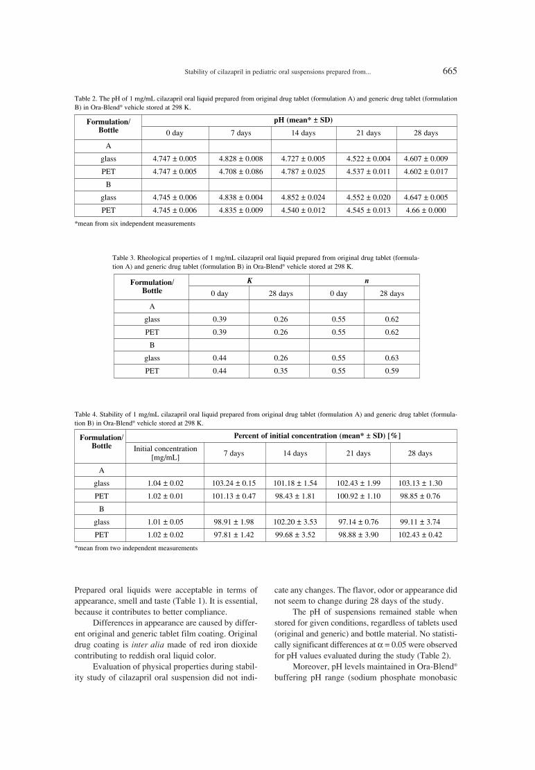
Stability of cilazapril in pediatric oral suspensions prepared from... 665
Prepared oral liquids were acceptable in terms ofappearance, smell and taste (Table 1). It is essential,because it contributes to better compliance.
Differences in appearance are caused by differ-ent original and generic tablet film coating. Originaldrug coating is inter alia made of red iron dioxidecontributing to reddish oral liquid color.
Evaluation of physical properties during stabil-ity study of cilazapril oral suspension did not indi-
cate any changes. The flavor, odor or appearance didnot seem to change during 28 days of the study.
The pH of suspensions remained stable whenstored for given conditions, regardless of tablets used(original and generic) and bottle material. No statisti-cally significant differences at α = 0.05 were observedfor pH values evaluated during the study (Table 2).
Moreover, pH levels maintained in Ora-BlendÆ
buffering pH range (sodium phosphate monobasic
Table 2. The pH of 1 mg/mL cilazapril oral liquid prepared from original drug tablet (formulation A) and generic drug tablet (formulationB) in Ora-BlendÆ vehicle stored at 298 K.
Formulation/ pH (mean* ± SD)
Bottle 0 day 7 days 14 days 21 days 28 days
A
glass 4.747 ± 0.005 4.828 ± 0.008 4.727 ± 0.005 4.522 ± 0.004 4.607 ± 0.009
PET 4.747 ± 0.005 4.708 ± 0.086 4.787 ± 0.025 4.537 ± 0.011 4.602 ± 0.017
B
glass 4.745 ± 0.006 4.838 ± 0.004 4.852 ± 0.024 4.552 ± 0.020 4.647 ± 0.005
PET 4.745 ± 0.006 4.835 ± 0.009 4.540 ± 0.012 4.545 ± 0.013 4.66 ± 0.000
*mean from six independent measurements
Table 3. Rheological properties of 1 mg/mL cilazapril oral liquid prepared from original drug tablet (formula-tion A) and generic drug tablet (formulation B) in Ora-BlendÆ vehicle stored at 298 K.
Formulation/ K nBottle 0 day 28 days 0 day 28 days
A
glass 0.39 0.26 0.55 0.62
PET 0.39 0.26 0.55 0.62
B
glass 0.44 0.26 0.55 0.63
PET 0.44 0.35 0.55 0.59
Table 4. Stability of 1 mg/mL cilazapril oral liquid prepared from original drug tablet (formulation A) and generic drug tablet (formula-tion B) in Ora-BlendÆ vehicle stored at 298 K.
Formulation/ Percent of initial concentration (mean* ± SD) [%]
Bottle Initial concentration7 days 14 days 21 days 28 days[mg/mL]
A
glass 1.04 ± 0.02 103.24 ± 0.15 101.18 ± 1.54 102.43 ± 1.99 103.13 ± 1.30
PET 1.02 ± 0.01 101.13 ± 0.47 98.43 ± 1.81 100.92 ± 1.10 98.85 ± 0.76
B
glass 1.01 ± 0.05 98.91 ± 1.98 102.20 ± 3.53 97.14 ± 0.76 99.11 ± 3.74
PET 1.02 ± 0.02 97.81 ± 1.42 99.68 ± 3.52 98.88 ± 3.90 102.43 ± 0.42
*mean from two independent measurements
666 BEATA J. STANISZ et al.
and citric acid), which is 3.5ñ5.0. Thus, it can beconcluded that buffering system of vehicle ensuredstable pH in oral liquid throughout the storage time.Rheological analyses were carried out at the day oforal liquids preparation and after 28 days long stor-age in amber glass and PET bottles at 298 K tem-perature. Such parameters as: flow curves,thixotropic curves were evaluated. In all rheologicaltests oral liquids were subjected to increasing shearrate and, as a consequence, liquid structure wasdestroyed and liquid started to flow. Nevertheless,when rate shear values decreased, suspension struc-ture was rebuilt, what can be clearly seen onthixotropy curves (Fig. 3). Moreover, from theshape of upwards part of thixotropic curve (the flowcurve) in can be concluded that the prepared sus-pensions are pseudoplastic, shear thinning fluids.Such rheological properties of prepared oral liquidsare owing to Ora-BlendÆ suspending agent. Ora-BlendÆ is compounded of synergistic blend of sus-pending agents, which form gel-like matrix counter-acting settling down of suspended drug particles.
For mathematical description of studied liquidsbehavior viscosity Ostwald model was used:
τ = K ∑ γn (1)where τ [N/m] is shear stress; γ [1/s] is shear rate; nsignifies flow behavior index and K fluid consisten-cy coefficient (13). As it results from data presentedin Table 3, flow behavior index value < 1 gives evi-dence that fluid is shear thinning. Changes betweenK and n parameters observed throughout storage inamber glass or plastic PET bottles through 28 daystime in 298 K temperature were minimal and indi-cated the evidence that 28 day storage did not affectrheological properties of prepared liquids.
Chemical stability
Cilazapril in oral suspensions retained itspotency during the storage, while concentrationremained within the limits specified in pharma-copoeia (±10%) (Table 4).
CONCLUSIONS
Presented study provides evidence on 28 daystability of cilazapril suspension oral liquid. Stable,
that means that throughout 28 day storage in cappedbottles of amber material glass or plastic PET retainswithin specified limits the same properties as at thetime of its manufacture.
Cilazapril oral suspensions in concentration of1 mg/mL are stable for 28 days in room temperature(298 K), regardless of tablets used (original orgeneric) and bottle material used.
Acknowledgment
The research was funded by the PolishNational Science Centre (grant no. NN405 050440).
REFERENCES
1. Szucs T.: Drugs 41 (Suppl. 1), 18 (1991).2. Deget F., Brogden R.N.: Drugs 41, 799 (1991).3. Schneeweiss A.: Pediatr. Cardiol. 11, 199
(1990).4. Momma K.: Paediatr. Drugs 81, 55 (2006).5. Harmon W.G., Sleeper L.A., Cuniberti L.,
Messere J., Colan S.D., Orav J.E., Towbin J.A.et al.: Am. J. Cardiol. 104, 281 (2009).
6. Mori Y., Nakazawa M., Tomimatsu H.,Momma K.: J. Am. Coll. Cardiol. 36, 270(2000).
7. Hazama K., Nakazawa M., Momma K.: Am. J.Cardiol. 88, 801 (2001).
8. Loyd V.A.: Clin. Ther. 30, 2102 (2008).9. Pharmacopoeia VIII, Polish Pharmaceutical
Society, Warszawa 2008.10. Gibson M.: Pharmaceutical preformulation and
formulation. Informa Healthcare, New York2009.
11. Paszun S.K., Stanisz B.J.: Reac. Kinet. Mech.Cat. 109, 285 (2013).
12. Paszun S.K., Stanisz B.J., Paw≥owski W.: ActaPol. Pharm. Drug Res. 70, 193 (2012).
13. Kemb≥owski Z.: Rheometry of non-newtonianfluid (Polish). WNT, Warszawa 1973.
Received: 7. 10. 2013
Acta Poloniae Pharmaceutica ñ Drug Research, Vol. 71 No. 4 pp. 667ñ675, 2014 ISSN 0001-6837Polish Pharmaceutical Society
Medicinal plants have always remained amajor target for drug development. Most of the tra-ditional medicines used nowadays are from plantorigin. In developing countries, medicinal plantsprovide an alternative therapy, which is cost effec-tive and easily available. It has been estimated thatone-third of the world population rely on traditionalmedicines for their health related needs (1).Traditional medicines though effective yet requireevaluation by scientific methods in order to be usedto their full extent.
The family Berberidaceae established asìBerberidesî is considered one of the most primitiveangiosperms (2). Berberidaceae is a small familythat contains 15 genera, approximately 650 speciesworldwide, distributed in temperate regions ofnorthern hemisphere (3). Berberis is represented by
3 genera and 20 species in Pakistan. Most of thespecies are found in mountainous regions of thecountry. Berberidaceae is characterized by perenni-al herbs and shrubs, leaves alternate or basal simpleor compound, flowers bisexual and actinomorphic(4).
Berberis orthobotrys (Family: Berberidaceae)commonly known as Ishkeen is a plant indigenousto Pakistan, found mainly in Gilgit Baltistan. Itsroots and stem bark has been used in the treatmentof wounds, infections, piles, jaundice, liver prob-lems, kidney stones, diabetes, sore throat and uterinetumors (5). In our previous study, antihypertensiveactivity of B.o. in rats was evaluated (6). This studywas therefore conducted to investigate the possiblemechanism of antihypertensive action of B.o. in iso-lated perfused rabbit heart.
PHARMACOLOGY
POSSIBLE MECHANISM OF CARDIAC DEPRESSANT ACTIVITY OF BERBERIS ORTHOBOTRYS ROOTS IN ISOLATED RABBIT HEART
ALAMGEER1*, MUHAMMAD SHOAIB AKHTAR1, QAISER JABEEN2, SAJID BASHIR1, MUHAMMAD NASIR HAYAT MALIK1 , SABEHA KARIM3, MUHAMMAD NAVEED MUSHTAQ1,
SHAHID RASOOL1, FOZIA LATIF4, NAZIA TABBASUM4, ABDUL QAYYUM KHAN1, HASEEB AHSAN1, WASIM KHAN1 and IBRAHIM JAVED1
1Faculty of Pharmacy, University of Sargodha, Sargodha, Pakistan2Faculty of Pharmacy and Alternative Medicine, Islamia University, Bahawalpur, Pakistan
3University College of Pharmacy, University of the Punjab, Lahore 54000, Pakistan4 The University of Lahore, Lahore, Pakistan
Abstract: Berberis orthobotrys Bien. ex Aitch. (B.o.) has been reported to have antihypertensive effect in dif-ferent experimental models. The aim of present study was to evaluate the possible antihypertensive mechanism.Aqueous methanolic extract of B.o. roots and its various fractions namely (ethyl acetate, n-butanol or aqueous)in different concentrations (10 ng/mL, 100 ng/mL, 1 µg/mL, 10 µg/mL, 100 µg/mL ) were evaluated in isolat-ed perfused rabbit heart to assess their effect on force of contraction, HR and perfusion pressure. The crudeextract of B.o. and its fractions exhibited a significant decrease in heart rate, contractility and perfusion pres-sure of isolated rabbit heart, however, butanolic fraction produced more prominent effect and was selected forfurther study. The effects of butanol fraction were not blocked by atropine (10-5 M) in isolated perfused heart.However, butanol fraction significantly blocked the effects of adrenaline (10-5 M). It is therefore conceivablethat cardiac depressant activity of B.o. butanol fraction might be due to the presence of certain β-blockingagents which might be responsible for antihypertensive effect However, further experiments are required to iso-late the active compound(s) and elucidate exact mechanism of action.
Keywords: Berberis orthobotrys roots, cardiac depressant, atropine, adrenaline, rabbit
667
* Corresponding author: e-mail: [email protected]; phone +92-343-7547785

668 ALAMGEER et al.
Figure 1. The effect of crude extract of B.o. and its fractions on perfusion pressure of isolated perfused rabbit heart (n = 6), where * = (p< 0.05), ** = (p < 0.01), and *** = (p < 0.001) vs. control
Figure 2. The effect of crude extract of B.o. and its fractions on contractility of isolated perfused rabbit heart (n = 6), where * = (p < 0.05)and *** = (p < 0.001) vs. control
Tracing (1) shows the effect of butanol fraction of B.o. on contractility of isolated perfused rabbit heart.

Possible mechanism of cardiac depressant activity of Berberis orthobotrys... 669
Figure 3. The effect of crude extract of B.o. and its fractions on heart rate of isolated perfused rabbit heart (n = 6), where * = (p < 0.05),** = (p < 0.01), and *** = (p < 0.001) vs. control
Tracing (2) shows the effect of butanol fraction of B.o on heart rate of isolated perfused rabbit heart.
Figure 4. The effect of butanol fraction of B.o. (10 µg/mL) on various cardiac parameters of isolated perfused rabbit heart both in theabsence and presence of atropine (10-5 M) (n = 6), where *** = (p < 0.001) vs. control

670 ALAMGEER et al.
Tracing (3) shows the effect of butanol fraction of B.o. (10µg/mL) on perfusion pressure of isolated perfused rabbit heart in the presenceof atropine (10-5M)
Tracing (4) shows the effect of butanol fraction of B.o. (10 µg/mL) on contractility of isolated perfused rabbit heart in the presence ofatropine (10-5M)
Tracing (5) shows the effect of butanol fraction of B.o. (10 µg/mL) on heart rate of isolated perfused rabbit heart in the presence of atropine(10-5M)

Possible mechanism of cardiac depressant activity of Berberis orthobotrys... 671
Figure 5. The effect of adrenaline (10-5 M) on various cardiac parameters of isolated perfused rabbit heart both in the absence and presenceof B.o. butanol fraction (10 µg/mL) (n = 6), where *** = (p < 0.001) vs. control
Tracing (6) shows the effect of adrenaline (10-5M) on perfusion pressure of isolated perfused rabbit heart both in the absence and presenceof B.o. butanol fraction (10 µg/mL)
Tracing (7)shows the effect of adrenaline (10-5M) on force of contraction of isolated perfused rabbit in the presence of B.o. butanol frac-tion (10 µg/mL)

672 ALAMGEER et al.
EXPERIMENTAL
Chemicals and drugs
Methanol, atropine and adrenaline were pur-chased from Sigma Chemicals Co. All the chemicalsand drugs used in the experiments were of standardgrade.
Animals
Both male and female rabbits of local strain(Oryctolagus cuniculus) weighing 1ñ1.5 kg wereused. All the animals were housed in controlledenvironment (23ñ25OC) at animal house ofDepartment of Pharmacy, University of Sargodha,Sargodha. All animals were treated according to thestandard procedures and the study protocol wasapproved by the local ethical committee.
Plant material
The roots of B.o. were collected from districtGilgit, Pakistan during June, 2011 and were identi-fied and authenticated by Dr. Shair Wali, AssistantProfessor of Botany, Karakurum InternationalUniversity, Gilgit, Baltistan, Pakistan. A voucher(no. BO-15-12) has been deposited in the herbarium,Faculty of Pharmacy, University of Sargodha forfuture reference.
Preparation of extract
Aqueous methanolic (70 : 30) extract of B. o.was prepared using cold maceration process. Thegrounded plant material (2 kg) was soaked in 5 L ofwater-methanol mixture (70 : 30) for 72 h at roomtemperature. After three days of occasional shaking,
the whole material was filtered and the filtrate evap-orated under reduced pressure using rotary evapora-tor. The crude extract was then air-dried to obtain asolid mass with a yield of 15% (7).
Fractionation of the extract
Activity directed fractionation of the crudeextract of B.o. was carried out by using differentorganic solvents in order to separate or concentratethe activities in anyone of the corresponding frac-tion. Fractionation was carried out by using differentorganic solvents (ethyl acetate, n-butanol, aqueous)based on their polarity order, as ethyl acetate < n-butanol < aqueous.
A known quantify of the crude extract was dis-solved in distilled water and mixed with equal vol-ume of organic solvents in a separating funnel, shak-en vigorously, with periodical removal of air. Themixture was allowed to separate for about 20ñ30min into two layers. The respective layer wasremoved, the same procedure was repeated twicemore times. Then, all the fractions were combinedand finally concentrated under reduced pressure onrotary evaporator to obtained the correspondingfraction. Similarly, the remaining layer (aqueous)was further treated with other organic solvents fortheir respective fractions. Finally, the remaininglayer was also evaporated and was considered asaqueous fraction (8).
Effect of crude extract and its fractions on
Langendorff perfused isolated rabbit heart
This experiment was carried out in accordancewith the Langendorff method (9). The rabbit was
Tracing (8) shows the effect of adrenaline (10-5M) on heart rate of isolated perfused rabbit in the presence of B.o. butanol fraction (10µg/mL)

Possible mechanism of cardiac depressant activity of Berberis orthobotrys... 673
injected with 1000 IU of heparin intravenouslythrough the marginal ear vein. Five min later, a blowon the neck of the rabbit made it unconscious. Thechest was opened and the heart was dissected outwith about 1 cm of aorta attached, and was quicklywashed with oxygenated Krebs-Henseleit solution.The isolated heart was gently squeezed several timesto remove as much residual blood as possible. Theheart was then transferred to the perfusion apparatus(Radnoti isolated heart system, AD Instrument,Australia) and tied to a glass cannula through theaorta. The perfusion fluid was Krebs-Henseleit solu-tion, which was continuously bubbled with a mix-ture of 95% oxygen and 5% carbon dioxide, and wasapplied at a constant flow mode. The temperaturewas continuously monitored and kept constant in therange of 36.5ñ37.5OC.
The heart was allowed to stabilize for 30 minbefore any drug interventions. The mechanicalresponses such as force of contraction of sponta-neously contracting isolated heart was recorded byattaching one end of a thread to the apex of theheart using a Palmer clip and the other end of thethread to a force transducer (MLT 844; ADInstruments, Australia). Heart rate was calculatedindirectly using LabChartpro5 software (ADInstruments, Australia). Perfusion pressure wasmeasured by using pressure transducer. The sig-nals from both pressure and force transducers werefiltered and amplified and sent to an analog-to-digital converter (Power Lab data acquisition andanalysis system, AD Instruments, Australia)attached to a computer. The signals recorded weresaved for later analysis. After stabilization, differ-ent doses of the crude extract and fractions (ethylacetate, n-butanol, aqueous) of B.o. (10 ng, 100ng, 1 µg, 10 µg, 100 µg/mL) were applied to assessvarious cardiac parameters i.e., heart rate(beats/min), force of contraction (g) and perfusionpressure (mm Hg) with each heart serving as itsown control. In order to explore the possiblemechanism of action, the effect of butanol fractionof the extract was assessed both in the absence andpresence of atropine 10-5 M (10). In addition, theeffects of adrenaline (10-5 M) were also assessedboth in the absence and presence of butanol frac-tion of B.o. (11).
Statistical analysis
The results were expressed as the means ±SEM. Statistical analysis was done by two wayANOVA followed by Bonferroni test using GraphPad Prism 5.0. Value of p < 0.05 was considered assignificant.
RESULTS
Effect of crude extract of B.o. and its fractions on
perfusion pressure, contractility and heart rate of
isolated perfused rabbit heart
The aqueous methanolic extract of B.o. at alldoses produced a significant (p < 0.01ñ0.001)decrease in force of contraction of isolated heartwhile a significant (p < 0.05ñ0.01) decrease in heartrate was observed only at doses from 10 ng/mL to 1µg/mL. The crude extract (1ñ100 µg/mL) alsoexhibited a significant (p < 0.001) decrease in per-fusion pressure. Ethyl acetate and aqueous fractionsof B.o. demonstrated a significant negative inotrop-ic and chronotropic effect at higher doses only.Similarly, both extracts produced a significantdecrease in perfusion pressure merely at 100 µg/mL.Moreover, butanol fraction of B.o. produced a sig-nificant (p < 0.05ñ0.001) decrease in perfusion pres-sure, heart rate and contractility of isolated heart atall the doses. A prominent effect in all the three car-diac parameters was observed at 10 µg/mL. Butanolfraction was chosen as the most potent of all of theextracts, hence 10 µg/mL of butanol fraction wasselected to elucidate its possible mechanism ofaction. The highest dose of the butanol fraction (100µg/mL) caused a maximum decrease in contractilityand heart rate but it also produced hardening of theheart and stoppage of beating of the heart.Therefore, this dose was not picked to explore itsdetailed mechanism (Figs. 1, 2 and 3).
Effect of butanol fraction of B.o. on perfusion
pressure, contractility and heart rate of isolated
perfused heart in the presence of atropine (10-5 M)
In the presence of atropine (10-5 M), thebutanol fraction of B.o. produced a significant (p <0.001) decrease in perfusion pressure, force of con-traction and heart rate of isolated heart. Atropine didnot block the negative inotropic and chronotropiceffects or reduced perfusion pressure of butanolfraction. The decrease in all the cardiac parameterswas quite similar in both presence and absence ofatropine (10-5 M) (Fig. 4).
Effect adrenaline (10-5 M) on perfusion pressure,
contractility and heart rate of isolated perfused
heart in the presence of butanol fraction of B.o. To find out the possible β-blocking activity,
the effects of adrenaline on various cardiac parame-ters were studied both in the presence and absenceof butanol fraction of B.o. (10 µg/mL). The findingsindicated that adrenaline (10-5 M) produced a signif-icant (p < 0.001) increase in force of contraction,

674 ALAMGEER et al.
heart rate and perfusion pressure of isolated heart inthe absence of butanol extract of B.o. (10 µg/mL).However, in the presence of butanol extract of B.o.(10 µg/mL), the effects of adrenaline (10-5 M) weresignificantly blocked as there was non-significantincrease in all the cardiac parameters of isolatedheart (Fig. 5).
DISCUSSION
Herbs were our first source of medicines. Noone knows when humans first used plants for medi-cine, but pollens of at least six medicinal plants werefound in a Neanderthal burial site estimated to be atleast 60,000 year old and some derivatives, e.g.,aspirin, reserpine, and digitalis, have become main-stays of human pharmacotherapy. Medicinal plantdrug discovery continues to provide new and impor-tant leads against various pharmacological targets.Several natural product drugs of plant origin haveeither recently been introduced in the market,including galantamine, nitisinone, and tiotropium,or are currently involved in late-phase clinical trials(12, 13). The medicinal values of traditional medic-inal plants cannot be disregarded and studies havebeen carried out in order to explore various activeprinciples of the extracts with intensive follow upstudies to establish their exact mechanism of action.One of the most important area in which compoundsfrom plant sources have contributed successfully, isthe cardiovascular research.
The present study revealed that aqueousmethanolic extract of B.o. exhibited a significantdecrease in perfusion pressure, force of contractionand heart rate at most of the doses. Similarly, all thefractions of B.o. produced a significant negativeinotropic and chronotropic effect in isolated heart.However, ethyl acetate and aqueous fractions of B.o.showed a significant decrease in perfusion pressureonly at 100 µg/mL. Butanol fraction of B.o. was foundto be much more potent than other extracts as it exhib-ited a significant decrease in all the cardiac parame-ters; this clearly indicates that the active principle(s)responsible for the said effects were more concentrat-ed in this fraction. Hence, butanol fraction of B.o. (10µg/mL) was selected for a detailed mechanism study.
Previously, it has been reported that negativeinotropic and chronotropic effect in isolated heartusually results from a cholinergic/histaminic stimu-lating activity or by β-receptor or calcium channelblocking effect (14, 15). A decrease in perfusionpressure indicates a coronary vasodilation effect,which is generally caused by three different media-tors that include β-receptor antagonist, cholinergic
receptor agonist and endothelium derived relaxingfactor (9, 15).
In the presence of atropine (10-5 M), butanolfraction of B.o. (10 µg/mL) produced a significantdecrease in perfusion pressure, heart rate and forceof contraction of isolated rabbit heart indicatingthat the extract did not mediated its actionsthrough cholinergic receptors. In order to investi-gate the β-blocking effect of butanol fraction ofB.o. (10 µg/mL), the effects of adrenaline (10-5 M)were studied in isolated rabbit heart. The findingsindicated that the butanol extract significantlyblocked the positive inotropic and chronotropiceffects of adrenaline (10-5 M). Similarly, theincreased perfusion pressure produced by adrena-line (10-5 M) was also significantly antagonized. Ithas been well established that adrenaline, a sym-pathomimetic drug, acts directly on β1 receptorsand produces an increased heart rate and contrac-tility. Propranolol blocks these receptors and pro-duces a negative inotropic effect and chronotropiceffect (16, 17). Hence, the blocking of adrena-lineís pharmacological effects by the butanol frac-tion of B.o. clearly indicated its β-receptor antag-onizing activity on isolated heart. However, theinvolvement of other receptors cannot be ruledout. Moreover, it has also been reported that β-blockers produce a cardiac inhibitory effect thatresults in decreased cardiac output and vasodila-tion, ultimately leading to a fall in blood pressure.Other blood pressure lowering mechanisms of β-adreneregic receptor blocking agents include adecrease in catecholamine release and an anti-renin activity (18). It is presumable therefore thatthis cardiac depressant activity of B.o. in isolatedperfused rabbit heart might also be responsible forits antihypertensive effect in rats.
CONCLUSION
It is concluded from this study that cardiacdepressant and coronary vasodilating activity of B.o.might be due to its antagonizing effects on β-recep-tors. Further studies are required to identify and iso-late the biologically active compounds in order todetermine the exact mechanism of action.
REFERENCES
1. Ali H., Qaiser M.: Pak. J. Bot. 41, 2009 (2009).2. Bruckner C.: Bot. Rev. 66, 155 (2000).3. Bottini M.C.J., Premoli A.C., Poggio L.: in XVI
International Botanical Congress, St. Louis,Missouri, USA 1999.

Possible mechanism of cardiac depressant activity of Berberis orthobotrys... 675
4. Jafri S.M.H.: in Flora of West Pakistan, NasirE., Ed., p. 1, Department of Botany, KarachiUniversity, Karachi, Pakistan 1975.
5. Khan S.W., Khatoon S.: Pak. J. Bot. 39, 699(2007).
6. Alamgeer., Akhtar M.S., Jabeen Q., Akram M.,Khan H.U., Karim S., Malik M.N.H. et al.:Trop. J. Pharm. Res. 12, 393 (2013).
7. Alamgeer., Malik M.N.H., Mushtaq M.N.,Bashir S., Ghumman S.A., Akram M., KhanH.U. et al.: J. Med. Plant Res. 6, 3222 (2012).
8. Williamson E., Okpako D.T., Evans F.J.: inSelection, preparation and pharmacologicalevaluation of plant material, p. 131, John Wileyand Sons Ltd., Chichester, England 1996.
9. Langendorff O.: Pflugerís Arch. Ges. Physiol.61, 291 (1895).
10. Khosla P., Gupta A., Singh J.: Indian J. Physiol.Pharmacol. 46, 241 (2002).
11. Ayinde B.A., Omogbai E.K.I., OnwukaemeD.N.: J. Pharmacol. Phytother. 1, 4 (2010).
12. Soleck R.S., Shanidar I.V.: Science 190, 880(1975).
13. Balunas M.J., Kinghorn A.D.: Life Sci. 78, 431(2005).
14. Mabe A.M., Hoover D.B.: Cardiovasc. Res. 82,93 (2009).
15. Mery P.F., Hove-Madsen L., Chesnais J.M.,Hartzell H.C., Fischmeister R.: Am. J. Physiol.270, 1178 (1996).
16. Bain W.A.: Exp. Physiol. 19, 297 (1929).17. Magnussen I., Kudsk F.N.: Acta Pharmacol.
Toxicol. 34, 141 (2009). 18. Gorre F., Vandekerckhove H.: Acta Cardiol. 65,
565 (2010).
Received: 18. 09. 2013


Acta Poloniae Pharmaceutica ñ Drug Research, Vol. 71 No. 4 pp. 677ñ682, 2014 ISSN 0001-6837Polish Pharmaceutical Society
Cardiovascular diseases (CVDs) have becomea major risk and their prevalence has graduallyincreased throughout the world. CVDs include coro-nary heart disease, cerebrovascular disease, hyper-tension, arrhythmias, rheumatic heart disease andheart failure. Hypertension refers to an increasedblood pressure and usually 140/90 mm Hg is takenas threshold. It has been estimated that high bloodpressure is responsible for 6% of deaths worldwideand remains the prominent risk factor for cardiovas-cular diseases affecting millions of people inPakistan (1). According to National Health Surveyof Pakistan, about one hundred thousand deathsoccur per year in the country due to CVDs. Manysynthetic drugs have been commonly used for thetreatment of hypertension in developed countries butherbal medicines still remain the popular choice inthe developing countries. The abundant use of these
herbal medicines has led to an extensive research inthis area to determine their potential efficacy andseveral modern cardiovascular drugs are now avail-able as natural/herbal products. Ethnobotanical sur-veys in our country have indicated their vast use inthe treatments of CVDs. For example, hydro-alco-holic extract of the leaves of Syzygium guineense,has produced a decrease in blood pressure of hyper-tensive rats. Similarly, Passiflora nepalensis Wallreported to be effective in hypertension. Moreover,the Chinese have used Ginko biloba, Stephaniatetandra and Uncaria rhynchophylla for the treat-ment of hypertension (2, 3).
Thymus linearis Benth. (Family: Lamiaceae)locally known as ìTumuro î is a perennial plantfound mostly in all alpine and subalpine pastures ofPakistan, India, Afghanistan and Nepal (4).Traditionally it has been widely used to treat a num-
PHARMACOLOGICAL EVALUATION OF ANTIHYPERTENSIVE EFFECT OF AERIAL PARTS OF THYMUS LINEARIS BENTH.
ALAMGEER1*, MUHAMMAD SHOAIB AKHTAR1, QAISER JABEEN2, HAFEEZ ULLAH KHAN1,SAFIRAH MAHEEN1, HAROON-UR-RASHID3, SABEHA KARIM4, SHAHID RASOOL1,
MUHAMMAD NASIR HAYAT MALIK1, KIFAYATULLAH KHAN1, MUHAMMAD NAVEED MUSHTAQ1, FOUZIA LATIF5, NAZIA TABASSUM5,
ABDUL QAYYUM KHAN1, HASEEB AHSAN1 and WASIM KHAN1
1Faculty of Pharmacy, University of Sargodha, Sargodha, Pakistan2Faculty of Pharmacy and Alternative Medicine, The Islamia University of Bahawalpur, Pakistan
3Department of Radiotherapy and Nuclear Medicine, Sargodha Medical College, University of Sargodha, Pakistan
4The University of Lahore, Lahore, Pakistan5University College of Pharmacy, University of the Punjab, Lahore 54000, Pakistan
Abstract: Traditionally Thymus linearis Benth. have been used for treatment of various diseases includinghypertension. The present study was conducted to evaluate the hypotensive and antihypertensive effect of aque-ous methanolic extract of aerial parts of Thymus linearis Benth. in normotensive and hypertensive rats. Acuteand subchronic studies were also conducted. The aqueous methanolic extract produced a significant decreasein SBP, DBP, MBP and heart rate of both normotensive and hypertensive rats. LD50 of the extract was foundto be 3000 mg/kg. The extract also exhibited a reduction in serum ALT, AST, ALP, cholesterol, triglyceridesand LDL levels, while a significant increase in HDL level was observed. It is conceivable therefore, thatThymus linearis Benth. contains certain active compound(s) that are possibly responsible for the observed anti-hypertensive activity. Moreover, these findings further authenticate the traditional use of this plant in folkloremedicine.
Keywords: Thymus linearis Benth., antihypertensive, isolated heart, LD50
677
* Corresponding author: e-mail: [email protected], phone: +92-343-7547785

678 ALAMGEER et al.
ber of ailments including high blood pressure,toothache, headache, cold, fever as well as skin, eye,and liver diseases (5, 6). In the present study, wehave endeavored to evaluate antihypertensiveeffects of aqueous methanolic extract of Thymus lin-earis Benth. in both normotensive and hypertensiverats.
MATERIALS AND METHODS
Chemicals
Glucose and methanol were purchased fromSigma Chemicals Co. All the chemicals used in theexperiments were of the best analytical grade.
Animals
Both male and female Sprague Dawley rats(200ñ300 g) and, albino mice (20ñ25 g) were used.The animals were housed in controlled environment(23ñ25OC) at animal house of University ofSargodha and were handled according to the stan-dard procedures. The study protocol was approvedby the local ethical committee Faculty of Pharmacy,University of Sargodha.
Collection and identification of plant material
The aerial parts of plant were collected from avillage shikyote district Gilgit, Gilgit Baltistan,Pakistan during the month of July, 2011 and wereidentified and authenticated by Dr Shair WaliAssistant Professor, Department of Botany,Karakuram International University, GilgitBaltistan. A voucher specimen with no. T.L.B=57-11 has been deposited in Faculty of Pharmacy,University of Sargodha for future reference.
Preparation of plant extract
Aqueous methanolic (70 : 30, v/v) extract ofaerial parts of Thymus linearis Benth. was preparedusing cold maceration process. The grounded plantmaterial (2 kg) was soaked in 7 liters of an aqueousmethanolic mixture (70 : 30) for 72 h at room tem-perature. After three days of occasional shaking, thewhole material was filtered and the filtrate wasevaporated under reduced pressure using rotaryevaporator and finally dried in lyophilizer.
Effect of extract of Thymus linearis Benth. on
blood pressure and heart rate of normotensive
rats
Sprague Dawley rats of either sex were usedfor these experiments and were randomly dividedinto three groups of five rats each. Group I received
100 mg/kg of the extract of Thymus linearis Benth.Group II and Group III were given 250 mg/kg and500 mg/kg of the aqueous methnolic extract ofThymus linearis Benth., respectively. Blood pres-sure and heart rate of each of these groups weredetermined at 0 hour, then after 2, 4 and 6 hoursafter administration of extract by using non-invasiveblood pressure (NIBP) measuring apparatus (NIBPController ñ AD Instruments) from the tail of rats.Each animal was placed in the restrainer and anappropriate cuff with sensor was mounted on theirtails and then warmed to about 33ñ35OC. The tailcuff was inflated to a pressure well above theexpected systolic blood pressure SBP (200 mmHg)and slowly released during which the pulse wasrecorded by using Power Lab data acquisition sys-tem and computer running Lab chart 5.0 software asdescribed by (3). SBP, MBP and heart rate weremeasured directly using pulse tracing while the dias-tolic blood pressure was calculated from SBP andMBP using the equation: DBP = (3MBPñSBP)/2.
Effect of aqueous methanolic extract of aerial
parts of Thymus linearis Benth. on blood pressure
and heart rate of cholesterol fed hypertensive rats
Sprague Dawley rats of either sex were ran-domly assigned into two groups of five rats each.Group I served as control and was treated with aspecially prepared egg feed diet in order to producecholesterol-induced hypertension. The diet was pre-pared by the addition of yolk of 12 eggs to 500 gstandard rat diet. The feed so prepared was dried inthe sunlight for 3 days. Animals were then fed onthis diet for 21 consecutive days. Group II served astreated group and received egg feed diet and aque-ous methanolic extract of Thymus linearis Benth.(500 mg/kg) for 21 consecutive days. BP and heartrate of these groups were measured on days: 0, 7, 14and 21 (7).
Effect of aqueous methanoloic extract of Thymuslinearis Benth. on blood pressure and heart rate in
glucose fed hypertensive rats
Sprague Dawley rats of either sex were divid-ed into two groups of five rats each. Group I servedas control and received 10% glucose solution for 21consecutive days. Animals in group II were given10% glucose solution and aqueous methanolicextract of aerial parts of Thymus linearis Benth. (500mg/kg) for 21 consecutive days. BP and heart rate ofeach of these groups were measured at 0, 1, 2, and 3weeks by placing them in a pre-warmed restrainer.SBP, MBP and heart rate were measured directlyusing pulse tracing while the diastolic blood pres-

Pharmacological evaluation of antihypertensive effect of aerial parts of... 679T
able
1.E
ffec
t of v
ario
us d
oses
of c
rude
ext
ract
of T
hym
us li
near
ison
SB
P, M
BP,
DB
P an
d he
art r
ate
of n
orm
oten
sive
rats
.
Dos
es
Tim
e10
0 m
g/kg
25
0 m
g/kg
50
0 m
g/kg
(h
)SB
PM
BP
DB
PH
RSB
PM
BP
DB
PH
RSB
PM
BP
DB
PH
R
0 12
7.5
± 2.
4410
4.2
± 0.
7892
.5±
0.90
383
± 10
.012
5.9
± 0.
4910
4.8
± 0.
7194
.3 ±
0.6
837
8 ±
9.80
125.
7 ±
1.00
105.
2 ±
0.36
95.9
± 0
.84
388
± 4.
56
2 12
5.3
± 2.
3210
0.9
± 0.
7990
.7±
0.68
370
± 6.
62*
122.
6 ±
1.62
100.
9 ±
0.59
93.5
± 0.
7835
6 ±
7.13
*11
4.5
± 3.
28*
99.2
± 2
.24
92.0
± 3
.75
360
± 4.
70**
4 12
0.5
± 4.
6995
.0 ±
2.5
5*88
.7±
1.08
365
± 4.
68**
116.
1 ±
2.48
*95
.3 ±
1.2
8*90
.9 ±
1.1
135
0 ±
8.65
**10
4.2
± 2.
76**
90.9
± 1
.48*
85.7
± 1
.51*
340
± 5.
19**
*
6 11
8 ±
1.82
**90
.0 ±
3.6
8**
85.5
± 0.
8936
0 ±
8.16
**11
3.6
± 2.
83**
80.6
4 ±
2.47
**85
.6 ±
0.8
3*34
0 ±
7.25
**10
0.4
± 1.
6***
86.6
± 1
.60*
*77
.3 ±
2.7
5**
320
± 5.
22**
*
Res
ults
are
exp
ress
ed a
s th
e m
ean
± SE
M (n
= 5
), O
ne w
ay A
NO
VA
follo
wed
by
Dun
nettí
s te
st h
ave
been
app
lied;
* =
(p <
0.0
5), *
*= (p
< 0
.01)
, ***
= (p
< 0
.001
) vs.
cont
rol 0
h.
Tab
le 2
.Eff
ect o
f Th
ymus
line
aris
on e
gg f
eed-
indu
ced
hype
rten
sive
rat
s.
Day
sSB
P (m
m H
g)
DB
P (m
m H
g)
MB
P (m
m H
g)
Hea
rt r
ate
(bea
ts/m
in)
Con
trol
T
reat
ed 5
00 m
g/kg
C
ontr
ol
Tre
ated
500
mg/
kg
Con
trol
T
reat
ed 5
00 m
g/kg
C
ontr
ol
Tre
ated
500
mg/
kg
0 12
1.6
± 3.
49
123.
8 ±
1.7
8 89
.60
± 2.
17
100.
8 ±
2.3
4 10
1.0
± 1.
58
108.
6 ±
2.07
36
1.0
± 5.
98
381.
0 ±
5.98
3rd
144.
2 ±
3.67
109.
2 ±
4.45
**10
1.4
± 1.
9390
.4 ±
2.4
5**
114.
4 ±
2.48
98.2
1 ±
2.22
*40
4.1
± 3.
3235
0.1±
6.1
2***
6th
149.
4 ±
3.67
100.
8 ±
5.97
**10
7.4
± 3.
8385
.2 ±
1.8
4**
120.
6 ±
3.60
90.4
0 ±
3.00
**43
1.0
± 4.
9632
0.2
± 3.
78**
*
9th
168.
0 ±
4.57
95.2
0 ±
3.88
***
110.
0 ±
4.28
80.6
± 2
.39*
**12
4.4
± 2.
1483
.60
± 2.
23**
455.
0 ±
5.47
300.
2 ±
4.93
***
Res
ults
are
exp
ress
ed a
s th
e m
ean
± SE
M (
n =
5), O
ne w
ay A
NO
VA
fol
low
ed b
y D
unne
ttís
test
hav
e be
en a
pplie
d; *
= (
p <
0.05
), *
* =
(p <
0.0
1), *
** =
(p
< 0.
001)
vs.
cont
rol d
ay 0
.
Tab
le 3
.Eff
ect o
f Th
ymus
line
aris
on g
luco
se in
duce
d hy
pert
ensi
ve r
ats.
Wee
ksSB
P (m
m H
g)
DB
P (m
m H
g)
MB
P (m
m H
g)
Hea
rt r
ate
(bea
ts/m
in)
Con
trol
T
reat
ed 5
00 m
g/kg
C
ontr
ol
Tre
ated
500
mg/
kg
Con
trol
T
reat
ed 5
00 m
g/kg
C
ontr
ol
Tre
ated
500
mg/
kg
0 12
4.6
± 2.
0112
2.6
± 1.
7387
.21
± 2.
7997
.80
± 2.
4298
.01±
1.8
510
8.2
± 2.
3638
3.6
± 4.
8539
1.6
± 2.
90
1st
139.
6 ±
3.05
10
2.6
± 3.
50**
93.2
5 ±
2.62
89.4
± 2
.05N
S10
9.2±
2.6
895
.60±
2.2
7*39
0.4
± 4.
3137
1.6
± 8.
18**
2nd
148.
4 ±
4.95
99.4
1 ±
3.94
**10
2.4
± 1.
7184
.60
± 2.
30*
116.
0± 2
.40
87.6
1± 3
.03*
*40
6.2
± 3.
9931
7.0
± 9.
10**
*
3rd
166.
4 ±
4.94
82.2
± 2
.60*
**11
2.4
± 2.
0775
.60
± 1.
82**
127.
1± 2
.76
77.0
0± 3
.13*
**41
0.4
± 3.
8929
6.6
± 5.
49**
*
Res
ults
are
exp
ress
ed a
s th
e m
ean
± SE
M (
n =
5), O
ne w
ay A
NO
VA
fol
low
ed b
y D
unne
ttís
test
hav
e be
en a
pplie
d; *
= (
p <
0.05
), *
* =
(p <
0.0
1), *
** =
(p
< 0.
001)
vs.
cont
rol (
wee
k 0)
.

680 ALAMGEER et al.
sure was calculated from SBP and MBP using theequation DBP = (3MBPñSBP)/2 (3).
Acute toxicity study
Albino mice of either sex were randomly divid-ed into five groups (n = 2). Group 1 served as controland received normal saline (10 mL/kg) while othergroups (Groups 2ñ9) were administered differentdoses of the extract in an ascending order i.e., 100,500 1000, 1500, 2000, 2500, 3000, 3500 mg/kg,respectively. The mortality rate was observed for 24h. All the doses were administered by intraperitonealroute. The highest dose, which did not kill any ani-mal, and the lowest dose, which killed only one ani-mal, was noted. LD50 was calculated from the geo-metric mean of these two doses (8).
Subchronic toxicity
Sprague Dawley rats of either sex were ran-domly divided into two groups (n = 6). The firstgroup received normal saline (10 mL/kg bodyweight p.o.) and the animals in group 2 received 500mg/kg body weight of the extract daily for 28 days.Food and water intake of animals were observedduring this period. At 29th day, blood was collectedfrom overnight fasted rats of each group by cardiacpuncture for the determination of serum biochemicalparameters. Then, the rats were sacrificed by cervi-cal dislocation for the study of various organs (heart,liver and kidney) weights (9).
Biochemical parameters
For the estimation of alanine transaminase(ALT), aspartate transaminase (AST), alkaline phos-
phatase (ALP), total cholesterol, triglycerides, lowdensity lipoprotein (LDL) and high density lipopro-tein (HDL) blood samples were collected in clotactivator gel tubes. The serum was separated by cen-trifuging the blood samples at 2000 r.p.m. for 10min. Serum biochemical parameters were thenmeasured by using commercially available reagentkits (9).
Statistical analysis
The results were expressed as the means ±SEM. One way ANOVA followed by Dunnettís testhave been applied with p < 0.05 considered as sig-nificant.
RESULTS
Effect of Thymus linearis Benth. extract on blood
pressure and heart rate of normotensive rats
The extract produced a significant decrease inthe SBP, MBP , DBP and heart rate with 100, 250and 500 mg/kg dose especially after 4 and 6 h ofdrug administration, however, more pronouncedeffect was produced by 500 mg/kg, hence, it wasselected for further study (Table 1).
Effect of extract Thymus linearis Benth on blood
pressure and heart rate of cholesterol fed hyper-
tensive rats
The extract of Thymus linearis Benth, at adose of 500 mg/kg produced a highly significant (p< 0.001) decrease in SBP, MBP, DBP and heartrate of cholesterol fed hypertensive rats. A highlypronounced effect in all the observed parameters
Table 4. Effect of Thymus linearis on body weights, organ weights and various biochemical parameters of rats.
Parameters Control Extract 500 mg/kg
Liver (g) 5.86 ± 0.10 5.85 ± 0.12NS
Kidney (g) 1.39 ± 0.14 1.38 ± 0.11NS
Heart (g) 1.47 ± 0.11 1.46 ± 0.12NS
Body weight (g) 280 ± 5.11 281 ± 4.50 NS
ALT (IU/L) 0.1 ± 2.29 36.9 ± 1.69*
AST(IU/L) 90.1 ± 1.06 80.2 ± 1.09*
ALP(IU/L) 70.0 ± 2.01 58.2 ± 1.94*
Triglycerides (mg/dL) 89.2 ± 1.92 75.0 ± 1.74*
Cholesterol (mg/dL) 60.5 ± 1.56 45.0 ± 1.02*
LDL (mg/dL) 20.09 ± 2.07 14.20 ± 1.09**
HDL (mg/dl) 33.4 ± 1.18 46.5 ± 2.02*
Values are expressed as the means ± SEM (n = 6); * = p < 0.05, ** = p < 0.01 and NS = Non-significant ascompared to control.

Pharmacological evaluation of antihypertensive effect of aerial parts of... 681
was seen at second and third weeks of treatment(Table 2).
Effect of extract Thymus linearis Benth. on blood
pressure and heart rate of glucose fed hyperten-
sive rats
The extract of Thymus linearis Benth. at a doseof 500 mg/kg produced a highly significant (p <0.001) decrease in SBP, MBP, DBP and heart rate ofglucose fed hypertensive rats with more significanteffects at week 2 and 3 (Table 3)
Acute and subchronic toxicity studies
The median lethal dose (LD50) of the extract inmice was found to be 3000 mg/kg. In subchronic toxic-ity studies, the extract did not cause any significantalteration in body weights or organs weights of rats. Theresults also revealed that the extract (500 mg/kg) pro-duced a significant reduction in serum ALT, AST andALP levels. The extract exhibited a significant decreasein total cholesterol, triglycerides and LDL levels whileincrease in HDL levels was observed (Table 4).
DISCUSSION
Plants have been used for the treatment of vari-ous diseases since ancient times. In the last few yearsthere has been an exponential growth in the field ofherbal medicine and these drugs are gaining populari-ty both in developing and developed countries becauseof their natural origin and less side effects (10).
In the present investigation in normotensiveand hypertensive rats, the extract produced a signif-icant decrease in SBP, MBP and DBP with maxi-mum effect at 500 mg/kg. Similarly, the extract pro-duced a significant decrease in the heart rate of bothnormotensive and hypertensive rats. It was observedthat the degree of antihypertensive response pro-duced by the extract of Thymus linearis Benth. wasmuch higher in hypertensive rats than in normoten-sive rats. This observation was in support of the gen-eral finding that hypertensive animals appear tohave an exaggerated response to depressor stimuli(11). A decrease in sympathetic activity is involvedin decrease in blood pressure (12). The extract alsoproduced a significant decrease in heart rate of nor-motensive rats possibly due to a reduction in sym-pathetic activity, which might have also beeninvolved in a decreased blood pressure.
Previous studies demonstrated that atherosclero-sis may contribute to an increase in blood pressure(13). In cholesterol fed hypertensive rats, the extractproduced a significant decrease in blood pressure andheart rate. Moreover, the extract also cause a decrease
in LDL, TG, cholesterol and an increase in HDLlevel, which could be responsible for its antihyperten-sive effect in cholesterol-induced hypertensive rats.
It has been reported that a high glucose intakecause an increase in blood pressure through the gen-eration of reactive oxygen species (ROS). TheseROS production have been found to be associatedwith an increased NADPH oxidase activity (14).Moreover, it has also been reported that an increasedglucose level is involved in the inhibition of nitricoxide synthase activity, ultimately resulting indecreased nitric oxide levels and an increased bloodpressure (15). Thus, the antihypertensive effect ofthe extract in glucose treated hypertensive rats mightbe due to an inhibition of NADPH oxidase activityor an increased level of nitric oxide.
During subchronic toxicity studies, the extractwas found to be safe and no signs of toxicity wereobserved. Food and water intake, body weights andorgan weights remained unaltered during the 28days of treatment with the extract. However, bio-chemical parameters related to hepatic functions,such as ALT, AST and ALP, were significantlydecreased when compared to control. The reductionof these enzymes indicated that the extract did notcause any toxic effects on both liver and heart tis-sues (16). The extract also revealed a significantdecrease in serum cholesterol, triglyceride and LDLlevels in rats as compared to control. The increase inHDL levels and a decrease in LDL levels by theextract indicated a possible reduction in cardiovas-cular risk factor that could lead to the death of ani-mals. These effects are quite similar to those of anti-hyperlipidemic drugs like statins (17). Therefore,the antihypertensive effect of aqueous methanolicextract of Thymus linearis could also be associatedwith its lipid lowering effects.
CONCLUSIONS
It is concluded from this study that the antihy-pertensive effect of Thymus linearis Benth. aerial partmight be due to certain biologically active compoundspresent in the plant extract. However, further studiesare needed to isolate the active antihypertensive prin-ciple(s) and assess its possible mechanism of action.
REFERENCES
1. Murray C.J.L., Lopez A.D.: Nat. Med. 4, 1241(1996).
2. Mahady G.: J Cardiovasc. Nurs. 16, 21 (2002).3. Ayele Y., Urg K., Engidawork E.: Phytother.
Res. 24, 1457 (2010).

682 ALAMGEER et al.
4. Jamzad Z.: Iranian J. Bot. 15, 51 (2009).5. Suresh K.G., Yildiz A.T.: Nepal J. Plant. Sci. 6,
56 (2009).6. Rana M.S.N., Samant S.S.: Indian J. Tradit.
Know. 10, 439 (2011).7. Saleem R., Ahmad M., Ahmed S.I., Azeem M.,
Khan RA., Rasool N.: Phytother. Res. 19, 881(2005).
8. Akhila J.S., Deepa S., Alwar M.C.: Curr. Sci.93, 917 (2007).
9. Biswas M., Kar B., Karan T.K., BhattacharyaS., Kumar R.B.S., Ghosh A.K., Haldar P.K.: J.Phytol. 2, 06 (2010).
10. Akhtar M.S., Nadeem M., Haroon-ur-Rashid.,Bashir S.: Can. J. Appl. Sci. 1, 16 (2011).
11. Bunang R.D., Eferakeya A.E., Langdon D.S.:Am. J. Physiol. 228, 217 (1975).
12. Salas L., Morales O.M.: Planta Med. 4, 56(1986).
13. Brantsma A.H., Bakker S.J.L., de Zeeuw D., deJong P.E., Gansevoort R.T.: J. Am. Soc.Nephrol. 17, 331 (2006).
14. Toyoshi I., Ping L., Fumio U., Hai Y.Y., MaikoK., Minako I.: Diabetes. 49, 1939 (2000).
15. Muniyappa R., Srinivas P.R., Ram J.L., WalshM.F., Sowers J.R.: Hypertension 31, 289(1998).
16. Ogwal-Okeng W.J., Obua C., AnokbonggoW.W.: Afr. Health Sci. 3, 124 (2003).
17. Barnett H.A., OíGara G.: in Diabetes and theHeart. p. 7, Churchill Livingstone, Edinburgh2003.
Received: 2. 10. 2013

Acta Poloniae Pharmaceutica ñ Drug Research, Vol. 71 No. 4 pp. 683ñ686, 2014 ISSN 0001-6837Polish Pharmaceutical Society
Many systemic diseases require anticoagulanttherapy. The conditions, which should be mentionedhere, include ischemic stroke, arterial embolism,pulmonary embolism, atrial fibrillation or preven-tive treatment of myocardial infarction, as well asmany others (1ñ3). Cardiovascular conditions, apartfrom neoplastic diseases, remain the major cause ofdeath in developed countries (4, 5). In 2010, theischemic heart disease led to 12.9 million deaths inthe world (4), which constitutes a quarter of the totalnumber of deaths (4, 6).
Introduction of acetylsalicylic acid combinedwith oral anticoagulants or antiplatelet drugs consid-erably reduced mortality and the number of subse-quent ischemic events in patients with history ofmyocardial infarction (7ñ9). In spite of optimalpharmacological treatment as well as compliancewith the rules of secondary prevention, several per-cent of patients annually develop new coronary
events or die (10, 11). Oral anticoagulants requireoften INR (International Normalized Ratio) testingand consequently, corrections of the drug dose as theINR increase above the therapeutic range raises therisk of hemorrhage complications (1, 2). SustainedINR level of 2ñ3.5 in patients treated with oral anti-coagulants results in low risk of bleeding (1, 12).
The number of patients on chronic antiplateletand anticoagulant therapy is increasing, which cre-ates a challenge for doctors performing proceduresaffecting tissue continuity. Due to the risk of hem-orrhage, the management algorithm compels doctorsto withdraw the drugs or use a bridge therapy forseveral days before and after the surgical procedure.
In a patient treated with anticoagulants, who isbeing prepared for an invasive procedure, the risk ofmajor bleeding with procedure-related death mustbe assessed and compared to the thromboembolicrisk caused by temporary withdrawal of drugs (13).
SAFETY AND EFFICACY OF BONE WAX IN PATIENTS ON ORAL ANTICOAGULANT THERAPY
MARTA KRASNY 1, KORNEL KRASNY 2 and PIOTR FIEDOR 3
1 Department of Orthodontics, Warsaw Medical University, Nowogrodzka 59, 02-005 Warszawa, Poland 2 Medicare Dental Practice, Warszawa, Ks. Jerzego Popie≥uszki 17a/102, 01-595 Warszawa, Poland
3 Department of General and Transplantation Surgery, Transplantation Institute, Medical University of Warsaw, Nowogrodzka 59, 02-006 Warszawa, Poland
Abstract: Cardiovascular conditions, apart from neoplastic diseases, remain the major cause of death in devel-oped countries; therefore, the number of patients receiving oral anticoagulants is constantly increasing.Anticoagulant therapy considerably reduced mortality in patients with history of myocardial infarction amongothers. Although many interventions may be performed without withdrawal of the anticoagulant and toothextraction was qualified as a procedure of low hemorrhage risk, a majority of dentists refer the patient to a car-diologist several days before the elective tooth extraction to withdraw anticoagulants. The aim of the study wasto evaluate the efficacy and safety of bone wax used to stop bleeding after dental procedures in a group ofpatients on chronic anticoagulant therapy and find an answer to a question, whether it is justified to temporar-ily withdraw anticoagulants for this type of procedures. The study involved 176 patients on chronic anticoagu-lant therapy undergoing tooth extraction (154 subjects) or surgical extraction of a retained tooth (48 subjects).After the procedure, in each case the alveolus was filled with bone wax to stop bleeding. In all patients involvedin the study bleeding from the alveolus was successfully stopped during the procedure. None of the subjectsreported increased bleeding from the operational site after coming back home. Bone wax is a good, efficient,and safe material to block bleeding from the alveolus following tooth extractions, also in patients on chronicanticoagulant therapy. The study demonstrated that withdrawal or adjustment of anticoagulant therapy is notnecessary before an elective tooth extraction.
Keywords: post-extraction bleeding, bone wax, anticoagulants, cardiovascular disease
683
* Corresponding author: e-mail: [email protected]; phone: 508-145-966
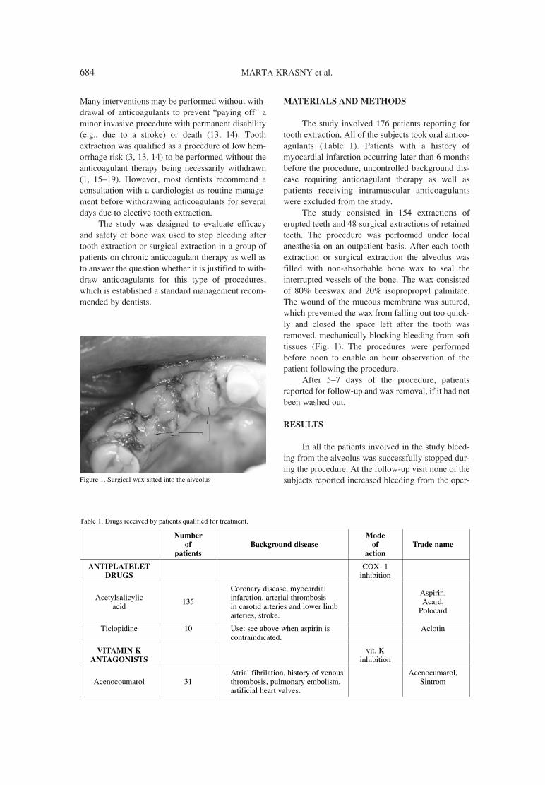
684 MARTA KRASNY et al.
Many interventions may be performed without with-drawal of anticoagulants to prevent ìpaying offî aminor invasive procedure with permanent disability(e.g., due to a stroke) or death (13, 14). Toothextraction was qualified as a procedure of low hem-orrhage risk (3, 13, 14) to be performed without theanticoagulant therapy being necessarily withdrawn(1, 15ñ19). However, most dentists recommend aconsultation with a cardiologist as routine manage-ment before withdrawing anticoagulants for severaldays due to elective tooth extraction.
The study was designed to evaluate efficacyand safety of bone wax used to stop bleeding aftertooth extraction or surgical extraction in a group ofpatients on chronic anticoagulant therapy as well asto answer the question whether it is justified to with-draw anticoagulants for this type of procedures,which is established a standard management recom-mended by dentists.
MATERIALS AND METHODS
The study involved 176 patients reporting fortooth extraction. All of the subjects took oral antico-agulants (Table 1). Patients with a history ofmyocardial infarction occurring later than 6 monthsbefore the procedure, uncontrolled background dis-ease requiring anticoagulant therapy as well aspatients receiving intramuscular anticoagulantswere excluded from the study.
The study consisted in 154 extractions oferupted teeth and 48 surgical extractions of retainedteeth. The procedure was performed under localanesthesia on an outpatient basis. After each toothextraction or surgical extraction the alveolus wasfilled with non-absorbable bone wax to seal theinterrupted vessels of the bone. The wax consistedof 80% beeswax and 20% isopropropyl palmitate.The wound of the mucous membrane was sutured,which prevented the wax from falling out too quick-ly and closed the space left after the tooth wasremoved, mechanically blocking bleeding from softtissues (Fig. 1). The procedures were performedbefore noon to enable an hour observation of thepatient following the procedure.
After 5ñ7 days of the procedure, patientsreported for follow-up and wax removal, if it had notbeen washed out.
RESULTS
In all the patients involved in the study bleed-ing from the alveolus was successfully stopped dur-ing the procedure. At the follow-up visit none of thesubjects reported increased bleeding from the oper-
Table 1. Drugs received by patients qualified for treatment.
Number Mode
of Background disease of Trade name
patients action
ANTIPLATELET COX- 1DRUGS inhibition
Coronary disease, myocardialAcetylsalicylic infarction, arterial thrombosis
Aspirin,
acid 135
in carotid arteries and lower limb Acard,
arteries, stroke. Polocard
Ticlopidine 10 Use: see above when aspirin is Aclotincontraindicated.
VITAMIN K vit. K ANTAGONISTS inhibition
Atrial fibrilation, history of venous Acenocumarol, Acenocoumarol 31 thrombosis, pulmonary embolism, Sintrom
artificial heart valves.
Figure 1. Surgical wax sitted into the alveolus

Safety and efficacy of bone wax in patients on oral anticoagulant therapy 685
ational site after coming back home. No case ofpurulent exudate from the wound was found. Innearly 1/3 of subjects (65 alveoli) the applied bonewax stayed within the site for 7 days and requiredmechanical removal.
Among the study group, 32 subjects did notreport for the follow-up visit at the scheduled dateand in 12 cases prolonged presence of bone waxcaused local inflammation within the healingwound. No case of allergic reaction was found.
DISCUSSION
Apart from wax, which is the oldest and thecheapest hemostatic material, bleeding control aftertooth extraction may be obtained by mouth rinsingwith solution of tranexamic acid q.i.d. for 2 days or25% solution of Â-aminocaproic acid (1, 2, 15) orlocal wound dressing with materials like spon-gostan, fibrin sealant or cellulosic acid (15, 19).
Patients treated with oral anticoagulants shouldnot receive NSAIDs or paracetamol as analgesicsduring the perioperative period due to increased riskof hemorrhage while taking both types of medica-tions simultaneously (1, 20). The first line anal-gesics in those patients are ibuprofen, naproxen ortramadol (15). INR testing should be made a daybefore the procedure and the value must be withinthe reference range. With INR > 3.5 the anticoagu-lant dose should be adjusted and 1 mg of vitamin Kshould be administered orally before the procedureor on the day of the procedure (1, 19). In case of pro-cedures with higher hemorrhage risk (multipleextractions, periodontal procedures) introduction oflow-molecular-weight heparin is required severaldays before and after the procedure.
This management algorithm generates numer-ous problems for the patients, who are often severe-ly ill, as it requires multiple blood taking to performa range of necessary tests. This management is evenmore burdensome in case of a patient reportingtoothache and taking anticoagulants at the sametime. It limits the time frame and urges the doctor toperform the procedure as soon as possible.
Although dental surgery procedures were qual-ified to the group of low post-operational hemor-rhage risk (3, 13, 14), this possibility must always beconsidered. Usually a visit at the dentistís surgeryinvolves stress and anxiety that may increase bloodpressure temporarily, which, in combination withreceived medications, may evoke hemorrhage. Inthis case, bone wax constitutes a very good protec-tion as it mechanically blocks vessel openings, in asimilar way to a tampon. In order to minimize the
risk of hemorrhage, it is advisable to perform singletooth extractions.
Apart from numerous advantages of bone wax,available literature provides many examples of com-plications related to its use. Already in the 1950s,Geary and Frantz observed hindered healing of bonefractures while using wax in dogs (21). Bone wax mayalso penetrate into the lungs and cause embolism (22).
Excessive pressure resulting from wax applica-tion during neurological procedures of the spinalcord may lead to chronic pain (23) and even irrita-tion of peripheral nerve function (24) or granuloma(25ñ27). Katre et al. described inferior alveolarnerve paralysis as complication after extraction ofthe wisdom tooth (28). Such a reaction was notfound in this study but in view of anatomic proxim-ity of the inferior alveolar nerve and the roots of themandibular third molar, special care should be takenwhile using wax to reduce postoperative bleeding.
The most common complication while usingthis type of hemostatic material in surgery is inflam-mation (23, 29, 30). Leaving the wax longer than itis required for covering the wound with epitheliummay result in irritation of the surrounding tissues,which was observed in this study. To avoid this con-dition, the wax should be precisely removed follow-ing 5ñ7 days of extraction without the risk of hem-orrhage from the wound.
CONCLUSIONS
This study implies that bone wax is a good,efficient, and safe material to block bleeding fromthe alveolus following tooth extractions. Its efficacywas confirmed also in the group of patients receiv-ing anticoagulants, eliminating the necessity of tem-porary withdrawal or adjustment of the anticoagu-lant therapy before the elective tooth extraction.
Due to possible allergic reactions to beeswax itis necessary to take medical history considering thatissue before the procedure is done. As the material isnot absorbed, it is important to instruct the patientabout the necessity to remove residue wax from thealveolus after several days of the procedure or toschedule a follow-up visit to remove the wax. If thewax was left behind, local inflammation may occurcaused by irritation of a foreign matter irritating thehealing tissues.
Apart from hemostatic properties of the wax,authors of this article observed decreased tissueedema resulting from limited exudate as well as pre-vention of alveolar osteitis after using the describedmaterial. However, these observations need furtherinvestigation.

686 MARTA KRASNY et al.
REFERENCES
1. Ansell J., Hirsh J., Poller L., Bussey H.,Jacobson A., Hylek E.: The pharmacology andmanagement of the vitamin K antagonists: theSeventh ACCP Conference on Antithromboticand Thrombolytic Therapy. Chest 126 (Suppl.3), 204S (2004). Erratum: Chest 127, 415(2005).
2. Geerts W.H., Pineo G.F., Heit J.A., BergqvistD., Lassen M.R., Colwell C.W., Ray J.G.:Prevention of venous thromboembolism: theSeventh ACCP Conference on Antithromboticand Thrombolytic Therapy. Chest 126 (Suppl.3), 338S (2004).
3. Spandorfer J.: Med. Clin. North Am. 85, 1109(2001).
4. Lozano R., Naghavi M., Foreman K., Lim S.,Shibuya K., Aboyans V., Abraham J. et al.:Lancet 380 (9859), 2095 (2012). Erratum in:Lancet 381 (9867), 628 (2013).
5. Murray C.J., Lopez A.D.: Lancet 349, 1498(1997).
6. Lim G.B.: Nat. Rev. Cardiol. 10, 59 (2013).7. Hurlen M., Abdelnoor M., Smith P., Erikssen J.,
Arnesen H.: N. Engl. J. Med. 347, 969 (2002).8. Wiviott S.D., Braunwald E., McCabe C.H.,
Montalescot G., Ruzyllo W., Gottlieb S.,Neumann FJ. et al.: N. Engl. J. Med. 357, 2001(2007).
9. Yusuf S., Zhao F., Mehta S.R. Chrolavicius S.,Tognoni G., Fox K.K.: N. Engl. J. Med. 345,494 (2001).
10. Fox K.A., Dabbous O.H., Goldberg R.J., PieperK.S., Eagle K.A., Van de Werf F., Avezum A.et al.: BMJ 333, 1091 (2006).
11. Jackevicius C.A., Tu J.V., Demers V., Melo M.,Cox J., Rinfret S., Kalavrouziotis D. et al.: N.Engl. J. Med. 359, 1802 (2008).
12. Levine M.N., Raskob G., Beyth R.J., HalperinJ.L.: Haemorrhagic compilcations of anticoagu-lant treatment: the Seventh ACCP Conferenceon Antithrombotic and Thrombolytic Therapy.Chest 126 (Suppl. 3), 287 (2004).
13. De Caterina R., Husted S., Wallentin R.,Agnelli G., Bachmann F., Baigent C., JespersenJ. et al.: Eur. Heart J. 28, 880 (2007).
14. Dunn A.S., Turpie A.G.: Arch. Intern. Med.163, 901 (2003).
15. Aframian D.J., Lalla R.V., Peterson D.E.: OralSurg. Med. Oral Pathol. Oral Radiol. Edod. 103,(Suppl.) 45, e1 (2007).
16. Chassot P.G., Delabays A., Spahn D.R.:Perioperative use of anti-platelet drugs. BestPract. Res. Clin. Anaesthesiol, 21, 241 (2007).
17. Cieúlik-Bielewska A., Pelc R., Cieúlik T.:Kardiol. Pol. 63, 137 (2005).
18. Du Breuil A.L., Umland E.M.: Am. Fam.Physician 75, 1031 (2007).
19. Hirsh J., Fuster V., Ansell J., Halperin J.L.: J.Am. Coll. Cardiol. 41, 1633 (2003).
20. MahÈ I., Bertrand N., Drouet L., Bal Dit SollierC., Simoneau G., Mazoyer E., Caulin C.,Bergmann J.F.: Haematologica 91, 1621(2006).
21. Geary J., Frantz V.K.: Ann. Surg. 132, 1128(1950).
22. Robicsek F., Masters T.N., Litman L.: Ann.Thorac. Surg. 31, 357 (1981).
23. Eser O., Cosar M., Aslan A., Sahin O.: Adv.Ther. 24 594 (2007).
24. Cirak B., Unal O.: J. Neurosurg. 92 (Suppl. 2),248 (2000).
25. Lavigne M., Boddu Siva Rama K.R., Doyon J.,Vendittoli P.A.: Can. J. Surg. 51, E58 (2008).
26. Ozdemir N., Gelal M.F., Minoglu M., Celik L.:Neurol. India, 57, 493 (2009).
27. Wolvius E.B., van der Wal K.G.: Int. J. OralMaxillofac. Surg., 32, 656 (2003).
28. Katre C., Triantafyllou A., Shaw R.J., BrownJ.S.: Int. J. Oral Maxillofac. Surg. 39, 511(2010).
29. Solomon L.B., Guevara C., B¸chler L., HowieD.W., Byard R.W., Beck M.: Clin. Orthop.Relat. Res. 470, 3207 (2012).
30. von Arx T., Jensen S.S., Hanni S., Schenk R.K.:Int. Endod. J. 39, 800 (2006).
Received: 16. 10 2013

Acta Poloniae Pharmaceutica ñ Drug Research, Vol. 71 No. 4 pp. 687ñ690, 2014 ISSN 0001-6837Polish Pharmaceutical Society
Ibuprofen, a propionic acid derivate, is a non-steroidal anti-inflammatory medication (NSAID)frequently used for the relief of fever, headaches,menstrual and other minor pains.
Patented in 1966, it was launched as a treat-ment in Great Britain by Boots Group in 1969 asBrufen (1). The patentís expiration in 1985 broughtmany generic drugs to the market.
At present, ibuprofen is one of the most wide-ly used analgesics, anti-pyretics and anti-infamma-tory drugs. It probably ranks after aspirin and para-cetamol on the non-prescription over-the-counter(OTC) sale, although the patterns of use of theseanalgesics vary considerably from country to coun-try (2).
buprofen is a non-selective cyclooxygenaseenzyme (COX) inhibitor, which inhibits COX-1 iso-
form responsible for physiologic action and COX-2isoenzyme stimulated by the inflammation process.The COX enzyme catalyses the conversion ofarachidonic acid to messenger molecules in theinflammation process: prostaglandins and throm-boxane (2).
The most common adverse effects of oralibuprofen are: dizziness, epigastric pain and heart-burn. Adverse reactions such as nausea, vomiting,diarrhoea, flatulence, constipation, dyspepsia,abdominal pain, melaena, hematemesis, ulcerativestomatitis, gastritis exacerbation of colitis andCrohnís disease have also been reported followingibuprofen administration. Adverse effects such aspancreatitis, hypersensitivity reactions and varioustype of rashes have also been reported very rarely.Data from epidemiological sources and from a large
GENERAL
SAFETY OF ORAL IBUPROFEN ñ ANALYSIS OF DATA FROM THE SPONTANEOUS REPORTING SYSTEM IN POLAND
ERNEST KUCHAR1*, STANIS£AW HAN2, KATARZYNA KAR£OWICZ-BODALSKA2,KATARZYNA MIåKIEWICZ1 and ELØBIETA KUTYCKA3
1Department of Pediatric Infectious Diseases, Wroclaw Medical University, Bujwida 44, 50-345 Wroc≥aw, Poland
2Department of Industrial Pharmacy, Wroclaw Medical University, Borowska 211A, 50-556 Wroc≥aw, Poland
3Pharmacovigilance Department, PPF HASCO-LEK S.A., Ømigrodzka 242E; 51-131 Wroclaw, Poland
Abstract: Ibuprofen is a popular over-the-counter, non-steroidal anti-inflammatory medication, frequently usedfor the relief of fever, headaches, menstrual and other minor pains as well as a major active ingredient in numer-ous cold preparations. We analyzed sales volume and data obtained from the monitoring of spontaneous reportson the adverse effects of IBUM soft capsules, IBUM Forte soft capsules, and IBUM oral suspension 100 mg/5mL collected by the manufacturer (PPF HASCO-LEK S.A. Wroc≥aw, Poland) and National Monitoring Centerin Warszawa in the period between October 2002 and June 2012. A total of 19,644,797 units of IBUM soft cap-sules 200 mg, 5,678,164 units of IBUM Forte soft capsules 400 mg and 4,333,325 units of IBUM oral suspen-sion 100 mg/5 mL (29,656,286 units altogether) produced by PPF HASCO-LEK S.A. Wroc≥aw, Poland weremarketed during the period analyzed. There were 5 spontaneous reports regarding these medications registeredin Poland in the period analyzed. Forms of oral ibuprofen are very safe medication rarely causing adverseeffects; nevertheless, the existing spontaneous monitoring system of adverse effects in Poland is not sensitiveenough to detect all adverse effects and needs improvement.
Keywords: pharmacoepidemiology, drug safety, phamacovigilance, NSAID, adverse event
Abbreviations: COX ñ cyclooxegenase, FDA ñ Food and Drug Administration, NSAID ñ non-steroidal anti-inflammatory drug, OTC ñ over-the-counter
687
* Corresponding author: e-mail: [email protected]

688 ERNEST KUCHAR et al.
number of controlled clinical trials performed incomparison with the newer coxib class of NSAIDshas shown that ibuprofen is amongst the NSAIDswith the lowest risk of serious gastrointestinalevents (i.e., peptic ulcer bleeds etc.) (3).
Post-marketing surveillance using tools such asthe data mining of spontaneous (passive) reports andthe investigation of case reports to identify adversedrug reactions are very important in drug safetymonitoring. Spontaneous reporting of suspectedAdverse Drug Reactions (ADR) is well establishedas the most effective method of discovering possiblenew adverse reactions after medicinal products havereceived market approval.
PPF HASCO-LEK S.A. manufactures medici-nal products with ibuprofen in the form of soft cap-sules (IBUM soft capsules 200 mg, IBUM Forte softcapsules 400 mg) as well as oral suspension (IBUMoral suspension 100 mg/5 mL). IBUM medicinalproducts have been on the Polish pharmaceuticalmarket since October 2002. The manufacturer, asthe Marketing Authorization Holder (MAH), has apharmacovigilance system at his disposal, which isbased on current EU legislation. As part of the phar-macovigilance system, the MAH monitors sponta-neous reporting i.e., adverse reaction reports sub-mitted by healthcare professionals and patients ortheir carers and world literature. PPF HASCO-LEKS.A. works closely with the Office for Registrationof Medicinal Products, Medical Devices andBiocidal Products based in Warszawa, which is theNational Monitoring Centre in Poland. For newdrugs, the monitoring actions aim to detect adversereactions and interactions as well as groups ofpatients with a more frequent occurrence of adversereactions.
Since no amount of pre-clinical and clinicaldata is sufficient to conclude the complete safety ofa drug, it is necessary to report any adverse reactionof any pharmaceutical product to assess its safety toensure patient health, which is the rationale of ourstudy.
The identification of new, unknown adverseeffects of oral ibuprofen and analysis of the safety oforal ibuprofen marketed by HASCO-LEK (IBUMsoft capsules 200 mg, IBUM Forte soft capsules 400mg, and IBUM oral suspension 100 mg/5 mL).
MATERIALS AND METHODS
We analyzed data obtained from the monitor-ing of spontaneous reports on adverse effects ofIBUM soft capsules, IBUM Forte soft capsules, andIBUM oral suspension 100 mg/5 mL collected by
the manufacturer (PPF HASCO-LEK S.A.Wroc≥aw, Poland) and National Monitoring Centrein Warszawa and sales volumes in the period fromOctober 2002 to June 2012.
RESULTS
A total of 19,644,797 units of IBUM soft cap-sules 200 mg, 5,678,164 units of IBUM Forte softcapsules 400 mg and 4,333,325 units of IBUM oralsuspension 100 mg/5 mL (29,656,286 units alto-gether) produced by PPF HASCO-LEK S.A.Wroclaw, Poland were marketed during the periodanalyzed.
There were 5 spontaneous reports regardingmedications registered in Poland in the period ana-lyzed. 1. An adult male reported a heavy burning sensa-
tion in his mouth as well as edema and irritationof the throat after chewing the IBUM Forte cap-sule, which is not recommended by the manufac-turer. The symptoms subsided within about 20min after gargling with water.
2. A 38-year old male complained of a burning sen-sation on his tongue after he tasted the IBUMoral suspension to check its taste.
3. An adult female complained of a burning sensa-tion on her tongue after she tasted the IBUM oralsuspension to check its taste.
The two above mentioned reports werereceived shortly after the manufacturer changed thecolor of the IBUM oral suspension from pink towhite, which triggered anxiety in parents of childconsumers.4. A 6-year old girl weighing 24 kg developed gen-
eralized itching and a rash, prominently on herbuttocks, knees and elbow folds after she hadbeen administered IBUM oral suspension at adaily dose of 30 mL (i.e., 600 mg; 25 mg/kg) for4 consecutive days. The girl was found to beallergic to the drug.
5. A 4-year old boy weighing 11 kg, developed gen-eralized urticaria (hives) shortly after a single 5mL dose of the IBUM suspension given forfever. The boy was found to be oversensitive toNSAIDs.
DISCUSSION
Following the thalidomide tragedy in 1961, theWHO pursued measures aiming at the prompt detec-tion and publication of information relating tounknown and poorly understood adverse effects,which, in 1978, contributed to the creation of a

Safety of oral ibuprofen - analysis of data from the spontaneous... 689
worldwide pharmacovigilance network that nowincludes more than 130 countries, known as theWHO Programme for International DrugMonitoring with its headquarters in The UppsalaMonitoring Centre, Uppsala, Sweden (4). Therefore,in most countries worldwide, drug-related adversereactions are monitored in order to increase the safe-ty of pharmacological therapy. A new field of sci-ence, called pharmacovigilance, came into being(5). As part of clinical trials preceding the launch ofa drug, the most frequent adverse reactions aredetected. Due to the number of subjects, very rareadverse effects with a frequency lower than 1 :10,000 are hardly detectable. Antihistamine drugsused to treat common allergies are ubiquitous reme-dies administered in millions of patients worldwideannually. With such extensive drug use, extremelyrare adverse events, even those < 1 per million, areunavoidable. Detecting these rare events is the issueto be addressed in the studies. Due to practical andfinancial reasons, active surveillance of millions ofpatients is not viable, thus pharmacovigilance isbased on the analysis of spontaneous reports fromhealthcare professionals and consumers. The assess-ment of the frequency of rare adverse events isbased on an equation with a numerator ñ the numberof events ñ and a denominator ñ the number of ther-apies. The problem is that not all adverse reactionsare reported, which results in decreased sensitivityand difficulties assessing the number of treatments.In practice, the surrogate for the number of therapiesis the number of drug packs available on the market.Another issue is the difficulty establishing drug usecausality (effect or coincidence), especially in thecase of concomitant drug use as discussed in thereport analyzed herein. In general, a causal relation-ship depends upon the nature and amount of evi-dence supporting an attribution hypothesis, such thatìmedication X causes adverse effect Yî. X can be asufficient cause of Y, meaning that X is always fol-lowed by Y; or a necessary cause, meaning that Ycannot occur without being preceded by X; or both.These deterministic concepts are not relevant topharmacovigilance. No drug is a sufficient cause ofany adverse effect, and there are no examples of nec-essary causes either. The trigger for modern pharma-covigilance, thalidomide, has come close to beingconsidered a necessary cause of a congenital defect,phocomelia, but this is not true. Phocomelia is veryrare in the absence of thalidomide but does happen;x-irradiation is one example of another cause (6).
Pharmacovigilance in action is essentiallyBayesian: a tentative prior probability for a hypoth-esis becomes modified up or down as further evi-
dence is obtained, to become a posterior probabilityfor new consideration. Such evidence may comefrom different sources and also the overall view ofthe data may use different logic. Individual casereports can provide excellent evidence on attributionin one or more specific patients (6)
Among the reported cases, the first oneoccurred due to medical error connected with theincorrect administration of IBUM Forte soft cap-sules 400 mg. A soft capsule is a pharmaceuticalform, which encloses medicines in a relatively sta-ble shell allowing them to be taken orally. It shouldbe swallowed not chewed by patients. Irritation con-nected with the drug crushed in the mouth can beconnected with a relatively high concentration of theactive substance or other ingredient, but it can alsobe a hypersensitivity reaction to one of the ingredi-ents. Two registered reports from suspicious parentsof a burning sensation on the tongue after tastingIBUM oral suspension were received shortly afterthe manufacturer changed the color of the drug fromred to white (a colorant had been removed). Thecolor change could have triggered anxiety in par-ents, who reported their own taste sensation.
Two reported adverse reactions connected withskin and soft tissue disorders were assessed ashypersensitivity reactions due to ibuprofen use.These cases were non-serious reactions.
Assessment of the occurrence probability ofthe reported reactions described above after ibupro-fen administration shows ibuprofen as a possiblecause according to WHO criteria (4).
Ibuprofen rarely causes skin-related adversereactions, especially fixed drug eruptions (7).However, in the literature there were describedeither general drug eruption like in one reported caseor urticaria in another one. Shelley and Shelley (8)described 4 cases of ibuprofen urticaria, which havebeen observed in 3 adults and a 14-year old girl.Diaz Jara et al. (9) reported urticaria after 120 mg ofibuprofen administration to 7-year old boy in thetreatment of a headache.
According to data from clinical trials, ibupro-fen is believed to have a very good clinical safetyprofile. In large randomized controlled clinical trialsin which 83,915 children participated, adverseevents were only observed in 79 children among54,785 treated with ibuprofen. The following wasnoted: abdominal pain in 4 children, asthma in 44children, multiforme erythema in 3 children, leu-copenia in 8 and gastritis in 20 (10). FDA acknowl-edge ibuprofen as a very safe NSAID (11).
In the 18-year period, a total of 5,042 adverseevent reports were received for ibuprofen from the

690 ERNEST KUCHAR et al.
USA. Of these, 768 (15.2%) were serious. Clinicallysignificant reports, such as GI bleeding and perfora-tion (71 cases) were infrequent over the 18-yearreporting period for the US population, which isestimated at 300 million. Of the 324 serious GIreports received during this 18-year period, deathwas noted in four, presumably due to GI bleeding.Two decades of post-marketing experience in theUnited States have established that the reporting fre-quency of serious GI events remained consistentlylow during this time, suggesting that ibuprofen,which is available over-the counter, is well toleratedin the general population (11).
Analyzing all safety signals reported in nearly10-years of marketing of IBUM drugs in Polandallows us to estimate that oral ibuprofen is a safemedication. But only 5 cases of adverse reactionsreported against almost 30 million treatments mightbe a signal of the low sensitivity of the existingmonitoring system. According to the FDA datatransferred into the Polish population, few reports ofadverse reactions are expected annually. Similarly,in the RxISK electronic database (https://www.rxisk.org/Default.aspx) only 20,590 reports werefound where ibuprofen was the suspected drug, cov-ering 78,271 reactions. The main reason for thesmall number of reports received might be due tomedical practitionersí poor awareness of the impor-tance of all safety signals, including all well-knownand common adverse reactions.
The weakness of this paper is that it is based onpassive adverse event reports and its strength isalmost 10- years of observation of the entire Polishpopulation.
Forms of oral ibuprofen are very safe rarelycausing adverse reactions. The scarce number ofsafety reports shows that the existing spontaneousmonitoring system in Poland seems not to be sensi-tive enough to detect all adverse effects and needsimprovement.
Conflicts of interest
Ernest Kuchar has received funding or hono-raria for lectures, conference attendance, or consul-tancy from a number of pharmaceutical companiesincluding PPF HASCO-LEK S.A. Stanis≥aw Han isthe President of the Management Board of PPFHASCO-LEK S.A. Katarzyna Kar≥owicz-Bodalskais a Research Director in PPF HASCO-LEK S.A.Eløbieta Kutycka is a PharmacovigilanceDepartment Manager in PPF HASCO-LEK S.A.
REFERENCES
1. Sittig M.: Pharmaceutical ManufacturingEncyclopedia, 2nd edn., Noyes Publications,Westwood, NJ 1988.
2. Rainsford K.D.: Infammopharmacology 17,275 (2009).
3. Rainsford K.D., Kean I.R.L., Kean W.F.:Gastro-intestinal complications of anti-rheu-matic drugs. in Handbook of systemic autoim-mune diseases, Font J., Ramos-Casals M.,Rhodes J. Eds., Ch. 18. pp. 243-275, Elsevier,Amsterdam 2008.
4. WHO: The Importance of Pharmacovigilance.Safety monitoring of medicinal products. WorldHealth Organization 2002 http://apps.who.int/medicinedocs/en/d/Js4893e/ (10. 2012).
5. Edwards I.R.: Drug Saf. 29, 461 (2006).6. Caster O., Edwards I.R.: Drug Saf. 33, 805
(2010).7. Kanwar A.J., Bharija S.C., Singh M., Belhaj
M.S.: Dermatologica 177, 274 (1988).8. Shelley E.D., Shelley W.B.: J. Am. Acad.
Dermatol. 17, 1057 (1987).9. DÌaz Jara M., PÈrez Montero A., Gracia Bara
M.T., Cabrerizo S., Zapatero L., MartÌnezMolero M.I.: Pediatr. Dermatol. 18, 66 (2001).
10. Lesko S.M, Mitchell A.A.: J. Am. Med. Assoc.273, 929 (1995).
11. FDA Meeting Topic: NDAC Meeting on Risksof NSAIDs, 2002, http://www.fda.gov/ohrms/dockets/ac/02/briefing/3882B2_04_Wyeth-Ibuprophen.htm (10. 2012).
Received: 26. 08. 2013

Acta Poloniae Pharmaceutica ñ Drug Research, Vol. 71 No. 4 pp. 691ñ697, 2014 ISSN 0001-6837Polish Pharmaceutical Society
Sunitinib (SU 11248) is an efficacious antineo-plastic and antiangiogenic drug from the group oftyrosine kinase inhibitors (TKIs). In 2006,SUTENTÆ (Pfizer) was registered by the US Foodand Drug Administration (FDA) for the treatment ofgastrointestinal stromal tumor (GIST) if the imatinibtherapy is unsuccessful and for the treatment ofrenal cell carcinoma (RCC) (1). In 2011, SUTENTÆ
was recommended by the FDA for the treatment ofprogressive pancreatic neuroendocrine tumors (2).Apart from the indications listed above, the drugmay also be efficacious in non-small-cell lung can-cer, breast cancer and neuroendocrine neoplasms(3). The drug achieves its antineoplastic effect byinhibition of the following receptors: platelet-derived growth factor receptors (PDGFRa andPDGFRb), vascular endothelial growth factor recep-tors (VEGFR1, VEGFR2, VEGFR3), stem cell fac-tor receptor (KIT), Fms-like tyrosine kinase recep-tor-3 (FLT3), colony stimulating factor receptor(CSF-1R), glial cell line-derived neurotrophic factorreceptor (RET). The pharmacokinetics of sunitinibwas studied in a number of investigations. Thebioavailability of the drug in animals is about 50%(4). The maximum concentration of the drug in theplasma (Cmax) is observed between 6 and 12 h afterthe administration (5). A daily dose of 50 mg ofsunitinib gives a possibility to reach the steady-state
concentration of 50ñ100 ng/mL, which influencesthe pharmacological effect of the drug (6). The timenecessary to achieve the steady state is 7ñ14 daysfor sunitinib and about 14 days for its active metabo-lite SU 12662 (N-desethylsunitinib). The adminis-tration of the drug once a day for 14 days causes theconcentration of sunitinib to increase 4.5 times; theconcentration of the active metabolite increases 10times and the complete fraction of the drug (suni-tinib plus SU 12662) increases 5 times (7). CYP3A4has been proved to be involved in the metabolism ofsunitinib and its major circulating metabolite (SU12662) (5, 6, 8), which unfortunately may be thecause of interaction with the inhibitors or inductorsof this enzyme when they are simultaneouslyapplied (9).
At present, there are only a few publicationsavailable, which support the opinion that sunitinibmay cross the blood-brain barrier. However, fewauthors confirm sunitinib activity in brain metas-tases from kidney cancer (10ñ13). Patyna et al. (14)showed rapid distribution of [14C]-sunitinib and itsmetabolite in the brain and spinal cord tissue afterintravenous or oral administration of the drug tomonkeys, rats and mice. The conclusion of theresearch was the need to continue it due to the anti-tumor activity in the brain. There is growing interestin the subject because of the efficaciousness of suni-
SHORT COMMUNICATION
THE PENETRATION OF SUNITINIB THROUGH THE BLOOD-BRAIN BARRIER AFTER THE ADMINISTRATION OF CIPROFLOXACIN
EDYTA SZA£EK1, AGNIESZKA KARBOWNIK1, KATARZYNA SOBA—SKA1, W£ODZIMIERZ P£OTEK2, TOMASZ GRABOWSKI3, MA£GORZATA NOWAK1
and EDMUND GRZEåKOWIAK1
1Department of Clinical Pharmacy and Biopharmacy, 2Department of Teaching Anaesthesiology and Intensive Therapy, Poznan University of Medical Sciences,
åw. Marii Magdaleny 14, 61-861 PoznaÒ, Poland3 Polpharma Biologics, Trzy lipy 3, 80-172 GdaÒsk, Poland
Keywords: sunitinib, blood-brain barrier, ciprofloxacin, P-gp, rabbits
691
* Corresponding author: e-mail: [email protected]; phone: +48 61 6687853

692 EDYTA SZA£EK et al.
tinib in the group of patients with brain metastasesfrom kidney cancer. P-glycoprotein (P-gp; ABCB1)plays a significant role in the transport of drugsthrough the blood-brain barrier (15ñ17). Numerousdata demonstrated that P-gp inhibition mediated bysome drugs could significantly increase brain fluidconcentrations by increasing its brain permeability(18, 19). Sunitinib and SU12662 are the substratesof ABCB1 (20, 21). Increased permeability of suni-tinib to the central nervous system (CNS) wasobserved in mice void of ABCB1 protein (20). Tanget al. proved higher accumulation of sunitinib in thebrain when it was administered with elacridar, P-gpinhibitor. However, this effect was not proved forSU12662 (22).
Ciprofloxacin is used in the treatment ofinfections caused especially by most Gram-nega-tive pathogens. It is still highly effective againstPseudomonas aeruginosa. The antibiotic is lessactive against Gram-positive pathogens (23, 24).Because of its good tissue penetration the fluoro-quinolone is efficacious in the treatment of uri-nary tract infections, skin and bone infections,gastrointestinal infections, lower respiratory tractinfections, febrile neutropenia, intraabdominalinfections (23, 25, 26). Ciprofloxacin is aninhibitor of cytochrome P450 CYP3A4 and caus-es numerous drug interactions (27ñ30). Theauthors proved a significant increase in the areaunder the curve (AUC) and Cmax of sunitinib afterthe administration of the TKI with ciprofloxacinto rabbits (31). The literature does not provideunequivocal data concerning the influence ofciprofloxacin on P-gp (32). Such fluoro-quinolones as grepafloxacin, levofloxacin andsparfloxacin inhibited the efflux of erythromycinin vitro (33). deLange et al. proved the inhibitionof ciprofloxacin on the transport of rhodamine, aP-gp substrate (34).
The current study was conducted to investi-gate the inhibitory effect of ciprofloxacin, a wide-ly used fluoroquinolone, on the penetration of suni-tinib into the cerebrospinal fluid. It is thought thatthe inhibition of P-gp at the blood-brain barriermay increase the cerebrospinal penetration of suni-tinib and in consequence increase its effectivenessin the treatment of brain metastases from kidneycancer. Due to the fact that there is high probabili-ty of combination of both drugs in patients withrenal cancer, ciprofloxacin was used in theresearch.
We searched the bibliographic database of theNational Library of Medicine (MEDLINEÆ) andfound no evidence of the effect in the literature.
EXPERIMENTAL
Reagents
Sunitinib and SU12662 were purchased fromLGC Standards (£omianki, Poland), HPLC gradeacetonitrile, ammonium acetate and acetic acid fromSigma-Aldrich and methanol from Merck. Waterused in the mobile phase was deionized, distilledand filtered through a Millipore system before use.SUTENTÆ was purchased (batch number P177H)from Pfizer Trading Polska Sp. z o.o., Warszawa,Poland.
Animals
Adult New Zealand male rabbits, weighing2.5ñ3.4 kg, were used for experiments. All the rab-bits were kept in individual metal cages located inthe animal laboratory of the PoznaÒ University ofMedical Sciences, Department and Unit of ClinicalPharmacy and Biopharmacy. They were acclima-tized for two weeks prior to the experiments andwere maintained under standard conditions of tem-perature (23 ± 2C) and humidity (56ñ60%) with analternating 12 h light/dark cycles. New Zealand rab-bits were provided with 100 g of commercial pellet-ed diet (Labofeed KBÆ: 9.8 MJ/kg metabolic energy,16.00% total protein, 0.65% vitamin P, 15,000 IUvitamin A, 1500 JU vitamin D3, and 65 mg vitaminE) and tap water ad libitum. All experimental proce-dures related to this study were approved by theLocal Ethics Committee of PoznaÒ University ofMedical Sciences.
Evaluation of sunitinib and SU12662 pharmaco-
kinetics
The rabbits were divided into two groups (7animals in each): I (control) ñ receiving sunitinib, IIñ receiving sunitinib and ciprofloxacin.Ciprofloxacin (ProxacinÆ 10 mg/mL; Polfa,Warszawa, Poland) as solution was administeredover 30 min intravenously with an infusion pumpthrough the left marginal ear vein at the doses of 20mg/kg/12 h (35). The steady-state was achieved bymultiple administration of ciprofloxacin. Afteradministration of the 7th dose of the antibiotic, suni-tinib (SUTENTÆ 25 mg) was administered per os(p.o.) at the single dose of 25 mg (36). The serumand CSF samples were taken from one rabbit at theparticular time points. The rabbits were anesthetizedwith intramuscular ketamine (30 mg/kg) andxylazine (4 mg/kg) into thigh. Ten minutes later,after the onset of anesthesia, the animals wereplaced in a lateral recumbent position in order tomaintain the patent airways. The rabbitsí respiratory

Penetration of sunitinib through the blood-brain barrier after... 693
movements were continuously observed to detectthe symptoms of respiratory insufficiency. Duringthe anesthesia, an 18-G catheter was inserted intothe central auricular artery for blood sampling. Thehair above the neck skin was removed and the skinsurface was disinfected. In the lateral recumbentposition, the head was flexed and the landmarks forneedle placement (the occipital protuberance and thecranial margins of the wings of the atlas) were pal-pated. A 20G (0.9 ◊ 40 mm) sterile needle (TerumoEurope N.V.) needle was inserted gently in the mid-line at 90 degrees to the vertebral column, and 0.5mL of the cerebrospinal fluid (CSF) was collectedinto a plastic container (SafeSeal microtube,Sarstedt). No syringe aspiration was applied. AfterCSF sampling, the animals were euthanized withintravenous sodium pentobarbital (Morbital,Vetoquinol-Biowet) 1 mL/kg administered into themarginal ear vein.
Blood (3 mL) and CSF (100 µL) samples forsunitinib, SU12662 assays were collected beforeand 2, 4, 6, 8, 12, 24 h following sunitinib adminis-tration. The blood samples were transferred intoheparinized tubes and they were centrifuged at 4000rpm for 8 min at 4OC. Next, the plasma was trans-ferred to propylene tubes and stored at ñ20OC untilanalysis.
The measurement of sunitinib concentration inthe blood plasma and CSF was made by means ofthe HPLC (high-performance liquid chromatogra-phy) method with UV detection, which was a modi-
fication of the method developed by Faivre et al.(37). Separation was achieved by isocratic elution ofthe mobile phase, ammonium acetate 20 mM pH 3.4(adjusted with acetic acid) ñ acetonitrile (60 : 40,v/v), at a flow rate of 1.0 mL/min through aSymmetryÆ RP-C8 column (250 mm ◊ 4.6 mm, 5.0mm particle size) (WatersÆ). The column tempera-ture was maintained at 40OC, the UV detectionwavelength was set at 431 nm. The total analysistime for each run was 6 min. The lower limit ofquantification (LLOQ) and limit of detection (LOD)for sunitinib and SU12662 were 1.0 ng/mL and 0.5ng/mL, respectively. Intra- and inter-day precisionand accuracy of the low quality control (2.5 ng/mL),medium quality control (25.0, 125.0 ng/mL), andhigh quality control (45.0, 200.0 ng/mL) were wellwithin the acceptable limit of 10% coefficient ofvariation (CV%) for SU12662 and sunitinib, respec-tively. The calibration for sunitinib was linear in therange 1.0ñ250.0 ng/mL (r = 0.999), for SU12662 inthe range 1.0ñ50.0 ng/mL (r = 0.998).
Pharmacokinetics analysis
Pharmacokinetic parameters for sunitinib andits metabolite were estimated by non-compartmentalmethods using validated software (PhoenixTM
WinNonlinÆ 6.3; Certara L.P., USA). The followingpharmacokinetic parameters were calculated forsunitinib in the blood and cerebrospinal fluid: max-imum observed plasma concentration (Cmax); time toreach maximum concentration (tmax); area under the
Figure 1. Sunitinib plasma concentrationñtime profiles following a single oral dose of sunitinib 25 mg in rabbits
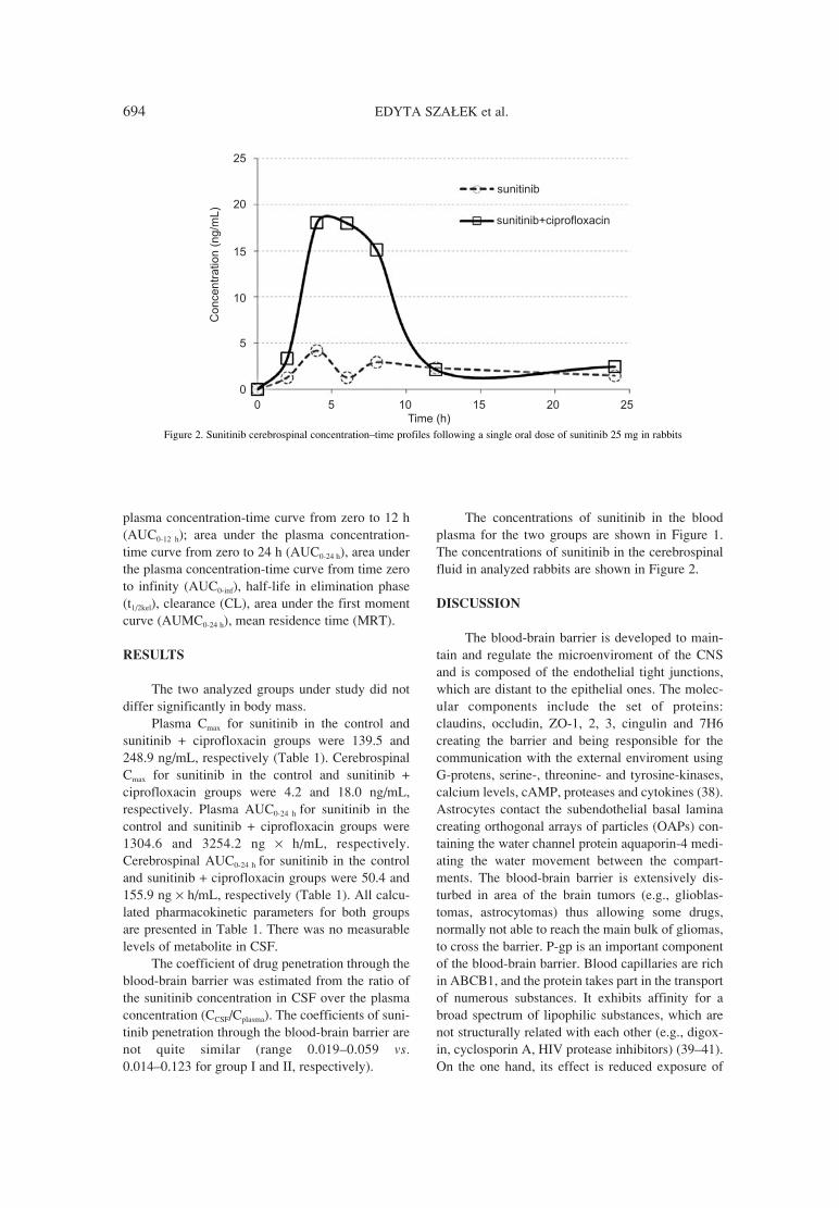
694 EDYTA SZA£EK et al.
plasma concentration-time curve from zero to 12 h(AUC0-12 h); area under the plasma concentration-time curve from zero to 24 h (AUC0-24 h), area underthe plasma concentration-time curve from time zeroto infinity (AUC0-inf), half-life in elimination phase(t1/2kel), clearance (CL), area under the first momentcurve (AUMC0-24 h), mean residence time (MRT).
RESULTS
The two analyzed groups under study did notdiffer significantly in body mass.
Plasma Cmax for sunitinib in the control andsunitinib + ciprofloxacin groups were 139.5 and248.9 ng/mL, respectively (Table 1). CerebrospinalCmax for sunitinib in the control and sunitinib +ciprofloxacin groups were 4.2 and 18.0 ng/mL,respectively. Plasma AUC0-24 h for sunitinib in thecontrol and sunitinib + ciprofloxacin groups were1304.6 and 3254.2 ng × h/mL, respectively.Cerebrospinal AUC0-24 h for sunitinib in the controland sunitinib + ciprofloxacin groups were 50.4 and155.9 ng × h/mL, respectively (Table 1). All calcu-lated pharmacokinetic parameters for both groupsare presented in Table 1. There was no measurablelevels of metabolite in CSF.
The coefficient of drug penetration through theblood-brain barrier was estimated from the ratio ofthe sunitinib concentration in CSF over the plasmaconcentration (CCSF/Cplasma). The coefficients of suni-tinib penetration through the blood-brain barrier arenot quite similar (range 0.019ñ0.059 vs.0.014ñ0.123 for group I and II, respectively).
The concentrations of sunitinib in the bloodplasma for the two groups are shown in Figure 1.The concentrations of sunitinib in the cerebrospinalfluid in analyzed rabbits are shown in Figure 2.
DISCUSSION
The blood-brain barrier is developed to main-tain and regulate the microenviroment of the CNSand is composed of the endothelial tight junctions,which are distant to the epithelial ones. The molec-ular components include the set of proteins:claudins, occludin, ZO-1, 2, 3, cingulin and 7H6creating the barrier and being responsible for thecommunication with the external enviroment usingG-protens, serine-, threonine- and tyrosine-kinases,calcium levels, cAMP, proteases and cytokines (38).Astrocytes contact the subendothelial basal laminacreating orthogonal arrays of particles (OAPs) con-taining the water channel protein aquaporin-4 medi-ating the water movement between the compart-ments. The blood-brain barrier is extensively dis-turbed in area of the brain tumors (e.g., glioblas-tomas, astrocytomas) thus allowing some drugs,normally not able to reach the main bulk of gliomas,to cross the barrier. P-gp is an important componentof the blood-brain barrier. Blood capillaries are richin ABCB1, and the protein takes part in the transportof numerous substances. It exhibits affinity for abroad spectrum of lipophilic substances, which arenot structurally related with each other (e.g., digox-in, cyclosporin A, HIV protease inhibitors) (39ñ41).On the one hand, its effect is reduced exposure of
Figure 2. Sunitinib cerebrospinal concentrationñtime profiles following a single oral dose of sunitinib 25 mg in rabbits

Penetration of sunitinib through the blood-brain barrier after... 695
the brain to toxic compounds. However, on the otherhand, the activity of P-gp may lead to lower effec-tiveness of the treatment of brain tumors, for exam-ple. Thus, it seems logical that the application of acombined therapy with a P-gp inhibitor should resultin better availability of the antineoplastic drug in thebrain and improved clinical efficacy in diseases ofthe CNS. The aim of the research was to determinethe influence of ciprofloxacin on the penetration ofsunitinib into the CSF. The influence ofciprofloxacin on P-gp has not been clearly deter-mined. Park et al. explain that the application of dif-ferent cell lines with P-gp overexpression may leadto different, usually contradictory, conclusionswhether or not a specific drug is a substrate of P-gp.In his research Park concludes that some cell lines(MDCKI and MDCKI-MDR1) indicate thatciprofloxacin is a substrate of P-gp, but data fromother cells (MDCKII, MDCKII-MDR1, LLC-PK1and L-MDR1) contradict it. Brain accumulation ofsunitinib and its metabolite is restricted by ABCB1(23). Additionally, sunitinib inhibits the function ofthe ATP-Binding Cassette (ABC) transporter P-gp(42), which may have influence on the availabilityof simultaneously applied drugs (42).
In the group of animals with ciprofloxacin theCmax of sunitinib in the CSF was 329% higher than
in the control group, whereas AUC0-24 h was 209%higher, which resulted from the higher parameters ofCmax and AUC of sunitinib in the blood in the groupwith the antibiotic (Table 1). Higher plasma concen-trations of active metabolite SU12662 correspondwith higher plasma levels of sunitinib. There was nomeasurable levels of metabolite in CSF. Theobtained values of the pharmacokinetic parametersin the plasma for sunitinib confirm the resultsobtained by the authors in their earlier research onthe influence of fluoroquinolones (ciprofloxacin,levofloxacin, moxifloxacin) on the pharmacokinet-ics of sunitinib (31), where values of mean maxi-mum TKI concentration in the sunitinib +ciprofloxacin group and in the control group were203.8 ± 50.6 vs. 111.8 ± 20.9 ng/mL, respectively.The results point to the inhibiting effect ofciprofloxacin on CYP3A4.
In view of the fact that in both analyzed groupsthe coefficients of sunitinib penetration through theblood-brain barrier are very low we can suppose thatthe influence of ciprofloxacin on P-gp was not sig-nificant.
The research was limited by a small number ofanimals. Nevertheless, even with this number of ani-mals we succeeded in proving visible differences in thePK parameters between the groups. The higher con-
Table 1. Plasma and CSF pharmacokinetic parameters for sunitinib and metabolite (SU12662) following a single oral dose of sunitinib 25 mg.
I II
PharmacokineticsCSF/plasma CSF/plasma
parametersa CSF plasmaratio
CSF plasmaratio
Sunitinib
AUC0-12h (ng◊h/mL) 27.2 869.2 0.031 128.2 1932.6 0.066
AUC0-24h (ng◊h/mL) 50.4 1304.6 0.039 155.9 3254.2 0.048
AUC0-∞∞ (ng◊h/mL) 88.0 1698.0 0.052 185.4 4111.1 0.045
Cmax (ng/mL) 4.2 139.5 0.030 18.0 248.9 0.072
tmax (h) 4.0 4.0 1.0 4.0 6.0 0.667
MRT (h) 11.3 9.4 1.20 8.3 10.5 0.762
AUMC0-24h (ng◊h2/mL) 570.1 12317.4 0.046 1297.0 34106.7 0.038
SU12662
AUC0-24h (ng◊h/mL) - 212.3 - - 258.2 -
AUC0-∞∞ (ng◊h/mL) - 274.6 - - 311.1 -
Cmax (ng/mL) - 12.4 - - 33.3 -
tmax (h) - 8 - - 8 -
AUMC0-24h (ng◊h2/mL) - 2362.7 - - 2611.4 -
I, sunitinib; II, sunitinib + ciprofloxacin; AUC0-12h ñ area under the plasma concentration-time curve from zero to 12h; AUC0-24h ñ area underthe plasma concentration-time curve from zero to 24h; AUC0-∞∞ ñ area under the plasma concentration-time curve from zero to infinity; Cmax
ñ maximum observed plasma concentration; tmax ñ time to reach maximum concentration; MRT ñ mean residence time; AUMC0-24h ñ areaunder the first moment curve; CSF, cerebrospinal fluid

696 EDYTA SZA£EK et al.
centration of sunitinib in the CSF is the consequence ofhigher TKI concentration in the plasma, which proba-bly results from the inhibiting effect of ciprofloxacinon the metabolism of sunitinib. The values of the coef-ficients of TKI penetration through the blood-brainbarrier in both groups seem to indicate that the antibi-otic has little influence on P-glycoprotein.
CONCLUSION
The study revealed increased concentrations ofsunitinib in the cerebrospinal fluid after the admin-istration of ciprofloxacin, but probably it was aneffect of the inhibition of CYP3A4 by the antibioticand increased sunitinib concentrations in the plasmarather than its influence on P-gp.
REFERENCES
1. Homsi J., Daud A.I.: Cancer Control 14, 285(2007).
2. Blumenthal G.M., Cortazar P., Zhang J.J., TangS., Sridhara R., Murgo A., Justice R. et al.:Oncologist 17, 1108 (2012).
3. Adams V.R., Leggas M.: Clin. Ther. 29, 1338(2007).
4. Haznedar J.O., Patyna S., Bello C.L., PengG.W., Speed W., Yu X., Zhang Q. et al.: CancerChemother. Pharmacol. 64, 691 (2009).
5. Houk B.E., Bello C.L., Kang D., Amantea M.:Clin. Cancer Res. 15, 2497 (2009).
6. Faivre S., Delbaldo C., Vera K., Robert C.,Lozahic S., Lassau N., Bello C. et al.: J. Clin.Oncol. 24, 25 (2006).
7. Britten C.D., Kabbinavar F., Hecht J.R., BelloC.L., Li J., Baum C., Slamon D.: CancerChemother. Pharmacol. 61, 515 (2008).
8. Patyna S., Haznedar J., Morris D., FreshwaterK., Peng G., Sukbuntherng J., Chmielewski G.et al.: Birth Defects Res. B Dev. Reprod.Toxicol. 86, 204 (2009).
9. Sza≥ek E., Karbownik A., Po≥om W.,Matuszewski M., SobaÒska K., Urjasz H.,Grabowski T. et al.: Pharmacol. Rep. 64, 1554(2012).
10. Gore M.E., Hariharan S., Porta C., Bracarda S.,Hawkins R., Bjarnason G.A., Oudard S. et al.:Cancer 117, 501 (2011).
11. Koutras A.K., Krikelis D., Alexandrou N.,Starakis I., Kalofonos H.P.: Anticancer Res. 27,4255 (2007).
12. Medioni J., Cojocarasu O., Belcaceres J.L.,Halimi P., Oudard S.: Ann. Oncol. 18, 1282(2007).
13. Zeng H., Li X., Yao J., Zhu Y., Liu J., Yang Y.,Qiang W.: Urol. Int. 83, 482 (2009).
14. Patyna S., Peng J.: Eur. J. Cancer 4, 21 (2006).15. Kaddoumi A., Choi U.P., Kinman L., Whit-
tington D., Tsai C.C., Ho R.J., Anderson B.D. etal.: Drug Metab. Dispos. 35, 1459 (2007).
16. Kemper E.M., van Zandbergen A.E., CleypoolC., Mos H.A., Boogerd W., Beijnen J.H., vanTellingen O.: Clin. Cancer Res. 9, 2849 (2003).
17. Ramakrishnan P.: Einstein Quart. J. Biol. Med.19, 160 (2003).
18. He L., Zhao C., Yan M., Zhang L.Y., Xia Y.Z.:Phytother. Res. 23, 933 (2009).
19. OíBrien F.E., Clarke G., Fitzgerald P., DinanT.G., Griffin B.T., Cryan J.F.: Br. J. Pharmacol.166, 1333 (2012).
20. Hu S., Chen Z., Franke R., Orwick S., Zhao M.,Rudek M.A., Sparreboom A. et al.: Clin. CancerRes. 15, 6062 (2009).
21. Kunimatsu S., Mizuno T., Fukudo M., KatsuraT.: Drug Metab. Dispos. 41, 1592 (2013).
22. Tang S.C., de Vries N., Sparidans R.W.,Wagenaar E., Beijnen J.H., Schinkel A.H.: J.Pharmacol. Exp. Ther. 346, 486 (2013).
23. Sza≥ek E., Tomczak H., KamiÒska A.,Grabowski T., Smuszkiewicz P., Matysiak K.,Wolc A. et al.: Adv. Med. Sci. 57, 217 (2012).
24. Zhanel G.G., Ennis K., Vercaigne L., WalktyA., Gin A.S., Embil J., Smith H. et al.: Drugs62, 13 (2002).
25. Sza≥ek E., KamiÒska A., Goüdzik-SpychalskaJ., Grzeúkowiak E., Batura-Gabryel H.: ActaPol. Pharm. Drug Res. 68, 777 (2011).
26. Sza≥ek E., Po≥om W., Karbownik A.,Grabowski T., Konko≥owicz A., Wolc A.,Matuszewski M. et al.: Pharmacol. Rep. 64, 673(2012).
27. Hedaya M.A., El-Afify D.R., El-MaghrabyG.M.: Biopharm. Drug Dispos. 27, 103 (2006).
28. McLellan R.A., Drobitch R.K., MonshouwerM., Renton K.W.: Drug Metab. Dispos. 24,1134 (1996).
29. Sawant R.D.: Can. J. Clin. Pharmacol. 16, 78(2009).
30. Shahzadi A., Javed I., Aslam B., MuhammadF., Asi M.R., Ashraf M.Y., Zia-ur-Rahman:Pak. J. Pharm. Sci. 24, 63 (2011).
31. Sza≥ek E., Karbownik A., Grabowski T.,SobaÒska K., Wolc A., Grzeúkowiak E.:Pharmacol. Rep. 65, 1383 (2013).
32. Park M.S., Okochi H., Benet L.Z.: Arch. DrugInf. 4, 1 (2011).
33. Sikri V., Pal D., Jain R., Kalyani D., MitraA.K.: Am. J. Ther. 11, 433 (2004).

Penetration of sunitinib through the blood-brain barrier after... 697
34. de Lange E.C., Marchand S., van den Berg D.,van der Sandt I.C., de Boer A.G., Delon A.,Bouquet S. et al.: Eur. J. Pharm. Sci. 12, 85(2000).
35. Bashir S., Jamshaid M., Ahmad B., Iqbal J.:Pak. J. Pharm. Sci. 21, 225 (2008).
36. Meisel J.A., Fallon E.M., Le H.D., Nehra D., deMeijer V.E., Rodig S.J., Puder M.: Surgery 150,32 (2011).
37. Faivre L., Gomo C., Mir O., Taieb F.,Schoemann-Thomas A., Ropert S., Vidal M. etal.: J. Chromatogr. B Analyt. Technol. Biomed.Life Sci. 879, 2345 (2011).
38. Wolburg H., Lippoldt A.: Vascul. Pharmacol.38, 323 (2002).
39. Didier A., Wenger J., Loor F.: AnticancerDrugs 6, 669 (1995).
40. Fenner K.S., Troutman M.D., Kempshall S.,Cook J.A., Ware J.A., Smith D.A., Lee C.A.:Clin. Pharmacol. Ther. 85, 173 (2009).
41. Zastre J.A., Chan G.N., Ronaldson P.T.,Ramaswamy M., Couraud P.O., Romero I.A.,Weksler B. et al.: J. Neurosci. Res. 87, 1023(2009).
42. Shukla S., Robey R.W., Bates S.E., AmbudkarS.V.: Drug Metab. Dispos. 37, 359 (2009).
Received: 19. 09. 2013


Submission of the manuscript
All papers (in duplicate and electronic version) should be submitteddirectly to Editor:
Editor Acta Poloniae Pharmaceutica ñDrug Research16 D≥uga St.00-238 WarsawPoland
We understand that submitted papers are original and not publishedelsewhere.
Authors submitting a manuscript do so on the understandingthat if it is accepted for publication, copyright of the article shall beassigned exclusively to the Publisher.
Scope of the Journal
Acta Poloniae Pharmaceutica - Drug Research publishes papers in allareas of research. Submitted original articles are published in the fol-lowing sections: Reviews, Analysis, Biopharmacy, Drug Biochemistry,Drug Synthesis, Natural Drugs, Pharmaceutical Technology, Pharmacology,Immunopharmacology, General. Any paper that stimulates progress indrug research is welcomed. Both, Regular Articles as well as ShortCommunications and Letters to the Editor are accepted.
Preparation of the manuscript
Articles should be written in English, double-spaced. Full name (first,middle initial, last) and address of authors should follow the title writ-ten in CAPITAL LETTERS. The abstract should be followed by key-words. We suggest the following structure of paper: l) introduction, 2)experimental, 3) results, 4) discussion and conclusion.
Instructions for citation of references in the e-journal:
1. In the text, sequential numbers of citations should be in order ofappearance (not alphabetically) in parentheses (...) not in brackets[Ö].2. In the list of references, for papers the correct order is: number ofreference with dot, family name and initial(s) of author(s), colon,proper abbreviation(s) for journal (Pubmed, Web of Science, no dotneither coma after one word journal name), number of volume, num-ber of issue (if necessary) in parantheses. first page or number of thepaper, year of publication (in parentheses), dot. For books: numberof reference with dot, family name and initial(s) of author(s), colon,title of chapter and/or book names and initials of editors (if any), edi-tion number, page(s) of corresponding information (if necessary),publisher name, place and year of publication.
EXAMPLES:
1. Gadzikowska M., Grynkiewicz G.: Acta Pol. Pharm. Drug Res.59, 149 (2002).
2. Gilbert A.M., Stack G.P., Nilakantan R., Kodah J., Tran M. etal.: Bioorg. Med. Chem. Lett. 14, 515 (2004).
3. Roberts S.M.: Molecular Recognition: Chemical andBiochemical Problems, Royal Society of Chemistry, Cambridge1989.
4. Salem I.I.: Clarithromycin, in Analytical Profiles of DrugSubstances And Excipients. Brittain H.G. Ed., pp. 45-85,Academic Press, San Diego 1996.
5. Homan R.W., Rosenberg H.C.: The Treatment of Epilepsy,Principles and Practices. p. 932 , Lea & Febiger, Philadelphia1993.
6. Balderssarini R.J.: in The Pharmacological Basis ofTherapeutics, 8th edn., Goodman L., Gilman A., Rall T.W., NiesA.S., Taylor P. Eds., Vol 1, p. 383, Pergamon Press, MaxwellMacmillan Publishing Corporation, New York 1985.
7. International Conference on Harmonization Guidelines,Validation of analytical procedures, Proceeding of theInternational Conference on Harmonisation (ICH), Commissionof the European Communities, Geneva 1996.
8. http://www.nhlbi.nih.gov/health/health-topics/topics/ms/(accessed on 03. 10. 2012).
Chemical nomenclature should follow the rules established bythe International Union of Pure and Applied Chemistry, theInternational Union of Biochemistry and Chemical Abstracts Service.Chemical names of drugs are preferred. If generic name is employed,its chemical name or structural formula should be given at point of firstcitation.
Articles should be written in the Past Tense and Impersonalstyle. I, we, me, us etc. are to be avoided, except in the Acknow-ledgment section.
Editor reserves the right to make any necessary corrections to apaper prior to publication.
Tables, illustrations
Each table, figure or scheme should be on a separate pagetogether with the relevant legend and any explanatory notes. Tablesideally should not have more than 70, and certainly not more than 140,characters to the line (counting spaces between columns 4 characters)unless absolutely unavoidable.
Good quality line drawings using black ink on plain A4 paperor A4 tracing paper should be submitted with all lettering etc., includ-ed. Good black and white photographs are also acceptable. Captions forillustrations should be collected together and presented on a separatesheet.
All tables and illustrations should be specially referred to in thetext.
Short Communications and Letters to the Editor
The same general rules apply like for regular articles, exceptthat an abstract is not required, and the number of figures and/or tablesshould not be more that two in total.
The Editors reserve the right to publish (upon agreement ofAuthor(s) as a Short Communication a paper originally submitted as afull-length research paper.
Preparation of the electronic manuscript
We encourage the use of Microsoft Word, however wewill accept manuscripts prepared with other software. Compact Disc -Recordable are preferred. Write following information on the disklabel: name the application software, and the version number used(e.g., Microsoft Word 2007) and specify what type of computer wasused (either IBM compatible PC or Apple MacIntosh).
Fee for papers accepted for publication
Since January 2013 there is a publication fee for papers accepted forpublication in Acta Poloniae Pharmaceutica Drug Research.The fee - 1000 PLN, should be paid before publication on the bankaccount: Polish Pharmaceutical Society, D≥uga 16, 00-238 WarszawaMillennium S.A. account no. 29 1160 2202 0000 0000 2770 0281with a note Ñpublication in Acta Pol. Pharm. Drug Res., paper no.ÖÖ..
For foreign authors the payment (250 �) should be done according tothe data:1. SWIFT Address: BANK MILLENNIUM SA, 02-593 WARSZA-
WA, POLAND, STANIS£AWA ØARYNA 2A St.2. SWIFT CODE: BIGBPLPWXXX3. Beneficiary account Number: PL 30 1160 2202 0000 0000 2777
0200 4. Bank Name: BANK MILLENNIUM SA5. Favoring: POLSKIE TOWARZYSTWO FARMACEUTYCZNE
(Polish Pharmaceutical Society), D£UGA 16, 00-238 WAR-SZAWA, Poland, NIP 526-025-19-54
6. Purpose of sending money: Publication in Acta Pol. Pharm. DrugRes., paper no. Ö..
For payments by Western Union, the name of recipient is KatarzynaTrembulak at the address of Polish Pharmaceutical Society (see above).
Instruction for Authors
