acs nl nl-2011-02559p 1. - semantic scholar€¦b dx.doi.org/10.1021/nl202559p | nano lett.xxxx,...
TRANSCRIPT
rXXXX American Chemical Society A dx.doi.org/10.1021/nl202559p |Nano Lett. XXXX, XXX, 000–000
PERSPECTIVE
pubs.acs.org/NanoLett
Gold Nanoparticles: A Revival in PreciousMetal Administration to PatientsA. S. Thakor,†,‡ J. Jokerst,† C. Zavaleta,† T. F. Massoud,†,‡ and S. S. Gambhir*,†,§
†Molecular Imaging Program at Stanford, Department of Radiology, Stanford University, California 94305-5427, United States‡Department of Radiology, University of Cambridge, Cambridge, CB2 2QQ, U.K.§Departments of Bioengineering & Materials Science and Engineering. Member Bio-X Program, Stanford University,California 94305-5427, United States
One of the most significant developments in recent years hasbeen the development of new materials in the nanometer
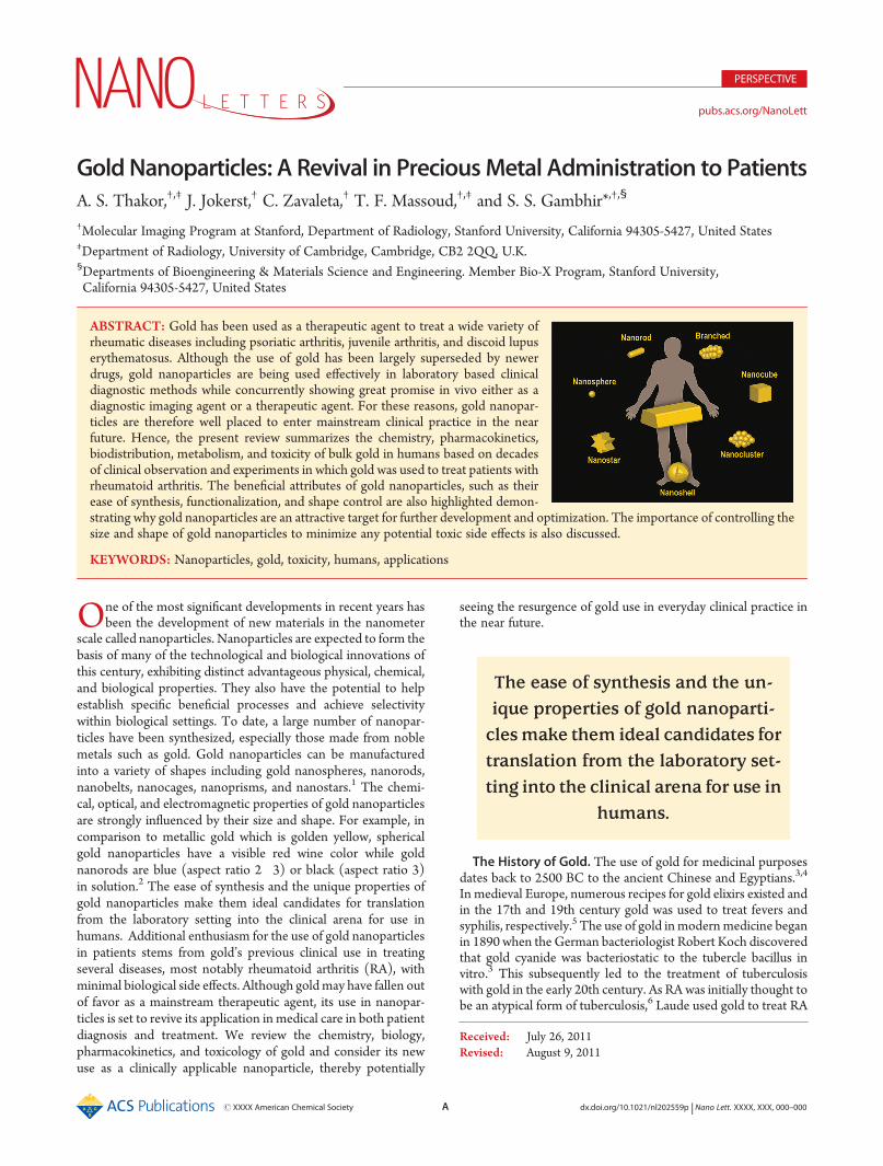
scale called nanoparticles. Nanoparticles are expected to form thebasis of many of the technological and biological innovations ofthis century, exhibiting distinct advantageous physical, chemical,and biological properties. They also have the potential to helpestablish specific beneficial processes and achieve selectivitywithin biological settings. To date, a large number of nanopar-ticles have been synthesized, especially those made from noblemetals such as gold. Gold nanoparticles can be manufacturedinto a variety of shapes including gold nanospheres, nanorods,nanobelts, nanocages, nanoprisms, and nanostars.1 The chemi-cal, optical, and electromagnetic properties of gold nanoparticlesare strongly influenced by their size and shape. For example, incomparison to metallic gold which is golden yellow, sphericalgold nanoparticles have a visible red wine color while goldnanorods are blue (aspect ratio 2�3) or black (aspect ratio 3)in solution.2 The ease of synthesis and the unique properties ofgold nanoparticles make them ideal candidates for translationfrom the laboratory setting into the clinical arena for use inhumans. Additional enthusiasm for the use of gold nanoparticlesin patients stems from gold’s previous clinical use in treatingseveral diseases, most notably rheumatoid arthritis (RA), withminimal biological side effects. Although goldmay have fallen outof favor as a mainstream therapeutic agent, its use in nanopar-ticles is set to revive its application in medical care in both patientdiagnosis and treatment. We review the chemistry, biology,pharmacokinetics, and toxicology of gold and consider its newuse as a clinically applicable nanoparticle, thereby potentially
seeing the resurgence of gold use in everyday clinical practice inthe near future.
The History of Gold. The use of gold for medicinal purposesdates back to 2500 BC to the ancient Chinese and Egyptians.3,4
In medieval Europe, numerous recipes for gold elixirs existed andin the 17th and 19th century gold was used to treat fevers andsyphilis, respectively.5 The use of gold inmodernmedicine beganin 1890 when the German bacteriologist Robert Koch discoveredthat gold cyanide was bacteriostatic to the tubercle bacillus invitro.3 This subsequently led to the treatment of tuberculosiswith gold in the early 20th century. As RA was initially thought tobe an atypical form of tuberculosis,6 Laude used gold to treat RA
The ease of synthesis and the un-ique properties of gold nanoparti-clesmake them ideal candidates fortranslation from the laboratory set-ting into the clinical arena for use in
humans.
Received: July 26, 2011Revised: August 9, 2011
ABSTRACT: Gold has been used as a therapeutic agent to treat a wide variety ofrheumatic diseases including psoriatic arthritis, juvenile arthritis, and discoid lupuserythematosus. Although the use of gold has been largely superseded by newerdrugs, gold nanoparticles are being used effectively in laboratory based clinicaldiagnostic methods while concurrently showing great promise in vivo either as adiagnostic imaging agent or a therapeutic agent. For these reasons, gold nanopar-ticles are therefore well placed to enter mainstream clinical practice in the nearfuture. Hence, the present review summarizes the chemistry, pharmacokinetics,biodistribution, metabolism, and toxicity of bulk gold in humans based on decadesof clinical observation and experiments in which gold was used to treat patients withrheumatoid arthritis. The beneficial attributes of gold nanoparticles, such as theirease of synthesis, functionalization, and shape control are also highlighted demon-strating why gold nanoparticles are an attractive target for further development and optimization. The importance of controlling thesize and shape of gold nanoparticles to minimize any potential toxic side effects is also discussed.
KEYWORDS: Nanoparticles, gold, toxicity, humans, applications

B dx.doi.org/10.1021/nl202559p |Nano Lett. XXXX, XXX, 000–000
Nano Letters PERSPECTIVE
in 1927. Although gold therapy proved to be ineffective fortuberculosis, a study by the Empire Rheumatism Council con-firmed gold to be effective in RA, with Forestier showingbeneficial results in RA patients in 1935.5,7 Gold has since beenused as a therapeutic agent to treat a wide variety of rheumaticdiseases including psoriatic arthritis,8 juvenile arthritis, anddiscoid lupus erythematosus;9,10 however, its use has been largelysuperseded by newer drugs. Gold has also been used in severalother areas of medicine including prostheses in dentistry11 andophthalmology,12 gene delivery,13 and gold coated coronary14 andrenal15 stents, to name a few (Table 1).The Chemistry of Gold. Gold is a noble metal found in group
1B of the periodic table with an atomic number of 197. Gold canexist in a number of oxidation states: (�I), (0), (I), (II), (III),(IV), and (V), however, only gold(0), (I), and (III) are stable inaqueous solution. Hence, in vivo, gold exists in equilibriumbetween its metallic ground state (gold(0)) and its oxidized states(gold(I) or gold(III)).6 Metallic gold does not oxidize or burn inair, even when heated, and has been shown to be inert to strongalkalis and acids thereby making it one of the least chemicallyreactive metals known to man.11 In contrast, gold(I) and (III) areunstable with respect to gold(0), with gold(III) being a strongoxidizing agent which is reduced to gold(I) by biologicallyoccurring reductants such as thiols.5 As gold(I) preferentiallyreacts with S-donors, rather than O- and N- donors, it can bestabilized by thiolate ligands. These resulting gold thiol com-pounds then undergo biological ligand exchange reactions whichaccount, in part, for their pharmacological activity.5
Gold in Humans. Humans contain a mean of 0.35 μg of gold(0) per gram of dry tissue weight16 which, according to calcula-tions byMerchant, equates to 2.45 mg of gold in an average 70 kgman.6 Blood gold concentrations in healthy human subjects havealso been reported to be around 0�0.001 ppm,17 with additionalstudies reporting small quantities of gold in hair (0.3 μg/g), skin(0.03 μg/g), and nails (0.17 μg/g).18�20 Up to 0.8 μg of gold perdry weight has also been measured in fingers beneath goldwedding rings of normal individuals.21 Interestingly, gold is alsosometimes used in food in very minute quantities in pastries,chocolates, and even alcoholic beverages.22
Gold Therapy.On the basis of the chemistry of gold, gold(I) isused as the main therapeutic agent as it is water soluble, is lessreactive than gold(III) and is easily stabilized in a complex by theaddition of ligands. Gold can be delivered to patients intrave-nously, intramuscularly, or orally with gold preparations specifi-cally designed for each particular route of administration.Accordingly, gold taken orally needs to be lipid soluble for it tobe absorbed within the gastrointestinal tract and will thereforehave different physiochemical, pharmacokinetic, and toxicologi-cal properties compared to water soluble gold that is injected.9,23
This is supported by experiments demonstrating only 1% ofinjectable gold is absorbed when given orally compared to 100%when given intramuscularly.24 Although gold has been recentlyused as an anticancer and antimicrobial agent,5 most of thestudies on the efficacy, toxicity, and pharmacokinetics of goldpreparations were previously performed in patients with RA whowere treated with gold in the late 20th century. However, despitegold being used in clinical practice for several decades there is stillconsiderable debate as to whether injectable or oral gold pre-parations are better for patients.25 Initially, oral gold treatmentsthat were developed in the 1980s, such as Auranofin, werethought to have improved pharmacokinetic profiles with lesstissue retention, less toxicity, and reduced serum gold levels thatwere maintained for longer.5 However, their clinical side effectprofile and fear of long-term immune suppression have resultedin injectable compounds, such as gold sodium thiomalate,remaining the preferred gold drugs for RA treatment.25
Gold Pharmacokinetics and Biodistribution. The bioavail-ability of gold in patients very much depends on the route ofadministration. While injectable gold compounds are fully ab-sorbed with maximum levels attained after about 2 h,26 only 20�25% of oral gold is absorbed.23,24,27 Furthermore, intermittentdosing regimens of injectable gold result in fluctuating bloodgold levels with high peak and low trough concentrations.28 Incontrast, oral gold preparations can be taken regularly and madewith prolonged blood half-life preparations, resulting in a nearlyconstant concentration of gold for the duration of a patient’streatment. However, with chronic daily oral administration theserum gold concentrations reaches a plateau, and in some cases,
Table 1. Examples of the Use of Bulk Gold in Clinical Practice
C dx.doi.org/10.1021/nl202559p |Nano Lett. XXXX, XXX, 000–000
Nano Letters PERSPECTIVE
despite constant dosing, gradually starts to decline.29 Variousmechanisms have been put forward to explain this phenomenonincluding insufficient drug compliance, increased drug clearance,or an increased distribution volume (i.e., a shift from protein-bound gold to cell-bound gold).29
Following absorption of gold, either from tissues or thegastrointestinal tract, approximately 95% is bound to albuminand/or globulin where it can remain within the plasma for severalmonths.4,30 Gold has also been found within the cellular com-partment of blood, primarily in the erythrocyte fraction.31,32
Here, gold has been shown to be within or attached to themembranes of red blood cells (RBCs),24,33 with uptake depen-dent on either the amount of gold available for red cell precursorsin the bone marrow or the gold binding capacity of plasmaproteins.32 Indeed, it has been shown that uptake into RBCsceases after 48 h even though there is still considerable gold in theplasma, presumably as all the gold by this time is tightly bound toplasma proteins. As gold uptake into RBCs differs among people,beingmore pronounced in smokers,34 this could explain the largevariability in gold distribution seen among patients. Gold iswidely distributed throughout the body with organs of thereticuloendothelial system, especially the lymph nodes, havingthe greatest affinity for this heavy metal.16 The liver and bonemarrow have each been shown to account for 25% of the totalbody gold burden with the skin and bone each accounting for20%.23 Furthermore, the exact form of gold in these locationsremains unknown although it appears to be inactive as it remainsdetectable in tissue samples taken from patients who had beentreated with gold years earlier.35 In general, gold has littleaffinity for keratinous tissue,18 but it can accumulate in the skindermis36,37 during intravenous administration, with negligiblelevels recorded when gold is given orally.28 At very high levels ofintravenous administration, gold has also been shown to depositin the cornea as detected by slit lamp examination.38
Gold Metabolism and Excretion. Gold is primarily excretedin the urine and feces and although the rate of excretion variesconsiderably from patient to patient, the basic pattern remainsthe same.30,36 Following intramuscular injection of gold, it hasbeen shown that urinary excretion was greatest during the firstday postinjection while fecal excretion was greatest during themiddle of the week.30 Although, the amount of gold excreted inthe urine and feces increases as the amount of injected goldincreases, the excretion rate was not directly proportional to theamount injected.30 The high binding capacity of albumin for goldmay explain the slow rate of gold clearance throughout the weekfollowing gold injection. When gold is given orally, 85�95% isexcreted in feces and the remaining 5�15% in urine, regardless ofdose.27,28 The majority of gold recovered in the feces representsnonabsorbed gold, gold breakdown products, gold shed frommucosal cells to which it was adsorbed and a minor contributionfrom the biliary tract.24,39 Once gold treatment is established, adynamic equilibrium is set up in the body with gold movingbetween the blood, body stores, urine, and feces.Gold Toxicity. Any toxicity associated with gold depends on
its oxidation state when given to patients. Metallic gold (gold(0))is an extremely inert metal which is widely used throughout theworld in both jewellery and prostheses. Indeed, most of the humanpopulation has had prolonged dermal contact with gold(0) in theform of jewelry, with only exceptionally rare cases of adversereactions or allergic contact dermatitis. Furthermore, approxi-mately half of the 1 billion people in the modern industrializedworld carry dental prostheses made of gold with relatively few
cases of oral lesions being reported despite the close prolongedcontact of the metal with the oral mucosa.6 However, gold(0)can, in very minute amounts, be converted to gold(I) by aminoacids contained in sweat and saliva which can then be absorbedthrough the skin or gingival mucosa and later enter phagocyticand antigen presenting cells.40 That notwithstanding, the meta-bolic impact of this is usually insufficient to evoke clinicalsymptoms.6 Moreover, as gold(0) is readily available, has a verylow toxicity profile, and can bemade into a consistently small sizeand shape, it has been used as a delivery vehicle for genetherapy.41 Indeed, microprojectile bombardment of cells withDNAon gold particles has been developed as an effective methodof high frequency gene transfer with minimal damage to livingcells.42 Experiments injecting naked gold beads into the epider-mis of pigs, whose skin is an excellent model for human skin,concluded that apart from acute impact physical effects, whichresulted in mild transient dermal irritation, there were no directtoxicities or adverse effects on health, survival, clinical chemistry,or hematology values related to the gold beads.6
Gold(I) is normally used as therapeutic agent in both inject-able and oral preparations. The toxicities surrounding gold(I)have been primarily understood by examining patients treatedwith gold for RA; however frustratingly, serum and urine levels ofgold have been of no value in predicting impending toxicity inpatients. The most common toxicity associated with gold treat-ment is skin and mucous membrane hypersensitivity reactions,with nonspecific pruritic erythematous, macular, and papularrashes appearing first. Other rarer skin reactions include cheilitis,eosinophilia, chronic papular eruptions, contact sensitivity, er-ythema nodosum, allergic contact purpura, exfoliative dermatitis,and pityriasis rosea.6 This diverse range of dermal reactionsappears not to depend on the gold concentrations in the skin andrarely occur in patients who receive less than 250 mg of goldsalts.11 In fact, it is generally regarded that these reactionsrepresent the balance between the total body burden of goldsalts and the patient’s genetic and metabolic makeup. Manage-ment involves the cessation of gold therapy with most casesresolving within 3months of onset depending on their extent andseverity. The most common form of gold-induced dermatitis isnonallergic, since following clearance of the original eruption,patients can be restarted on gold treatment without developingfurther dermatitis.43 In contrast, allergic contact dermatitis occursat a lower incidence and represents an immune reactivity to goldwhich usually necessitates total cessation of gold therapy.44
Diarrhea is also frequently associated with administration of goldcomplexes, but with a greater incidence when patients use oral goldpreparations.45 Less frequently, gold has been associated withnephrotoxicity as demonstrated byminor and transient proteinuriain people treated with injectable gold complexes.3,46 Occasionally,this may progress to glomerulonephritis with nephritic syndromealthough patients usually recover fully within a few months.9,47
Hematological abnormalities can also be sometimes produced bygold complexes and include eosinophilia, thrombocytopenia,9 andrarely aplastic anemia probably as a result of a direct inhibition ofmyelopoiesis.48 Several other reports have also mentioned the rareconsequences of using gold complexes including entercolitis withbloody diarrhea,49,50 diffuse inflammatory lung reactions,51 andneurotoxicity.52 Gold therapy is not recommended duringpregnancy,53 as animal studies have shown it to be teratogenic.54
Caution is also advised in the puerperium, despite conflictingreports as to whether significant absorption occurs in the infant,since gold can also be found in breast milk.55

D dx.doi.org/10.1021/nl202559p |Nano Lett. XXXX, XXX, 000–000
Nano Letters PERSPECTIVE
Gold(III) is rarely used as a primary therapeutic agent as it is astrong oxidizing agent and thus very reactive. However, gold(I)can transform into gold(III) within phagolysosomes, which mayaccount, in part, for some of its toxic effects. In brief, gold(I) isoxidized to gold(III) via a redox system involving myeloperox-idase and other lysosomal enzymes within phagolysosomescontaining gold (i.e., aurosomes). Gold(III) then diffuses awayfrom its site of generation where it can interact with and denature“self-proteins” surrounding proteins, thereby possibly explainingwhy autoimmunity occurs during a few cases of gold therapy.Gold Nanoparticle Synthesis. Gold nanoparticles can be
synthesized into a variety of different sizes and shapes (Figure 1)by different strategies;56,57 however, the most common methodis by chemical or electrochemical reduction of a gold(III)precursor. Control over shape and size is achieved throughcareful experimental conditions including the specific reducingagent, reaction time, temperature, and use of a capping agent, thelatter binds to select nanoparticle faces and blocks growthbeyond a certain nanometer range.58 The most commonmethodto prepare gold spherical nanoparticles is a single-phase water-based reduction method using citrate reduction as described byTurkevich59 and Frens.60 By variation of the concentration ofcitrate and gold for the thermal reduction, spherical particles ofdifferent sizes can be made.61 Newer methods, such as UVinitiated particle growth, have also recently been introduced toimprove the size distribution and spherical shape of larger sizedgold nanoparticles (i.e., 9�120 nm).61 As spherical gold nano-particles have absorption peaks near 540 nm, the nanoparticlesize and shape can be modulated to bring this peak closer to theoptical window of tissue which is between 700 and 800 nm.Furthermore, the surface plasmon resonance (SPR) peaks of goldnanoparticles can also now be optimally tuned,62 thereby allowingmultiplexing of gold nanoparticles. Recently, significant pro-gress has been made in synthesizing nonspherical gold nanopar-ticles using seed-mediated growth.56,63 For gold nanorods, a goldspherical nanoparticle seed (i.e., diameter of 1�5 nm) is firstsynthesized. Next, a solution containing more gold ions, cetyltetrammonium bromide (CTAB), and ascorbic acid (a mildreducing agent) is added to selectively reduce gold(III) to gold(I).A seed solution containing citrate-capped, penta-twinned goldnanoparticles then catalyzes the reduction of gold(I) ions on theirsurface with a careful choice of experimental conditions enablingthe seed to grow and elongate into a nanorod.56 The aspect ratio iscontrolled by the concentration of silver nitrate in the growthsolution. Branched gold nanoparticles are more difficult to synthe-size reproducibly; however, their sharp edges and correspondinghigh localization of SPRmodesmake them excellent candidates forbiological applications.56 Gold nanostars with a magnetic core areable to couple polarized resonance with spatial control.64 Whengold nanostars are placed in an external magnetic field, theorientation of the points which make up the star shape (and hencefield enhancement) is controlled. Like spheres, gold nanoprisms65
maintain an aspect ratio near unity, yet have red-shifted absorptionresonance peaks. Nanoshells are created by coating a silica orpolymeric core with a thin gold layer,66 the thickness of whichcontrols the optical properties of the nanoparticle which can besubsequently tuned for efficient heating when irradiated with anear-infrared (NIR) laser. A final class of gold nanoparticle isthe gold nanocluster, which contain hundreds of gold atoms(e.g., Au102) and behave as intermediates of nanoparticles andmolecular gold.67 As gold nanoparticles can be easily functionalized
and thus targeted, they are therefore ideal particles for in vivo genedelivery, biological imaging, diagnostics and disease treatment.GoldNanoparticle Toxicity.The toxicity of nanoparticles has
also been suggested to differ dramatically from their correspond-ing bulk material. The small size of nanoparticles will affect theirmode of endocytosis and cellular processing.68 In addition, theirhigh surface area to volume ratio can dramatically alter theirchemical and physical properties resulting in them possessingunexpected toxicities and biological interactions. Since nanopar-ticles will also have a greater amount of their surface in directcontact with the body, they are therefore more reactive to boththemselves and their surrounding environment.69 The mainmolecular mechanism by which nanoparticles incur toxicity hasbeen hypothesized to be from an increase in oxidative stress as aresult of free radical formation.68 These reactive species areexceedingly toxic in vivo, especially within intracellular compart-ments, resulting in the oxidation and damage of lipids, proteins,and DNA. While the slow clearance and tissue accumulation ofthese free radical producing nanoparticles makes organs of thereticuloendothelial system (i.e., the liver and spleen) targets fortoxicity, the high blood flow through organs such as the kidneyand lungs also place these organs at high risk of oxidative damage.When nanoparticles are introduced into the systemic circulation,they can also interact with blood components to cause hemolysisand thrombosis69 and with the immune system to causeimmunotoxicity.70 Furthermore, nanoparticle aggregation fol-lowing systemic administration not only leads to a loss ofnanoparticle function but also can cause end organ damage fromcapillary occlusion.Studies by Chithrani and Chan have shown gold nanoparticles
enter cells via a receptor-mediated clathrin-dependent endocy-tosis pathway, with 50 nm nanoparticles taken up at a faster rateand higher concentration compared to other nanoparticlesizes.71,72 In general, they demonstrated that the rate of uptakeof gold nanoparticles into cells is lower with an increasing aspectratio. In addition, gold spherical nanoparticles have a greaterefficiency of uptake compared to gold nanorods due to thethermodynamic driving forces for membrane wrapping andreceptor diffusion kinetics.71 Despite this, gold nanorods havebeen shown to be more toxic compared to spherical particles.73
One possible explanation for this could lie in their method ofsynthesis with the cationic surfactant CTAB; however, reportsregarding this have been conflicting.69 As the size of the goldnanoparticles decreases, their rate of exocytosis from cells
Figure 1. Schematic representations of gold nanoparticles used inclinical practice.
E dx.doi.org/10.1021/nl202559p |Nano Lett. XXXX, XXX, 000–000
Nano Letters PERSPECTIVE
dramatically and linearly increases, with 14 nm nanoparticlesleaving cells twice as fast compared to particles of 100 nm size.71
Furthermore the fraction of gold nanorods exocytosed washigher than spherical-shaped nanostructures. Studies by Panand colleagues have also shown that the cytotoxicity of goldnanoparticles primarily depends on their size, with particles1�2 nm in diameter being toxic whereas larger 15 nm goldparticles are comparatively nontoxic, irrespective of the cell typetested.74 Furthermore, particles of 1.4 nm were found to behighly toxic as they irreversibly bind to the major grooves ofB-DNA, an effect not observed with larger or smaller particlesdue to steric reasons. Although there are only a limited number ofin vivo studies investigating the systemic effects of gold nano-particles, 13 nm PEG-coated gold nanoparticles have beenshown to have long circulating times, eventually accumulatingin the liver where they can induce acute inflammation andapoptosis.75 The surface charge of gold nanoparticles has alsobeen shown to be important in determining particle toxicity, withcationic gold nanoparticles exhibiting moderate toxicity owing tothe electrostatic binding of the particles to the negatively chargedcell membrane. In contrast, anionic particles have no toxicity asthey are repelled from the membrane.76
Taken together, the size, shape, and surface charge of goldnanoparticles need to be carefully considered when designinggold nanoparticles for human use in order to optimize theirtherapeutic function, while concurrently decreasing their toxicityprofile by minimizing their cellular uptake and interactions. Oneway to reduce any potential toxicity from gold nanoparticles is bythe addition of surface poly(ethylene glycol) (PEG). PEG is acoiled polymer of repeating ethylene ether units with dynamicconformations which are inexpensive, versatile, and FDAapproved.77 In both drug delivery and imaging applications,the addition of PEG to nanoparticles reduces uptake by thereticuloendothelial system and increases circulation time versusuncoated counterparts.78 Recent studies have also shown thatPEGylated nanoparticles generally have lower accumulationin the liver compared to non-PEGylated nanoparticles and highertumor accumulation versus background.79 Aggregation of nano-particles also decrease following the addition of PEG due topassivation of the nanoparticle surface and the reduction inthe coating of serum and tissue proteins, resulting in so-called
“stealth” behavior. PEG also increases the solubility of nanopar-ticles in buffer and serum due to the hydrophilic ethylene glycolrepeats and the enhanced permeability and retention (EPR)effect.80,81 Alternative passivating polymers which can be addedto gold nanoparticles besides PEG include chitosan, dextran,poly(vinylpyrrolidone) (PVP) as well as the copolymer poly-lacticcoglycolic acid (PLGA).Clinical Use of Gold Nanoparticles.Over the past few years,
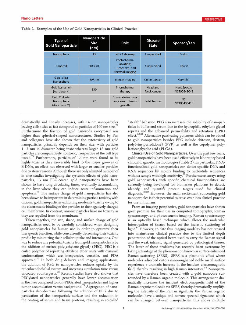
gold nanoparticles have been used effectively in laboratory basedclinical diagnostic methodologies (Table 2). In particular, DNA-functionalized gold nanoparticles can detect specific DNA andRNA sequences by rapidly binding to nucleotide sequenceswithin a sample with high sensitivity.82 Furthermore, arrays usinggold nanoparticles with specific chemical functionalities arecurrently being developed for biomarker platforms to detect,identify, and quantify protein targets used for clinicaldiagnosis.58,83 However, the main excitement concerning goldnanoparticles is their potential to cross over into clinical practicefor use in humans.From an imaging perspective, gold nanoparticles have shown
great promise for their use in computed tomography, Ramanspectroscopy, and photoacoustic imaging. Raman spectroscopyis an optically based technique which allows the molecularinterrogation of tissues based on the inelastic scattering oflight.84 However, to date this imaging modality has not crossedinto mainstream clinical practice due to the limited depthpenetration of the optical beam used to carry the Raman signaland the weak intrinsic signal generated by pathological tissues.The latter of these problems has recently been overcome bytaking advantage of the phenomenon known as surface enhancedRaman scattering (SERS). SERS is a plasmonic effect wheremolecules adsorbed onto a nanoroughened noble metal surfaceexperience a dramatic increase in the incident electromagneticfield, thereby resulting in high Raman intensities.85 Nanoparti-cles have therefore been created with a gold nanocore sur-rounded by a Raman organic molecule. This arrangement dra-matically increases the incident electromagnetic field of theRaman organic molecule via SERS, thereby dramatically amplify-ing the intensity of the Raman signal. As the Raman organicmolecules have a unique and narrow spectral signature, whichcan be changed between nanoparticles, this allows multiple
Table 2. Examples of the Use of Gold Nanoparticles in Clinical Practice
F dx.doi.org/10.1021/nl202559p |Nano Lett. XXXX, XXX, 000–000
Nano Letters PERSPECTIVE
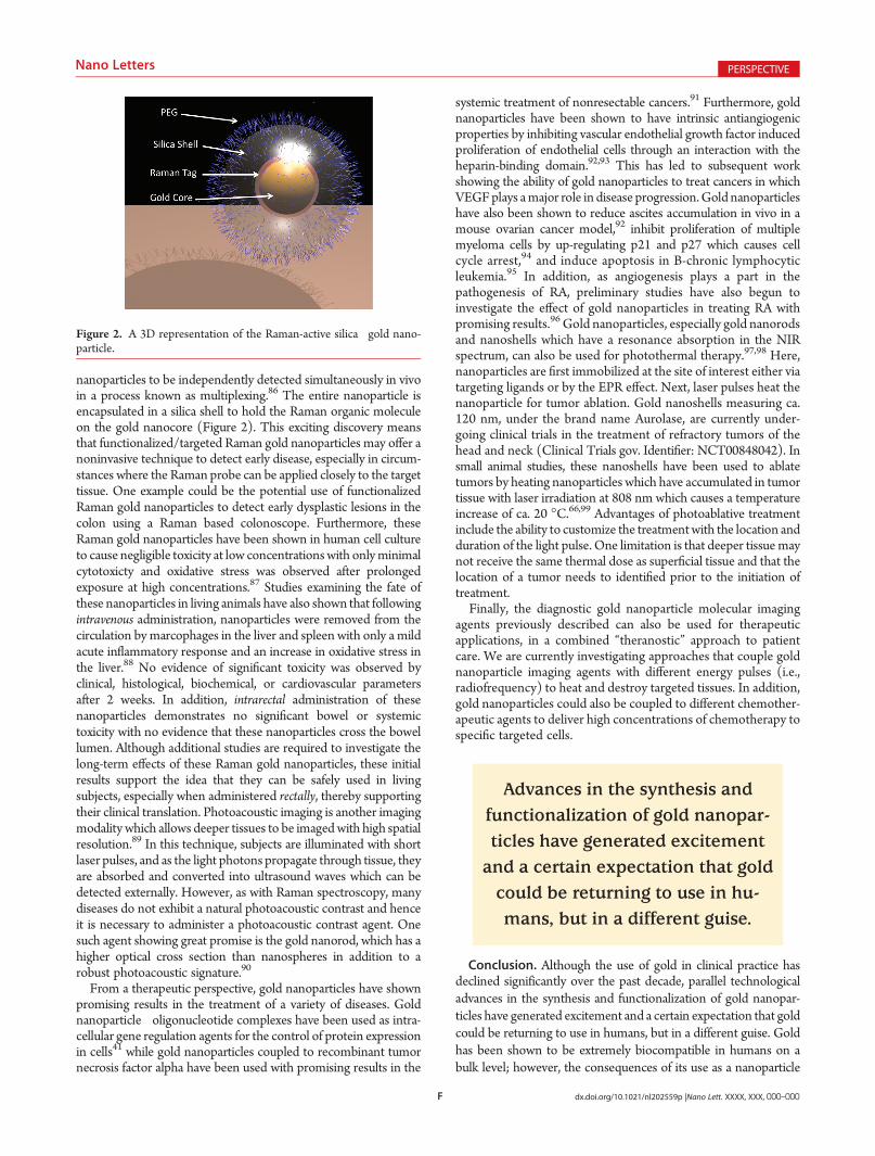
nanoparticles to be independently detected simultaneously in vivoin a process known as multiplexing.86 The entire nanoparticle isencapsulated in a silica shell to hold the Raman organic moleculeon the gold nanocore (Figure 2). This exciting discovery meansthat functionalized/targeted Raman gold nanoparticles may offer anoninvasive technique to detect early disease, especially in circum-stances where the Raman probe can be applied closely to the targettissue. One example could be the potential use of functionalizedRaman gold nanoparticles to detect early dysplastic lesions in thecolon using a Raman based colonoscope. Furthermore, theseRaman gold nanoparticles have been shown in human cell cultureto cause negligible toxicity at low concentrationswith onlyminimalcytotoxicty and oxidative stress was observed after prolongedexposure at high concentrations.87 Studies examining the fate ofthese nanoparticles in living animals have also shown that followingintravenous administration, nanoparticles were removed from thecirculation by marcophages in the liver and spleen with only a mildacute inflammatory response and an increase in oxidative stress inthe liver.88 No evidence of significant toxicity was observed byclinical, histological, biochemical, or cardiovascular parametersafter 2 weeks. In addition, intrarectal administration of thesenanoparticles demonstrates no significant bowel or systemictoxicity with no evidence that these nanoparticles cross the bowellumen. Although additional studies are required to investigate thelong-term effects of these Raman gold nanoparticles, these initialresults support the idea that they can be safely used in livingsubjects, especially when administered rectally, thereby supportingtheir clinical translation. Photoacoustic imaging is another imagingmodality which allows deeper tissues to be imagedwith high spatialresolution.89 In this technique, subjects are illuminated with shortlaser pulses, and as the light photons propagate through tissue, theyare absorbed and converted into ultrasound waves which can bedetected externally. However, as with Raman spectroscopy, manydiseases do not exhibit a natural photoacoustic contrast and henceit is necessary to administer a photoacoustic contrast agent. Onesuch agent showing great promise is the gold nanorod, which has ahigher optical cross section than nanospheres in addition to arobust photoacoustic signature.90
From a therapeutic perspective, gold nanoparticles have shownpromising results in the treatment of a variety of diseases. Goldnanoparticle�oligonucleotide complexes have been used as intra-cellular gene regulation agents for the control of protein expressionin cells41 while gold nanoparticles coupled to recombinant tumornecrosis factor alpha have been used with promising results in the
systemic treatment of nonresectable cancers.91 Furthermore, goldnanoparticles have been shown to have intrinsic antiangiogenicproperties by inhibiting vascular endothelial growth factor inducedproliferation of endothelial cells through an interaction with theheparin-binding domain.92,93 This has led to subsequent workshowing the ability of gold nanoparticles to treat cancers in whichVEGFplays amajor role in disease progression.Gold nanoparticleshave also been shown to reduce ascites accumulation in vivo in amouse ovarian cancer model,92 inhibit proliferation of multiplemyeloma cells by up-regulating p21 and p27 which causes cellcycle arrest,94 and induce apoptosis in B-chronic lymphocyticleukemia.95 In addition, as angiogenesis plays a part in thepathogenesis of RA, preliminary studies have also begun toinvestigate the effect of gold nanoparticles in treating RA withpromising results.96 Gold nanoparticles, especially gold nanorodsand nanoshells which have a resonance absorption in the NIRspectrum, can also be used for photothermal therapy.97,98 Here,nanoparticles are first immobilized at the site of interest either viatargeting ligands or by the EPR effect. Next, laser pulses heat thenanoparticle for tumor ablation. Gold nanoshells measuring ca.120 nm, under the brand name Aurolase, are currently under-going clinical trials in the treatment of refractory tumors of thehead and neck (Clinical Trials gov. Identifier: NCT00848042). Insmall animal studies, these nanoshells have been used to ablatetumors by heating nanoparticles which have accumulated in tumortissue with laser irradiation at 808 nm which causes a temperatureincrease of ca. 20 �C.66,99 Advantages of photoablative treatmentinclude the ability to customize the treatmentwith the location andduration of the light pulse. One limitation is that deeper tissue maynot receive the same thermal dose as superficial tissue and that thelocation of a tumor needs to identified prior to the initiation oftreatment.Finally, the diagnostic gold nanoparticle molecular imaging
agents previously described can also be used for therapeuticapplications, in a combined “theranostic” approach to patientcare. We are currently investigating approaches that couple goldnanoparticle imaging agents with different energy pulses (i.e.,radiofrequency) to heat and destroy targeted tissues. In addition,gold nanoparticles could also be coupled to different chemother-apeutic agents to deliver high concentrations of chemotherapy tospecific targeted cells.
Conclusion. Although the use of gold in clinical practice hasdeclined significantly over the past decade, parallel technologicaladvances in the synthesis and functionalization of gold nanopar-ticles have generated excitement and a certain expectation that goldcould be returning to use in humans, but in a different guise. Goldhas been shown to be extremely biocompatible in humans on abulk level; however, the consequences of its use as a nanoparticle
Figure 2. A 3D representation of the Raman-active silica�gold nano-particle.
Advances in the synthesis andfunctionalization of gold nanopar-ticles have generated excitementand a certain expectation that goldcould be returning to use in hu-mans, but in a different guise.

G dx.doi.org/10.1021/nl202559p |Nano Lett. XXXX, XXX, 000–000
Nano Letters PERSPECTIVE
may be determined by alternative chemical and biological properties.Thus, gold nanoparticles for use in humans should be designed basedon data from both bulk gold treatment in humans and in vivo goldnanoparticle validation experiments. Taken together, nanoparticlesmade ofmetallic gold (gold(0)) which are spherical in shape, anionic,and of a size greater than 20 nm would be expected to have the leasttoxicity in humans. Furthermore, the gold nanoparticle preparationshould be optimized depending on its method of delivery (i.e.,intravenous vs oral vs intrarectal) to decrease systemic absorptionand distribution while increasing urinary and fecal excretion. Futureresearch will need to determine the optimal gold nanoparticles foreach potential human application, and inevitably, trade-offs will haveto be made regarding some of their diagnostic and therapeuticproperties vis-a-vis their associated toxicity profile. Overall, goldnanoparticles are ideally placed to make the transition from thelaboratory benchtop to the clinical bedside in the very near future.
’AUTHOR INFORMATION
Corresponding Author*E-mail: [email protected].
’ACKNOWLEDGMENT
Supported by the NCI Center for Cancer NatotechnologyExcellence (CCNE) U54 CA119367 (S.S.G.), NCI Network forTranslational Research (NTR): Optical Imaging in Multimod-ality Platforms (S.S.G.), the NCI In vivo cellular and MolecularImaging Centers (ICMIC) P50 CA114747 (S.S.G.), the CanaryFoundation (S.S.G.), the American Cancer Society (A.S.T.), theEuropean Association for Cancer Research (A.S.T.), and the PEELMedical Research Trust (A.S.T.), NIHR Cambridge BiomedicalResearch Centre (AST and T.F.M.), and Ben & Catherine IvyFoundation (SSG).
’REFERENCES
(1) Yang, D. P.; Cui, D. X.Chem.—Asian J. 2008, 3 (12), 2010–2022.(2) Burda, C.; Chen, X.; Narayanan, R.; El-Sayed, M. A. Chem. Rev.
2005, 105 (4), 1025–1102.(3) Antonovych, T. T. Ann. Clin. Lab. Sci. 1981, 11 (5), 386–391.(4) Lewis, A. J.; Walz, D. T. Prog. Med. Chem. 1982, 19, 1–58.(5) Fricker, S. P. Gold Bull. 1996, 29 (2), 53.(6) Merchant, B. Biologicals 1998, 26 (1), 49–59.(7) Clark, P.; Tugwell, P.; Bennet, K.; Bombardier, C.; Shea, B.;
Wells, G.; Suarez-Almazor, M. E. Cochrane Database Syst. Rev. 2000, No.2, CD000520.(8) Ujfalussy, I.; Koo, E.; Sesztak, M.; Gergely, P. Z. Rheumatol.
2003, 62 (2), 155–160.(9) Champion, G. D.; Graham, G. G.; Ziegler, J. B. Baillieres Clin.
Rheumatol. 1990, 4 (3), 491–534.(10) Dalziel, K.; Going, G.; Cartwright, P. H.; Marks, R.; Beveridge,
G. W.; Rowell, N. R. Br. J. Dermatol. 1986, 115 (2), 211–216.(11) Belies, R. P. Patty’s Industrial Hygeine and Toxicology, 4th ed.;
John Wiley & Sons Inc: New York, 1994; pp 2021�2032.(12) Bair, R. L.; Harris, G. J.; Lyon, D. B.; Komorowski, R. A.
Ophthalmol. Plast. Reconstr. Surg. 1995, 11 (3), 209–214.(13) Pertmer, T. M.; Eisenbraun, M. D.; McCabe, D.; Prayaga, S. K.;
Fuller, D. H.; Haynes, J. R. Vaccine 1995, 13 (15), 1427–1430.(14) Kastrati, A.; Schomig, A.; Dirschinger, J.; Mehilli, J.; vonWelser,
N.; Pache, J.; Schuhlen, H.; Schilling, T.; Schmitt, C.; Neumann, F. J.Circulation 2000, 101 (21), 2478–2483.(15) Nolan, B. W.; Schermerhorn, M. L.; Powell, R. J.; Rowell, E.;
Fillinger, M. F.; Rzucidlo, E. M.; Wyers, M. C.; Whittaker, D.; Zwolak,R. M.; Walsh, D. B.; Cronenwett, J. L. J. Vasc. Surg. 2005, 42 (1), 40–46.
(16) Gottlieb, N. L.; Smith, P. M.; Smith, E. M. Arthritis Rheum.1972, 15 (1), 16–22.
(17) Plantin, L. L’analyse par radioactivations; Presses universitairesde France: Paris, 1964.
(18) Gottlieb, N. L.; Smith, P. M.; Penneys, N. S.; Smith, E. M.Arthritis Rheum. 1974, 17 (1), 56–62.
(19) Tipton, I. H.; Cook, M. J. Health Phys. 1963, 9, 103–145.(20) Petushkov, A. A.; Linekin, D. M.; Balcius, J. F.; Brownell, G. L.
J. Nucl. Med. 1969, 10 (12), 730–731.(21) Brown, D. H.; Smith,W. E.; Fox, P.; Sturrock, R. D. Inorg. Chim.
Acta 1982, 67, 27–30.(22) Russell, M. A.; King, L. E., Jr.; Boyd, A. S. N. Engl. J. Med. 1996,
334 (9), 603.(23) Gottlieb, N. L. Scand. J. Rheumatol. Suppl. 1983, 51, 10–14.(24) Walz, D. T.; DiMartino, M. J.; Griswold, D. E.; Intoccia, A. P.;
Flanagan, T. L. Am. J. Med. 1983, 75 (6A), 90–108.(25) Kean, W. F.; Kean, I. R. Inflammopharmacology 2008, 16 (3),
112–125.(26) Palmer, D. G.; Dunckley, J. V. Aust. N. Z. J. Med. 1973, 3 (5),
461–466.(27) Blocka, K.; Furst, D. E.; Landaw, E.; Dromgoole, S.; Blomberg,
A.; Paulus, H. E. J. Rheumatol. Suppl. 1982, 8, 110–119.(28) Gottlieb, N. L. J. Rheumatol. Suppl. 1982, 8, 99–109.(29) Van Riel, P. L.; Gribnau, F. W.; Van de Putte, L. B.; Arts, C. W.;
Van Aernsbergen, A. Clin. Rheumatol. 1987, 6 (1), 50–54.(30) Mascarenhas, B. R.; Granda, J. L.; Freyberg, R. H. Arthritis
Rheum. 1972, 15 (4), 391–402.(31) Smith, P. M.; Smith, E. M.; Gottlieb, N. L. J. Lab. Clin. Med.
1973, 82 (6), 930–937.(32) Pedersen, S. M.; Graabaek, P. M.Ann. Rheum. Dis. 1980, 39 (6),
576–579.(33) Walz, D. T.; Griswold, D. E.; DiMartino, M. J.; Bumbier, E. E.
J. Rheumatol. Suppl. 1979, 5, 56–60.(34) Graham, G. G.; Champion, G. D.; Haavisto, T. M.; McNaught,
P. J. Ann. Rheum. Dis. 1981, 40 (2), 210.(35) Grahame, R.; Billings, R.; Laurence, M.; Marks, V.; Wood, P. J.
Ann. Rheum. Dis. 1974, 33 (6), 536–539.(36) Gottlieb, N. L.; Smith, P. M.; Smith, E. M. Arthritis Rheum.
1972, 15 (6), 582–592.(37) Penneys, N. S.; Kramer, K.; Gottlieb, N. L. J. Invest. Dermatol.
1975, 65 (3), 331–333.(38) Hashimoto, A.; Maeda, Y.; Ito, H.; Okazaki, M.; Hara, T.
Arthritis Rheum. 1972, 15 (3), 309–315.(39) Weisman, M. H.; Hardison, W. G.; Walz, D. T. J. Rheumatol.
1980, 7 (5), 633–638.(40) Rapson, W. S. Contact Dermatitis 1985, 13 (2), 56–65.(41) Rosi, N. L.; Giljohann, D. A.; Thaxton, C. S.; Lytton-Jean, A. K.;
Han, M. S.; Mirkin, C. A. Science 2006, 312 (5776), 1027–1030.(42) Particle bombardment technology for gene transfer; Yang, N.-S.,
Christou, P., Eds.; Oxford University Press: Oxford, 1994.(43) Penneys, N. S.; Ackerman, A. B.; Gottlieb, N. L. Arch. Dermatol.
1974, 109 (3), 372–376.(44) Rasanen, L.; Kaipiainen-Seppanen,O.;Myllykangas-Luosujarvi, R.;
Kasnanen, T.; Pollari, P.; Saloranta, P.; Horsmanheimo, M. Br. J. Dermatol.1999, 141 (4), 683–688.
(45) Heuer, M. A.; Pietrusko, R. G.; Morris, R. W.; Scheffler, B. J.J. Rheumatol. 1985, 12 (4), 695–699.
(46) Horton, R. J. Scand. J. Rheumatol. Suppl. 1983, 51, 100–110.(47) Clark, P.; Tugwell, P.; Bennett, K.; Bombardier, C. J. Rheumatol.
1989, 16 (4), 442–447.(48) Hamilton, J. A.;Williams, N. J. Rheumatol. 1985, 12 (5), 892–896.(49) Langer, H. E.; Hartmann, G.; Heinemann, G.; Richter, K. Ann.
Rheum. Dis. 1987, 46 (10), 787–792.(50) Rocha, M. P.; Burrichter, P. J.; Blodgett, R. C. Semin. Arthritis
Rheum. 1987, 16 (4), 294–299.(51) Evans, R. B.; Ettensohn, D. B.; Fawaz-Estrup, F.; Lally, E. V.;
Kaplan, S. R. Semin. Arthritis Rheum. 1987, 16 (3), 196–205.

H dx.doi.org/10.1021/nl202559p |Nano Lett. XXXX, XXX, 000–000
Nano Letters PERSPECTIVE
(52) Levine, J. D.; Moskowitz, M. A.; Basbaum, A. I. Neurosci. Lett.1988, 87 (1�2), 200–202.(53) Preston, S.; Needs, C. Baillieres Clin. Rheumatol. 1990, 4 (3),
687–698.(54) Szabo, K. T.; Guerriero, F. J.; Kang, Y. J. Vet. Pathol. Suppl.
1978, 15 (5), 89–96.(55) Jones, G.; Brooks, P. M. Br. J. Rheumatol. 1996, 35 (11),
1154–1158.(56) Grzelczak, M.; Perez-Juste, J.; Mulvaney, P.; Liz-Marzan, L. M.
Chem. Soc. Rev. 2008, 37 (9), 1783–1791.(57) Murphy, C. J.; Sau, T. K.; Gole, A. M.; Orendorff, C. J.; Gao, J.;
Gou, L.; Hunyadi, S. E.; Li, T. J. Phys. Chem. B 2005, 109 (29),13857–13870.(58) Baptista, P.; Pereira, E.; Eaton, P.; Doria, G.; Miranda, A.;
Gomes, I.; Quaresma, P.; Franco, R. Anal. Bioanal. Chem. 2008, 391 (3),943–950.(59) Turkevich, J. Discuss. Faraday Soc. 1951, 55.(60) Frens, G. Nat. Phys. Sci. 1973, 241, 20–22.(61) Kimling, J.; Maier, M.; Okenve, B.; Kotaidis, V.; Ballot, H.;
Plech, A. J. Phys. Chem. B 2006, 110 (32), 15700–15707.(62) Hu, M.; Chen, J.; Li, Z. Y.; Au, L.; Hartland, G. V.; Li, X.;
Marquez, M.; Xia, Y. Chem. Soc. Rev. 2006, 35 (11), 1084–1094.(63) Nikoobakht, B.; El-Sayed, M. A. Chem. Mater. 2003, 15 (10),
1957–1962.(64) Wei, Q.; Song, H. M.; Leonov, A. P.; Hale, J. A.; Oh, D.; Ong,
Q. K.; Ritchie, K.; Wei, A. J. Am. Chem. Soc. 2009, 131 (28), 9728–9734.(65) Jin, R.; Cao, Y.; Mirkin, C. A.; Kelly, K. L.; Schatz, G. C.; Zheng,
J. G. Science 2001, 294 (5548), 1901–1903.(66) Hirsch, L. R.; Stafford, R. J.; Bankson, J. A.; Sershen, S. R.;
Rivera, B.; Price, R. E.; Hazle, J. D.; Halas, N. J.; West, J. L. Proc. Natl.Acad. Sci. U.S.A. 2003, 100 (23), 13549–13554.(67) Jadzinsky, P. D.; Calero, G.; Ackerson, C. J.; Bushnell, D. A.;
Kornberg, R. D. Science 2007, 318 (5849), 430–433.(68) Lanone, S.; Boczkowski, J. Curr. Mol. Med. 2006, 6 (6),
651–663.(69) Aillon, K. L.; Xie, Y.; El-Gendy, N.; Berkland, C. J.; Forrest,
M. L. Adv. Drug. Deliv. Rev. 2009.(70) Dobrovolskaia, M. A.; McNeil, S. E. Nat. Nanotechnol. 2007, 2
(8), 469–478.(71) Chithrani, B. D.; Chan, W. C. Nano Lett. 2007, 7 (6),
1542–1550.(72) Chithrani, B. D.; Ghazani, A. A.; Chan,W. C.Nano Lett. 2006, 6
(4), 662–668.(73) Wang, S.; Lu, W.; Tomvmachenko, O.; Rai, U. S.; Yu, H.; Ray,
P. C. Chem. Phys. Lett. 2008, 463, 145–149.(74) Pan, Y.; Neuss, S.; Leifert, A.; Fischler, M.; Wen, F.; Simon, U.;
Schmid, G.; Brandau, W.; Jahnen-Dechent, W. Small 2007, 3 (11),1941–1949.(75) Cho,W. S.; Cho, M.; Jeong, J.; Choi, M.; Cho, H. Y.; Han, B. S.;
Kim, S. H.; Kim, H. O.; Lim, Y. T.; Chung, B. H. Toxicol. Appl.Pharmacol. 2009, 236 (1), 16–24.(76) Goodman, C. M.; McCusker, C. D.; Yilmaz, T.; Rotello, V. M.
Bioconjugate Chem. 2004, 15 (4), 897–900.(77) Knop, K.; Hoogenboom, R.; Fischer, D.; Schubert, U. S. Angew.
Chem., Int. Ed. 2010, 49 (36), 6288–308.(78) van Vlerken, L. E.; Vyas, T. K.; Amiji, M. M. Pharm. Res. 2007,
24 (8), 1405–1414.(79) Gref, R.; Minamitake, Y.; Peracchia, M. T.; Trubetskoy, V.;
Torchilin, V.; Langer, R. Science 1994, 263 (5153), 1600–1603.(80) Kanaras, A. G.; Kamounah, F. S.; Schaumburg, K.; Kiely, C. J.;
Brust, M. Chem. Commun. (Cambridge, U.K.) 2002, 20, 2294–2295.(81) Kwon, G. S. Crit. Rev. Ther. Drug Carrier Syst. 2003, 20 (5),
357–403.(82) Mirkin, C. A.; Letsinger, R. L.; Mucic, R. C.; Storhoff, J. J.
Nature 1996, 382 (6592), 607–609.(83) You, C. C.; Miranda, O. R.; Gider, B.; Ghosh, P. S.; Kim, I. B.;
Erdogan, B.; Krovi, S. A.; Bunz, U. H.; Rotello, V. M. Nat. Nanotechnol.2007, 2 (5), 318–323.
(84) Hanlon, E. B.; Manoharan, R.; Koo, T. W.; Shafer, K. E.; Motz,J. T.; Fitzmaurice, M.; Kramer, J. R.; Itzkan, I.; Dasari, R. R.; Feld, M. S.Phys. Med. Biol. 2000, 45 (2), R1–59.
(85) Banholzer, M. J.; Millstone, J. E.; Qin, L.; Mirkin, C. A. Chem.Soc. Rev. 2008, 37 (5), 885–897.
(86) Zavaleta, C. L.; Smith, B. R.; Walton, I.; Doering, W.; Davis, G.;Shojaei, B.; Natan, M. J.; Gambhir, S. S. Proc. Natl. Acad. Sci. U.S.A. 2009,106 (32), 13511–13516.
(87) Thakor, A. S.; Paulmurugan, R.; Kempen, P.; Zavaleta, C.;Sinclair, R.; Massoud, T. F.; Gambhir, S. S. Small 2011, 7 (1), 126–136.
(88) Thakor, A. S.; Luong, R.; Paulmurugan, R.; Lin, F. I.; Kempen,P.; Zavaleta, C.; Chu, P.; Massoud, T. F.; Sinclair, R.; Gambhir, S. S. Sci.Transl. Med. 2011, 3 (79), 79ra33.
(89) De la Zerda, A.; Zavaleta, C.; Keren, S.; Vaithilingam, S.;Bodapati, S.; Liu, Z.; Levi, J.; Smith, B. R.; Ma, T. J.; Oralkan, O.; Cheng,Z.; Chen, X.; Dai, H.; Khuri-Yakub, B. T.; Gambhir, S. S.Nat. Nanotechnol.2008, 3 (9), 557–562.
(90) Chen, Y. S.; Frey, W.; Kim, S.; Kruizinga, P.; Homan, K.;Emelianov, S. Nano Lett. 2011, 11 (2), 348–354.
(91) Libutti, S. K.; Paciotti, G. F.; Byrnes, A. A.; Alexander, H. R., Jr.;Gannon, W. E.; Walker, M.; Seidel, G. D.; Yuldasheva, N.; Tamarkin, L.Clin. Cancer Res. 2010, 16 (24), 6139–6149.
(92) Mukherjee, P.; Bhattacharya, R.; Wang, P.; Wang, L.; Basu, S.;Nagy, J. A.; Atala, A.; Mukhopadhyay, D.; Soker, S. Clin. Cancer Res.2005, 11 (9), 3530–3534.
(93) Bhattacharya, R.; Mukherjee, P. Adv. Drug Delivery Rev. 2008,60 (11), 1289–1306.
(94) Bhattacharya, R.; Patra, C., R.; Verma, R.; Kumar, P., R.; Greipp,P.; Mukherjee, P. Adv. Mater. 2007, 19, 711–716.
(95) Mukherjee, P.; Bhattacharya, R.; Bone, N.; Lee, Y. K.; Patra,C. R.; Wang, S.; Lu, L.; Secreto, C.; Banerjee, P. C.; Yaszemski, M. J.;Kay, N. E.; Mukhopadhyay, D. J Nanobiotechnology 2007, 5, 4.
(96) Tsai, C. Y.; Shiau, A. L.; Chen, S. Y.; Chen, Y. H.; Cheng, P. C.;Chang, M. Y.; Chen, D. H.; Chou, C. H.;Wang, C. R.; Wu, C. L. ArthritisRheum. 2007, 56 (2), 544–554.
(97) von Maltzahn, G.; Park, J. H.; Agrawal, A.; Bandaru, N. K.; Das,S. K.; Sailor, M. J.; Bhatia, S. N. Cancer Res. 2009, 69 (9), 3892–3900.
(98) Gobin, A. M.; Lee, M. H.; Halas, N. J.; James, W. D.; Drezek,R. A.; West, J. L. Nano Lett. 2007, 7 (7), 1929–1934.
(99) Schwartz, J. A.; Shetty, A. M.; Price, R. E.; Stafford, R. J.; Wang,J. C.; Uthamanthil, R. K.; Pham, K.; McNichols, R. J.; Coleman, C. L.;Payne, J. D. Cancer Res. 2009, 69 (4), 1659–1667.