acquired resistance to the trk inhibitor entrectinib in ... · acquired resistance to the trk...
TRANSCRIPT
1
Acquired resistance to the TRK inhibitor
entrectinib in colorectal cancer
Mariangela Russo1,2,3*, Sandra Misale2*, Ge Wei4*, Giulia Siravegna1,2,
Giovanni Crisafulli2, Luca Lazzari1,2, Giorgio Corti2, Giuseppe Rospo2, Luca
Novara2, Benedetta Mussolin2, Alice Bartolini2, Nicholas Cam4, Roopal Patel4,
Shunqi Yan4, Robert Shoemaker4, Robert Wild4, Federica Di Nicolantonio1,2,
Andrea Sartore Bianchi5, Gang Li4, Salvatore Siena5,6, §, Alberto Bardelli1,2, §,#
1University of Torino, Department of Oncology, SP 142, Km 3.95, 10060 Candiolo, Torino, Italy;
2Candiolo Cancer Institute – FPO, IRCCS, Candiolo, Torino, Italy; 3 FIRC Institute of Molecular
Oncology (IFOM), Milano, Italy; 4 Ignyta, Inc. San Diego, CA, USA; 5Department of Hematology and
Oncology, Niguarda Cancer Center, Ospedale Niguarda Ca’ Granda, Milan, Italy; 6Università degli Studi
di Milano, Milan, Italy
* Drs. Russo, Misale and Wei contributed equally to this article; § Drs. Bardelli and Siena are co-senior
author; # Address correspondence to:
Alberto Bardelli at: [email protected]
University of Torino, Department of Oncology, FPO, IRCCS,
SP 142, Km 3.95, Candiolo (TO) , ZIP 10060 , Italy
Phone: +39-011-9933548
Running Title:
TRKA mutations and resistance to entrectinib in CRC
Key words:
Colorectal, acquired resistance, NTRK1, TRK inhibition, ctDNA
Conflict of interest
Ignyta co-authors are full time emplyees of Ignyta, and own Ignyta stocks. All
the other authors do not have conflict of interest to declare.
Research. on November 6, 2015. © 2015 American Association for Cancercancerdiscovery.aacrjournals.org Downloaded from
Author manuscripts have been peer reviewed and accepted for publication but have not yet been edited. Author Manuscript Published OnlineFirst on November 6, 2015; DOI: 10.1158/2159-8290.CD-15-0940
2
Abstract
Entrectinib is a first-in-class pan-TRK kinase inhibitor currently undergoing
clinical testing in colorectal cancer and other tumor types. A patient with
metastatic colorectal cancer harboring an LMNA-NTRK1 rearrangement
displayed a remarkable response to treatment with entrectinib, which was
followed by the emergence of resistance. To characterize the molecular bases
of the patient’s relapse, circulating tumor DNA (ctDNA) was collected
longitudinally during treatment and a tissue biopsy, obtained before entrectinib
treatment, was transplanted in mice (xenopatient), which then received the
same entrectinib regimen until resistance developed. Genetic profiling of
ctDNA and xeno-patient samples showed acquisition of two point mutations in
the catalytic domain of NTRK1, p.G595R and p.G667C. Biochemical and
pharmacological analysis in multiple preclinical models confirmed that either
mutation renders the TRKA kinase insensitive to entrectinib. These findings
can be immediately exploited to design next generation TRKA inhibitors.
Significance
We provide proof of principle that analyses of xenopatients (avatar) and liquid
biopsies allow the identification of drug resistance mechanisms in parallel with
clinical treatment of individual patient. We describe for the first time that
p.G595R and p.G667C TRKA mutations drive acquired resistance to
entrectinib in colorectal cancers carrying NTRK1 rearrangements.
Research. on November 6, 2015. © 2015 American Association for Cancercancerdiscovery.aacrjournals.org Downloaded from
Author manuscripts have been peer reviewed and accepted for publication but have not yet been edited. Author Manuscript Published OnlineFirst on November 6, 2015; DOI: 10.1158/2159-8290.CD-15-0940

3
Introduction
TRK receptors are a family of tyrosine kinases that comprises three members:
TRKA, TRKB and TRKC, encoded by the NTRK1 (neurotrophic tyrosine
kinase receptor, type 1), NTRK2 and NTRK3 genes, respectively. Genomic
rearrangement is the most common mechanism of oncogenic activation for
this family of receptors, resulting in sustained cancer cell proliferation through
activation of MAPK and AKT downstream pathways (1). Rearrangements of
the NTRK1, NTRK2 and NTRK3 genes occur across different tumors
including colorectal cancers (CRCs) (2).
Entrectinib (RXDX-101, previously known as NMS-E628) is a potent pan-
TRK, ALK, ROS1 inhibitor, currently undergoing phase I clinical trial(3).
During treatment with entrectinib a patient with metastatic colorectal cancer
harboring an LMNA-NTRK1 rearrangement showed a remarkable response.
We reasoned that, as it has been shown for most targeted agents, response
to entrectinib might be limited in time due to the emergence of acquired
resistance. Nothing is presently known on the mechanisms of resistance to
entrectinib and consequently further lines of treatment are not available. We
postulated that it might be possible to identify the resistance mechanism(s)
while the patient was being treated by analyzing circulating tumor DNA
(ctDNA) and developing a xenopatient (avatar).
Results
Research. on November 6, 2015. © 2015 American Association for Cancercancerdiscovery.aacrjournals.org Downloaded from
Author manuscripts have been peer reviewed and accepted for publication but have not yet been edited. Author Manuscript Published OnlineFirst on November 6, 2015; DOI: 10.1158/2159-8290.CD-15-0940

4
Acquired resistance to TRKA inhibition in a CRC patient.
A molecular screen identified a genetic rearrangement involving exon 10 of
NTRK1 and exon 11 of the LMNA genes(4) in a patient with metastatic
colorectal cancer (mCRC) whose disease was intrinsically resistant to 1st line
FOLFOX, 2nd line FOLFIRI/cetuximab and 3rd line Irinotecan. We and others
have previously reported that CRC cell models harboring NTRK1
translocations are sensitive to NTRK1 silencing and to TRKA (protein
encoded by NTRK1 gene) kinase inhibition (5-7). Based on this, the patient
was enrolled in the phase I ALKA clinical trial (EudraCT Number 2012-
000148-88) of the pan-TRK kinase inhibitor entrectinib, a first-in-class drug
currently undergoing clinical testing(3) . The patient received entrectinib on an
intermittent dosing schedule of 4 days on/3 days off for three weeks followed
by a week break in every 28-day cycle(4). Treatment was remarkably effective
and well tolerated, leading to a partial response (PR) with 30% tumor
shrinkage of multiple liver metastases that was demonstrated by an early CT
scan assessment performed after 30 days of treatment. The clinical response
lasted four months, followed by the emergence of drug resistance as
evaluated by RECIST (Response Evaluation Criteria in Solid Tumor)
progression (Fig. 1 upper panels).
Emergence of NTRK1 mutations in ctDNA during entrectinib treatment
To unveil the molecular basis of acquired resistance to TRKA inhibition we
analyzed circulating tumor DNA (ctDNA), a form of liquid biopsy (8) we
Research. on November 6, 2015. © 2015 American Association for Cancercancerdiscovery.aacrjournals.org Downloaded from
Author manuscripts have been peer reviewed and accepted for publication but have not yet been edited. Author Manuscript Published OnlineFirst on November 6, 2015; DOI: 10.1158/2159-8290.CD-15-0940

5
previously optimized to detect and monitor drug resistance in patients treated
with targeted agents (9,10).
ctDNA extracted from plasma samples collected before treatment initiation
and at clinical relapse was subjected to molecular profiling using the IRCC-
TARGET panel, an NGS-platform based on 226 cancer related genes (10).
Profiling of ctDNA at entrectinib resistance revealed two novel NTRK1 genetic
alterations in the kinase domain of the protein, p.G595R and p.G667C, which
were not detected in ctDNA obtained before initiation of therapy
(Supplementary Tables S1, S2). To monitor the NTRK1 mutated alleles in the
plasma of the patient collected through the treatment, droplet digital PCR
(ddPCR) (11,12) assays were designed for both mutations. As a mean of
tracking the overall disease, a ddPCR assay was also optimized to detect the
LMNA-NTRK1 rearrangement in ctDNA.
Longitudinal analysis of plasma revealed that the p.G595R and p.G667C
mutated alleles were initially absent in ctDNA but emerged in the circulation
as early as 4 weeks upon initiation of treatment with entrectinib (Fig. 1).
NTRK1 mutations frequencies continued to increase in ctDNA and peaked
when clinical progression was radiologically confirmed (16 weeks after
initiation of treatment). The profile of the LMNA-NTRK1 rearrangement in
ctDNA paralleled tumor response and resistance to entrectinib (Fig. 1;
Supplementary Table S3).
Secondary resistance to entrectinib in CRC Xenopatient
Research. on November 6, 2015. © 2015 American Association for Cancercancerdiscovery.aacrjournals.org Downloaded from
Author manuscripts have been peer reviewed and accepted for publication but have not yet been edited. Author Manuscript Published OnlineFirst on November 6, 2015; DOI: 10.1158/2159-8290.CD-15-0940

6
To functionally evaluate the mechanistic basis of resistance to entrectinib, a
biopsy specimen gathered before initiation of treatment was transplanted
subcutaneously in an immunocompromised mouse (xenopatient) (see
Supplementary Methods). Upon successful engraftment, the tumor was
expanded in multiple mice, which were treated with dosage levels and
schedules that matched clinically relevant exposure achievable in patients.
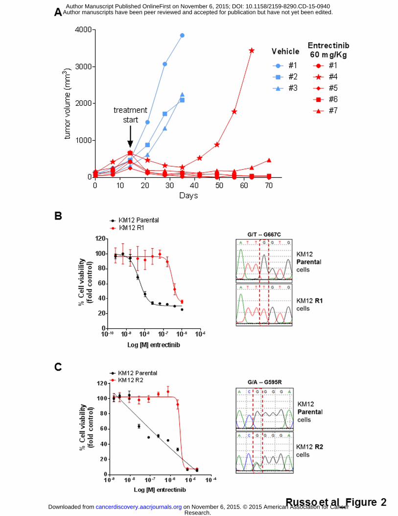
Entrectinib induced remarkable tumor shrinkage in the xenopatient while
vehicle treated tumors grew exponentially (Fig. 2A). After 3 weeks of drug
dosing, one of the tumors treated with entrectinib rapidly developed resistance
to TRKA inhibition (Fig. 2A). NGS-based molecular profiling of this resistant
sample using the IRCC-TARGET panel unveiled the LMNA-NTRK1
rearrangement peculiar of the patient and the NTRK1 p.G595R mutation,
which could not be detected in the untreated tumor (Supplementary Fig. S1A,
B; Supplementary Tables S1, S2) .
Secondary resistance to entrectinib in cells carrying NTRK1
rearrangements
To assess whether the mechanism of resistance was patient-specific or
contingent on the peculiar NTRK1 rearrangements, independent models of
acquired resistance to entrectinib were established. The KM12 CRC cell line
harbors a distinct genetic rearrangement involving exon 10 of NTRK1 and
exon 7 of TPM3 gene (5,7) and is also highly sensitive to entrectinib (Fig.
2B,C). Independent batches of parental (sensitive) KM12 cells were exposed
to either acute constant dose (R2) or escalating doses (R1) of entrectinib until
resistant derivatives emerged (Fig. 2B,C) (see Supplementary Methods).
Research. on November 6, 2015. © 2015 American Association for Cancercancerdiscovery.aacrjournals.org Downloaded from
Author manuscripts have been peer reviewed and accepted for publication but have not yet been edited. Author Manuscript Published OnlineFirst on November 6, 2015; DOI: 10.1158/2159-8290.CD-15-0940
7
Molecular profiling of the cells that became resistant to lower concentrations
of entrectinib (30-100 nM) (named KM12 R1) revealed the missense mutation
p.G667C in the kinase domain of NTRK1, previously identified also in the
plasma of the patient (Fig. 2B). When cells were made resistant to a higher
doses (1-2 µM) of the drug (named KM12 R2), the NTRK1 p.G595R alteration
was detected (Fig. 2C). The experiment was repeated multiple times, the two
mutations were never concomitantly detected in the same resistant
populations indicating they occurred in independent cells.
To further evaluate the mechanisms of entrectinib resistance we engineered
Ba/F3 cells to express ETV6-TRKA. In this model system the ETV6 domain
mimics the dimerization effect of TRK fusion partners that occur in human
tumors. Ba/F3 cells engineered to express ETV6-TRKA became exquisitely
sensitive to entrectinib (Supplementary Fig.S2). ETV6-TRKA Ba/F3 cells were
then exposed to entrectinib treatment until resistant derivatives emerged and
analyzed as described above. Remarkably upon development of resistance
Ba/F3 also acquired p.G595R mutation in the kinase domain of TRKA when a
high dose of entrectinib was applied, while the p.G667C allele emerged in the
presence of a lower dose of the drug (Supplementary Fig.S2). Analogously to
what we observed in KM12, the two mutations were found in independent
pools of Ba/F3 cells indicating they do not co-occur in the same cells.
NTRK1 p.G595R and p.G667C mutations drive resistance to TRK
inhibitors
Research. on November 6, 2015. © 2015 American Association for Cancercancerdiscovery.aacrjournals.org Downloaded from
Author manuscripts have been peer reviewed and accepted for publication but have not yet been edited. Author Manuscript Published OnlineFirst on November 6, 2015; DOI: 10.1158/2159-8290.CD-15-0940
8
We then examined the impact of the p.G595R and p.G667C variants on the 3-
dimensional (3D) structure of the TRKA catalytic domain (see Supplementary
Methods). The binding model of entrectinib with wild type (WT) TRKA
highlighted that entrectinib makes extensive hydrogen bonding as well as
hydrophobic interactions with the protein in the ATP pocket where p.G595 and
p.G667 residues are located (Fig. 3A). The p.G595R and p.G667C mutations
create steric hindrance that either abrogates binding (p.G595R) or reduces
the binding affinity (p.G667C) of entrectinib to the TRKA catalytic pocket (Fig.
3B,C respectively).
We next assessed whether and to what extent mutations in the kinase domain
of NTRK1 drive resistance to TRKA inhibition. We engineered Ba/F3 cells
expressing wild type , p.G595R or p.G667C TPM3-TRKA fusion proteins. We
then measured the sensitivity of NTRK1 mutated cells to TRK inhibitors
currently in clinical development. LOXO-101 is a TRK inhibitor in phase 1 trial
for patients with advanced solid tumors with NTRK alterations
(NCT02122913); TSR-011 is presently undergoing a phase 1 trial for patients
with advanced solid tumors or lymphomas with NTRK alterations
(NCT02048488).
As shown in Supplementary Fig.S3, Ba/F3 cells harboring the NTRK1
translocation become highly sensitive to TRK inhibitors (Supplementary Fig.
S3 A,B; Supplementary Table S4). On the contrary NTRK1 p.G595R or
p.G667C mutations are resistant to entrectinib, LOXO-101 and TSR-011
(Supplementary Fig. S3 C,D respectively). Of potential clinical relevance, and
Research. on November 6, 2015. © 2015 American Association for Cancercancerdiscovery.aacrjournals.org Downloaded from
Author manuscripts have been peer reviewed and accepted for publication but have not yet been edited. Author Manuscript Published OnlineFirst on November 6, 2015; DOI: 10.1158/2159-8290.CD-15-0940

9
in line with previous results, NTRK1 p.G595R appears to be more potent in
conferring resistance than p.G667C.
These results are indeed consistent with the observation that entrectinib and
LOXO-101 retain a partial effect on p.G667C (IC50=61 nM; IC50= 524nM
respectively) but are totally ineffective on p.G595R (IC50>1000 nM) in Ba/F3
engineered cells (Supplementary Table S4).
Alignment of the TRKA kinase domain with clinically targeted tyrosine
kinases, such as ALK, ROS, EGFR, MET and KIT, showed that the glycine
residues at position 595 and 667 lie in a conserved region (Supplementary
Fig. S4A and S4B respectively), and are analogous to residues previously
found to be associated with secondary resistance to other kinase inhibitors
such as erlotinib, crizotinib and imatinib (Fig.3D,E respectively).
Biochemical characterization of NTRK1 p.G595C and p.G667C in
xenopatient derived cells.
To mechanistically study the impact of NTRK1 resistant alleles, we
established two cell lines, one from the xenopatient treated with vehicle, and
the other from the xenopatient that became resistant to entrectinib (Fig. 4A).
Both cell lines displayed the LMNA-NTRK1 translocation found in the patient
tumor (Figure 4B), but only cells derived from the xenotumor that had become
resistant to entrectinib carried the p.G595R allele (Figure 4C). Both cell lines
displayed a pharmacological response to entrectinib analogous to that
observed in the corresponding xenopatients (Fig.4D). Biochemical
Research. on November 6, 2015. © 2015 American Association for Cancercancerdiscovery.aacrjournals.org Downloaded from
Author manuscripts have been peer reviewed and accepted for publication but have not yet been edited. Author Manuscript Published OnlineFirst on November 6, 2015; DOI: 10.1158/2159-8290.CD-15-0940
10
characterization confirmed that NTRK1 secondary mutations render the
corresponding proteins insensitive (or only marginally sensitive) to entrectinib
and capable of activating downstream signaling in the presence of the drug
(Fig. 4E, F). We next asked whether the tumor cell that had become resistant
remained dependent on the expression of TRKA. Indeed, siRNA-mediated
suppression of mutant NTRK1 in resistant cells induced apoptosis, similarly to
the knockdown of WT NTRK1 in sensitive cells (Fig. 4G).
Discussion
A subset of CRCs carries NTRK1 translocations, which also occur in other
tumor types such as lung tumors and thyroid carcinomas (6,13-15). The TRK
inhibitor entrectinib induced a remarkable clinical response in a patient with a
metastatic colorectal cancer carrying a LMNA-NTRK1 translocation, whose
disease was intrinsically refractory to three prior lines of therapy including
anti-EGFR inhibition(4) . However, after four months of treatment, resistance
developed in this patient. The entrectinib half-life is 17-44h and the
intermittent dosing regimen may have promoted or anticipated the
development of resistance due to incomplete treatment coverage of the
patient. Nevertheless, it is still unknown whether or not continuous dosing will
affect the emergence and/or the type of acquired mutations.
In this work, we sought to identify mechanisms of resistance to entrectinib, as
this is key to development of additional lines of therapy for patients carrying
NTRK1 rearrangements. The most commonly used approach to study
Research. on November 6, 2015. © 2015 American Association for Cancercancerdiscovery.aacrjournals.org Downloaded from
Author manuscripts have been peer reviewed and accepted for publication but have not yet been edited. Author Manuscript Published OnlineFirst on November 6, 2015; DOI: 10.1158/2159-8290.CD-15-0940
11
resistance to targeted therapies involves molecular profiling of tissue biopsy
obtained at progression. However, tumor heterogeneity and tissue sampling
limit the effectiveness of this strategy. In addition, tissues biopsies are not
always feasible and are associated with non-negligible risks (16). Most
importantly, even when the biopsy reveals emergence of alleles that were not
present before treatment, their functional role in driving resistance remains to
be formally established using functional assays. This requires significant
experimental efforts, and the timeframe is not compatible with further
treatment of the patient from whom the biopsy was obtained. We find that
coupling pharmacological analyses of xenopatients with molecular profiles of
liquid biopsies allows the identification of resistance mechanisms in parallel
with clinical treatment of individual patients, thus potentially enabling
decisions on following treatment options.
We report for the first time that acquisition of p.G595R and p.G667C
mutations in the kinase domain of TRKA drive secondary resistance to TRK
inhibition in CRC cells carrying NTRK1 rearrangements. Both mutations were
detected in patient plasma obtained at progression, suggesting that both are
indeed associated with acquired resistance to entrectinib in the clinical setting.
We found a remarkable concordance among results obtained in clinical
samples and preclinical models. Genomic profiling of patient derived samples
and multiple cell models pointed to the p.G595R or p.G667C NTRK1
mutations as the only common mechanism of resistance to entrectinib.
Research. on November 6, 2015. © 2015 American Association for Cancercancerdiscovery.aacrjournals.org Downloaded from
Author manuscripts have been peer reviewed and accepted for publication but have not yet been edited. Author Manuscript Published OnlineFirst on November 6, 2015; DOI: 10.1158/2159-8290.CD-15-0940

12
Analysis of a larger number of patients will ultimately be needed to determine
the clinical impact of the findings. Based on data obtained with other
anticancer therapies based on kinase inhibitors, it is possible that other
mechanisms of resistance to entrectinib could occur, including activation of
parallel pathways able to bypass TRKA inhibition.
Interestingly, we found that the emergence of each of the two mutations might
be dependent on the entrectinib concentration used. NTRK1 p.G667C
emerged when cells were exposed to a low concentration of the inhibitor,
while it was absent from cells made resistant to higher dose.
Structural model-based characterization also indicates that the potency of the
p.G667C mutation in conferring resistance to entrectinb is weaker than
p.G595R. Both mutations fall in the ATP binding pocket and are analogous to
resistance mutations which have been described for other clinically druggable
tyrosine kinase fusions. While p.G595C completely abrogates the binding of
entrectinib to TRKA, the p.G667C only reduces affinity of binding. In line with
this, homology alignment showed that TRKA p.G595R is analogous to ALK
p.G1202R, while TRKA p.G667C to ALK p.G1269A. As for ALK p.G1269A
which mediates resistance to crizotinib, this alteration can be overcome with
second generation ALK inhibitors, such as ceritinib and alectinib, while both
are ineffective on ALK p.G1202R(17-19); entrectinib at clinically achievable
exposure still retain a partial effect on p.G667C but not on p.G595R TRKA.
Of note, while we found that cells that develop entrectinib’s resistance remain
dependent on the expression of TRKA, none of the TRK inhibitors current
being tested in the clinic (LOXO-101 and TSR-011) can overcome resistance
Research. on November 6, 2015. © 2015 American Association for Cancercancerdiscovery.aacrjournals.org Downloaded from
Author manuscripts have been peer reviewed and accepted for publication but have not yet been edited. Author Manuscript Published OnlineFirst on November 6, 2015; DOI: 10.1158/2159-8290.CD-15-0940

13
driven by p.G595R. Accordingly, the biochemical and pharmacological
characterization of the preclinical models described here highlight the need of
developing next generation TRKA inhibitors that do not rely on specific spatial
accommodation of the drug-target interaction around G595 region, aimed at
overcoming resistance driven by the p.G595R variant.
In addition to providing clues for the development of second generation TRK
inhibitors, our finding offer means of tracking –non invasively- the emergence
of resistance to entrectinib. Monitoring of NTRK1 resistant variants (p.G595R
and p.G667C) in the plasma of patients treated with entrectinib could be
valuable to predict recurrences.
Material and Methods
Cells lines authentication
KM12 CRC cells were obtained from NCI60 cell line bank and authenticated
in May 2011. The genetic identity of cell line was last checked no less than
three months before performing experiments by Cell ID™ System and by
Gene Print® 10 System (Promega), throught Short Tandem Repeats (STR) at
10 different loci (D5S818, D13S317, D7S820, D16S539, D21S11, vWA,
TH01, TPOX, CSF1PO and amelogenin). Amplicons from multiplex PCRs
were separated by capillary electrophoresis (3730 DNA Analyzer, Applied
Biosystems) and analyzed using GeneMapperID software from Life
Research. on November 6, 2015. © 2015 American Association for Cancercancerdiscovery.aacrjournals.org Downloaded from
Author manuscripts have been peer reviewed and accepted for publication but have not yet been edited. Author Manuscript Published OnlineFirst on November 6, 2015; DOI: 10.1158/2159-8290.CD-15-0940
14
Technologies. Cell lines were tested and resulted negative for mycoplasma
contamination with Venor GeM Classic Kit (Minerva Biolabs).
Establishment of primary colorectal cancer cell line
Primary colorectal cancer cell lines were established from tumor tissues
obtained from patient-derived xenografts. Tumor tissues were dissociated into
single-cell suspension by mechanical dissociation using the gentleMACS
Dissociator (Miltenyi Biotec) and enzymatic degradation of the extracellular
matrix using the Tumor Dissociation Kit (Miltenyi Biotec) according to
manufacturer’s instructions. The cell suspension was then centrifuged at 1200
rpm for 5 minutes. Supernatants were removed and cell pellets were
resuspended with DMEM/F12 medium containing 10% FBS. This process
was repeated 3 times. Then, cell suspensions were filtered through a 70µm
cell strainer (Falcon) and resuspended with culture media DMEM-F12
containing 2 mM L-glutamine, antibiotics (100 U/mL penicillin and 100 µg/mL
streptomycin), gentamicin 50 µg/ml and 10 μM ROCK inhibitor Y-27632
(Selleck Chemicals Inc.).
Ba/F3-TPM3-TRKA WT , G595R and G667C generation
To generate Ba/F3 cells expressing TPM3-TRKA WT, TPM3-TRKA G595R
and TPM3-TRKA G667C the cDNAs were cloned from KM12 cells by RT-
PCR and inserted into a lentiviral vector pVL-EF1a-MCS-IRES-Puro
(BioSettia, San Diego, CA). After confirmation by direct sequencing, VSVG-
pseudo-typed lentiviruses were introduced into the murine IL-3 dependent
pro-B cell Ba/F3. The transduced Ba/F3 cells were selected at 1 µg/mL of
Research. on November 6, 2015. © 2015 American Association for Cancercancerdiscovery.aacrjournals.org Downloaded from
Author manuscripts have been peer reviewed and accepted for publication but have not yet been edited. Author Manuscript Published OnlineFirst on November 6, 2015; DOI: 10.1158/2159-8290.CD-15-0940
15
puromycin in the murine IL-3 containing RPMI and 10% FBS media for two
weeks. The stable cell pools were further selected in RPMI and 10% FBS
media without murine IL-3 for 4 weeks.
Drugs
Entrectinib, LOXO-101(20) and TSR-011(21) were obtained from Ignyta, San
Diego (CA, USA).
Patient’s samples collection
Patient’s plasma and tumor biopsy were obtained through protocols approved
by local Ethical Committee at Ospedale Niguarda Ca ' Granda, Milano, Italy.
The study was conducted according to the provisions of the Declaration of
Helsinki, and patient signed and provided his/her informed consent before
sample collection. The liver biopsy was subcutaneously implanted in NOD-
SCID mouse and experiments were performed according to a study protocol
approved by Ethical Committee at Ospedale Niguarda Ca ' Granda, Milano,
Italy.
Droplet digital PCR analysis
Isolated circulating free DNA was amplified using ddPCR™ Supermix for
Probes (Bio-Rad) with LMNA-NTRK1 translocation, NTRK1 p.G595R and
NTRK1 p.G667C assays (sequences of custom designed probes are listed in
Supplementary Table S5). ddPCR was then performed according to
manufacturer’s protocol and the results reported as percentage or fractional
abundance of mutant DNA alleles to total (mutant plus wild type) DNA alleles.
Research. on November 6, 2015. © 2015 American Association for Cancercancerdiscovery.aacrjournals.org Downloaded from
Author manuscripts have been peer reviewed and accepted for publication but have not yet been edited. Author Manuscript Published OnlineFirst on November 6, 2015; DOI: 10.1158/2159-8290.CD-15-0940
16
8–10 µl of DNA template was added to 10 µl of ddPCR Supermix for Probes
(Bio-Rad) and 2 µl of the primer and probe mixture. This reaction mix was
added to a DG8 cartridge together with 60 µl of Droplet Generation Oil for
Probes (Bio-Rad) and used for droplet generation. Droplets were then
transferred to a 96-well plate (Eppendorf) and then thermal cycled with the
following conditions: 5 min at 95 °C, 40 cycles of 94 °C for 30 s, 55 °C for 1
min followed by 98 °C for 10 min (Ramp Rate 2 °C/s). Droplets were analyzed
with the QX200 Droplet Reader (Bio-Rad) for fluorescent measurement of
FAM and HEX probes. Gating was performed based on positive and negative
controls, and mutant populations were identified. The ddPCR data were
analyzed with QuantaSoft analysis software (Bio-Rad) to obtain fractional
abundance of the mutated alleles in the wild-type or normal background. The
quantification of the target molecule was presented as number of total copies
(mutant plus WT) per sample in each reaction. The number of positive and
negative droplets is used to calculate the concentration of the target and
reference DNA sequences and their Poisson-based 95% confidence intervals,
as previously shown(22). ddPCR analysis of normal control plasma DNA
(from cell lines) and no DNA template controls were always included.
Samples with too low positive events were repeated at least twice in
independent experiments to validate the obtained results.
Next Generation Sequencing analysis
Libraries were prepared with Nextera Rapid Capture Custom Enrichment Kit
(Illumina Inc., San Diego, CA, USA), according to the manufacturer’s protocol.
Preparation of libraries was performed using up to 150ng of plasma ctDNA
Research. on November 6, 2015. © 2015 American Association for Cancercancerdiscovery.aacrjournals.org Downloaded from
Author manuscripts have been peer reviewed and accepted for publication but have not yet been edited. Author Manuscript Published OnlineFirst on November 6, 2015; DOI: 10.1158/2159-8290.CD-15-0940

17
and 100 ng of gDNA from both cells and avatar fresh tissue. gDNA was
fragmented using transposons, adding simultaneously adapter sequences.
For ctDNA libraries preparation was used NEBNext® Ultra™ DNA Library
Prep Kit for Illumina® (New England BioLabs Inc., Ipswich MA), with optimized
protocol. Purified gDNA after the tagmentation step, and ctDNA were used as
template for subsequent PCR to introduce unique sample barcodes.
Fragments’ size distribution of the DNA was assessed using the 2100
Bioanalyzer with a High Sensitivity DNA assay kit (Agilent Technologies,
Santa Clara, CA). Equal amount of DNA libraries were pooled and subjected
to targeted panel hybridization capture. Libraries were then sequenced using
Illumina MiSeq sequencer (Illumina Inc., San Diego, CA, USA).
Bioinformatic analysis
FastQ files generated by Illumina sequencer were mapped to the human
reference (assembly hg19) using BWA-mem algorithm(23); PCR duplicates
were then removed using the SAMtools package(24). Xenome software(25)
was applied to remove murine sequences from xenopatient samples prior to
alignment. We used a custom script pipeline for NGS in order to call somatic
variations when supported by at least 1.5% allelic frequency and 5%
significance level obtained with a Fisher's Test. Mutational analyses were the
result of comparison between pre- and post-treatment samples.
Detection of LMNA-NTRK1 rearrangement in ctDNA
ctDNA obtained from blood draw collected before entrectinib treatment started
was analyzed by NGS as described above. To unveil the specific LMNA-
Research. on November 6, 2015. © 2015 American Association for Cancercancerdiscovery.aacrjournals.org Downloaded from
Author manuscripts have been peer reviewed and accepted for publication but have not yet been edited. Author Manuscript Published OnlineFirst on November 6, 2015; DOI: 10.1158/2159-8290.CD-15-0940
18
NTRK1 genetic rearrangement, a combination of BWA (v. 0.7.10) and BLAT
(v. 35) was used. BWA was first used to align reads to the hg19 human
reference genome with default options. The reads with a non perfect
alignment from BWA, potentially containing translocations, were extracted and
aligned using BLAT (tileSize 11 and stepSize 5). The resulting PSL alignment
was then post-processed to detect chimeric alignments. Gene fusion calling
was performed using the following criteria: i) each fusion partner must have at
least 25 mapped bases on the respective end of the read; ii) the fusion
partners must map to two different genes; iii) each reported fusion breakpoint
must be supported by at least 10 reads. Based on the fusion sequence
identified by NGS analysis, specific ddPCR primers and probes for the LMNA-
NTRK1 rearrangement were designed using Primer3 Input (version 0.4.0)
following BioRad instructions available on the website. Primers and probes
sequences are listed in Supplementary Table S5.
Kinase domain alignment
The aminoacidic sequences of human TRKA [P04629], ALK [Q9UM73],
ROS1 [P08922], EGFR [P00533], KIT [P10721] and MET [P08581] were
obtained from UniprotKB database (26). Their kinase domains were aligned
using the MUSCLE tool (27) and results were post-processed using Jalview
(28).
Acknowledgments
Research. on November 6, 2015. © 2015 American Association for Cancercancerdiscovery.aacrjournals.org Downloaded from
Author manuscripts have been peer reviewed and accepted for publication but have not yet been edited. Author Manuscript Published OnlineFirst on November 6, 2015; DOI: 10.1158/2159-8290.CD-15-0940

19
Supported by the European Community’s Seventh Framework Programme
under grant agreement no. 602901, MErCuRIC (A.B.); Innovative Medicines
Initiative grant no.115749, CANCER-ID (A.B.) Associazione Italiana per la
Ricerca sul Cancro (AIRC) IG grant no. 12812 (A.B.); AIRC 2010 Special
Program Molecular Clinical Oncology 5 per mille, project no. 9970 (A.B.);
FPRC 5 per mille 2010 and 2011 Ministero della Salute (F.D.N. and A.B.);
Ministero dell’Istruzione, dell’Università e della Ricerca, progetto PRIN (A.B.).
Investigators at Niguarda Cancer Center (ASB, SS) are also supported by
Fondazione Oncologia Niguarda Onlus.
References 1. Vaishnavi A, Le AT, Doebele RC. TRKing down an old oncogene in a new era of targeted therapy. Cancer Discov 2015;5(1):25-34. 2. Shaw AT, Hsu PP, Awad MM, Engelman JA. Tyrosine kinase gene rearrangements in epithelial malignancies. Nat Rev Cancer 2013;13(11):772-87. 3. Siena S, Drilon AE, Ou S-HI, Farago AF, Patel M, Bauer TM, et al. Entrectinib (RXDX-101), an oral pan-Trk, ROS1, and ALK inhibitor in patients with advanced solid tumors harboring gene rearrangements. European Journal of Cancer 2015;50:Supplement 3. 4. Sartore-Bianchi A, Ardini E, Bosotti R, Amatu A, Valtorta E, Somaschini A, et al. Sensitivity to Entrectinib Associated With a Novel LMNA-NTRK1 Gene Fusion in Metastatic Colorectal Cancer. J Natl Cancer Int 2016;108. [Epub ahead of print]. 5. Ardini E, Bosotti R, Borgia AL, De Ponti C, Somaschini A, Cammarota R, et al. The TPM3-NTRK1 rearrangement is a recurring event in colorectal carcinoma and is associated with tumor sensitivity to TRKA kinase inhibition. Mol Oncol 2014;8(8):1495-507. 6. Vaishnavi A, Capelletti M, Le AT, Kako S, Butaney M, Ercan D, et al. Oncogenic and drug-sensitive NTRK1 rearrangements in lung cancer. Nat Med 2013;19(11):1469-72. 7. Medico E, Russo M, Picco G, Cancelliere C, Valtorta E, Corti G, et al. The molecular landscape of colorectal cancer cell lines unveils clinically actionable kinase targets. Nat Commun 2015;6:7002. 8. Siravegna G, Bardelli A. Genotyping cell-free tumor DNA in the blood to detect residual disease and drug resistance. Genome Biol 2014;15(8):449.
Research. on November 6, 2015. © 2015 American Association for Cancercancerdiscovery.aacrjournals.org Downloaded from
Author manuscripts have been peer reviewed and accepted for publication but have not yet been edited. Author Manuscript Published OnlineFirst on November 6, 2015; DOI: 10.1158/2159-8290.CD-15-0940

20
9. Misale S, Yaeger R, Hobor S, Scala E, Janakiraman M, Liska D, et al. Emergence of KRAS mutations and acquired resistance to anti-EGFR therapy in colorectal cancer. Nature 2012;486(7404):532-6. 10. Siravegna G, Mussolin B, Buscarino M, Corti G, Cassingena A, Crisafulli G, et al. Clonal evolution and resistance to EGFR blockade in the blood of colorectal cancer patients. Nat Med 2015;21(7):795-801. 11. Hindson BJ, Ness KD, Masquelier DA, Belgrader P, Heredia NJ, Makarewicz AJ, et al. High-throughput droplet digital PCR system for absolute quantitation of DNA copy number. Anal Chem 2011;83(22):8604-10. 12. Reinert T, Schøler LV, Thomsen R, Tobiasen H, Vang S, Nordentoft I, et al. Analysis of circulating tumour DNA to monitor disease burden following colorectal cancer surgery. Gut 2015. [Epub ahead of print] 13. Stransky N, Cerami E, Schalm S, Kim JL, Lengauer C. The landscape of kinase fusions in cancer. Nat Commun 2014;5:4846. 14. Wiesner T, He J, Yelensky R, Esteve-Puig R, Botton T, Yeh I, et al. Kinase fusions are frequent in Spitz tumours and spitzoid melanomas. Nat Commun 2014;5:3116. 15. Bounacer A, Schlumberger M, Wicker R, Du-Villard JA, Caillou B, Sarasin A, et al. Search for NTRK1 proto-oncogene rearrangements in human thyroid tumours originated after therapeutic radiation. Br J Cancer 2000;82(2):308-14. 16. Overman MJ, Modak J, Kopetz S, Murthy R, Yao JC, Hicks ME, et al. Use of research biopsies in clinical trials: are risks and benefits adequately discussed? J Clin Oncol 2013;31(1):17-22. 17. Friboulet L, Li N, Katayama R, Lee CC, Gainor JF, Crystal AS, et al. The ALK inhibitor ceritinib overcomes crizotinib resistance in non-small cell lung cancer. Cancer Discov 2014;4(6):662-73. 18. Katayama R, Friboulet L, Koike S, Lockerman EL, Khan TM, Gainor JF, et al. Two novel ALK mutations mediate acquired resistance to the next-generation ALK inhibitor alectinib. Clin Cancer Res 2014;20(22):5686-96. 19. Zou HY, Friboulet L, Kodack DP, Engstrom LD, Li Q, West M, et al. PF-06463922, an ALK/ROS1 Inhibitor, Overcomes Resistance to First and Second Generation ALK Inhibitors in Preclinical Models. Cancer Cell 2015;28(1):70-81. 20. Doebele RC, Davis LE, Vaishnavi A, Le AT, Estrada-Bernal A, Keysar S, et al. An Oncogenic NTRK Fusion in a Patient with Soft-Tissue Sarcoma with Response to the Tropomyosin-Related Kinase Inhibitor LOXO-101. Cancer Discov 2015;5(10):1049-57. 21. Wilcoxen KM, inventor; Tesaro, Inc., assignee. Modulating certain tyrosine kinases. United States patent US wo2013074518a1. 2013 May 23. 22. Hayden RT, Gu Z, Ingersoll J, Abdul-Ali D, Shi L, Pounds S, et al. Comparison of droplet digital PCR to real-time PCR for quantitative detection of cytomegalovirus. J Clin Microbiol 2013;51(2):540-6. 23. Li H, Durbin R. Fast and accurate long-read alignment with Burrows-Wheeler transform. Bioinformatics 2010;26(5):589-95. 24. Li H, Handsaker B, Wysoker A, Fennell T, Ruan J, Homer N, et al. The Sequence Alignment/Map format and SAMtools. Bioinformatics 2009;25(16):2078-9.
Research. on November 6, 2015. © 2015 American Association for Cancercancerdiscovery.aacrjournals.org Downloaded from
Author manuscripts have been peer reviewed and accepted for publication but have not yet been edited. Author Manuscript Published OnlineFirst on November 6, 2015; DOI: 10.1158/2159-8290.CD-15-0940

21
25. Conway T, Wazny J, Bromage A, Tymms M, Sooraj D, Williams ED, et al. Xenome--a tool for classifying reads from xenograft samples. Bioinformatics 2012;28(12):i172-8. 26. Wu CH, Apweiler R, Bairoch A, Natale DA, Barker WC, Boeckmann B, et al. The Universal Protein Resource (UniProt): an expanding universe of protein information. Nucleic Acids Res 2006;34(Database issue):D187-91. 27. Edgar RC. MUSCLE: a multiple sequence alignment method with reduced time and space complexity. BMC Bioinformatics 2004;5:113. 28. Waterhouse AM, Procter JB, Martin DM, Clamp M, Barton GJ. Jalview Version 2--a multiple sequence alignment editor and analysis workbench. Bioinformatics 2009;25(9):1189-91. Figure Legends Figure 1. Tracking resistance to TRKA inhibition in ctDNA of a CRC
patient.
CT scans of a patient with metastatic colorectal cancer harboring an LMNA-
NTRK1 rearrangement were recorded at baseline (March 2014), at the time of
partial response to pan-TRK inhibitor entrectinib (April 2014) and upon
disease progression (July 2014) (upper panels). Longitudinal analysis of
plasma ctDNA collected at different time points throughout the treatment is
shown in the lower panel. Red bars indicate absolute LMNA-NTRK1 copies in
1 ml of plasma; blue and black lines represent NTRK1 p.G595R and p.G667C
mutated alleles (%), respectively. Average ± SD of 3 independent
experiments is reported.
Research. on November 6, 2015. © 2015 American Association for Cancercancerdiscovery.aacrjournals.org Downloaded from
Author manuscripts have been peer reviewed and accepted for publication but have not yet been edited. Author Manuscript Published OnlineFirst on November 6, 2015; DOI: 10.1158/2159-8290.CD-15-0940

22
Figure 2. Resistance to entrectinib in xenopatient and CRC cell models
carrying NTRK1 translocations. (A) Bioptic specimen obtained from a thin
needle biopsy of a patient with metastatic colorectal cancer harboring an
LMNA-NTRK1 rearrangement was first implanted subcutaneously in an
immunocompromised mouse and then expanded in multiple mice upon
successful engraftment. Mice were treated with dosage levels and schedules
(60mg/kg, 4 days/week) that yielded clinically relevant exposure achievable
in the patients. After 3 weeks of treatment a mouse (#4) in the treated arm
relapsed. Blue and red lines indicate vehicle and entrectinib treated mice,
respectively. (B) Proliferation assay of KM12 (carrying an TPM3-NTRK1
rearrangement) R1 cells made resistant to low dose entrectinib (300nM). Cell
viability was assessed by measuring ATP content after 5 days of treatment.
Sanger sequencing electropherogram of KM12 R1 shows NTRK1 p.G667C
mutation. (C) Proliferation assay of KM12 (carrying an TPM3-NTRK1
rearrangement) R2 cells made resistant to a high dose of entrectinib (2µM).
Cell viability was assessed by measuring ATP content after 5 days of
treatment. Sanger sequencing electropherogram of KM12 R2 shows an
NTRK1 p.G595R mutation.
Figure 3. 3D modeling and homology alignment of NTRK1 p.G595 and
p.G667 variants
(A-C) Modeled binding mode of entrectinib with wildtype TRKA (A), TRKA
p.G595R (B) and TRKA p.G667C (C). G595R and G667C mutants create
steric hindrance directly with entrectinib, making it a much weaker binder with
both mutants than the wild type. The alignments of amino acid sequences
Research. on November 6, 2015. © 2015 American Association for Cancercancerdiscovery.aacrjournals.org Downloaded from
Author manuscripts have been peer reviewed and accepted for publication but have not yet been edited. Author Manuscript Published OnlineFirst on November 6, 2015; DOI: 10.1158/2159-8290.CD-15-0940

23
show that NTRK1 mutation p.G595 (D) and p.G667 (E) are conserved among
6 clinically relevant tyrosine kinases listed in the figure. Both alterations are
located in a residue homologous to amino acids changes involved in acquired
resistance to therapies targeting other tyrosine kinases.
Figure 4. Biochemical and pharmacological characterization of
xenopatient derived CRC cells.
(A) CRC cells were established from a vehicle treated xenopatient (sensitive
to entrectinib) and from the tumor grown in xenopatient #4 that became
resistant to entrectinib treatment in vivo. (B) Sanger sequencing
electropherogram shows LMNA-NTRK1 genetic rearrangements in both
xenopatient-derived cell lines. (C) Cells derived from the xenopatient that
developed resistance to entrectinib display NTRK1 p.G595R mutation. (D)
Drug proliferation assay of LMNA-NTRK1 rearranged CRC cells. Entrectinib
sensitive cells established from vehicle treated xenopatient are indicated with
black line; entrectinib resistant cells established from resistant xenopatient are
indicated with red line. Cell viability was assessed by measuring ATP content
after 5 days of treatment. (E) Sensitive and resistant xenopatient-derived cells
were treated with 1 µM entrectinib for 16h; after that, protein lysates were
analyzed by western blot. (F) Sensitive and resistant xenopatient-derived cells
were treated with 1 µM entrectinib for 48h; after that, protein lysates were
analyzed by western blot. (G) RNAi knockdown of WT NTRK1 in xenopatient
derived sensitive cells and mutated NTRK1 in xenopatient derived resistant
cells induces apoptosis as shown by cleaved PARP. Protein lysates were
Research. on November 6, 2015. © 2015 American Association for Cancercancerdiscovery.aacrjournals.org Downloaded from
Author manuscripts have been peer reviewed and accepted for publication but have not yet been edited. Author Manuscript Published OnlineFirst on November 6, 2015; DOI: 10.1158/2159-8290.CD-15-0940

24
analyzed by western blot 3 days after transfection with NTRK1-specific pooled
siRNAs, scrambled siRNA, or transfection reagent (mock).
Research. on November 6, 2015. © 2015 American Association for Cancercancerdiscovery.aacrjournals.org Downloaded from
Author manuscripts have been peer reviewed and accepted for publication but have not yet been edited. Author Manuscript Published OnlineFirst on November 6, 2015; DOI: 10.1158/2159-8290.CD-15-0940

Published OnlineFirst November 6, 2015.Cancer Discov Mariangela Russo, Sandra Misale, Ge Wei, et al. colorectal cancerAcquired resistance to the TRK inhibitor entrectinib in
Updated version
10.1158/2159-8290.CD-15-0940doi:
Access the most recent version of this article at:
Material
Supplementary
http://cancerdiscovery.aacrjournals.org/content/suppl/2015/11/06/2159-8290.CD-15-0940.DC1.htmlAccess the most recent supplemental material at:
Manuscript
Authoredited. Author manuscripts have been peer reviewed and accepted for publication but have not yet been
E-mail alerts related to this article or journal.Sign up to receive free email-alerts
Subscriptions
Reprints and
To order reprints of this article or to subscribe to the journal, contact the AACR Publications
Permissions
To request permission to re-use all or part of this article, contact the AACR Publications
Research. on November 6, 2015. © 2015 American Association for Cancercancerdiscovery.aacrjournals.org Downloaded from
Author manuscripts have been peer reviewed and accepted for publication but have not yet been edited. Author Manuscript Published OnlineFirst on November 6, 2015; DOI: 10.1158/2159-8290.CD-15-0940