ack koshia final no ch - saurashtra university etheses...
TRANSCRIPT

Saurashtra University Re – Accredited Grade ‘B’ by NAAC (CGPA 2.93)
Koshia, Hemant G., 2010, “Studies on effect of medicated oil formulations of
Karanj Oil (Derris Indica)”, thesis PhD, Saurashtra University
http://etheses.saurashtrauniversity.edu/id/eprint/781 Copyright and moral rights for this thesis are retained by the author A copy can be downloaded for personal non-commercial research or study, without prior permission or charge. This thesis cannot be reproduced or quoted extensively from without first obtaining permission in writing from the Author. The content must not be changed in any way or sold commercially in any format or medium without the formal permission of the Author When referring to this work, full bibliographic details including the author, title, awarding institution and date of the thesis must be given.
Saurashtra University Theses Service http://etheses.saurashtrauniversity.edu
© The Author

STUDIES ON EFFECT OF MEDICATED OIL FORMULATIONS OF KARANJ OIL
(DERRIS INDICA)
A THESIS
SUBMITTED TO SAURASHTRA UNIVERSITY, RAJKOT
FOR THE AWARD OF DEGREE OF
DOCTOR OF PHILOSOPHY
IN
SUBJECT OF PHARMACY UNDER FACULTY OF MEDICINE
By
KOSHIA HEMANT GORDHANBHAI M. PHARM.
RESEARCH GUIDE
Dr. L. D. PATEL
M. Pharm., Ph.D.
C.U. SHAH COLLEGE OF PHARMACY & RESEARCH WADHWAN-363030, DIST.: SURENDRANAGAR
INDIA
NOVEMBER 2010

ii
Certificate
This is the bonafide thesis on “Studies on effect of
medicated oil formulations of Karanj oil (Derris Indica)” by Mr.
Koshia Hemant Gordhanbhai for the Ph. D. degree in the Subject
of Pharmacy under Faculty of Medicine at Saurashtra University.
The work mentioned in this thesis was carried out under my
guidance and supervision at the Department of Pharmacognosy,
C.U. Shah College of Pharmacy & Research, Wadhwan (Gujarat).
The work has not been submitted to any university for the award of
diploma or degree.
Dr. L.D. Patel M. Pharm., Ph.D.,
Director & Professor, Forwarded through C.U. Shah College of Pharmacy & Research, Wadhwan.-380009,
Dist.: Surendranagar INDIA Dr. L.D. Patel
M. Pharm., Ph.D., Director & Professor, C.U. Shah College of Pharmacy & Research, Wadhwan.-380009, Dist.: Surendranagar, INDIA
iii
Name of the Candidate : Koshia Hemant Gordhanbhai Title of Thesis : Studies on effect of medicated oil
formulations of Karanj oil (Derris Indica) Subject : Pharmacy Faculty : Medicine Registration Number : 3474 Date of Registration : 09/09/2006 Name of the Guide : Prof. Dr. L.D. Patel Place of work : Department of Pharmacognosy,
C.U. Shah College of Pharmacy & Research, Wadhwan-363030, Dist.: Surendranagar, India.
iv
ACKNOWLEDGEMENT
It is aptly said that “The sum of human happiness lies at door of he,
who always works hard, wishes to acquire more knowledge and always
finds it.” I have been fortunate to undertake this journey of learning and
my voyage has been made richer with the contribution, support and
assistance of my well-wishers. I would like to acknowledge my sincere
indebtedness for their valuable support and express my sincere gratitude.
It becomes my first duty to salute The almighty, my Country and
my institutions, which provided me the infrastructure and platform to
carry out my present research work.
With great pleasure and profound sense of reverence, I express my
gratitude and thanks to my esteemed guide and preceptor Dr. L. D. Patel,
M. Pharm., Ph.D., Director and Professor, C.U. Shah College of
Pharmacy & Research, Wadhwan for his erudite guidance and untiring
encouragement. My work and process of learning are enriched by him.
He is Dr. L. D. Patel who has synergized my efforts to bring this
dissertation in the present form. His valuable ideas and suggestions
contributed significantly towards my research work. Besides being a
guide for my work, Dr. Patel is my teacher and path leader. I am
immensely grateful for his wise counsel and support which is incredible.
His brilliant creativity and enthusiasm have guided my research work
from its conception to completion.
v
I express my sincere thanks to Shri Jay Narayan Vyas, Honorable
Health Minister, Government of Gujarat, Gandhinagar for the valuable
guidance and inspiration for the research work.
I am thankful to Shri Rajesh Koshore, Principal Secretary, Health
& Family Welfare Department, and Shri S. P. Adeshara, Former -
Commissioner, Food and Drugs Control Administration, Government of
Gujarat, Gandhinagar for the support and guidance for the research work.
I have no words in justifying their undefined role by taking deep interest
and providing necessary permission and inspiration.
I express my sincere thanks to Dr. M. C. Gohel, Principal &
Professor, L.M. College of Pharmacy for the moral support, valuable
suggestions and divine blessings. I am highly indebted to Dr.
Jigneshkumar L. Patel and Dr. Yagnesh L. Patel for timely help and
indispensable support in completion of this work. I am thankful to
laboratory, office and library staff of C. U. Shah College of Pharmacy &
Research for their support and help in the present work.
This account would be incomplete without acknowledging the
co-operation extended by Dr. Bhowmick, Research and Development
Adviser, Sun Pharma. I also cannot forget the valuable inputs, help and
suggestions made by my friends from Food and Drug Administration,
pharmaceutical industries and pharmacy colleges.
This account would be incomplete without the mention of my
beloved parents and family members. I owe much to their initiative and
vi
enthusiasm without which I could not have become what I am today.
Through the many long and uncertain hours that I had to put in during the
course of my research, they brightened my life with their patient
understanding and unstinted confidence in my abilities. Particularly my
wife, Bhairavi, who has been a pillar of support, made the entire journey
less tedious and certainly a truly rewarding experience. The love, support
and encouragement I received from my son, Arth, can not be expressed in
any words and the sacrifice they have given, the time they allowed me to
utilize in my research work will be a lifelong and the best gift I consider
most invaluable.
November 29, 2010 Koshia H.G.
vii
INDEX
Chapter Title Page1 Introduction to Karanj (Derris Indica) and
Objectives
1
2 Review of Literature 27
3 Studies on wound healing, antimicrobial, and
hemostatic activity of karanj oil and its formulations
74
4 Summary 134
5 Publications and Presentations 142
CHAPTER 1
INTRODUCTION
TO
KARANJ (DERRIS INDICA)
&
OBJECTIVES
Introduction & Objectives
2
CHAPTER 1
INTRODUCTION TO KARANJ (DERRIS INDICA) AND
OBJECTIVES
Section CONTENT
1.1 Introduction to Karanj (Derris Indica)
1.2 Botanic description
1.3 Functional uses
1.4 Specification for Karanj Oil
1.5 Objectives
1.6 References
Introduction & Objectives
3
LIST OF FIGURES
Sr. No. Title of Figure
Fig. 1.1 A 10-year old Pongamia pinnata tree
Fig. 1.2 The pongamia nursery become a new income-generating activity in the village
Fig. 1.3 Usage of Pongamia pinnata
Fig. 3.4 Karanj Pods
Fig. 3.5 Karanj Seeds
Fig. 3.6 Karanj Oil
Fig. 3.7 Histogram of Wound Healing Efficiency of Karanj oil
Fig. 3.8 Histogram of Antibacterial Activity of Karanj Oil
Fig. 3.9 Curve of Hemostatic activity of Karanj oil and its formulations
Introduction & Objectives
4
1.1. Introduction to Karanj (Derris Indica)
Karanj tree is a wonderful tree almost like neem tree.
Derris indica (Lamk.) Bennet
Family: Fabaceae
Local Name: Karanj
Species identity (1, 2)
Taxonomy:
Current name: Pongamia pinnata
Family: Fabaceae - Papilionoideae
Synonym(s):
Derris indica (Lam.) Bennett
Millettia novo-guineensis Kane. & Hat.
Pongamia glabra Vent.
Pongamia pinnata Merr.
Common names:
(Arabic) : um al shuur
(Bengali) : karanj, karanja, s[aa]m hoa
(Cantonese) : honge
(English) : Indian beech, karum tree, oil tree, pongam, pongam oil
tree, poonga-oil-tree, seashore mempari
(Filipino) : báni

Introduction & Objectives
5
(French) : arbre de pongolote
(Hindi) : kanji, karanj, karanja, papar
(Indonesian) : bangkong, biansu, ki pahang laut, kranji, melapari
(Javanese) : bangkong
(Lao (Sino-Tibetan)) : (do:k) ko:m ko:y, dok kom koi
(Malay) : biansu, kacang kayu laut, malapari, mempare, mempari,
pari-pari, pongu
(Nepali) : karanji, karauini
(Sanskrit) : karanj, karanja
(Sinhala) : karanda, kona
(Tamil) : dalkaramcha, ponga, pongam, pungam, punku
(Thai) : khayi, ko:m ko:y, yi-nam
(Trade name) : karanga, pongam
(Vietnamese) : d[aa]y kim, d[aa]y m[aas]u, day lim, day mau,
kh[oor], kh[oor]s[aa]m hoa, s[aa]m hoa
1.2. Botanic description (3)
Pongamia pinnata is a medium-sized evergreen or briefly
deciduous, glabrous shrub or tree 15-25 m high, with straight or crooked
trunk 50-80 cm or more in diameter and broad crown of spreading or
drooping branches. Bark is grey-brown, smooth or faintly vertically
fissured. Branchlets are hairless with pale stipule scars. Leaves are
alternate, imparipinnate with long slender leafstalk, hairless, pinkish-red
when young, glossy dark green above and dull green with prominent
veins beneath when mature. Leaflets are 5-9, paired except at end, short-
stalked, ovate elliptical or oblong, obtuse-acuminate at apex, rounded to

Introduction & Objectives
6
cuneate at base, not toothed at the edges, slightly thickened. Inflorescence
is raceme-like, axillary, 6-27 cm long, bearing pairs of strongly fragrant
flowers; calyx campanulate, 4-5 mm long, truncate, finely pubescent.
Flower clusters at base of and shorter than leaves, to 15 cm long, slender,
drooping. Flowers are 2-4 together, short-stalked, pea-shaped, 15-18 mm
long. Calyx is campanulate, 4-5 mm long, truncate, finely pubescent;
corolla white to pink, purple inside, brownish veined outside, 5-toothed,
standard rounded obovate 1-2 cm long, with basal auricles, often with
green central blotch and thin silky hairs on back; wings oblong, oblique,
slightly adherent to obtuse keel.
Figure 1.1: A 10-year old Pongamia pinnata tree
Introduction & Objectives
7
Pods borne in quantities, smooth, oblique oblong to ellipsoid,
flattened but slightly swollen, slightly curved with short, curved point
(beaked), brown, thick-walled, thick leathery to subwoody, hard,
indehiscent, 1-2 seeded, short stalked. Seeds are compressed ovoid or
elliptical, bean-like with a brittle coat long, flattened, dark brown, oily.
This species has been placed alone in its genus Pongamia, derived
from the Malabar local name (pongam). In 1972, S. R. Bennet, an Indian
taxonomist gave the pongam a new name, Derris indica (Lamk.) Bennet,
but this change has not been generally adopted. The name Derris, derived
from Greek, means ‘leather covering or skin’; the specific name ‘indica’
obviously means of India.
History of cultivation (4)
Pongamia pinnata is originated from India and occurs naturally and
is naturalized from Pakistan, India and Sri Lanka throughout south-east
Asia to north-eastern Australia, Fiji and Japan. It is planted in the humid
tropical lowlands around the world, and has been introduced in Egypt and
the United States.
Natural Habitat: P. pinnata is native to humid and sub-tropic
environments; common along waterways or seashores, with its roots in
fresh or saltwater. It is very tolerant of saline conditions and alkalinity,
and occurs naturally in lowland forest on limestone and rocky coral
outcrops on the coast, along the edges of mangrove forest and along tidal
streams and rivers. It is a shade bearer and can grow under the shade of

Introduction & Objectives
8
other trees; it is, however, not a shade demander and grows well even
with full overhead light. It is also drought resistant and well adapted to
adverse climatic conditions and soil moisture conditions; prolonged
drought may however kill seedlings. In its natural habitat, the species
tolerates a wide temperature range. Mature trees withstand light frost,
water logging and tolerate temperatures of up to 50°C. In addition to rain,
trees require a dry season of 2-6 months.
Geographic distribution:
Native: Bangladesh, India, Myanmar, Nepal, Thailand.
Exotic : Australia, China, Egypt, Fiji, Indonesia, Japan, Malaysia,
Mauritius, New Zealand, Pakistan, Papua New Guinea, Philippines,
Samoa, Seychelles, Solomon Islands, Sri Lanka, Sudan, Tonga, United
States of America.
Biophysical limits: Altitude: 0-1 200 m, Mean annual temperature: 1-16
to 27-50°C, Mean annual rainfall: 500-2 500 mm, Soil type: P. pinnata
can grow on most soil types; best growth is found on deep well-drained
sandy loams with assured moisture, but it grows on sandy soils and heavy
swelling clay soils. It does not do well on dry sands, although it tolerates
saline conditions, alkalinity and waterlogged soils.
Reproductive Biology: In Florida, it sheds its leaves in April and
develops new leaves and flowers from May onwards. In India, seed
ripens from February to May. Pod production starts 5-7 years after
sowing. They do not open naturally, and must decay before seeds can
germinate.
Introduction & Objectives
9
Propagation methods: Natural reproduction is profuse by seed and
common by root suckers. Spontaneous seedlings and root suckers are
produced and may cause serious weed problems. Direct sowing is
common and most successful. Seeds require no pre-treatment and
germinate within 7 days to 1 month of sowing. Germination is hypogeal
and the radicle develops quickly before the plumule emerges. In the
nursery, it can be planted at a close spacing, as young plants tolerate
shade well; in India a spacing of 7.5 x 15 cm is recommended. Seedlings
attain a height of 25-30 cm in their first growing season. Transplanting to
the field should occur at the beginning of the next rainy season when
seedlings are about 60 cm in height. Seedlings have large root systems
and soil should be retained around the roots during transplanting. Easily
established by direct seeding or by planting nursery-raised seedlings or
stump cuttings of 1-2 cm root-collar diameter. Propagation by branch
cuttings and root suckers is also possible.
Tree Management: Seedling survival and growth benefit from annual
weed control for the first 3 years after transplanting. Growth of young
trees is fairly slow. Trees coppice well and can also be pollarded. When
planted as a shade or ornamental tree, pruning may be necessary to obtain
a trunk of appropriate height. The spacing adopted in avenue planting is
about 8 m between plants. In block plantings, the spacing can range from
2 x 2 m to 5 x 5 m. The lateral spread of roots on this species, about 9 m
in 18 years, is greater than most other species; moreover it produces root
suckers profusely. Because of these characteristics, pongam is unsuitable
for agroforestry and has the potential to become a weed if not managed
carefully. Individual trees yield 9-90 kg of pods annually, while mature
trees yield 8-24 kg of seeds annually.

Introduction & Objectives
10
Germplasm Management: Seed storage behaviour is orthodox and seeds
remain viable for about a year when stored in air-tight containers. There
are 1 500-1 700 seeds/kg.
1.3. Functional uses (5-7)
Figure 1.2: The pongamia nursery become a new income-generating activity in the village
Products: Karanja tree is wonderful tree almost like neem tree.
Fodder: The leaves can be eaten by cattle and are readily consumed by
goats however it is not common. The leaves contain 43% dry matter, 18%
crude protein, 62% neutral detergent fibre, and in vitro dry matter
digestibility of 50%. The press cake (seed residue) after oil extraction is
bitter and unfit for use as a sole animal feed. It is high in protein, but
posses several toxic flavonoids including 1.25% karanjin, and 0.85%
Introduction & Objectives
11
pongamol alkaloid, resin, mucilage, sugar and tannin. These toxins are oil
soluble and most of the toxins are removed during solvent extraction of
oil from cake with hexane. The de-oiled cakes could be used in
compound cattle feed. It is suggested as a short-term substitute for other
protein sources but never serving as more than a 75% replacement. The
cake contains nitrogen (N) 5.1%, phosphorous (P 2 O 5) 1.1% and potash
(K 2 0) 1.3.% and has been reported as a useful organic manure for
sugarcane, coffee, oranges, and paddy. It repeals red ants and also
reported effective for nematode controls. It also possesses nitrification
inhibition property and cake is used in blending with nitrogenous
fertilizers.
Apiculture: P. pinnata flowers are considered a good source of pollen for
honeybees in India and they yield adequate nectar.
Fuel: With a calorific value of 4 600 kcal/kg, it is commonly used as a
fuel wood. The seed oil was formerly indispensable as an illuminant in
lamps, but has been largely replaced by kerosene.
Timber: Wood varies from white to yellowish-grey with no distinct
heartwood; beautifully grained and medium to coarse textured. Although
it is a moderately strong timber that is relatively easy to saw, turn and
finish, the wood is not considered a quality timber because it is not
durable, tends to split and warp during seasoning and is susceptible to
insect attack. The wood is used for cabinet making, cartwheels, posts,
agricultural implements, tool handles and combs.
Tannin or dyestuff: Roots yield a natural pigment, pinnatin, which was
synthesized in 1967. The wood ash is employed in dyeing. Oil from the
seeds is used for leather dressing in tanning industries.
Introduction & Objectives
12
Lipids: Oil is the most important product of the pongam tree and vast
amounts of seeds are collected in India for commercial processing of
industrial uses. It has been found that the seed contains 27-40% of thick,
yellow or reddish-brown oil and that 2 kg of mature pods will yield about
1 kg of husked kernels. Extracted oil amounts to 13.4% of the whole seed
pod; 26.97% of the kernels. The oil has a bitter taste, a disagreeable
aroma and a specific gravity of 0.9371 at 15 deg. C. It is used as a
lubricant, varnish, water-paint binder and in soap making. It is one of the
few nitrogen-fixing trees to produce seeds containing oil.
Poison: The press cake, when applied to the soil is valued as a pesticide,
particularly against nematodes. In rural areas, dried leaves are stored with
grain to repel insects. Pounded and roasted seeds used to be utilized as
fish poison.
Medicine: The seed oil is rubbed as liniment on skin diseases and
rheumatic parts. Internally, it is given as a stomachic and cholagogue in
dyspepsia and cases of sluggish liver. A seed powder is given as an
expectorant in bronchitis and whooping cough, and is also prescribed as a
ferbifuge and tonic. Seed paste is spread on sores and rheumatic parts. An
infusion of the leaves is used to relieve rheumatism, a decoction is a
cough remedy, expressed juice is used on herpes and itches, and when
they are crushed, applied as a poultice for the treatment of parasitic skin
diseases. The flowers are claimed to have anti-diabetic action. Fresh stem
bark is applied to reduce the enlargement of the spleen. It is astringent
and taken internally to relieve bleeding heamorrhoids while a poultice of
young leaves is laid on externally. The root bark contains a bitter alkaloid
and is employed by the people of Guimaras Island in the Philippines as an

Introduction & Objectives
13
abortifacient. The antiseptic root juice is put on sores and ulcers and used
to clean teeth. Other products: In India, the tree is a host for the useful lac
insect. It is also used as a host for the hemiparasitic sandalwood,
Santalum album L.
According to Ayurveda, Karanj is anthelmintic, alexipharmic and
useful in diseases of eye, vagina, skin. It is good for tumour, wounds,
ulcers, itching, ascites, enlargement of spleen and abdomen, urinary
discharges. It also cures biliousness, piles, head pains, leucoderma, skin
diseases and wounds. According to Unani system of medicine, seeds are
acrid and carminative, purify and enrich blood, relieves inflammations,
cure earache, chest complaints, lumbago, chronic fever and hydrocele.
Oil is styptic and anthelmintic. It is good in scabies, leprosy, piles,
lumbago, chronic fever, liver pain etc.
The natives and traditional healers of Chhattisgarh use different
parts of Karanj as medicine very frequently. In case of joint pains, they
use Karanj roots and barks externally. These parts are powdered and with
the help of water, aqueous paste is prepared. This paste is applied
externally on painful joints. According to them, the roots are more
effective as compared to barks. In order to stop, the vomiting, the natives
use the roasted seeds of Karanj internally. It is used under supervision of
traditional healers because overdose can create problems. The healers
suggest the patients to take the pieces of seeds in place of whole seeds.
To check intense pain of hydrocele, the traditional healers of Chhattisgarh
Plains use Karanj in many ways. They apply the aqueous paste of Karanj,
seeds externally on hydrocele for quick relief. They also use the aqueous

Introduction & Objectives
14
paste prepared from Karanj roots by mixing it in rice water. They prefer
the second combination, as its use is safe as well as effective. The seed oil
possess valuable medicinal properties, the natives use the oil externally in
treatment of skin troubles. They add one or more herbal oils or herbs in
this oil, in order to make it more effective. Although during field works,
there is less chance of poisonous bites due to rats, but in such cases the
field workers immediately apply the aqueous paste prepared by mixing
equal quantity of Karanj bark and seeds in affected parts. The healers
always keep the seeds with them for its use in off-seasons. The traditional
healers specialized in treatment of liver related troubles, use different
parts of Karanj as medicine, alone or in combination with other herbs.
They suggest the patients to take fresh juice of bark or take bark powder
with water. It is considered as good liver tonic. In treatment of Leprosy
and leucoderma, the traditional healers of Pendra region, use aqueous
paste of bark and seed oil externally as treatment. The traditional healer
of Mudpar village uses the poultice of Karanj, Neem, and Nirgundi
(Vitex negundo) leaves, to destroy the harmful worms present in wounds.
It is frequently used as veterinary medicine. In treatment of Adhasisi
(Migraine), the healer suggested the patients to burn the seeds and inhale
the fumes during attack, for quick and long lasting relief. The problem of
Filaria is increasing in Chhattisgarh, the traditional healers specialized in
treatment of filaria, suggested the patients to take fresh juice of leaves
daily as it acts as both preventive and curative. There is a need for
dissemination of this knowledge in Chhattisgarh in order to check, the
further spread of this problematic disease. The traditional healers of
Narharpur region use the flower buds of Karanj in treatment of acidity.
Introduction & Objectives
15
The patients roast the buds in ghee and take it internally after
lunch. This intake induces vomiting and after one or two vomiting, the
patients get complete relief from acidity. The use of Karanj seeds in
treatment of Jalodari is already described in previous article on Jalodari.
Karanj is a boon for the patients having the problem of Bavasir (Piles). It
is used both internally and externally. Internally, the leaves are roasted in
ghee and given with whey (Mattha). Externally, the leaves and seeds are
burnt, the patients are suggested to expose the piles in fumes. The healers
claim that both treatments, if taken simultaneously, can root out the
problem of piles within no time. The traditional healers of other parts of
Chhattisgarh are also aware of this use. The traditional healers of
Chhattisgarh Plains, advise the patient to apply aqueous paste of Karanj
leaves externally on piles. The healers of Janjgir region, prepare a herbal
combination using Karanj seeds and use it very commonly in treatment of
stomach ache. According to them this combination is capable of checking
the pain in most of the cases. The healers of Nagri - Sihawa region,
specialized in treatment of fevers, use the leaves of Karanj with Kali
Mirch (Black Pipper). But as other promising alternatives are available,
they use this combination less frequently. The presence of Karanj in the
surrounding is must, in order to live safe and healthy life.
The main use of Karanja oil is in the tanning industry for dressing
of E.I. leathers. The Karanja oil is mainly used as a raw material for
manufacturing of washing soaps, candles and for illumination purposes.
The main constraints for usage of karnanja oil in soaps are its colour and
odour. Oleic acid is the principal unsaturated fatty acid averaging 45 to
70% of all the commercially available oils from Tree Borne Oil Seeds

Introduction & Objectives
16
like Neem and Sal, Karanja containing maximum oleic acid, is a valuable
raw material for oleic acid derived oleo chemicals apart from other
traditional uses.
The karanj based products exhibited outstanding antifungal activity
against the soil-borne phytophagous fungus Sclerotium rolfsii (Sacc.).
Karanj oil in the karanjin was more active than karanj oil without
karanjin. Karanjin, however, exhibited moderate antifungal activity.
Karanjic acid and their three esters exhibited significant antifungal
activity. Karanjic acid showed the highest antifungal activity (EC50
52.97 ppm) followed by chloroacetyl ester (EC50 50.19 ppm). Karanj
ketone and its oxime derivaties also exhibited outstanding antifungal
activity. The activity was enhanced when karanj ketone was converted to
its oxime derivatives (8).
Pongamia pinnata is a leguminous tree i.e. nitrogen fixing (NFTS)
which produces seeds containing 30-40% oil. It is often planted as an
ornamental and shade tree. This species is commonly called pongam,
karanja, or a derivation of these names. 'Pongamia' name is derived from
Tamil name of the plant viz. ' pongam' or ‘ pungam'. In latin “ pinnata'
means 'feathered' and glabra’ means without hairs. It is found to grow in
areas with annual rainfall ranging from 500 mm to over 2500 mm. It is
drought resistant. It occurs in a wide range of soil conditions, sandy and
saline, clayey soils, alkaline soils, but it does not grow well on very dry
sand. Best growth has been observed on deep sandy loams with abundant
moisture. It can occur in areas with poor as well as good drainage. It does
well with full overhead light in its early stages. Natural reproduction is
Introduction & Objectives
17
profuse by seed and common by root suckers. Bark is thick grey mottled
with brown dull yellow inside. It is indigenous through out India from
Himalayan foot-hills to Kanyakumari. Fruits have a viability period of
one year with a germination percent of 60-80% and seed number varies
between 810- 1410 per kilogram. Seed of karanj yields an average of 25-
30% of yellowish brown oil. It is a leguminous tree yielding nonedible oil
which can be used as biodiesel i.e. substitute of diesel to run engine at the
same efficiency. Traditionally, it has been used to burn household oil
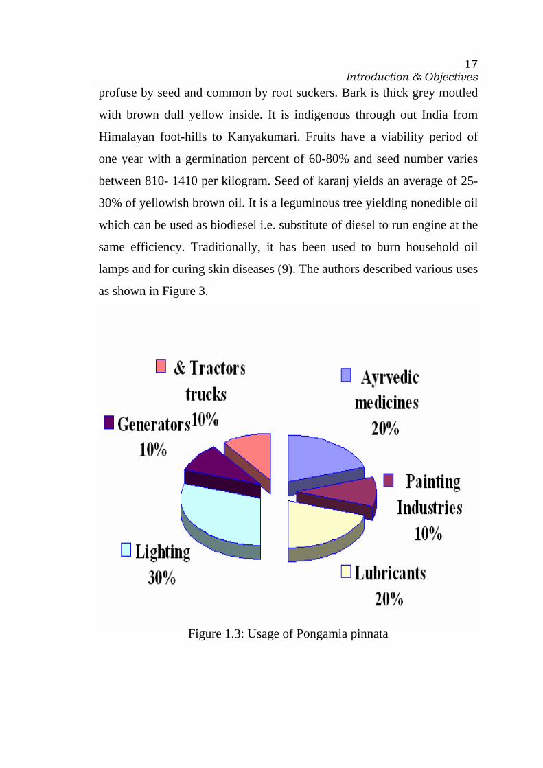
lamps and for curing skin diseases (9). The authors described various uses
as shown in Figure 3.
Figure 1.3: Usage of Pongamia pinnata

Introduction & Objectives
18
Warm oil of Karanj is applied on affected parts, thrice a day for 2
weeks, for curing skin diseases. In case of toothache, plant twig is used as
toothbrush, twice a day for one month. Oil is applied before hair wash
once a week for two months for long or black hair. During chest pain,
warm oil is massaged on the chest twice a day for 2 weeks (10).
Karanj has nutritive value of major dry season feed resources
available for Goats in Bhilwara and Udaipur Districts. Leaves are used as
fodder. The composition (% dry basis) is CP (Crude protein) 10 to 13,
NDF (neutral detergent fibre) 35 to 41, ash 6, EE (ether extract) 3 to 5,
lignin 11, TP (Total Phenols) 3. It has medium to high fermentability,
and it appeared to be very deficient in fermentable protein, but may be a
source of by-pass protein (11).
T.L.C. of Alcoholic extract of the karanj bark powder on silica gel
'G' plate using Toluene: Ethyl acetate (9:1) shows under UV light (366
nm.) eleven fluorescent zones at Rf. 0.04 (blue), 0.08 (greenish blue),
0.13(sky blue) 0.18 (blue) 0.25 (sky blue), 0.31 (sky blue), 0.37 (greenish
yellow), 0.42 (sky blue), 0.47 (greenish yellow), 0.51 (light blue), 0.80
(light blue). On exposure to iodine vapours nine spots appear at Rf. 0.09,
0.18, 0.31, 0.37, 0.47, 0.47, 0.51, 0.80 and 0.98 (all yellow). The
constituents are Ponganone I to XI, flavones, kanugin and demethoxy
kanugin (12).
Pongam is a fast-growing evergreen tree which reaches 40 feet in
height and spread, forming a broad, spreading canopy casting moderate
shade. The three-inch-long, pinnately compound, glossy green leaves are

Introduction & Objectives
19
briefly deciduous, dropping for just a short period of time in early spring
but being quickly replaced by new growth. In spring, Pongam is at its
finest when the showy, hanging clusters of white, pink, or lavender, pea-
like, fragrant blossoms appear, the clusters up to 10 inches long. These
beautiful blossoms and the glossy, nearly-evergreen leaves help make
Pongam a favorite for use as a specimen, shade, or windbreak. It has also
been planted as a street tree, but dropping pods often litter the ground.
However, the seeds which are contained within the oval, 1.5- inch-long,
brown seedpods are poisonous, a fact which should be considered in
placing the tree in the landscape, if many children are present (13).
Physico-chemical properties of crude oil established suitability of
P. pinnata for its use as a potential biofuel crop. The total mono
unsaturated fatty acid (oleic acid 46%) present in seed oil was more in
comparison to polyunsaturated fatty acid (33%) as analyzed by GC–MS.
Seed oil also showed inhibition against the tested fungal and bacterial
cultures. However, the efficacy of antimicrobial activity of the seed oil at
four concentration levels (50%, 80%, 90% and 100%) against various
pathogenic indicators was found to be concentration-dependent. The
obtained results confirmed the use of seed oil from well characterized
elite genotype of Pongamia as diesel fuel and in pharmaceuticals (14).
Karanj is a small to medium-sized deciduous or nearly evergreen
tree reaching 8-15 m in height, with a straight or crooked trunk 50-70 cm
in diameter and a broad crown of spreading or drooping branches. The
wood is used for cabinetwork, cart wheels, posts, and fuel. Bark fiber can
be made into rope. The oil extracted from the seeds is used as a lubricant,

Introduction & Objectives
20
fuel, leather dressing, and in the manufacture of soaps, varnish, paints and
medicine. Seed residue has insecticidal and pesticidal properties; it can be
served to poultry or used as fertilizer. The leaves can be browsed, used as
fodder or plowed under as green manure. Leaves, flowers, bark, seeds,
and sap has medicinal properties. The tree is planted for erosion control,
for afforestation in drier areas, as hedges and as ornamentals. It is
mentioned as a possible agroforestry species. The growing period is
perennial. The common names include as Papar, Kanji, Karanja, Karanj,
Honge, Ponga, Pongam, Kanga, Thinwin, Oil tree, Kona, Pari-pari,
Karanda, Pongamia, Ponga oil tree, Karum tree, Dona, Kuro-yona zoku,
Bani, Melapari, Langi poka. It often reaches an adult height of 8 m within
4 or 5 years. It is indigenous to the Indian subcontinent. It can be found at
elevations between sea level and 1200 m. The tree is drought resistant.
Because of its spontaneous seedlings and root suckers it may run wild and
create serious weed problems (15).
Introduction & Objectives
21
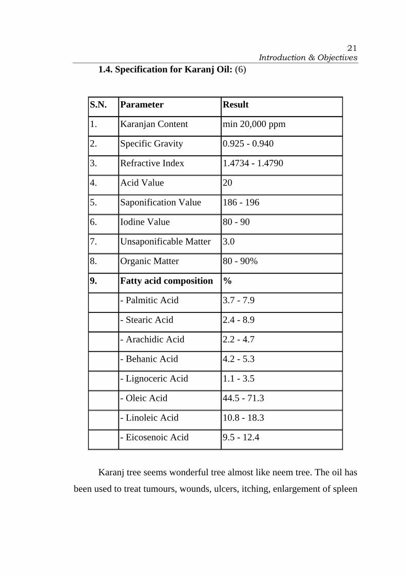
1.4. Specification for Karanj Oil: (6)
S.N. Parameter Result
1. Karanjan Content min 20,000 ppm
2. Specific Gravity 0.925 - 0.940
3. Refractive Index 1.4734 - 1.4790
4. Acid Value 20
5. Saponification Value 186 - 196
6. Iodine Value 80 - 90
7. Unsaponificable Matter 3.0
8. Organic Matter 80 - 90%
9. Fatty acid composition %
- Palmitic Acid 3.7 - 7.9
- Stearic Acid 2.4 - 8.9
- Arachidic Acid 2.2 - 4.7
- Behanic Acid 4.2 - 5.3
- Lignoceric Acid 1.1 - 3.5
- Oleic Acid 44.5 - 71.3
- Linoleic Acid 10.8 - 18.3
- Eicosenoic Acid 9.5 - 12.4
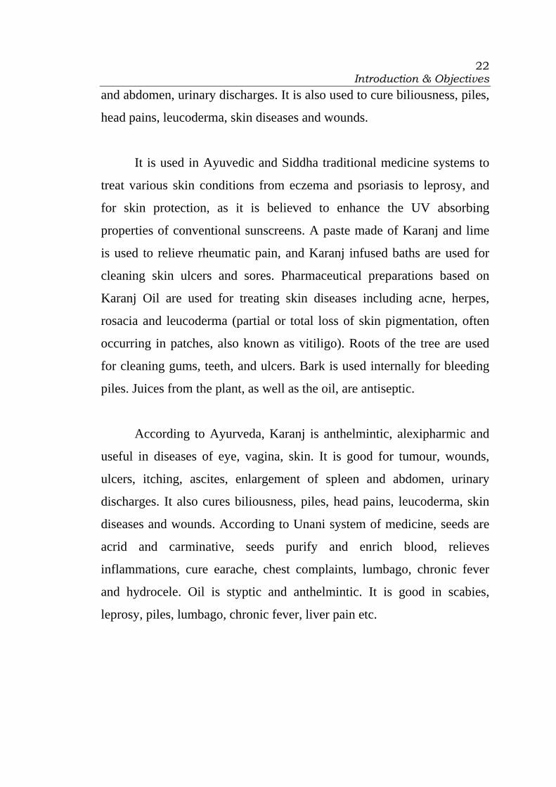
Karanj tree seems wonderful tree almost like neem tree. The oil has
been used to treat tumours, wounds, ulcers, itching, enlargement of spleen
Introduction & Objectives
22
and abdomen, urinary discharges. It is also used to cure biliousness, piles,
head pains, leucoderma, skin diseases and wounds.
It is used in Ayuvedic and Siddha traditional medicine systems to
treat various skin conditions from eczema and psoriasis to leprosy, and
for skin protection, as it is believed to enhance the UV absorbing
properties of conventional sunscreens. A paste made of Karanj and lime
is used to relieve rheumatic pain, and Karanj infused baths are used for
cleaning skin ulcers and sores. Pharmaceutical preparations based on
Karanj Oil are used for treating skin diseases including acne, herpes,
rosacia and leucoderma (partial or total loss of skin pigmentation, often
occurring in patches, also known as vitiligo). Roots of the tree are used
for cleaning gums, teeth, and ulcers. Bark is used internally for bleeding
piles. Juices from the plant, as well as the oil, are antiseptic.
According to Ayurveda, Karanj is anthelmintic, alexipharmic and
useful in diseases of eye, vagina, skin. It is good for tumour, wounds,
ulcers, itching, ascites, enlargement of spleen and abdomen, urinary
discharges. It also cures biliousness, piles, head pains, leucoderma, skin
diseases and wounds. According to Unani system of medicine, seeds are
acrid and carminative, seeds purify and enrich blood, relieves
inflammations, cure earache, chest complaints, lumbago, chronic fever
and hydrocele. Oil is styptic and anthelmintic. It is good in scabies,
leprosy, piles, lumbago, chronic fever, liver pain etc.
Introduction & Objectives
23
1.5. Objectives
The local name of the oil of Derris Indica is Karanj oil. Medicated
karanj oil is used widely by Vaidraj in rural areas of Gujarat for the
treatment of rheumatism and curing skin infections. It is used for healing,
antiseptic, and haemostatic effect externally by Vaidraj.
To the best of our knowledge, no systemic information is available
on the efficacy of karanj oil. The present investigation was aimed to study
systematically and scientifically the efficiency of Karanj oil. The efficacy
of Karanj oil formulations was compared with karanj oil and medicated
karanj oil. The formulations of the oil were prepared using
pharmaceutical methodology. The activity of the selected formulated
product was studied for wound healing, antibacterial, and haemostatic
efficiency.
Introduction & Objectives
24
1.6. References
1. Downloaded through www.worldagrofprestry.org.
2. Downloaded through http://www.bicco.com/herb_photo.html
3. Pankaj Oudhia, Research Note downloaded through
http://www.botanical.com/site/column_poudhia/152_karanj.htm
4. http://trifed.nic.in/productdetails.asp?productid=205&id=prod, by
Ministry of Tribal affairs
5. http://www.newdirectionsaromatics.com/karanj-seed-p-266.html
6. http://www.organicneem.com/karanja_intro.htm
7. SP Wani and TK Sreedevi, Pongamia’s Journey from Forest to
Micro-enterprise for Improving Livelihoods, International Crops
Research Institute for the Semi-Arid Tropics (ICRISAT),
Patancheru 502 324, Andhra Pradesh, India, Downloaded through
www.icrisat.org
8. Shailja Vohra, Ph.D. Thesis, Chemical Investigation and
Modification of Compound(s) Isolated from Pongamia glabra as
Potential Nitrification Inhibitors, Department of Chemistry, Jamia
Millia Islamia, New Delhi-110012.
Introduction & Objectives
25
9. BIO FUELS, ENVIS Newsletter, Vol.1 No.3, October 2005,
Department of Forests, Ecology & Environment, Government of
Karnataka.
10. Amia Tirkey, Some ethnomedicinal plants of family-Fabaceae of
Chhattisgarh state, Indian Journal of Traditional Knowledge, Vol.
5(4), October 2006, pp. 551-553.
11. C D Wood, R Matthewman, V C Badve, and C Conroy, A review
of the nutritive value of dry season feeds for ruminants in southern
Rajasthan, Bulletin of BAIF Development Research Foundation,
Central Research Station, Uruli Kanchan-412 202, District Pune,
India.
12. THE SIDDHA PHARMACOPOEIA OF INDIA, PART – I,
VOLUME – I, First Edition, p.181-182, by GOVERNMENT OF
INDIA, MINISTRY OF HEALTH AND FAMILY WELFARE
DEPARTMENT OF AYURVEDA, YOGA & NATUROPATHY,
UNANI, SIDDHA AND HOMOEOPATHY (AYUSH).
13. Edward F. Gilman and Dennis G. Watson, Pongamia pinnata, Fact
Sheet ST-498, Forest service, department of Agriculure, Southern
Group of State Foresters, October 1994.
14. Vigya Kesari, Archana Das, and Latha Rangan, Physico-chemical
characterization and antimicrobial activity from seed oil of

Introduction & Objectives
26
Pongamia pinnata, a potential biofuel crop, Biomass and
Bioenergy, Volume 34, Issue 1, January 2010, Pages 108-115.
15. View crop, Food and Agriculture Organization of the UN - Helping
to build a world without hunger, Downloaded through
http://ecocrop.fao.org/ecocrop/srv/en/dataSheet?id=5250

CHAPTER 2
REVIEW
OF
LITERATURE

Review of Literature
28
CHAPTER 2
REVIEW OF LITERATURE
Section CONTENT
2.1 Introduction to Karanj (Derris Indica)
2.2 Dosage forms
2.3 Wound healing activity
2.4 Antimicrobial activity
2.5 Hemostatic activity
2.6 References
Review of Literature
29
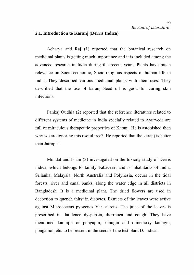
2.1. Introduction to Karanj (Derris Indica)
Acharya and Raj (1) reported that the botanical research on
medicinal plants is getting much importance and it is included among the
advanced research in India during the recent years. Plants have much
relevance on Socio-economic, Socio-religious aspects of human life in
India. They described various medicinal plants with their uses. They
described that the use of karanj Seed oil is good for curing skin
infections.
Pankaj Oudhia (2) reported that the reference literatures related to
different systems of medicine in India specially related to Ayurveda are
full of miraculous therapeutic properties of Karanj. He is astonished then
why we are ignoring this useful tree? He reported that the karanj is better
than Jatropha.
Mondal and Islam (3) investigated on the toxicity study of Derris
indica, which belongs to family Fabaceae, and is inhabitants of India,
Srilanka, Malaysia, North Australia and Polynesia, occurs in the tidal
forests, river and canal banks, along the water edge in all districts in
Bangladesh. It is a medicinal plant. The dried flowers are used in
decoction to quench thirst in diabetes. Extracts of the leaves were active
against Micrococcus pyogenes Var. aureus. The juice of the leaves is
prescribed in flatulence dyspepsia, diarrhoea and cough. They have
mentioned karanjin or pongapin, kanugin and dimethoxy kanugin,
pongamol, etc. to be present in the seeds of the test plant D. indica.

Review of Literature
30
Shailja Vohra (4) isolated chemical compound using methanolic
extract of Pongamia glabra and analysed for its karanjin and other
flavonoid and hydrocarbon content by electrospray ionisation (ESI) mass
spectroscopy. It showed the presence of six flavonoids namely karanjin,
desmethoxykanugin, pongachalcone, pongapin, glabrachromene I,
glabrachromene II and six hydrocarbons namely octadecane, undecane,
decane, octane, undecanol and tetradecanol. The karanj based products
exhibited outstanding antifungal activity against the soil-borne
phytophagous fungus Sclerotium rolfsii (Sacc.). Karanj oil in the karanjin
was more active than karanj oil without karanjin. Karanjin, however,
exhibited moderate antifungal activity. Karanjic acid and their three esters
exhibited significant antifungal activity. Karanjic acid showed the highest
antifungal activity.
Manigauha and Patel (5) investigated the anticonvulsant effect of
the leaf extract of Pongamia pinnata using pentylene tetrazole induced
convulsion (PTZ) in rats. Pongamia pinnata is an indigenous plant
belonging to the family Fabaceae (Papilionaceae) commonly known as
Karanj. Freshly powdered leaves were extracted with 70% ethanol. The
convulsion is induced by administration of pentylene tetrazole (80 mg/kg,
i.p.) to wistar albino rats and those showing response were divided into
three groups of six animals each. The group I treated with 1% normal
saline (1ml/100gm, orally), Groups II treated with phenytoin sodium (25
mg/kg, i.p.) and Groups III treated with ethanolic extract of PPLE at a
dose of (250 mg/kg, i.p.). The ethanolic extract showed significant
anticonvulsant activity by lowering the duration of extension phase (3.72
Review of Literature
31
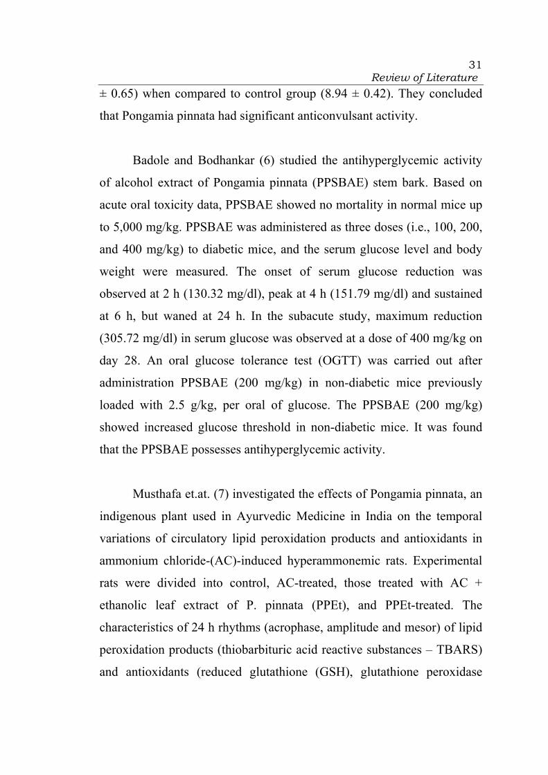
± 0.65) when compared to control group (8.94 ± 0.42). They concluded
that Pongamia pinnata had significant anticonvulsant activity.
Badole and Bodhankar (6) studied the antihyperglycemic activity
of alcohol extract of Pongamia pinnata (PPSBAE) stem bark. Based on
acute oral toxicity data, PPSBAE showed no mortality in normal mice up
to 5,000 mg/kg. PPSBAE was administered as three doses (i.e., 100, 200,
and 400 mg/kg) to diabetic mice, and the serum glucose level and body
weight were measured. The onset of serum glucose reduction was
observed at 2 h (130.32 mg/dl), peak at 4 h (151.79 mg/dl) and sustained
at 6 h, but waned at 24 h. In the subacute study, maximum reduction
(305.72 mg/dl) in serum glucose was observed at a dose of 400 mg/kg on
day 28. An oral glucose tolerance test (OGTT) was carried out after
administration PPSBAE (200 mg/kg) in non-diabetic mice previously
loaded with 2.5 g/kg, per oral of glucose. The PPSBAE (200 mg/kg)
showed increased glucose threshold in non-diabetic mice. It was found
that the PPSBAE possesses antihyperglycemic activity.
Musthafa et.at. (7) investigated the effects of Pongamia pinnata, an
indigenous plant used in Ayurvedic Medicine in India on the temporal
variations of circulatory lipid peroxidation products and antioxidants in
ammonium chloride-(AC)-induced hyperammonemic rats. Experimental
rats were divided into control, AC-treated, those treated with AC +
ethanolic leaf extract of P. pinnata (PPEt), and PPEt-treated. The
characteristics of 24 h rhythms (acrophase, amplitude and mesor) of lipid
peroxidation products (thiobarbituric acid reactive substances – TBARS)
and antioxidants (reduced glutathione (GSH), glutathione peroxidase
Review of Literature
32
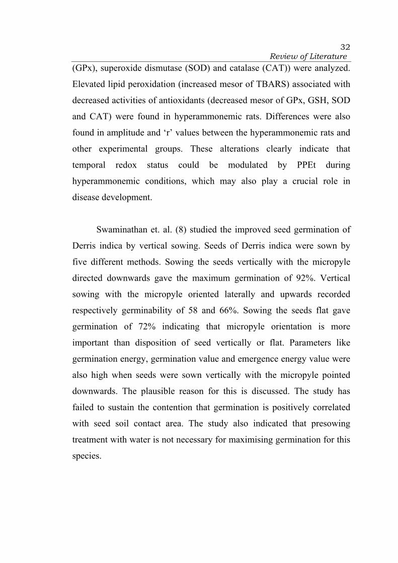
(GPx), superoxide dismutase (SOD) and catalase (CAT)) were analyzed.
Elevated lipid peroxidation (increased mesor of TBARS) associated with
decreased activities of antioxidants (decreased mesor of GPx, GSH, SOD
and CAT) were found in hyperammonemic rats. Differences were also
found in amplitude and ‘r’ values between the hyperammonemic rats and
other experimental groups. These alterations clearly indicate that
temporal redox status could be modulated by PPEt during
hyperammonemic conditions, which may also play a crucial role in
disease development.
Swaminathan et. al. (8) studied the improved seed germination of
Derris indica by vertical sowing. Seeds of Derris indica were sown by
five different methods. Sowing the seeds vertically with the micropyle
directed downwards gave the maximum germination of 92%. Vertical
sowing with the micropyle oriented laterally and upwards recorded
respectively germinability of 58 and 66%. Sowing the seeds flat gave
germination of 72% indicating that micropyle orientation is more
important than disposition of seed vertically or flat. Parameters like
germination energy, germination value and emergence energy value were
also high when seeds were sown vertically with the micropyle pointed
downwards. The plausible reason for this is discussed. The study has
failed to sustain the contention that germination is positively correlated
with seed soil contact area. The study also indicated that presowing
treatment with water is not necessary for maximising germination for this
species.
Review of Literature
33
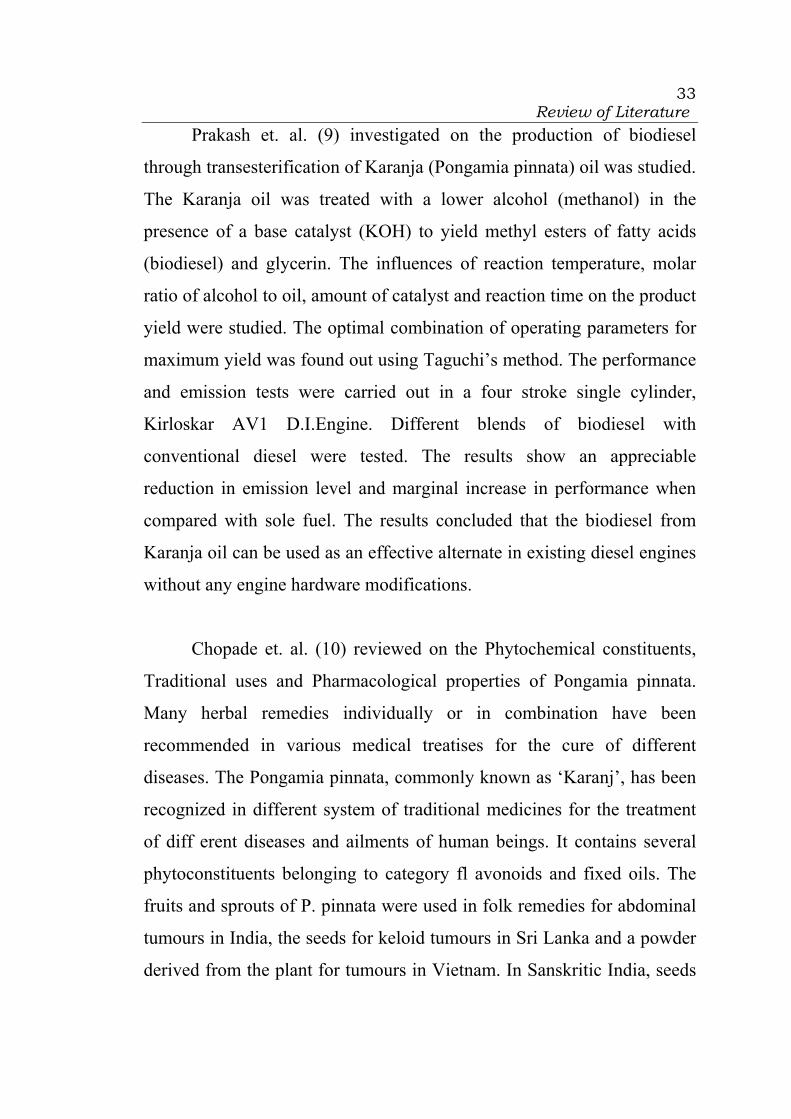
Prakash et. al. (9) investigated on the production of biodiesel
through transesterification of Karanja (Pongamia pinnata) oil was studied.
The Karanja oil was treated with a lower alcohol (methanol) in the
presence of a base catalyst (KOH) to yield methyl esters of fatty acids
(biodiesel) and glycerin. The influences of reaction temperature, molar
ratio of alcohol to oil, amount of catalyst and reaction time on the product
yield were studied. The optimal combination of operating parameters for
maximum yield was found out using Taguchi’s method. The performance
and emission tests were carried out in a four stroke single cylinder,
Kirloskar AV1 D.I.Engine. Different blends of biodiesel with
conventional diesel were tested. The results show an appreciable
reduction in emission level and marginal increase in performance when
compared with sole fuel. The results concluded that the biodiesel from
Karanja oil can be used as an effective alternate in existing diesel engines
without any engine hardware modifications.
Chopade et. al. (10) reviewed on the Phytochemical constituents,
Traditional uses and Pharmacological properties of Pongamia pinnata.
Many herbal remedies individually or in combination have been
recommended in various medical treatises for the cure of different
diseases. The Pongamia pinnata, commonly known as ‘Karanj’, has been
recognized in different system of traditional medicines for the treatment
of diff erent diseases and ailments of human beings. It contains several
phytoconstituents belonging to category fl avonoids and fixed oils. The
fruits and sprouts of P. pinnata were used in folk remedies for abdominal
tumours in India, the seeds for keloid tumours in Sri Lanka and a powder
derived from the plant for tumours in Vietnam. In Sanskritic India, seeds
Review of Literature
34
were used for skin aliments. Today, the oil is used as a liniment for
rheumatism. Leaves are active against Micrococcus; their juice is used for
cold, cough, diarrhoea, dyspepsia, fl atulence, gonorrhoea and leprosy.
Roots are used for cleaning gums, teeth and ulcers. Bark is used internally
for bleeding piles. Juices from the plant as well as oil are antiseptic. In the
traditional systems of medicines, such as Ayurveda and Unani, the P.
pinnata plant is used for anti-infl ammatory, anti-plasmodial, anti-
nonciceptive, anti-hyperglycaemics, anti-lipidoxidative, anti-diarrhoeal,
anti-ulcer, anti-hyperammonic and antioxidant. Its oil is a source of
biodiesel. It has also alternative source of energy, which is renewable,
safe and non-pollutant.
Meher et. al. (11) studied the transesterification of Pongamia
pinnata oil by means of methanol to study the feasibility of methanolysis
process by using potassium hydroxide catalysts. The yield of biodiesel
obtained was >97 per cent by using oil/methanol molar ratio 12:1,
potassium hydroxide as catalyst, at 65oC and stirring at 360 rpm in 3 h.
The biodiesel was characterized by TLC and HPLC analysis to determine
the fatty acid methyl esters, mono-, di- and triglycerides and glycerol.
The properties like viscosity, flash point, cloud point, and pour point have
been determined for accessing the fuel quality of karanja based biodiesel.
Naghmana Rashid et. al. (12) isolated Karanjachromene,
C21H18O4, from the seed oil of Pongamia pinnata. Karanjachromene is a
fluorescent pyranoflavonoid. Such compounds are reported to have many
interesting pharmacological and industrial applications. They reported on
its isolation in significant yield and X-ray crystal structure.
Review of Literature
35
Kalidhar et. al. (13) investigated the chemical composition of the
roots of Pongamia pinnata. Four compounds, karanjin, pongachromene,
pongapin and demethoxykanugin, were characterized from the
methanolic extract of its roots by them.
Chauhan and Cauhan (14) characterized two new flavone
glycosides from the seeds of Pongamia pinnata on the basis of chemical
and spectral evidence.
Kumar et. al. (15) covered a review on the chemical composition of
P. pinnata, and the insecticidal, antimicrobial, nematicidal and medicinal
properties of compounds isolated from P. pinnata.
Hemlata et. al. (16) characterized two flavone glycosided from P.
Pinnata. It is used to treat eye diseases, lucoderma [leukoderma], piles,
wounds, ulcers, skin diseases and rheumatic joints. Two new flavone
glycosides from the ethanol extract of the heartwood (collected from
Allahabad, India) were characterized on the basis of chemical and
spectral evidence as 3,5,7,4'-tetrahydroxy-6,8,3'-trimethoxyflavone-3-O-
[alpha-L-arabinopyranosyl(13)]-beta-D-galacto pyranoside and 3,5,7,4'-
tetrahydroxy-6,8,3'-trimethoxyflavone-3-O-[{-alpha-L-arabinopyranosyl
(13)}{-beta-D-galactopyranosyl(16)}-beta-D-galactopyranoside].
Punitha and Manoharan (17) evaluated the antihyperglycemic and
antilipid peroxidative effect of ethanolic extract of Pongamia pinnata
(Linn.) Pierre (Leguminosae) flowers (PpEt) in normal rats and alloxan
Review of Literature
36
induced diabetic rats. Hyperglycaemia, elevated lipid peroxidation
[thiobarbituric acid reactive substances (TBARS)] and disturbed
nonenzymatic [Vitamin E, Vitamin C and glutathione] and enzymatic
antioxidants status were noticed in alloxan induced diabetic rats. The oral
administration of ethanolic extract of Pongamia pinnata flowers (300
mg/kg bw) showed significant antihyperglycemic, and
antilipidperoxidative effects and enhancement in antioxidants defence
system in alloxan induced diabetic rats. However, no significant
characteristic changes were noticed in blood glucose level as well as in
lipid peroxidation and antioxidant status in normal rats treated with
"PpEt" alone. We have also observed that the "PpEt" considerably
reduced the blood glucose concentration in a similar extent to that of the
reference drug glibenclamide (600 micro g/kg bw) in alloxan induced
diabetic rats. Our results thus suggested that the "PpEt" could be used as a
safe alternative antihyperglycemic drug for diabetic patients
Gopinath et. al. (18) characterized from fruits of Pongamia pinnata
four new furanoflavonoids, pongapinnol A-D (1-4), and a new
coumestan, pongacoumestan along with thirteen known compounds 6-18.
Compounds 16 and 17 are isolated for the first time from this plant. The
structures of isolated compounds were elucidated on the basis of
spectroscopic data interpretation.
Vismaya et. al. (19) studied the gastroprotective properties of
Karanj seed and found promising results for the treatment of gastric
ulcers. Karanjin, a furano-flavonoid has been evaluated for anti-
ulcerogenic property by employing adult male albino rats. Karanjin

Review of Literature
37
(>95% pure) was administered to these rats in two different
concentrations, i.e. 10 and 20 mg/kg. Ulcers were induced in the
experimental animals by swim and ethanol stress. Serum, stomach and
liver-tissue homogenates were assessed for biochemical parameters.
Karanjin inhibited 50 and 74% of ulcers induced by swim stress at 10 and
20 mg/kg, respectively. Gastric mucin was protected up to 85% in case of
swim stress, whereas only 47% mucin recovery was seen in ethanol stress
induced ulcers. H+,K+-ATPase activity, which was increased 2-fold in
ulcer conditions, was normalized by Karanjin in both swim/ethanol
stress-induced ulcer models. Karanjin could inhibit oxidative stress as
evidenced by the normalization of lipid peroxidation and antioxidant
enzyme (i.e. catalase, peroxidase and superoxide dismutase) levels.
Karanjin at concentrations of 20 mg/kg, when administered orally for 14
days, did not indicate any lethal effects. There were no significant
differences in total protein, serum glutamate pyruvate transaminase,
serum glutamate oxaloacetate transaminase and alkaline phosphatase
between normal and Karanjin-treated rats indicating no adverse effect on
major organs. During treatment schedule, animals remained as healthy as
control animals with normal food and water intake and body weight gain.

Review of Literature
38
2.2. Dosage forms
Vlad and Joseph (20) filed a European Patent on the formulations
of clear, thermodynamically stable and concentrated oil-in-water
microemulsions. The oil-in-water microemulsion was comprising : a) at
least 30% by weight of oil; b) from 1 to 30% by weight of a surfactant
system having a hydrophilic lipophilic balance, HLB, comprised between
9 and 18 Surfactants useful for the purpose of the present invention are
approved for use in food type products and include notably Tween® 20,
Tween® 40, Tween® 60, Tween® 80, Glycosperse® L-20 (polyethylene
glycol sorbitan laurate), Glycosperse® O-20 (hexaethylene glycol
sorbitan monooleate), Glycosperse® S-20 (polyoxyethylene sorbitan
stearate), Polyaldo® 10-1-O K (decaglyceryl monooleate), Polyaldo® 10-
2-O K (decaglyceryl dioleate), Glycosperse® TS-20 (polyoxyethylene
sorbitan tristearate), Lonzest® SMO-20 (monodehydrosorbitol
monooleate), Span® 20, Span® 40, c) less than 20% by weight of co-
solvent, propylene glycol, ethanol, mono- and di-saccharide sugars and
sugar alcohols such as sorbitol, xylitol and mannitol. They used
propylene glycol; and d) at least 30% by weight of water; relative to the
total weight of the microemulsion.
Bauer et. al. (21) filed a US patent on their invention concerning
with a microemulsion of the oil-in water type, containing at least one
polyglycerol ester as the emulsifier and at least one lipophilic substance
as the internal phase. The emulsifier contains a triglycerol monofatty acid
ester and the lipophilic substance is one from the group carotenoids,
Review of Literature
39
especially, β-carotene, vitamins A, D, E and K and their derivatives and
polyunsaturated fatty acids.
Rosano (22) filed a US patent on the unique method for the
preparation of oil-in-water microemulsions is described. In carrying out
this method, microemulsions are prepared in a four-step process: (1) a
surfactant is selected which is just barely soluble in the oil phase; (2) the
surfactant thus selected is dissolved in the oil to be emulsified in an
amount effective to yield a fine emulsion of the emulsified oil in an
aqueous phase; and (3) the oil, together with its dissolved surfactant, is
added to the water phase and shaken or stirred; and (4) finally, there is
provided a second surfactant in the water phase which is somewhat more
soluble in water than the first surfactant to produce a substantially clear
microemulsion of oil in water.
Owen and Yiv (23) filed a US patent on water-in-oil (w/o)
microemulsion which readily converts to an oil-in-water (o/w) emulsion
by the addition of aqueous fluid to the w/o microemulsion, whereby any
water-soluble biologically-active material in the aqueous phase is
released for absorption by the body. The w/o microemulsion is
particularly useful for storing proteins and the like for long periods of
time at room temperature and above until they are ready for use, at which
time the addition of aqueous fluid converts the microemulsion to an o/w
emulsion and releases the protein.
Faiyaz Shakeel et. al. (24) investigated the potential of a
nanoemulsion formulation for transdermal delivery of aceclofenac.

Review of Literature
40
Various oil-in-water nanoemulsions were prepared by the spontaneous
emulsification method. The nanoemulsions area was identified by
constructing pseudoternary phase diagrams. The prepared nanoemulsions
were subjected to different thermodynamic stability tests. The
nanoemulsions formulations that passed thermodynamic stability tests
were characterized for viscosity, droplet size, transmission electron
microscopy, and refractive index. Transdermal permeation of aceclofenac
through rat abdominal skin was determined by Franz diffusion cell. The
in vitro skin permeation profile of optimized formulations was compared
with that of aceclofenac conventional gel and nanoemulsion gel. A
significant increase in permeability parameters such as steady-state flux
(Jss), permeability coefficient (Kp), and enhancement ratio (Er) was
observed in optimized nanoemulsion formulation.
Carli et. al. (25) studied on the nanoemulsified composite (NECTM
) delivery system, which is a patented technology based on the
incorporation of a double microemulsion into microporous carrier. This
approach was applied on the very water insoluble ubidecarenone drug.
The resulting composite powder showed good technological properties
such as flowability; also good stability was evidentiated, with size of the
nano-droplets released from the systems maintained equal to the starting
size also after a long storage. Furthermore very good biopharmaceutical
properties were originated, with water solubility concentrations up to 50-
fold higher than pure ubidecarenone and oral absorption in rats up to
three-fold greater than standard commercial products in terms of plasma
levels and AUC.

Review of Literature
41
Peira et.al. (26) reported on the pig-skin permeation and skin
accumulation of miconazole nitrate (MCZ) from positively charged
microemulsions containing water, 1-decanol/1-dodecanol (2:1, w/w),
lecithin and/or decyl polyglucoside at different weight ratios, propylene
glycol, 1,2 hexanediol and a cationic charge-inducing agent (stearylamine
(ST), l-alanine benzyl ester (ALAB) or cetyltrimethylammonium bromide
(CTAB)). Zeta-potential values of the positively charged microemulsions
ranged from 14.2 to 37.5mV and mean droplet size from 6.0 to 16.8 nm.
In vitro pig-skin permeation of MCZ after a single 24 h application was
negligible for all microemulsions; accumulation from positively charged
microemulsions was nearly twice that from their negatively charged
counterparts. The increased accumulation might be ascribed to the
interaction between positive microemulsive systems and negatively
charged skin sites; no significant difference was observed among the
various cationic charge-inducing agents. Skin accumulation from the
microemulsion containing most lecithin was lower than those of other
microemulsions; this was ascribed to the phase transformation from
microemulsion to a liquid crystal system after skin contact. These results
suggest that positively charged microemulsions could be used to optimize
drug targeting without a concomitant increase in systemic absorption;
ALAB, an ester of a natural aminoacid, is an appropriate cationic charge-
inducing agent.
Review of Literature
42
2.3. Wound healing activity
Karodi et. al. (27) explored the wound healing activity of R.
cordifolia crude extract as well as its gel formulation against the specific
microorganisms which generally infect wound. Rubia cordifolia Linn.
(Rubiaceae) is popular all over the world for its medicinal uses in skin
diseases like eczema, dermatitis, skin ulcers, etc. In India, it is used
traditionally for various types of skin diseases. Hence, the present study
was aimed to evaluate its scientific validity. The alcoholic extract and the
hydrogel of same were investigated for the evaluation of its healing
efficiency on excision wound model in mice. A different formulation of
alcoholic extract was topically applied on the excision wound surface as a
single dose. Wound area and histopathology were used to evaluate the
effect on wound healing. The effect produced by gel, in terms of wound
contracting ability, wound closure, decrease in surface area of wound,
tissue regeneration at the wound site and histopathological characteristics
were significant (p<0.01) in treated mice. The present study thus provides
a scientific rationale for the traditional use of this plant in the
management of the wounds.
Jain et. al. (28) investigated the effect of Ageratum conyzoides root
extract on wound healing and a comparable study of effect of polyherbal
formulation containing Ageratum conyzoides, Ficus religiosa,Curcuma
longa and Tamarindus indica, on wound healing process with respect to
intact plant formulation through topical route. The wound healing
property of Ageratum conyzoides appears to be due to the presence of its
active principles, which accelerates the healing process and confers

Review of Literature
43
breaking strength to the healed wound. Further, wound healing activity by
polyherbal formulation was found to be better than ageratum treated
groups in rat. It may be attributed to the synergistic action of ageratum
constituent and the constituent of other plants present in the polyherbal
formulation.
Nalwaya et.al. (29) studied the wound healing activity of latex of
calotropis gigantean, The entire wound healing process is a complex
series of events that begins at the moment of injury and can continue for
months to years. The stages of wound healing are inflammatory phase,
proliferation phase, fibroblastic phase and maturation phase. The Latex of
Calotropis gigantean (200 mg/kg/day) was evaluated for its wound
healing activity in albino rats using excision and incision wound models.
Latex treated animals exhibit 83.42 % reduction in wound area when
compared to controls which was 76.22 %. The extract treated wounds are
found to epithelize faster as compared to controls. Significant (p<0.001)
increase in granuloma breaking strength (485±34.64) was observed. The
Framycetin sulphate cream (FSC) 1 % w/w was used as standard.
Shahbhag et. al. (30) studied the wound healing activity of
alcoholic extract of kaempferia galanga in wistar rats. Three wound
models viz. incision, excision and dead space wounds were used in this
study. The parameters studied were breaking strength in case of incision
wounds, epithelialisation and wound contraction in case of excision
wound and granulation tissue dry weight, breaking strength and
hydroxyproline content in case of dead space wound. The dexamethasone
treated group showed a significant (P<0.001) reduction in the wound
Review of Literature
44
breaking strength when compared to control group in incision type of
wound model. Coadministration of K. galanga with dexamethasone had
significantly (P<0.001) increased the breaking strength of dexamethasone
treated group. In excision wound model, the percentage of the wound
contraction was significantly (P<0.05) increased by K. galanga only on
16th day and also it reversed the dexamethasone suppressed wound
contraction on the 16 day. K. galangal significantly (P<0.001) reduced
the time required for epithelialization and reversed the epithelialization
delaying effect of dexamethasone significantly (P<0.001).
Sharma et. al. (31) assessed the wound healing activity of benzene
extracts of Momordica charantia fruit.Methanol extract of Momordica
Charantia fruit was examined for wound healing potential in the form of
1%w/v solution in the excision wound created on the dorsal side of
experimental animals, the 1% w/v extract should considerable difference
in wound models and the result were compatable to that of the standard
drug betadine (5% w/w) in terms of wound contracting ability and wound
closure time. Antibacterial activity of methanol extract of the plant was
also carried out as a supporting evidence for its wound healing potential.
The mean percentage wound closure was calculated on the 4 th, 8 th, 12
th, 16 th and 15 th wounding days. The extract treated animals showed
tastes epithelisation of wound (17.86 ± 0.19) then the control. The period
of epithelisation was 21.23±0.37 in case of standard drug 5% betadine
ointment.
Kristine et. al. (32) studied the effect of barium sulfate on wound
healing in the gastrointestinal tract of the rat. Sixty rats weighing
Review of Literature
45
approximately 320 g were divided into four groups: Fifteen control rats
had gastric, small-bowel, and colonic incisions; 15 rats had gastric
incision; 15 rats had small-bowel incision; and 15 rats had colonic
incision. Barium sulfate was placed into the incision before closure in all
rats except those in the control group, and the effects were documented
clinically and histopathologically for 3 months. Autopsy was performed
in five rats from each group at 1, 4, and 12 weeks. The incisions in the
rats receiving barium sulfate were compared with those in the control
rats. There was no difference in the clinical course (weight gain, activity,
and viability) between the control and experimental groups. Early and
late autopsy findings and histopathologic grading of healing and
inflammatory response were similar for both the control and experimental
groups. Under the conditions of this study, the effect of barium sulfate on
visceral transmural wound healing in the gastrointestinal tract of the rat
was minimal.
Nadagouda Smitha G. et. al. (33) evaluated the anti-inflammatory
activity of aqueous extract of Pongamia pinnata stem bark (PPSB) in
acute and chronic models of inflammation in albino rats. Oral
administration of PPSB (400, 800 mg/kg) exhibited significant anti-
inflammatory activity in acute (carrageenin induced hind paw edema) and
chronic (cotton pellet granuloma) models of inflammation. PPSB did not
show any sign of toxicity and mortality up to a dose level of 8000 mg/kg,
p.o. in rats. Both acute as well as chronic administration of PPSB (400
and 800 mg/kg, p.o.) did not produce any gastric lesion in rats. These
results indicated that PPSB hold significant anti-inflammatory activity

Review of Literature
46
without ulcerogenic activity. Thus it can be used as anti-inflammatory
agent in the treatment of various inflammatory diseases.
Vigya Kesari et. al. (34) carried out oil analysis and antimicrobial
activity from seeds of elite genotype of Pongamia pinnata. The highest oil
yield (33%) from seeds was recovered in n-Hexane. Physico-chemical
properties of crude oil established suitability of P. pinnata for its use as a
potential biofuel crop. The total mono unsaturated fatty acid (oleic acid
46%) present in seed oil was more in comparison to polyunsaturated fatty
acid (33%) as analyzed by GC–MS. Seed oil also showed inhibition
against the tested fungal and bacterial cultures. However, the efficacy of
antimicrobial activity of the seed oil at four concentration levels (50%,
80%, 90% and 100%) against various pathogenic indicators was found to
be concentration- dependent. The obtained results confirmed the use of
seed oil from well characterized elite genotype of Pongamia as diesel fuel
and in pharmaceuticals.
Kotade and Mohammed (35) verified the effect of S. indicum seeds
oil on experimentally induced excision wound, incision wound, burn
wound and dead space wound models in rats. Aloe vera was used as
standard wound healing agent. A formulation of seeds and oil was
prepared in carbopol at 2.5% and 5% concentrations and applied to the
wounds. In the excision and burn wound models, the so treated animals
showed significant reduction in period of epithelization and wound
contraction (50%). In the incision wound model a significant increase in
the breaking strength was observed. Seeds and oil treatment (250 mg and
500 mg/kg; po) in dead space wound model, produced a significant

Review of Literature
47
increase in the breaking strength, dry weight and hydroxyproline content
of the granulation tissue. The results suggest that S. indicum seeds and oil
applied topically or administered orally possesses wound healing activity.
Agrawal et. al. (36) investigated Plantain banana (M. sapientum
var. paradisiaca, MS) has been shown to possess ulcer healing activity.
The present work with plantain banana was undertaken with the premise
that the drug promoting ulcer healing could have effect on wound healing
also. Wound healing activity of MS was studied in terms of (i) percent
wound contraction, epithelization period and scar area; (ii) wound
breaking strength and (iii) on granulation tissue antioxidant status
[estimation of superoxide dismutase (SOD) and reduced glutathione
(GSH), free radical (lipid peroxidation, an indicator of tissue damage) and
connective tissue formation and maturation (hexuronic acid,
hydroxyproline and hexosamine levels)] in excision, incision and dead
space wound models respectively. The rats were given graded doses (50-
200 mg/kg/day) of aqueous (MSW) and methanolic (MSE) extracts of
MS orally for a period of 10-21 days depending upon the type of study.
Both extracts (100 mg/kg) when studied for incision and dead space
wounds parameters, increased wound breaking strength and levels of
hydroxyproline, hexuronic acid, hexosamine, superoxide dismutase,
reduced glutathione in the granulation tissue and decreased percentage of
wound area, scar area and lipid peroxidation when compared with the
control group. Both the extracts showed good safety profile. Plantain
banana thus, favoured wound healing which could be due to its
antioxidant effect and on various wound healing biochemical parameters.
Review of Literature
48
Mathivanan et. al. (37) found that Morinda leaf and fruit extracts
are effective in healing the wounds using the animal model. The
application of chloroform fruit extract of M. tinctoria topically on the
excision wound surface of two different doses accelerated the wound
healing process by decreasing the surface area of the wound. The fruit
extracts of M. tinctoria at 20 mg/ml significantly healed the wound in rats
within 15 days where complete healing was observed against 60% in
untreated control. On day 3, all the treated animals exhibited considerable
increase in the percentage of wound contraction as compared to control.
The wound contraction was significantly increased in the subsequent days
due to treatment of fruit extract at 10 and 20 mg/ml as compared to
control.
Devi and Meera (38) investigated antioxidant and anti
inflammatory and wound healing effects of Litsea glutinosa in rats. The
aqueous extract of Litsea glutinosa (250 and 500 mg/kg body weight) was
studied for antinflammatory in animal models. The activity was studied in
some acute models Viz carragenan, histamine and dextrin induced rats
paw edema against indomethacin as standard, and it showed significant
anti inflammatory ctivity in all the three models. The preliminary
phytochemical analysis was carried out for different extracts. It was
found that flavone glycosides, reducing sugars, aminoacids and tannins.
The ethanolic extract was screened for wound healing activity by excision
and incison wound model, in the form of an ointment having 2
concentrations( 3 and 5 % w/w) of leaf extract in simple ointment base.
Both the concentrations of the ethanolic extract showed significant
response in both the wound types tested when compared with the control
Review of Literature
49
group. Nitrofurazone ointment (0.2%w/w) was used as an standard drug.
Antioxidant activity was determined by two in vitro methods- DPPH and
H2O2 radical scavenging. The extract having significant (p<0.001)
antioxidant activity was compared to control. BHT and Ascorbic acid
were used as reference standard for antioxidant activity. On the basis of
the results obtained suggest marked that extracts have significant Anti
oxidant, Anti inflammatory and wound healing activity of Litsea
glutinosa. The results supported the traditional use of this plant in some
painful and inflammatory conditions.
Singh et. al. (39) evaluated the wound healing activity of the leaf
extracts and deoxyelephantopin isolated from Elephantopus scaber Linn.
Using excision, incision, and dead space wound models in rats. The
wound-healing activity was assessed by the rate of wound contraction,
period of epithelialization, skin-breaking strength, weight of the
granulation tissue, and collagen content. The wound-healing activity was
more significant in deoxyelephantopin-treated animals. Histological
studies of the granulation tissue also evidenced the healing process by the
presence of a lesser number of chronic inflammatory cells, lesser edema,
and increased collagenation than the control.
Review of Literature
50
2.4. Antimicrobial activity
Baswa et. al. (40) assessed the antibacterial activity of Karanj
(Pongamia pinnata) and Neem (Azadirachta indica) seed oil in vitro
against fourteen strains of pathogenic bacteria. Using the tube dilution
technique, it was observed that 57.14 and 21.42% of the pathogens were
inhibited at 500 microl/ml; 14.28 and 71.42% at 125 microl/ml; and 28.57
and 7.14% at 250 microl/ml of Karanj and Neem oils, respectively. The
activity with both the oils was bactericidal and independent of
temperature and energy. Most of the pathogens were killed more rapidly
at 4 degrees C than 37 degrees C. The activity was mainly due to the
inhibition of cell-membrane synthesis in the bacteria.
Kesari et. al. (41) carried out the study on oil analysis and
antimicrobial activity from seeds of elite genotype of Pongamia pinnata.
The highest oil yield (33%) from seeds was recovered in n-Hexane.
Physico-chemical properties of crude oil established suitability of P.
pinnata for its use as a potential biofuel crop. The total mono unsaturated
fatty acid (oleic acid 46%) present in seed oil was more in comparison to
polyunsaturated fatty acid (33%) as analyzed by GC–MS. Seed oil also
showed inhibition against the tested fungal and bacterial cultures.
However, the efficacy of antimicrobial activity of the seed oil at four
concentration levels (50%, 80%, 90% and 100%) against various
pathogenic indicators was found to be concentration-dependent. The
obtained results confirmed the use of seed oil from well characterized
elite genotype of Pongamia as diesel fuel and in pharmaceuticals.
Review of Literature
51
Gislene et. al. (42) evaluated the antimicrobial activity of plant
extracts and phytochemicals with antibiotic susceptible and resistant
microorganisms. In addition, the possible synergistic effects when
associated with antibiotics were studied. Extracts from the following
plants were utilized: Achillea millifolium (yarrow), Caryophyllus
aromaticus (clove), Melissa offficinalis (lemon-balm), Ocimun basilucum
(basil), Psidium guajava (guava), Punica granatum (pomegranate),
Rosmarinus officinalis (rosemary), Salvia officinalis (sage), Syzygyum
joabolanum (jambolan) and Thymus vulgaris (thyme). The
phytochemicals benzoic acid, cinnamic acid, eugenol and farnesol were
also utilized. The highest antimicrobial potentials were observed for the
extracts of Caryophyllus aromaticus and Syzygyum joabolanum, which
inhibited 64.2 and 57.1% of the tested microorganisms, respectively, with
higher activity against antibiotic-resistant bacteria (83.3%). Sage and
yarrow extracts did not present any antimicrobial activity. Association of
antibiotics and plant extracts showed synergistic antibacterial activity
against antibiotic-resistant bacteria. The results obtained with
Pseudomonas aeruginosa was particularly interesting, since it was
inhibited by clove, jambolan, pomegranate and thyme extracts. This
inhibition was observed with the individual extracts and when they were
used in lower concentrations with ineffective antibiotics.
Kuete et. al. (43) used the crude methanolic extracts from Treculia
africana and Treculia acuminata, three compounds isolated from T.
africana and identified as, Phyllocoumarin (1), Catechin (2) and 6, 9-
dihydroxy-megastigmane-3-one (3), four compounds namely 2, 3, 2,3-
dihydroxypropyl-heptadecanoate (4), and Ferulic acid (5) isolated from T.
Review of Literature
52
acuminata were tested for their antimicrobial activity against Gram-
positive bacteria (six species), Gramnegative bacteria (12 species) and
three Candida species. The micro-dilution method for the determination
of the minimal inhibition concentration (MIC) and the Minimal
microbicidal concentration (MMC) was used. The MIC values obtained
with the crude extracts varied from 78 to 156 μg/ ml against 12 (57.14%)
and 20 (95.24%) of the 21 tested microorganisms respectively for T.
acuminata and T. africana. Apart from compound 2 that prevented the
growth of all the tested microorganisms, other bioactive compounds
showed selective activity. The obtained results provide promising
baseline information for the potential use of these crude extracts as well
as some of the isolated compounds in the treatment of bacterial and
fungal infections.
Abdullahil et. al. (44) isolated two phenolic compounds,
methylkarranjic (1) acid and pongamol (2) from the petroleum ether
soluble fraction of an ethanolic extract of Derris indica seeds. The
structures of these compounds were confirmed by LC-MS and a series of
1D and 2D NMR data. The solvent solvent partionates of the ethanolic
extract and compounds (1 and 2) exhibited moderate antibacterial activity
against several test organisms. The minimum inhibitory concentrations
(MICs) of 1 and 2 were found to be in the range of 32-128 μg/ml against
Bacillus megatorium, Streptococcus β-haemolyticus, Shigella dysenteriae
and E. coli.
Nair and Chandra (45) screened 20 plants for antimicrobial
activity. Medicinal plants are an important therapeutic aid for various
Review of Literature
53
ailments. Scientific experiments on the antimicrobial properties of plant
components were first documented in the late 19th
century. In India, from
ancient times, different parts of medicinal plants have been used to cure
specific ailments. Today, there is widespread interest in drugs derived
from plants. This interest primarily stems from the belief that green
medicine is safe and dependable, compared with costly synthetic drugs
that have adverse effects. Natural antimicrobials can be derived from
plants, animal tissues, or microorganisms. The shortcomings of the drugs
available today, propel the discovery of new pharmacotherapeutic agents
in medicinal plants. To determine the potential and promote the use of
herbal medicine, it is essential to intensify the study of medicinal plants
that find place in folklore. In this study, some selected plants of
Saurashtra region, India, were screened for potential antibacterial activity.
Pritima and Pandian (46) studied the antibacterial activity of
Azadirachta indica. It is a commonly seen medicinal tree in India, which
is considered Holy. The antibacterial potential of leaves of this tree was
tested against certain reproductive tract infection (RTI) causing microbes,
seen among women. Crude extracts were made using ethanol, methanol,
acetone and water as solvents. Highest degree of activity was observed
for ethanol followed by methanol, acetone and water extracts, which
exhibited the same degree of activity. Escherichia coli and Enterococcus
faecalis were highly susceptible to neem extracts of all solvents followed
by Proteus mirabilis, Bacillus cereus and Staphylococcus aureus. Activity
of Klebsiella pneumoniae was weak, while Neisseria gonohorreae was
found to be relatively resistant to all forms of neem extract.
Review of Literature
54
Kaushik et. al. (47) evaluated the antibacterial potential of dry
fruits extracts of E. cardamomum against some selected bacterial species.
Elettaria cardamomum can potentially be used in the treatment of various
infectious diseases caused by microorganisms that are showing resistance
to currently available antibiotics. Furthermore, active plant extracts can
be subjected to various chemical evaluations by several methods.
Wafaa A. Helmy et. al. (48) studied the Biological and Anti-
microbial Activities of Aqueous Extracts from Neem Tree (Azadirachta
indica . The aqueous extraction of neem leaves, seeds and seed hulls were
carried out at 80 °C under acidic, neutral and alkaline conditions. The
sulphation of nine crude extracts is carried out by chlorosulphonic acid as
sulfating agent in formamide. Anticoagulation and fibrinolytic activities,
as well as antimicrobial effect of aqueous extracts and their
corresponding sulphates are demonstrated. The anticoagulation and
fibrinolytic properties of total 9 crude extracts as well as their sulfated
derivatives at 2mg/ml of extract were tested. No one of native and
modified extracts showed anticoagulation activity. On the other hand,
native water seed extract, and alkaline extracts of seed, seed-hulls and
leaves as well as sulfated extracts activate the fibrinolytic processes. In
addition, all of these native extracts and their orresponding sulfates at
concentration of 20 ìg/disc gave various inhibition activities against the
Grampositive bacteria Staphylococcus aureus, the Gram-negative bacteria
Escherichia coli, the yeasts Candida albicans, yeast-like Fungus) the
fungi Aspergillus niger and Penicillium citrinum. In the light of these
results we conclude that level of fibrinolytic and antimicrobial activities
of the various extracts depends on both the protein and carbohydrate

Review of Literature
55
contents. Generally, the high level of protein and carbohydrate contents
of extract had better fibrinolytic and antimicrobial activities. Moreover,
the sulfated extracts increase with the respect of the sulphate groups
present.
Jerajani et. al. (49) described that Purim tablet is a polyherbal
formulation indicated for management of chronic dermatitis and it
contains extracts of Curcuma longa, Cassia fistula, Psoralea corylifolia,
Saussurea lappa, Picrorhiza kurroa, Azadirachta indica, Tinospora
cordifolia, Crataeva magna, Eclipta alba, Andrographis paniculata,
Emblica ribes, Emblica officinale, Terminalia chebula and Terminalia
belerica. This study was planned to evaluate efficacy and safety of Purim
tablet in chronic dermatitis. Aatopic dermatitis is an expression of
immunologic malfunction, which manifests as a spectrum of complicated
clinical manifestations. This clinical trial observed highly significant
rapid symptomatic relief and clinical improvement, with Purim tablets
and there were no reported or observed adverse events. This study
concluded that Purim tablets were clinically highly effective and safe in
all types of chronic dermatitis.
Okoli et. al. (50) evaluated the potentials of the leaves of the
haemorrhage plant, Aspilia africana C. D Adams (Compositae) in wound
care using experimental models. A. africana, which is widespread in
Africa, is used in traditional medicine to stop bleeding from wounds,
clean the surfaces of sores, in the treatment of rheumatic pains, bee and
scorpion stings and for removal of opacities and foreign bodies from the
eyes. The present study was undertaken to evaluate the potentials for use

Review of Literature
56
of leaves of this plant in wound care. The leaves of A. africana possess
constituents capable of arresting wound bleeding, inhibiting the growth of
microbial wound contaminants and accelerating wound healing which
suggest good potentials for use in wound care.
Rath et. al. (51) assesses the antibacterial activity of Jasmine
(Jasminum sambac L.) flower hydro steam distilled essential oil,
synthetic blends and six major individual components against Escherichia
coli (MTCC-443) strain. The activity was bactericidal. Minimum
inhibitory concentration was determined by tube dilution technique, and
the Minimum inhibitory concentration ranged between 1.9-31.25 μl/ml.
Phenol coefficient of the oil, synthetic blends and components varied
between 0.6-1.7. The activity of the chemicals was possibly due to the
inhibition of cell membrane synthesis.
Bose et.al. (52) reported that the bark of Polyalthia longifolia is
traditionally reputed to lower blood pressure, stimulate respiration and
help in fever and skin diseases, diabetes, hypertension and vitiated
conditions of vata and pitta. Preliminary phytochemical investigation of
various extracts of the bark of Polyalthia longifolia showed the presence
of flavonoids, alkaloids, steroids and carbohydrates. An attempt has been
made to isolate flavonoids and perform antioxidant and antimicrobial
activity of the same. We carried out DPPH radical scavenging assay,
nitric oxide scavenging assay, metal chelating activity and reducing
power activity in antioxidant activity. In antimicrobial activity, we used
six microorganisms, which included two Gram positive, two Gram
negative bacteria and two fungi. Both the isolated flavonoids exhibited a
Review of Literature
57
concentration-dependant free radical scavenging capacity. Isolated
compounds showed promising results against various micro-organisms in
comparison with standard drugs (Penicillin, Gentamicin and
Ketoconazol).
Umamaheswari and Shreevidya (53) studied the qualitative
analysis of phytochemicals and antimicrobial activity of various solvent
extracts of Boerhaavia diffusa L. (Family: Nyctaginaceae) leaves. The
antimicrobial activity of different solvent extracts of B. diffusa L. leaves
was tested against the Gram-positive and Gram-negative bacterial strains
by observing the zone of inhibition. The Gram-positive bacteria used in
the test were Staphylococcus aureus, Bacillus subtilis, Streptococcus
faecalis and Micrococcus luteus, and the Gram-negative bacteria were
Escherichia coli, Pseudomonas aeruginosa, Salmonella typhii, Klebsiella
pneumoniae, Proteus vulgaris, Serratia marcescens, Shigella flexneri and
Vibrio cholerae. It was observed that ethanol, methanol, chloroform,
ethylacetate and aqueous extracts showed activity against Gram-positive
and Gram-negative bacteria. The ethanol extract of B. diffusa L. leaves
showed more activity against Gram-positive (e.g. S. aureus, zone of
diameter 11 mm) and Gram-negative bacteria (e.g. E. coli, zone of
diameter 9 mm) when compared to other solvent extracts except V.
cholerae. The results confirmed the presence of antibacterial activity of B.
diffusa L. leaves extract against various human pathogenic bacteria.
Pavithra et. al. (54) reported on antibacterial activity of Delonix
elata, Enicostemma axillare, Merremia tridentata, Mollugo cerviana and
Solanum incanum whiche are used in traditional Indian medicine for the

Review of Literature
58
treatment of various ailments like rheumatism, piles fever, skin diseases
and snake bite. The antibacterial activity of organic solvent extracts of
these plants were determined by disc diffusion and broth dilution
techniques against gram-positive bacterial strains (Bacillus subtilis,
Staphylococcus aureus) and gram-negative bacterial strains (Escherichia
coli, Klebsiella pneumoniae, Pseudomonas aeruginosa). Results revealed
that the chloroform and methanol extracts of D. elata and methanol
extracts of M. cerviana exhibited significant antibacterial activity against
gram-positive and gram-negative strains with minimum bactericidal
concentration (MBC) ranging from 1.5 to 100 mg/ml. Methanol extracts
of M. tridentata exhibited activity only against gram-positive bacterial
strains with MBC ranging from 12.5 to 100 mg/ml. Extracts of E. axillare
and S. incanum showed activity only against B. subtilis and were not
bactericidal at 100 mg/ml. The most susceptible organism to the organic
extracts from all the studied plants was B. subtilis and the most resistant
organism was P. aeruginosa. The presence of phytochemicals such as
alkaloids, tannins, triterpenoids, steroids and glycosides in the extracts of
these plants supports their traditional uses as medicinal plants for the
treatment of various ailments. The present study reveals potential use of
these plants for developing new antibacterial compounds against
pathogenic microorganisms.

Review of Literature
59
2.5. Hemostatic activity
Shamsher et. al. (55) investigated the feasibility of delivering
aspirin transdermally from eudragit and polyvinyl acetate (PVA) matrix-
type patches to enhance its antithrombotic efficiency of aspirin.
Transdermal films containing mixture of eudragit RL: eudragit RS and
polyvinyl acetate were fabricated. Eudragit RL: eudragit RS (5:1) films
containing 30 mg/ transdermal patch aspirin showed maximum release
(11.89�1.1�g/cm2) after 24 hrs as compared to PVA films. With regards
to appearance eudragit films were also wrinkle free, uniform, flexible and
transparent with good adhesion property to skin. The effect of turpentine
oil and lemon oil at different concentrations on the in vitro percutaneous
absorption of aspirin from eudragit copolymer patches through rat skin
was investigated. Two formulation containing 50 mg/transdermal patch
ASA with 0.042 ml turpentine oil and 0.042 ml lemon oil showed a
significantly higher flux of ASA 4.22 µg/cm2/hr and 38.52 �g/cm2/hr
respectively. The optimized formulations influenced the blood
coagulation parameters (bleeding time, prothrombin time, Activated
partial prothrombin time) significantly by means of affecting both the
extrinsic coagulation system and the intrinsic coagulation system as
compared to orally administered and control gel formulations.
Kamal et. al. (56) described that the prothrombin time (PT) and
activated partial thromboplastin time (APTT) are among the most
commonly ordered coagulation tests. In 2005, more than 140,000 PT and
more than 95,000 APTT tests were performed at Mayo Clinic. The most
common indications for ordering these tests include anticoagulant
Review of Literature
60
monitoring, initial evaluation of hemorrhage, and, although not generally
indicated, routine preoperative screening. In addition, the bleeding time
(BT) test, which is infrequently performed, is still available in certain
institutions. Abnormal results from these tests (prolonged PT, APTT, and
BT), especially from tests conducted for initial evaluation of hemorrhage
or for preoperative screening, may pose a diagnostic dilemma to the
nonhematologist. We review the essential factors affecting test results;
provide a practical approach to the evaluation of a prolonged PT, APTT,
and BT; and offer suggestions on which reflexive tests are appropriate
and when to consider a subspecialty consultation.
Mustard and Murphy (57) studied blood coagulation, platelet
survival and turnover, and blood lipids in a group of seven patients
maintained successively on each of three rigidly controlled diets for a
period of four or more weeks each. They concluded that dietary control
influences not only serum lipid values but also blood coagulation in vitro
and platelet economy in vivo.
Saxena et. al. (58) described that it is important to go in a stepwise
approach to diagnose spectrum of bleeding disorders, so that minimum
tests are undertaken to make a definitive diagnosis and to avoid
unnecessary tests and laboratory load. Depending on the abnormalities
observed in the short screening, extended screening tests can be
performed followed by specialized diagnostic tests. Bleeding time is
prolonged in thrombocytopenia and platelet function disorders (PFD). If
platelet count is normal, extended screening tests such as RVVT, PF3
availability and clot retraction can be performed. Russel viper venom
Review of Literature
61
directly activates FX, in presence of PF3,is an indicator of common
pathway of coagulation. However, if there is deficiency of PF3 as
obtained in PFD and APTT PT are normal, its prolongation indicates
PFD. These can be tested invitro by performing RVVT with and without
inosithin it is highly suggestive of underlying PFD. In such cases,
diagnostic tests for PFD such as platelet aggregation with ADP, ADR,
AA, Collagen and Ristocetin can be performed followed by electron
microscopy if possible. Few of the interesting cases also have been
discussed in the text.
Kale et. al. (59) screened Various extracts of the leaves of Tridax
procumbens Linn for hemostatic activity by studying the clotting time of
10 human volunteers employing Lee-White’s method performed in vitro.
Out of the ethanolic extract, fresh leaf juice and petroleum extract,
ethanolic extract showed positive activity. The experiment was
successfully tested for statistical significance by employing the paired t-
test.
Bhagat et. Al. (60) designed to determine the antiproliferative
activity of three extracts (alcoholic, hydro-aqueous and aqueous) and
their fractions from the root part of Calotropis procera using human oral
(KB) and central nervous system (SNB-78) cancer cell lines as a model
system. KB and SNB-78 cells were cultured in the presence of extracts
and fractions at various concentrations (10, 30 and100 μg/ml) for 48 h,
and the percentage of cell viability was evaluated by the sulforhodamine-
B (SRB) assay. Our result indicates that out of the three extracts of C.
procera (root), alcoholic extract had shown greater potential for growth

Review of Literature
62
inhibition followed by hydro-aqueous extract at three different
concentration of 10 μg/ml, 30 μg/ml and 100 μg/ml in a dose-dependent
manner, whereas aqueous extract was found to be least active against
both oral and CNS human cancer lines. On evaluation of the fractions
prepared from alcoholic and hydro-aqueous extracts, it was observed that
chloroform fraction from alcoholic extract was antiproliferative for oral
(KB) cancer cell line and n-butanol fraction from alcoholic extract was
antiproliferative for CNS cancer cell line than remaining fractions at three
different concentration of 10 μg/ml, 30 μg/ml, 100 μg/ml in a dose-
dependent manner. The result indicated that the root part of C. procera
possess in vitro cytotoxicity against oral and CNS human cancer cell
lines. Further investigations are required to obtain the clinically important
lead molecules for the drug development.
Gupta et. al. (61) compared the postoperative blood loss in patients
not receiving magnesium after cardiopulmonary bypass (CPB) with the
group who received magnesium and to studied the requirement of blood,
fresh frozen plasma (FFP) and platelets within 24 hours after surgery.
This prospective randomized controlled study was conducted in 80 adult
patients on oral aspirin undergoing elective CABG requiring CPB.
Hypomagnesaemia is a common complication after cardiopulmonary
bypass (CPB) and predisposes to the development of cardiac arrhythmias.
Previous studies showed that intravenous magnesium reduces the
incidence of postoperative cardiac arrhythmias but it also inhibits platelet
function. Group A patients had not received magnesium infusion after
recovery from CPB. Group B patients received magnesium infusion after
recovery from CPB. Postoperative bleeding was assessed in both the
Review of Literature
63
groups. All the data were statistically analyzed. There was a insignificant
increase in 24 hours postoperative drainage in magnesium recipient group
compared to control group (p>0.05). Requirements of blood and blood
products to maintain haematocrit and coagulation profile revealed
insignificant (p >0.05). Increase in requirement of PRC, FFP and platelets
in magnesium recipient patients than the control group. Incidence of atrial
fibrillation (Gr A 2.5%, Gr B 2.5%) and atrial extrasystoles (Gr A 2.5%,
Gr B 10%) revealed comparable (p>0.05) between the groups, but
incidence of ventricular arrhythmias were significantly ( p<0.05) high in
the patients of Gr A (17.5%) than Gr B (5%). Magnesium may be
administered to patients who continue pre-operative aspirin to undergo
on-pump CABG surgery.
Sharma and Kulshrestha (62) performed comparative evaluation of
enfenamic acid and few NSAID on biochemical parameters like blood
sugar and uric acid and on baematological parameters like clotting time,
platelet count, euglobulin clot lysis time and plasma fibrinogen content in
rabbits. Aspirin and ibuprofen per se induced hyperglycemia while
tolmetin and indometbacin produced hypoglycaemic response. However,
with enfenamic acid, hypoglycaemlc resposne was more marked in higher
doses. Aspirin and tolmetin exhibited significant hypouricaemic effect,
enfenamic acid and ibuprofen were less potent in this respect. The study
showed increased plasma fibrinogen content and euglobulin clot lysis
time with aspirin, tolmetin and indomethacin while ibuprofen produced
an increase in plasma fibrinogen content and had no effect on euglobulin
clot lysis time, enfenamic acid had no effect on both the parameters.

Review of Literature
64
Phatnaik and Parabar (63) investigated that the anti-inflammatory
drug, Indomethacin. was found to have no significant effects on whole
blood clotting time. one-stage prothrombin time, qualitative test for
fibrinolysis time, clot retraction fibrinogen; visual thrombelastographic
observations. The results indicated that the drug has no adverse effect on
blood coagulation.

Review of Literature
65
2.6 References
1. Deepak Acharya and M K Rai, Traditional knowledge for curing
various ailments among Gonds and Bharias of Patalkot valley,
M.P., India, http://www.selfgrowth.com/articles/Acharya12.htm.
2. Pankaj Oudhia, On-line article at http://botanical.com/site/column
_poudhia/poudhia_index.html.
3. Omar Ali Mondal and Nurul Islam, Toxicity of chloroform extracts
of Derris indica Bennet. Against Callosobruchus maculatus (F.)
adults, Univ. J. Zool. Rajshahi Univ., Vol. 27, 95-96, 2008.
4. Shailja Vohra, Ph.D. Thesis by, Jamia Millia Islamia, New Delhi-
110025. Chemical Investigation and Modification of Compound(s)
Isolated from Pongamia glabra as Potential Nitrification Inhibitors.
5. Ashish Manigauha, and Sunita Patel, Anticonvulsant study of
pongamia pinnata against pentylenetetrazole induced convulsion in
rats, Inter. J. Pharma and Bio Sciences, Vol. 1(2), 23-29, 2010.
6. Sachin L. Badole, Subhash L. Bodhankar, Antihyperglycemic
Activity of Pongamia pinnata Stem Bark in Diabetic Mice ,
Pharmaceutical Biology, Volume 46 (12), 900-905, Dec. 2008.
7. Mohamed Musthafa, Mohamed Essa and Perumal Subramanian,
Effects of Pongamia pinnata on Lipid Peroxidation Products and
Antioxidants in Hyperammonemic Rats: with Reference to
Circadian Variations, Iranian J. pharmacology & therapeutics, Vol.
6 (2), 119-123, July 2007.
8. C. Swaminathan, R. S. Vinaya Rai, K. K. Suresh, and K.
Sivaganam, Improving seed germination of Derris Indica by
vertical sowing, J. Tropical Forest Science, 6(2): 152-158, 1992.
Review of Literature
66
9. N Prakash, A Arul Jose, M G Devanesan, and T Viruthagiri,
Optimization of Karanja oil transesterification, Indian Journal of
Chemical Technology, Vol. 13, 505-509, September 2006.
10. V.V. Chopade, A.N. Tankar, V.V. Pande, A.R. Tekade, N.M.
Gowekar, S.R. Bhandari, S.N. Khandake, Pongamia pinnata:
Phytochemical constituents, Traditional uses and Pharmacological
properties: A review, Inter. J. Green Pharmacy, 72-75, April-June
2010.
11. L.C. Meher, S.N. Naik and L.M. Das, Methanolysis of Pongamia
pinnata (karanja) oil for production of biodiesel, J. Scientific &
Industrial Research, Vol. 63, 913-918, November 2004.
12. Naghmana Rashid, Muhammad S. A. Abbasi, Muhammad K.
Tahir, Nurdiyana M. Yusuf, and Bohari M. Yamin, Isolation and
Crystal Structure of Karanjachromene, ANALYTICAL
SCIENCES, Vol. 24, p.21-22, 2008.
13. Meera, Bimla, Kalidhar-SB, Phenolic constituents from the roots of
Pongamia pinnata, Journal of Medicinal and Aromatic Plant
Sciences, 25(4):974-975, 2003.
14. Chauhan JS, and Chauhan-D, Flavonoid glycosides from Pongamia
pinnata, Pharmaceutical Biology, 40(3):171-174, 2002.
15. Kumar S, Meera, Bimla, and Kalidhar SB, A review of the
chemistry and biological activity of Pongamia pinnata, Journal of
Medicinal and Aromatic Plant Sciences, 25(2):411-465, 2003.
16. Hemlata, Singh J, Agrawal B., Two new flavone glycosides from
[heartwood] of Pongamia pinnata, International Journal of
Pharmacognosy, 31(4):305-310, 1993.
Review of Literature
67
17. Punitha R, and Manoharan S, Antihyperglycemic and
antilipidperoxidative effects of Pongamia pinnata (Linn.) Pierre
flowers in alloxan-induced diabetic rats, Journal of
Ethnopharmacology, 105(1/2):39-46, 2006.
18. Gopinath G, Ahmad G, and Yadav PP, Furanoflavonoids from
Pongamia pinnata fruits, Phytochemistry, 65(4):439-443, 2004.
19. Vismaya, Srikanta M. Belagihally, Sindhu Rajashekhar, Vinay B.
Jayaram, Shylaja M. Dharmesh, and Sindhu Kanya C.
Thirumakudalu, Gastroprotective Properties of Karanjin from
Karanja (Pongamia Pinnata) Seeds; Role as Antioxidant and
H+,K+-ATPase Inhibitor, Downloaded from http://ecam.
oxfordjournals.org by on May 29, 2010.
20. Vlad, Florin Joseph, Transparent high oil loaded microemulsions,
European Patent EP1243185, Publication date: 03/21/2007.
21. Bauer Kurt, Neuber Clarissa, Schmid Axel, and Völker Karl
Manfred, United States Patent 4146499, Publication Date:
07/30/2002, Oil in water microemulsion.
22. Rosano, Henri L., Application Number: 05/818965, Publication
Date: 03/27/1979, Method for preparing microemulsions.
23. Owen, Albert J., Yiv, Seang H., US Patent No. 5633226 filed on
04/20/1995, Convertible microemulsion formulations.
24. Faiyaz Shakeel, Sanjula Baboota, Alka Ahuja, Javed Ali,
Mohammed Aqil, and Sheikh Shafiq, Nanoemulsions as Vehicles
for Transdermal Delivery of Aceclofenac, AAPS PharmSciTech, 8
(4) Article 104, 2007.
Review of Literature
68
25. F. Carli, E.E. Chiellini, B. Bellich, S. Macchiavelli, G. Cadelli,
Ubidecarenone nanoemulsified composite systems, Inter. J.
Pharm., 291, 113–118, 2005.
26. Elena Peira, M. Eugenia Carlotti, Chiara Trotta,Roberta Cavalli,
Michele Trotta, Positively charged microemulsions for topical
application, Inter. J. Pharm., 346, 119–123, 2008.
27. Karodi R, Jadhav M, Rub R, Bafna A, Evaluation of the wound
healing activity of a crude extract of Rubia cordifolia L. (Indian
madder) in mice, International Journal of Applied Research in
Natural Products, Vol. 2(2), 12-18, June-July 2009.
28. Jain Sachin, Jain Neetesh, Tiwari A, Balekar N, and Jain D K,
Simple Evaluation of Wound Healing Activity of Polyherbal
Formulation of Roots of Ageratum conyzoides Linn, Asian J.
Research Chem., 2(2), 135-138, April.-June 2009.
29. Narendra Nalwaya, Gaurav Pokharna, Lokesh Deb, Naveen kumar
Jain, Wound healing activity of latex of calotropis gigantean,
International Journal of Pharmacy and Pharmaceutical Sciences,
Vol. 1, Issue 1, 176-18, July-Sep. 2009.
30. Shanbhag Tara V., Sharma Chandrakala, Adiga Sachidananda,
Bairy Laxminarayana kurady, Shenoy Smita and Shenoy Ganesh,
Wound healing activity of alcoholic extract of kaempferia galanga
in wistar rats, Indian J Physiol Pharmacol, 50 (4) : 384–390, 2006.
31. S. SHARMA, M. C. SHARMA* D. V. KOHLI, Wound healing
activity of the ether-chloroform benzene-95% extract of
momordica charantia fruits s in albino rats, Journal of
Optoelectronics and Biomedical Materials, Vol. 1, Issue 4, 389–
392, December 2009.
Review of Literature
69
32. Kristine L. Shackleton, Edward T. Stewart, James D. Henderson,
Michael J. Demeure, Gordon L. Telford, Effect of Barium Sulfate
On Wound Healing in the Gastrointestinal Tract of the Rat,
Radiology, Volume 214, Number 2, 563-567, February 2000.
33. Nadagouda Smitha G., Karigar Asif A., Sikarwar Mukesh S.,
Geetanjali S.S., Anti-inflammatory activity of Pongamia Pinnata
stem bark in rats, Journal of Pharm. Research, 3(4),828-830, 2010.
34. Vigya Kesari, Archana Das, Latha Rangan, Physico-chemical
characterization and antimicrobial activity from seed oil of
Pongamia pinnata, a potential biofuel crop, Biomass and
Bioenergy, 34, 108-115, 2010.
35. Kotade Kiran, and Mohammed Asad, Wound healing activity of
Sesamum indicum L seed and oil in rats, Indian Journal of
Experimental Biology, Vol. 46, 777-782, November 2008.
36. P K Agarwal, A Singh, K Gaurav, Shalini Goel, H D Khanna, and
R K Goel, Evaluation of wound healing activity of extracts of
plantain banana (Musa sapientum var. paradisiaca) in rats, Indian
Journal of Experimental Biology, Vol. 47, 32-40, January 2009.
37. N. Mathivanan, G. Surendiran, K. Srinivasan, E. Sagadevan and K.
Malarvizhi, Review on the current scenario of Noni research:
Taxonomy,distribution, chemistry, medicinal and therapeutic
values of Morinda citrifolia, Intl. J. Noni Res., 1(1), 1-17, 2005.
38. P.Devi and R.Meera, Study of antioxdant, antiinflammatory and
woundhealing activity of extracts of Litsea glutinosa, J. Pharm. Sci.
& Res., Vol.2(2), 155-163, 2010.
39. S. D. J. Singh, V. Krishna1, K. L. Mankani, B. K. Manjunatha, S.
M. Vidya, Y. N. Manohara, Wound healing activity of the leaf

Review of Literature
70
extracts and deoxyelephantopin isolated from Elephantopus scaber
Linn., Indian J Pharmacol., Vol. 37 (4), 238-242, August 2005.
40. M Baswa; C C Rath; S K Dash; R K Mishra, Antibacterial activity
of Karanj (Pongamia pinnata) and Neem (Azadirachta indica) seed
oil: a preliminary report., Microbios, Volume: 105, 183-189, 2001.
41. Vigya Kesari, Archana Das, and Latha Rangan, Physico-chemical
characterization and antimicrobial activity from seed oil of
Pongamia pinnata, a potential biofuel crop, Biomass and
Bioenergy, Volume 34, Issue 1, 108-115, January 2010.
42. Gislene G. F. Nascimento, Juliana Locatelli, Paulo C. Freitas, and
Giuliana L. Silva, Antibacterial activity of plant extracts and
phytochemicals on antibioticresistant bacteria, Brazilian Journal of
Microbiology, 31:247-256, 2000.
43. V. Kuete, R. Metuno, B. Ngameni, A.T. Mbaveng, F. Ngandeu, M.
Bezabih, F.X. Etoa, B.T. Ngadjui, B.M. Abegaz, V.P. Beng,
Antimicrobial activity of the methanolic extracts and compounds
from Treculia africana and Treculia acuminata (Moraceae), South
African Journal of Botany, 74, 111–115, 2008.
44. M. Abdullahil Baki, Golam Sadik, K. A. M. Shahadat Hossain
Mondal, M. Ashilk Mosaddik
and M. Mukhlesur Rahman,
Methylkarranjic acid and Pongamol from Derris indica Seeds and
their Antibacterial Activity, Dhaka Univ. J. Pharm. Sci. 6(1): 9-13,
June 2007.
45. Nair, and S. Chanda, Activity of some medicinal plants against
pathogenic bacterial strain, Indian J Pharmacol, Vol 38(2), 142-
144, April 2006.

Review of Literature
71
46. R. Annie Pritima, and R. Selvaraj Pandian, Antibacterial potency
of crude extracts of Azadirachta indica A.juss (Leaf) against
microbes causing reproductive tract infections among women,
Current Biotica, Vol. 2(2), 193-205, 2008.
47. Purshotam Kaushik, Pankaj Goyal, Abhishek Chauhan and Garima
Chauhan, In Vitro Evaluation of Antibacterial Potential of Dry
Fruit Extracts of Elettaria cardamomum Maton (Chhoti Elaichi),
Irranian J. Pharm. Res., 9(3), 234-245, 2010.
48. Wafaa A. Helmy, Hassan Amer and Nefisa M.A. EL-Shayeb,
Biological and Anti-microbial Activities of Aqueous Extracts from
Neem Tree (Azadirachta indica A. Juss., Meliaceae), Journal of
Applied Sciences Research, 3(10): 1050-1055, 2007.
49. Jerajani, H.R., Rachita S. Dhurat, Kolhapure, S.A., Evaluation of
efficacy and safety of Purim Tablets in chronic dermatitis, with
special reference to atopic dermatitis, Medicine Update, 12(2), 33-
48, 2004.
50. CO Okoli, PA Akah1 and AS Okoli, Potentials of leaves of Aspilia
africana (Compositae) in wound care: an experimental evaluation,
BMC Complementary and Alternative Medicine, 7:24, 1-7, 2007.
51. C. C. Rath, S. Devi, S. K. Dash, AND R. K. Mishra, Antibacterial
Potential Assessment of Jasmine Essential Oil against E. coli,
Indian Journal of Pharmaceutical Sciences, 238-241, March - April
2008.
52. Sankhadip Bose, Vivek V. Byahatti, Marina D’Souza, Arti Bose,
Antioxidant and antimicrobial activities of isolated constituents
from the bark of Polyalthia longifolia, Inter. J. Green Pharmacy,
93-97, April-June 2010.
Review of Literature
72
53. A. Umamaheswari, A. Nuni, R. Shreevidya, Evaluation of
antibacterial activity of Boerhaavia diffusa L. leaves, Inter. J.
Green Pharmacy, 75-78, April-June 2010.
54. Pavithra P. S., Janani V. S., Charumathi K. H., Indumathy R.,
Sirisha Potala, Rama S. Verma, Antibacterial activity of plants
used in Indian herbal medicine, Inter. J. Green Pharmacy, 22-28,
Jan-March 2010.
55. Areeg A. Shamsher, Naseem A. Charoo, Kanchan Kohli, Krishna
Pillai, Ziyaur Rahman, Effect of Transdermally Delivered Aspirin
on Blood Coagulation Parameters, Am. J. Biomed. Sci. 2(2), 129-
141, 2010; doi: 10.5099/ aj100200129.
56. Arif H. Kamal, Ayalew Tefferi, and Rajiv K. Pruthi, How to
Interpret and Pursue an Abnormal Prothrombin Time, Activated
Partial Thromboplastin Time, and Bleeding Time in Adults, Mayo
Clin Proc., 82(7), 864-873, July 2007.
57. J. F. Mustard, and E. A. Murphy, Effect of different dietary fats on
blood coagulation, platelet economy, and blood lipids, BRITISH
MEDICAL JOURNAL, 1651-1655, June 1962.
58. Renu Saxena, Meganathan Kannan and Ved P Choudhry,
Laboratory Studies in Coagulation Disorders, Indian Journal of
Pediatrics, Volume 74, 649-655, July 2007.
59. Mayura A. Kale1, Sadhana R. Shahi1, Vijay G. Somani1, Prashant
B. Shamkuwar1, A. S. Dhake, Hemostatic activity of the leaves of
Tridax procumbens Linn, International Journal of Green Pharmacy,
Vol.2 (1), 54-55, January-March 2008.
60. Madhulika Bhagat, Jatinder Singh Arora1, Ajit Kumar Saxena, In
vitro cytotoxicity of extracts and fractions of Calotropis procera
Review of Literature
73
(Ait.) roots against human cancer cell lines, International Journal of
Green Pharmacy, 36-40, January-March 2010.
61. Sampa Dutta Gupta, Soumendu Pal, Anupam Goswami, Sandip
Bhadra, Sripurna Mandal, Chandashi Naskar, Sudakshina
Mukherjee, Debasish Boral, A Comparative Study to Determine
the Effect of Intravenous Magnesium on Postoperative Bleeding
after on Pump CABG in Patients Receiving Pre-Operative Aspirin,
Indian Journal of Anaesthesia, 53(2), 197-203, 2009.
62. Sadhna, V. V. Sharma, AND V. K. Kulshrestha, Comparative
biochemical and haematological study of enfenamic acid and some
nonsteroidal antiinflammatory drugs, Indian J. Pharmac., 1987, 19,
94-99.
63. B. S. Phatnaik, and G. C. Parabar, Coagulation profile of
indomethacin, Ind. J. Pharmae., 14 (4), 355-356, 1982.
CHAPTER 3
STUDIES
ON
WOUND HEALING
ANTIMICROBIAL
AND
HEMOSTATIC ACTIVITY
OF
KARANJ OIL
AND
ITS FORMULATIONS
Wound Healing, Antimicrobial & Hemostatic activity of Karanj Oil & its Formulations
75
CHAPTER 3
STUDIES ON WOUND HEALING, ANTIMICROBIAL, AND
HEMOSTATIC ACTIVITY OF KARANJ OIL AND ITS
FORMULATIONS
Section CONTENT
3.1 Introduction
3.2 Materials and Methods
3.2.1 Materials
3.2.2 Methods
3.2.2.1 Preparation of Karanj oil formulations
3.2.2.2 Wound healing activity
3.2.2.3 Antimicrobial activity
3.2.2.4 Hemostatic activity
3.3 Result and Discussion
3.3.1 Formulation Development of Karanj oil Emulsion
3.3.2 Formulation development of Microemulsion of
Karanj Oil
3.3.3 Wound healing activity of Karanj oil Formulations
3.3.4 Antimicrobial activity of Karanj oil Formulations
3.3.5 Hemostatic activity of Karanj oil Formulations
3.4 Conclusion
3.5 References

Wound Healing, Antimicrobial & Hemostatic activity of Karanj Oil & its Formulations
76
LIST OF TABLES
Sr. No. Title of Table
Table 3.1 List of Materials Table 3.2 Formulation Composition of Emulsion containing Karanj
Oil Using Gum Acacia Table 3.3 Composition of Batches of Factorial Design for Karanj oil
Emulsion Table 3.4 Formulation of Karanj Oil Emulsion Table 3.5 Composition of Batches of Factorial Design for Karanj oil
Microemulsion Table 3.6 Formulation of Karanj Oil Microemulsion Table 3.7 Formulation of Cream of Karanj Oil Table 3.8 Results of Wound Healing Activity of Karanj oil
Formulations Table 3.9 Results of % Wound Closure of Karanj oil Formulations Table 3.10 Results of Zone of Inhibition of Karanj oil and its
Formulations Table 3.11 Results of Hemostatic Activity of Karanj oil Formulations Table 3.12 Composition and results of evaluation parameters for
emulsion containing Karanj oil and gum acacia Table 3.13 Experimental Runs and the Measured Responses for
Batches S1-S9 Table 3.14 Summary of Results of Regression Analysis for Batches
S1-S9 Table 3.15 Observed and Predicted Results of Check points for
Karanj oil Emulsion Table 3.16 Experimental Runs and Measured Responses for Karanj
oil Microemulsion Batches F1 to F27 Table 3.17 Summary of Results of Regression Analysis for Karanj oil
Microemulsion Batches F1 to F27 Table 3.18 Desirability values for Responses for Batches F1 to F27
Wound Healing, Antimicrobial & Hemostatic activity of Karanj Oil & its Formulations
77
LIST OF FIGURES
Sr. No. Title of Figure
Fig. 3.1 Karanj Tree Fig. 3.2 Karanj Flowers Fig. 3.3 Karanj Fruits Fig. 3.4 Karanj Pods Fig. 3.5 Karanj Seeds Fig. 3.6 Karanj Oil Fig. 3.7 Contour Plot for Cream Height for Batches S1-S9 Fig. 3.8 Contour Plot for Number of Inversions for Batches S1-S9 Fig. 3.9 Contour Plot for Globule Size for Batches S1-S9 Fig. 3.10 Structure of Microemulsion Fig. 3.11 Histogram of Wound Healing Efficiency of Karanj oil
Formulations Fig. 3.12 One-way ANOVA Results of Wound Healing Activity at 0
day of Karanj oil Formulations Fig. 3.13 One-way ANOVA Results of Wound Healing Activity at 4
day of Karanj oil Formulations Fig. 3.14 One-way ANOVA Results of Wound Healing Activity at 8
day of Karanj oil Formulations Fig. 3.15 One-way ANOVA Results of Wound Healing Activity at 16
day of Karanj oil Formulations Fig. 3.16 One-way ANOVA Results of Period of Epithelialisation of
Karanj oil Formulations Fig. 3.17 Histogram of Antibacterial Activity of Karanj Oil
formulations Fig. 3.18 One-way ANOVA Results of Antibacterial Activity against
B. subtilis of Karanj oil Formulations Fig. 3.19 One-way ANOVA Results of Antibacterial Activity against
S. Aureus of Karanj oil Formulations Fig. 3.20 One-way ANOVA Results of Antibacterial Activity against
E. Coli of Karanj oil Formulations Fig. 3.21 One-way ANOVA Results of Antibacterial Activity against
S. Typhi of Karanj oil Formulations Fig. 3.22 Curve of Hemostatic activity of Karanj oil formulations

Wound Healing, Antimicrobial & Hemostatic activity of Karanj Oil & its Formulations
78
3.1. Introduction
The function of skin is to serve as a protective barrier against the
environment. Wounds are physical injuries that result in an opening or
breaking of the skin. The wound may be defined as a loss or breaking of
cellular and anatomic or functional continuity of living tissues. Healing is
a complex biological process initiated in response to an injury that
restores the function and integrity of damaged tissues. Healing process
can be broadly categorized into three stages: inflammatory phase
(consisting of the establishment of homeostasis and inflammation);
proliferate phase (consisting of granulation, contraction and
epithelialisation) and finally the remodeling phase which ultimately
determines the strength and appearance of the healed tissue.
Microbial infection of wounds delays healing (1) and causes a
more pronounced acute inflammatory reaction (2) which can lead to
further tissue injury and damage. The antimicrobial activity may partly
contribute to the wound healing effect by eliminating infection thus
allowing the natural tissue repair processes to start. The antimicrobial
activity may also play a useful role in accelerating the healing of old
wounds by eradicating already established infection. The antimicrobial
activity of honey and Melaleuca alternifolia is believed to underlie their
usefulness as alternative therapy in wound healing (3-4). Since wound
healing and antimicrobial activity are also interrelated with blood
coagulation in the wound, the study of hemostatic activity and their
influence on each other is of significant important.

Wound Healing, Antimicrobial & Hemostatic activity of Karanj Oil & its Formulations
79
In India, there has been interest in the potential of medicinal plant
for development of drugs with wound healing efficiency as mentioned in
a popular form of Indian medicine known as Ayurveda (5). Medicinal
herbs are an indispensible part of traditional medicine. All parts of the
plant Pongamia pinnata, also called as Derris indica, have been used as a
crude drug for the treatment of tumours, piles, skin diseases, itches,
abscess, painful rheumatic joints, wounds, ulcers, diarrhea, etc. (6).
Pongamia pinnata plant, known as Karanj, is used for anti-infl ammatory,
anti-plasmodial, anti-nonciceptive, anti-hyperglycaemics, anti-
lipidoxidative, anti-diarrhoeal, anti-ulcer, anti-hyperammonic and
antioxidant. Its oil is a source of biodiesel (7).
Fig. 3.1: Karanj Tree
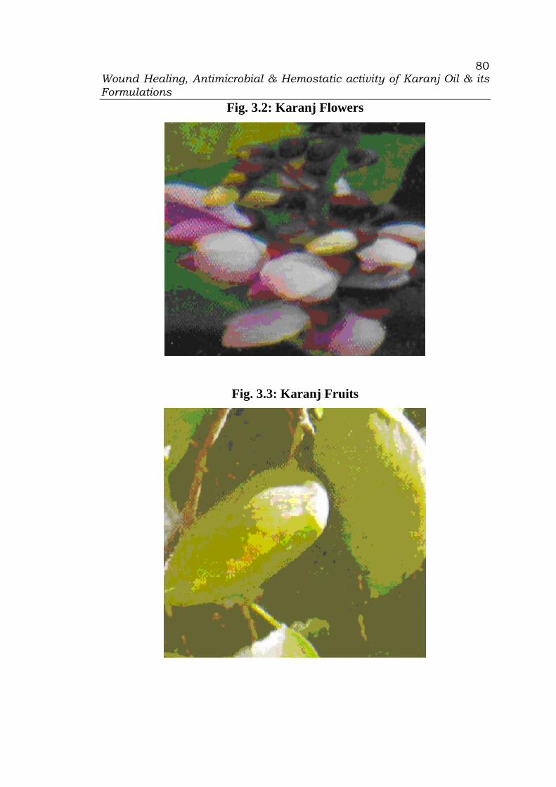
Wound Healing, Antimicrobial & Hemostatic activity of Karanj Oil & its Formulations
80
Fig. 3.2: Karanj Flowers
Fig. 3.3: Karanj Fruits

Wound Healing, Antimicrobial & Hemostatic activity of Karanj Oil & its Formulations
81
Fig. 3.4: Karanj Pods
Fig. 3.5: Karanj Seeds
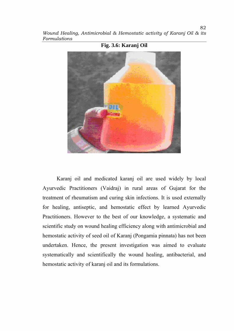
Wound Healing, Antimicrobial & Hemostatic activity of Karanj Oil & its Formulations
82
Fig. 3.6: Karanj Oil
Karanj oil and medicated karanj oil are used widely by local
Ayurvedic Practitioners (Vaidraj) in rural areas of Gujarat for the
treatment of rheumatism and curing skin infections. It is used externally
for healing, antiseptic, and hemostatic effect by learned Ayurvedic
Practitioners. However to the best of our knowledge, a systematic and
scientific study on wound healing efficiency along with antimicrobial and
hemostatic activity of seed oil of Karanj (Pongamia pinnata) has not been
undertaken. Hence, the present investigation was aimed to evaluate
systematically and scientifically the wound healing, antibacterial, and
hemostatic activity of karanj oil and its formulations.
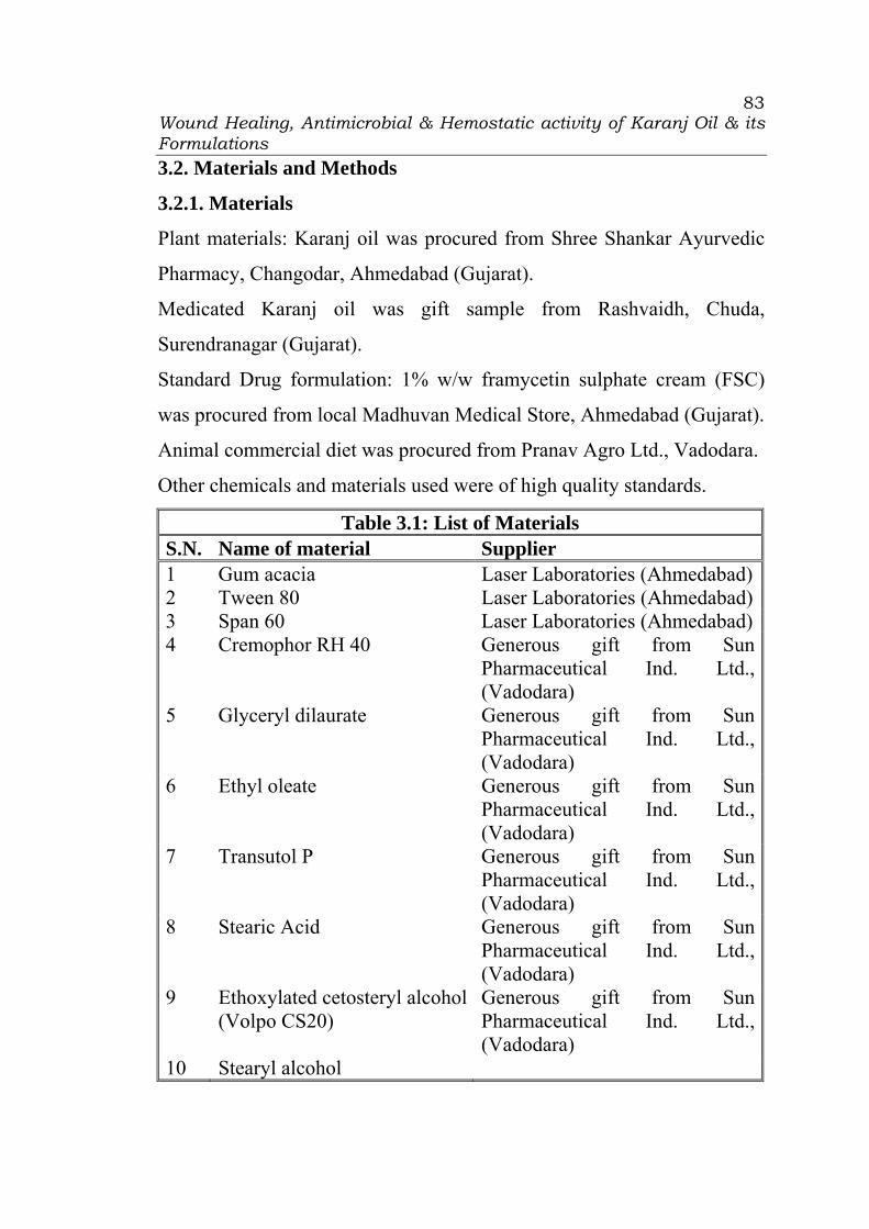
Wound Healing, Antimicrobial & Hemostatic activity of Karanj Oil & its Formulations
83
3.2. Materials and Methods
3.2.1. Materials
Plant materials: Karanj oil was procured from Shree Shankar Ayurvedic
Pharmacy, Changodar, Ahmedabad (Gujarat).
Medicated Karanj oil was gift sample from Rashvaidh, Chuda,
Surendranagar (Gujarat).
Standard Drug formulation: 1% w/w framycetin sulphate cream (FSC)
was procured from local Madhuvan Medical Store, Ahmedabad (Gujarat).
Animal commercial diet was procured from Pranav Agro Ltd., Vadodara.
Other chemicals and materials used were of high quality standards.
Table 3.1: List of Materials S.N. Name of material Supplier 1 Gum acacia Laser Laboratories (Ahmedabad)2 Tween 80 Laser Laboratories (Ahmedabad)3 Span 60 Laser Laboratories (Ahmedabad)4 Cremophor RH 40 Generous gift from Sun
Pharmaceutical Ind. Ltd., (Vadodara)
5 Glyceryl dilaurate Generous gift from Sun Pharmaceutical Ind. Ltd., (Vadodara)
6 Ethyl oleate Generous gift from Sun Pharmaceutical Ind. Ltd., (Vadodara)
7 Transutol P Generous gift from Sun Pharmaceutical Ind. Ltd., (Vadodara)
8 Stearic Acid Generous gift from Sun Pharmaceutical Ind. Ltd., (Vadodara)
9 Ethoxylated cetosteryl alcohol (Volpo CS20)
Generous gift from Sun Pharmaceutical Ind. Ltd., (Vadodara)
10 Stearyl alcohol

Wound Healing, Antimicrobial & Hemostatic activity of Karanj Oil & its Formulations
84
11 Propylene glycol Generous gift from Sun Pharmaceutical Ind. Ltd., (Vadodara)
12 Agar Generous gift from Sun rise Remedies (Ahmedabad)
13 Peptose Generous gift from Sun rise Remedies (Ahmedabad)
14 Lab lemco (Beef Extract) Generous gift from Sun rise Remedies (Ahmedabad)
15 Sodium chloride Laser Laboratories (Ahmedabad)16 Sodium hydroxide Laser Laboratories (Ahmedabad)17 Light ether Generous gift from Sun
Pharmaceutical Ind. Ltd., (Vadodara)
18 Sodium pentobarbital Generous gift from Sun Pharmaceutical Ind. Ltd., (Vadodara)
19 100 mM citrate buffer pH 4.5 Generous gift from Sun Pharmaceutical Ind. Ltd., (Vadodara)
20 Human thromboplastin (Thromborel®)
Generous gift from Sun Pharmaceutical Ind. Ltd., (Vadodara)
21 Human placenta lipid extract (Pathrombin®)
Generous gift from Sun Pharmaceutical Ind. Ltd., (Vadodara)
22 Diethylbarbiturate-citrate buffer, pH 7.6
Generous gift from Sun Pharmaceutical Ind. Ltd., (Vadodara)
23 Bovine test-thrombin 30 IU/ml
Generous gift from Sun Pharmaceutical Ind. Ltd., (Vadodara)
24 Calcium chloride Loba Chemie Pvt. Ltd (Bombay)

Wound Healing, Antimicrobial & Hemostatic activity of Karanj Oil & its Formulations
85
3.2.2 Methods
3.2.2.1 Preparation of Karanj oil formulations
3.2.2.1.1 Karanj oil:
Karanj oil was procured from Shree Shankar Ayurvedic Pharmacy,
Changodar, Ahmedabad (Gujarat). It was stored in well closed container
at room temperature.
3.2.2.1.2 Medicated Karanj oil:
Medicated Karanj oil was gift sample from Rashvaidh, Chuda,
Surendranagar (Gujarat).
3.2.2.1.3 Emulsion of Karanj oil:
Emulsion of the karanj oil was prepared by dry gum method using
gum acacia, Tween 80, or/and Span 20 as emulsifiers. The required
quantity of karanj oil was weighed and transferred in a glass beaker.
Weighted quantity of gum, emulgent, or/and co-emulgent (Acacia, Tween
80, Span 20) were mixed in the oil with stirring into a mortar. The
measured volume of distilled water was added in portion with trituration.
Remaining quantity of distilled water was added dropwise with
trituration. The creamy thick emulsion was obtained. The emulsion was
stored in white glass bottle at RT.
The composition of emulsion containing 20% to 40% karanj oil
prepared using gun acacia is presented in Table 3.2.
Table 3.2: Formulation Composition of Emulsion containing Karanj Oil Using Gum Acacia
Batch Code Ingredient (%w/w) E1 E2 E3 E4 E5
Karanj oil 20 25 30 35 40 Gum acacia 5 6.25 7.25 8.75 10 Dist. Water q.s. to 100 100 100 100 100
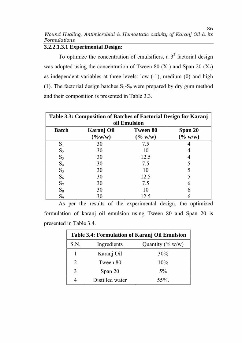
Wound Healing, Antimicrobial & Hemostatic activity of Karanj Oil & its Formulations
86
3.2.2.1.3.1 Experimental Design:
To optimize the concentration of emulsifiers, a 32 factorial design
was adopted using the concentration of Tween 80 (X1) and Span 20 (X2)
as independent variables at three levels: low (-1), medium (0) and high
(1). The factorial design batches S1-S9 were prepared by dry gum method
and their composition is presented in Table 3.3.
Table 3.3: Composition of Batches of Factorial Design for Karanj oil Emulsion
Batch Karanj Oil (%w/w)
Tween 80 (% w/w)
Span 20 (% w/w)
S1 30 7.5 4 S2 30 10 4 S3 30 12.5 4 S4 30 7.5 5 S5 30 10 5 S6 30 12.5 5 S7 30 7.5 6 S8 30 10 6 S9 30 12.5 6 As per the results of the experimental design, the optimized
formulation of karanj oil emulsion using Tween 80 and Span 20 is
presented in Table 3.4.
Table 3.4: Formulation of Karanj Oil Emulsion S.N. Ingredients Quantity (% w/w)
1 Karanj Oil 30% 2 Tween 80 10% 3 Span 20 5% 4 Distilled water 55%.
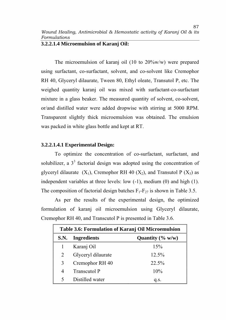
Wound Healing, Antimicrobial & Hemostatic activity of Karanj Oil & its Formulations
87
3.2.2.1.4 Microemulsion of Karanj Oil:
The microemulsion of karanj oil (10 to 20%w/w) were prepared
using surfactant, co-surfactant, solvent, and co-solvent like Cremophor
RH 40, Glyceryl dilaurate, Tween 80, Ethyl oleate, Transutol P, etc. The
weighed quantity karanj oil was mixed with surfactant-co-surfactant
mixture in a glass beaker. The measured quantity of solvent, co-solvent,
or/and distilled water were added dropwise with stirring at 5000 RPM.
Transparent slightly thick microemulsion was obtained. The emulsion
was packed in white glass bottle and kept at RT.
3.2.2.1.4.1 Experimental Design:
To optimize the concentration of co-surfactant, surfactant, and
solubilizer, a 33 factorial design was adopted using the concentration of
glyceryl dilaurate (X1), Cremophor RH 40 (X2), and Transutol P (X3) as
independent variables at three levels: low (-1), medium (0) and high (1).
The composition of factorial design batches F1-F27 is shown in Table 3.5.
As per the results of the experimental design, the optimized
formulation of karanj oil microemulsion using Glyceryl dilaurate,
Cremophor RH 40, and Transcutol P is presented in Table 3.6.
Table 3.6: Formulation of Karanj Oil Microemulsion
S.N. Ingredients Quantity (% w/w) 1 Karanj Oil 15% 2 Glyceryl dilaurate 12.5% 3 Cremophor RH 40 22.5% 4 Transcutol P 10% 5 Distilled water q.s.
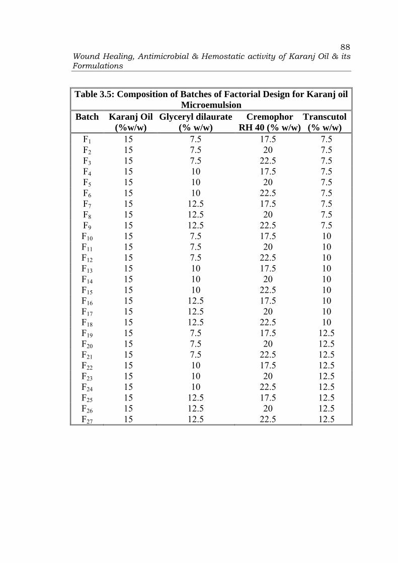
Wound Healing, Antimicrobial & Hemostatic activity of Karanj Oil & its Formulations
88
Table 3.5: Composition of Batches of Factorial Design for Karanj oil Microemulsion
Batch Karanj Oil (%w/w)
Glyceryl dilaurate (% w/w)
Cremophor RH 40 (% w/w)
Transcutol (% w/w)
F1 15 7.5 17.5 7.5 F2 15 7.5 20 7.5 F3 15 7.5 22.5 7.5 F4 15 10 17.5 7.5 F5 15 10 20 7.5 F6 15 10 22.5 7.5 F7 15 12.5 17.5 7.5 F8 15 12.5 20 7.5 F9 15 12.5 22.5 7.5 F10 15 7.5 17.5 10 F11 15 7.5 20 10 F12 15 7.5 22.5 10 F13 15 10 17.5 10 F14 15 10 20 10 F15 15 10 22.5 10 F16 15 12.5 17.5 10 F17 15 12.5 20 10 F18 15 12.5 22.5 10 F19 15 7.5 17.5 12.5 F20 15 7.5 20 12.5 F21 15 7.5 22.5 12.5 F22 15 10 17.5 12.5 F23 15 10 20 12.5 F24 15 10 22.5 12.5 F25 15 12.5 17.5 12.5 F26 15 12.5 20 12.5 F27 15 12.5 22.5 12.5

Wound Healing, Antimicrobial & Hemostatic activity of Karanj Oil & its Formulations
89
3.2.2.1.5 Cream of Karanj oil:
The cream containing karanj oil was prepared by hot emulsification
method. The weighed quantity of propylene glycol was added in distilled
water in a glass beaker. Ethoxylated cetosteryl alcohol was weighed and
added. The mass was heated at 70°C on a water bath and transferred to a
mortar. The measured quantity of karanj oil, stearic acid, and stearyl
alcohol were heated to 70°C in a beaker. The hot oil phase was added
slowly to hot water phase with trituration. The mixture was stirred till
temperature came down to room temperature. The cream was packed in a
container and kept at RT. The formulation of karanj oil cream is depleted
in Table 3.7.
Table 3.7: Formulation of Cream of Karanj Oil S.N. Ingredient Quantity (%w/w)
1 Karanj Oil 30 2 Stearic Acid 05 3 Ethoxylated cetosteryl alcohol (Volpo CS20) 10 4 Stearyl alcohol 05 5 Propylene glycol 10 6 Purified water 40
3.2.2.1.6 Evaluation of Karanj oil Formulations: Freeze Thawing
The formulations were subjected to 3 to 4 freeze-thaw cycles, which
included freezing at -4°C for 24 hours followed by thawing at 40°C for
24 hours. Centrifugation was performed at 3000 rpm for 5 minutes. The
formulations (n=3) were then observed for phase separation.

Wound Healing, Antimicrobial & Hemostatic activity of Karanj Oil & its Formulations
90
Droplet Size Measurement
One hundred microliters of the formulation was diluted with distilled
water to 250 mL in a beaker and gently mixed using a glass rod. The
resultant diluted formulation was then subjected to particle size analysis
(Malvern Mastersizer, Worchestershire, UK, equipped with 2000 Hydro
MU) with a particle size measurement range of 0.02 to 2000 μm. Particle
size was calculated from the volume size distribution (n=3).
Optical birefringence
The microemulsion was checked visually to confirm absence of other
phases. The microemulsion was characterized for % Transmittance
measured by UV Spectrophotometer at 520nm. Distilled water was
employed as the reference solution with a transmittance of 100% (n=3).
Measurement of zeta potential
The zeta potential of the microemulsion was measured using a Zetasizer
Nano-ZS (Malvern Instruments, UK). The electrophoretic mobility
(µm/s) was measured using small volume disposable zeta cell and
converted to zeta potential by in-built software using Helmholtz-
Smoluchowski equation. The measurement was done at 25°C in triplicate.
Viscosity determination
The viscosity of each formulation was measured using a DV-E
viscometer (Brookfield Engineering Laboratories, Middleboro, MA,
USA) at 25˚C in triplicate. The samples (n=3) were contained in
cylindrical bottles, 2.4 cm in diameter and 4.5 cm in height. The
measurement was performed with a No. 16 spindle and the shear rate was
set at 100 RPM.

Wound Healing, Antimicrobial & Hemostatic activity of Karanj Oil & its Formulations
91
3.2.2.2 Wound healing activity:
Wistar rats of either sex weighing 150-200 gm were used for the
study (8). All the animals were maintained under controlled room
temperature (22±2°C) and relative humidity (50±5%) with 12:00 hr light,
12:00 hr dark cycle. The animals were housed in colony cages (three
animals per cage) with free access to commercial diet (Pranav Agro Ltd.,
Baroda) and water ad libitum during the investigation period. All the
animals were acclimatized to the laboratory environment for 5 days
before the experimentation. Six animals (rats) per group comprising of
three males and three females were used in each experiment set, unless
otherwise specified. The study protocol was approved by the Institutional
Animal Ethics Committee (IAEC) of C.U. Shah College of Pharmacy and
Research, Wadhwan.
Excision wound model was used to evaluate the wound healing
efficiency. The rats were inflicted with excision wounds as described by
Morton and Malone (9) under light ether anesthesia. A circular wound of
about 300 sq. mm and 2 mm depth was made on depilated ethanol
sterilized dorsal thoracic region of the rats. The animals were divided into
seven groups of six each. The animals of group I was left untreated and
considered as control. The rats of Group II were served as reference
standard and received 1% w/w framycetin sulphate cream (FSC), the
animal group III and IV were treated with Karanj oil and medicated
karanj oil, respectively. The animal group V, VI and VII were treated
with Karanj oil emulsion, Karanj oil microemulsion and karanj oil cream,
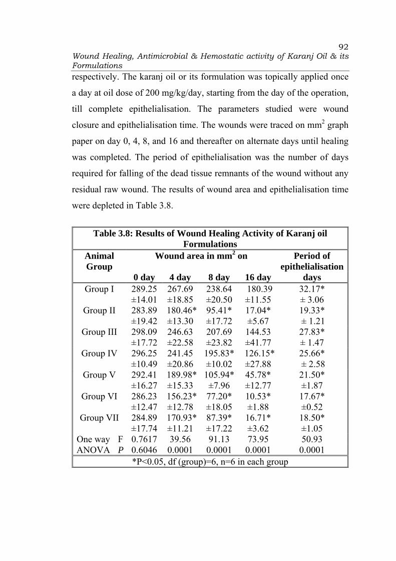
Wound Healing, Antimicrobial & Hemostatic activity of Karanj Oil & its Formulations
92
respectively. The karanj oil or its formulation was topically applied once
a day at oil dose of 200 mg/kg/day, starting from the day of the operation,
till complete epithelialisation. The parameters studied were wound
closure and epithelialisation time. The wounds were traced on mm2 graph
paper on day 0, 4, 8, and 16 and thereafter on alternate days until healing
was completed. The period of epithelialisation was the number of days
required for falling of the dead tissue remnants of the wound without any
residual raw wound. The results of wound area and epithelialisation time
were depleted in Table 3.8.
Table 3.8: Results of Wound Healing Activity of Karanj oil
Formulations Wound area in mm2 on Period of
epithelialisationAnimal Group
0 day 4 day 8 day 16 day days Group I 289.25
±14.01 267.69 ±18.85
238.64 ±20.50
180.39±11.55
32.17* ± 3.06
Group II 283.89 ±19.42
180.46*±13.30
95.41* ±17.72
17.04* ±5.67
19.33* ± 1.21
Group III 298.09 ±17.72
246.63 ±22.58
207.69 ±23.82
144.53 ±41.77
27.83* ± 1.47
Group IV 296.25 ±10.49
241.45 ±20.86
195.83* ±10.02
126.15*±27.88
25.66* ± 2.58
Group V 292.41 ±16.27
189.98*±15.33
105.94* ±7.96
45.78* ±12.77
21.50* ±1.87
Group VI 286.23 ±12.47
156.23*±12.78
77.20* ±18.05
10.53* ±1.88
17.67* ±0.52
Group VII 284.89 ±17.74
170.93*±11.21
87.39* ±17.22
16.71* ±3.62
18.50* ±1.05
F 0.7617 39.56 91.13 73.95 50.93 One way ANOVA P 0.6046 0.0001 0.0001 0.0001 0.0001
*P<0.05, df (group)=6, n=6 in each group
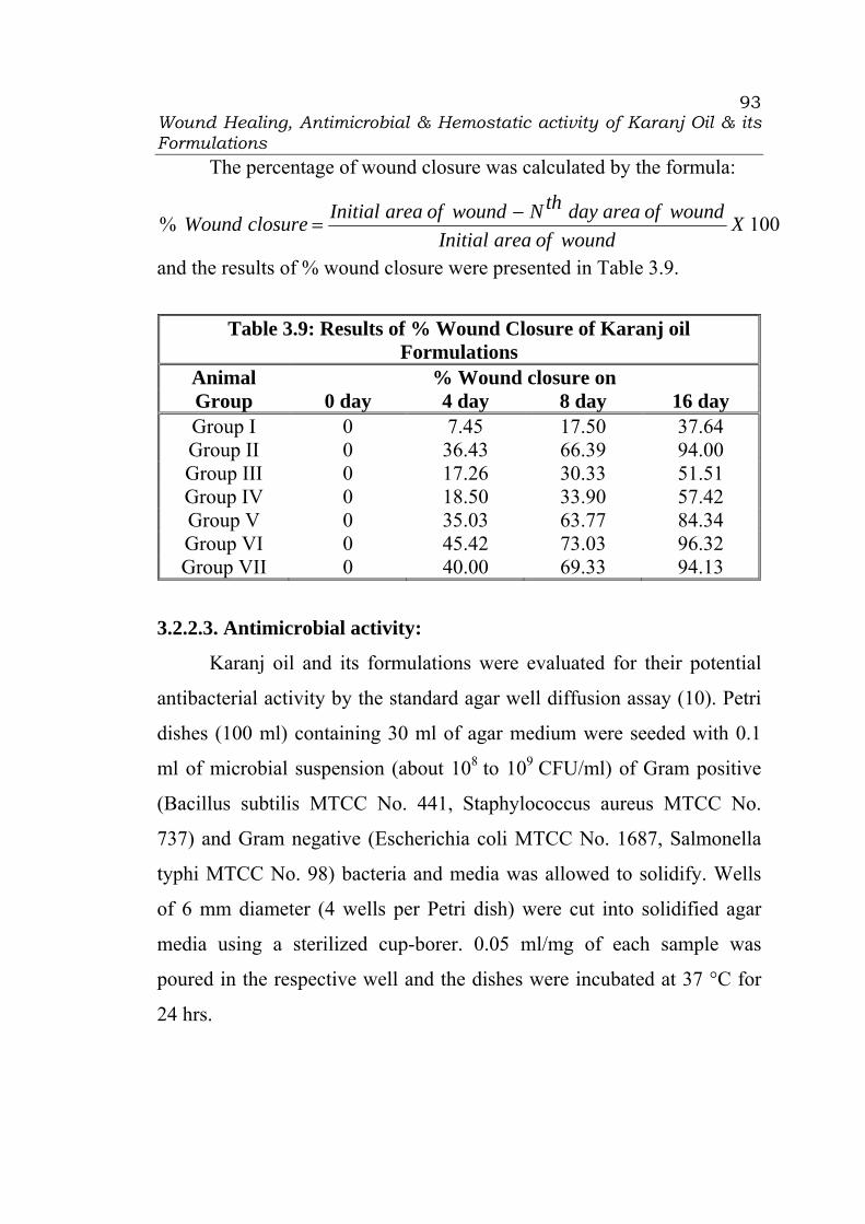
Wound Healing, Antimicrobial & Hemostatic activity of Karanj Oil & its Formulations
93
The percentage of wound closure was calculated by the formula:
100% XwoundofareaInitial
woundofareadaythNwoundofareaInitialclosureWound −=
and the results of % wound closure were presented in Table 3.9.
Table 3.9: Results of % Wound Closure of Karanj oil
Formulations % Wound closure on Animal
Group 0 day 4 day 8 day 16 day Group I 0 7.45 17.50 37.64 Group II 0 36.43 66.39 94.00 Group III 0 17.26 30.33 51.51 Group IV 0 18.50 33.90 57.42 Group V 0 35.03 63.77 84.34 Group VI 0 45.42 73.03 96.32 Group VII 0 40.00 69.33 94.13
3.2.2.3. Antimicrobial activity:
Karanj oil and its formulations were evaluated for their potential
antibacterial activity by the standard agar well diffusion assay (10). Petri
dishes (100 ml) containing 30 ml of agar medium were seeded with 0.1
ml of microbial suspension (about 108 to 109 CFU/ml) of Gram positive
(Bacillus subtilis MTCC No. 441, Staphylococcus aureus MTCC No.
737) and Gram negative (Escherichia coli MTCC No. 1687, Salmonella
typhi MTCC No. 98) bacteria and media was allowed to solidify. Wells
of 6 mm diameter (4 wells per Petri dish) were cut into solidified agar
media using a sterilized cup-borer. 0.05 ml/mg of each sample was
poured in the respective well and the dishes were incubated at 37 °C for
24 hrs.

Wound Healing, Antimicrobial & Hemostatic activity of Karanj Oil & its Formulations
94
The antibacterial activity was expressed in terms of the mean of
diameter of zone of inhibition including 6mm diameter of well (in mm
measured horizontally and vertically) produced by each sample at the end
of incubation period. Sterilized distilled water was served as negative
control. 1gm of 1% W/W framycetin sulphate cream (FSC) ointment
diluted and mixed with 9ml of sterilized distilled water was used as
standard i.e. positive control. The microbial assay was performed in
triplicate and the results were presented in Table 3.10 as mean of the
three values ± standard deviation.
Table 3.10: Results of Zone of Inhibition of Karanj oil and its Formulations
Diameter of the zone of inhibition (mm) Gram positive Gram negative
Bacteria
Bacillus subtilis
Staphylococcus aureus
Escherichia coli
Salmonella typhi
Negative control
No No No No
Positive control
34.33 ±5.03
32.67 ±5.51
29.33 ±4.16
24.67 ±4.51
Karanj oil 10.33* ±1.53
14.33* ±4.16
11.67 ±3.51
10.67 ±4.04
Medicated Karanj oil
12.33* ±3.06
19.33* ±4.73
13.67 ±4.16
11.67 ±4.04
Emulsion 17.33* ±3.51
22.33 ±4.51
16.33 ±3.51
15.33 ±3.51
Micro emulsion
24.33* ±2.08
24.33 ±5.03
18.67 ±6.51
17.67 ±5.03
Cream 15.67* ±1.15
19.67* ±3.51
14.67 ±6.03
13.67 ±3.51
F 25.98 5.36 5.202 4.547 One way ANOVA P 0.000 0.008 0.009 0.0147
*P<0.05, df (group)=5, n=3 in each group

Wound Healing, Antimicrobial & Hemostatic activity of Karanj Oil & its Formulations
95
3.2.2.4. Hemostatic activity:
Purpose and rationale blood coagulation test:
The coagulation cascade consists of a complex network of
interactions resulting in thrombin-mediated cleavage of fibrinogen to
fibrin which is one major component of a thrombus. The coagulation
cascade can be initiated either by the “exogenous pathway”, the release of
thromboplastin (tissue factor) leading to activation of factor VII to the
tissue factor/factor VIIa complex, or by the “endogenous pathway”, so-
called contact activation leading via factors XII, XI and IX to the
assembly of the tenase complex consisting of activated factors VIII and
IX and Ca2+ on a phospholipids surface. Both complexes can activate
factor X which induces the formation of the prothrombinase complex
consisting of factor Xa, factor Va and Ca2+ on a phospholipid surface.
The latter leads to the activation of thrombin which in turn cleaves
fibrinogen to fibrin. The three coagulation tests (PT, APTT and TT) allow
differentiating between effects on the exogenous or endogenous pathway
or on fibrin formation. The influence of compounds on the plasmatic
blood coagulation is determined by measuring the coagulation parameters
prothrombin time (PT), activated partial thromboplastin time (APTT) and
thrombin time (TT) ex vivo (11).
Method of measurement of Hemostatic activity
Rats were slightly ether anesthetized, and abdominal hair was
carefully removed with electric clipper, they were fasted overnight. The

Wound Healing, Antimicrobial & Hemostatic activity of Karanj Oil & its Formulations
96
animals were divided into six groups of six rats in each. The animals of
group I was left untreated and considered as control. The rats of Group II,
III, IV, V, and VI were applied topically Karanj, medicated karanj oil,
emulsion, microemulsion, and cream, respectively, on the abdominal area
of 9 cm2 gently at a dose of 200 mg/kg (11). Rats were anesthetized by
intravenous injection of 60 mg/kg sodium pentobarbital. The caudal caval
vein was exposed by a mid-line incision and 1.8 ml blood were collected
at 0 hr and 24 hrs into a plastic syringe containing 0.2 ml 100 mM citrate
buffer pH 4.5. The sample was immediately agitated and centrifuged in a
plastic tube at 1500 rpm for 10 min. Plasma was transferred to another
plastic tube and the coagulation tests for the determination of
thromboplastin time (PT), activated partial thromboplastin time (APTT)
and thrombin time (TT) were performed within 3 h. In general, citrated
plasma was coagulated by the addition of the respective compounds (see
below for PT, APTT or TT) and the time in seconds to clot formation was
determined in the coagulometer (12-13).
Prothrombin Time (PT): An aliquot of 0.1 ml of citrated plasma was
incubated for 1 min at 37°C. Then 0.2 ml of thromboplastin was added
and the coagulometer was started. The time to clot formation was
measured. The PT measures effects on the exogenous pathway of
coagulation.
Activated Partial Thromboplastin Time (APTT): To 0.1 ml of citrated
plasma, 0.1 ml of placenta lipid extract was added and the mixture was
incubated for 2 min at 37°C. The coagulation process was initiated by the

Wound Healing, Antimicrobial & Hemostatic activity of Karanj Oil & its Formulations
97
addition of 0.1 ml 25 mM calcium chloride when the coagulometer was
started and the time to clot formation was determined. The APTT
measures effects on the endogenous pathway of coagulation.
Thrombin Time (TT): To 0.1 ml of citrated plasma, 0.1 ml of
diethylbarbiturate-citrate buffer, pH 7.6 was added and the mixture was
incubated for 1 min at 37 °C. Then 0.1 ml of bovine test-thrombin was
added and the coagulometer was started. The time to clot formation was
measured. The TT measures effects on fibrin formation.
The mean values of PT, APTT, and TT were calculated and the
results are presented in Table 3.11.
Table 3.11: Results of Hemostatic Activity of Karanj oil
Formulations PT
(Seconds) APTT
(Seconds) TT
(Seconds) Animal Group
Initial 24 hrs Initial 24 hrs Initial 24 hrs Group I 9.83
±0.759.67
±0.82 29.16 ±1.17
28.83 ±1.33
14.83 ±1.17
15.17 ±1.17
Group II 9.67 ±0.52
9.50 ±0.55
28.00 ±0.89
27.50 ±1.05
14.17 ±0.75
14.00 ±0.63
Group III 9.33 ±0.52
8.67 ±0.52
27.17*±0.75
27.00 ±0.89
13.83 ±1.33
13.66* ±1.03
Group IV 9.17±0.75
8.83 ±0.41
27.00*±1.79
26.67* ±1.37
13.67 ±1.37
13.17* ±0.75
Group V 9.00±0.89
8.67 ±0.52
27.17*±0.75
26.50* ±0.55
13.83 ±1.33
12.83* ±0.75
Group VI 9.17±0.75
8.50* ±0.55
26.83*±0.75
26.33* ±1.03
13.67 ±1.21
13.00* ±0.89
One way F 1.242 4.356 4.000 4.488 0.8182 6.152 ANOVA P 0.3145 0.0042 0.0067 0.0036 0.5464 0.0005
P< 0.05, df=3, n=6

Wound Healing, Antimicrobial & Hemostatic activity of Karanj Oil & its Formulations
98
3.3. Results and Discussion
3.3.1 Formulation Development of Karanj oil Emulsion:
With an objective to fix oil phase concentration, emulsion of the
karanj oil was prepared initially using gum acacia as a model emulsifier.
The required quantity of karanj oil was weighed and transferred in a
mortar. Weighted quantity of gum acacia were added in karanj oil with
trituration. The measured volume of distilled water was added in portion
with trituration. Remaining quantity of distilled water was added
dropwise with trituration. The creamy slightly thick emulsion was
obtained. The emulsion was evaluated for phase separation by
centrifugation at 1500 RPM for 10 minutes in a glass test tube (15ml) and
storing at RT overnight. The test tube containing the emulsion was gently
inverted till homogenous emulsion was achieved and the number of
inversions were counted in triplicate. The results of cream height and
number of inversion for batch E1 to E5 were presented in Table 3.12.
Table 3.12: Composition and results of evaluation parameters for emulsion containing Karanj oil and gum acacia
Batch Code Ingredient (%w/w) E1 E2 E3 E4 E5 Karanj oil 20 25 30 35 40 Gum acacia 5 6.25 7.25 8.75 10 Dist. Water q.s. to 100 100 100 100 100 Parameters Creaming height Phase
separation5cm 1.3 cm 2.5cm 3.4 cm
Number of inversion
Not performed
Remained separated
23 68 Thick cream
Wound Healing, Antimicrobial & Hemostatic activity of Karanj Oil & its Formulations
99
The results (Table 3.12) revealed that the emulsion containing 20%
karanj oil (Batch E1) showed separation of oil phase on centrifugation,
while the emulsion containing 25% karanj oil (Batch E2) showed cream
height of 5 cm and emulsion containing 30% karanj oil (Batch E3)
showed least phase separation (1.3cm). Emulsion of batch E3 was
redispered evenly by 23 inversions, while of batch E4 by 68 inversions.
The emulsion of Batch E5 was a thick cream, and it was not possible to
redisperse it. Hence, 30% concentration of karanj oil (Batch E3) was
selected for further studies of formulation optimization of emulsion.
Formulation optimization of karanj oil emulsion using Tween 80 and
Span 20 as emulsifiers:
Externally used emulsions do not contained gum acacia as an
emulsifying agent due to its stickiness and grittiness. Surfactants were
widely used to increase dissolution and bioavailability of drug by
lowering interfacial tension, which allowed better wetting and penetration
of the drug from the dosage form. The combination of hydrophilic Tween
80 with lipophilic Span 20 is widely preferred in formulation
development of externally used emulsions by formulation pharmacist by
varying the proportion as to produce o/w or w/o emulsion. When Tween
80 is added, it orients at the interface such that part of its hydrocarbon tail
is in the oil phase, and the remainder of the chain, together with the
sorbitan ring and the polyoxyethylene chains, is located in the water
phase, this orientation results in effective van der Waals attraction. The
interfacial film is strengthened and the stability of o/w emulsion is
increased against particle coalescence (14). To our knowledge, no
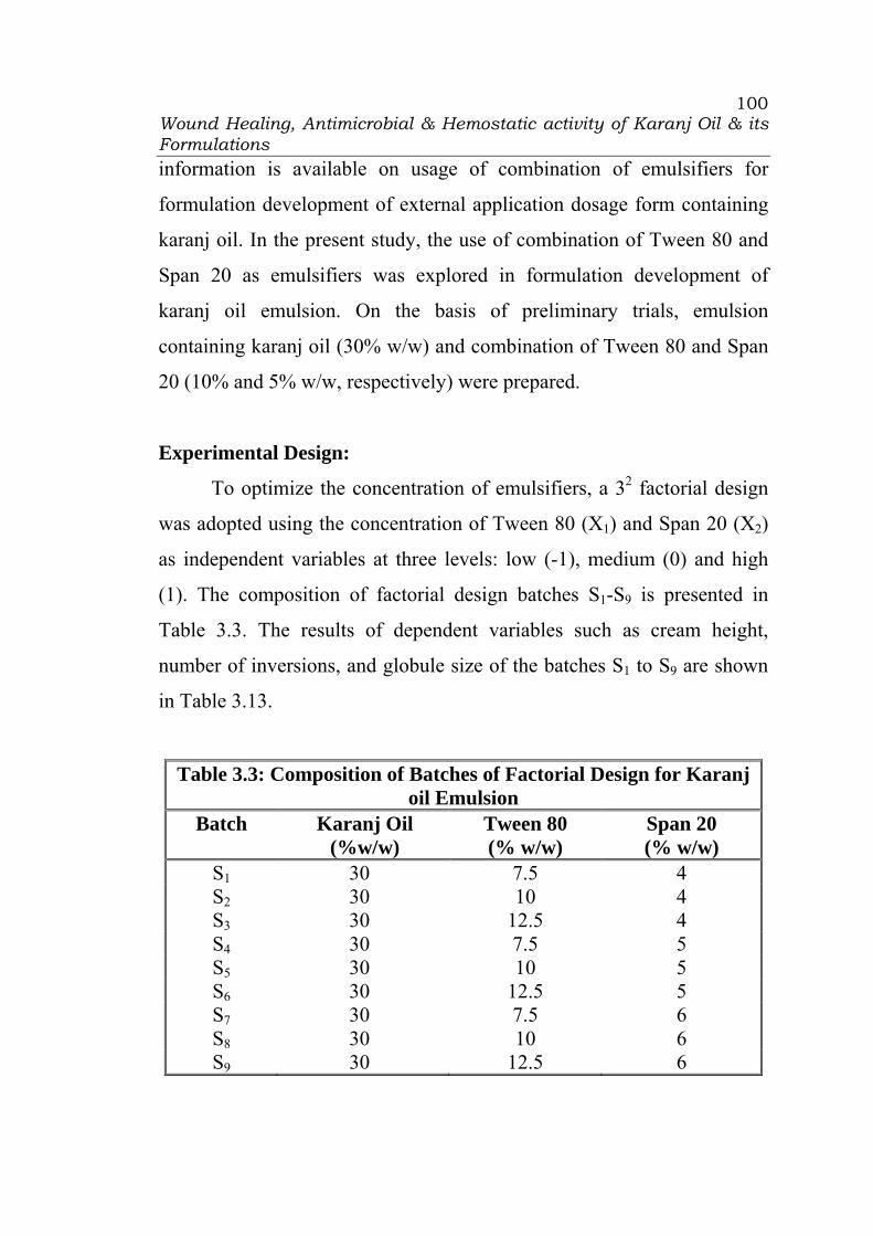
Wound Healing, Antimicrobial & Hemostatic activity of Karanj Oil & its Formulations
100
information is available on usage of combination of emulsifiers for
formulation development of external application dosage form containing
karanj oil. In the present study, the use of combination of Tween 80 and
Span 20 as emulsifiers was explored in formulation development of
karanj oil emulsion. On the basis of preliminary trials, emulsion
containing karanj oil (30% w/w) and combination of Tween 80 and Span
20 (10% and 5% w/w, respectively) were prepared.
Experimental Design:
To optimize the concentration of emulsifiers, a 32 factorial design
was adopted using the concentration of Tween 80 (X1) and Span 20 (X2)
as independent variables at three levels: low (-1), medium (0) and high
(1). The composition of factorial design batches S1-S9 is presented in
Table 3.3. The results of dependent variables such as cream height,
number of inversions, and globule size of the batches S1 to S9 are shown
in Table 3.13.
Table 3.3: Composition of Batches of Factorial Design for Karanj oil Emulsion
Batch Karanj Oil (%w/w)
Tween 80 (% w/w)
Span 20 (% w/w)
S1 30 7.5 4 S2 30 10 4 S3 30 12.5 4 S4 30 7.5 5 S5 30 10 5 S6 30 12.5 5 S7 30 7.5 6 S8 30 10 6 S9 30 12.5 6
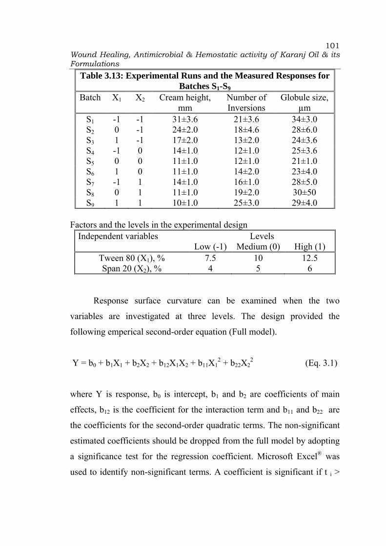
Wound Healing, Antimicrobial & Hemostatic activity of Karanj Oil & its Formulations
101
Table 3.13: Experimental Runs and the Measured Responses for Batches S1-S9
Batch X1 X2 Cream height, mm
Number of Inversions
Globule size, µm
S1 -1 -1 31±3.6 21±3.6 34±3.0 S2 0 -1 24±2.0 18±4.6 28±6.0 S3 1 -1 17±2.0 13±2.0 24±3.6 S4 -1 0 14±1.0 12±1.0 25±3.6 S5 0 0 11±1.0 12±1.0 21±1.0 S6 1 0 11±1.0 14±2.0 23±4.0 S7 -1 1 14±1.0 16±1.0 28±5.0 S8 0 1 11±1.0 19±2.0 30±50 S9 1 1 10±1.0 25±3.0 29±4.0
Factors and the levels in the experimental design
Independent variables Levels Low (-1) Medium (0) High (1)
Tween 80 (X1), % 7.5 10 12.5 Span 20 (X2), % 4 5 6
Response surface curvature can be examined when the two
variables are investigated at three levels. The design provided the
following emperical second-order equation (Full model).
Y = b0 + b1X1 + b2X2 + b12X1X2 + b11X12 + b22X2
2 (Eq. 3.1)
where Y is response, b0 is intercept, b1 and b2 are coefficients of main
effects, b12 is the coefficient for the interaction term and b11 and b22 are
the coefficients for the second-order quadratic terms. The non-significant
estimated coefficients should be dropped from the full model by adopting
a significance test for the regression coefficient. Microsoft Excel® was
used to identify non-significant terms. A coefficient is significant if t i >

Wound Healing, Antimicrobial & Hemostatic activity of Karanj Oil & its Formulations
102
tcrit (v), where v is the degrees of freedom of residual variance. The refined
model may be used for calculation of residuals or for drawing the contour
plot. The full and refined models (p < 0.05) of factorial design batches S1-
S9 for cream height, number of inversions, and globule size are shown in
Table 3.14.
Table 3.14: Summary of Results of Regression Analysis for Batches
S1-S9 Response b0 b1 b2 b11 b22 b12 r2
FM 11.44 -3.50 -6.17 0.83 5.83 2.5 0.9688 Cream
height,
mm
RM 12.00 -3.50 -6.17 - 5.83 - 0.9042
FM 12.33 0.50 1.33 0.50 6.00 4.25 0.9807 Number
of
inversions
RM 12.67 0.50 1.33 - 6.00 4.25 0.9776
FM 22.44 -1.83 0.17 0.83 5.83 2.75 0.9125 Globule
size, µm RM 23.00 -1.83 0.17 - 5.83 2.75 0.9205
FM = Full model, RM = Refined model
Cream height: The refined model showed that the second-order term,
b11, and interaction term, b12, were insignificant. The P value was found
to be close to 0.03 for the second order term (X22). It was concluded that
the coefficients, b1 and b2, carry a negative sign and therefore addition of
each surfactant can decrease the cream height of the emulsion. The lowest
cream height was observed when both the emulgent were used at medium
levels. The poor emulsion stability may be attributed to poor
emulsification and wetting when emulgent was used at low level. The
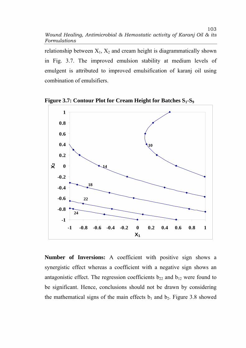
Wound Healing, Antimicrobial & Hemostatic activity of Karanj Oil & its Formulations
103
relationship between X1, X2 and cream height is diagrammatically shown
in Fig. 3.7. The improved emulsion stability at medium levels of
emulgent is attributed to improved emulsification of karanj oil using
combination of emulsifiers.
Figure 3.7: Contour Plot for Cream Height for Batches S1-S9
-1
-0.8
-0.6
-0.4
-0.2
0
0.2
0.4
0.6
0.8
1
-1 -0.8 -0.6 -0.4 -0.2 0 0.2 0.4 0.6 0.8 1X1
X2
24
18
22
14
10
Number of Inversions: A coefficient with positive sign shows a
synergistic effect whereas a coefficient with a negative sign shows an
antagonistic effect. The regression coefficients b22 and b12 were found to
be significant. Hence, conclusions should not be drawn by considering
the mathematical signs of the main effects b1 and b2. Figure 3.8 showed
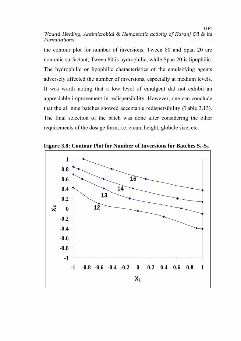
Wound Healing, Antimicrobial & Hemostatic activity of Karanj Oil & its Formulations
104
the contour plot for number of inversions. Tween 80 and Span 20 are
nonionic surfactant; Tween 80 is hydrophilic, while Span 20 is lipophilic.
The hydrophilic or lipophilic characteristics of the emulsifying agents
adversely affected the number of inversions, especially at medium levels.
It was worth noting that a low level of emulgent did not exhibit an
appreciable improvement in redispersibility. However, one can conclude
that the all nine batches showed acceptable redispersibility (Table 3.13).
The final selection of the batch was done after considering the other
requirements of the dosage form, i.e. cream height, globule size, etc.
Figure 3.8: Contour Plot for Number of Inversions for Batches S1-S9
-1-0.8-0.6-0.4-0.2
00.20.40.60.8
1
-1 -0.8 -0.6 -0.4 -0.2 0 0.2 0.4 0.6 0.8 1
X1
X2 12
1314
16

Wound Healing, Antimicrobial & Hemostatic activity of Karanj Oil & its Formulations
105
Globule size:
The refined model indicates that only the second-order terms (b11) is
insignificant. Tween 80 seems to be more effective in controlling globule
size since the coefficient b1 (-1.83) is greater in magnitude compared with
the coefficient b2 (0.17). The HLB value of Tween 80 is 15 and Span 20
is 4.5. The lower the HLB number, more lipophilic the compound and
vice versa. The non-significant interaction term indicated that Tween 80
did not affect the functionality of Span 20. The contour plot (Fig. 3.9)
showed that lower values of globule size can be obtained even at high to
medium level of X1. The plot indicates that to obtain globule size of 22 µ,
at least a medium level of X2 is required.
Figure 3.9: Contour Plot for Globule Size for Batches S1-S9
-1
-0.8
-0.6
-0.4
-0.2
0
0.2
0.4
0.6
0.8
1
-1 -0.8 -0.6 -0.4 -0.2 0 0.2 0.4 0.6 0.8 1
X1
X 2
222426
28
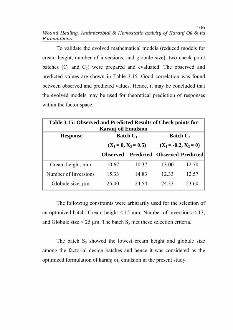
Wound Healing, Antimicrobial & Hemostatic activity of Karanj Oil & its Formulations
106
To validate the evolved mathematical models (reduced models for
cream height, number of inversions, and globule size), two check point
batches (C1 and C2) were prepared and evaluated. The observed and
predicted values are shown in Table 3.15. Good correlation was found
between observed and predicted values. Hence, it may be concluded that
the evolved models may be used for theoretical prediction of responses
within the factor space.
Table 3.15: Observed and Predicted Results of Check points for Karanj oil Emulsion
Batch C1
(X1 = 0, X2 = 0.5)
Batch C2
(X1 = -0.2, X2 = 0)
Response
Observed Predicted Observed Predicted
Cream height, mm 10.67 10.37 13.00 12.70
Number of Inversions 15.33 14.83 12.33 12.57
Globule size, µm 25.00 24.54 24.33 23.60
The following constraints were arbitrarily used for the selection of
an optimized batch: Cream height < 15 mm, Number of inversions < 13,
and Globule size < 25 µm. The batch S5 met these selection criteria.
The batch S5 showed the lowest cream height and globule size
among the factorial design batches and hence it was considered as the
optimized formulation of karanj oil emulsion in the present study.

Wound Healing, Antimicrobial & Hemostatic activity of Karanj Oil & its Formulations
107
3.3.2 Formulation development of Microemulsion of Karanj Oil:
The concept of microemulsion was introduced by Hoar and
Schulman in 1943 (15). They prepared the first microemulsions by
dispersing oil in an aqueous surfactant solution and adding an alcohol as a
co-surfactant, leading to a transparent, stable formulation. The existence
of this theoretical structure was later confirmed by use of various
technologies, and it is defined by Attwood (16) as “a microemulsion is a
system of water, oil, and amphiphilic compounds (surfactant and co-
surfactant) which is a transparent, single optically isotropic, and
thermodynamically stable liquid”.
These dispersed systems are isotropic, as opposed to liquid crystals
which are anisotropic. The main difference between emulsion and
microemulsion lies in the size and shape of the particles dispersed in the
continuous phase: these are at least an order of magnitude smaller in the
case of microemulsions (10–200 nm) than those of conventional
emulsions (1–30 μm). Emulsions consist of roughly spherical droplets of
one phase dispersed into the other, microemulsions constantly evolve
between various structures ranging from droplet like swollen micelles to
bicontinuous structures, making the usual o/w and w/o. Microemulsions
are formed when and only when the interfacial tension at the oil/water
interface is brought to a very low level and the interfacial layer is kept
highly flexible and fluid. These two conditions are usually met by a
careful and precise choice of the components and of their respective
proportions, and by the use of a co-surfactant which brings flexibility to
the oil/water interface. These conditions lead to a thermodynamically

Wound Healing, Antimicrobial & Hemostatic activity of Karanj Oil & its Formulations
108
optimized structure, which is stable as opposed to conventional emulsions
and does not require high input of energy (i.e. through agitation) to be
formed. As the size of the particles is much smaller than the wavelength
of visible light, microemulsions are transparent and their structure cannot
be observed through an optical microscope.
In topical formulations, microemulsions have been proved to
increase the cutaneous absorption of both lipophilic and hydrophilic
API’s when compared to conventional vehicles (emulsions, pure oils,
aqueous solutions, etc.). Many topical formulations currently marketed
are based on microemulsions. Figure 3.10: Structure of Microemulsion
Wound Healing, Antimicrobial & Hemostatic activity of Karanj Oil & its Formulations
109
Formulation optimization of microemulsion of Karanj oil:
The main problem in microemulsion formulations is to identify
surfactant combinations that would produce a thermodynamically stable
formulation. It is essential to establish the phase behavior of the particular
combination of chosen components. The formulation of microemulsions
is still highly empirical and time consuming and full characterization of
such systems is a tedious task and requires a large number of
experiments. Accordingly, the aim of the present study was to develop
microemulsion formulations using experimental design which may
bypass the need to study the phase behavior of microemulsion systems
and thus minimize the experimental efforts using desirability function.
To our knowledge, no information is available on usage of
combination of co-surfactant, surfactant, and solubilizer for the
formulation development of external application dosage form containing
karanj oil. In the present study, the use of combination of Glyceryl
dilaurate, Cremophor RH 40, and Transcutol P as co-surfactant,
surfactant, and solubilizer respectively was explored in formulation
development of karanj oil microemulsion. On the basis of preliminary
trials, microemulsions containing karanj oil (15% w/w) and combination
of Glyceryl dilaurate, Cremophor RH 40, and Transcutol P (10%, 20%
and 10% w/w, respectively) were prepared using distilled water.
Experimental Design:
To optimize the concentration of co-surfactant, surfactant, and
solubilizer, a 33 factorial design was adopted using the concentration of
Glyceryl dilaurate (X1), Cremophor RH 40 (X2), and Transutol P (X3) as
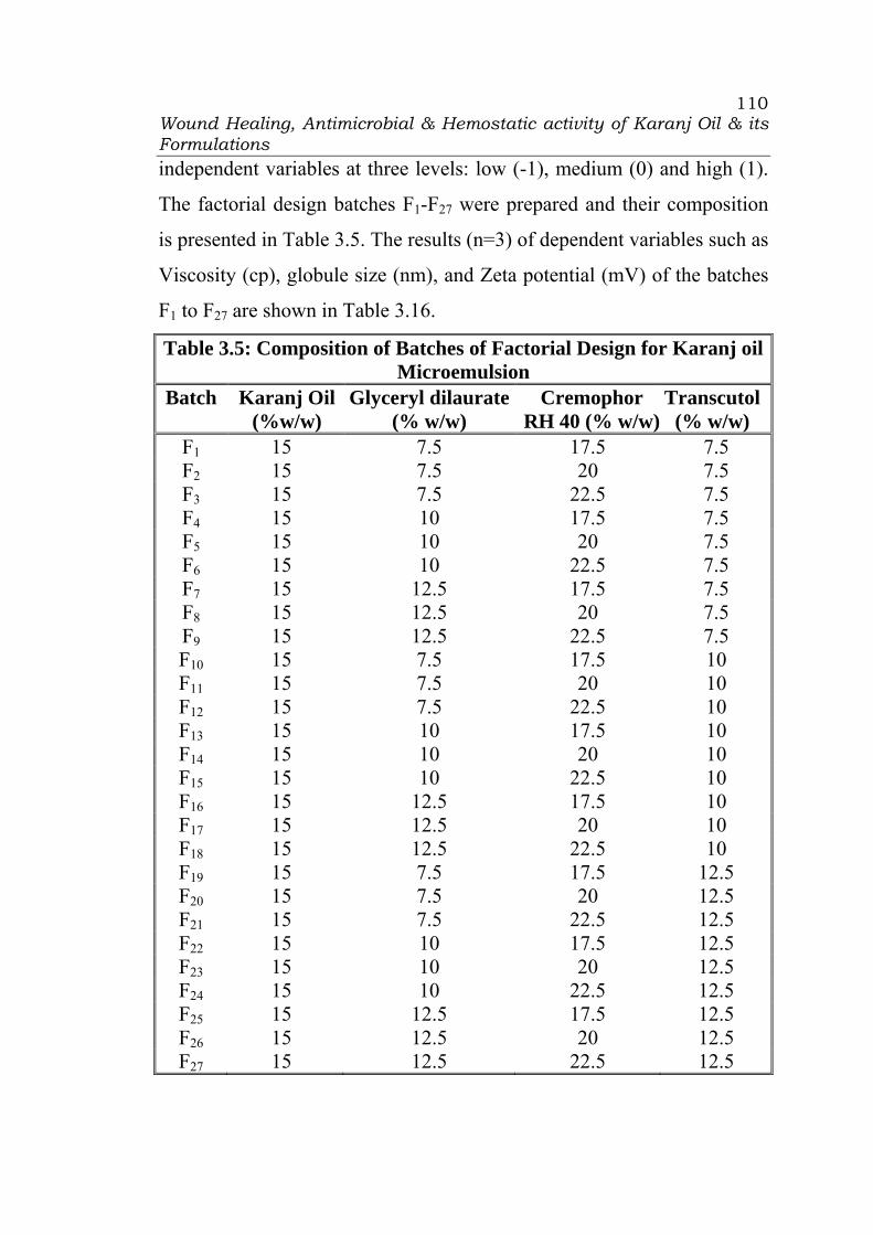
Wound Healing, Antimicrobial & Hemostatic activity of Karanj Oil & its Formulations
110
independent variables at three levels: low (-1), medium (0) and high (1).
The factorial design batches F1-F27 were prepared and their composition
is presented in Table 3.5. The results (n=3) of dependent variables such as
Viscosity (cp), globule size (nm), and Zeta potential (mV) of the batches
F1 to F27 are shown in Table 3.16.
Table 3.5: Composition of Batches of Factorial Design for Karanj oil Microemulsion
Batch Karanj Oil (%w/w)
Glyceryl dilaurate (% w/w)
Cremophor RH 40 (% w/w)
Transcutol (% w/w)
F1 15 7.5 17.5 7.5 F2 15 7.5 20 7.5 F3 15 7.5 22.5 7.5 F4 15 10 17.5 7.5 F5 15 10 20 7.5 F6 15 10 22.5 7.5 F7 15 12.5 17.5 7.5 F8 15 12.5 20 7.5 F9 15 12.5 22.5 7.5 F10 15 7.5 17.5 10 F11 15 7.5 20 10 F12 15 7.5 22.5 10 F13 15 10 17.5 10 F14 15 10 20 10 F15 15 10 22.5 10 F16 15 12.5 17.5 10 F17 15 12.5 20 10 F18 15 12.5 22.5 10 F19 15 7.5 17.5 12.5 F20 15 7.5 20 12.5 F21 15 7.5 22.5 12.5 F22 15 10 17.5 12.5 F23 15 10 20 12.5 F24 15 10 22.5 12.5 F25 15 12.5 17.5 12.5 F26 15 12.5 20 12.5 F27 15 12.5 22.5 12.5
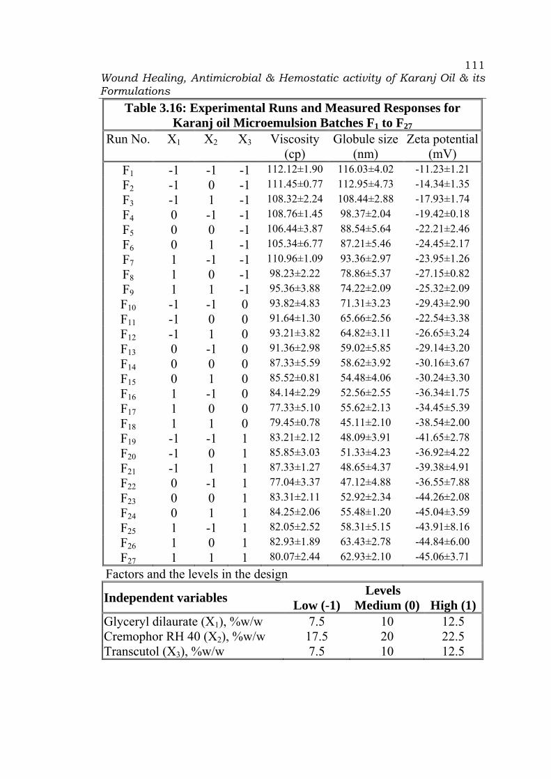
Wound Healing, Antimicrobial & Hemostatic activity of Karanj Oil & its Formulations
111
Table 3.16: Experimental Runs and Measured Responses for Karanj oil Microemulsion Batches F1 to F27
Run No. X1 X2 X3 Viscosity (cp)
Globule size (nm)
Zeta potential(mV)
F1 -1 -1 -1 112.12±1.90 116.03±4.02 -11.23±1.21 F2 -1 0 -1 111.45±0.77 112.95±4.73 -14.34±1.35 F3 -1 1 -1 108.32±2.24 108.44±2.88 -17.93±1.74 F4 0 -1 -1 108.76±1.45 98.37±2.04 -19.42±0.18 F5 0 0 -1 106.44±3.87 88.54±5.64 -22.21±2.46 F6 0 1 -1 105.34±6.77 87.21±5.46 -24.45±2.17 F7 1 -1 -1 110.96±1.09 93.36±2.97 -23.95±1.26 F8 1 0 -1 98.23±2.22 78.86±5.37 -27.15±0.82 F9 1 1 -1 95.36±3.88 74.22±2.09 -25.32±2.09 F10 -1 -1 0 93.82±4.83 71.31±3.23 -29.43±2.90 F11 -1 0 0 91.64±1.30 65.66±2.56 -22.54±3.38 F12 -1 1 0 93.21±3.82 64.82±3.11 -26.65±3.24 F13 0 -1 0 91.36±2.98 59.02±5.85 -29.14±3.20 F14 0 0 0 87.33±5.59 58.62±3.92 -30.16±3.67 F15 0 1 0 85.52±0.81 54.48±4.06 -30.24±3.30 F16 1 -1 0 84.14±2.29 52.56±2.55 -36.34±1.75 F17 1 0 0 77.33±5.10 55.62±2.13 -34.45±5.39 F18 1 1 0 79.45±0.78 45.11±2.10 -38.54±2.00 F19 -1 -1 1 83.21±2.12 48.09±3.91 -41.65±2.78 F20 -1 0 1 85.85±3.03 51.33±4.23 -36.92±4.22 F21 -1 1 1 87.33±1.27 48.65±4.37 -39.38±4.91 F22 0 -1 1 77.04±3.37 47.12±4.88 -36.55±7.88 F23 0 0 1 83.31±2.11 52.92±2.34 -44.26±2.08 F24 0 1 1 84.25±2.06 55.48±1.20 -45.04±3.59 F25 1 -1 1 82.05±2.52 58.31±5.15 -43.91±8.16 F26 1 0 1 82.93±1.89 63.43±2.78 -44.84±6.00 F27 1 1 1 80.07±2.44 62.93±2.10 -45.06±3.71
Factors and the levels in the design Levels Independent variables Low (-1) Medium (0) High (1)
Glyceryl dilaurate (X1), %w/w 7.5 10 12.5 Cremophor RH 40 (X2), %w/w 17.5 20 22.5 Transcutol (X3), %w/w 7.5 10 12.5

Wound Healing, Antimicrobial & Hemostatic activity of Karanj Oil & its Formulations
112
Response surface curvature can be examined when the variables
are investigated at three levels. The design provides the following
emperical second-order equation (Full model, FM):
Y = b0 + b1X1 + b2X2 + b3X3 + b11 X1
2 + b22 X22 + b33 X3
2 +b12 X1X2 +
b23X2X3 + b13X1X3 + b123X1X2X3 (Eq. 3.2)
where Y is response, b0 is the intercept, b1, b2 and b3 are regression
coefficient of main effects and b12, b23, b13 and b123 are regression
coefficient for the interaction terms. The coefficients with second-order
terms (b11, b22 and b33) indicate the quadratic nature. Microsoft Excel®
may be used to identify the non-significant terms. The refined model
(RM) may be used for calculation of residuals or for drawing contour
plots. The full and refined models (p < 0.05) for Viscosity (cp), globule
size (nm), and Zeta potential (mV) are shown in Table 3.17.
Table 3.17: Summary of Results of Regression Analysis for Karanj oil Microemulsion Batches F1 to F27
Viscosity (cp) Globule size (nm) Zeta potential (mV) FM RM FM RM FM RM
b0 86.64 87.09 56.44 56.05 -30.57 -31.16 b1 -4.25 -4.25 -5.72 -5.72 -4.42 -4.42 b2 -1.37 -1.37 -2.34 -2.38 -1.17 -1.17 b3 -12.66 -11.72 -20.54 -20.54 -9.96 -10.65 b11 -0.068 - 3.787 3.787 0.184 - b22 0.738 - -0.575 - -0.583 - b33 7.52 7.52 16.21 16.21 -0.479 - b12 -1.83 -1.83 -0.704 - -0.256 - b23 2.68 2.68 4.286 4.286 0.478 - b13 1.41 - 10.63 10.63 -1.03 - b123 0.712 - 1.95 - -1.09 - r2 0.9599 0.9555 0.9859 0.9827 0.9505 0.9423

Wound Healing, Antimicrobial & Hemostatic activity of Karanj Oil & its Formulations
113
The results of regression analysis (Table 3.17) revealed that the
sign of main effects, i.e. b1, b2, and b3 is negative which indicated that
addition of Glyceryl dilaurate, Cremophor, and Transcutol P decreased
the viscosity, globule size, and Zeta potential of the formulations. The
value of regression coefficient is nearer to 0.99 which indicated the good
correlation among the results.
Desirability function approach
Process and product optimization are common objectives in
industrial research. The overall quality of a product can be defined by
many performance characteristics which are commonly referred as
responses in the statistical literatures. Often a decision maker is faced
with the problem of balancing one response against the other. The
decision maker must weigh each response according to their relative
importance and compromise when these responses are in opposition in
order to achieve the most desirable or optimum solution (17).
Desirability function approach is powerful tools for solving the
multiple performance characteristics optimization problems, where all the
objectives are to attain a definite goal simultaneously. The basic idea of
the approach is to convert a multiple performance characteristics
optimization problem into a single response optimization problem with
the objective function of overall desirability. The general approach is to
first convert each response, yi, into an individual desirability function, di,
that may vary over the range 0 ≤ di ≤1, where if the response Yi meets the
goal or target value T, then di = 1; and if the response falls beyond the
acceptable limit T, then di = 0. The target T is located between the lower

Wound Healing, Antimicrobial & Hemostatic activity of Karanj Oil & its Formulations
114
L and upper U limits. The next step is to select the parameter combination
that will maximize overall desirability D using equations mentioned as
under:
D = (d1.d2.d3….dm)1/m (Eqn. 3.3)
where, m= number of responses.
The individual desirability functions are calculated by equations shown
below:
If the objective or target T for the response Y is a maximum value,
D = 0, if y < L,
D = r
LTLY⎟⎠⎞
⎜⎝⎛
−− , L ≤ Y ≤ T, (Eqn. 3.4)
D = 1, if Y>T.
When the weight r =1, the desirability function is linear. Selecting r>1
places more emphasis on being close to the target value, and selecting 0<r
<1 makes this less important.
If the target T for the response Y is a minimum value,
D = 1, if y < T,
D = r
TUYU⎟⎠⎞
⎜⎝⎛
−− , T ≤ Y ≤ U, (Eqn. 3.5)
D = 0, if Y> U.
For optimization of the microemulsion formulation using 33
factorial design, the parameters such as viscosity and globule size are to
be minimized, while zeta potential is to be optimized. First of all, the
individual desirability is calculated as per the equations 3.5 and shown in
the Table 3.18. The most acceptable dimensions for viscosity, globule

Wound Healing, Antimicrobial & Hemostatic activity of Karanj Oil & its Formulations
115
size, and zeta potential have been chosen as 77.33, 45.11 and - 45.06
respectively. The overall desirability value have been calculated by using
equation 3.3 and shown in the Table 3.18.
Table 3.18: Desirability values for Responses for Batches F1 to F27 Run No.
Viscosity (cp)
Globule size
(nm)
Zeta potential
(mV)
DVS DGS DZP DOV
F1 112.12 116.03 -11.23 0.0000 0.0000 0.0000 0.0000 F2 111.45 112.95 -14.34 0.0193 0.0434 0.0919 0.0425 F3 108.32 108.44 -17.93 0.1092 0.1070 0.1980 0.1323 F4 108.76 98.37 -19.42 0.0966 0.2490 0.2421 0.1799 F5 106.44 88.54 -22.21 0.1633 0.3876 0.3246 0.2739 F6 105.34 87.21 -24.45 0.1949 0.4064 0.3908 0.3140 F7 110.96 93.36 -23.95 0.0333 0.3197 0.3760 0.1588 F8 98.23 78.86 -27.15 0.3993 0.5241 0.4706 0.4618 F9 95.36 74.22 -25.32 0.4817 0.5895 0.4165 0.4909 F10 93.82 71.31 -29.43 0.5260 0.6306 0.5380 0.5630 F11 91.64 65.66 -22.54 0.5887 0.7102 0.3343 0.5190 F12 93.21 64.82 -26.65 0.5435 0.7221 0.4558 0.5635 F13 91.36 59.02 -29.14 0.5967 0.8039 0.5294 0.6333 F14 87.33 58.62 -30.16 0.7126 0.8095 0.5596 0.6860 F15 85.52 54.48 -30.24 0.7646 0.8679 0.5619 0.7198 F16 84.14 52.56 -36.34 0.8043 0.8950 0.7422 0.8114 F17 77.33 55.62 -34.45 1.0000 0.8518 0.6864 0.8362 F18 79.45 45.11 -38.54 0.9391 1.0000 0.8073 0.9118 F19 83.21 48.09 -41.65 0.8310 0.9580 0.8992 0.8945 F20 85.85 51.33 -36.92 0.7551 0.9123 0.7594 0.8058 F21 87.33 48.65 -39.38 0.7126 0.9501 0.8321 0.8259 F22 77.04 47.12 -36.55 1.0083 0.9717 0.7484 0.9018 F23 83.31 52.92 -44.26 0.8281 0.8899 0.9764 0.8961 F24 84.25 55.48 -45.04 0.8011 0.8538 0.9994 0.8809 F25 82.05 58.31 -43.91 0.8643 0.8139 0.9660 0.8792 F26 82.93 63.43 -44.84 0.8390 0.7417 0.9935 0.8519 F27 80.07 62.93 -45.06 0.9212 0.7487 1.0000 0.8836

Wound Healing, Antimicrobial & Hemostatic activity of Karanj Oil & its Formulations
116
As per the desirability function approach, it converts multiple
objectives into a single objective function. This objective function is
called as overall desirability function. To get an optimum process
condition, the overall desirability is being optimized. The results of the
Table 3.18 revealed that the maximum D value for viscosity (DVS) is 1 for
batch F17, for globule size (DGS) is 1 for batch F18, and for zeta potential
(DZP) is 1 for batch F27. On the basis of individual microemulsion
characteristics, the respective batches may be considered as optimized for
the respective parameters. The overall desirability D value (DOV) for
batch S18 is 0.9118 which is maximum. Hence on the basis of overall
optimum characteristics, the batch F18 may be considered as optimized
batch in microemulsion formulation prepared using glyceryl dilaurate,
Cremophor RH 40, and Transcutol P. It was concluded that with a
minimum number of experimental runs, the desirability function
approach is a quite efficient one for solution of optimization problem in
microemulsion formulation.
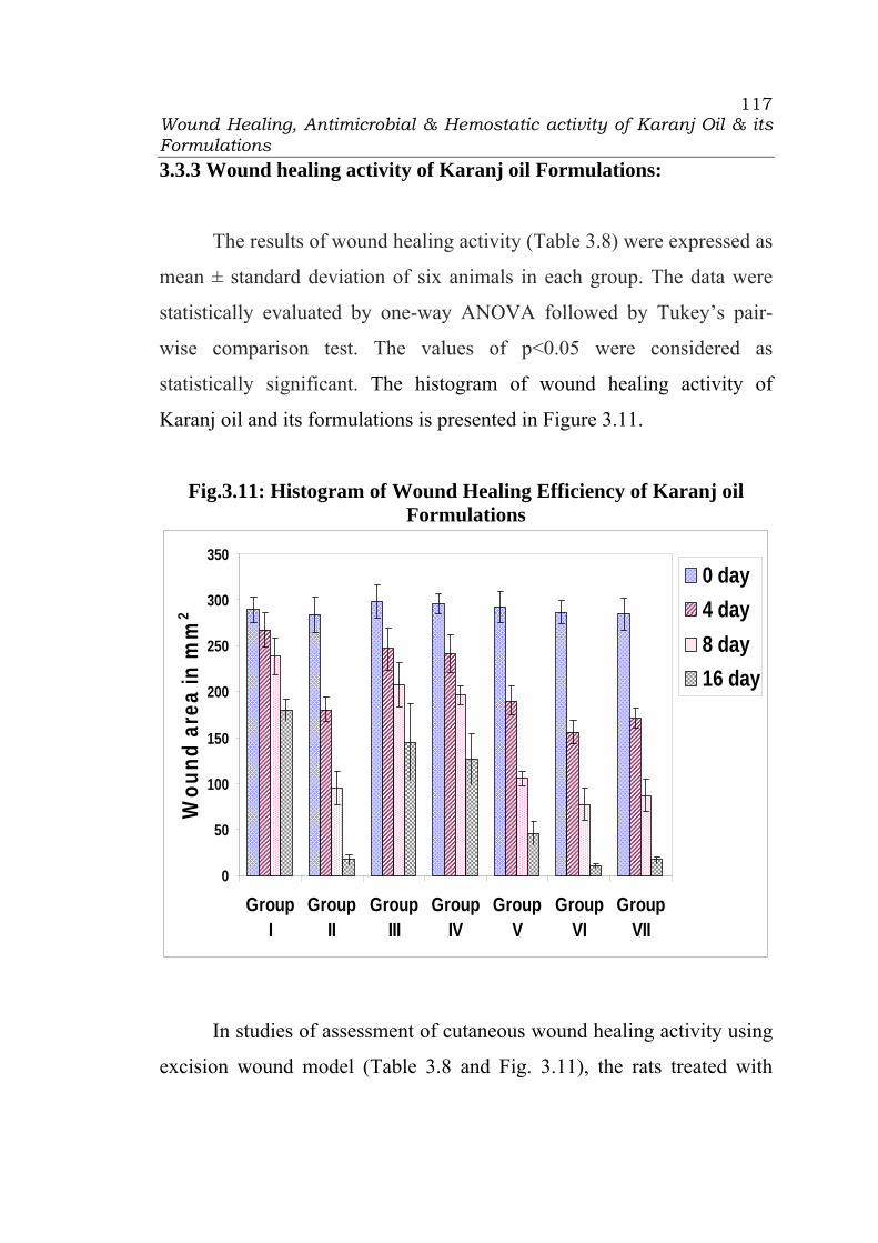
Wound Healing, Antimicrobial & Hemostatic activity of Karanj Oil & its Formulations
117
3.3.3 Wound healing activity of Karanj oil Formulations:
The results of wound healing activity (Table 3.8) were expressed as
mean ± standard deviation of six animals in each group. The data were
statistically evaluated by one-way ANOVA followed by Tukey’s pair-
wise comparison test. The values of p<0.05 were considered as
statistically significant. The histogram of wound healing activity of
Karanj oil and its formulations is presented in Figure 3.11.
Fig.3.11: Histogram of Wound Healing Efficiency of Karanj oil Formulations
0
50
100
150
200
250
300
350
GroupI
GroupII
GroupIII
GroupIV
GroupV
GroupVI
GroupVII
Wou
nd a
rea
in m
m2
0 day4 day8 day16 day
In studies of assessment of cutaneous wound healing activity using
excision wound model (Table 3.8 and Fig. 3.11), the rats treated with

Wound Healing, Antimicrobial & Hemostatic activity of Karanj Oil & its Formulations
118
cream containing 1% w/w framycetin sulphate showed wound healing
area of 283.89±19.42 mm2 on initial day and 17.04±5.67 mm2 on 16th day.
The karanj oil treated group III and medicated karanj oil treated group IV
showed wound healing area of 144.53±41.77 mm2 and 126.15±27.88
mm2 on 16th day as compared to untreated control animals (180.39±11.55
mm2 on 16th day). The wound healing area of 45.78±12.77 mm2,
10.53±1.88 mm2, and 16.71±3.62 mm2 on 16th day was exhibited by
emulsion treated group V, microemulsion treated group VI, and cream
treated group VII, respectively.
Wound healing Activity Groups Group I Untreated (Control) Group II Standard (1% Framycin) Group III Karanj Oil Group IV Medicated Karanj oil Group V Emulsion (30% Karanj oil) Group VI Microemulsion (15% Karanj oil) Group VII Cream (30% Karanj oil)
The one-way ANOVA results for wound healing activity (Fig.3.12)
showed that the p value for Turkey’s pair wise comparison test for wound
healing at 0 day for group II to VII were 0.9967, 0.9566, 0.9864, 0.9998,
0.9999, and 0.9990 respectively with reference to group I, where all p
values were greater than 0.05 (p>0.05) which indicated that the results of
wound healing at 0 day were similar. The p value for wound healing at 4
day (Fig.3.13) for the group III and IV were 0.3435 and 0.1315 with
reference to group I, which were greater than 0.05 (p>0.05) which
indicated that the results are non-significant meaning no difference in the
wound healing activity. But for group II, V, VI, and VII, the p value was
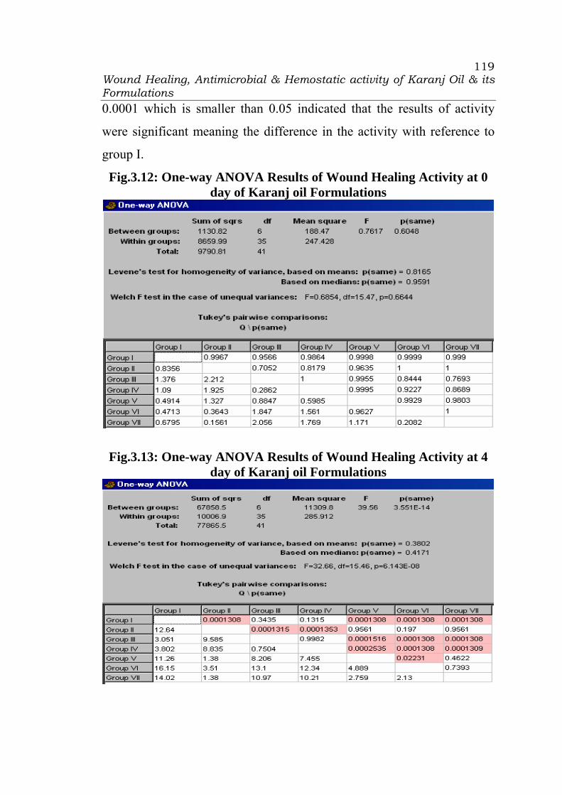
Wound Healing, Antimicrobial & Hemostatic activity of Karanj Oil & its Formulations
119
0.0001 which is smaller than 0.05 indicated that the results of activity
were significant meaning the difference in the activity with reference to
group I.
Fig.3.12: One-way ANOVA Results of Wound Healing Activity at 0 day of Karanj oil Formulations
Fig.3.13: One-way ANOVA Results of Wound Healing Activity at 4 day of Karanj oil Formulations

Wound Healing, Antimicrobial & Hemostatic activity of Karanj Oil & its Formulations
120
The One-way ANOVA results of wound healing at 8 day
(Fig.3.14) and 16 day (Fig.3.15), the p value for the group III were
0.0528 and 0.0572 respectively with reference to group I which was
greater than 0.05 indicated that the difference is non-significant meaning
no difference in the wound healing activity. For group II, V, VI, and VII,
the p value was 0.0001 which is <0.05 indicated that the results of wound
healing activity at 8 day and 16 day were significant meaning the
difference in the activity with reference to group I. The p value less than
0.05 indicated that the karanj oil formulations showed significant wound
healing activity.
Fig.3.14: One-way ANOVA Results of Wound Healing Activity at 8
day of Karanj oil Formulations

Wound Healing, Antimicrobial & Hemostatic activity of Karanj Oil & its Formulations
121
Fig.3.15: One-way ANOVA Results of Wound Healing Activity at 16
day of Karanj oil Formulations
The results of Table 3.8 revealed that the standard drug treated
animal group II (94.00%) showed significantly greater % wound closure
as compared to control group I (37.64%) and karanj oil treated group III
(51.51%) on 16 day. The animal group IV treated with medicated karanj
oil showed 57.42% wound closure on 16 day. The animal group V treated
with emulsion of karanj oil showed 84.34% wound closure on 16 day.
The animal group VI and VII treated respectively with microemulsion
and cream of karanj oil showed 96.32% and 94.13% wound closure on 16
day which comparable with that of standard group II.

Wound Healing, Antimicrobial & Hemostatic activity of Karanj Oil & its Formulations
122
The period of epithelialisation was 19.33 ± 1.21 days in the case of
standard drug treated animal group II (1% w/w framycetin sulphate
cream). The period of epithelialisation was 32.17±3.06 days for untreated
group I, while same was 27.83±1.47 and 25.67±2.58 days for karanj oil
group III and medicated karanj oil treated group IV respectively. The
period of epithelialisation was 21.50±1.87, 17.67±0.52, and 18.50±1.05
days for emulsion treated group V, microemulsion treated group VI, and
cream treated group VI respectively. For group II, V, VI, and VII, the p
value was 0.0001 which is <0.05 indicated that the one-way ANOVA
results of period of epithelialisation (Fig.3.16) were significant meaning
the difference in the activity with reference to group I. The results of the
wound healing activity revealed that the significant promotion of wound
healing was observed by karanj oil and its formulations as compared to
control group.
Fig.3.16: One-way ANOVA Results of Period of Epithelialisation of Karanj oil Formulations

Wound Healing, Antimicrobial & Hemostatic activity of Karanj Oil & its Formulations
123
3.3.4 Antimicrobial activity of Karanj oil Formulations:
All the experimentation of antibacterial activity was carried out in
aseptic area under laminar air-flow cabinet. The results of antibacterial
activity of the positive control i.e. 1% w/w framycin cream, karanj oil and
medicated karanj oil, formulations of karanj oil (Emulsion,
Microemulsion and cream) were presented in Table 3.10. The comparison
of the antibacterial activity of oils with the standard drug was presented in
Fig. 3.17. Sterilized distilled water was served as negative control. 1gm
of 1% W/W framycetin sulphate cream (FSC) ointment diluted and mixed
with 9ml of sterilized distilled water was used as standard i.e. positive
control.
Fig. 3.17: Histogram of Antibacterial Activity of Karanj Oil
Formulations
0
5
10
15
20
25
30
35
40
B.Subtilis S.Aureus E.Coli S.Typhi
Zone
of I
nhib
ition
in m
m
PositiveKaranj oilMed KOEmulsionMicroemulsionCream
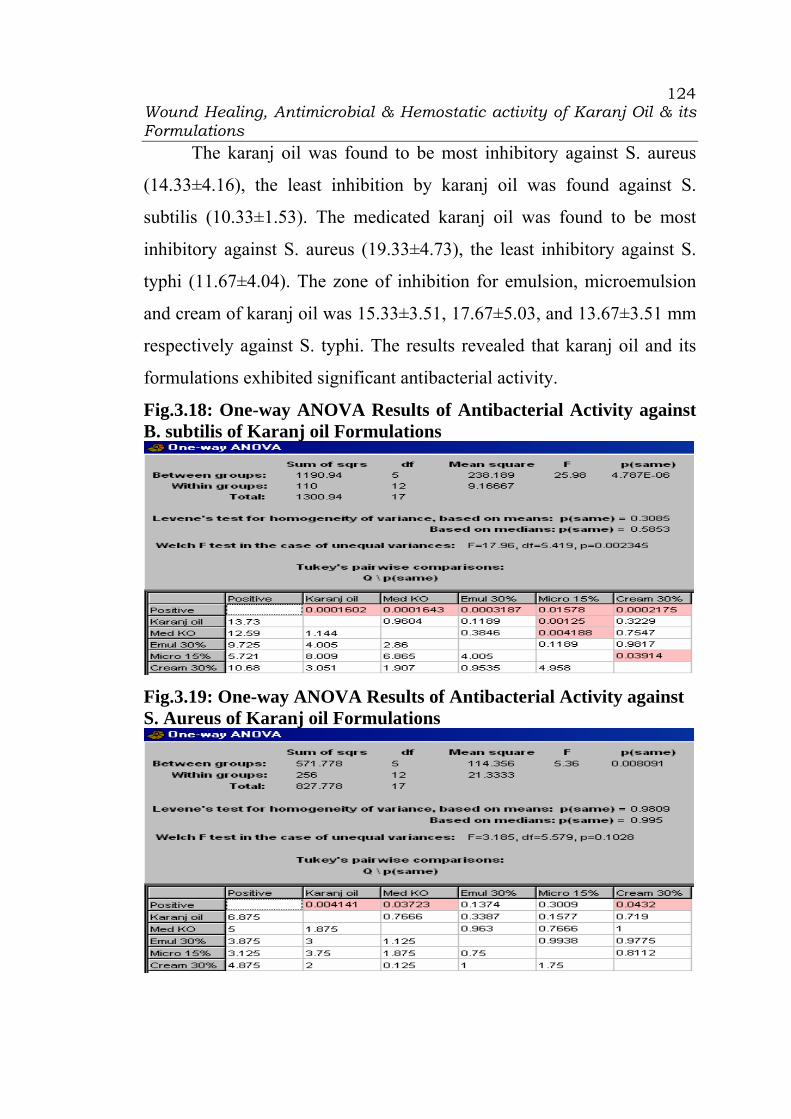
Wound Healing, Antimicrobial & Hemostatic activity of Karanj Oil & its Formulations
124
The karanj oil was found to be most inhibitory against S. aureus
(14.33±4.16), the least inhibition by karanj oil was found against S.
subtilis (10.33±1.53). The medicated karanj oil was found to be most
inhibitory against S. aureus (19.33±4.73), the least inhibitory against S.
typhi (11.67±4.04). The zone of inhibition for emulsion, microemulsion
and cream of karanj oil was 15.33±3.51, 17.67±5.03, and 13.67±3.51 mm
respectively against S. typhi. The results revealed that karanj oil and its
formulations exhibited significant antibacterial activity.
Fig.3.18: One-way ANOVA Results of Antibacterial Activity against B. subtilis of Karanj oil Formulations
Fig.3.19: One-way ANOVA Results of Antibacterial Activity against S. Aureus of Karanj oil Formulations
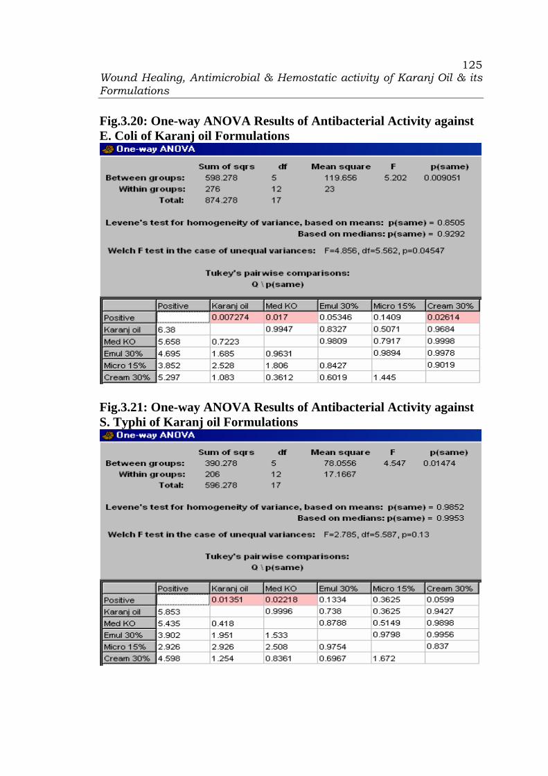
Wound Healing, Antimicrobial & Hemostatic activity of Karanj Oil & its Formulations
125
Fig.3.20: One-way ANOVA Results of Antibacterial Activity against E. Coli of Karanj oil Formulations
Fig.3.21: One-way ANOVA Results of Antibacterial Activity against S. Typhi of Karanj oil Formulations
Wound Healing, Antimicrobial & Hemostatic activity of Karanj Oil & its Formulations
126
The one-way ANOVA results showed that the p value for Turkey’s
pairwise comparison test for antibacterial activity against B. Subtilis (Fig.
3.18) for karanj oil, medicated karanj oil, emulsion, microemulsion and
cream of karanj oil was 0.0002, 0.0002, 0.0003, 0.0157, and 0.0002
respectively which is <0.05 indicated that the results of antibacterial
activity against B. Subtilis were significant meaning the difference in the
activity with reference to negative control. Similarly, the results of one-
way ANOVA for S. aureus, E. coli, and S. typhi (Fig. 3.19-3.21)
indicated significant antibacterial activity of karanj oil formulations. The
p value less than 0.05 indicated that the karanj oil formulations showed
significant antibacterial activity.
Microbial infection of wounds delays healing (1) and causes a
more pronounced acute inflammatory reaction (2) which can lead to
further tissue injury and damage. The antimicrobial activity of honey and
the essential oil of Melaleuca alternifolia reported to underlie their
usefulness as alternative therapy in wound healing (3). Thus, the
antimicrobial activity of karanj oil and its formulations on these wound
isolates may partly contribute to the wound healing effect by eliminating
infection thus allowing the natural tissue repair processes to start. It can
also be concluded that the karanj oil may also play a useful role in
accelerating the healing of old wounds by eradicating already established
infection.
Wound Healing, Antimicrobial & Hemostatic activity of Karanj Oil & its Formulations
127
3.3.5 Hemostatic activity of Karanj oil Formulations:
Influence of karanj oil on PT
The test measures the clotting time of plasma in the presence of an
optimal concentration of tissue extract (thromboplastin) and indicate the
overall efficiency of the extrinsic clotting system (Factors II, V, VII and
X). The test is also known to depend on reactions with factors V, VII and
X, and on the fibrinogen concentration of the plasma. The coagulation
process is triggered by incubation of plasma with the optimal amount of
thromboplastin and calcium. The time to formation of a fibrin clot is then
measured.
It can be seen from Table 3.11 that the medicated karanj oil
application resulted sight change in prothrombin time (9.33± 0.52 secs at
initial and 8.67±0.52 secs at 24 hrs) as compared to karanj oil (9.67±0.52
secs at initial and 9.50±0.55 secs at 24 hrs) and control group (9.83±0.75
secs at initial and 9.67±0.82 at 24 hrs). The prothrombin time for animals
treated with emulsion, microemulsion, and cream formulations of karanj
oil was 8.83±0.41, 8.67±0.52, and 8.50±0.55 at 24 hrs respectively.
Influence of karanj oil on APTT
The test measures the clotting time of plasma after the activation of
contact factors but without added tissue thromboplastin, and so indicates
the overall efficiency of the intrinsic pathway (Factors VIII and IX as
well as the contact factors). Incubation of plasma with the optimal

Wound Healing, Antimicrobial & Hemostatic activity of Karanj Oil & its Formulations
128
quantity of phospholipids and a surface activator leads to activation of
factors of the intrinsic coagulation system. The addition of calcium ions
trigger the coagulation process, the time to formation of a fibrin clot is
measured.
APTT for control group was 29.16±1.17 and 28.83±1.33 secs at
initial and at 24 hrs respectively. APTT was shortened after the medicated
karanj oil application to 27.17 ± 0.75 seconds at initial and 27.00±0.89 at
24 hrs (Table 3.11 and Figure 3.22) as compared to the control group
(29.16±1.17 secs at initial and 28.83±1.33 secs at 24 hrs) and karanj oil
(28.00±0.89 secs at initial and 27.50±1.05 secs at 24 hrs). Similarly, the
application of emulsion, microemulsion, and cream resulted in slight
shortening of APTT to 26.67±1.37, 26.50±0.55, and 26.33±1.03 secs at
24 hrs in the present study.
Fig. 3.22: Curve of Hemostatic activity of Karanj oil formulations
05
10152025
3035
Control
Karanj Oil
Med KO
Emulsion
Microem
ulsion
Cream
Group
Tim
e in
Sec
onds
PT at 0 hrPT at 24 hrsAPTT at 0 hrAPTT at 24 hrsTT at 0 hrTT at 24 hrs
Wound Healing, Antimicrobial & Hemostatic activity of Karanj Oil & its Formulations
129
Influence of karanj oil on TT
TT was shortened after the medicated karanj oil application to
13.83±1.33 seconds at initial and 13.66±1.03 at 24 hrs (Table 3.11 and
Figure 3.13) as compared to the control group (14.83±1.17 secs at initial
and 15.17±1.17 secs at 24 hrs) and karanj oil (14.17±0.75 secs at initial
and 14.00±0.63 secs at 24 hrs). The TT was slightly shortened on the
application of emulsion, microemulsion, and cream to 13.17±0.75,
12.83±0.75, and 13.00±0.89 secs at 24 hrs in the present investigation.
The results of present investigation indicated that the karanj oil had
shortened the PT, APTT and TT indicating its hemostatic activity.
3.4. Conclusions
The wound healing activity of Karanj oil and its formulations was
studied by using excision wound model and significant wound healing
activity was observed. The seed oil of Karanj tree inhibited microbial
growth of known wound contaminants and accelerated wound healing
process. It also exhibited hemostatic activity with accelerated blood
coagulation process. The wound healing, antimicrobial and hemostatic
activity are interrelated. The results of the present study indicated that
karanj oil had good potentials for use in wound care and further provided
a rationale for the use of the plant in wound management in traditional
medicine practice.
Wound Healing, Antimicrobial & Hemostatic activity of Karanj Oil & its Formulations
130
In addition to inhibiting the growth of the micro-organisms and
shortening the blood coagulation time, the karanj oil effectively reduced
the epithelialisation period of experimentally-induced wounds which is an
index of pro-healing activity. The precise aspect as well as the exact
mechanism of wound healing effect of the karanj oil is yet to be
elucidated with respect to antimicrobial as well as hemostatic activity. In
the tissue repair process, inflammatory cells promote the migration and
proliferation of endothelial cells, leading to neovascularisation of
connective tissue cells which synthesize extracellular matrices including
collagen, and of keratinocytes resulting to re-epithelialisation of the
wounded tissue (18). In the wound healing process, collagen formation
peaks at day 7 and epithelialisation occurs in 48 hrs under optimal
conditions (19). The extent to which the karanj oil and its formulations
interacted with these processes is not known to us and it needs to be
clinically investigated systematically and scientifically.

Wound Healing, Antimicrobial & Hemostatic activity of Karanj Oil & its Formulations
131
3.5 References
1. Bowler P.G., Duerden B.I., Armstrong D.G.: Wound microbiology
and associated approaches to wound management, Clin Microbiol
Rev., 14, 244-269, 2001.
2. Whaley K., Burt A.D.: Inflammation, healing and repair. In Muir's
Textbook of Pathology, 13th edition, Edited by: Mac Sween RMN,
Whaley K., London, Arnold, 112-165, 1996.
3. Molan P.C., The role of honey in the management of wounds, J.
Wound Care, 8, 415-418, 1999.
4. Carson C.F., Riley T.V., Cookson B.D., Efficacy and safety of tea
tree oil as a topical antimicrobial agent, J. Hosp. Infect., 40, 175-
178, 1998.
5. Narendra Nalwaya, Gaurav Pokharna, Lokesh Deb, Naveenkumar
Jain, Wound healing activity of latex of Calotropis Gigantea,
International Journal of Pharmacy and Pharmaceutical Sciences,
Vol. 1, Issue 1, 176-181, July-Sep. 2009.
6. Meera B., Kumar S., Kalidhar S.B., A review of the chemistry and
biological activity of Pongamia pinnata, Journal of Medicinal and
Aromatic Plant Science, 25, 441–65, 2003.
Wound Healing, Antimicrobial & Hemostatic activity of Karanj Oil & its Formulations
132
7. V.V. Chopade, A.N. Tankar1, V.V. Pande1, A.R. Tekade1, N.M.
Gowekar1, S.R. Bhandari1, S.N. Khandake, Pongamia pinnata:
Phytochemical constituents, Traditional uses and Pharmacological
properties: A review, International Journal of Green Pharmacy, 72-
75, April-June 2008.
8. Manjunatha B.K., Vidya S.M., Rashmi K.V., Mankani K.L., Shilpa
H.J., Jagadeesh Singh S.D., Evaluation of wound-healing potency
of Vernonia arborea Hk, Indian Journal of Pharmacology, Vol. 37,
Issue 4, 223-226, August 2005.
9. Morton J.J., Malone M.H., Evaluation of vulnerary activity by an
open wound procedure in rats, Arch. Int. Pharmocodyn. Ther., 196,
117-126, 1972.
10. Pertz C., Pauli M., Bazerque P., An antibiotic assay by the agar-
well diffusion method., Acta Biol. Med. Exp., 15, 113-115, 1990.
11. Effect of Transdermally Delivered Aspirin on Blood Coagulation
Parameters, Areeg A. Shamsher, Naseem A. Charoo, Kanchan
Kohli, Krishna Pillai, Ziyaur Rahman , American Journal of
Biomedical Sciences, Am. J. Biomed. Sci., 2(2), 129-141, 2010.
12. Palmer RL, Laboratory diagnosis of bleeding disorders: Basic
screening tests, Post grad. Med., 76:137–148, 1984.
Wound Healing, Antimicrobial & Hemostatic activity of Karanj Oil & its Formulations
133
13. Nilsson IM (1987): Assessment of blood coagulation and general
haemostasis. In: Bloom AL, Thomas DP (eds) Haemostasis and
Thrombosis, 2nd edition, Longman Group (UK) Limited, pp. 922.
14. Physical Pharmacy, Alfred Martin, B.I. Waverly Pvt. Ltd., New
Delhi, Fourth Edition, 1997, pp. 489.
15. Hoar T.P. and Schulman J. H., Transparent water in oil dispersions:
the oleopathic hydromicelle, Nature, 152, 102–103, 1943.
16. Attwood D., Microemulsions in Colloidal drug delivery systems,
(J. Kreuter ed.), Marcel Dekker, New York 1994.
17. B. C. Routara, A. Bandyopadhyay, P. Sahoo, Use of desirability
function approach for optimization of multiple performance
characteristics of the surface roughness parameters in CNC turning,
ICME07-AM-16, Proceedings of the International Conference on
Mechanical Engineering 2007 (ICME2007), 29- 31 December
2007, Dhaka, Bangladesh.
18. Clark RAF, Cutaneous wound repairs, In Physiology, Biochemistry
and Molecular Biology of Skin, Edited by Goldsmith L.A., New
York, Oxford University Press, 576, 1991.
19. Graber A.M., General surgery: Wound management, 2002
(http://wound-science.com).

CHAPTER 4
SUMMARY
Summary
135
Summary
The local name of the oil of Derris Indica is Karanj oil. Medicated
karanj oil is used widely by Ayurvedic Practitioners in rural areas of
Gujarat for the treatment of rheumatism and curing skin infections. It is
used for healing, antiseptic, and haemostatic effect externally by
Ayurvedic Practitioners.
Systemic information is not available on the efficacy of karanj oil.
The present investigation was aimed to study systematically and
scientifically the efficiency of Karanj oil. The formulations of the karanj
oil were prepared using pharmaceutical methodology. The activity of the
karanj oil, medicated karanj oil, selected optimized formulation of
emulsion, microemulsion and cream of karanj oil was studied for wound
healing, antibacterial, and haemostatic efficiency.
Formulation optimization:
Emulsion of karanj oil:
To optimize the concentration of emulsifiers for emulsion of karanj
oil, a 32 factorial design was adopted using the concentration of Tween 80
(X1) and Span 20 (X2) as independent variables at three levels: low (-1),
medium (0) and high (1). The factorial design batches were prepared by
dry gum method. The dependent variables were cream height, number of
inversions, and globule size. Response surface curvature can be examined
when the two variables are investigated at three levels. The design
provided the following emperical second-order equation (Full model):

Summary
136
Y = b0 + b1X1 + b2X2 + b12X1X2 + b11X12 + b22X2
2
where Y is response, b0 is intercept, b1 and b2 are coefficients of main
effects, b12 is the coefficient for the interaction term and b11 and b22 are
the coefficients for the second-order quadratic terms. The non-significant
estimated coefficients should be dropped from the full model by adopting
a significance test for the regression coefficient. Microsoft Excel® was
used to identify non-significant terms. A coefficient is significant if t i >
tcrit (v), where v is the degrees of freedom of residual variance. The refined
model may be used for calculation of residuals or for drawing the contour
plot. The full and refined models (p < 0.05) of factorial design batches for
cream height, number of inversions, and globule size were obtained and
contour plots were constructed.
To validate the evolved mathematical models (reduced models for
cream height, number of inversions, and globule size), two check points
were prepared and evaluated. Good correlation was found between
observed and predicted values. Hence, it was concluded that the evolved
models may be used for theoretical prediction of responses within the
factor space. As per the results of the 32 experimental design, the
optimized formulation of karanj oil emulsion contained 30% w/w of
karanj oil, 10% w/w/ of Tween 80 and 5% w/w of Span 20.
Microemulsion of karanj oil:
The microemulsion of karanj oil were prepared using surfactant,
co-surfactant, solvent, and co-solvent like Cremophor RH 40, Glyceryl
dilaurate, Tween 80, Ethyl oleate, Transutol P, etc. To optimize the

Summary
137
concentration of co-surfactant, surfactant, and solubilizer, a 33 factorial
design was adopted using the concentration of glyceryl dilaurate (X1),
Cremophor RH 40 (X2), and Transutol P (X3) as independent variables at
three levels: low (-1), medium (0) and high (1). The dependent variables
were Viscosity (cp), globule size (nm), and Zeta potential (mV).
Response surface curvature can be examined when the variables
are investigated at three levels. The design provides the following
emperical second-order equation (Full model, FM):
Y = b0 + b1X1 + b2X2 + b3X3 + b11 X1
2 + b22 X22 + b33 X3
2 +b12 X1X2 +
b23X2X3 + b13X1X3 + b123X1X2X3
where Y is response, b0 is the intercept, b1, b2 and b3 are regression
coefficient of main effects and b12, b23, b13 and b123 are regression
coefficient for the interaction terms. The coefficients with second-order
terms (b11, b22 and b33) indicate the quadratic nature. Microsoft Excel®
may be used to identify the non-significant terms. The full and refined
models (p < 0.05) for Viscosity (cp), globule size (nm), and Zeta potential
(mV) were obtained.
Desirability function approach was used for solving the multiple
performance characteristics optimization problems, where all the
objectives are to attain a definite goal simultaneously. As per the results
of the 33 experimental design on the basis of overall optimum
characteristics, the optimized formulation of karanj oil microemulsion
contained 15%w/w of karanj oil, 12.5% w/w of Glyceryl dilaurate, 22.5%
w/w/ of Cremophor RH 40, and 10%w/w of Transcutol P.
Summary
138
Cream of karanj oil:
The cream containing karanj oil was prepared by hot emulsification
method. It contained 30% w/w/ of karanj oil, 5%w/w of stearic Acid,
10%w/w of ethoxylated cetosteryl alcohol, 5%w/w/ of stearyl alcohol,
and 10% w/w/ of propylene glycol.
Wound healing activity of Karanj oil Formulations:
Wistar rats of either sex weighing 150-200 gm were used for the
study. All the animals were maintained under controlled room
temperature (22±2°C) and relative humidity (50±5%) with 12:00 hr light,
12:00 hr dark cycle. The animals were housed in colony cages (three
animals per cage) with free access to diet and water ad libitum during the
investigation period. All the animals were acclimatized to the laboratory
environment for 5 days before the experimentation. Six animals (rats) per
group comprising of three males and three females were used in each
experiment set. The study protocol was approved by the Institutional
Animal Ethics Committee (IAEC) of C.U. Shah College of Pharmacy and
Research, Wadhwan (Gujarat).
Excision wound model was used to evaluate the wound healing
efficiency. The rats were inflicted with excision wounds by under light
ether anesthesia. A circular wound of about 300 sq. mm and 2 mm depth
was made on depilated ethanol sterilized dorsal thoracic region of the
rats. The animals were divided into seven groups of six each. The animals
of group I was left untreated and considered as control. The rats of Group
II were served as reference standard and received 1% w/w framycetin

Summary
139
sulphate cream (FSC), the animal group III and IV were treated with
karanj oil and medicated karanj oil, respectively. The animal group V, VI
and VII were treated with karanj oil emulsion, karanj oil microemulsion
and karanj oil cream, respectively. The karanj oil or its formulation was
topically applied once a day, starting from the day of the operation, till
complete epithelialisation. The parameters studied were wound closure
and epithelialisation time. The wounds were traced on mm2 graph paper
on day 0, 4, 8, and 16 and thereafter on alternate days until healing was
completed. The period of epithelialisation was the number of days
required for falling of the dead tissue remnants of the wound without any
residual raw wound
The results of wound healing activity were expressed as mean ±
standard deviation of six animals in each group. The data were
statistically evaluated by one-way ANOVA followed by Tukey’s pair-
wise comparison test. The values of p<0.05 were considered as
statistically significant. The histogram of wound healing activity of
Karanj oil and its formulations were presented. The results of the wound
healing activity revealed that the significant promotion of wound healing
was observed by karanj oil and its formulations as compared to control
group.
Antimicrobial activity of Karanj oil Formulations:
Karanj oil and its formulations were evaluated for their potential
antibacterial activity by standard agar well diffusion assay. Petri dishes
containing agar medium were seeded with microbial suspension of

Summary
140
Bacillus subtilis, Staphylococcus aureus, Escherichia coli, and Salmonella
typhi and media was allowed to solidify. Wells were cut into solidified
agar media using a sterilized cup-borer. Each sample was poured in the
respective well and the dishes were incubated at 37 °C for 24 hrs. The
antibacterial activity was expressed in terms of the mean of diameter of
zone of inhibition at the end of incubation period. Sterilized distilled
water was served as negative control. 1% w/w framycetin sulphate cream
(FSC) ointment diluted and mixed sterilized distilled water was used as
standard i.e. positive control.
The karanj oil was found to be most inhibitory against S. aureus,
the least inhibition by karanj oil was found against S. subtilis. The
medicated karanj oil was found to be most inhibitory against S. aureus,
the least inhibitory against S. typhi. The results revealed that karanj oil
and its formulations exhibited significant antibacterial activity.
Hemostatic activity of Karanj oil Formulations:
Rats were slightly ether anesthetized, and abdominal hair was
carefully removed with electric clipper, they were fasted overnight. The
animals were divided into six groups of six rats in each. The animals of
group I was left untreated and considered as control. The rats of Group II,
III, IV, V, and VI were applied topically karanj oil, medicated karanj oil,
emulsion, microemulsion, and cream, respectively, on the abdominal area
gently. Rats were anesthetized by intravenous injection of sodium
pentobarbital. The caudal caval vein was exposed by a mid-line incision
and blood was collected at 0 hr and 24 hrs into a plastic syringe
Summary
141
containing citrate buffer pH 4.5. The sample was immediately agitated
and centrifuged in a plastic tube at 1500 rpm for 10 min. Plasma was
transferred to another plastic tube and the coagulation tests for the
determination of thromboplastin time (PT), activated partial
thromboplastin time (APTT) and thrombin time (TT) were performed
within 3 h. The results of present investigation indicated that the karanj
oil had shortened the PT, APTT and TT indicating its hemostatic activity.
Conclusions:
The wound healing activity of Karanj oil and its formulations was
studied by using excision wound model and significant wound healing
activity was observed. The seed oil of Karanj tree inhibited microbial
growth of known wound contaminants and accelerated wound healing
process. It also exhibited hemostatic activity with accelerated blood
coagulation process. The wound healing, antimicrobial and hemostatic
activity are interrelated. The results of the present study indicated that
karanj oil had good potentials for use in wound care and further provided
a rationale for the use of the plant in wound management in traditional
medicine practice.

CHAPTER 5
PUBLICATIONS
AND
PRESENTATIONS
Publications & Presentations
143
CHAPTER 5 PUBLICATIONS AND PRESENTATIONS
Sr. No. Details 5.1 62nd IPC 2010 Poster presentation Acceptance 5.2 62nd IPC 2010 Oral presentation acceptance

Publications & Presentations
144
5.1: 62nd IPC 2010 Poster presentation Acceptance: 16 Oct 2010
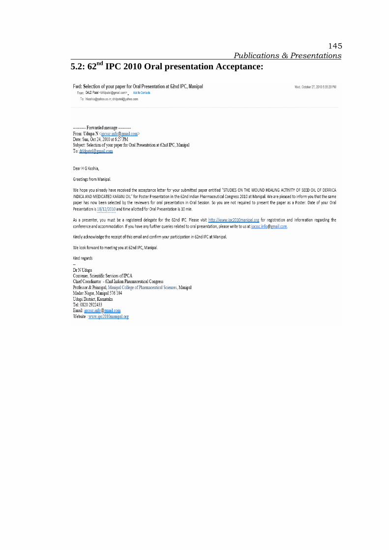
Publications & Presentations
145
5.2: 62nd IPC 2010 Oral presentation Acceptance: