acceleration of conduction velocity linked to …acceleration of conduction velocity linked to...
TRANSCRIPT
Acceleration of conduction velocity linked to clusteringof nodal components precedes myelinationSean A. Freemana,b,c,1, Anne Desmazièresa,b,c,1, Jean Simonneta,b,c, Marie Gattaa,b,c, Friederike Pfeiffera,b,c,Marie Stéphane Aigrota,b,c, Quentin Rappeneaua,b,c, Serge Guerreiroa,b,c, Patrick Pierre Michela,b,c, Yuchio Yanagawad,e,Gilles Barbina,b,c, Peter J. Brophyf, Desdemona Frickera,b,c, Catherine Lubetzkia,b,c,g,1,2, and Nathalie Sol-Foulona,b,c,1,2
aSorbonne Universités Université Pierre et Marie Curie University of Paris 06, UMR_S 1127, Institut du Cerveau et de la Moelle–Groupe Hospitalo-UniversitairePitié-Salpêtrière, F-75013, Paris, France; bINSERM U1127, F-75013, Paris, France; cCNRS UMR7225, F-75013, Paris, France; dDepartment of Genetic andBehavioral Neuroscience, Gunma University Graduate School of Medicine, Maebashi 371-8511, Japan; eJapan Science and Technology Agency, Tokyo102-8666, Japan; fCentre for Neuroregeneration, University of Edinburgh, Edinburgh EH16 ASB, United Kingdom; and gAssistance Publique-Hôpitaux de Paris,Hôpital Pitié-Salpêtrière, 75013, Paris, France
Edited by William A. Catterall, University of Washington School of Medicine, Seattle, WA, and approved December 16, 2014 (received for reviewOctober 6, 2014)
High-density accumulation of voltage-gated sodium (Nav) chan-nels at nodes of Ranvier ensures rapid saltatory conduction alongmyelinated axons. To gain insight into mechanisms of node assem-bly in the CNS, we focused on early steps of nodal protein cluster-ing. We show in hippocampal cultures that prenodes (i.e., clustersof Nav channels colocalizing with the scaffold protein ankyrinGand nodal cell adhesion molecules) are detected before myelindeposition along axons. These clusters can be induced on purifiedneurons by addition of oligodendroglial-secreted factor(s), whereasankyrinG silencing prevents their formation. The Nav isoformsNav1.1, Nav1.2, and Nav1.6 are detected at prenodes, with Nav1.6progressively replacing Nav1.2 over time in hippocampal neuronscultured with oligodendrocytes and astrocytes. However, the oli-godendrocyte-secreted factor(s) can induce the clustering of Nav1.1and Nav1.2 but not of Nav1.6 on purified neurons. We observedthat prenodes are restricted to GABAergic neurons, whereas clus-tering of nodal proteins only occurs concomitantly with myelinensheathment on pyramidal neurons, implying separate mecha-nisms of assembly among different neuronal subpopulations. Toaddress the functional significance of these early clusters, we usedsingle-axon electrophysiological recordings in vitro and showedthat prenode formation is sufficient to accelerate the speed of ax-onal conduction before myelination. Finally, we provide evidencethat prenodal clusters are also detected in vivo before myelination,further strengthening their physiological relevance.
node of Ranvier | sodium channel | myelination | GABAergic neuron |conduction velocity
Voltage-gated sodium (Nav) channels are highly enriched atthe axon initial segment (AIS) and the node of Ranvier,
allowing generation and rapid propagation of action potentialsby saltatory conduction in myelinated fibers. These axonaldomains also contain cell adhesion molecules [e.g., neurofascin186 (Nfasc186)] and the scaffolding proteins ankyrinG (AnkG)and βIV spectrin, which provide a potential link with the actincytoskeleton (1). Flanking the nodes are the paranodes, whereaxoglial junctions between paranodal myelin loops and the axonare formed through interactions between axonal contactin-associated protein (Caspr)/contactin and glial Nfasc155 (2, 3).Although the mechanisms of nodal assembly are best character-ized in the peripheral nervous system (4–9), less is known aboutthe cellular and molecular mechanisms underlying node assemblyin the CNS. ECM proteins, adhesion molecules, such as Nfasc186,and also, axoglial paranodal junctions have been shown to triggerCNS nodal clustering, although their respective roles remainuncertain (10–19). Moreover, axonal clustering of Nav channelsbefore myelin deposition and oligodendroglial contact has beenshown to occur in retinal ganglion cell (RGC) cultures, wherethese clusters were induced by oligodendroglial-secreted factor(s)(20, 21).
Here, we have investigated the cellular and molecular mech-anisms underlying nodal protein assembly in hippocampal neu-ron cultures. We first showed that evenly spaced clusters of Navchannels, colocalizing with Nfasc186 and AnkG, are detectedalong axons before myelination. Strikingly, this prenode assem-bly is restricted to GABAergic interneurons, suggesting the ex-istence of different mechanisms of nodal assembly. The prenodalclustering can be induced on purified neurons by the addition ofoligodendroglial-secreted factor(s) and also depends on intrinsiccues, such as AnkG. Furthermore, we also provide evidence thatthese clusters are detected in vivo before myelination on hip-pocampal tissue sections. Finally, to gain insight into theirfunctional significance, we performed in vitro simultaneous so-matic and axonal recordings and showed that the presence ofprenodes increases the speed of action potential propagationalong axons before myelination.
ResultsClusters of Nodal Proteins Are Detected Before Myelination onHippocampal GABAergic Axons. To gain insight into the chronol-ogy of axonal domain assembly, we first analyzed the distributionof Nav channels in mixed hippocampal cultures (i.e., neuronswith oligodendrocytes and astrocytes) from embryonic day 18(E18) rat embryos at different time points before myelination.
Significance
Cellular and molecular mechanisms underlying the assembly ofnodes of Ranvier of myelinated axons in the CNS are still onlypartly understood. Our study shows the influence of intrinsiccues and glial extrinsic factors for nodal protein clustering beforemyelination on specific hippocampal neuronal subpopulationsand extends to electrophysiological understandings and in vivorelevance. Although conduction velocity along axons has longbeen thought to mostly rely on the insulating properties ofmyelin, we here show that nodal protein aggregation can in-crease this speed in the absence of such insulation. These resultshighlight the role of nodal clusters per se on conduction velocityby uncoupling it from myelination.
Author contributions: S.A.F., A.D., J.S., G.B., D.F., C.L., and N.S.-F. designed research; S.A.F.,A.D., J.S., M.G., F.P., M.S.A., Q.R., S.G., and N.S.-F. performed research; P.P.M., Y.Y., andP.J.B. contributed new reagents/analytic tools; S.A.F., A.D., J.S., D.F., C.L., and N.S.-F.analyzed data; and S.A.F., A.D., C.L., and N.S.-F. wrote the paper.
The authors declare no conflict of interest.
This article is a PNAS Direct Submission.1S.A.F., A.D., C.L., and N.S.-F. contributed equally to this work.2To whom correspondence may be addressed. Email: [email protected] [email protected].
This article contains supporting information online at www.pnas.org/lookup/suppl/doi:10.1073/pnas.1419099112/-/DCSupplemental.
www.pnas.org/cgi/doi/10.1073/pnas.1419099112 PNAS | Published online January 5, 2015 | E321–E328
NEU
ROSC
IENCE
PNASPL
US
Dow
nloa
ded
by g
uest
on
June
29,
202
0

Hippocampal neuronal cultures contain both glutamatergic py-ramidal cells, identified by Ca2+/calmodulin-dependent proteinkinase II immunolabeling (75% ± 5% of neurons), and GABAreleasing interneurons, identified by glutamate decarboxylaseisoform of 67 kDa (GAD67) expression (24.5% ± 3.9% of neu-rons). As might be predicted, Nav channel accumulation wasdetected at the AIS of all neurons during the first week of cultureand colocalized with AnkG and Nfasc186, whereas expressionalong the axon was diffuse and barely visible. Surprisingly, incontrast, at 17 days in vitro (DIV), evenly spaced clusters of Navchannels AnkG and Nfasc186 were found exclusively localized onGABAergic neurons (67% ± 15% of total GAD67+ neurons)(Fig. 1 and Fig. S1). These clusters, which were formed in theabsence of myelin ensheathment as indicated by the lack ofproteolipid protein (PLP) immunostaining (Fig. 1 A, C, and D),were named prenodes. The percentage of hippocampal axons withprenodes increased as a function of time in vitro from 4.1% ±3.1% at 14 DIV to 15.8% ± 3.5% and 17.1% ± 7.1% at 17 and 21DIV, respectively. We also observed that the number of prenodesdistributed along each axon increased from 14 to 21 DIV(Fig. S1B).Because hippocampal interneurons are highly diverse (22), we
further investigated whether neurons with prenodes are re-stricted to a particular interneuron subtype by characterizingcalcium-binding protein (i.e., calbindin, calretinin, and parvalbumin)and somatostatin expression. Most neurons with prenodeswere somatostatin+ and/or parvalbumin+ (81.3% ± 6.8% and57.5% ± 3.5, respectively) (Fig. S2), suggesting an overlap inprenodal formation between varying interneuron subtypes, par-ticularly those that express both somatostatin and parvalbumintogether (23).
Prenode Assembly Requires Oligodendroglial Cues. To gain insightinto the role of glial cells in prenode formation, we used purifiedhippocampal neuronal cultures through elimination of dividing
cells by antimitotic treatment, which were virtually devoid ofoligodendrocytes and contained a small percentage of astrocytes(less than 5%). We observed that purified neurons with nodalprotein clusters along axons were rare (1.1% ± 0.7%) (Fig. 2 Aand F). However, when we prepared highly purified populationsof oligodendroglial lineage cells and added oligodendrocytes oroligodendroglial-conditioned medium (OCM) to purified neu-ronal cultures, the percentage of axons with prenodes increased,respectively from 1.1% ± 0.7% to 10.8% ± 3.3% and 11.9% ±3.9% (Fig. 2F). Under these conditions, Nav was observed tocolocalize with AnkG and Nfasc186 in prenodes (Fig. 2 B and D).Furthermore, similar to mixed cultures, prenodes were restricted toGAD67+ neurons (Fig. 2C). The number of prenodes along eachaxon was decreased compared with mixed cultures (Fig. S1C), andthe spacing between clusters was shorter in OCM-treated purifiedneurons compared with neurons in mixed cultures (9.6 ± 1.1 and15.8 ± 1.8 μm, respectively; P = 0.028) (Fig. 2E). In addition, nodalprotein clustering occurred preferentially along axons with highlyphosphorylated neurofilaments, suggesting an association withneuronal maturity (Fig. 2D). Altogether, our results show that glialcues are necessary and that a diffusible oligodendroglial factor(s)participate(s) in prenode formation along GABAergic axons.
Fig. 1. Prenodes are formed before myelination on hippocampal GABAergicneurons in culture. Immunostaining of mixed hippocampal rat cultures at 17DIV. (A) Clustering of Nav (green) in the absence of myelin (PLP−; blue) alonga GABAergic axon (GAD67+; red), (B) contrasting with the absence of pre-nodal clusters (AnkG; red) on the axons of pyramidal cells Ca2+/calmodulin-dependent protein kinase II+ (CaMKII+; green). Nav clusters (green) colo-calized with (C) Nfasc186 (Nfasc) and (D) AnkG (plot profile is in E) in redalong unmyelinated axons (PLP−; blue). (Scale bars: 25 μm.)
Fig. 2. A secreted oligodendroglial factor promotes nodal protein cluster-ing. Immunostaining of purified hippocampal neurons cultured in the (A)absence or (B–D) presence of OCM. (A–D) The AIS is detected in all con-ditions, but (B–D) clusters of Nav (green), AnkG (red), and Nfasc (green) areonly detected in the presence of OCM. (C) Prenodes are formed on GAD67+
neurons treated with OCM. (D) Neuronal cell body and neurites stained withan Ab targeting phosphorylated neurofilaments (PNfil; blue). (Scale bars:25 μm.) (E) Distance between prenodes measured on axons in mixed cultureor purified neurons cocultured with oligodendrocytes (oligo) or OCM. Meanspacing between clusters (micrometers) ± SEM of four independent experi-ments is shown. *P = 0.028 (Mann–Whitney test). (F) Percentage of axons(AIS+) having at least two Nav clusters at 21 DIV in purified neuron cultures,purified neurons cocultured with oligodendrocytes (oligo), or OCM. Themeans ± SDs of 4 (for oligo) and 10 (for OCM) independent experiments areshown. For each experiment, at least 100 neurons were analyzed. *P =0.0187; ***P = 0.0003 (Mann–Whitney test).
E322 | www.pnas.org/cgi/doi/10.1073/pnas.1419099112 Freeman et al.
Dow
nloa
ded
by g
uest
on
June
29,
202
0

Hippocampal GABAergic and Pyramidal Neurons Assemble NodalProteins by Different Mechanisms. To further examine if oligo-dendrocytes were contacting prenodes, we used mixed hippo-campal cultures from CNP-EGFP embryos, in which all cells inthe oligodendrocyte lineage express EGFP (24). Although oli-godendrocyte processes contacted some axons with prenodes,most clusters were not (Fig. 3B), suggesting that nodal proteinclustering on GABAergic interneurons does not depend on strictoligodendroglial contact. In addition, prenodes were formedindependently of paranodal aggregation as indicated by the dif-fuse Caspr expression (Fig. 3A).We also examined, at later time points (24–28 DIV), whether
neurons were eventually myelinated by analyzing mixed hippo-campal cultures treated with differentiation medium to enhancethe myelination process. Myelinated axons were detected withNav channel clusters flanked by paranodal Caspr clusters, formingeither heminodes or nodes of Ranvier at the tips of myelin-ated internodes (Fig. 3C). Myelinated internodes were de-tected on both GABAergic and pyramidal neurons (Fig. 3D andFig. S3), suggesting that clustering at the node on glutamatergicaxons coincides with myelin deposition, in contrast to GABAergic
axons. Within GABAergic myelinated axons, internodal lengthwas 32.7 ± 3.1 μm (i.e., twofold longer than the distance betweenclusters observed in nonmyelinated GABAergic axons pres-ent at the same time point in the same cultures) (Fig. 3E).To determine if prenodal clustering is restricted to neuronsdestined to be myelinated, we used dopaminergic neurons inculture, which are known to remain unmyelinated in vivo. Theseneurons exhibited Nav clustering at the AIS but not along axons(Fig. 3 F and G).Taken together, our results suggest that the mechanisms of
nodal assembly differ between neuron subtypes. Indeed, whereasmost GABAergic neurons assemble prenodes before myelina-tion, hippocampal pyramidal cells initiate nodal clustering con-comitantly to myelination.
Nav Channel Isoforms Are Differentially Regulated in Prenodes overTime in Vitro. Because CNS nodes of Ranvier express differentNav α-subunits (25, 26), we also analyzed prenodal expression ofNav α-subunits before myelination. Nav1.1 was detected in morethan 90% of AnkG+ clusters at all time points analyzed (Fig. 4D),whereas Nav1.2 was mainly detected at 14 DIV (Fig. 4) andprogressively disappeared at later time points. Nav1.6, which
Fig. 3. Prenodes are formed on myelin-competent GABAergic neurons.(A and B) Immunostaining of mixed hippocampal culture at 17 DIV. (A) Navclusters (green) observed in the absence of Caspr aggregation (red) and theabsence of myelin (PLP−; blue). (B) Hippocampal culture from CNP-EGFP em-bryos shows an oligodendrocyte (asterisk) stained with an anti-GFP Abwith processes contacting the axon (arrows). Numerous AnkG clusters (red)are observed at a distance from these processes on a GAD67+ neuron (blue).(C and D) Immunostaining of mixed hippocampal culture at 24 DIV. (C) My-elinated axons (PLP+; blue) with nodes (Nav; green) and paranodes (Caspr;red). A higher magnification of a node with flanking paranodes is shown. (D)Myelinated segments (PLP+; green) with nodes (AnkG+; red) on GABAergicaxons [Parvalbumin+ (Parv); blue]. (Scale bars: 25 μm.) (E) Measure of thedistance between prenodes on unmyelinated axons and between nodes ofRanvier on myelinated axons at different time points. Values are the meanspaces between clusters (micrometers) ± SEMs of four independent experi-ments. *P = 0.028 (Mann–Whitney test). (F and G) Immunostaining of mes-encephalic cultures at 21 DIV. (F) Dopaminergic neurons tyrosine hydroxylase(TH+; red) do not form Nav clusters downstream of the AIS (Nav
+; green),whereas (G) Nav clusters are formed on a nondopaminergic (TH−) axon.*Neuronal cell body. (Scale bars: 25 μm.)
Fig. 4. Prenodes express different Nav isoforms. (A–C) Hippocampal neuroncultures at (A) 14, (B) 17, or (C) 24 DIV immunostained for Nav1.1, Nav1.2,Nav1.6, AnkG, GAD, and/or PLP as indicated, illustrating the different Navα-subunit isoform expression in prenodes at different time points. *Neuronalcell body. (Scale bar: 25 μm.) (D) Percentage of axons with clusters of AnkG,which are colocalized with Nav1.1, Nav1.2, or Nav1.6 at different DIVs.Quantifications were performed in mixed cultures and cultures of purifiedneurons with OCM. The means ± SDs of three independent experiments areshown. For each experiment, at least 100 neurons were analyzed.
Freeman et al. PNAS | Published online January 5, 2015 | E323
NEU
ROSC
IENCE
PNASPL
US
Dow
nloa
ded
by g
uest
on
June
29,
202
0

appeared at 17 DIV, was detected at 21 DIV before myelinationon 32.2% ± 13.3% of axons with prenodes (Fig. 4 B and D). Incontrast, in pure hippocampal neuronal cultures treated withOCM, whereas Nav1.1 and/or Nav1.2 were expressed in prenodes,Nav1.6 was not detected (Fig. 4D). These results show that Navchannel isoforms described at nodes of Ranvier in myelinatedfibers (26) are expressed in prenodal clusters on unmyelinatedfibers in hippocampal cultures. Moreover, oligodendrocyte-secreted factors can induce the clustering of Nav1.1 and Nav1.2but not Nav1.6 isoforms in prenodes.
Nfasc Is Not Necessary for Prenode Assembly. In the CNS, Nfasc186can induce nodal clustering of Nav channels in the absence ofintact paranodal junctions (19), and it has been proposed re-cently that Nfasc186 participates in nodal protein clustering inthe presence of nodal ECM molecules (12). To address the roleof Nfasc186 as the neuronal organizer of prenodes, we preparedmixed hippocampal cultures from Nfasc−/− or WT mice. Therewere no differences at 17 and 21 DIV in prenode (i.e., Nav andAnkG) clustering between Nfasc−/− and WT cultures (Fig. 5 andFig. S4 A and B). Furthermore, when purified Nfasc−/− hippo-campal neurons were cultured and treated with OCM, prenodalclustering was induced on GAD67+ neurons as in WT cultures(Fig. S4C). Therefore, the neuronal neurofascin isoform is notnecessary for prenode assembly, and clustering of prenodes byOCM does not rely on an Nfasc186-based mechanism.
Prenode Assembly Requires AnkG. The cytoskeletal architecturehas been seen to play an important role in the maintenance andpossible clustering of the nodal complex (12, 18). We then ad-dressed whether prenodal formation was dependent on AnkGby silencing AnkG expression within hippocampal cultures. Be-
cause AnkG is required for assembly and maintenance of theAIS (27, 28), transfection with either AnkG miRNA or controlmiRNA plasmids coexpressing GFP was performed at 6 DIV(i.e., after AIS assembly). In neurons expressing AnkG miRNA(GFP+), AnkG was weakly detectable at the AIS, illustrating theefficiency of the miRNA (Fig. 6 B and C). Because AnkG re-moval may alter neuronal polarity (21), we analyzed Nav clus-tering at a time point when microtubule associated protein 2(MAP2) immunostaining was still restricted to the somatoden-dritic domain (Fig. 6B). In AnkG knockdown, GAD67+ neurons,some Nav and Nfasc immunoreactivity was detected at the AIS,but Nav clustering along the axon was completely prevented (Fig.6 A, C, and E and Fig. S5). In contrast, in neurons transfectedwith the control plasmid, axonal clusters of AnkG, Nav (Fig. 6 Dand E), and Nfasc were formed.Taken together, these results show that premyelination clus-
tering of Nav channels and Nfasc along axons of hippocampalneurons depends on AnkG.
Fig. 5. Prenodes are formed in the absence of Nfasc expression. Immunos-taining of hippocampal neuron cultures of Nfasc−/− mice at 20 DIV showingclusters of Nav (green) and AnkG (red) along an axon with phosphorylatedneurofilaments (PNfil; blue) in A and in the absence of myelin (PLP−; red) ona GAD67+ neuron (blue) in B. *Neuronal cell body. (Scale bars: 25 μm.)
Fig. 6. Prenode assembly requires AnkG expression. (A–C) Immunostainingof hippocampal cell cultures transfected at 6 DIV with AnkG miRNA andfixed at 18 DIV. Transfected neurons are GFP+ (green). (A) Representativeimage of a transfected GAD67+ neuron (blue); Nav expression (red) is weakat the AIS (arrowheads), and no prenodes are observed. (B) TransfectedAnkG miRNA GFP+ neuron with somatodendritic expression of MAP2 (blue)and a weak AnkG (red) staining of the AIS (arrowheads). (C) TransfectedAnkG miRNA GFP+ neuron with weak expression of AnkG (red) and Nav(blue) at the AIS and no prenodes, whereas prenodes are observed in theneighboring nontransfected neuron (arrowheads). *Neuronal cell body. (D)Image of a neuron transfected with control miRNA showing AnkG (red) andNav (blue) expression at the AIS and prenodes. (E) Quantification of axonsforming Nav clusters in hippocampal cell cultures transfected (+) or not (−)with control miRNA (miCT) or AnkG miRNA (miAnkG). The means ± SDs ofthree independent experiments are shown. For each experiment, at least 50neurons were analyzed. (Scale bars: 25 μm.)
E324 | www.pnas.org/cgi/doi/10.1073/pnas.1419099112 Freeman et al.
Dow
nloa
ded
by g
uest
on
June
29,
202
0
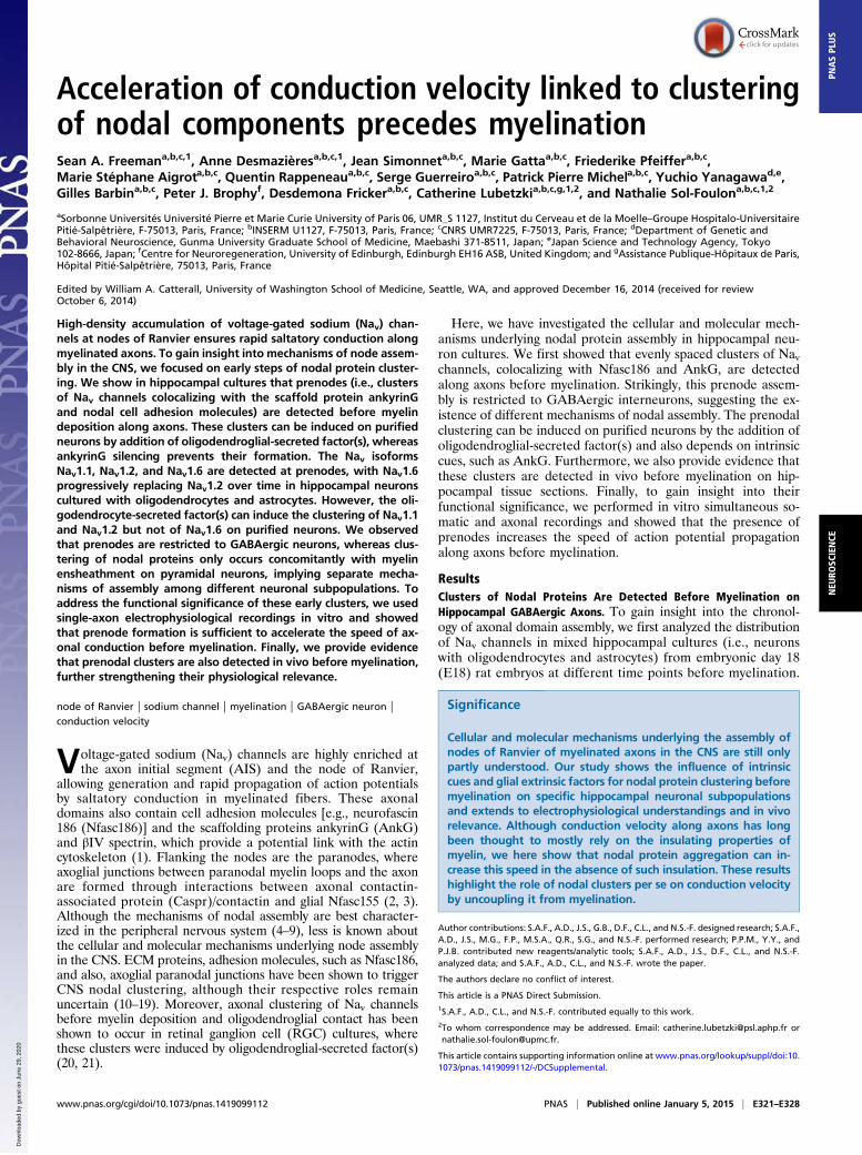
Conduction Velocity Is Increased on Axons with Prenodes in the Absenceof Myelination. We first investigated whether unmyelinated in-terneurons with and without prenodes had different intrinsicelectrophysiological properties by performing somatic whole-cellpatch-clamp recordings on mixed hippocampal cultures fromGAD67-GFP knockin mice (29) (Fig. S6A). We observed nosignificant difference between the mean resting membrane po-tential, action potential (AP) threshold, AP rise time, AP half-width, and AP decay in cells with prenodes and those without(Fig. S6B). However, we observed that neurons with prenodestended to exhibit a significantly lower input resistance comparedto neurons without [with prenodes = 67.5 ± 24.5 MΩ; withoutprenodes = 175.1 ± 74.4 MΩ; n = 7 (each group); P = 0.01].We then examined whether prenodal aggregates might accel-
erate axonal conduction velocity before myelination. Therefore,we compared electrical conduction on cultured rat mixed hip-pocampal GABAergic interneurons with and without prenodesusing simultaneous recordings from the soma and axon of thesame neuron from 10 to 19 DIV. AIS and prenodes were iden-tified using live staining with an Ab targeting the extracellulardomain of Nfasc (Fig. 7 A and B). Cell-attached patch-clamprecordings of action currents from the soma and axon were madeduring spontaneous firing of neurons at a minimum distance of150 μm between the two recording electrodes (Fig. 7 C and D).At longer intervals between the somatic and axonal recordingelectrodes, we observed that the latencies between spikesrecorded in the axon and at the soma increased in both neuronswith and without prenodes (Fig. 7E). Interestingly, the meanapparent conduction velocity was significantly higher in neuronswith prenodes compared to neurons without prenodes (Fig. 7F)(1.23 ± 0.09 m/s, n = 10; 0.72 ± 0.05 m/s, n = 8, respectively; P =0.0002). Because the mean axonal diameter was significantlylarger in neurons with prenodes compared to neurons withoutprenodes (1.96 ± 0.18 μm, n = 10; 1.26 ± 0.12 μm, n = 8, re-spectively; P = 0.008), one possibility might have been that in-creased conduction velocity was caused by larger axonal dia-meter. To test this alternative hypothesis, we selected neuronswith and without prenodes having similar mean axonal diameters(1.71 ± 0.10 μm, n = 3; 1.68 ± 0.05 μm, n = 3, respectively) andshowed that the mean conduction velocity was significantlyhigher in neurons with prenodes (1.24 ± 0.12 m/s, n = 3) com-pared to neurons without prenodes (0.74 ± 0.03 m/s, n = 3; P =0.016). Taken together, these results show that clusters of nodalproteins increase axonal conduction velocity in hippocampalGABAergic neurons before myelination.
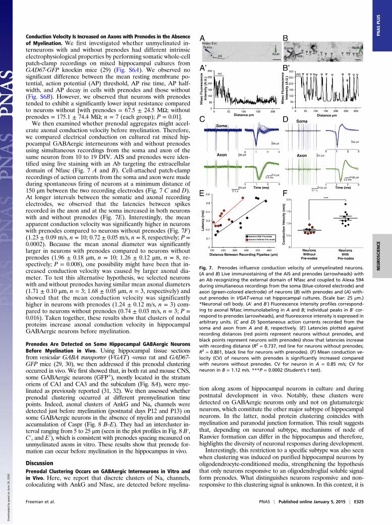
Prenodes Are Detected on Some Hippocampal GABAergic NeuronsBefore Myelination in Vivo. Using hippocampal tissue sectionsfrom vesicular GABA transporter (VGAT) -venus rat and GAD67-GFP mice (29, 30), we then addressed if this prenodal clusteringoccurred in vivo. We first showed that, in both rat and mouse CNS,some GABAergic neurons (GFP+), mostly located in the stratumoriens of CA1 and CA3 and the subiculum (Fig. 8A), were mye-linated as previously reported (31, 32). We then assessed whetherprenodal clustering occurred at different premyelination timepoints. Indeed, axonal clusters of AnkG and Nav channels weredetected just before myelination (postnatal days P12 and P13) onsome GABAergic neurons in the absence of myelin and paranodalaccumulation of Caspr (Fig. 8 B–E). They had an intercluster in-terval ranging from 5 to 25 μm (seen in the plot profiles in Fig. 8 B′,C′, and E′), which is consistent with prenodes spacing measured onunmyelinated axons in vitro. These results show that prenode for-mation can occur before myelination in the hippocampus in vivo.
DiscussionPrenodal Clustering Occurs on GABAergic Interneurons in Vitro andin Vivo. Here, we report that discrete clusters of Nav channels,colocalizing with AnkG and Nfasc, are detected before myelina-
tion along axons of hippocampal neurons in culture and duringpostnatal development in vivo. Notably, these clusters weredetected on GABAergic neurons only and not on glutamatergicneurons, which constitute the other major subtype of hippocampalneurons. In the latter, nodal protein clustering coincides withmyelination and paranodal junction formation. This result suggeststhat, depending on neuronal subtype, mechanisms of node ofRanvier formation can differ in the hippocampus and therefore,highlights the diversity of neuronal responses during development.Interestingly, this restriction to a specific subtype was also seen
when clustering was induced on purified hippocampal neurons byoligodendrocyte-conditioned media, strengthening the hypothesisthat only neurons responsive to an oligodendroglial soluble signalform prenodes. What distinguishes neurons responsive and non-responsive to this clustering signal is unknown. In this context, it is
Fig. 7. Prenodes influence conduction velocity of unmyelinated neurons.(A and B) Live immunostaining of the AIS and prenodes (arrowheads) withan Ab recognizing the external domain of Nfasc and coupled to Alexa 594during simultaneous recordings from the soma (blue-colored electrode) andaxon (green-colored electrode) of neurons (B) with prenodes and (A) with-out prenodes in VGAT-venus rat hippocampal cultures. (Scale bar: 25 μm.)*Neuronal cell body. (A′ and B′) Fluorescence intensity profiles correspond-ing to axonal Nfasc immunolabeling in A and B; individual peaks in B′ cor-respond to prenodes (arrowheads), and fluorescence intensity is expressed inarbitrary units. (C and D) Spontaneous action currents recorded from thesoma and axon from A and B, respectively. (E) Latencies plotted againstrecording distances (red points represent neurons without prenodes, andblack points represent neurons with prenodes) show that latencies increasewith recording distance (R2 = 0.737, red line for neurons without prenodes;R2 = 0.801, black line for neurons with prenodes). (F) Mean conduction ve-locity (CV) of neurons with prenodes is significantly increased comparedwith neurons without prenodes. CV for neuron in A = 0.85 m/s; CV forneuron in B = 1.12 m/s. ***P = 0.0002 (Student’s t test).
Freeman et al. PNAS | Published online January 5, 2015 | E325
NEU
ROSC
IENCE
PNASPL
US
Dow
nloa
ded
by g
uest
on
June
29,
202
0

of note that the induction of Nav clustering by a soluble oligo-dendroglial factor has been identified previously by Kaplan et al.(20, 21) on RGCs, which are glutamatergic neurons, suggestingthat the clustering permissivity does not depend on the type ofneurotransmitter. An attractive hypothesis is that prenodal for-mation might be associated with the need for early establishmentof neuronal connections during development on axons with longtrajectories. In favor of this hypothesis, these two types of axons withprenodes are known to be characterized by long-range projectingaxons: RGC axons extend from the retina to the superior colliculus,and our results reveal that most axons with clusters in vivo extendedfrom the CA1 region of the hippocampus, which has been found toproject to farther interregional areas, such as the medial septum,the subiculum, or the retrosplenial cortex (31, 33, 34).Lastly, whether these nodal protein clusters disappear or fuse
to form nodes of Ranvier when oligodendrocyte processes con-tact the axon, such as described for heminodes (35, 36), remainsto be determined. Indeed, the increase in the spacing of nodalclusters upon myelination in vitro and in vivo along GABAergicaxons suggests that spatial rearrangements could occur.
Oligodendrocyte-Secreted Factor Induces Prenodal Clustering. Ourresults on purified hippocampal neurons extend previous datathat a proteinaceous factor(s) secreted by oligodendroglia inducesclustering of nodal proteins (20, 21). Whether this factor(s)induces Nav clustering through interactions with the axonalmembrane or stimulation of neuronal protein synthesis has yetto be fully understood. Gliomedin expressed by Schwann cells isknown to induce nodal clustering in the peripheral nervous sys-tem (7). However, not only has CNS expression of gliomedin notbeen reported, but also, we found that gliomedin was undetect-able both in oligodendroglial cultures and at prenodes. Thevarious ECM proteins enriched at the CNS nodes mainly playa role in buffering nodal environment and stabilizing the nodes(10–12). Whether they participate in the induction of prenodalassembly remains to be seen. However, our results provide thatclustering of prenodes through OCM is not mediated through anNfasc-based mechanism, implying that other cell adhesion mol-ecules may interact with the oligodendroglial-secreted factor.Furthermore, we addressed whether this oligodendroglial clus-tering effect might be related to oligodendrocyte-induced neu-ronal maturation and survival (37). This hypothesis was partlysupported by the facts that clusters were mostly detected in vitroon large-diameter axons with highly phosphorylated neurofila-ments, a hallmark of maturation, that also, in vivo, prenodeswere found more frequently at P12 and P13 compared withearlier time points, and lastly, that neurons with prenodesexhibited a lower input resistance compared to neurons withoutclusters. In accordance with these findings, it has been observedthat there is a significant decrease in the input resistance as micematured from P9–P11 to P12–P16 (38). However, addition ofmaturation/growth factors [glial cell derived neurotrophic factor(GDNF), BDNF, ciliary neurotrophic factor (CNTF), leukemiainhibitory factor (LIF), or NGF] to pure neuron cultures whileinducing neurofilament phosphorylation did not trigger Navclustering, suggesting that axonal maturation is not sufficient toinduce nodal protein clustering. We also addressed the role ofelectrical activity by using tetrodotoxin or veratridine (blockerand activator of Nav channels, respectively), which did not affectprenode formation.
AnkG Is Essential for Prenodal Formation, Whereas Nfasc Is Dispensable.In the CNS, several mechanisms are involved in nodal assembly:clustering of Nfasc186 through interactions with ECM, restrictionof diffusion through paranodal junctions, and stabilization by thecytoskeletal scaffold. Numerous studies suggest that these threemechanisms are alternative or complementary to induce nodalassembly (8, 9). Direct axoglial contacts established at the para-nodal junctions have been suggested to induce nodal proteinclustering through restriction of their diffusion (12, 13). Never-theless, other studies have indicated that, when paranode assemblyis impaired by inactivation of genes coding for the paranodalproteins (Caspr, contactin, or Nfasc155) or the myelin proteins, thetiming and number of developing nodes occur normally, suggestingthat formation of paranodal junctions may be sufficient but notnecessary for nodal assembly (14–19). Recently, by using a geneticstrategy to determine their requirement, Susuki et al. (12) haveshown that disruption of at least two of these three mechanisms isnecessary to affect node formation. We show here that Nav clusterscan be formed without direct axoglial contact, which has beenpreviously found (20, 21). We also show that AnkG is required forthe formation of Nav and Nfasc186 clusters along unmyelinatedaxons. AnkG, which likely provides a link by establishing inter-actions with the intracellular domains of Nfasc186 and Navα-subunit as well as βIV spectrin and kinesin motors (39, 40), mayeither initiate or stabilize prenodal protein clustering before mye-lination. In contrast, Nfasc is not necessary for Nav and AnkGprenodal clustering. This result suggests that Nfasc186 plays arole in late rather than early stages of node of Ranvier assembly.
Fig. 8. Prenodes can be detected in vivo during postnatal development inboth mouse and rat. (A and B) Immunostaining of sagittal sections of P13VGAT-venus rat hippocampus shows the presence of AnkG+ nodes ofRanvier on myelinated fibers (PLP+) in A as well as AnkG axonal clusters inabsence of myelin (PLP−) in B on some GABAergic neurons stained with ananti-GFP Ab. (Scale bars: 10 μm.) Intensity profiles corresponding to AnkGexpression in (A′) myelinated and (B′) nonmyelinated fibers. Arrowheadsindicate AnkG axonal clusters. (C and D) Immunostaining of sagittal sectionsof P12 GAD-GFP mouse hippocampus shows the presence of (C) Nav and (D)AnkG clusters (arrowheads) in the absence of myelin (PLP−). In D, the boxedarea is shown as a zoomed-in view. (Scale bars: 10 μm.) (E) Immunostainingof P12 GAD-GFP hippocampus shows that Caspr is not clustered aroundprenodal AnkG before myelination (arrowheads), whereas it is accumulatedat hemiparanodes flanking nodal AnkG in myelinated fibers (arrow). *Neu-ronal cell body. (Scale bar: 20 μm.) (C′ and E′) Intensity profiles correspondingto (C) Nav staining and (D) AnkG show isolated peaks corresponding toprenodes (arrowheads).
E326 | www.pnas.org/cgi/doi/10.1073/pnas.1419099112 Freeman et al.
Dow
nloa
ded
by g
uest
on
June
29,
202
0

Nav Clustering Is Differentially Regulated in Prenodes. We observedthat distinct Nav channel isoforms are targeted differentially inprenodes along different time points in vitro. Whereas the Nav1.1isoform is found at all time points, the immature isoform Nav1.2progressively disappears, and Nav1.6 is recruited in ∼35% of neuronswith prenodes in mixed culture. One noteworthy finding is that Nav1.6was found in the absence of myelination, but Nav1.6 does not appear inaxonal clusters on purified hippocampal neurons treated with OCM,the latter of which are similar to those previously reported on purifiedRGCs treated with OCM (21). Interestingly, the timing of Nav1.6expression at prenodes coincided with late in vitro culture time points,wherein its first appearance started at 17 DIV and gradually increasedup to 24 DIV. These results may implicate either the transition ofimmature to mature oligodendrocytes in culture and/or that neuronalmaturation is necessary to synthesize and recruit Nav1.6 to prenodes.Although these results differ from a previous report of Nav1.6 re-cruitment requiring myelin compaction during optic nerve de-velopment (25), these results nevertheless point to an important rolefor the physical presence of oligodendrocytes to target Nav1.6 atprenodes. Because we did observe some CNP-EGFP+ oligoden-drocytes contacting prenodal clusters, it would be interesting to in-vestigate the role of oligodendroglial contact and their influence onthe distribution of sodium channel subtypes, which may shed lightonto additional cues necessary for Nav1.6 targeting in the CNS.
What Are the Functional Roles of Prenodes? What is the functionalgain provided by axonal Nav clusters in the absence of myelin?Axonal propagation velocity of the action potential is known todepend on the axon diameter and the presence of a myelinsheath (41). Here, our whole-cell patch-clamp recordings andsingle-axon electrophysiological results strongly suggest that thepresence of regularly spaced Nav clusters increases action po-tential propagation in the absence of myelination independent ofaxonal caliber, and these data suggest another level of influenceby oligodendrocytes on the regulation of conduction velocitybeyond the production of a myelin sheath. In addition, theshorter interval between prenodes compared with mature nodesof Ranvier could compensate for a lack of insulation in the ab-sence of myelin. Of note, on GABAergic neurons with prenodalclusters, recorded apparent conduction velocities were in a similarrange to those reported for myelinated axons of cortical pyramidalneurons (42, 43). Prenodes with high-density sodium channelclusters could serve as acceleration points, which was predicted bytheoretical modeling calculations (44, 45). It is tempting to spec-ulate that optimization of propagation of electrical signaling ata given developmental stage will be especially crucial for highlyconnected GABAergic hub neurons, which have a widespreadaxonal arborization, and for long-range projection neurons, andprovides a means to impact network activities (33). Moreover, Navclustering may help to overcome axonal branch point failures andmaintain reliable propagation of action potentials (41).In multiple sclerosis, within lesions undergoing remyelination,
clustering of Nav channels has been observed on PLP− (i.e., non-remyelinated) fibers (46), suggesting that, such as for develop-mental myelination, nodal protein clustering might precede mye-lin repair. Although the mechanisms of axonal domain reassemblyduring remyelination are still poorly understood, it can be hypoth-esized that, similar to early nodal clustering during developmentalmyelination, these clusters accelerate conduction velocity beforeremyelination and therefore, participate in functional recovery.
Materials and MethodsAnimals. The care and use of rats and mice in all experiments conformed toinstitutional policies and guidelines (UPMC, INSERM, and European Com-munity Council Directive 86/609/EEC). The following rat and mouse strainswere used in this study: Sprague–Dawley or Wistar rats (Janvier BreedingCenter), VGAT-venus Wistar rats (30), Nfasc−/− mice (1), GAD67-GFP knockinmice (29), CNP-EGFP mice (24), and C57bl/6 WT mice.
Cell Cultures. Mixed hippocampal cultures (containing neurons and glial cells) atE18 were prepared according to procedures described previously (47) withmodifications. Briefly, pooled hippocampi were dissociated enzymatically bytrypsin (0.1%; Worthington) treatment for 20 min with DNase (50 μg/mL). Aftertrypsin neutralization, cells were centrifuged at 400 × g for 5 min, resuspended,and then seeded on polyethylenimine precoated glass coverslips at a densityof 5.0 × 10⁴ cells/35 mm2. Cultures were maintained for 24 h in a 1:1 mixture ofDMEM (11880; Gibco) with 10% FCS (100 IU/mL), penicillin-streptomycin (100 IU/mL),and neuron culture medium (NCM). Culture medium was replaced by a 1:1mixture of Bottenstein–Sato (BS) medium with PDGF-A (0.5%) and NCM, andthen, one-half of the medium was changed every 3 d and replaced by NCM. Toincrease myelination in cultures maintained until 24–28 DIV, differentiationmedium was added at 18 DIV. Compositions of NCM, BS medium, and differ-entiation medium are detailed in SI Materials and Methods. Purified hippo-campal neuronal cultures were obtained by adding (24 h after isolation) theantimitotic agents uridine and 5-fluorodeoxyuridine (5 μM; Sigma) to the NCMfor 36 h. Cultures of dopaminergic neurons were prepared from Wistar rats atE15.5 according to procedures described previously (48). Glial cell cultures wereprepared using cerebral cortices from P2Wistar rats dissected free of meninges,then incubated for 45 min in papain (30 U/mL; Worthington) supplemented withL-cysteine (0.24 mg/mL) and DNase (50 μg/mL) in DMEM at 37 °C, and mechan-ically homogenized. Cells were resuspended in DMEM with 10% FCS and peni-cillin-streptomycin and dispensed into T175 culture flasks at a density of 105 cells/cm2. After 12–15 d in culture, flasks were shaken for 1 h at 180 rpm to removemicroglial cells, medium was replaced, and flasks were shaken on a rotary shakerovernight at 230 rpm at 37 °C. Detached cells were used for immunopanning(SI Materials and Methods) to isolate oligodendrocytes. All cultures were main-tained at 37 °C and 5% CO2.
Preparation of OCM. Oligodendrocytes were cultured for 24 h in BS medium,and then, medium was replaced with NCM and collected after 48 h. OCMwas filtered through a 0.22-μm filter. Protein concentration (4.1 ± 2.4 μg/μL,mean ± SD of four OCMs) was measured using the bicinchoninic acid (BCA)protein assay (Pierce).
Purified Hippocampal Neurons Cultured with OCM or Oligodendrocytes. OCM(500 μL/well) or oligodendrocytes (2.5 × 10⁴ cells/well) were added to puri-fied hippocampal neuronal cultures at 3 DIV after removal of antimitoticagents. Then, one-half of the medium was changed with NCM every 5 d.Percentages of neurons (AnkG+ cells), astrocytes (GFAP+ cells), and oligo-dendrocytes (O4+ and PLP+ cells) were determined at 21 DIV. Mixed hip-pocampal cultures contained 43.7% ± 2.7% of neurons, 42.5% ± 5.4% ofastrocytes, and 11.2% ± 2.7% of oligodendrocytes, whereas purified neu-ronal cultures contained 94.2% ± 2.2% of neurons, 3.6% ± 1.7% of astro-cytes, and 0.4% ± 0.6% of oligodendrocytes; purified neurons cultured withOCM contained 92% ± 2.6% of neurons, 4.3% ± 3.4% of astrocytes, and0.5% ± 0.5% of oligodendrocytes; and purified neurons cocultured witholigodendrocytes contained 77.5% ± 3.5% of neurons, 4.5% ± 4.2% ofastrocytes, and 17.3% ± 0.4% of oligodendrocytes (mean ± SD; n = 3, 150cells at least were counted for each experiment).
Tissue Sections. GAD67-GFP mice and VGAT-venus rats were anesthetized at P12and P13 with lethal doses of Imalgen 500 (Merial) combined with 2% Rompun(Bayer). They were transcardially perfused with 1% or 4% paraformaldehyde(PFA), and brains were dissected and postfixed in 1% or 4% PFA followed by PBSwashes and subsequent equilibration in sucrose (5–30%) solution overnight at4 °C for cryostat sectioning. The next day, brains were embedded in OCT (Tissue-Tek), frozen, and then cryosectioned (CM 3050; Leica) at 35 μm. Serial sectioningof the hippocampus was in the sagittal plane.
Abs and Immunofluorescence. All Abs used can be found listed in SI Materialsand Methods. Cell cultures were fixed with 4% PFA for 10 min or for Navchannel staining, 1% PFA for 10 min at room temperature (RT) and then in-cubated with methanol for 10 min at −20 °C. Coverslips were then washedwith PBS one time. After fixation, cells were incubated with blocking buffer(BB; 1× PBS, 5% normal goat serum, 0.1% Triton) for 15 min and then primaryAb (diluted in BB) for 2 h at RT or 4 °C overnight. Coverslips were then washedand incubated with secondary Ab at RT for 1 h. After last wash, Hoechst stain(1 mM) was placed on cells at RT for 5 min. Floating brain cryostat sectionswere pretreated with ethanol at −20 °C for 10 min and then washed with PBS.They were then treated with BB with 0.2% Triton for 1 h at RT before in-cubation with Ab overnight at 4 °C. Sections were then washed, and secondaryAbs were added for 2 h at RT. Coverslips and sections were mounted onsuperfrost glass slides with Fluoromount G (Southern Biotech).
Freeman et al. PNAS | Published online January 5, 2015 | E327
NEU
ROSC
IENCE
PNASPL
US
Dow
nloa
ded
by g
uest
on
June
29,
202
0

Image Acquisition and Analysis. Slides from hippocampal cultures and cryostatsections were visualized using an Olympus FV-1000 Upright Confocal Mi-croscope or a Zeiss AxioImager-Apotome. Z series of optical sections wasperformed at 0.3-μm increments for qualitative analysis. Blue, green, red,and far-red fluorescence was acquired sequentially. Maximum orthogonalprojection of images and plot profiles of immunofluorescence intensity werecarried out using ImageJ software (NIH).
Quantification of Neurons with Clusters, Sizes of Clusters, Intervals, andMyelination. At least 100 neurons per coverslip, identified by the presence ofan AIS, were counted, and the percentage of neurons with Nav/AnkG clusterswas determined for at least two coverslips per condition. Results wereexpressed as means ± SDs of at least three independent experiments. An Navcluster was defined by its size and mean intensity of area (i.e., size of clustersvaried from 1 to 8 μm, and mean value of cluster area was at least 2.5 that ofthe adjacent part of the axon). Intervals between clusters were measured onacquired images by using ImageJ software. The mean intervals were calcu-lated for one neuron; then, the mean of all neurons for one experiment wasdetermined, and results were expressed as means ± SEMs of four in-dependent experiments. Myelination was assessed at 24 DIV and identifiedas bright PLP+ double lines interrupted at nodes of Ranvier. Statisticalanalyses were performed using the Mann–Whitney test or Student’s t testwith Prism software (GraphPad software).
Design and Transfection of miRNA. To generate AnkG miRNA constructs, weused the Block-it PolII miR RNAi Expression Vector Kits (K4936-00) (SI Materialsand Methods). Hippocampal cells in 24-well plates were transfected at 6 DIV
with 0.5 μg DNA/well using the lipofectamine 2000 reagent according to themanufacturer’s protocol (11668–019; Invitrogen). Transfected cells were fixedat 18 or 20 DIV. The two miRNAs used gave similar results.
Electrophysiology: Simultaneous Soma/Axon Recordings. Rat hippocampalcultures were used between 10 and 19 DIV for electrophysiological record-ings. Nfasc external Ab (cloneA12/18; Neuromab) was coupled to Alexa 594according to the manufacturer’s protocol (A10474; Invitrogen) and thenincubated on hippocampal cultures for 30 min at 37 °C with a 1:15 dilution inculture medium. Labeled cultures were transferred to the recordingchamber on the stage of an Axioskop 2 FS plus microscope (Zeiss) andcontinuously superfused with artificial cerebrospinal fluid. Simultaneouspatch recordings were performed with a Multiclamp 700B amplifier (Mo-lecular Devices) and digitized with pClamp10 software (Axon; MolecularDevices). Spontaneous action currents were recorded in cell-attached modefrom both the somatic and axonal electrodes (SI Materials and Methods).
ACKNOWLEDGMENTS. We thank François Couraud, Rock Levinson, and EliorPeles for the gift of Abs. We also thank Bernard Zalc, Charles-Felix Calvo, andmembers of the laboratory of C.L. for discussions and critical reading of themanuscript. We thank Plate-forme d’Imagerie Cellulaire de la Pitié-Salpêtrièrefor support with image acquisition. This work was supported by INSERM,French Multiple Sclerosis Research Foundation association pour la recherchesur la sclérose en plaques (ARSEP) Program “Investissements d’avenir” ANR-10-IAIHU-06, and the Bouvet–Labruyère prize (to N.S.-F.). S.A.F. and A.D. weresupported by the French Medical Research Foundation, and C.L. was sup-ported by the Sobeck Prize. P.J.B. was supported by the Wellcome Trust.
1. Sherman DL, Brophy PJ (2005) Mechanisms of axon ensheathment and myelin growth.Nat Rev Neurosci 6(9):683–690.
2. Charles P, et al. (2002) Neurofascin is a glial receptor for the paranodin/Caspr-con-tactin axonal complex at the axoglial junction. Curr Biol 12(3):217–220.
3. Sherman DL, et al. (2005) Neurofascins are required to establish axonal domains forsaltatory conduction. Neuron 48(5):737–742.
4. Lustig M, et al. (2001) Nr-CAM and neurofascin interactions regulate ankyrin G andsodium channel clustering at the node of Ranvier. Curr Biol 11(23):1864–1869.
5. Zhang Y, et al. (2012) Assembly and maintenance of nodes of ranvier rely on distinctsources of proteins and targeting mechanisms. Neuron 73(1):92–107.
6. Feinberg K, et al. (2010) A glial signal consisting of gliomedin and NrCAM clustersaxonal Na+ channels during the formation of nodes of Ranvier. Neuron 65(4):490–502.
7. Eshed Y, et al. (2005) Gliomedin mediates Schwann cell-axon interaction and themolecular assembly of the nodes of Ranvier. Neuron 47(2):215–229.
8. Chang KJ, Rasband MN (2013) Excitable domains of myelinated nerves: Axon initialsegments and nodes of Ranvier. Curr Top Membr 72(2013):159–192.
9. Leterrier C, Brachet A, Dargent B, Vacher H (2011) Determinants of voltage-gatedsodium channel clustering in neurons. Semin Cell Dev Biol 22(2):171–177.
10. Dours-Zimmermann MT, et al. (2009) Versican V2 assembles the extracellular matrixsurrounding the nodes of ranvier in the CNS. J Neurosci 29(24):7731–7742.
11. Bekku Y, Rauch U, Ninomiya Y, Oohashi T (2009) Brevican distinctively assembles ex-tracellular components at the large diameter nodes of Ranvier in the CNS. J Neurochem108(5):1266–1276.
12. Susuki K, et al. (2013) Three mechanisms assemble central nervous system nodes ofRanvier. Neuron 78(3):469–482.
13. Rasband MN, et al. (1999) Dependence of nodal sodium channel clustering on par-anodal axoglial contact in the developing CNS. J Neurosci 19(17):7516–7528.
14. Mathis C, Denisenko-Nehrbass N, Girault JA, Borrelli E (2001) Essential role of oligo-dendrocytes in the formation and maintenance of central nervous system nodal re-gions. Development 128(23):4881–4890.
15. Bhat MA, et al. (2001) Axon-glia interactions and the domain organization of mye-linated axons requires neurexin IV/Caspr/Paranodin. Neuron 30(2):369–383.
16. Boyle ME, et al. (2001) Contactin orchestrates assembly of the septate-like junctions atthe paranode in myelinated peripheral nerve. Neuron 30(2):385–397.
17. Ishibashi T, et al. (2002) A myelin galactolipid, sulfatide, is essential for maintenanceof ion channels on myelinated axon but not essential for initial cluster formation.J Neurosci 22(15):6507–6514.
18. Jenkins SM, Bennett V (2002) Developing nodes of Ranvier are defined by ankyrin-G clusteringand are independent of paranodal axoglial adhesion. Proc Natl Acad Sci USA 99(4):2303–2308.
19. Zonta B, et al. (2008) Glial and neuronal isoforms of Neurofascin have distinct roles in theassembly of nodes of Ranvier in the central nervous system. J Cell Biol 181(7):1169–1177.
20. Kaplan MR, et al. (1997) Induction of sodium channel clustering by oligodendrocytes.Nature 386(6626):724–728.
21. Kaplan MR, et al. (2001) Differential control of clustering of the sodium channelsNa(v)1.2 and Na(v)1.6 at developing CNS nodes of Ranvier. Neuron 30(1):105–119.
22. Freund TF, Buzsáki G (1996) Interneurons of the hippocampus. Hippocampus 6(4):347–470.23. Jinno S, Kosaka T (2000) Colocalization of parvalbumin and somatostatin-like im-
munoreactivity in the mouse hippocampus: Quantitative analysis with optical dis-sector. J Comp Neurol 428(3):377–388.
24. Yuan X, et al. (2002) Expression of the green fluorescent protein in the oligoden-drocyte lineage: A transgenic mouse for developmental and physiological studies.J Neurosci Res 70(4):529–545.
25. Boiko T, et al. (2001) Compact myelin dictates the differential targeting of two so-dium channel isoforms in the same axon. Neuron 30(1):91–104.
26. Duflocq A, Le Bras B, Bullier E, Couraud F, Davenne M (2008) Nav1.1 is predominantlyexpressed in nodes of Ranvier and axon initial segments. Mol Cell Neurosci 39(2):180–192.
27. Hedstrom KL, et al. (2007) Neurofascin assembles a specialized extracellular matrix atthe axon initial segment. J Cell Biol 178(5):875–886.
28. Hedstrom KL, Ogawa Y, Rasband MN (2008) AnkyrinG is required for maintenance ofthe axon initial segment and neuronal polarity. J Cell Biol 183(4):635–640.
29. Tamamaki N, et al. (2003) Green fluorescent protein expression and colocalizationwith calretinin, parvalbumin, and somatostatin in the GAD67-GFP knock-in mouse.J Comp Neurol 467(1):60–79.
30. Uematsu M, et al. (2008) Quantitative chemical composition of cortical GABAergicneurons revealed in transgenic venus-expressing rats. Cereb Cortex 18(2):315–330.
31. Jinno S, et al. (2007) Neuronal diversity in GABAergic long-range projections from thehippocampus. J Neurosci 27(33):8790–8804.
32. Meier S, Bräuer AU, Heimrich B, Nitsch R, Savaskan NE (2004) Myelination in the hip-pocampus during development and following lesion. Cell Mol Life Sci 61(9):1082–1094.
33. Picardo MA, et al. (2011) Pioneer GABA cells comprise a subpopulation of hub neu-rons in the developing hippocampus. Neuron 71(4):695–709.
34. Melzer S, et al. (2012) Long-range-projecting GABAergic neurons modulate inhibitionin hippocampus and entorhinal cortex. Science 335(6075):1506–1510.
35. Vabnick I, Novakovi�c SD, Levinson SR, Schachner M, Shrager P (1996) The clusteringof axonal sodium channels during development of the peripheral nervous system.J Neurosci 16(16):4914–4922.
36. Custer AW, et al. (2003) The role of the ankyrin-binding protein NrCAM in node ofRanvier formation. J Neurosci 23(31):10032–10039.
37. Du Y, Dreyfus CF (2002) Oligodendrocytes as providers of growth factors. J NeurosciRes 68(6):647–654.
38. Holter NI, Zuber N, Bruehl C, Draguhn A (2007) Functional maturation of developinginterneurons in the molecular layer of mouse dentate gyrus. Brain Res 1186:56–64.
39. Bennett V, Baines AJ (2001) Spectrin and ankyrin-based pathways: Metazoan in-ventions for integrating cells into tissues. Physiol Rev 81(3):1353–1392.
40. Barry J, et al. (2014) Ankyrin-G directly binds to kinesin-1 to transport voltage-gatedNa+ channels into axons. Dev Cell 28(2):117–131.
41. Debanne D, Campanac E, Bialowas A, Carlier E, Alcaraz G (2011) Axon physiology.Physiol Rev 91(2):555–602.
42. Palmer LM, Stuart GJ (2006) Site of action potential initiation in layer 5 pyramidalneurons. J Neurosci 26(6):1854–1863.
43. Popovic MA, Foust AJ, McCormick DA, Zecevic D (2011) The spatio-temporal charac-teristics of action potential initiation in layer 5 pyramidal neurons: A voltage imagingstudy. J Physiol 589(Pt 17):4167–4187.
44. Johnston WL, Dyer JR, Castellucci VF, Dunn RJ (1996) Clustered voltage-gated Na+channels in Aplysia axons. J Neurosci 16(5):1730–1739.
45. Zeng S, Tang Y (2009) Effect of clustered ion channels along an unmyelinated axon.Phys Rev E Stat Nonlin Soft Matter Phys 80(2 Pt 1):021917.
46. Coman I, et al. (2006) Nodal, paranodal and juxtaparanodal axonal proteins duringdemyelination and remyelination in multiple sclerosis. Brain 129(Pt 12):3186–3195.
47. Banker GA, Cowan WM (1977) Rat hippocampal neurons in dispersed cell culture.Brain Res 126(3):397–445.
48. Toulorge D, et al. (2011) Neuroprotection of midbrain dopamine neurons by nicotineis gated by cytoplasmic Ca2+. FASEB J 25(8):2563–2573.
E328 | www.pnas.org/cgi/doi/10.1073/pnas.1419099112 Freeman et al.
Dow
nloa
ded
by g
uest
on
June
29,
202
0