acanthocolpidae (digenea) of marine ... - folia.paru.cas.cz · 36 small acicular intercalary spines...
TRANSCRIPT

35
Folia Parasitologica 58[1]: 35–47, 2011issN 0015-5683 (print), issN 1803-6465 (online)
© institute of Parasitology, Biology centre ascrhttp://www.paru.cas.cz/folia/
address for correspondence: r.a. Bray, Department of Zoology, Natural History Museum, cromwell road, london sW7 5BD, UK. Phone: +44 (0)20 7942 5563; Fax: +44 (0)20 7942 5151; E-mail: [email protected]
in this paper we are adding to the sparse knowledge of the acanthocolpids of fishes from the coast of New caledonia. Durio and Manter (1969) reported two Steph-anostomum species, S. japonocasum Durio et Manter, 1969 and S. casum (linton, 1910) and Justine et al. (2010) recorded the former again. Bray and Justine (2007) men-tioned an immature Stephanostomum in a balistid. We have identified members of three acanthocolpid genera and report on one new species of Acaenodera Manter et Pritchard, 1960, one species of Pleorchis railliet, 1896 and five species of Stephanostomum looss, 1899, includ-ing one new species.
Materials and MethodsMost fish were caught by hook and line, a few were bought
at the fish market or spear-fished and the conger was taken in a baited cage for the collection of Nautilus at a depth of 400 m. Digeneans were collected live, immediately fixed in nearly boil-ing saline and then transferred to 80% ethanol (cribb and Bray 2010). Whole-mounts were stained with Mayer’s paracarmine, cleared in beechwood creosote and mounted in canada balsam. Measurements were made through a drawing tube on an olym-pus BH-2 microscope, using a Digicad Plus digitising tablet and carl Zeiss Ks100 software adapted by imaging associates, and are quoted in micrometres. the following abbreviations are used: BMNH, British Museum (Natural History) collection at the
Natural History Museum, london, UK; MNHN JNc, Muséum National d’Histoire Naturelle, Paris, France; iPcas, institute of Parasitology, Biology centre of the academy of sciences of the Czech Republic, České Budějovice, Czech Republic.
resultsacanthocolpidae lühe, 1906Acaenodera Manter et Pritchard, 1960
Acaenodera nautili sp. n. Figs. 1–4description. Based on 7 whole-mounts, 4 measured,
measurements and ratios in Table 1. Specimens difficult to mount exactly dorso-ventrally. Body elongate, nar-row, cylindrical, narrower forebody (Fig. 1). No eyespots seen. Forebody spination complicated (Figs. 2–3). about 19 rows of three (usually) or four large spines in mid-ventral region. these spines are irregular, fairly blunt, and gradually reduce in size to merge with usual body-spines at about level of pharynx; anteriormost spines 60–76 long, spines in 19th row 32–43 long. Each lateral region with about 19 rows of 5 to 6 spines, similar in structure to those of mid-ventral region; anteriormost spines 50–74 long. large spines apparently overlain by layer of tegu-ment. Between mid-ventral and lateral large spine fields, and dorsally between lateral large spine fields are rows of
Acanthocolpidae (Digenea) of marine fishes off New Caledonia, with the descriptions of two new species
rodney a. Bray1 and Jean-lou Justine2,3
1 Department of Zoology, Natural History Museum, cromwell road, london sW7 5BD, UK;2 UMr 7138 systématique, adaptation, Évolution, Muséum National d’Histoire Naturelle, 57, rue cuvier, 75231 Paris cedex 05, France;
3 aquarium des lagons, B.P. 8185, 98807 Nouméa, Nouvelle-calédonie
abstract: the following acanthocolpid species are reported from New caledonia. Acaenodera nautili sp. n. from Conger cinereus rüppel differs from other Acaenodera species in details of the body-spination, the sucker-ratio and the bipartite seminal vesicle; Stephanostomum murielae sp. n. from Carangoides hedlandensis (Whitley) differs from most species of Stephanostomum in the average of 36 circum-oral spines, the circum-oral spine rows with a ventral hiatus and the anterior extent of the vitellarium being >10% of the hindbody length from ventral sucker. the species is distinguished from the three other species with these characters in a detailed review. the other species reported are: Stephanostomum aaravi Bray et cribb, 2003 from Lethrinus miniatus and L. ru-brioperculatus; Stephanostomum ditrematis (Yamaguti, 1939) from Gnathanodon speciosus; Stephanostomum japonocasum Durio et Manter, 1969 from Cephalopholis urodeta, Epinephelus areolatus, E. chlorostigma, E. maculatus, E. retouti, Lethrinus miniatus and Variola louti; Stephanostomum uku Yamaguti, 1970 and Pleorchis uku Yamaguti, 1970 from Aprion virescens.
Keywords: Digenea, acanthocolpidae, Acaenodera, Pleorchis, Stephanostomum, New caledonia

36
small acicular intercalary spines about 11–20 long, more or less in line with the large spine rows, apparently eas-ily lost, particularly dorsally. Hindbody spination reaches to about anterior testis. oral sucker oval, terminal. oral spination formed of irregular rows of small acicular spines, about three ventrally and up to about six dorsally. Ventral sucker rounded, slightly protuberant. Prepharynx long, straight. Pharynx oval. oesophagus short. intestinal bifurcation in posterior forebody. caeca reach to posterior extremity: uroproct not apparent.
testes two, oval, entire, tandem, separated, in poste-rior half of hindbody. cirrus-sac long, claviform, sinuous, reaches to about mid-distance between ventral sucker and ovary (Fig. 4). seminal vesicle large, bipartite. Pars pro-statica short, not highly glandular. Ejaculatory duct lined throughout with pavement of thin-walled irregular cupo-las. genital atrium short. genital pore median, immedi-ately anterior to ventral sucker.
ovary oval, entire, pre-testicular, separated from ante-rior testis. Uterine seminal receptacle in proximal uterus.
Figs. 1–4. Acaenodera nautili sp. n. from Conger cinereus. Fig. 1. Ventral view of holotype, uterus in outline, detail of forebody spination omitted. Fig. 2. Ventral view of forebody spination. Fig. 3. Dorsal view of forebody spination. Fig. 4. lateral view of ter-minal genitalia, lining of metraterm omitted for clarity. scale bars: Fig. 1 = 1000 µm; Figs. 2, 3 = 500 µm; Fig. 4 = 200 µm.

37
Mehlis’ gland antero-dextral to ovary. laurer’s canal opens dorsally to ovary. Uterus mostly intercaecal be-tween ovary and ventral sucker. Eggs numerous, tanned, operculate. Metraterm about half length of cirrus-sac, lined in similar fashion to ejaculatory duct. Vitellarium follicular, reaching from about mid-way between ventral sucker and ovary anteriorly and posterior extremity; in lateral fields, ventral, lateral and dorsal to caeca, over-laps gonads, almost confluent or confluent between testes, confluent in post testicular region.
Excretory pore terminal. Excretory vesicle not traced.
t y p e - h o s t : Conger cinereus Rüppell, Congridae, longfin african conger.
s i t e : Digestive tract.t y p e - l o c a l i t y : Deep sea 400 m, near Passe de Dumbéa,
off Nouméa, New caledonia (03/07/2009). P r e v a l e n c e : 1 of 1.s p e c i m e n s : Holotype MNHN JNc2993B-1, paratypes
MNHN JNc2993B-2-4; BMNH 2010.9.29.1-3; iPcas D-689.
E t y m o l o g y : Named after Nautilus. the host was captured in a Nautilus trap.
discussion. the type-species of this genus, Acaenod-era placophora Manter et Pritchard, 1960, is known from the longfin African conger Conger cinereus rüppell (syn. Conger marginatus Valenciennes) from off Hawaii (Man-ter and Pritchard 1960, Yamaguti 1970). the only other confirmed report of this genus is that of A. spinosior Etch-egoin, lanfranchi, cremonte et timi, 2006 from the ar-gentine conger Conger orbignianus Valenciennes, off Mar del Plata, argentina (Etchegoin et al. 2006). Manter and Pritchard (1960) thought that the species Stephanostomum robustum (Maccallum, 1917) from the European conger eel Conger conger (linnaeus) from New York aquarium (Maccallum 1917) probably belonged in Acaenodera, but the material was too poor to be certain. the spination of the forebody is unusual in this genus and confusingly de-scribed, but well illustrated, for A. placophora. Manter and Pritchard (1960) stated ‘three distinct sets of spines present: (1) small, sharp body spines decreasing in both size and number from a level anterior to pharynx to level of ovary; (2) 2 lateral groups of 12 to 18 conspicuously enlarged, recurved spines extending from just posterior to oral sucker to level of eyespots, decreasing in size to merge with body spines … ; (3) 17 flat plates in 7 midven-tral rows (2-3-2-3-2-3-2) beginning just posterior to oral sucker and decreasing in size to level of eyespots … . No peribuccal spines. spines, particularly the large ones, may be lost in macerated specimens; lateral spines may leave basal stumps only’. Yamaguti (1970) said ‘although Man-ter & Pritchard (1960) stated in their original description that there are no peribuccal spines, i have clearly observed five alternating, circumoral rows of sharp spines; the spines are more distinct (up to 13–18 µ long) on the dorsal side than on the ventral side where the anterior spines may be missing or rudimentary. Body spines sharply pointed,
9–12 µ long, decreasing in size and number posteriorly. Of the five lateral cervical spines the anterior two are larger than the posterior three, up to 60–160 µ long; vent-rolateral spines are arranged in five transverse rows of two or three each, beginning just posterior to oral sucker, up to 80–170 µ long; they are followed by several transverse rows of distinctly smaller spines.’ We have illustrated and described the situation in our material and as far as we can tell the basic pattern is similar to that described by Manter and Pritchard (1960) and Yamaguti (1970). as the spines in the mid-ventral rows decrease in size gradually until they are indistinguishable from the regular body-spines, it is not easy to accurately assess how many rows there are. We reckon that in our specimens there are about 19. Man-ter and Pritchard (1960) described and illustrated 7 rows, and Yamaguti (1970) illustrated about 14 rows, the pos-terior 7 of which are distinctly smaller than the anterior seven. We are confused by Yamaguti’s (1970) description and cannot ascertain how many mid-ventral rows he is de-scribing. We reckon that, in our specimens, the spines in the anterior 12 rows are of similar size and the diminution in size occurs between about rows 13 and 19. Etchegoin et al. (2006) described 18–20 rows of enlarged mid-ventral spines and illustrated about 12 rows of large hooks and reduced size posterior to that. this arrangement is similar to that in our specimens. thus in this feature A. nautili differs from A. placophora, but not A. spinosior. in the il-lustrations in Yamaguti (1970) and Etchegoin et al. (2006) the hooks appear acicular, rather than blunt and curved as in our specimens.
Acaenodera nautili is similar to the described species but is distinctly narrower, with a relatively longer fore-body (see width ratios in table 1) (it should be noted, however, that our specimens are not flattened). The ven-tral sucker is relatively smaller (see sucker ratios in ta-ble 1) and the testes are distinctly separated. in addition A. nautili differs from A. spinosior in the relatively shorter cirrus-sac (see ratios in table 1). the seminal vesicle in A. nautili appears bipartite, a condition not described in the other species.
Pleorchis railliet, 1896Pleorchis uku Yamaguti, 1970
H o s t : Aprion virescens Valenciennes, Lutjanidae, green jobfish.s i t e : Digestive tract.l o c a l i t y : récif snark off Nouméa, New caledonia
(22°26′S, 166°25′E, 05/06/2008).P r e v a l e n c e : 1 of 2.Vo u c h e r s : MNHN JNc2568.
discussion. all specimens are immature. this spe-cies has been reported in Aprion virescens from Hawaii (Yamaguti 1970), off Xisha islands, china (gu and shen 1983) and from lizard island on the great Barrier reef (Bray et al. 2005). the host is unusual for a member of this genus, in that most species are parasites of sciaenids
Bray, Justine: acanthocolpidae from New caledonia

38
(Bray 2005). Pleorchis uku has also been reported from the crimson jobfish Pristipomoides filamentosus (Valen-ciennes) (Lutjanidae) and the redbelly yellowtail fusilier Caesio cuning (Bloch) (caesionidae) off Xisha islands, china (gu and shen 1983). Molecular phylogenies pre-
sented by Miller et al. (2007) indicated that the caesioni-dae is embedded within the Lutjanidae, so these findings suggest that P. uku is a specific parasite of lutjanids. The record of P. uku from the camouflage grouper Epinephe-lus polyphekadion (Bleeker) (serranidae) from off the
table 1. Measurements and ratios of Acaenodera spp.
species Acaenodera nautili sp. n. Acaenodera placophora
Acaenodera placophora
Acaenodera spinosior
Host Conger cinereus Conger cinereus Conger cinereus Conger orbignyanussource original Manter and Pritch-
ard (1960)Yamaguti (1970) Etchegoin et al. (2006)
locality New caledonia Hawaii Hawaii argentinan 4 5 31 12
length 4,546–4,608 (4,575) 2,370–3,351 2,300–5,650 5,140–7,140 (6,170)Width 393–453 (430) 438–533 300–770 560–1,000 (714)Forebody 1,615–1,761 (1,662) 1,160–2,330 (1,742)Ventral spine row number 18–19 (19) 7 [about 14] 18–20Ventral spine 46–63 (55) ‘ventrolateral’
up to 80–17090–130 (110), reducing to 46–65 (58)
intercalary small spines 15–21 (19)oral sucker 121–132 × 161–178 (128 × 171) 94–119 × 60–73 50–130 × 80–130 120–168 × 160–184 (147 × 167)Prepharynx 840–987 (907) 400–550 180–750 740–1,240 (956)Pharynx 154–179 × 122–152 (167 × 132) 95–146 × 88–121 80–150 × 80–130 168–240 × 136–200 (207 × 160)oesophagus 138–202 (173) 50–300 60–136 (107)iB to Vs 246–296 (269)Ventral sucker 221–235 × 210–254 (226 × 223) 190–241 × 180–238 360–512 × 328–500 (412 × 395)cirrus-sac 784–913 × 90–111 (849 × 102) 400–720 × 70–110 1,660–1,900 × 85–300 (1,838 × 207)cirrus-overlap into hindbody 464–510 (486)Vs to vitellarium 226–357 (285) 130–650 (299)Vs to ovary 969–1,001 (988) 1,330–2,300 (1,717)ovary 212–244 × 161–183 (227 × 174) 133–161 × 124–175 100–250 × 100–250 176–340 × 248–400 (267 × 305)ovary to anterior testis 28–88 (65)anterior testis 257–331 × 162–227 (306 × 195) 289–380 × 162–241 250–900 × 170–350
(both testes)488–900 × 240–600 (711 × 416)
Distance between testes 156–214 (181) 0–230 (73)Posterior testis 455–471 × 201–268 (461 × 228) 316–496 × 170–212 250–900 × 170–350
(both testes)504–1,300 × 272–580 (891 × 435)
Post-testicular distance 387–419 (398) 260–780 (472)Post-caecal distance 31–91 (60) 75–460 (181)Eggs 77–92 × 38–44 (85 × 42) 60–74 × 46–61 67–81 × 46–58 65–95 × 48–59 (75 × 53)Width%* 8.61–9.87 (9.40) {15.9–18.5} {13.0–13.6} {10.9–14.0}Forebody%* 35.4–38.2 (36.3) [27] [25] {22.6–32.6}sucker-length ratio 1:1.67–1.87 (1:1.76) {1:2.0} [1:3] 1:2.4–3.4 (1:2.7)sucker-width ratio 1:1.20–1.50 (1:1.31) 1:2.0–2.4 {1:3.0–3.3} [1:2.1] 1:2.0–2.8 (1:2.4)cirrus-sac length%* 17.2–19.9 (18.5) {12.7–17.4} 29.8Vs-cs%Vs-ov 46.3–50.9 (49.2) [33] [42]Vs to vitellarium%* 4.91–7.83 (6.24) [7.5] [0] {2.5–9.1}Vs-ov%* 21.0–22.0 (21.6) [31] [21] {26–32}ov-at%* 0.62–1.92 (1.41) 0 [0.9] [2.3]Distance between testes%* 3.38–4.69 (3.96) [1.5] 0–? [0] {0–3.2}Post-testicular region%* 8.41–9.15 (8.71) [6] [8] {5.1–10.9}oral sucker length%* 2.66–2.91 (2.80) {3.6–4.0} {2.2–2.3} {2.3–2.4}Ventral sucker length%* 4.85–5.11 (4.93) {7.2–8.0} [8] {7.0–7.2}Prepharynx%* 18.3–21.4 (19.8) {16.4–16.9} {7.8–13.3} {14.4–17.4}Pharynx length%* 3.37–3.94 (3.64) {4.0–4.4} {2.7–3.5} {3.3–3.4}anterior testis length%* 5.61–7.28 (6.69) {11.3–12.2} [13.6] {9.5–12.6}Posterior testis length%* 9.92–10.2 (10.1) {13.3–14.8} [17.4] {9.8–18.2}ovary length%* 4.66–5.36 (4.96) {4.8–5.6} {4.3–4.4} {3.4–4.8}Hindbody (HB) 2,612–2,734 (2,687) 3,932–4,900 (3,932)Hindbody%* 56.7–59.7 (58.7) [67] [67] {69–76}Vs-Vit % HB 8.66–13.1 (10.6) [11] [0] {3.3–13.3}
* percentage of body-length; {within curly brackets are estimates derived from published ranges}, [within square brackets are estimates derived from published illustrations]

39
Maldives Island in the Indian Ocean needs confirmation (lorber et al. 2006).
Stephanostomum looss, 1899Bray and cribb (2003) listed the then known species of
Stephanostomum looss, 1899. since then several species have been erected (the circum-oral spine number (cos) and information on the presence (VH) or absence (Vc, i.e. ventral continuum) of a ventral hiatus in the cos rows are indicated in parentheses, LHH signifies two lateral hiatus-es): S. qatarense saoud, Nahhas, al Kuwari et ramadan, 2002 (cos 35–35, Vc), S. beukelaardori Bray et reimer, 2004 (cos 33–38, Vc), S. euzeti Bartoli et Bray, 2004 (cos 49–51, Vc), S. tantabiddii Bray et cribb, 2004 (cos 38–45, VH), S. fijiensis Nahhas, Nasser et tam, 2004 (cos 44–48, Vc), S. talakitok Bray et cribb, 2006 (cos 34–40, Vc), S. adlardi Bray, cribb, Waeschenbach et littlewood, 2007 (cos 30–31, lHH), S. gibsoni shau-kat et Bilqees, 2007 (cos 38–41, Vc), S. lamothei Bray et cribb, 2008 (cos 50–55, Vc) and S. tupatupa Bray et cribb, 2008 (cos 34–36, Vc) (saoud et al. 2002, Bartoli and Bray 2004, Bray and cribb 2004, 2006, 2008, Bray and reimer 2004, Nahhas et al. 2004, Bray et al. 2007, shaukat and Bilqees 2007).
Stephanostomum murielae sp. n. Figs. 5–7description. Based on 9 whole-mount preparations,
6 measured. Measurements and ratios in table 2. Body elongate, narrow, widest in region of gonads (Fig. 5). tegument spinous, unarmed patch immediately posterior to oral sucker, spines large, acuminate in forebody, small-er in hindbody, becoming sparse in hindbody, detectable to varying levels in hindbody, in some cases to posterior testis. oral sucker terminal, distinctly wider than long. circum-oral spines in double ring, with distinct ventral hiatus (Fig. 6). Ventral sucker rounded, in anterior fifth of body. Prepharynx long. Pharynx pyriform. oesophagus short. intestinal bifurcation in posterior forebody. caeca long, narrow, terminations often obscured by vitellarium, but uroproct sometimes detected.
testes 2, rounded to oval, entire, tandem, separated by vitelline follicles. Post-testicular region short. cirrus-sac elongate (Figs. 5, 7), reaches well into hindbody; ante-rior extremity dorsal to about mid-ventral sucker. semi-nal vesicle claviform, undivided, narrows anteriorly. Pars prostatica relatively short, lined with anuclear cell-like bodies, surrounded by gland-cells. Ejaculatory duct long, wide, lined with closely packed small cupolas, with round bases seen as circles on wall of duct, with distinct, but short, naked region distally. genital atrium reaching to mid-ventral sucker. genital pore median, slit-like, imme-diately anterior to ventral sucker.
ovary oval, entire, widely separated from anterior tes-tis. Mehlis’ gland antero-dorsal to ovary. laurer’s canal opens dorsally to ovary. Uterine seminal receptacle not
seen. Uterus narrow, intercaecal, pre-ovarian. Eggs large, tanned, operculate, in one specimen only. Metraterm slightly shorter than cirrus-sac, lining lacking cupolas or with weakly developed cupolas. Vitellarium follicular, just overlaps posterior end of cirrus-sac; fields confluent ventrally and dorsally to uterus, gaps lateral to gonads, confluent dorsally and ventrally between gonads and in post-testicular region.
Excretory pore terminal. Vesicle i-shaped, anterior ex-tent not detected.
t y p e - h o s t : Carangoides hedlandensis (Whitley), carangi-dae, bumpnose trevally.
s i t e : Digestive tract.t y p e - l o c a l i t y : Nouméa Fish Market, New caledonia
(13/03/2009).P r e v a l e n c e : 1 of 1.s p e c i m e n s : Holotype: MNHN JNc2883-1, paratypes
JNc2883-2-5, JNc2883c, BMNH 2010.9.29.8-9.E t y m o l o g y : this species is named after the mother of the
senior author, who died during the preparation of this paper.
discussion. according to Bray and cribb (2003) there are three species of Stephanostomum with 10% or more of the hindbody devoid of vitelline follicles, the circum-oral spine count around 30 and with a ventral hiatus, namely S. megacephalum Manter, 1940, S. bicoronatum (stossich, 1883), and S. madhaviae Bray et cribb, 2003. None of the species described since Bray and cribb (2003) show these features.
Stephanostomum megacephalum was described from the crevalle jack Caranx hippos (linnaeus) (carangidae), off Bahia Honda, on the Pacific coast of Panama, off San Francisco on the coast of Ecuador and off White Friars on the Pacific coast of Mexico (Manter 1940), as hav-ing 30–32 circum-oral spines, almost always 32 (based on 11 specimens). Manter (1940) also reported a mac-erated specimen of this species from the horse-eye jack Caranx latus agassiz (carangidae) with 30 oral spines. the species has been reported several times since mainly from the gulf of Mexico or caribbean sea in a variety of carangids (Manter 1947, sparks 1958, Nahhas and ca-ble 1964, Nahhas and short 1965, overstreet 1969, Nah-has and Powell 1971, Fischthal 1977). it has also been reported from carangids in the red sea (Parukhin 1970) and off cochin and Krusadai, india (Zhukov 1977) and in C. hippos and the dwarf mullet Mugil curvidens (Va-lenciennes) (Mugilidae) off ghana in the eastern atlantic ocean (Fischthal and thomas 1968). Manter’s original description of S. megacephalum indicates that it differs from S. murielae in being much smaller (1,431–2,212 × 375–465 vs. 5,107–6,645 × 184–283), much broader (width 21–26% of body-length vs. 3–4%) and with a rela-tively longer forebody (25–33% of body-length vs. 17–21%). the cirrus-sac is not illustrated in detail, but the cir-rus (?ejaculatory duct) is ‘spined, extending only slightly posterior to’ the ventral sucker, whereas in S. murielae the
Bray, Justine: acanthocolpidae from New caledonia

40
ejaculatory duct is long and reaches well into the hind-body. the gonads in S. megacephalum are close together or contiguous, with no intervening vitelline follicles and continuous lateral bands of follicles at the level of the go-
nads; in S. murielae the gonads are well separated, with intervening vitelline follicles and with the vitelline fields interrupted at the level of the gonads.
Stephanostomum bicoronatum is a reasonably well-studied widespread parasite, mainly infecting members of the sciaenidae. the type-host is the shi drum Umb-rina cirrosa (linnaeus) (sciaenidae), from off trieste (stossich 1883). our comparison of this species is based mainly on its redescription from the brown meagre Sci-aena umbra linnaeus (sciaenidae) off corsica, France by Bartoli and Bray (2001), where they list many of the previous records. later records are by lozano et al. (2001) who reported S. bicoronatum from off the southern iberian coast in the meagre Argyrosomus regius (asso) (sciaenidae) giving some dimensions, and by Bray and cribb (2003) who described the worm from off the south-ern Queensland coast, australia in the Madagascar mea-gre Argyrosomus hololepidotus (lacepède) (sciaenidae). over 80% of all records are from sciaenids. Bartoli and Bray’s (2001) study indicates that S. bicoronatum dif-fers from S. murielae in being relatively wider (width about 9% of body length vs. 3–4%) with a longer fore-body (20–30% of body-length vs. 17–21%). the genital atrium is longer, reaching to about the posterior margin of the ventral sucker (vs. about mid-ventral sucker). the cirrus-sac reaches 55–57% of the ventral sucker to ova-ry distance (vs. 32–38%). the gonads are only slightly separated or contiguous (vs. distinctly separated). the post-testicular region is shorter (4–5% of body length vs. 10–11%). Judging by the description in Bray and cribb (2003) S. bicoronatum is relatively wider (width 6–10% of body-length), with a similar forebody length (15–19% of body-length), a longer genital atrium, a longer reach of the cirrus-sac into the hindbody (43–54% of ventral sucker to ovary distance), contiguous testes and with the ovary contiguous or very close to the anterior testis, and a short post-testicular region (3–4% of body-length). the vitelline fields are not interrupted laterally at the level of the gonads in S. bicoronatum.
Stephanostomum madhaviae was described from three specimens from the giant trevally Caranx ignobilis (For-sskål) (carangidae) by Bray and cribb (2003) from off Hope island, southern Queensland, australia. they con-sidered this to be the same form as described as ‘Steph-anostomum orientalis (srivastava, 1939)’ by Madhavi (1976) from the Malabar trevally Carangoides malabari-cus (Bloch et schneider) (carangidae) and the longnose trevally Carangoides chrysophrys (cuvier) (carangidae) off the Waltair coast, Bay of Bengal. it differs from S. mu-rielae, judging by Bray and Cribb (2003), in being shorter (2,606–3,936 long vs. 5,107–5,645), wider (width 8–9% of body length vs. 3–4%), with a longer forebody (28–30% of body-length vs. 17–21%), contiguous gonads (the testes may occasionally be very slightly separated), and a shorter post-testicular region (5–6% of body length vs.
Figs. 5–7. Stephanostomum murielae sp. n. from Carangoides hedlandensis. Fig. 5. Ventral view of holotype, uterus in out-line. Fig. 6. Ventral view of oral-sucker and forebody spina-tion. Fig. 7. lateral view of terminal genitalia. scale bars: Fig. 5 = 1000 µm; Fig. 6 = 200 µm; Fig. 7 = 500 µm.
41
Bray, Justine: acanthocolpidae from New caledonia
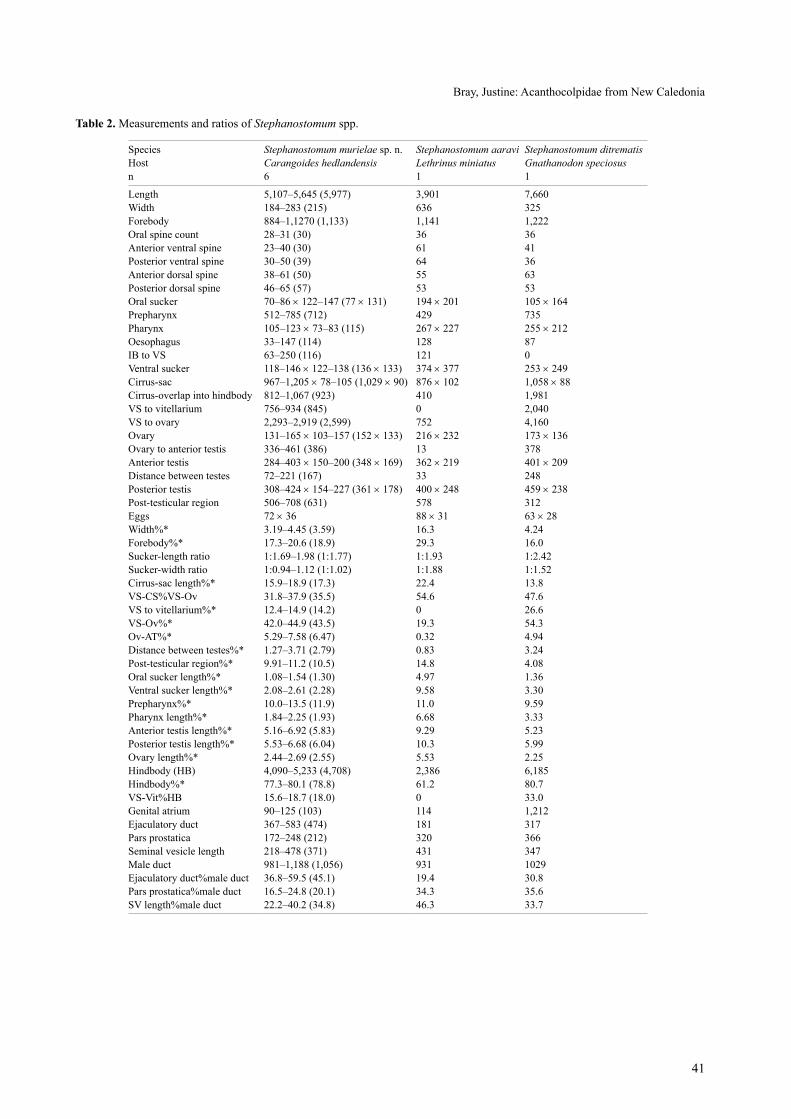
table 2. Measurements and ratios of Stephanostomum spp.
species Stephanostomum murielae sp. n. Stephanostomum aaravi Stephanostomum ditrematisHost Carangoides hedlandensis Lethrinus miniatus Gnathanodon speciosusn 6 1 1
length 5,107–5,645 (5,977) 3,901 7,660 Width 184–283 (215) 636 325 Forebody 884–1,1270 (1,133) 1,141 1,222 oral spine count 28–31 (30) 36 36 anterior ventral spine 23–40 (30) 61 41 Posterior ventral spine 30–50 (39) 64 36 anterior dorsal spine 38–61 (50) 55 63 Posterior dorsal spine 46–65 (57) 53 53 oral sucker 70–86 × 122–147 (77 × 131) 194 × 201 105 × 164Prepharynx 512–785 (712) 429 735 Pharynx 105–123 × 73–83 (115) 267 × 227 255 × 212oesophagus 33–147 (114) 128 87 iB to Vs 63–250 (116) 121 0 Ventral sucker 118–146 × 122–138 (136 × 133) 374 × 377 253 × 249cirrus-sac 967–1,205 × 78–105 (1,029 × 90) 876 × 102 1,058 × 88cirrus-overlap into hindbody 812–1,067 (923) 410 1,981 Vs to vitellarium 756–934 (845) 0 2,040 Vs to ovary 2,293–2,919 (2,599) 752 4,160 ovary 131–165 × 103–157 (152 × 133) 216 × 232 173 × 136ovary to anterior testis 336–461 (386) 13 378 anterior testis 284–403 × 150–200 (348 × 169) 362 × 219 401 × 209Distance between testes 72–221 (167) 33 248 Posterior testis 308–424 × 154–227 (361 × 178) 400 × 248 459 × 238Post-testicular region 506–708 (631) 578 312 Eggs 72 × 36 88 × 31 63 × 28Width%* 3.19–4.45 (3.59) 16.3 4.24Forebody%* 17.3–20.6 (18.9) 29.3 16.0sucker-length ratio 1:1.69–1.98 (1:1.77) 1:1.93 1:2.42sucker-width ratio 1:0.94–1.12 (1:1.02) 1:1.88 1:1.52cirrus-sac length%* 15.9–18.9 (17.3) 22.4 13.8Vs-cs%Vs-ov 31.8–37.9 (35.5) 54.6 47.6Vs to vitellarium%* 12.4–14.9 (14.2) 0 26.6Vs-ov%* 42.0–44.9 (43.5) 19.3 54.3ov-at%* 5.29–7.58 (6.47) 0.32 4.94Distance between testes%* 1.27–3.71 (2.79) 0.83 3.24Post-testicular region%* 9.91–11.2 (10.5) 14.8 4.08oral sucker length%* 1.08–1.54 (1.30) 4.97 1.36Ventral sucker length%* 2.08–2.61 (2.28) 9.58 3.30Prepharynx%* 10.0–13.5 (11.9) 11.0 9.59Pharynx length%* 1.84–2.25 (1.93) 6.68 3.33anterior testis length%* 5.16–6.92 (5.83) 9.29 5.23Posterior testis length%* 5.53–6.68 (6.04) 10.3 5.99ovary length%* 2.44–2.69 (2.55) 5.53 2.25Hindbody (HB) 4,090–5,233 (4,708) 2,386 6,185 Hindbody%* 77.3–80.1 (78.8) 61.2 80.7Vs-Vit%HB 15.6–18.7 (18.0) 0 33.0genital atrium 90–125 (103) 114 1,212 Ejaculatory duct 367–583 (474) 181 317Pars prostatica 172–248 (212) 320 366seminal vesicle length 218–478 (371) 431 347Male duct 981–1,188 (1,056) 931 1029Ejaculatory duct%male duct 36.8–59.5 (45.1) 19.4 30.8Pars prostatica%male duct 16.5–24.8 (20.1) 34.3 35.6sV length%male duct 22.2–40.2 (34.8) 46.3 33.7

42
10–11%). Usually the ejaculatory duct is relatively short-er (36% of male-duct vs. 37–59 (45)). The vitelline fields are not interrupted laterally at the level of the gonads in S. madhaviae.
Stephanostomum aaravi Bray et cribb, 2003
H o s t s : Lethrinus miniatus Forster, lethrinidae, trumpet em-peror; Lethrinus rubrioperculatus sato, lethrinidae, spotch-eek emperor.
s i t e s : Digestive tract, stomach.l o c a l i t i e s : L. miniatus, Récif Toombo (22°33′S, 166°27′E,
20/11/2007), L. rubrioperculatus, Récif Toombo (22°26′S, 166°33′E, 27/06/2006), Shallow, Interior Lagoon near Récif Toombo (22°33′S, 166°29′E, 25/11/2008), all off Nouméa, New caledonia.
P r e v a l e n c e : L. miniatus 3.7% (1 of 27); L. rubriopercula-tus, 12% (2 of 17).
Vo u c h e r s : L. miniatus MNHN JNc2402; L. rubriopercula-tus, MNHN JNc1885, JNc2773.
discussion. These specimens fit comfortably into the original and only description of this species from Lethri-nus miniatus off Heron island, Queensland (Bray and cribb 2003), although those from L. rubrioperculatus are distorted and not measured (see table 2 for measurements of specimen from L. miniatus).
Stephanostomum ditrematis (Yamaguti, 1939) Manter, 1947
H o s t : Gnathanodon speciosus (Forsskål), carangidae, golden trevally.
s i t e : Digestive tract.l o c a l i t y : Nouméa Fish Market (05/12/2008).P r e v a l e n c e : 25% (1 of 4).Vo u c h e r s : MNHN JNc2819c, BMNH 2010.9.29.4.
discussion. these worms (see table 2) are indistin-guishable from those described as this species from the same species of host off Heron and lizard islands on the great Barrier reef by Bray and cribb (2008). the type host was reported as Ditrema temmincki Bleeker (Embi-otocidae) from the inland sea of Japan (Yamaguti 1939), but most subsequent reports have been from carangids.
Stephanostomum japonocasum Durio et Manter, 1969 Figs. 8–13
H o s t s : Cephalopholis urodeta (Forster), Serranidae, darkfin hind; Epinephelus areolatus (Forsskål), serranidae, areolate grouper; Epinephelus chlorostigma (Valenciennes), serrani-dae, brownspotted grouper; Epinephelus maculatus (Bloch), Serranidae, highfin grouper; Epinephelus retouti Bleeker, serranidae, red-tipped grouper; Lethrinus miniatus (Forster), lethrinidae, trumpet emperor; Variola louti (Forsskål), ser-ranidae, yellow-edged lyretail.
s i t e : intestine, digestive tract.
l o c a l i t i e s : C. urodeta, shallow, interior lagoon near Récif Toombo (22°33′S, 166°29′E, 04/11/2008); E. areola-tus, Off Ilôt Brun et Baie des Citrons (22°17′S, 166°25′E, 29/04/2008); E. chlorostigma, off récif toombo, deep-sea (22°34′S, 166°28′E, 04/01/2008); E. maculatus, récif Toombo (22°26′S, 166°33′E, 14/12/2005), interior lagoon near Récif Toombo (22°33′S, 166°29′E, 30/04/2009), Near Récif Toombo (22°34′S, 166°29′E, 16/09/2009); E. retouti, Récif Kué, External slope (22°35′S, 166°30′E, 19/06/2007); L. miniatus, Récif Kué, External slope (22°35′S, 166°30′E, 21/06/2007, 22/06/2007), External slope of récif toombo (22°34′S, 166°27′E, 09/10/2007), Off Récif Kué, Middle of Reef (22°36′S, 166°32′E, 09/12/2008); V. louti, récif Kué, External slope (22°35′S, 166°30′E, 21/06/2007), all off Nou-méa, New caledonia.
P r e v a l e n c e : C. urodeta, 25% (1 of 4); E. areolatus, 20% (1 of 5); E. chlorostigma, 33% (1 of 3); E. maculatus, 12% (3 of 26); E. retouti, 33% (1 of 3); L. miniatus, 19% (5 of 27); V. louti, 8% (1 of 12).
Vo u c h e r s : C. urodeta, MNHN JNc2748; E. areolatus, MNHN JNc2494, JNc3053; E. chlorostigma, MNHN JNc2446; E. maculatus, MNHN JNc1684, JNc2930, BMNH 2010.9.29.5; E. retouti, MNHN JNc2181B; L. min-iatus, MNHN JNc2205, JNc2207, JNc2300, JNc2822B, BMNH 2010.9.29.6-7; V. louti, JNc2198.
discussion. this species is known only from the origi-nal record by Durio and Manter (1969) from Epinephelus sp. and an ‘unidentified serranid’ off New Caledonia and reports from six serranid species off New caledonia by Justine et al. (2010). We here record the species in six serranid species and one lethrinid (table 3). the latter is a surprising host as in the lethrinids of the great Barrier reef the similar species, S. pagrosomi (Yamaguti, 1939), is reported from Lethrinus miniatus, the spangled em-peror L. nebulosus (Forsskål) and the Pacific yellowtail emperor L. atkinsoni seale, off Heron island (Bray and cribb 2003). Stephanostomum pagrosomi, in lethrinids, is reported as having 49–59 uninterrupted circum-oral spines and vitelline fields that reach to about the poste-rior margin of the ventral sucker. according to our data S. japonocasum has 44–53 circum-oral spines and vitel-line fields that reach to about the anterior margin of the ventral sucker or just into the forebody and the fields are confluent in the anterior region. The vitelline configura-tion is a convincing distinction between S. japonocasum and S. pagrosomi. Durio and Manter (1969) reported 40–44 circum-oral spines in S. japonocasum, distinguish-ing this species from S. japonicum (Yamaguti, 1934) by circum-oral spine number, i.e. 40–44 vs. 46. our data cast doubt on this distinction. No spines were described in the ejaculatory duct or metraterm of S. japonicum. We ob-served cupolas with circular bases on the ejaculatory duct and metraterm walls, which we take to be the same as the spines with ‘spherical base’ as described by Durio and Manter (1969). the cirrus of S. japonicum is described by Yamaguti (1934) as joining ‘the metraterm near the conspicuous genital pore lying immediately’ anterior to

43
the ventral sucker. in S. japonocasum the ejaculatory duct (= cirrus) joins the metraterm at about mid-ventral sucker level, and there is a distinct elongate genital atrium. these features associated with the terminal genitalia may serve to distinguish S. japonicum from S. japonocasum. there is also no evidence that the vitelline fields in S. japonicum are confluent in the anterior region or in the posterior fore-body, as in S. japonocasum. Stephanostomum japonicum has been reported only in the spinyhead sculpin Dasycott-us setiger Bean and Cottunculus sp. (Psychrolutidae) and the hookhorn sculpin Artediellus pacificus gilbert (cotti-dae) from toyama Bay, Japan (Yamaguti 1934). We disa-gree with Machida (1984) who considered S. japonicum a synonym of S. baccatum (Nicoll, 1907), a widespread northern temperate species, originally described from the atlantic halibut Hippoglossus hippoglossus (linnaeus)
(Pleuronectidae) off scotland (Nicoll 1907). in S. bac-catum the vitellarium does not reach into the forebody (Nicoll 1907, 1913, Manter 1926, Wolfgang 1955, Zhu-kov 1960, Machida 1984).
all specimens of Lethrinus miniatus and several of the serranids were taken outside the barrier reef in rela-tively deep water and, therefore, they share a common environment and probably common prey items which act as second intermediate hosts of S. japonocasum. Lethri-nus miniatus is a relatively large predator as are most of the serranids (apart from Cephalopholis urodeta). the prevalence of S. japonocasum in L. miniatus was 19%, and in serranids the prevalence varied between 8% and 33%, suggesting that L. miniatus is not an accidental host. other lethrinid species from within the lagoon apparently do not harbour this digenean, but they are smaller species.
Bray, Justine: acanthocolpidae from New caledonia
Figs. 8–13. Stephanostomum japonocasum Durio et Manter, 1969 ex Lethrinus miniatus (Figs. 8–10) and ex Epinephelus areolatus (Figs. 11–13). Figs. 8, 11. Ventral view, uterus in outline. Figs. 9, 12. Ventral view of oral-sucker and forebody spination. Figs. 10, 13. lateral view of terminal genitalia. scale bars: Fig. 8 = 500 µm; Figs. 9, 10, 12, 13 = 200 µm. Fig. 11 = 1000 µm.

44
table 3. Measurements and ratios of Stephanostomum japonocasum.
speciesStephanostomum japonocasum
Stephanostomum japonocasum
Stephanostomum japonocasum
Stephanostomum japonocasum
Host Cephalopholis urodeta Epinephelus areolatus Epinephelus chlorostigma Epinephelus maculatusn 1 1 1 2
length 2,247 2,741 3,786 2193–2,289Width 293 466 492 367–396Forebody 814 788 942 655–802oral spine count 48 44 45 46–51anterior ventral spine 32 31 36 36–40Posterior ventral spine 39 35 43 36–37anterior dorsal spine 35 18 43 22–27Posterior dorsal spine 49 25 59 36–37oral sucker 137 × 171 145 × 171 161 × 214 140–151 × 157–168Prepharynx 359 262 320 220–290Pharynx 150–138 177 × 198 185 × 195 164–170 × 161–175oesophagus 74 91 131 61–105iB to Vs 93 103 145 75–89Ventral sucker 234 × 188 228 × 214 306 × 273 215–229 × 167–229cirrus-sac 504 × 54 494 × 97 670 × 79 467–492 × 57–88cirrus-overlap into hindbody 206 364 529 315–356Vs to vitellarium 0 0 0 0–51Vs to ovary 384 566 920 410–471ovary 140 × 108 192 × 179 239 × 208 92–147 × 92–118ovary to anterior testis 64 35 136 14–26anterior testis 171 × 122 179 × 132 291 × 192 150–152 × 112–154Distance between testes 0 0.51 0 0Posterior testis 173 × 119 176 × 119 310 × 179 138–144 × 107–144Post-testicular region 264 523 616 390–444Eggs 75 × 32 64 × 44 81 × 36 52–75 × 32–38Width%* 13.1 17.0 13.0 16.8–17.3Forebody%* 36.2 28.8 24.9 29.9–35.0sucker-length ratio 1:1.70 1:1.57 1:1.90 1:1.52–1.53sucker-width ratio 1:1.10 1:1.25 1:1.28 1:1.06–1.36cirrus-sac length%* 22.4 18.0 17.7 21.3–21.5Vs-cs%Vs-ov 53.8 64.3 57.5 75.6–76.8Vs to vitellarium%* 0 0 0 0–2.2Vs-ov%* 17.1 20.7 24.3 18.7–20.6ov-at%* 2.83 1.29 3.60 0.64–1.1Distance between testes%* 0.00 0.51 0.00 0.00Post-testicular region%* 11.7 19.1 16.3 17.0–20.3oral sucker length%* 6.12 5.29 4.25 6.4–6.6Ventral sucker length%* 10.4 8.31 8.09 9.79–10.0Prepharynx%* 16.0 9.56 8.47 10.0–12.7Pharynx length%* 6.69 6.47 4.88 7.41–7.48anterior testis length%* 7.61 6.54 7.68 6.62–6.84Posterior testis length%* 7.69 6.43 8.19 6.01–6.56ovary length%* 6.23 6.99 6.30 4.03–6.72Hindbody (HB) 1,200 1,725 2,537 1,259–1,324 Hindbody%* 53.4 62.9 67.0 55.0–60.3Vs-Vit%HB 0 0 0 0–4.1genital atrium 72 141 199 126–136Ejaculatory duct 273 266 376 219–316Pars prostatica 132 227 99 139–172seminal vesicle length 143 110 199 72–127Male duct 548 603 674 485–560Ejaculatory duct%male duct 49.9 44.1 55.7 45.3–56.5Pars prostatica%male duct 24.1 37.6 14.7 28.6–30.7sV length%male duct 26.0 18.3 29.6 12.8–26.2
(continued)

45
Bray, Justine: acanthocolpidae from New caledonia
table 3. continued.
species Stephanostomum japonocasum Stephanostomum japonocasum Stephanostomum japonocasumHost Epinephelus retouti Lethrinus miniatus Variola loutin 1, poor specimen 3 1
length 2,933 2,618–3,362 (3,007) 2558Width 513 370–469 (422) 383Forebody 776 730–941 (838) 783oral spine count 46 45–53 (48) 50anterior ventral spine 30 26–34 (30) 32Posterior ventral spine 33 28–34 (31) 33anterior dorsal spine 41 24–33 (28) 32Posterior dorsal spine 49 34–43 (39) 35oral sucker 147 × ? 107–160 × 148–198 (129 × 174) 129 × 187Prepharynx 281 261–369 (321) 338.2Pharynx 171 × 197 126–177 × 141–171 (150 × 161) 139 × 143oesophagus 36 68–107 (91) 88iB to Vs 142 136–168 (149) 0Ventral sucker 295 × ? 206–298 × 203–253 (255 × 224) 221 × 216cirrus-sac ? 593–686 × 91–102 (639 × 96) 562 × 79cirrus-overlap into hindbody ? 373–525 (449) 368Vs to vitellarium ? 42–100 (76) 0Vs to ovary 625 587–809 (677) 514ovary 150 × 157 144–217 × 97–158 (175 × 132) 117 × 106ovary to anterior testis 85 44–92 (61) 100.3anterior testis 206 × 185 205–285 × 167–220 (235) 179 × 143Distance between testes 24 0 93.9Posterior testis 222 × 185 230–283 × 172–217 (259 × 192) 162 × 135Post-testicular region 365 327–486 (409) 377.8Eggs 80 × 35 75–79 × 34–35 (77 × 34) 74 × 29Width%* 17.5 12.7–15.4 (14.1) 15.0Forebody%* 26.5 27.7–28.0 (27.9) 30.6sucker-length ratio 1:2.00 1:1.86–2.15 (1:1.98) 1:1.71sucker-width ratio ?? 1:1.23–1.37 (1:1.29) 1:1.16cirrus-sac length%* ? 19.5–20.4 (19.9) 22.0Vs-cs%Vs-ov ? 58.8–65.0 (61.9) 71.5Vs to vitellarium%* 0 1.40–3.23 (2.53) 0Vs-ov%* 21.3 20.8–24.1 (22.4) 20.1ov-at%* 2.89 1.31–3.50 (2.12) 3.92Distance between testes%* 0.80 0 3.67Post-testicular region%* 12.4 12.3–16.0 (13.6) 14.8oral sucker length%* 5.02 3.97–4.76 (4.27) 5.04Ventral sucker length%* 10.1 7.88–8.86 (8.43) 8.63Prepharynx%* 9.58 9.98–11.0 (10.6) 13.2Pharynx length%* 5.82 4.80–5.28 (4.97) 5.43anterior testis length%* 7.01 7.11–8.48 (7.81) 7.01Posterior testis length%* 7.55 8.43–8.79 (8.63) 6.35ovary length%* 5.11 5.41–6.47 (5.79) 4.56Hindbody (HB) 1,861 1,682–2,123 (1,915) 1,554 Hindbody%* 63.5 63.1–64.2 (63.7) 60.8Vs-Vit % HB ? 2.19–5.02 (3.97) 0genital atrium ? 98–157 (136) 183Ejaculatory duct ? 245–327 (284) 260Pars prostatica ? 126–216 (162) 221seminal vesicle length ? 141–316 (251) 145Male duct ? 530–858 (696) 626Ejaculatory duct%male duct ? 38.1–46.3 (41.4) 41.6Pars prostatica%male duct ? 18.0–27.0 (23.4) 35.3sV length%male duct ? 26.7–42.1 (35.2) 23.2
46
Bartoli P., Bray r.a. 2001: contribution to the knowledge of species of the genus Stephanostomum looss, 1899 (Digenea: acanthocolpidae) from teleosts of the Western Mediterranean, with the description of S. gaidropsari n. sp. syst. Parasitol. 49: 159–188.
Bartoli P., Bray r.a. 2004: Four species of Stephanostomum looss, 1899 (Digenea, acanthocolpidae) from Seriola dumerili (risso) (teleostei, carangidae) in the Western Mediterranean, including S. euzeti n. sp. syst. Parasitol. 58: 41–62.
Bray r.a. 2005: Family acanthocolpidae lühe, 1906. in: a. Jones, r.a. Bray and D.i. gibson (Eds.), Keys to the trema-toda. Volume 2. caBi Publishing and the Natural History Mu-seum, Wallingford, pp. 603–619.
Bray r.a., CriBB t.H. 2003: species of Stephanostomum looss, 1899 (Digenea: Acanthocolpidae) from fishes of Australian and South Pacific waters, including five new species. Syst. Parasi-tol. 55: 159–197.
Bray r.a., CriBB t.H. 2004: Stephanostomum tantabiddii n. sp. (Digenea: acanthocolpidae) from Carangoides fulvoguttatus (Forsskål, 1775) (Perciformes: carangidae), from Ningaloo reef, Western australia. Zootaxa 457: 1–8.
Bray r.a., CriBB t.H. 2006: Stephanostomum talakitok n. sp. (Digenea: acanthocolpidae) from Gnathanodon speciosus (Perciformes: carangidae) from Ningaloo reef, Western aus-tralia. Zootaxa 1104: 59–68.
Bray r.a., CriBB t.H. 2007: Monostephanostomum nolani sp. n. and M. krusei reimer, 1983 (Digenea: acanthocolpidae) from carangid fishes from coral reef waters off Australia. Folia Para-sitol. 54: 19–26.
Bray r.a., CriBB t.H. 2008: Stephanostomum spp. (Digenea: acanthocolpidae) from scombrids and carangids (Perciformes) from the great Barrier reef, with the description of two new species. rev. Mex. Biodivers. 79: 49s–68s.
Bray r.a., CriBB t.H., WaesCHenBaCH a., littleWood d.t.J. 2007: a new species of Stephanostomum looss, 1899 (Dige-nea: acanthocolpidae) with a bizarre oral sucker: S. adlardi n. sp. from the common coral trout Plectropomus leopardus (lacepède, 1802) (Perciformes: serranidae) from lizard is-land, great Barrier reef. acta Parasitol. 52: 206–212.
Bray r.a., Justine J.-l. 2007: Pseudopycnadena tendu sp. nov. (Digenea, Opecoelidae) in the yellow-spotted triggerfish Pseudobalistes fuscus (Perciformes, Balistidae) and additional opecoelids parasitizing fishes from the waters off New Caledo-nia. acta Parasitol. 52: 13–17.
Bray r.a., reimer l.W. 2004: two species of Stephanostomum Looss, 1899 (Digenea: Acanthocolpidae) from marine fishes off
Namibia, including S. beukelaardori n. sp. syst. Parasitol. 58: 209–216.
Bray r.a., WeBster B.l., Bartoli P., littleWood d.t.J. 2005: relationships within the acanthocolpidae lühe, 1906 and their place among the Digenea. acta Parasitol. 50: 281–291.
CriBB t.H., Bray r.a. 2010: gut wash, body soak, blender, and heat-fixation: approaches to the effective collection, fixation and preservation of trematodes of fishes. Syst. Parasitol. 55: 45–52.
durio W.o., manter H.W. 1969: some digenetic trematodes of marine fishes of New Caledonia. III. Acanthocolpidae, Haplo-poridae, gyliauchenidae, and cryptogonimidae. J. Parasitol. 55: 293–300.
etCHegoin J.a., lanfranCHi a.l., Cremonte f., timi J.t. 2006: a new species of Acaenodera (Digenea: acanthocolpidae) parasitizing Conger orbignyanus (Pisces: congridae) from the coasts of argentina. Parasitol. int. 55: 291–293.
fisCHtHal J.H. 1977: Some digenetic trematodes of marine fishes from the Barrier reef and reef lagoon of Belize. Zool. scripta 6: 81–88.
fisCHtHal J.H., tHomas J.d. 1968: Digenetic trematodes of ma-rine fishes from Ghana: Families Acanthocolpidae, Bucephali-dae, Didymozoidae. Proc. Helminthol. soc. Wash. 35: 237–247.
gu C.-d., sHen J.-W. 1983: Digenetic trematodes of fishes from the Xisha islands, guangdong Province, china. i. stud. Mar. sin. 20: 157–184. (in chinese.)
Justine J.-l., Beveridge i., BoxsHall g.a., Bray r.a., moraveC f., trilles J.-P., WHittington i.d. 2010: an an-notated list of parasites (isopoda, copepoda, Monogenea, Dige-nea, cestoda and Nematoda) collected in groupers (serranidae, Epinephelinae) in New caledonia emphasizes parasite biodiver-sity in coral reef fish. Folia Parasitol. 57: 237–262 .
lorBer J., CriBB t., moraveC f., KiKinger r., KoneCny r. 2006: Endoparasiten von Fischen der Malediven. in: Helminthologische Fachgespräche 2006 ‘Von Würmern und Wirten’, Naturhistorisches Museum, Wien, pp. 4–5.
lozano C., uBeda J.m., de roJas m., ariza C., guevara d.C. 2001: Estudio de digénidos de peces marinos del sur de la Península ibérica. res. rev. Parasitol. 61: 103–116.
maCCallum g.a. 1917: some new forms of parasitic worms. Zoopathologica 1: 43–75.
maCHida m. 1984: Trematodes of marine fishes from depth of 200–400 m off Yamagata, the Japan sea. Mem. Natl. sci. Mus., tokyo 17: 101–110.
madHavi r. 1976: Digenetic trematodes from marine fishes of Waltair coast, Bay of Bengal. Family acanthocolpidae. riv. Parassitol. 37: 115–128.
Stephanostomum uku Yamaguti, 1970
H o s t : Aprion virescens Valenciennes, Lutjanidae, green jobfish.s i t e : Posterior intestine.L o c a l i t y : Reef near Îlot La Regnière (22°19′S, 166°20′E,
05/07/2005), off Nouméa, New caledonia.P r e v a l e n c e : 1 of 2.Vo u c h e r s : MNHN JNc1557c.
discussion. this species is known only from A. vires-cens, in Hawaii (Yamaguti 1970) and a report of immature
specimens from lizard island, great Barrier reef (Bray et al. 2005).
Acknowledgements. Many students and colleagues were in-volved in the fishing expeditions and the parasitological survey, especially aude sigura, charlotte schoelinck, cyndie Dupoux, Isabelle Mary, Frank Moravec, Eva Řehulková and Naďa Musi-lová. Xavier Neyrat (Aquarium des Lagons, Nouméa) collected the conger. The identification of several fishes was confirmed, from photographs, by ichthyologists: conger: Bernard séret (MNHN, Paris); carangids: ronald Fricke (staatliches Museum für Naturkunde, stuttgart); groupers: John E. randall (Bishop Museum, Hawaii).
REFERENCES

47
Bray, Justine: acanthocolpidae from New caledonia
manter H.W. 1926: Some North American fish trematodes. Ill. Biol. Monogr. 10: 7–138.
manter H.W. 1940: Digenetic trematodes of fishes from the Galapagos Islands and the neighboring Pacific. Allan Hancock Pacif. Exped. 2: 325–497.
manter H.W. 1947: The digenetic trematodes of marine fishes of tortugas, Florida. am. Midl. Nat. 38: 257–416.
manter H.W., PritCHard m.H. 1960: some digenetic trema-todes of eels of Hawaii. J. Parasitol. 46: 651–658.
miller t.l., CriBB t.H. 2007: Phylogenetic relationships of some common Indo-Pacific snappers (Perciformes: Lutjanidae) based on mitochondrial DNa sequences, with comments on the taxonomic position of the caesioninae. Mol. Phylogenet. Evol. 44: 450–460.
naHHas f.m., CaBle r.m. 1964: Digenetic and aspidogastrid trematodes from marine fishes of Curaçao and Jamaica. Tulane stud. Zool. 11: 169–228.
naHHas f.m., nasser H., tam J. 2004: Digenetic trematodes of marine fishes from Suva, Fiji: families: Acanthocolpidae, lepocreadiidae, Bivesiculidae, Zoogonidae, Monorchiidae and description of a new species. riv. Parassitol. 21: 33–48.
naHHas f.m., PoWell e.C. 1971: Digenetic trematodes of marine fishes from the Floridian northern Gulf of Mexico. Tulane Stud. Zool. Bot. 17: 1–9.
naHHas f.m., sHort r.B. 1965: Digenetic trematodes of marine fishes from Apalachee Bay, Gulf of Mexico. Tulane Stud. Zool. 12: 39–50.
niColl W. 1907: a contribution towards a knowledge of the En-tozoa of British marine fishes. Part 1. Ann. Mag. Nat. Hist., 7 ser., 19: 66–94.
niColl W. 1913: Trematode parasites from food-fishes of the North sea. Parasitology 6: 188–194.
overstreet r.m. 1969: Digenetic trematodes of marine teleost fishes from Biscayne Bay, Florida. Tulane Stud. Zool. Bot. 15: 119–176.
ParuKHin a.m. 1970: [On the study of trematode fauna in fish from the red sea and aden Bay]. Biol. Morya, Kiev 20: 187–213. (in russian.)
saoud m.f.a., naHHas f.m., al KuWari K.s.r., ramadan m.m. 2002: Helminth parasites of fishes from the Arabian gulf: 10. trematodes of the genus Stephanostomum looss, 1899 (Digenea: acanthocolpidae lühe, 1901), with descrip-tion of Stephanostomum qatarense n. sp. and redescription of Stephanostomum triacanthi Madhavi, 1976. riv. Parassitol. 29 (63): 87–103.
sHauKat n., Bilqees f.m. 2007: a new species of the genus Stephanostomum looss, 1899 (Digenea: acanthocolpidae) from the fish Pomadasys olivaceum off Karachi coast. Proc. Parasitol. 44: 45–67.
sParKs a.K. 1958: Some digenetic trematodes of fishes of Grand isle, louisiana. Proc. la. acad. sci. 20: 71–82.
stossiCH m. 1883: Brani di elmintologia tergestina. serie prima. Boll. soc. adriat. sci. Nat. 8: 111–121.
Wolfgang r.W. 1955: studies on the trematode Stephanostomum baccatum (Nicoll, 1907). iii. its life cycle. can. J. Zool. 33: 113–128.
yamaguti s. 1934: studies on the helminth fauna of Japan. Part 2. Trematodes of fishes, I. Jpn. J. Zool. 5: 249–541.
yamaguti s. 1939: studies on the helminth fauna of Japan. Part 26. Trematodes of fishes, VI. Jpn. J. Zool. 8: 211–230.
yamaguti s. 1970: Digenetic trematodes of Hawaiian fishes. Kei-gaku, tokyo, 436 pp.
zHuKov e.v. 1960: Endoparasitic worms of the fishes in the Sea of Japan and south-Kuril shallow-waters. trud. Zool. inst., len-ingr. 28: 3–146. (in russian.)
zHuKov e.v. 1977: contribution to the knowledge of trematodes of marine fishes of India. Parazitol. Sb. 27: 51–79. (In Russian.)
received 2 august 2010 accepted 6 october 2010