absolute lymphocyte count and risk of short-term infection in patients with immune thrombocytopenia
TRANSCRIPT
ORIGINAL ARTICLE
Absolute lymphocyte count and risk of short-term infectionin patients with immune thrombocytopenia
Ming-Hung Hu & Yuan-Bin Yu & Yu-Chung Huang & Jyh-Pyng Gau &
Liang-Tsai Hsiao & Jin-Hwang Liu & Ming-Huang Chen & Tzeon-Jye Chiou &
Po-Min Chen & Cheng-Hwai Tzeng & Chun-Yu Liu
Received: 9 July 2013 /Accepted: 7 January 2014# Springer-Verlag Berlin Heidelberg 2014
Abstract Patients with immune thrombocytopenia (ITP) maybe at increased risk of infection because of the steroids andother immunosuppressive agents used in its treatment. Thisstudy aimed to identify events that are associated with infec-tion within 6 months of diagnosis and the impact that infectionhas on survival. We retrospectively evaluated 239 patients(107 men, 132 women; median age 61 years) diagnosedbetween January 1997 and August 2011. Every patient re-ceived steroid treatment according to the platelet count and theextent of bleeding. Logistic regression analysis was used toidentify risk factors associated with the development of infec-tion within 6 months of ITP being diagnosed. Sixty-two
patients (25.9 %) developed an infection within 6 months ofdiagnosis. Multivariate analysis revealed that a lower absolutelymphocyte count (ALC) at diagnosis (<1×10⁹/l) was anindependent risk factor for infection (P=0.039; 95 % confi-dence interval, 1.033–3.599; odds ratio, 1.928). The time toinfection event is significant shorter in those of low ALC,compared with those of higher ALC (P=0.032). Furthermore,the 1-year mortality rate after ITP diagnosis was signifi-cantly higher in those patients who developed an infection(P=0.001). ITP patients with a low absolute lymphocytecount at diagnosis have an increased risk of infection, andthose who develop infections have lower 1-year survival.
Keywords Immune thrombocytopenia (ITP) . Absolutelymphocyte count (ALC) . Lymphopenia . Infection
Introduction
Adult patients with idiopathic or immune thrombocytopenia(ITP) are generally considered to have the same mortalityrisk as the general population [1]. However, considerablemorbidity and mortality may still occur in these patients,particularly in those with severe thrombocytopenia thatpersists despite treatment [1]. It seems likely that patientswith ITP will have an increased risk of infection assteroids and other immunosuppressive agents are the pri-mary treatment for this condition. Indeed, in a study ofhigh-dose steroid treatment for ITP patients younger than60 years of age by the GIMEMA group, the infection riskwas reported to be 2.7 % [2]. In a Danish population-based cohort study, the infection risk during the 1-yearfollow-up period was found to be 15.2 %, and an evenhigher infection rate was noted in patients who were olderthan 60 years [3]. In another single-institute cohort studyof 47 consecutive elderly ITP patients (also aged over
M.<H. Hu :Y.<B. Yu :Y.<C. Huang : J.<P. Gau : L.<T. Hsiao :J.<H. Liu :M.<H. Chen : P.<M. Chen : C.<H. Tzeng : C.<Y. Liu (*)Division of Haematology and Oncology, Department of Medicine,Taipei Veterans General Hospital, No. 201, Sec. 2, Shih-Pai Road,Beitou Dist., Taipei City 11217, Taiwan, Republic of Chinae-mail: [email protected]
C.-Y. Liue-mail: [email protected]
T.<J. ChiouDivision of Transfusion Medicine, Department of Medicine, TaipeiVeterans General Hospital, Taipei, Taiwan, Republic of China
M.<H. Hu :Y.<B. Yu :Y.<C. Huang : J.<P. Gau : L.<T. Hsiao :J.<H. Liu :M.<H. Chen : T.<J. Chiou : P.<M. Chen : C.<H. Tzeng :C.<Y. LiuNational Yang-Ming University School of Medicine, Taipei, Taiwan,Republic of China
Y.<C. HuangDivision of Haematology and Oncology, Department of Medicine,Taoyuan Veterans Hospital, Taoyuan City, Taiwan,Republic of China
M.<H. HuDivision of Haematology and Oncology, Department of Medicine,Cardinal Tien Hospital, New Taipei City, Taiwan
Ann HematolDOI 10.1007/s00277-014-2014-3
60 years), the incidence of bacterial infection after corti-costeroid treatment was 17 % [4]. Moreover, in additionto life-threatening bleeding complications, infection hasbeen considered a risk of morbidity and mortality in thelong-term follow-up of patients with ITP [1, 2]. Mostprevious studies have addressed the success or failure ofdifferent therapies, or hematologic events and death afterITP, but far fewer have focused on the infection risk andrelated complications. Infection in patients with ITP maycomplicate therapy, increase the length of hospital stay orthe need for admission, and influence their quality of life.
Interestingly, a low absolute lymphocyte count (ALC) hasbeen suggested as a poor prognostic factor in lymphomapatients [5–7], but whether it is also a factor associated withthe development of infection is unclear. However, a low ALCmight imply an immune deficient status and could be apotential surrogate marker for infection risk. Indeed, ourrecent study showed that the combination of a low ALCand rituximab treatment was associated with a high risk ofinterstitial pneumonia in patients with diffuse large B celllymphoma [5]. The aim of this study was to determinewhich factors are associated with the risk of short-terminfection (within 6 months after diagnosis) in patients withITP, particularly with respect to ALC, and to assess the 1-yearmortality rate in these patients.
Materials and methods
Participants
The retrospective study enrolled adult patients (≥18 years) withITP, who were diagnosed and treated in Taipei Veteran GeneralHospital during the period between January 1, 1997 and August30, 2011. ITP was diagnosed in accordance with the guidelinesof the American Society of Hematology, based principally onthe clinical history, physical examination, complete blood count,and examination of the peripheral smear in order to excludeother causes of thrombocytopenia [8]. Moderate thrombocyto-penia was defined as an initial platelet count of between 30.0×10⁹/l and 100.0×10⁹/l without a further reduction to below30.0×109/l during the first 3 months of observation [1]. Severethrombocytopenia was defined as a platelet count below 30.0×10⁹/l at presentation or an initial count between 30.0×10⁹/l and100.0×109/l with a subsequent reduction to below 30.0×10⁹/lduring the following 3 months [1]. Bone marrow examinationwas performed when necessary, including cases of unexplaineddeterioration or severe thrombocytopenia, in patients older than60 years for whom the possibility of myelodysplastic syndromeneeded to be considered or in patients for whom etiologies otherthan ITP were suspected clinically.
Patients who were found to have malignancies, systemicautoimmune diseases, lymphoproliferative disorders, or other
secondary thrombocytopenia within 3 months of ITP diagno-sis were excluded, as were patients who did not receive steroidtreatment after ITP diagnosis. Comorbidities including diabe-tes mellitus (DM), hypertension (HTN), chronic kidney dis-ease (CKD), congestive heart failure (CHF), coronary arterydisease (CAD), cerebral vascular accident (CVA), and chronicobstructive pulmonary disease (COPD) were also recorded.
Management
Patients with ITP in this study were treated in accordance withthe American Society of Hematology and British Committeefor Standards in Haematology guidelines, whereby patientswith platelet counts less than 30.0×10⁹/l or with bleedingshould receive treatment [8, 9]. In this retrospective study, allpatients received corticosteroid as a first-line treatment. In mostcases, this consisted of a standard dose of methylprednisolone(1–2mg/kg/day), although somewith severe thrombocytopeniareceived high-dose steroids (dexamethasone 40 mg/day for4 days or methylprednisolone 15 mg/kg/day for 4 days) [10,11]. In addition to standard steroid therapy, intravenous immu-noglobulin (1 g/kg for 2 days), azathioprine, and danazol wereused as first-line treatments in combination with steroids or assecond-line managements. Splenectomy was performed as asalvage therapy when severe thrombocytopenia or bleedingpersisted after steroid treatment. Alternative managementsincluding cyclosporine, cyclophosphamide, vincristine, andrituximab were used as salvage therapy on an individualbasis at the discretion of the attending physician [12–14].
Definition of infection and the analysis of potential riskfactors associated with infection
Infectious complications that occurred within 6 months of theinitial diagnosis were documented. These included bloodstreaminfections, urinary tract infections (UTIs), pneumonia, soft tissueinfections/abscesses, intra-abdominal abscesses, cholangitis, os-teomyelitis, pseudomembranous colitis, and oral infections (in-cluding oral candidiasis, acute suppurative periodontitis, andacute suppurative tonsillitis). A series of infections affectingthe same patient at different times were considered to beindividual events. Infection was defined clinically (symptomsand physical signs compatible with the infectious process)and confirmed by a positive urine (leukocyturia with≥10,000 cells/mm3 and/or bacteriuria), blood culture, sputumsmear/culture, or tissue culture test and/or a positive findingon radiography or another imaging modality. Herpes zosterinfection was also screened for. The final diagnosis wasbased on the care physicians’ and patients’ documentation.In order to evaluate the potential risk factors for infection,patients were divided into two groups depending on whetheror not they had had at least one episode of infection within6 months of ITP being diagnosed.
Ann Hematol

To explore lowALC as a potential risk of infection, we pre-defined a cutoff value of ALC<1×10⁹/l. Previously, low ALCat the values of <1×10⁹/l has been suggested as a poorprognostic factor in patients with lymphoid malignancies [5,6, 15–17]. In addition, in our previous study, ALC<1×10⁹/lwas an important surrogate marker for predicting the occur-rence of interstitial pneumonia [5]. ALC at time of ITP diag-nosis and at time of infection were both documented, but onlyALC at time of ITP diagnosis was considered in risk factoranalysis. The cutoff point selected for ALC (1×10⁹/l) wassupported both by our data and those of previous studies[5, 6, 15–17]. Other risk factors tested included age(younger than 65 vs. 65 years or older), disease severityat diagnosis (moderate vs. severe), and whether or not thepatient had any comorbidity including DM, HTN, CKD,CHF, CAD, CVA, and COPD; received a first-line treat-ment of high dose steroids; had Evan’s syndrome; and hadundergone a splenectomy.
Treatment responses and follow-up
Treatment responses were evaluated according to the consen-sus definition of the InternationalWorking Group [18]. “Com-plete response” (CR) was defined as a platelet count equal toor greater than 100.0×10⁹/l. “Response” (R) was defined as aplatelet count of more than 30.0×10⁹/l and at least a doublingof the baseline count, and “no response” (NR) was defined asa platelet count lower than 30.0×10⁹/l or less than a doublingof the baseline count. The definition of response also requiredthe concurrent resolution of bleeding symptoms [18].
For each patient, the observation period started on the dayof the initial diagnosis. Patients were followed until theobservation period ended on August 30, 2011 or until death(due to any cause) if it occurred before this date. One-yearsurvival after diagnosis was used for statistical analysis.
Statistical analysis
Categorical variables were compared using the χ2 test or theFisher exact test between patients with or without infectionafter treatment, and the log-rank test was used to compare thesurvival curves. Logistic regression was applied for univariateand multivariate analyses to determine the potential risk fac-tors associated with infection. Variables with P values<0.1 inunivariate analyses were entered into multivariate analyses.A P value of <0.05 was regarded as statistically significantin two-sided tests. Kaplan–Meier methods were used toevaluate time to infection event and 1-year survival afterITP diagnosis. Log-rank tests were used for comparisons.A Cox proportional hazards model was used to analyzethe contribution of ALC on the time to infection event.All statistical analyses were performed using SPSS statisticalsoftware version 18 (SPSS, Chicago, IL, USA).
Results
Demographic and clinical characteristics of ITP patients
The medical records of 294 patients with ITP were reviewed.Of these, 55 patients were excluded because of secondarydisease, including autoimmune disease, malignancy, or lym-phoproliferative disorder. Table 1 summarizes the generalcharacteristics of the 239 enrolled patients. Among these,107 (44.8 %) were men and 132 (55.2 %) were women. Themedian age at diagnosis was 61 years (range, 18–97 years),and the median follow-up time was 19.36 months (range 0.1–155.3 months). The majority of patients had severe thrombo-cytopenia (197 patients, 82.4 %). A total of 98 (41.0 %)patients had at least one comorbidity, including 72 (30.1 %)patients with HTN, 35 (14.6 %) with DM, 11 (4.6 %) withCKD, 9 (3.8 %) with CHF, 12 (5.0 %) with a history of CVA,and 8 (3.3 %) with COPD (Table 1). Among the 35 patientswith DM, most of them were under oral hypoglycemic agentcontrol (94.3 %). Only two patients (5.7 %) received insulintreatment.
All patients received steroids as a first-line treatment. Atotal of 18 patients (7.5 %) received splenectomy as a salvagetherapy for severe and refractory disease during their course oftreatment (median time to splenectomy after diagnosis was2.4 months, range 0.23–18.6 months).
Response to steroid treatment
Of the 236 patients included in this study, 155 (65.7 %)displayed CR, 44 (18.6 %) displayed R, and 37 (15.7 %)displayed NR to treatment. Among the 37 NR patients, eightpatients received more than two different medications as asecond-line treatment. Two of these patients remained refrac-tory after splenectomy and second-line management, and theremaining six patients subsequently developed treatment-dependent chronic ITP.
Clinical characteristics of infection
Sixty-two patients (25.9 %) had a total of 73 infections within6 months of ITP diagnosis and treatment, including ten pa-tients who had two separate episodes of infection and onepatient who had three separate episodes of infection. Amongthese infections, 32 (43.9 %) were pneumonia, 13 (17.8 %)were UTIs, 9 (12.3 %) were caused by herpes zoster, 11(15.1 %) were soft tissue infection, and 8 (10.9 %) were otherinfections, including oral candidiasis, cholangitis, fungemia,bacteremia, intra-abdominal abscess, osteomyelitis, andpseudomembranous colitis (Table 2).
In 30 (41.1 %) of the 73 infections, a specific pathogencould be identified by culture, including 16 cases where twopathogens were identified. Regarding the time at which the
Ann Hematol

infection was first noted, 23 (32.9 %) occurred in the firstmonth after diagnosis, and the remaining 19 (26.0 %), 7(9.6 %), 8 (11.0 %), 7 (9.6 %), and 9 (12.3 %) infectionsoccurred in the second, third, fourth, fifth, and sixth monthafter diagnosis, respectively (Table 2).
Among these 62 patients with infection, mean ALC count atdiagnosis and at infection was 1.469×10⁹/l and 1.329×10⁹/l,respectively (P=0.286). There was no significant differencewith regards to the proportion of patients with lymphopenia(ALC<1×10⁹/l) at time of ITP diagnosis and at time of infec-tion among these 62 patients (41.9 % at ITP diagnosis vs.51.6 % at time of infection, P=0.123). Compared with ITPpatients without infections, patients who had infections within6 months after ITP diagnosis had older age, more comorbidi-ties, and lower ALC count (Table 3).
Table 1 Demographic and clinical characteristics of patients with im-mune thrombocytopenia
Patient characteristic All patients (n=239)
Sex, no. of patients (%)
Male 107 (44.8 %)
Female 132 (55.2 %)
Medium age, years (range) 61.0 (18∼97)Medium follow-up period, months (range) 19.36 (0.1∼155.3)Thrombocytopeniaa no. of patients (%)
Severe 197 (82.4 %)
Moderate 42 (17.6 %)
Evan’s syndrome, no. of patients (%) 21 (8.8 %)
Splenectomy, no. of patients (%) 18 (7.5 %)
Comorbidity, no. of patients (%)
DM 35 (14.6 %)
HTN 72 (30.1 %)
CKD 11 (4.6 %)
CHF 9 (3.8 %)
CVA 12 (5.0 %)
COPD 8 (3.3 %)
Any 98 (41.0 %)
First-line treatment
Standard dose methylprednisolone 149 (62.3 %)
High-dose methylprednisolone 90 (37.7 %)
Other treatment
Immunoglobulin
First line 17 (7.1 %)
Salvage 9 (3.8 %)
Azathioprine
First line 20 (8.4 %)
Salvage 38 (15.9 %)
Danazol
First line 3 (1.3 %)
Salvage 6 (2.5 %)
Cyclosporine
First line 0 (0.0 %)
Salvage 7 (2.9 %)
Cyclophosphamide
First line 0 (0.0 %)
Salvage 17 (7.1 %)
Vincristine
First line 0 (0.0 %)
Salvage 6 (2.5 %)
Rituximab
First line 0 (0.0 %)
Salvage 7 (2.9 %)
DM diabetes mellitus, HTN hypertension, CKD chronic kidney disease,CHF congestive heart failure, CVA cerebral vascular accident, COPDchronic obstructive pulmonary diseaseaModerate thrombocytopenia=platelet counts between 30.0×10⁹ /l and100.0×109 /l; severe thrombocytopenia counts below 30.0×10⁹ /l
Table 2 Characteristics of infections
Patient characteristic No. of events (%)
Type of infection
Total infections 73
Pneumonia 32 (43.8 %)
UTI 13 (17.8 %)
Herpes zoster 9 (12.3 %)
Soft tissue infectiona 11 (15.1 %)
Othersb 8 (11.0 %)
Pathogens
Event with definite cultured pathogen 30 (41.1 %)
Polymicrobial 16 (21.9 %)
Staphylococcus aureus 9 (12.5 %)
E. coli 4 (5.6 %)
Pseudomonas aeruginosa 5 (7.0 %)
Klebsiella pneumonia 6 (8.3 %)
Stenotrophomonas maltophilia 4 (5.6 %)
Yeastc 6 (8.3 %)
Othersd 12 (16.7 %)
Infection event date
1st month 23 (31.5 %)
2nd month 19 (26.0 %)
3rd month 7 (9.6 %)
4th month 8 (11.0 %)
5th month 7 (9.6 %)
6th month 9 (12.3 %)
a Soft tissue infection include cellulitis (eight), facial abscess (one), acutesuppurative periodontitis (one), and acute suppurative tonsillitis (one)b Other infection includes oral candidiasis (two), cholangitis (one),fungemia (one), bacteremia (one), intra-abdominal abscess (one), osteo-myelitis (one), and pseudomembranous colitis (one)c Including Candida albicans and Aspergillusd Other pathogen including Acinetobacter baumannii (four cases),Enterobacter spp. (three cases), Streptococcus agalactiae (one case),Serratiaspp. (one case)Clostridium dificile (one case),Chryseobacteriummeningosepticum (one case), and gram-positive cocci (one case)
Ann Hematol
Factors associated with infection in ITP patients
Univariate analysis revealed that an ALC of less than 1×10⁹/l atdiagnosis (P=0.008; odds ratio, 2.263), being older than 65 years(P=0.004; odds ratio (OR), 2.416), and having at least onecomorbidity (P=0.050; OR, 1.794) were risk factors for infec-tion within 6 months of ITP diagnosis. Multivariate analysisrevealed that a low ALC was the most significant risk factor(P=0.039; 95% confidence interval, 1.033–3.599; OR, 1.928)associated with short-term infection in patients with ITP.
Analysis of 1-year survival after ITP treatment
A total of 11 patients died during the follow-up period afterITP diagnosis, of whom ten died of infectious disease and theremaining patient died of a complication resulting from
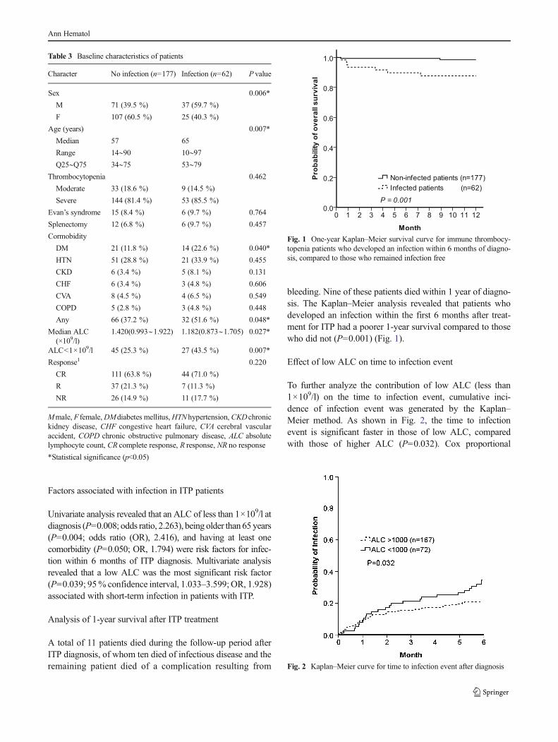
bleeding. Nine of these patients died within 1 year of diagno-sis. The Kaplan–Meier analysis revealed that patients whodeveloped an infection within the first 6 months after treat-ment for ITP had a poorer 1-year survival compared to thosewho did not (P=0.001) (Fig. 1).
Effect of low ALC on time to infection event
To further analyze the contribution of low ALC (less than1×109/l) on the time to infection event, cumulative inci-dence of infection event was generated by the Kaplan–Meier method. As shown in Fig. 2, the time to infectionevent is significant faster in those of low ALC, comparedwith those of higher ALC (P=0.032). Cox proportional
Table 3 Baseline characteristics of patients
Character No infection (n=177) Infection (n=62) P value
Sex 0.006*
M 71 (39.5 %) 37 (59.7 %)
F 107 (60.5 %) 25 (40.3 %)
Age (years) 0.007*
Median 57 65
Range 14∼90 10∼97Q25∼Q75 34∼75 53∼79
Thrombocytopenia 0.462
Moderate 33 (18.6 %) 9 (14.5 %)
Severe 144 (81.4 %) 53 (85.5 %)
Evan’s syndrome 15 (8.4 %) 6 (9.7 %) 0.764
Splenectomy 12 (6.8 %) 6 (9.7 %) 0.457
Cormobidity
DM 21 (11.8 %) 14 (22.6 %) 0.040*
HTN 51 (28.8 %) 21 (33.9 %) 0.455
CKD 6 (3.4 %) 5 (8.1 %) 0.131
CHF 6 (3.4 %) 3 (4.8 %) 0.606
CVA 8 (4.5 %) 4 (6.5 %) 0.549
COPD 5 (2.8 %) 3 (4.8 %) 0.448
Any 66 (37.2 %) 32 (51.6 %) 0.048*
Median ALC(×109/l)
1.420(0.993∼1.922) 1.182(0.873∼1.705) 0.027*
ALC<1×10⁹/l 45 (25.3 %) 27 (43.5 %) 0.007*
Response1 0.220
CR 111 (63.8 %) 44 (71.0 %)
R 37 (21.3 %) 7 (11.3 %)
NR 26 (14.9 %) 11 (17.7 %)
Mmale, F female,DMdiabetes mellitus,HTNhypertension,CKDchronickidney disease, CHF congestive heart failure, CVA cerebral vascularaccident, COPD chronic obstructive pulmonary disease, ALC absolutelymphocyte count, CR complete response, R response, NR no response
*Statistical significance (p<0.05)
Fig. 1 One-year Kaplan–Meier survival curve for immune thrombocy-topenia patients who developed an infection within 6 months of diagno-sis, compared to those who remained infection free
Fig. 2 Kaplan–Meier curve for time to infection event after diagnosis
Ann Hematol

hazards analysis showed that patient with low ALC hadhigher risk of infection as compared with those withhigher ALC (HR 1.723, 1.040∼2.853).
Discussion
ITP has previously been shown to be associated with anincreased risk of infection relative to normal populations [3].Therapy for ITP typically involves the use of steroids and/orother immunosuppressive agents that might contribute to theincreased risk of infection. Interestingly, our study demon-strated that a low ALC may be an important risk factor forinfection in ITP patients. Moreover, the mean ALC value andthe proportion of lymphopenia at time of ITP diagnosis werenot significantly differed from those at time of infectionamong ITP patients with infections suggesting that a lowALC may be a reliable predictive factor for risk of infectionsin ITP patients. Nevertheless, the mechanisms beneath a lowALC and the association with risk of infections remain un-known, and further studies are necessary. It is possible thatALC may be a surrogate marker, reflecting an underlyingimmune deficiency and/or dysfunction. There is growingevidence that T cells play a vital role in the onset of ITP[19–28]. As the immune system undergoes dysregulation,predisposed individuals develop a Th1/Th2 imbalance thatfavors the induction of organ-specific autoimmunity [21]. Areduced number of CD4+ CD25+ T-regulatory cells, whichelicit peripheral tolerance and downregulate T-effector cellresponses, have also been reported in ITP patients [24, 26].Several studies have demonstrated that ALC is a good predic-tor of CD4 count, which in turn is a strong predictor ofopportunistic infection in human immunodeficiency virus-positive patients [29–32]. Furthermore, treatment with steroidshas been shown to induce lymphopenia [33–36], which mayfurther complicate a preexisting low ALC status. In this study,we have identified a new risk factor associated with infectionin patients with ITP, but further studies will be required toconfirm this and to elucidate the underlying mechanisms.
The CR rate after initial steroid treatment for ITP waspreviously reported to be 51.9 % [37], while in our study itwas 65.7 %. However, compared with this previous study, theinfection risk in our study was higher (25.9 %), which wasprobably related to the older median age (61 years) in ourpatient group (a total of 112 patients (46.8 %) were older than65 years in our study). Moreover, old age was a univariate riskfactor associated with infection in our study, although itshowed only a borderline significance inmultivariate analysis.Aging of the immune system, or immunosenescence, mayrender the elderly more susceptible to infection and associatedmortality [38]. Furthermore, immunosenescence usually in-volves thymus involution, which is thought to contribute toautoimmune diseases including ITP [39, 40].
Refractory ITP is known to increase the risk of bleeding[41] and mortality, with a reported mortality rate in refractorydisease of about 15–20 % [12]. Bleeding and infection bothcontribute to the death of patients with ITP [1], although theinfection-attributable mortality in patients with ITP may varyand is not well-defined. Our study demonstrated that patientswho suffered an infection after treatment had a significantlypoorer 1-year survival than those who did not develop aninfection (Fig. 1). This suggests that the dose of steroid orother immunosuppressive agents should be reduced as soon asa treatment response has been achieved in order to avoidadditional infections. Furthermore, if combination chemother-apy is being considered, the short- and long-term risk shouldbe weighed against the potential benefits.
Our study had several limitations. First, the retrospectivedesign and relatively small patient number may have failed toidentify other potentially important risk factors. Second, thesteroid treatment varied according to each individual’s clinicalcondition. It is therefore difficult to be sure of an associationbetween the cumulative steroid dosage and duration andinfection risk, especially in elderly patients. In addition,other viral infections such as cytomegalovirus and Epstein–Barr virus were not routinely screened during the diseasecourse of ITP. Moreover, common viral infections such as“common cold” or flu are generally self-limited and may beresolved before detection, therefore causing difficulty indocumentation.
In conclusion, our results show that ITP patients with aninfection occurring within 6 months of diagnosis have apoorer 1-year survival than patients who do not succumb toinfection. Furthermore, a low ALC may be associated with anincreased risk of infection within this period. In addition tomonitoring for bleeding, individualized management withcautious monitoring and surveillance for the onset of infectionis also needed if better outcomes are to be achieved.
Acknowledgments This study was supported by the Taiwan ClinicalOncology Research Foundation and a grant from Taipei Veterans GeneralHospital V101A-004.
Conflict of interests The authors declare no conflict of interest.
References
1. Portielje JE, Westendorp RG, Kluin-Nelemans HC et al (2001)Morbidity and mortality in adults with idiopathic thrombocytopenicpurpura. Blood 97:2549–2554
2. Mazzucconi MG, Fazi P, Bernasconi S et al (2007) Therapy withhigh-dose dexamethasone (HD-DXM) in previously untreatedpatients affected by idiopathic thrombocytopenic purpura: a GIMEMAexperience. Blood 109:1401–1407
3. NorgaardM, JensenAO, EngebjergMC et al (2011) Long-term clinicaloutcomes of patients with primary chronic immune thrombocytopenia:a Danish population-based cohort study. Blood 117:3514–3520
Ann Hematol
4. Daou S, Federici L, Zimmer J et al (2008) Idiopathic thrombocyto-penic purpura in elderly patients: a study of 47 cases from a singlereference center. Eur J Intern Med 19:447–451
5. Huang YC, Liu CJ, Liu CY et al (2011) Low absolute lymphocytecount and addition of rituximab confer high risk for interstitialpneumonia in patients with diffuse large B-cell lymphoma. AnnHematol 90:1145–1151
6. Siddiqui M, Ristow K, Markovic SN et al (2006) Absolute lympho-cyte count predicts overall survival in follicular lymphomas. Br JHaematol 134:596–601
7. Cox MC, Nofroni I, Ruco L et al (2008) Low absolute lymphocytecount is a poor prognostic factor in diffuse-large-B-cell-lymphoma.Leuk Lymphoma 49:1745–1751
8. George JN, Woolf SH, Raskob GE et al (1996) Idiopathic thrombo-cytopenic purpura: a practice guideline developed by explicitmethods for the American Society of Hematology. Blood 88:3–40
9. Neunert C, LimW, CrowtherM et al (2011) The American Society ofHematology 2011 evidence-based practice guideline for immunethrombocytopenia. Blood 117:4190–4207
10. GodeauB, Chevret S, Varet B et al (2002) Intravenous immunoglobulinor high-dose methylprednisolone, with or without oral prednisone, foradults with untreated severe autoimmune thrombocytopenic purpura: arandomised, multicentre trial. Lancet 359:23–29
11. Cheng Y, Wong RS, Soo YO et al (2003) Initial treatment of immunethrombocytopenic purpura with high-dose dexamethasone. N Engl JMed 349:831–836
12. Figueroa M, Gehlsen J, Hammond D et al (1993) Combinationchemotherapy in refractory immune thrombocytopenic purpura. NEngl J Med 328:1226–1229
13. Boruchov DM, Gururangan S, Driscoll MC et al (2007) Multiagentinduction and maintenance therapy for patients with refractory im-mune thrombocytopenic purpura (ITP). Blood 110:3526–3531
14. Ghanima W, Godeau B, Cines DB et al (2012) How I treat immunethrombocytopenia: the choice between splenectomy or a medicaltherapy as a second-line treatment. Blood 120:960–969
15. Huang JJ, JiangWQ, Lin TYet al (2011) Absolute lymphocyte countis a novel prognostic indicator in extranodal natural killer/T-celllymphoma, nasal type. Ann Oncol 22:149–155
16. HungMH, Yu YB, Hsiao LTet al (2013) Absolute lymphocyte countpredicts response to rituximab-containing salvage treatment forrelapsed/refractory B-cell non-Hodgkin’s lymphoma with prior ritux-imab exposure. J Chin Med Assoc 76:195–200
17. Oki Y, Yamamoto K, Kato H et al (2008) Low absolute lymphocytecount is a poor prognostic marker in patients with diffuse large B-celllymphoma and suggests patients’ survival benefit from rituximab.Eur J Haematol 81:448–453
18. Rodeghiero F, Stasi R, Gernsheimer T et al (2009) Standardization ofterminology, definitions and outcome criteria in immune thrombocy-topenic purpura of adults and children: report from an internationalworking group. Blood 113:2386–2393
19. Panitsas FP, Theodoropoulou M, Kouraklis A et al (2004) Adultchronic idiopathic thrombocytopenic purpura (ITP) is the manifesta-tion of a type-1 polarized immune response. Blood 103:2645–2647
20. Wang T, Zhao H, Ren H et al (2005) Type 1 and type 2 T-cell profilesin idiopathic thrombocytopenic purpura. Haematologica 90:914–923
21. Semple JW, Provan D (2012) The immunopathogenesis of immunethrombocytopenia: T cells still take center-stage. Curr Opin Hematol19:357–362
22. Semple JW (1998) Immunobiology of T helper cells and antigen-presenting cells in autoimmune thrombocytopenic purpura (ITP).Acta Paediatr Suppl 424:41–45
23. Nishimoto T, Satoh T, Takeuchi T et al (2012) Critical role of CD4(+)CD25(+) regulatory T cells in preventing murine autoantibody-mediated thrombocytopenia. Exp Hematol 40:279–289
24. Aboul-Fotoh Lel M, Abdel Raheem MM, El-Deen MA et al (2011)Role of CD4+ CD25+ T cells in children with idiopathic thrombo-cytopenic purpura. J Pediatr Hematol Oncol 33:81–85
25. Johnsen J (2012) Pathogenesis in immune thrombocytopenia: newinsights. Hematology Am Soc Hematol Educ Program 2012:306–312
26. Sakakura M,Wada H, Tawara I et al (2007) Reduced Cd4+ Cd25+ Tcells in patients with idiopathic thrombocytopenic purpura. ThrombRes 120:187–193
27. Semple JW (2002) Immune pathophysiology of autoimmune throm-bocytopenic purpura. Blood Rev 16:9–12
28. Cines DB, Bussel JB, Liebman HA et al (2009) The ITP syndrome:pathogenic and clinical diversity. Blood 113:6511–6521
29. Blatt SP, Lucey CR, Butzin CA et al (1993) Total lymphocyte countas a predictor of absolute CD4+ count and CD4+ percentage in HIV-infected persons. JAMA 269:622–626
30. Shapiro NI, Karras DJ, Leech SH et al (1998) Absolute lymphocytecount as a predictor of CD4 count. Ann Emerg Med 32:323–328
31. Napoli AM, Fischer CM, Pines JM et al (2011) Absolute lymphocytecount in the emergency department predicts a low CD4 count inadmitted HIV-positive patients. Acad Emerg Med 18:385–389
32. Napoli AM, Maughan B, Murray R et al (2013) Use of the relation-ship between absolute lymphocyte count and CD4 count to improveearlier consideration of pneumocystis pneumonia in HIV-positiveemergency department patients with pneumonia. J Emerg Med 44:28–35
33. Meibohm B, Derendorf H, Mollmann H et al (1999) Mechanism-based PK/PD model for the lymphocytopenia induced by endoge-nous and exogenous corticosteroids. Int J Clin Pharmacol Ther 37:367–376
34. Zweiman B, Atkins PC, Bedard PM et al (1984) Corticosteroideffects on circulating lymphocyte subset levels in normal humans. JClin Immunol 4:151–155
35. Ginsburg C, Guillevin L, Sauvaget F et al (1993) Systemic diseasestreated by corticoids, plasma exchange and cyclophosphamide: in-fectious complications and lymphopenia. Ann Med Interne (Paris)144:15–19
36. Craddock CG (1978) Corticosteroid-induced lymphopenia, immuno-suppression, and body defense. Ann Intern Med 88:564–566
37. Pamuk GE, Pamuk ON, Baslar Z et al (2002) Overview of 321patients with idiopathic thrombocytopenic purpura. Retrospectiveanalysis of the clinical features and response to therapy. AnnHematol 81:436–440
38. Boren E, Gershwin ME (2004) Inflamm-aging: autoimmunity, andthe immune-risk phenotype. Autoimmun Rev 3:401–406
39. Prelog M (2006) Aging of the immune system: a risk factor forautoimmunity? Autoimmun Rev 5:136–139
40. Hasler P, Zouali M (2005) Immune receptor signaling, aging, andautoimmunity. Cell Immunol 233:102–108
41. Cohen YC, Djulbegovic B, Shamai-Lubovitz O et al (2000) Thebleeding risk and natural history of idiopathic thrombocytopenicpurpura in patients with persistent low platelet counts. Arch InternMed 160:1630–1638
Ann Hematol