aberrant ventricular conduction types and concealed conduction
DESCRIPTION
Aberrant Ventricular ConductionTRANSCRIPT
ABERRANT VENTRICULAR CONDUCTION TYPES AND CONCEALED CONDUCTION
Others denominations: Aberrancy, ventricular aberration.
Aberrant ventricular conduction definition: It is a term applied to alterations
in QRS contour of supraventricular beats resulting from impulse transmition
during periods of physiologic refractoriness and/or depressed conductivity. The
supraventricular electrical impulse is conducted abnormally through the
ventricular conducting system. This results in a wide QRS complex that may be
confused with a ventricular ectopic beat.
RELATIVE FREQUENCY OF EXPERIMENTAL ABERRATION1
Aberrant ventricular conduction was induced in 44 subjects by introduction of
atrial premature beats through a transvenous catheter-electrode. Multiple
patterns of aberrant ventricular conduction were obtained in 32 patients and, in
the whole group, 116 different configurations were recorded. Of these, 104
showed a classical pattern of mono- or biventricular conduction disturbance.
Right Bundle Branch Block (RBBB) 24%, RBBB combined with Left Anterior
Fascicular Block (LAFB) 18%, LAFB 15%, RBBB combined with Left Posterior
Fascicular Block (LPFB) 10%, LPFB 9%, LBBB 5%, Incomplete LBBB (ILBBB)
6%, trivial changes of the QRS contour 6% and marked anterior displacement
or Prominent Anterior Forces( LSFB) 4%.
Totals: RBBB: 52, LAFB: 33, LPFB: 19, LBBB: 14, trivial modifications of the
QRS contour in 7 of them. In the other 5 instances, aberrant conduction
manifested itself by a conspicuous anterior displacement of the QRS loop
(Prominent Anterior Forces = PAF), with increased duration of anterior forces.
The latter observation is worthy of notice, as it indicates that, in the differential
diagnosis of the VCG pattern characterized by, conduction disturbances should
be considered a possible etiological factor in addition to right ventricular
hypertrophy, and true posterior wall myocardial infarction (or lateral MI in the
new Bayes de Luna nomenclature concept).
1
RBBB aberrancy pattern is observed in 80 % to 85% of cases. In sick
population LBBB aberrancy is near 33% of cases2.
The mechanisms of aberrancy with changing cycle length are3; 4:
1) Premature arrival of the supraventricular impulse before full recovery of
the right bundle branch (RBB);
2) Inadequate or unequal refractoriness of conducting tissue resulting in
local delay or block of dromotropism;
3) Prolongation of Action Potential (AP) secondary to lengthiness of the
preceding cycle duration;
4) Unsuccessful of restitutions of transmembrane electrolyte concentration
during relaxation and dilatation of the ventricles;
5) Failure of the refractory period to shorten in response to acceleration of
the heart rate( HR);
6) A reduced take-off potential secondary to diastole depolarization;
7) Concealed transseptal conduction with delay or block of bundle branch
conduction and
8) Diffuse depression of Intraventricular conduction including that of
specialized as well as contractile myocardial.
The three forms of ventricular aberration
Type A: It is the common form and due to fascicular refractoriness. It is caused
by supraventricular (atrial or junctional) premature beat or accelerated of the
sinus rhythm. The early impulse reach the RBB when still in refractory period
and it has been unable to respond and conduct
Type B: It is due to anomalous supraventricular activation
Type C: It is due to paradoxical critical rate.
In cases of stable wide QRS-complex tachycardia the evaluation of the ECG
without further information in prehospital emergency-medicine leads to
unsatisfactory results. The correct diagnosis in wide QRS-complex tachycardia
can be improved by using additional data but the diagnostic accuracy is still low.
2

Therefore, the differential diagnosis of stable wide QRS-complex tachycardia in
preclinical emergency-medicine cannot be recommended. Until proven
otherwise, any stable wide QRS-complex tachycardia should be managed as if
it were VT5.
I) ABERRANCY SECONDARY TO GOUAUX-ASHMAN PHENOMENON OR ASHMAN PHENOMENON6
Although most Premature Atrial Contractions (PACs) or Premature Junctional
Contractions (PJCs) (premature supraventricular beats) are conducted to the
ventricles normally (i.e., with a narrow QRS complex), this is not always the
case. Instead, PACs or PJCs may sometimes occur so early in the cycle as to
be "blocked" (i.e., non-conducted), because the conduction system is still in an
absolute refractory period (ARP). Other times, premature beats may occur
during the relative refractory period (RRP), in which case aberrant conduction
(with a widened QRS) occurs. Practically speaking, aberrant conduction is most
likely to take the form of some type of bundle branch block (BBB)/fascicular
pattern most commonly RBBB. Attention to QRS morphology may help to
distinguish between aberrancy and ventricular premature contractions( VPCs).
The refractory period in cardiac physiology is related to the ion currents which,
in cardiac cells as in nerve cells, flow into and out of the cell. The flow of ions
translates into a change in the voltage of the inside of the cell relative to the
extracellular space. As in nerve cells, this characteristic change in voltage is
referred to as an AP. Unlike nerve cells, the cardiac AP duration is closer to 100
ms (with variations depending on cell type, autonomic tone, etc.). After an AP
initiates, the cardiac cell is unable to initiate another AP for some duration of
time (which is slightly shorter than the "true" action potential duration). This
period of time is referred to as the refractory period.
Classically, the cardiac refractory period is separated into an ARP and a RRP.
During the ARP, a new AP cannot be elicited. During the RRP, a new AP can
be elicited under the correct circumstances. During RRP a second AP can be
evoked, but only if the stimulus strength is increased.
3
The aberrant conduction depends on the RRP of the conduction tissues. The
refractory period depends on the heart rate (HR). Action potential duration
(APd) (ie, refractory period) changes with the R-R interval of the preceding
cycle; shorter duration of action potential (AP) is associated with a short R-R
interval and prolonged duration of AP is associated with a long R-R interval. A
longer cycle lengthens the ensuing refractory period, and, if a shorter cycle
follows, the beat ending it is likely to be conducted with aberrancy.
Aberrant conduction results when a supraventricular impulse reaches the His-
Purkinje system while one of its branches is still in the RRP or ARP. This results
in slow or blocked conduction through this bundle branch and delayed
depolarization through the ventricular muscles, causing a bundle-branch block
configuration pattern on the surface ECG, in the absence of bundle-branch
pathology. A RBBB pattern is more common than a LBBB pattern because of
the longer refractory period of the RBB.
4
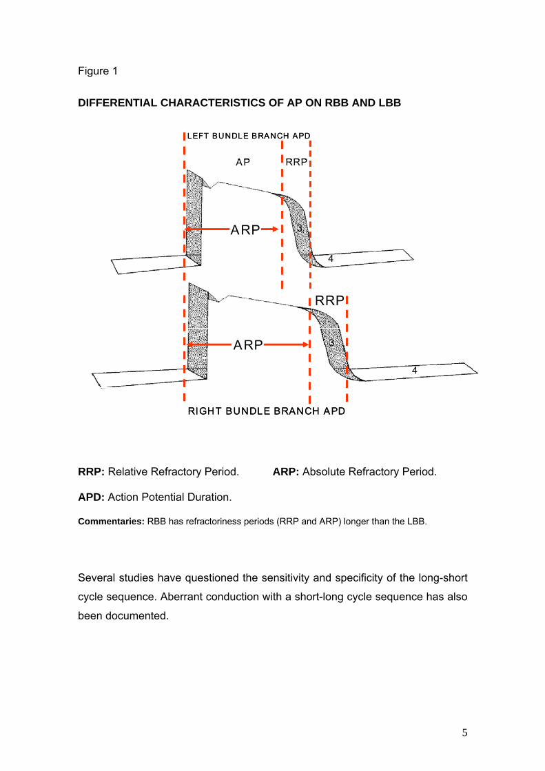
Figure 1
DIFFERENTIAL CHARACTERISTICS OF AP ON RBB AND LBB
PA
PA
ARP
ARP
RRPAP
4
4
3
3
LEFT BUNDLE BRANCH APD
RIGHT BUNDLE BRANCH APD
RRP
PA
PA
ARP
ARP
RRPAP
4
4
3
3
LEFT BUNDLE BRANCH APD
RIGHT BUNDLE BRANCH APD
RRP
RRP: Relative Refractory Period. ARP: Absolute Refractory Period.
APD: Action Potential Duration.
Commentaries: RBB has refractoriness periods (RRP and ARP) longer than the LBB.
Several studies have questioned the sensitivity and specificity of the long-short
cycle sequence. Aberrant conduction with a short-long cycle sequence has also
been documented.
5

Gouaux-Ashman phenomenon or Ashman phenomenon6 is an intraventricular
conduction abnormality restricted to the His-Purkinje system, caused by a
change in the HR. This is dependent on the effects of rate on the
electrophysiological properties of the heart and can be modulated by metabolic
and electrolyte abnormalities and the effects of drugs. Conditions causing an
altered duration of the refractory period of the bundle branch or the ventricular
tissue cause Ashman phenomenon. These conditions are commonly observed
in:
1) Atrial fibrillation( AF): Figure 2;
2) Atrial tachycardia;
3) Premature Atrial Contractions.
Figure 2
LONG CYCLE SHORT CYCLELONG CYCLE SHORT CYCLE
In this case aberration occurs when a short cycle follows a long one. Aberration follows a long-
short sequence during AF. The last 2 complex are aberrant with LBBB pattern: Gouaux-Ashman
phenomenon or Ashman phenomenon.
Ashman phenomenon is an aberrant ventricular conduction due to a change in
QRS cycle length. In 1947, Gouaux and Ashman reported that in AF, when a
relatively long cycle was followed by a relatively short cycle, the beat with a
short cycle often has RBBB morphology. This causes diagnostic confusion with
premature ventricular complexes (PVCs). If a sudden lengthening of the QRS
cycle occurs, the subsequent impulse with a normal or shorter cycle length may
be conducted with aberrancy.
6
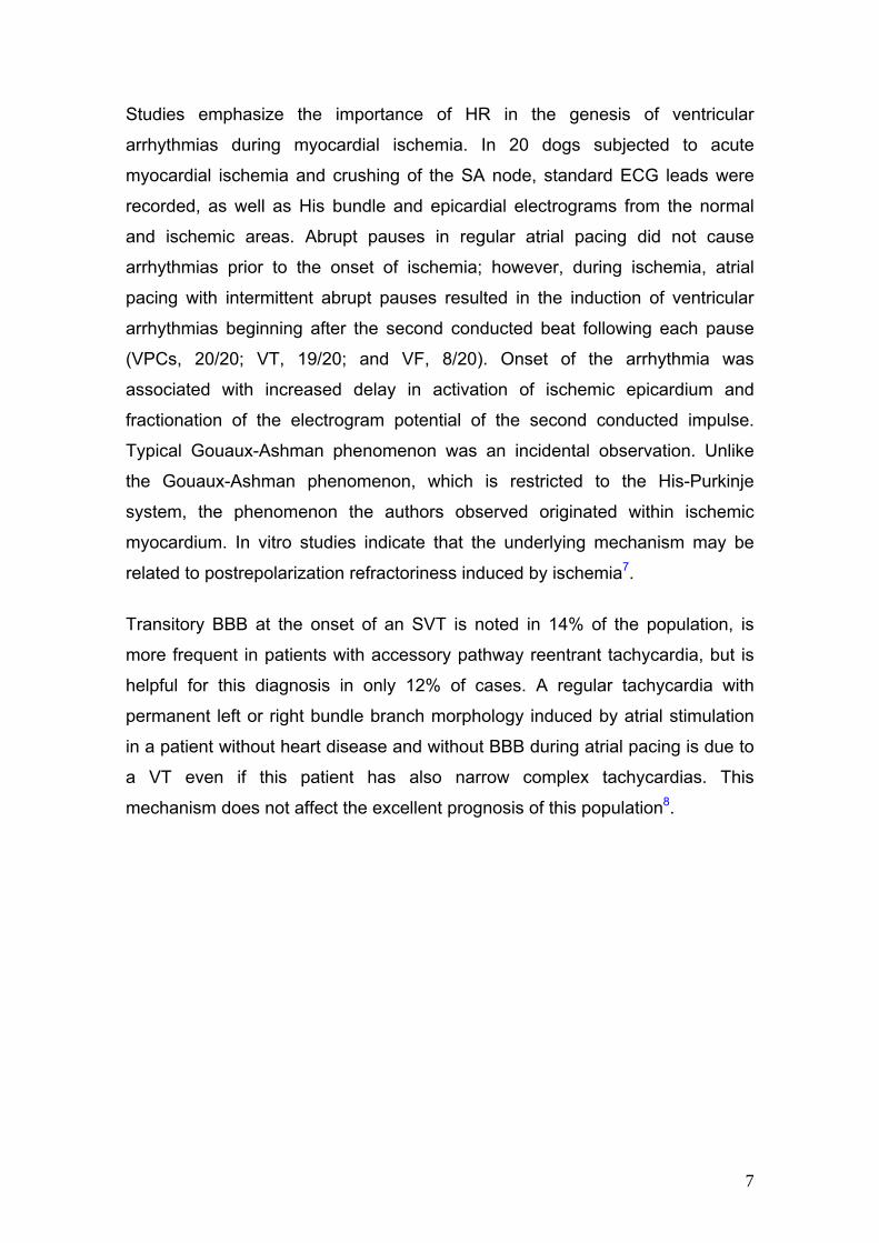
Studies emphasize the importance of HR in the genesis of ventricular
arrhythmias during myocardial ischemia. In 20 dogs subjected to acute
myocardial ischemia and crushing of the SA node, standard ECG leads were
recorded, as well as His bundle and epicardial electrograms from the normal
and ischemic areas. Abrupt pauses in regular atrial pacing did not cause
arrhythmias prior to the onset of ischemia; however, during ischemia, atrial
pacing with intermittent abrupt pauses resulted in the induction of ventricular
arrhythmias beginning after the second conducted beat following each pause
(VPCs, 20/20; VT, 19/20; and VF, 8/20). Onset of the arrhythmia was
associated with increased delay in activation of ischemic epicardium and
fractionation of the electrogram potential of the second conducted impulse.
Typical Gouaux-Ashman phenomenon was an incidental observation. Unlike
the Gouaux-Ashman phenomenon, which is restricted to the His-Purkinje
system, the phenomenon the authors observed originated within ischemic
myocardium. In vitro studies indicate that the underlying mechanism may be
related to postrepolarization refractoriness induced by ischemia7.
Transitory BBB at the onset of an SVT is noted in 14% of the population, is
more frequent in patients with accessory pathway reentrant tachycardia, but is
helpful for this diagnosis in only 12% of cases. A regular tachycardia with
permanent left or right bundle branch morphology induced by atrial stimulation
in a patient without heart disease and without BBB during atrial pacing is due to
a VT even if this patient has also narrow complex tachycardias. This
mechanism does not affect the excellent prognosis of this population8.
7
Differential diagnosis
1) Atrial fibrillation and premature ventricular contractions
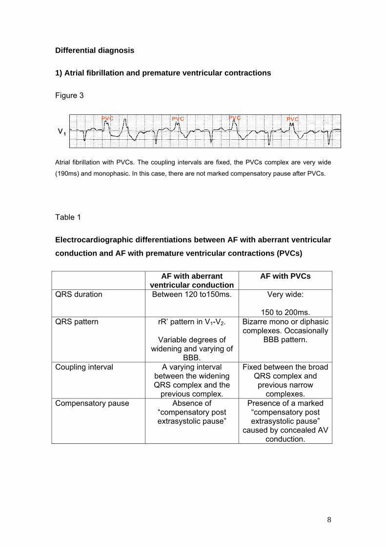
Figure 3
V1
PVCPVC PVC PVC
V1
PVCPVC PVC PVC
Atrial fibrillation with PVCs. The coupling intervals are fixed, the PVCs complex are very wide
(190ms) and monophasic. In this case, there are not marked compensatory pause after PVCs.
Table 1
Electrocardiographic differentiations between AF with aberrant ventricular conduction and AF with premature ventricular contractions (PVCs)
AF with aberrant ventricular conduction
AF with PVCs
QRS duration Between 120 to150ms. Very wide:
150 to 200ms. QRS pattern rR’ pattern in V1-V2.
Variable degrees of widening and varying of
BBB.
Bizarre mono or diphasic complexes. Occasionally
BBB pattern.
Coupling interval A varying interval between the widening QRS complex and the
previous complex.
Fixed between the broad QRS complex and previous narrow
complexes. Compensatory pause Absence of
“compensatory post extrasystolic pause”
Presence of a marked “compensatory post extrasystolic pause”
caused by concealed AV conduction.
8
II) ACCELERATION-DEPENDENT ABERRANCY, TACHYCARDIA-DEPENDENT, IN PHASE 3 ABERRANCY, OR PHASE 3 ABERRATION
Resulting from the occurrence of impaired intraventricular conduction as the
heart attains a specific critical rate. At a critical HRs, impaired ventricular
conduction results in aberrancy. The appearance and disappearance often
depends on very small changes in cycle length. Aberrancy often appears at
relatively slow rates, frequently below 75 beats/min.
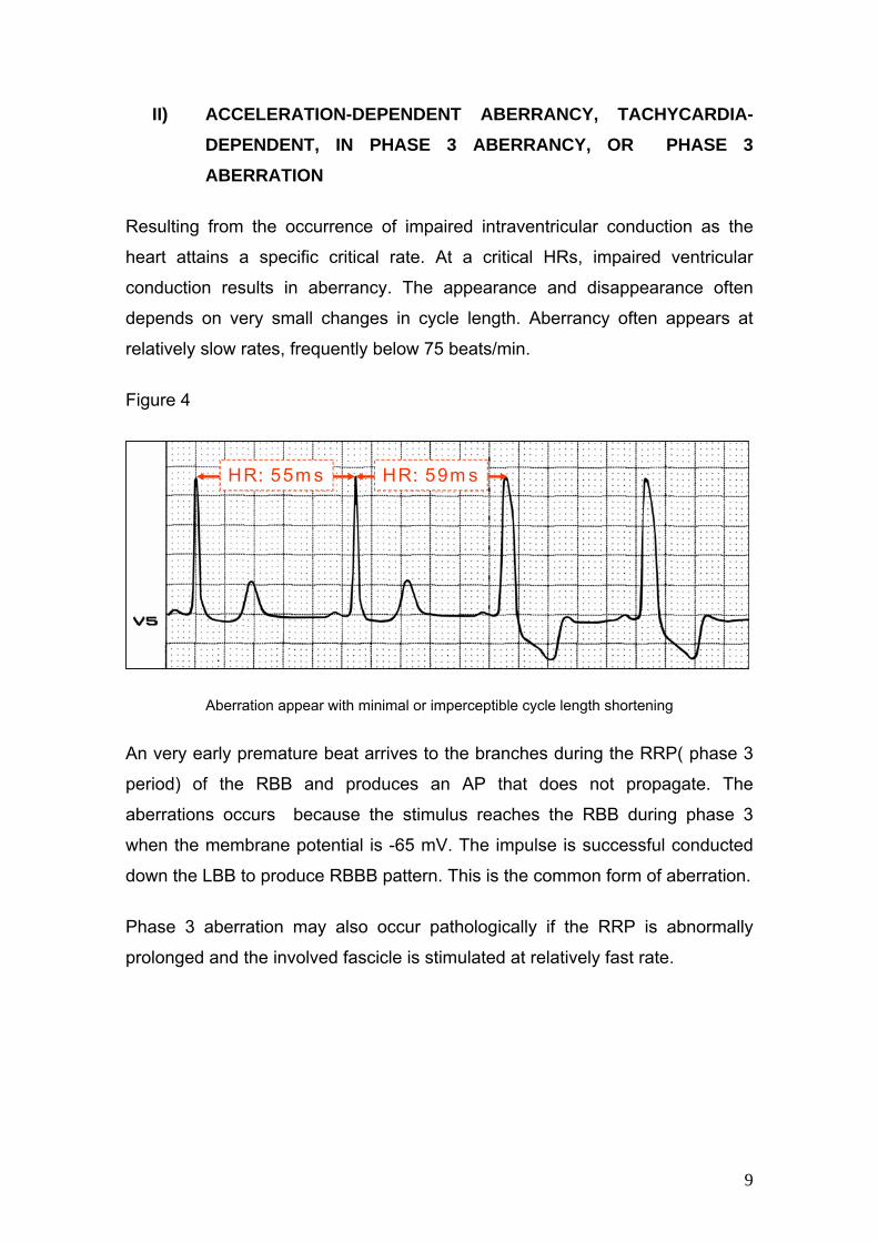
Figure 4
HR: 55m s HR: 59m sHR: 55m s HR: 59m s
Aberration appear with minimal or imperceptible cycle length shortening
An very early premature beat arrives to the branches during the RRP( phase 3
period) of the RBB and produces an AP that does not propagate. The
aberrations occurs because the stimulus reaches the RBB during phase 3
when the membrane potential is -65 mV. The impulse is successful conducted
down the LBB to produce RBBB pattern. This is the common form of aberration.
Phase 3 aberration may also occur pathologically if the RRP is abnormally
prolonged and the involved fascicle is stimulated at relatively fast rate.
9
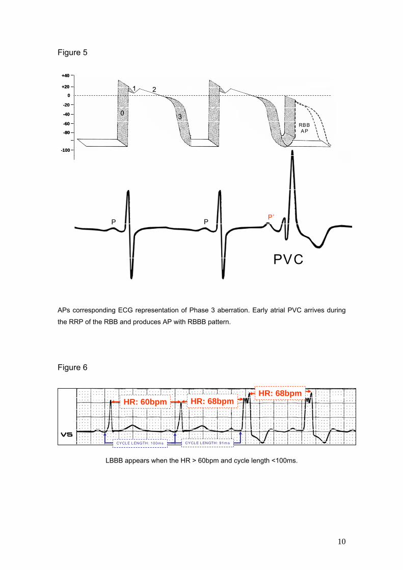
Figure 5
+40
+20
0
-20
-40
-60
-80
-100
0
1 2
3
PP’
P
RBBAP
PVC
+40
+20
0
-20
-40
-60
-80
-100
0
1 2
3
PP’
P
RBBAP
PVC
APs corresponding ECG representation of Phase 3 aberration. Early atrial PVC arrives during
the RRP of the RBB and produces AP with RBBB pattern.
Figure 6
HR: 60bpm HR: 68bpmHR: 68bpm
CYCLE LENGTH: 100m s CYCLE LENGTH: 91m s
HR: 60bpm HR: 68bpmHR: 68bpm
CYCLE LENGTH: 100m s CYCLE LENGTH: 91m s
LBBB appears when the HR > 60bpm and cycle length <100ms.
10

Patients who present with atypical chest pain in whom rate-dependent LBBB
develops on the treadmill are significantly less likely to have coronary artery
disease than patients who present with classic angina. The onset of LBBB at a
HR of ≥125 beats/min is highly correlated with the presence of normal coronary
arteries, regardless of patient presentation. Patients with angina in whom both
chest pain and LBBB develop during exercise may have normal coronary
arteries9.
The LBBB itself can produce T-wave inversions in the right precordial leads
during the normal conduction phase which may simulate an AMI. Persistent
deep T wave inversions are seem after return of normal depolarization. The
phenomenon is named cardiac memory-persistent T wave changes10;11;12
Figure 7
Name: VGA. Sex: Male. Age: 54 y. Race: Caucasian. Weight: 70 Kg Height: 1.75 m Date: 02/05/2006.
Clinical diagnosis: Coronary insufficiency. Acute myocardial infarction.
ECG diagnosis: Intermittent LBBB independent from heart rate with bigeminy sequential beat-
to-beat: normal conduction and LBBB alternatively. During the normal conduction, ischemic
changes masked by the LBBB are seen. Elevated value of biochemical markers was observed.
11
When the LBBB is intermittent it might be possible to diagnose the AMI during
those periods when the conduction is normal. Patients with clinical picture of
AMI associated with high HR and LBBB pattern, on the assumption that the
block might be rate-dependent, carotid massage with secondary diminution of
HR, the LBBB eventually disappear and during the normal conduction ischemic
changes masked by the LBBB are clearly seen13.
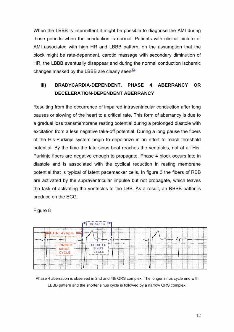
III) BRADYCARDIA-DEPENDENT, PHASE 4 ABERRANCY OR DECELERATION-DEPENDENT ABERRANCY
Resulting from the occurrence of impaired intraventricular conduction after long
pauses or slowing of the heart to a critical rate. This form of aberrancy is due to
a gradual loss transmembrane resting potential during a prolonged diastole with
excitation from a less negative take-off potential. During a long pause the fibers
of the His-Purkinje system begin to depolarize in an effort to reach threshold
potential. By the time the late sinus beat reaches the ventricles, not at all His-
Purkinje fibers are negative enough to propagate. Phase 4 block occurs late in
diastole and is associated with the cyclical reduction in resting membrane
potential that is typical of latent pacemacker cells. In figure 3 the fibers of RBB
are activated by the supraventricular impulse but not propagate, which leaves
the task of activating the ventricles to the LBB. As a result, an RBBB patter is
produce on the ECG.
Figure 8
HR: 42bpm
LONGERSINUS CYCLE
HR: 54bpm
SHORTERSINUS CYCLE
HR: 42bpm
LONGERSINUS CYCLE
HR: 54bpm
SHORTERSINUS CYCLE
Phase 4 aberration is observed in 2nd and 4th QRS complex. The longer sinus cycle end with
LBBB pattern and the shorter sinus cycle is followed by a narrow QRS complex.
12

Phase 4 aberration is rare and associated with organic heart disease. Phase 4
aberrancy needs one or more of following situations:
1) The presence of slow diastolic depolarization which need not be
enhanced;
2) A shift in threshold potential toward zero.
3) A deterioration in membrane resposiveness so that significant conduction
impairment develops at -75mV instead of -65mV;
4) Hypopolarization ( the lost of maximum diastolic potential)
RBBB block occurring on alternate beats during regular sinus rhythm, can
disappeared during hyperventilation induced increase in HR, and reappeared
with slight slowing of the sinus rate due to carotid sinus massage an be caused
by bradycardia-dependent RBBB14.
IV) CRITICAL RATE BUNDLE BRACH BLOCK
This situation is defined as the rate at which BBB develops during acceleration
or disappears during slowing. At the fast rate the refractory period shortens;
normal conduction tends to be preserved because of this response.
V) CONCEALED INTRAVENTRICULAR CONDUCTION
Concealed Intraventricular conduction is defined as the manifestations of
concealed conduction into the bundle branch system or the effect of a non-
propagated impulse conduction of a subsequently propagated impulse.
Conduction of an impulse through a part of the heart without directs evidence of
its presence in the ECG; conduction is inferred only because of its influence on
the subsequent cardiac cycle.
Concealed conduction in the human heart usually occurs in the AV node or
Hiss-Purkinje system or both. Tissue stimulation without direct effect (such as
causing contraction in another chamber), but little is know about the underlying
mechanisms.
13

In AV node concealed conduction acts as a resetting mechanism of the
excitability cycle in the slow and fast pathways similar to that expected form a
conducted beat15.
A common example would be interpolated PVC during normal sinus rhythm; the
PVC does not cause an atrial contraction, because the retrograde impulse form
the PVC does not completely penetrate the AV node. However this AV node
stimulation ( which is not visible no ECG by itself, hence “concealed”) can cause
a delay in subsequent AV conduction by modifying the AV node´s subsequent
conduction characteristic. Hence, the PR interval after the PVC is longer than
the baseline PR interval16.
Anterograde concealed conduction into the concealed accessory AV pathway
has been postulated to be one of the factors preventing the reciprocicating
process via the accessory pathway in patients with the concealed Wolff-
Parkinson White syndrome17. In these cases, the parallel accessory tract is only
capable of conducting the stimulus in a retrograde fashion; i.e. ventriculo-atrial,
which overshadows the presence of pre-excitation, given that ventricular
activation is processed by the normal Nodo-Hisian normal pathway, originating
a normal-duration PR interval. It is important to know the WPW syndrome,
because it may predispose the appearance of Supraventricular Paroxysmal
Tachycardia runs of the orthodromic macro-reentry type, which use the Node-
Hisian system in anterograde fashion and the parallel pathway in retrograde
fashion (narrow QRS complexes).
Finally, another variation on concealed conduction concept is seen in atrial
flutter. As a result of the rapid atrial rate, some of the atrial activity fails to get
through the AV node in an anterograde direction but can alter the rate at which
a subsequent atrial impulse is conduced. In this circumstance, an alteration on
the “F” wave to QRS relationship is seen.
14
The following are the possible mechanisms 18:
1) Trans-septal retrograde concealed intraventricular conduction
responsible for:
(1a) Perpetuation of functional BBB initiated by a premature supraventricular
impulse;
(1b) Alternation of aberrant ventricular conduction in supraventricular bigeminy;
(1c) Normalization of intraventricular conduction with acceleration or rate in
bradycardia-dependent BBB, and
(1d) Prevention of the manifestation of Wenckebach periods of conduction in a
bundle branch or fascicle.
2) Anterograde concealed intraventricular conduction responsible for
(2a) Prevention of expected aberrant ventricular conduction when a short cycle
follows a long one, and;
(2b) Exceptions to the "rule of bigeminy".
3. Retrograde concealed intraventricular conduction of a ventricular
escape in association with unidirectional bundle branch or fasciular block
responsible for:
(3a) Resumption of AV conduction in "paroxysmal AV block" with BBB;
(3b) Facilitation (due to supernormality) of conduction in type II AV block due to
bilateral BBB.
4. Concealed intraventricular conduction of a premature ventricular impulse
responsible for
(4a) Initiation or termination of a re-entrant ventricular tachycardia;
(4b) Resetting of an idioventricular pacemaker, and
15
(4c) Pseudo-intraventricular or pseudo-AV block.
VI) ABERRANCY SECONDARY TO DRUGS AND METABOLIC OR ELECTROLYTE DISORDERS
Aberrant conduction is common during infusion of the I(kr)-blocker almokalant
(Class III drugs) during AF, and seems to be more frequent in females and in
patients with more advanced myocardial disease19.
In severe hyperpotasemia (serum potassium between 8 to 9 mEq/l) is frequent
observe progressive rhythm and conduction disturbances such as bradycardia,
spiked and narrow T waves, widening QRS complex, widening and flattening P
wave, disappearance of the P wave, and cardiac arrest. Diffuse QRS
complexes widening, similar to left or RBBB, associated to anterior or posterior
fascicular block by extreme shift of SAQRS in the FP to left or right is frequently
observed. This QRS complex widening is differentiated of genuine branch
blocks, because in them, the delay is final or middle, while in hyperpotasemia is
always global or diffuse20.
16
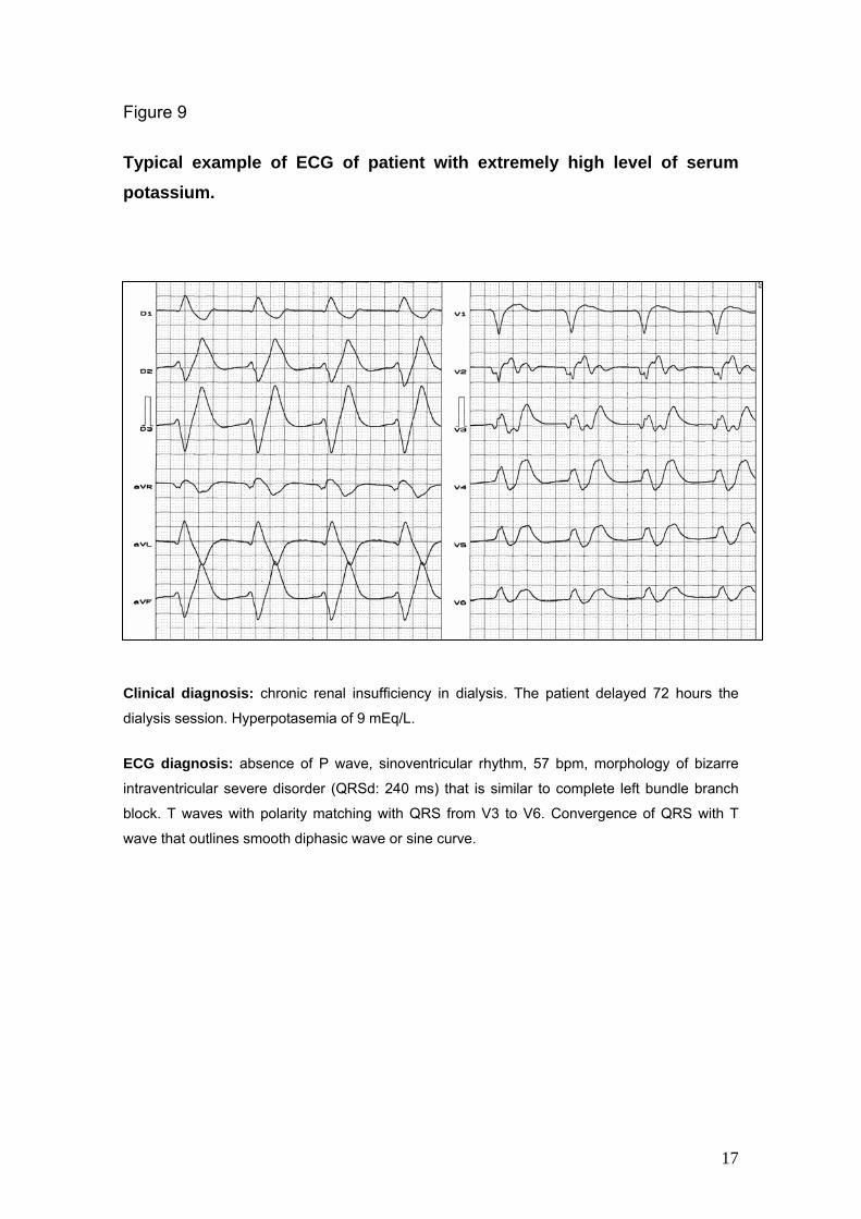
Figure 9
Typical example of ECG of patient with extremely high level of serum potassium.
Clinical diagnosis: chronic renal insufficiency in dialysis. The patient delayed 72 hours the
dialysis session. Hyperpotasemia of 9 mEq/L.
ECG diagnosis: absence of P wave, sinoventricular rhythm, 57 bpm, morphology of bizarre
intraventricular severe disorder (QRSd: 240 ms) that is similar to complete left bundle branch
block. T waves with polarity matching with QRS from V3 to V6. Convergence of QRS with T
wave that outlines smooth diphasic wave or sine curve.
17
VIII) POSTPAUSAL ABERRANCY OR POSTEXTRASYSTOLIC ABERRATION
This variant is caused probably to slow diastolic depolarization, unequal
recovery of conducting or myocardial tissue, or increased diastolic volume.
VENTRICULAR TACHYCARDIA VERSUS SUPRAVENTRICULAR TACHYCARDIA WITH ABERRATION: WIDE-COMPLEX OR BROAD QRS
TAQUICARDIAS
Wide-complex tachycardia (WCT) or broad QRS complex tachycardia is defined
as a rhythm disturbance with a rate greater than 100 beats/min and a QRS
complex duration of 120 ms or more in the adult patient in the pediatric patient,
both rate and QRS complex width are age related and caused by various
mechanisms, either supraventricular with aberrant intraventricular conduction or
ventricular21. It is important to differentiate between ventricular (VT) and
supraventricular (SVT) because it will determine treatment and prognosis of
patients. Unconscious patients with wide complex tachycardia should be treated
in a standard cardiac arrest approach. VT is frequently mistaken for
supraventricular tachycardia with aberration (SVT). Ectopy is much more
common than aberration.
Although up to 10% of cases will defy differentiation, VT and SVT with aberrant
conduction can be distinguished utilizing history, physical examination, and
ECG criteria.
The positive features in favor of SVT with aberration are:
1) Signs of AV dissociation are infrequent in SVT. Infrequently SVT may
also have AV dissociation when retrograde conduction occurs from the
junctional focus. The signs of AV dissociation are: irregular cannon
waves in the jugular pulse, varying intensity of the first heart sound, and
best-to beat changes in systolic blood pressure.
2) Triphasic pattern rsR´ in V1. ( monophophasic or diphasic QRS complex
in V1 are suggestive of VT);
18
3) qRs pattern in V6;
4) Small, narrow r wave in V1 –V2 during LBBB aberration, if an r wave is
present;
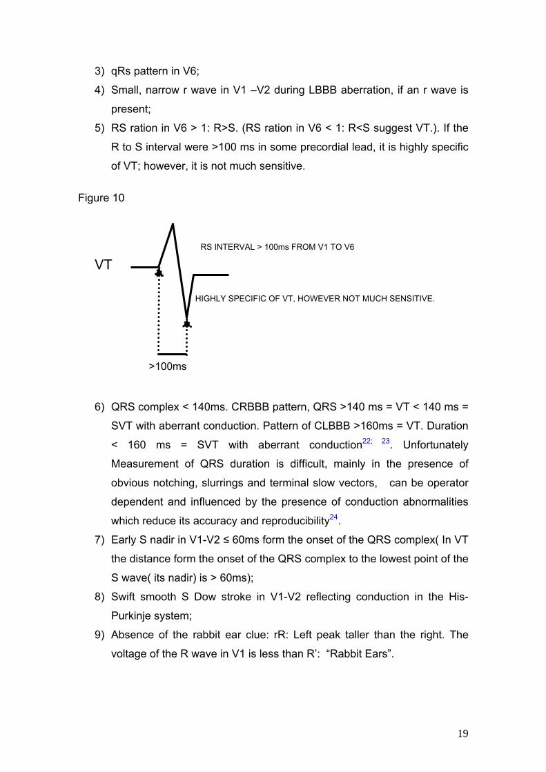
5) RS ration in V6 > 1: R>S. (RS ration in V6 < 1: R<S suggest VT.). If the
R to S interval were >100 ms in some precordial lead, it is highly specific
of VT; however, it is not much sensitive.
Figure 10
RS INTERVAL > 100ms FROM V1 TO V6
6) QRS complex < 140ms. CRBBB pattern, QRS >140 ms = VT < 140 ms =
SVT with aberrant conduction. Pattern of CLBBB >160ms = VT. Duration
< 160 ms = SVT with aberrant conduction22; 23. Unfortunately
Measurement of QRS duration is difficult, mainly in the presence of
obvious notching, slurrings and terminal slow vectors, can be operator
dependent and influenced by the presence of conduction abnormalities
which reduce its accuracy and reproducibility24.
7) Early S nadir in V1-V2 ≤ 60ms form the onset of the QRS complex( In VT
the distance form the onset of the QRS complex to the lowest point of the
S wave( its nadir) is > 60ms);
8) Swift smooth S Dow stroke in V1-V2 reflecting conduction in the His-
Purkinje system;
9) Absence of the rabbit ear clue: rR: Left peak taller than the right. The
voltage of the R wave in V1 is less than R’: “Rabbit Ears”.
VT
HIGHLY SPECIFIC OF VT, HOWEVER NOT MUCH SENSITIVE.
>100ms
19
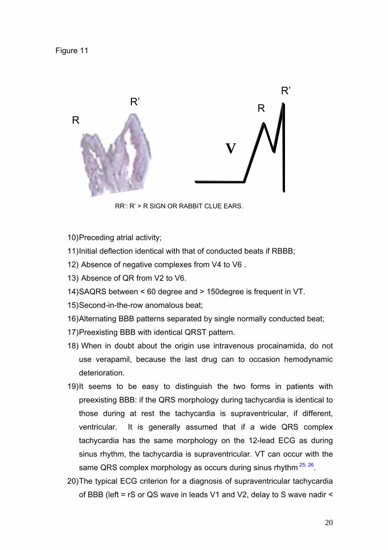
Figure 11
R’ R’
10) Preceding atrial activity;
11) Initial deflection identical with that of conducted beats if RBBB;
12) Absence of negative complexes from V4 to V6 .
13) Absence of QR from V2 to V6.
14) SAQRS between < 60 degree and > 150degree is frequent in VT.
15) Second-in-the-row anomalous beat;
16) Alternating BBB patterns separated by single normally conducted beat;
17) Preexisting BBB with identical QRST pattern.
18) When in doubt about the origin use intravenous procainamida, do not
use verapamil, because the last drug can to occasion hemodynamic
deterioration.
19) It seems to be easy to distinguish the two forms in patients with
preexisting BBB: if the QRS morphology during tachycardia is identical to
those during at rest the tachycardia is supraventricular, if different,
ventricular. It is generally assumed that if a wide QRS complex
tachycardia has the same morphology on the 12-lead ECG as during
sinus rhythm, the tachycardia is supraventricular. VT can occur with the
same QRS complex morphology as occurs during sinus rhythm 25; 26.
20) The typical ECG criterion for a diagnosis of supraventricular tachycardia
of BBB (left = rS or QS wave in leads V1 and V2, delay to S wave nadir <
V
RR
RR’: R’ > R SIGN OR RABBIT CLUE EARS.
20
70 ms, and R wave and no Q wave in lead V6; right = rSR' wave in lead
V1 and an RS wave in lead V6, with R wave height greater than S wave
depth) These criteria, which require only knowledge of typical bundle
branch block patterns, were highly sensitive for the important diagnosis
of VT27.
Observation: Fascicular VT Left ventricular idiopathic VT and bundle branch
reentry VT may also have the triphasic pattern in V1 and relatively narrow QRS
complex.
There are several causes of broad-complex tachycardia, even in patients with
previous myocardial infarction, and, where doubt exists, electrophysiologic
studies should be performed. Antidromic atrioventricular reentrant tachycardia
can to mimic a VT28.
Atriofascicular pathway-mediated antidromic tachycardia using atriofascicular
pathway as the antegrade limb and the atrioventricular nodal pathway as
retrograde limb should be considered in differential diagnosis of all cases of
wide complex tachycardia with LBB morphology and left axis29. The most
common type of accessory pathway causing a wide complex tachycardia is the
atrioventricular bypass tract. Distinguishing the accessory pathway-mediated
tachycardia from VT or SVT with aberrancy is often difficult, but has important
clinical consequences. Despite sophisticated atrial and ventricular pacing
techniques used during electrophysiologic study, the exact mechanism of some
wide-complex tachycardia remain with inconclusive results from diagnostic. In
this situation, eliminating conduction from specific tissue and observing the
effect on the tachycardia can be diagnostic. Such a maneuver can be
performed using the technique of ice mapping, which entails reversible cooling
of tissue to test function prior to delivery of a permanent ablation lesion30.
If the ECG reveal wide QRS tachycardia with a narrow complex beat during a
wide complex tachycardia suggests a capture or fusion beat in the setting of VT.
However, there are situations where SVT can also manifest this way31.
Patients who are misdiagnosed with VT because of ECG artifact may be
subjected to unnecessary procedures.
Physicians (n = 766) were surveyed with a case simulation that included a two-
lead electrocardiographic monitor tracing of artifact simulating a wide-complex
tachycardia. The rhythm strip was not recognized as artifact by 52 of the 55
21
internists (94%), 128 of the 221 cardiologists (58%), and 186 of the 490
electrophysiologists (38%). 156 of the 181 electrophysiologists (88%), 67 of the
126 cardiologists (53%), and 14 of the 15 internists (31%) who misdiagnosed
the rhythm as VT recommended an invasive procedure for further evaluation or
therapy. ECG artifact that mimics VT may frequently result in patients being
subjected to unnecessary invasive cardiac procedures. Physicians should
include artifact in their differential diagnosis of wide complex tachycardias to
minimize unneeded procedures32.
References
1) Kulbertus HE, de Lava-Rutten F, Casters P; Vectorcardiographic study of
aberrant conduction anterior displacement of QRS: another form of
intraventricular block. Br Heart J. 1976; 38: 549-557.
2) Sandler LA, Marriott HJ .The Differential Morphology of Anomalous
Ventricular Complexes of RBBB-Type in Lead V; Ventricular Ectopy
Versus Aberration.Circulation. 1965; 31:551-556.
3) Fisch C. Aberration: seventy five years after Sir Thomas Lewis.
Br Heart J. 1983; 50:297-302.
4) Fisch C. Knoebel SB. Vagaries of acceleration dependent aberration.
Br Heart J. 1992; 67: 16-24.
5) Ohlow MA, Beierlein A, Müller S, von Korn H, Geller JC, Yu J, Lauer B.
Stable tachycardia with wide QRS complex in pre-hospital emergency
medicine. Dtsch Med Wochenschr. 2005;130:2694-2698.
6) Gouaux JL, Ashman R. Auricular fibrillation with aberration simulating
ventricular paroxysmal tachycardia. Am Heart J 1947; 34:366.
7) Hope RR, Lazzara R, Scherlag, BJ.The induction of ventricular
arrhythmias in acute myocardial ischemia by atrial pacing with long-short
cycle sequences. Chest. 1977; 71: 651-658.
8) Brembilla-Perrot B, Beurrier D, Houriez P, Claudon O, Rizk J, Lemoine
C, Gregoire P, Nippert M.Transitory or permanent regular wide QRS
complex tachycardia induced by atrial stimulation in patients without
22
apparent heart disease. Significance. Ann Cardiol Angeiol (Paris). 2003
Aug;52(4):226-231.
9) Vasey C, O'Donnell J, Morris S, McHenry P.Exercise-induced left bundle
branch block and its relation to coronary artery disease. Am J Cardiol.
1985; 56: 892-895.
10) Gould L, Reddy CV, Singh B, Zen B. T-wave changes with intermittent
left bundle branch block. Angiology. 1980;31:66-68.
11) Rosenbaum MB, Blanco HH, Elizari MV, Lázzari JO, Davidenko JM.
Electrotonic modulation of the T wave and cardiac memory. Am J
Cardiol. 1982 Aug;50(2):213-22. .
12) Kolb JC. Cardiac memory-persistent T wave changes after ventricular
pacing. J Emerg Med. 2002; 23:191-197.
13) Almong C, Gabizon D, Bezeishli I. Carotid massage as a means of ECG
diagnosis of acute myocardial infarction in the presence of the Left
bundle branch block. Chest 1975; 67:249-250.
14) Carbone V, Tedesco MA.Bundle branch block on alternate beats: by
what mechanism? J Electrocardiol. 2002; 35: 147-152.
15) Xu B, Billete J, Lavallée M. Concealed conduction in nodal dual
pathways: depressed conduction, prolonged refractoriness, or reset
excitability cycle ? Heart Rhythm. 2006:3: 212-221.
16) Udyavar AR, Pandurangui UM. Blocked or delayed atrioventricular nodal
conduction due to concealed conduction due to interpolated ventricular
ectopics.J Postgrad Med 2007; 53: 148-149.
17) Suzuki F, Kawara T, Tanaka K, Harada TO, Endoh T, Kanazawa Y,
Okishige K, Hirao K, Hiejima K. Electrophisyological demonstration of
anterograde concealed conductionin accessory atriventricular pathways
capable only of retrograde conduction. Pacinng Clin Electrophysiol.
1989; 12: 591-603.
18) Langendorf R, Pick A.Concealed intraventricular conduction in the
human heart. Adv Cardiol. 1975; 14:40-50.
19) Houltz B, Darpö B, Crijns HJ, Swedberg K, Blomström P, Jensen SM,
Svernhage E, Edvardsson N. QRS aberration during atrial fibrillation at
rest and during exercise. Effect of a selective potassium channel
blocking agent. J Electrocardiol. 2002; 35:201-212.
23

20) Ochoa-Gomez J, Villar-Arias A, Aresti I, Marco-Aguilar P. A case of
severe hyperkalaemia and compartment syndrome due to
rhabdomyolysis after drugs abuse. Resuscitation. 2002; 54: 103-105.
21) Hollowell H, Mattu A, Perron AD, Holstege C, Brady WJ. Wide-complex
tachycardia: beyond the traditional differential diagnosis of ventricular
tachycardia vs supraventricular tachycardia with aberrant conduction. Am
J Emerg Med. 2005; 23: 876-889.
22) Wellens HJJ, Bar FWHM, Brugada P; Ventricular tachycardia; the
clinical problem. In Josephson ME, editor: Ventricular tachycardia:
mechanisms and management, Mt Kisco, NY, 1982, Futura.
23) Wellens HJ. Electrophysiology: Ventricular tachycardia: diagnosis of
broad QRS complex tachycardia. Heart 2001; 86: 579-585.
24) Sarubbi B, Li W, Somerville J.QRS width in right bundle branch block.
Accuracy and reproducibility of manual measurement. Int J Cardiol.
2000; 75: 71-74.
25) Olshansky B.Ventricular tachycardia masquerading as supraventricular
tachycardia: a wolf in sheep's clothing. J Electrocardiol. 1988;21:377-
384.
26) Tomcsányi J, Somlói M, Tenczer J, Karlócai K.Ventricular tachycardia
masquerading as supraventricular tachycardia. Orv Hetil. 1998; 139:
2779-2781.
27) Griffith MJ, Garratt CJ, Mounsey P, Camm AJ.Ventricular tachycardia as
default diagnosis in broad complex tachycardia. Lancet. 1994; 343: 386-
388.
28) Dagres N, Clague JR, Kottkamp H, Breithardt G, Borggrefe M.
Antidromic atrioventricular reentrant tachycardia mimicking ventricular
tachycardia in the setting of previous myocardial infarction. Clin Cardiol.
2000; 23: 63-65.
29) Latent atriofascicular pathway participating in a wide complex
tachycardia: differentiation from ventricular tachycardia. Pacing Clin
Electrophysiol. 2006; 29:1434-1437.
30) Gula LJ, Skanes A, Krahn AD, Klein GJ. Novel approach to diagnosis of
a wide-complex tachycardia. J Cardiovasc Electrophysiol. 2004; 15: 466-
469.
24

31) Rosman J, Tawil J, Hanon S, Schweitzer P. Wide QRS tachycardia: what
is the rhythm? Ann Noninvasive Electrocardiol. 2006; 11: 354-356.
32) Knight BP, Pelosi F, Michaud GF, Strickberger SA, Morady F.Physician
interpretation of electrocardiographic artifact that mimics ventricular
tachycardia.
25