aberrant methylation of c-myc and c-fos protooncogenes and ... · suppressor genes has been found...
TRANSCRIPT

Received 01-06-2003; Accepted 13-06-2003
Author and address for correspondence:
Panagiota Matsouka, MDUniversity Hospital of PatrasDepartment of Internal MedicineHematology Division265 00 Rio, PatrasGreeceTel: +30 2610 999255, +30 2610 999495Fax:+30 2610 993950E-mail: [email protected]
Journal of BUON 8: 341-350, 2003© 2003 Zerbinis Medical Publications. Printed in Greece
ORIGINAL ARTICLE
Aberrant methylation of c-myc and c-fos protooncogenes and p53 tumor suppres-sor gene in myelodysplastic syndromes and acute non-lymphocytic leukemia
P.C. Papaggeli1, A.C. Kortsaris2, P.T. Matsouka3
1University of Thessaly, Department of Medicine, Larissa; 2University of Thrace, Department of Medicine, Laboratory of Biochemistry,Alexandroupolis; 3University Hospital of Patras, Hematology Division, Department of Internal Medicine, Rio, Patras, Greece
Summary
Purpose: Aberrant methylation, as an epigenetic phe-nomenon, may precede and regulate the expression of genesinvolved in transformation mechanisms that lead to leuke-mogenesis of hemopoietic cells. The genes involved mostlyencode transcription factors and cell cycle specific inhibi-tors. The aim of this project was to study the DNA methyla-tion pattern of c-myc, c-fos and p53 in myelodysplastic syn-dromes (MDS) and in acute non-lymphocytic leukemias(ANLL).
Patients and methods: DNA was isolated from themonocyte cell layer harvested from bone marrow or periph-eral blood samples of 44 patients suffering from MDS andANLL. Genomic DNA was digested with methylation-specif-ic enzymes, and was electrophoresed and hybridized withprobes specific for human c-myc, c-fos and p53 genes.
Results: In MDS, the c-myc gene in exons 2 and 3 wasregionally hypomethylated, whereas exon 2 in ANLL was
hypermethylated and exon 3 hypomethylated. The c-fos genewas hypomethylated in ANLL type 4 and presented aber-rant hypomethylation in the different types of MDS. The p53anti-oncogene appeared extensively hypomethylated inMDS.
Conclusion: Aberrant DNA methylation pattern of thec-myc, c-fos and p53 tumor suppressor gene seems to be aprimary event in the transformation process from myelodys-plasia to acute leukemia, affecting their expression, and,consequently, altering the proliferation, differentiation orapoptosis of hemopoietic precursor cells. The p53 hypome-thylation predisposes to critical mutations that enhance thetransformation process of myelodysplasia to leukemia. Therecognition of altered methylation of these genes in myelo-dysplasia may have prognostic implications and may leadto novel therapeutic modalities.
Key words: acute nonlymphocytic leukemia, leukemogen-esis, methylation, myelodysplasia, p53, protooncogenes
Introduction
Methylation is an important key-regulator mech-anism of gene expression in mammals. Sites of meth-ylation in the genome are the CpG-islands, locatedmainly in the promoter region of the genes [1].
DNA methylation influences the transcriptionactivity in 3 ways. Firstly, DNA methylation interfereswith the binding of specific transcription factors (e.g.c-Myc, AP1) to their respective DNA binding sites,which are located at various promoter sites. Second-ly, methylation can directly induce gene silencing bysuspending the binding of specific transcriptional in-hibitors to methylated DNA sites. The formation ofinactive chromatin structure is the third mechanismthrough which DNA methylation represses gene ex-pression [2,3]. Hypomethylation enhances the tran-scription of genes. Hypomethylation has been char-acterized as an epigenetic mechanism, which con-tributes to tumorigenesis through the mutated or over-expressed genes, due to the increased incidence ofmutations and the subsequent increased transcriptionalrate [3,4].
342
Primary evidence regarding correlation betweenDNA methylation and cancer is the finding that celllines, which were derived from tumors, were lessmethylated than normal tissues [2,5,6]. Another fea-ture of neoplastic cells is the DNA methylation im-balance, which is consisted of widespread hypome-thylation, regional hypermethylation and increasedcellular rate for methylation [6-8].
MDS are a class of disorders characterized byineffective hematopoiesis and excessive programmedcell death resulting in peripheral cytopenias. Since twoor more hematopoietic lineages are affected, thesedisorders occur at the level of the pluripotent stemcell [9]. Aberrant expression of many genes or tumorsuppressor genes has been found in the multistep pro-cess that evolves to leukemogenesis in MDS [10].These genes/oncoproteins regulate proliferation, dif-ferentiation and apoptosis in primitive and differenti-ated hemopoietic cells, in both normal and leukemichematopoiesis [10,11].
The cumulative nature of genetic abnormalitieson hemopoietic stem cells or in myeloid and lymphoidprogenitors are characteristic findings in the differenttypes of MDS and particularly in the types which rep-resent the transition state to leukemia [11]. Deletions,mutations and loss of expression of different genesmay be the consequence of abnormal methylation incritical regions of the protooncogenes or tumor sup-pressor genes involved [12].
The network of events responsible for the leu-kemic transformation in the different types of MDSinvolves aberrant proliferation, block of differentia-tion and increased apoptosis occurring in the stemcell progenitors of hemopoietic cells [10]. Apoptosisis triggered by a variety of extracellular and intracel-lular signals, which use different pathways in order toactivate transcription factors, which regulate cell pro-liferation and programmed cell death [11].
The c-myc and c-fos protooncogenes are tran-scription factors that respond to intra or extracellularsignals via specific sequences located mainly in thepromoter region. C-Myc and c-Fos proteins controlproliferation, differentiation or apoptosis in various celltypes [13,14]. On the other hand the p53 gene, as ananti-oncogene, regulates the entrance of cell to thecell cycle if the DNA is normal. In case of severedamage in the structure of DNA, p53 leads the cell toapoptosis, whereas in case of less important damage,p53 inhibits the progression of the cell cycle and ini-tiates the repair of the damaged DNA [15].
We studied the methylation profile of CpG sitesof the human protooncogenes c-myc and c-fos andthat of the tumor suppressor gene p53 as well. The
methylation status of DNA of hemopoietic cells har-vested from normal individuals and from patients suf-fering from MDS of all types (refractory anemia (RA),refractory anemia with ring sideroblasts (RARS), re-fractory anemia with excess of blasts (RAEB), andchronic myelo-monocytic leukemia (CMML)) andANLL of M1, M2, M3, M4 types according to theFAB classification was analysed.
Patients and methodsPatients
Samples of 44 patients were analysed in thisstudy. We studied 19 patients with MDS and 25 pa-tients with ANLL, types M1, M2, M3, M4, accordingto the FAB classification. The myelodysplastic groupincluded 4 patients with RA, 4 patients with RARS, 8with RAEB and 3 with CMML, according to the FABclassification.
Bone marrow and peripheral blood cells were har-vested at initial diagnosis, after the informed consentof the patients at the Hematology Division, UniversityHospital, Patras. Peripheral blood from 12 healthy vol-unteers was harvested after informed consent.
It should be pointed out that the methylation pat-tern of the digested and hybridized DNA of the sam-ples under investigation, with the different probes ofthe protooncogenes c-myc, c-fos and the antionco-gene p53, was compared to a normal sample, elec-trophoresed and hybridized under the same experi-mental conditions.
DNA digestion
Bone marrow and peripheral blood cells werecollected in sodium heparin diluted in RPMI 1:10. Cellswere washed in RPMI and PBS 1% (Sigma, St. Louis,MO, USA). Mononuclear cell layers from bone mar-row cells or peripheral blood were isolated by cen-trifugation on a Ficoll-Hypaque density gradient (Sig-ma, St. Louis, MO, USA) [16]. Genomic DNA wassubsequently extracted using the standard organicextraction procedure [17]. In samples with low num-ber of cells, the extraction of DNA was performedusing isopropanol-fractionation with concentrated so-dium iodide and SDS [18]. The amount of the ex-tracted DNA was calculated in the Hitachi U-1100fluorometer, and DNA from each sample was divid-ed into equal aliquots. 10μg of purified DNA weredigested with EcoRI restriction enzyme (12u/μl,Promega, Madison, WI, USA) at 37° C for 4h. Then,the digested DNA was further digested with the me-
343
thylation insensitive restriction enzyme MspI (10u/μl,Promega, Madison, WI, USA) and the other half ofthe sample with the methylation sensitive restrictionenzyme HpaII (10u/μl, Promega, Madison, WI, USA).MspI restriction enzyme recognizes and digests theCCGG sequence, whereas, when cytosine is methy-lated, it does not digest the following sequences: mC-CGG, GGCmCGGm. HpaII restriction enzyme rec-ognizes and digests the CCGG sequence, though, whencytosine is double-methylated, it cannot digest thesame sequence CmCGG, mCmCGG [19]. In general,the restriction enzyme MspI cuts unmethylated ormethylated DNA, whereas HpaII cuts only unmeth-ylated DNA. Digestion was performed at 37° C for4h, and 20u of the restriction enzyme per μg of DNAwere used. All the experiments were performed us-ing an excess of enzyme quantities to avoid partialdigestion’s artifacts. In order to monitor the completedigestion of the DNA, parallel digestion of lambdaphage DNA was performed under the same experi-mental conditions.
Southern blot analysis of DNA
The digested DNA was subjected to electro-phoresis in 0.8% agarose gel, in 1× TBE running buff-er (10×TBE: 106 g/l Tris, 55 g/l boric acid, 9.3 g/lEDTA), at 30V (overnight running). λ phage DNAdigested with the restriction enzyme Bgl II (36u/μl,Promega, Madison, WI, USA), was used as a molec-ular weight marker.
The fractionated DNA was transferred to Hy-bond-N membranes (Amersham, Germany) in 10× SSCbuffer (20× SSC:175.3 g/l NaCl, pH 7 and 88.3 g/l sodi-um citrate) for 18h, according to the Southern protocol[20]. The membranes, protected with Whatmann 3mmpaper, were air-dried and baked at 80° C for 2h.
Hybridization of DNA
The specific probes used in these experimentswere: (1) the first exon of human c-myc gene (pMyc6514-1); (2) the second exon of c-myc gene (pMyc6514-2); (3) the third exon of c-myc gene probe(pMyc 6514-R3); (4) the human c-fos gene probe[pc-fos (human)-1]; and (5) the p53 gene probe(pSP65). The Japanese Cancer Research Resourc-es Bank (JCRB) provided the cDNAs of the above-mentioned probes (Table 1). For the hybridization ofDNA, membranes were labeled with high probe spec-ificity (at least 1×10⁶ cpm/μg DNA) with 32P dCTP(5.000 Ci/mmol, Amersham, Germany) following thenick translation method [21]. Hybridization was per-
formed at 65° C for at least 18h. Subsequentwashings were carried out according to the pro-tocol of Sambrook et al. [17]. Hybridized mem-branes were exposed to Kodak X-OMAT AR(or Agfa Curix XP) film at –70° C for a varyingperiod of time.
Results
1. c-myc
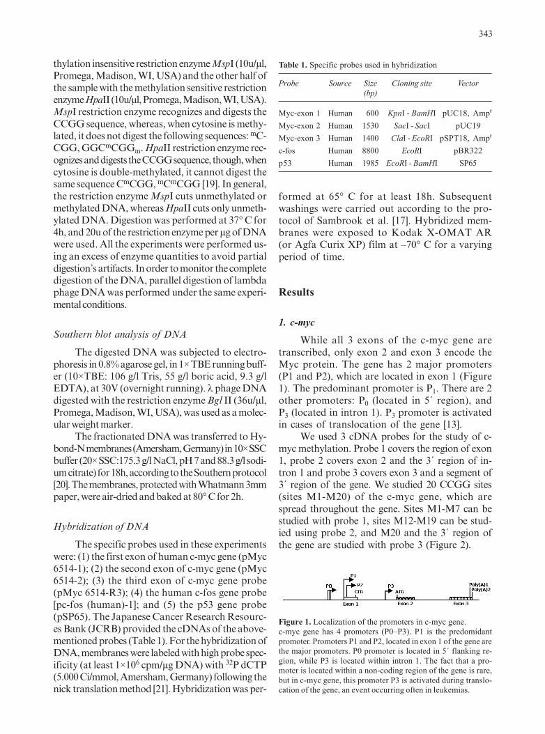
While all 3 exons of the c-myc gene aretranscribed, only exon 2 and exon 3 encode theMyc protein. The gene has 2 major promoters(P1 and P2), which are located in exon 1 (Figure1). The predominant promoter is P1. There are 2other promoters: P0 (located in 5´ region), andP3 (located in intron 1). P3 promoter is activatedin cases of translocation of the gene [13].
We used 3 cDNA probes for the study of c-myc methylation. Probe 1 covers the region of exon1, probe 2 covers exon 2 and the 3´ region of in-tron 1 and probe 3 covers exon 3 and a segment of3´ region of the gene. We studied 20 CCGG sites(sites M1-M20) of the c-myc gene, which arespread throughout the gene. Sites M1-M7 can bestudied with probe 1, sites M12-M19 can be stud-ied using probe 2, and M20 and the 3´ region ofthe gene are studied with probe 3 (Figure 2).
Table 1. Specific probes used in hybridization
Probe Source Size Cloning site Vector(bp)
Myc-exon 1 Human 600 KpnI - BamHI pUC18, Ampr
Myc-exon 2 Human 1530 SacI - SacI pUC19Myc-exon 3 Human 1400 ClaI - EcoRI pSPT18, Ampr
c-fos Human 8800 EcoRI pBR322p53 Human 1985 EcoRI - BamHI SP65
Figure 1. Localization of the promoters in c-myc gene.c-myc gene has 4 promoters (P0–P3). P1 is the predomidantpromoter. Promoters P1 and P2, located in exon 1 of the gene arethe major promoters. P0 promoter is located in 5´ flanking re-gion, while P3 is located within intron 1. The fact that a pro-moter is located within a non-coding region of the gene is rare,but in c-myc gene, this promoter P3 is activated during translo-cation of the gene, an event occurring often in leukemias.

344
a) Sites of methylation of exon 1 in c-myc gene
Following digestion with the enzymes MspIand HpaII, 2 distinct bands of 1.5 and 2.1 Kbpsize were observed, both in normal and in RAEBsamples. These bands were recovered from diges-tion of the CCGG sites M5 and M7 in exon 1(Figure 2, lanes 1, 2, 7, and 8; panel A). RARSsamples showed only one band in 1.5 Kbp, sug-gesting digestion in the M7 CCGG site of exon 1(Figure 2, lanes 5, 6; panel A). ANLL samplesgave a 2.1 Kbp band resulting from the M5 CCGGsite digestion (Figure 2, lanes 9, 10; panel A). Adifferent pattern was obtained after the digestionof RA samples. These samples gave a 2.6 Kbpband, which became obvious after the digestionof the M1 site at the 5�end of exon 1 (Figure 2,lanes 3, 4; panel A). These results indicate that inthe 5� region of c-myc gene – including exon 1 –the sites of hypomethylation in RA are differentfrom RARS and ANLL, and only the RAEB sam-ple gives a pattern similar to the normal one.
In exon 1, 2 promoters of the gene are lo-cated, and this hypomethylated region may en-hance the expression of the gene in RAEB. Thesamples of ANLL, RARS and RA give only oneband, of different molecular weights and they
seem to be regionally methylated (or hyperme-thylated) in exon 1.
b) Sites of methylation of exon 2 in c-myc gene
Using probe 2, which covers exon 2 and the3� flanking region in intron 1 of the c-myc gene,we studied the methylation pattern of exon 2. The3 CCGG sites (M17 – M19) in exon 2 and 5 CCGGsites (M12 – M16) in intron 1 (Figure 3, panel B)were uncovered by probe 2. In double digestion ofgenomic DNA (EcoRI-HpaII enzymes) fromRAEB samples, 3 bands of approximately 4.3, 2.4and 1.9 Kbp were detected. Double digestion withEcoRI-MspI of the same RAEB sample revealed2 bands sized 1.9 and 1.5 Kbp. The bands 2.4 and1.9 Kbp indicate that CCGG sites M17 (exon 2)and M15 (intron 1) are hypomethylated in RAEBsamples as well as in normal controls. The differ-ence between normal and RAEB samples appearsin a 4.3 Kbp band that has resulted from the di-gestion of CCGG site M7 (exon 1) and M20 (exon3) (Figure 3, lane 6; panel A). This finding indi-cates that CCGG site M7 (exon 1) is hypomethy-
Figure 2. DNA methylation pattern of exon 1 of c-myc gene.Panel A: Fragments of hyper or hypomethylation observed indifferent molecular weights with the restriction enzymes MspI(M) and HpaII (H). Normal (lane 1, 2), RA (lane 3, 4), RARS(lane 5, 6), RAEB (lane 7, 8), and ANLL (lane 9, 10).Panel B: The methylation map of exon 1 of c-myc recognized byprobe 1. M1-M7 CCGG sites correspond to methylation sitesdigested by the 2 restriction enzymes.
Figure 3. DNA methylation pattern of exon 2 of c-myc gene.Panel A: Methylated fragments of DNA observed in the differ-ent molecular weights with the use of the restriction enzymesMspI (M) and HpaII (H). Normal (lane 1, 2), RA (lane 3, 4),RAEB (lane 5, 6), and ANLL (lane 7,8).Panel B: The methylation map of exon 2 and intron 1 of c-mycas recognized by probe 2. M17-M19 and M12-M16 CCGGsites respectively are indicated by the arrows.
Lane
Lane
Panel A
Panel A
Panel B
Panel B

345
lated in RAEB samples, but not in normal sam-ples (Figure 3, lanes 1,2 5, 6; panel A).
In RA samples 2 bands of approximate size4.8 and 4.6 Kbp were detected upon double diges-tion with EcoRI - Hpa II (Figure 3, lane 3, 4; panelA). The 1.5 Kbp band was derived from the Eco RI- Msp I cleavage. This 1.5 Kbp band was the resultof digestion in the CCGG sites M10 and M19 withinintron 1 and exon 2, respectively. The 4.8 Kbp and4.6 Kbp bands may result from the cleavage ofCCGG sites M8 and M10 within intron 1 (Figure3, lanes 3, 4; panel A). The data above indicatethat there are different sites of hypomethylation inthe RA and RAEB samples regarding the secondexon of the c-myc gene, that is, the CCGG site M17in RA and M19 in RAEB samples (Figure 3, lanes3, 4, 5, 6; panel A). It was observed that in intron 1and exon 2, the CCGG sites M8, M10 and M15were also hypomethylated in RA and RAEB sam-ples. Normal samples seemed to be hypomethylat-ed only in the CCGG sites M15 (intron 1) and M17(exon 2) (Figure 3, lanes 1, 2; panel A).
ANLL gave the same pattern of methyla-tion as in normal controls (Figure 3, lanes 1, 2,7, 8; panel A). The density of the band (1.9 Kb)was stronger in both pairs of enzymes used fordigestion in ANLL samples (Figure 3, lanes 7,8; panel A), compared to the normal samples.
c) Sites of methylation of exon 3 in c-myc gene
Probe 3 covers the sequence of exon 3 anddetects the methylation of the CCGG site M20of exon 3 and the 3�region of the gene, where 4critical CCGG sites (M21 – M24) are located.Analysis of the methylation pattern obtainedfrom the double digestion of the DNA with theenzymes EcoRI-MspI in all MDS (RA, RARS,RAEB) and ANLL samples revealed mainly 4bands of approximate sizes 8.6, 6.0, 4.7 and 1.0Kbp (Figure 4, lanes 3-12; panel A). DoubleEcoRI-HpaII digestion gave rise to the follow-ing bands: 8.6, 6.0, 4.7, 3.6 and 1 Kbp, in alltypes of MDS samples. According to the genet-ic map of c-myc gene, the 8.6 and 6.0 Kbp bandsfound in these experiments resulted from the di-gestion of M20 site (exon 3) and the CCGG sitesM21 and M23 (in the 3�region of the gene), re-spectively, which are hypomethylated in thesame experiments. A different band of 3.1 Kbpmol. weight was found in normal samples afterdouble digestion with EcoRI-HpaII enzymes(Figure 4, lanes 1, 2; panel A). This result sug-
gests that exon 3 is methylated in normal sam-ples, whereas the MDS samples reveal an ex-tensive hypomethylation pattern. RA samplespresent 2 more bands of hypomethylation: 2.1and 1.0 Kb, compared to RAEB and ANLL sam-ples. ANLL and RAEB samples were found lesshypomethylated compared to RA and RARSsamples (Figure 4, lanes 7-12; panel A). TheMDS samples in the CCGG site M21 and M23in the 3� region are hypomethylated and theCCGG sites M10 and M19 (in intron 1 and exon2) were found hypomethylated by hybridizationwith probes 2 and 3.
In summary, intron 1, exon 2 and exon 3methylation sites of the c-myc gene are hypom-ethylated in MDS, but the rate of methylation isincreased in RAEB and ANLL, albeit c-myc ap-pears to be less methylated compared to the nor-mal samples (Table 2).
Figure 4. DNA methylation pattern of exon 3 of c-myc gene.Panel A: Methylated fragments of DNA observed in the differ-ent molecular weights with the use of restriction enzymes MspI(M) and HpaII (H). Normal (lane 1, 2), RA (lane 3, 4, 5, 6),RARS (lane 7, 8), RAEB (lane 9, 10), and ANLL (lane 11,12).Panel B: The arrows indicate the CCGG methylation sites ofexon 3 and 3´ region of the c-myc gene.
Lane Panel A
Panel B

346
2. Sites of methylation in c-fos gene
The probe used for the study of the methyla-tion status of c-fos protooncogene, covers thewhole gene sequence. C-fos gene has a plethoraof CpG sites found across the entire sequence ofthe gene, an excess of which is located near the 5’region of the gene [22]. In our experiments, densebands of methylation were observed in all of thesamples examined, as well as a smear of multiplebands. The pattern of DNA methylation in myel-odysplasia gave regions of hypomethylated andhypermethylated DNA (Figure 5, panel A).
After double digestion with EcoRI-MspI, the MDSsamples (RARS and RAEB) showed distinct bands of6.5, 5.0 and 4.3 Kb in high molecular weights and otherbands in low molecular weights (1.2 and 1.0 Kbp). WhenMDS samples were analysed with the double digestionEcoRI-HpaII, smear and intense bands of approximatesizes 6.5, 5.0 and 4.3 Kbp were observed (Figure 5,lanes 3-6; panel A). The CMML samples showed adifferent pattern compared to the rest of the MDS sub-types, forming an intense distinct band of hypomethyla-tion in high molecular weight (6.5 Kbp), after both dou-ble digestions (Figure 5, lanes 7, 8; panel A).
The normal samples (Figure 5, lanes 1,2; panelA) revealed smears of bands without distinct bandsof methylated DNA independently of the enzymesused (MspI, HpaII). ANLL samples gave a smear ofbands which was more intense in the M4 subtype ofANLL, compared to the M2 subtype of ANLL, sug-gesting broad hypomethylation in the M4 samples (Fig-ure 5, lanes 9-12; panel A). The hypomethylated sitescompared to the normal samples were numerous.
In conclusion, c-fos in RARS and RAEBwas found hypomethylated in more sites than innormal controls. In CMML, the c-fos gene wasgenerally hypermethylated with one distinct re-
gion of hypomethylation. The M2 subtype ofANLL was regionally hypomethylated and theM4 subtype was extensively hypomethylated(Table 2).
3. Sites of methylation in p53 gene
Digestion of RARS and RAEB sampleswith MspI and HpaII enzymes showed multiplebands of hypomethylation in the p53 sequence(Figure 6, lanes 3-6; panel A). Normal DNA ap-peared to be methylated and gave only 2 bands(Figure 6, lane 2; panel A).
As a result, the normal samples digested with EcoRI/MspI exhibited 2 bands of 5.4 and 1.8 Kb of hy-pomethylation in the whole DNA sequence of thep53 gene. The RARS and RAEB samples digestedwith EcoRI/MspI and EcoRI/HpaII exhibited mul-tiple hypomethylated sites in high (8.4, 6.3, 5.4, 4.5
Table 2. Schematic presentation of the methylation profile ofthe studied genes
¶Regionally hypomethylated gene; *regionally hypermethylatedgene; §the methylation status of the genes was not studied.For abbreviations, see text
Figure 5. DNA methylation pattern of c-fos gene.Panel A: Methylated fragments of DNA observed in the differ-ent molecular weights with the use of restriction enzymes MspI(M) and HpaII (H). Normal (lane 1, 2), RARS (lane 3, 4), RAEB(lane 5, 6), CMML (lane 7, 8), ANLL type M2 (lane 9, 10) andANLL type M4 (lane 11, 12).Panel B: CCGG methylation sites of c-fos DNA sequence. No-tice the clustering of CCGG sites in the regions of the 1st intronand 1st exon of the gene where the promoter and the SRE ele-ment is located.
Lane Panel A
Panel B

347
Kbp) and low molecular weight regions of the gene(2.0, 1.8, 1.3 and 1.2 Kb). More hypomethylatedCCGG sites were observed in the RARS samplesthan in the RAEB samples. In conclusion, p53 isextensively hypomethylated in RARS and less hy-pomethylated in RAEB samples (Figure 6, lanes 5,6; panel A) compared to normal samples (Table 2).
Discussion
The methylation status of DNA in differ-ent genes, and particularly the methylation ofCpG sites located within the promoter region orother critical regions of DNA, are inversely re-lated to gene expression [3].
Methylation in CpG-rich genes may serveas a locking-off mechanism, or precede otherevents that turn a gene off [22]. There is strongevidence that hypomethylation, areas of hyper-methylation and increased DNA methyltrans-ferase activity are components of methylationimbalance in the genome, contributing to neoplas-tic transformation and tumor progression [7,23].
In the relevant literature there are multiple
paradigms of gene activation or silencing, func-tion of which is directly related to leukemogen-esis or tumorigenesis in lymphomas, chronic andacute leukemias, as well as in solid tumors [4-6,24,25].
Hypermethylation of p15-ink4B tumor suppres-sor gene has been found in 50% of the high risk groupof MDS (RAEB and RAEB-t) compared to 8% ofhypermethylation found in low risk MDS (RA andRARS) [26]. The percentage of cases carrying hy-permethylated inhibitor of cyclin kinase (p15ink) in-creases to 78% in leukemia secondary to MDS [27].In addition, 75% of ANLL patients had hypermethy-lation of multiple genes related to cell cycle regula-tion, and 95% of them had hypermethylation atleast of one gene. These genes were mainly cellcycle regulators and cancer type-specific [8, 28].
c-myc, c-fos and p53 genes, transcriptionfactors that are directly involved in hemopoie-sis were studied and aberrant patterns of meth-ylation in the different syndromes of leukemictransformation were found.
The c-myc protooncogene regulates prolifera-tion and programmed cell death of normal cells in re-sponse to endogenous and extracellular signals [9,13,29]. Since 1984 it is known that altered methylation,mainly hypomethylation, of the c-myc gene is foundin cancer cell lines, in tumor cells from solid cancersand in multiple myeloma cells [1-5, 30-32]. In CMMLand in ANLL secondary to myelodysplasia, c-myc wasfound to be hypomethylated in the 3� region of thegene [6].
Our study revealed more CCGG sites of hypom-ethylation, particularly in exon 2 and 3 of the gene andintron 1 in MDS of RA, RARS and RAEB type. Hy-pomethylated regions were found in exon 3 in ANLLcases, but they were less than in the MDS cases.
The c-myc gene has several primers (P0-P3, Fig-ure 1), even though primer P1 is the dominant one.Meanwhile, we have to notice that primer P3, locatedin intron 1, is activated in cases of translocation ob-served in cases of lymphomas. It is known that exon 1or exon 2 possibly encode serum response elements[13,29]. Our data showed that CCGG sites M5, M7(and in some cases M1) of exon 1 appeared unmethy-lated in MDS cases. The fact that MDS are transitionstages to leukemic transformation may explain whyMDS methylation patterns resemble to normal in theregion of exon 1 of c-myc gene. Ohtsuki et al. [33]studying the methylation status of the c-myc gene inhuman myeloma cell lines found that exon 1 re-gions were hypomethylated. Because sequencesnearby and within exon 1 act as transcription in-
Figure 6. DNA methylation pattern of p53 gene.Panel A: Methylated fragments of DNA observed in the differ-ent molecular weights with the use of restriction enzymes MspI(M) and HpaII (H). Normal (lane 1, 2), RARS (lane 3, 4), RAEB(lane 5, 6).Panel B: The methylation map of p53 gene. CCGG sites of p53gene are multiple across the sequence of the gene, indicated bythe vertical short lines.
Lane Panel A
Panel B

348
hibitors [13], changes of methylation status of thisregion may influence the c-myc transcription rate.
The CpG islands in the region of exon 2 seem tobe hypomethylated in MDS as other studies also haveshown [1,5,6]. The hypomethylated regions of exon 2of c-myc may be prone to translocations, affectingthe function of the gene and promoting leukemogen-esis [34].
In exon 3, one CCGG site (M20) and M21-M24 sites of the 3�region appear to be hypometh-ylated in all MDS subtypes. Several studies haveshown similar results in human myeloma cell lines,leukemias and liver cancers [5,6,30,35]. In exon 3,there are sequences, which regulate the additionof polyA tails in RNA transcripts. Furthermore,exon 3 encodes for the domain of c-Myc protein,which is responsible for the nuclear localization ofthe protein [13]. The fact that exon 3 is hypometh-ylated in MDS may indicate that c-myc is not onlyactively transcribed, but also that the c-myc mRNAis stable, the protein translocates to the nucleus andall these facts may promote programmed cell deathor proliferation of hemopoietic precursors cells.
We conclude that hypomethylation of c-mycin multiple sites is an early event in leukemogen-esis. It is more prominent in low risk MDS whereits expression is high, participating in increasedapoptosis of hemopoietic cells [11]. The gene isonly regionally hypomethylated in high risk MDSand in ANLL, resulting in a decline of its expres-sion and of the rate of apoptosis [11,36,37]. It isalso probable that the regional restricted hypom-ethylation in exon 3 of c-myc in ANLL permitsgrowth advantage to the leukemic clone.
The c-fos protooncogene is a member of theAP-1 transcription factor complex, and the constitu-tive expression of the c-fos protein regulates the ho-meostasis of monocytic and myeloid lineage throughdifferentiation and programmed cell death [14]. Theserum response element of c-fos responds to extra-cellular stimuli for growth or apoptosis. C-fos activa-tion is the earliest response of cells to genotoxic agentsfor DNA by inducing programmed cell death of thedamaged cells [14].
Recently Bakin et al. have found that c-fosmay transform cells through alterations in DNAmethylation and histone acetylation [38]. Alter-ations in DNA methylation of c-fos gene havebeen described in gliomas, compared to normaltissues, both in the promoter region and in theencoding sequence of the c-fos protein [39].
In our study, in low risk MDS (RA, RARS)there are regions of extensive hypomethylation
throughout the gene, with a different distribution ofmethylation sites compared to normal c-fos. The pat-tern of hypomethylation in low risk MDS (RA,RARS) may indicate inhibition of differentiation andaccelerated apoptosis of mature hemopoetic cells,particularly of the monocytoid lineage. The gene isgenerally methylated in CMML, with a focus ofrestricted hypomethylation in the promoter region.It could be hypothesized that hypomethylated pro-moter of c-fos in CMML leukemic cells could re-spond to extracellular or intracellular signals foruncontrolled proliferation or inhibition of apopto-sis, resulting in accumulation of the monocytic leu-kemic clone [14]. This pattern of CMML methyla-tion is consistent with the high expression of c-fosgene that was found in CMML compared to RA,RARS and RAEB (data not shown). In the ANLLsamples, and particularly the M4 subtype, wherethe c-fos gene appears extensively hypomethylat-ed, the c-fos protein is probably the key protein forthe transformation process, inducing acceleratedproliferation of the monocytic leukemic clone.
The p53 tumor suppressor gene is consid-ered as the “gatekeeper” of the cell cycle in nor-mal cells, inducing apoptosis when DNA dam-age is recognized [40-42]. In the literature it isrecognized that the p53 mutant protein is onco-genic and in leukemias and lymphomas p53 ishighly mutated [42,43].
Our findings support that p53 gene is hypome-thylated in different regions in both low (RARS) andhigh risk (RAEB) MDS, compared to normal sam-ples where it appears methylated.
The widespread hypomethylation of the p53 genefound in our experiments may predispose to muta-tions in critical regions of the gene. The hypomethy-lated state of the p53 gene confers eligibility to muta-tions and to leukemic transformation of the mutantgene-carrying cell [44].
Another mechanism that the mutated p53 pro-tein may gain oncogenic functions is by interferingwith the p53-dependent apoptosis and cell cycle ar-rest [45]. The extended hypomethylation of p53 foundin our experiments may confer loss of normal p53function on hemopoetic progenitor cells, enhancingapoptosis in low risk MDS (RARS).
From our study and from the results of otherinvestigators it is obvious that a preceding event inleukemic transformation is an “instability” of the me-thylation in the DNA of regulatory genes, with re-gional hypo- or hypermethylated sequences [46]. Thisunstable methylation probably affects the expressionand function of genes that regulate cell cycle, differ-

349
entiation and apoptosis of hemopoietic cells. The func-tional activity of tumor suppressor genes or antionco-genes is abolished in leukemia and lymphoma by hy-permethylation of their encoding DNA [47].
Changes of the methylation pattern of these cellcycle regulatory genes probably act as a precedingevent in a series of methylation changes in the ge-nome during leukemic transformation [47-49]. Thenumber of cases studied in each group of MDS issmall, but the methylation pattern of the genes underinvestigation was identical in each experiment com-pared to normal controls.
There is a perspective that the methylation pro-file of many genes in MDS, leukemias and lympho-mas, may have prognostic value [8, 49] and methyla-tion modifiers may be beneficial for the treatment ofhematological diseases.
Acknowledgements
We thank Dr. C. Giannakenas for his invaluabletechnical assistance, Prof. Dimitriadis for helpful dis-cussions, and E. Karligiotou for her valued help inproofreading the manuscript.
References1. Bird AP. CpG-rich islands and the function of DNA meth-
ylation. Nature 1986; 321: 209-213.2. Zing JM, Jones PA. Genetic and epigenetic aspects of DNA
methylation on genome expression, evolution, mutation andcarcinogenesis. Carcinogenesis 1997; 18: 869-882.
3. Baylin SB, Makos M, Wu J et al. Abnormal patterns ofDNA methylation in human neoplasia: potential consequenc-es for tumor progression Cancer Cells 991; 3: 383-390.
4. Jones PA. Methylation, mutation and cancer. Bioassays1992; 14: 33-36.
5. Gama-Sosa MA, Slaghel VA, Trewyn RW et al. The 5-methylcytosine content of DNA from human tumors. Nu-cleic Acids Res 1983; 11: 6883-6894.
6. Jones PA. DNA methylation errors and cancer. Cancer Res1996; 261: 2463-2467.
7. Singall R, Ginder GD. DNA methylation. Blood 1999; 93:4059-4070.
8. Bird AP. The relationship of DNA methylation to cancer.Cancer Survey 1996; 28: 87-101.
9. Rosenfield C, List A. A hypothesis for the pathogenesis ofmyelodysplastic syndromes : implications for new thera-pies. Leukemia 2000; 14:2-8.
10. Delforge M, Verhoff G, Boogerts M. Understanding thepathogenesis of myelodysplastic syndromes. 5th CongrEur Haematol Assoc, Birmingham, U K, 25-28 June 2000.Educ program, pp 5-7.
11. Rajapalska R, Ginzton N, Rott L, Greenberg PL. Alteredoncoprotein expression and apoptosis in myelodysplasticsyndrome marrow cells. Blood 1996; 88: 4275-4287.
12. Baylin S, Herman J, Graff J, Vertino P, Issa JP. Alterationsin DNA methylation: a fundamental aspect of neoplasia.Adv Cancer Res 1998; 72: 141-196.
13. Marcu KB, Bossone SA, Patel AJ. myc function and regu-lation. Ann Rev Biochem 1992; 61: 809-860.
14. Liebermann DA, Gregory B, Hoffman B. AP-1 (Fos/Jun)transcription factors in hematopoiesis, differentiation andapoptosis. Int J Oncol 1998; 12: 685-700.
15. Symonds H, Krall L, Remington L et al. p53-dependentapoptosis suppresses tumor growth and progression invivo. Cell 1994; 78: 703-711.
16. Madyastha P, Madyastha KR, Wade T, Levine D. An im-proved method for rapid layering of Ficoll-Hypaque dou-ble density gradients suitable for granulocyte separation. JImmunol Methods 1982; 48: 281-286.
17. Sambrook J, Fritsch EF, Maniatis T (eds). Molecular clon-ing laboratory manual (2nd edn). Cold Spring Harbor Press,New York, USA, 1989, pp 9.14-9.23 and 9.47-9.58.
18. Wang L, Hirayasu K, Ishihawa M, Kobayashi Y. Purifica-tion of genomic DNA from human whole blood by isopro-panol-fractionation with concentrated NaI and SDS. Nu-cleic Acids Res 1994; 22: 1774-1775.
19. Waalwijk C, Flavell RA. Msp I: an isoschizomer of Hpa II,which cleaves unmethylated and methylated Hpa II sites.Nucleic Acids Res 1978; 5: 3231-3236.
20. Southern EM. Detection of specific sequences among DNAfragments separated by gel electrophoresis. Biotechnology1975; 24: 122-139.
21. Rigby PWI, Dieckman M, Rhodes C, Berg P. Labeling deo-xyribonucleic acid to high specific activity in vitro by nicktranslation with DNA polymerase I. J Mol Biol 1977; 113:237-251.
22. Tazi J, Bird AP. Alternative chromatin structure at CpGislands. Cell 1990; 60: 909-920.
23. Bird AP. DNA methylation; how important in gene con-trol? Nature 1986; 307: 503-504.
24. Pinyol M, Codo F, Bea S et al. p16IN K4a gene inactivityby deletions, mutations and hypermethylation is associat-ed with transformed and aggressive variants of non-Hodgkin’s lymphomas. Blood 1998; 91: 2977-2984.
25. Asimakopoulos FA, Stheper PJ, Krishevsky S et al. ABL1methylation is a distinct molecular event associated withclonal evolution of chronic myeloid leukemia. Blood 1999;94: 2452-2460.
26. Cleary HJ, Plumb M. Allelic loss and promoter hyperme-thylation of the p15IN K4b gene features in mouse radia-tion – induced lymphoid but not myeloid leukaemias. Leu-kemia 1999; 13: 2049-2052.
27. Uchida T, Kinoshita T, Nagai H et al. Hypermethylation ofp15IN K4b gene in myelodysplastic syndromes. Blood1997; 90: 1403-1409.
28. Melki JR, Vincent P, Clarck S. Concurrent DNA hyperme-thylation of multiple genes in acute myeloid leukemia. Can-cer Res 1999; 59: 3730-3740.
29. Evan GI, Littlewood TD. The role of c-myc in cell growth.Curr Opin Genetics Development 1993; 3: 44-49.
30. Kaneko Y, Shibuya M, Nakayama T et al. Hypomethyla-tion of c-myc and epidermal growth factor receptor genesin human hepatocellular carcinoma and fetal liver. Jpn JCancer Res 1985; 76: 1136-1140.
31. Sardi I, Dal Canto M, Bartoletti R, Montali E. Abnormal c-myconcogene DNA methylation in human bladder cancer: possible

350
role in tumor progression. Eur Urol 1997; 31: 224-230.32. Cheah MSC, Wallace D, Robert MH. Hypomethylation of
DNA in human cancer cells: a site-specific change in the c-myc oncogene. J Natl Cancer Inst 1984; 73: 1057-1061.
33. Ohtsuki T, Nishitani K, Hatamochi A, Yawata Y, NambaM. Analysis of methylation in the c-myc gene in five hu-man myeloma cell lines. Br J Haematol 1991; 77: 172-179.
34. Del Senno L, Maestri I, Piva R. Differential hypomethyla-tion of the c-myc protooncogene in bladder cancers at dif-ferent stages and grades. J Urol 1989; 142: 146-149.
35. Stephenson J, Akdag R, Ozbek N, Mufti GJ. Methylationstatus within exon 3 of the c-myc gene as a prognosticmarker in myeloma and leukemia. Leukemia Res 1992;17:291-293.
36. Tsukamoto N, Morita K, Karasawa M, Omine M. Methy-lation status of c-myc oncogene in leukemic cells: hypom-ethylation in acute leukemia derived from myelodysplasticsyndromes. Experim Hematol 1992; 20: 1061-1064.
37. Askew DS, Ihle JN, Cleveland JL. Activation of apoptosisassociated with enforced myc expression in myeloid progeni-tor cells is dominant to the suppression of apoptosis by inter-leukin-3 or erythropoietin. Blood 1993; 82: 2079-2087.
38. Bakin AV, Curran T. Role of DNA 5-methylocytosine trans-ferase in cell transformation by fos. Science 1999; 283: 387-390.
39. Uyeno S, Komura J, Tawa R et al. Alteration of c-fos genemethylation in human gliomas. Molec Carcinogenesis 1996;16: 91-100.
40. Donehaower LA, Brandley A. The tumor suppressor genep53. Biochimica Biophysica Acta 1993; 1155: 181-205.
41. Perry ME, Levine AJ. Tumor suppressor p53 and the cellcycle. Curr Opin Genetics Development 1993; 3: 50-54.
42. Haffner R, Oren M. Biochemical properties and biological ef-fects of p53. Curr Opin Genetics Development 1995; 5: 84-90.
43. Sigal A, Rotter V. Oncogenic mutations of the p53 tumorsuppressor: the demons of the guardian of the genome.Cancer Res 2000; 60: 6788-6793.
44. Magewn AN, Jones PA. Ubiquitous and tenacious methy-lation of the CpG site in codon 248 of the p53 gene mayexplain its frequent appearance as a mutational hot spot inhuman cancer. Mol Cell Biol 1994; 14: 4225-4232.
45. Scarpa A, Moore PS, Rigand G et al. Molecular features ofprimary mediastinal-B cell lymphoma: involvement ofp16IN K4A, p53 and c-myc. Br J Haematol 1999; 107:106-113.
46. Redner RL, Wang J, Lin JM. Chromatin remodeling andleukemia: a new therapeutic paradigm. Blood 1999; 94:417-428.
47. Melki JR, Vincent PC, Clarck SJ. Cancer-specific region ofhypermethylation identified within the HIC1 putative tu-mour suppressor gene in acute myeloid leukaemia. Leuke-mia 1999; 13: 877-883.
48. Wong IHN, NG MHL, Huang DP, Lee JC K. Aberrant p15promoter methylation in adult and childhood acute leuke-mias of nearly all morphologic subtypes: potential prog-nostic implications. Blood 2000; 95: 1942-1949.
49. Toyota M, Kopecky KJ, Toyota MO, Jair KW, WillmanCL, Issa JP. Methylation profiling in acute myeloid leuke-mia. Blood 2001; 97: 2823-2829.