abdominal tb - university of cape · pdf fileabdominal tb first described ... diagnostic and...
TRANSCRIPT

Abdominal TB Louise Cooke
ESPGHAN goes AFRICA
October 2013

Abdominal TB
First described 1843
Small proportion of cases of childhood TB
Increasing incidence with immunosuppression
Diagnostic and therapeutic challenge
“even in centres of excellence, early diagnosis and
appropriate treatment is not infrequently delayed
because of the non-specific and deceptive clinical
presentation of abdominal TB”
Am J Gastroenterol 1993;88:744-50
Overview
1. TB – burden of disease
2. Types of Abdominal TB
Pathology/pathophysiology
3. “Who gets TB”
4. Clinical presentation
5. “Making the diagnosis”
6. Investigations
7. Management
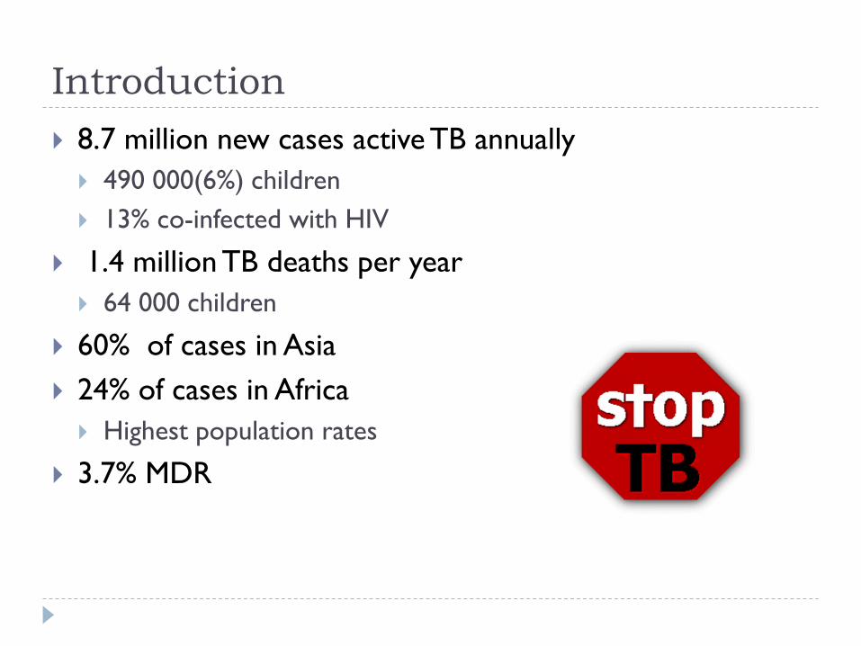
Introduction
8.7 million new cases active TB annually
490 000(6%) children
13% co-infected with HIV
1.4 million TB deaths per year
64 000 children
60% of cases in Asia
24% of cases in Africa
Highest population rates
3.7% MDR
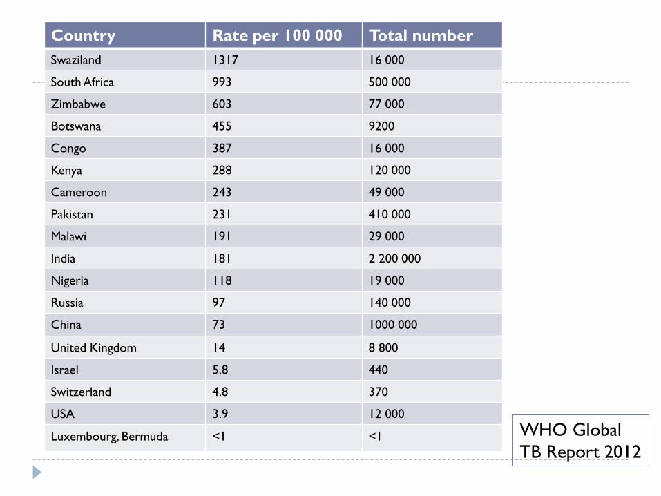
Country Rate per 100 000 Total number
Swaziland 1317 16 000
South Africa 993 500 000
Zimbabwe 603 77 000
Botswana 455 9200
Congo 387 16 000
Kenya 288 120 000
Cameroon 243 49 000
Pakistan 231 410 000
Malawi 191 29 000
India 181 2 200 000
Nigeria 118 19 000
Russia 97 140 000
China 73 1000 000
United Kingdom 14 8 800
Israel 5.8 440
Switzerland 4.8 370
USA 3.9 12 000
Luxembourg, Bermuda <1 <1 WHO Global
TB Report 2012
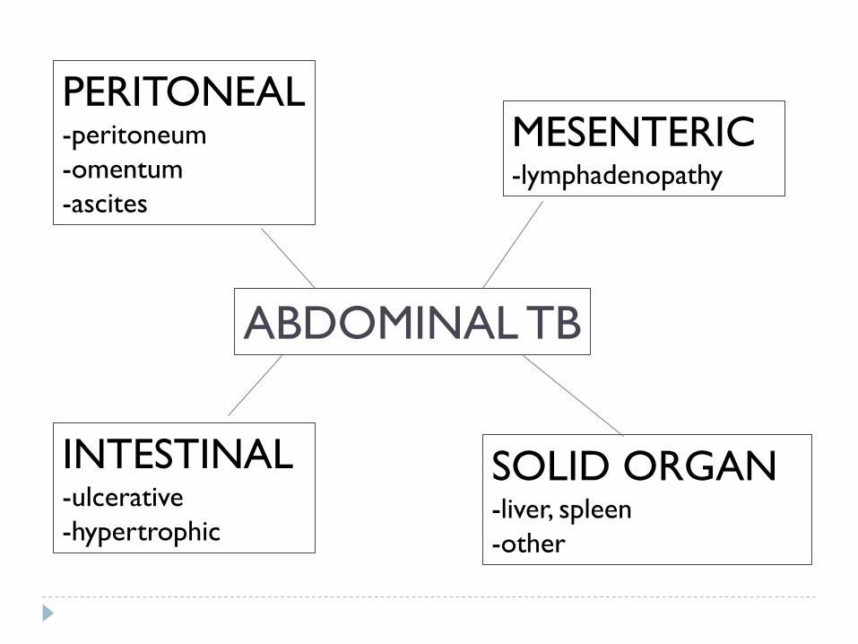
PERITONEAL -peritoneum
-omentum
-ascites
MESENTERIC -lymphadenopathy
INTESTINAL -ulcerative
-hypertrophic
SOLID ORGAN -liver, spleen
-other
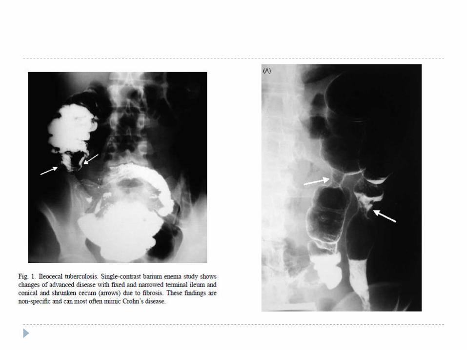
ABDOMINAL TB

Pathogenesis/Pathology:
1. Intestinal (1)
Organism:
Majority mycobacterium tuberculosis
Rarely mycobacterium bovis
Route of infection:
1. Swallowed sputum
2. Lymphohaematogenous: miliary/primary TB infection
3. Direct from adjacent organs(rare)
Areas involved
Can lodge anywhere in intestine
Distal ileum-caecum(90%): most well developed lymphoid follicles
Rarely: jejunum-ileum, colon, ano-rectum duodenum, stomach,
oesophagus

Pathogenesis/Pathology:
1. Intestinal (2)
Bacilli penetrate mucosa and infect submucosal lymphoid
tissue : results in epithelioid tubercle
1. ulcerative form
2-4 weeks later: mucosal sloughing and ulceration
Multiple small irregular ulcers, transverse
Progresses to granulomas at base, caseous necrosis, fibrosis
2. hypertropic form
Abundant inflammatory response
Multinodular mass

Pathogenesis/Pathology:
1. Intestinal (3) Rarer forms
1. Oesophagus
Extrinsic compression by lymph nodes: fistula
2. Gastric
Antral narrowing, wall thickening, fistula
3. Duodenum
Extrinsic compression from lymph nodes, matted mass
4. Jejunem and ileum
ulcers and strictures
5. Colon
Short segment(<5cm)
Long segment :contiguous with caecum
Shortening, distortion, narrowing, ulcers

Pathogenesis:
2. Peritoneal (1)
More frequent than intestinal TB in children
Possible mechanisms:
Reactivation of latent foci from haematogenous spread
Lympho-haematogenous spread from active TB
Spread to peritoneum from ruptured mesenteric nodes
Contiguous spread from intestinal TB
Involves: peritoneum, mesentery, omentum
Ascites common

Pathology:
2. Peritoneal (2)
Widespread tuberculous nodules
Peritoneum and omentum thickened and hyperaemic
Fibrous bands and adhesions
Ascites
lattice like septa
Omentum
Nodular
Smudged: ill defined infiltration
Caked: soft tissue replacement

Pathology:
2. Peritoneal (3)
1. “wet “ type (66%)
1. Large amount viscous ascitic fluid
Diffuse or loculated ascites
2. Whitish miliary nodules(<5mm)
2. “fibrotic-fixed” type(21%)
1. Large omental masses
2. Matted and tethered bowel loops with adhesions
3. Fibro-adhesive “dry” or “plastic” type (13%)
1. Caseous nodules(cheesy yellow)
2. Fibrous peritoneal reaction
3. Dense adhesions

Pathogenesis/Pathology:
3.Mesenteric Lymphadenitis
Routes: ingestion sputum(lymphatic drainage),
haematogenous or adjacent organs
Lymph nodes involved:
mesenteric
para-aortic
periportal
May not have peritoneal or intestinal disease
Pattern varies:
Increased normal size nodes
Local clusters of enlarged nodes
Large conglomerated masses

Tuberculous lymphadenitis

Tuberculous lymphadenitis
Pathogenesis/Pathology:
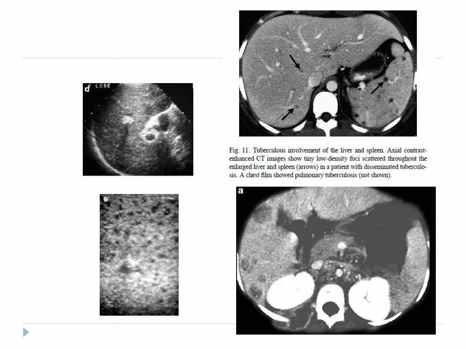
4. Solid organ
Haematogenous from pulmonary or miliary TB
From GI lesions through portal vein
Usually non-specific hepato-splenomegaly
Ultrasound
Diffuse hyperechogenicity
Multiple hypoechoic lesions
CT scan
Multiple hypodense small masses throughout organs from 1 to
3 mm
Early: contrast enhancement

Complications of abdominal TB
Small bowel obstruction
Peritoneal or omental adhesions, adhesions to enlarged lymph
nodes, compression by nodes or hyperplastic intestinal Tb with
strictures
Perforation
Abscess formation
Fistulae: entercutaneous, rectal, TOF
Intussusception
Vascular: intestinal ischaemia, pseudoaneurysms,
haemorrhage, portal vein thrombosis
Lymphatic: chylous peritonitis, intestinal lymphangiectasia

Abdominal TB epidemiology
“Who gets abdominal TB?”
Peak incidence 3rd and 4th decade
10% of cases under 10 years of age
Age range: newborn infant to adolescent
Mean age 9 years
SA: >50% under 5 years
Developed world - immigrants

Clinical presentation(1)
Varied and non-specific
Most sub-acute/insidious
Symptoms:
Common :
abdominal pain,
abdominal distension
LOW
Other : vomiting, constipation, diarrhoea, anorexia
Rare: GI Bleed: haematochezia

Clinical presentation(2)
Clinical exam:
Systemically ill
Malnourished
Pale
Low grade fever
Ascites, abdominal mass
Hepatomegaly, +/- splenomegaly
“doughy” abdomen(10%)
Bowel obstruction
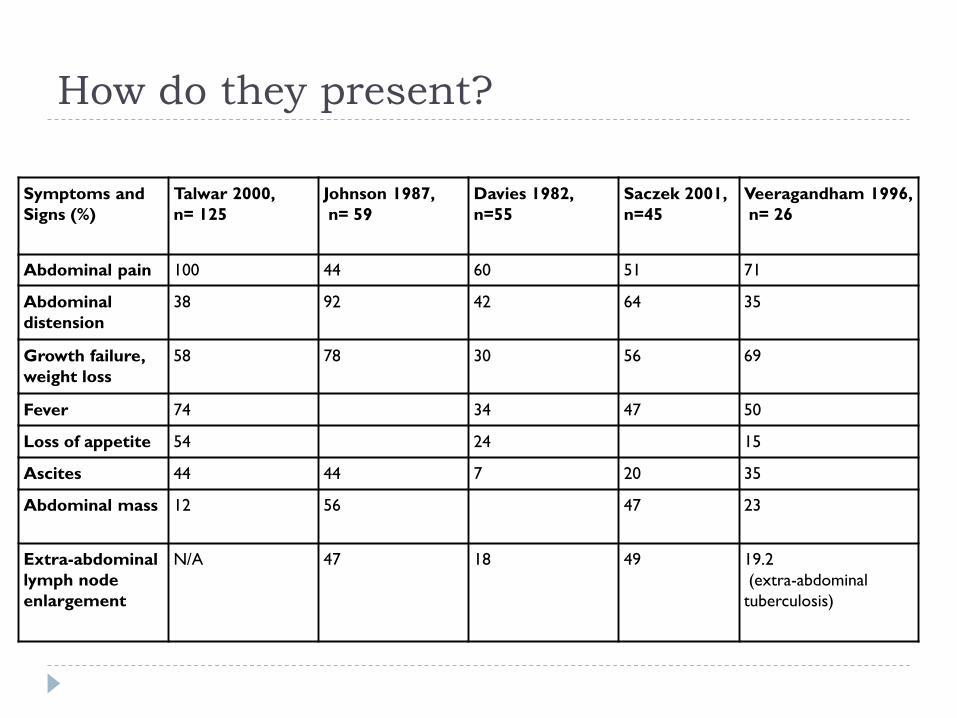
How do they present?
Symptoms and
Signs (%)
Talwar 2000,
n= 125
Johnson 1987,
n= 59
Davies 1982,
n=55
Saczek 2001,
n=45
Veeragandham 1996,
n= 26
Abdominal pain 100 44 60 51 71
Abdominal
distension
38 92 42 64 35
Growth failure,
weight loss
58 78 30 56 69
Fever 74 34 47 50
Loss of appetite 54 24 15
Ascites 44 44 7 20 35
Abdominal mass 12 56 47 23
Extra-abdominal
lymph node
enlargement
N/A 47 18 49 19.2
(extra-abdominal
tuberculosis)

Making the diagnosis………..
High index of suspicion
1. History and clinical examination
Positive household contact: varies from 30-80%
2. Laboratory features of systemic chronic disease
Elevated ESR, normochromic anaemia, thrombocytosis
3.TB investigations
4.Ascitic fluid analysis
5. Imaging
6. Surgical methods
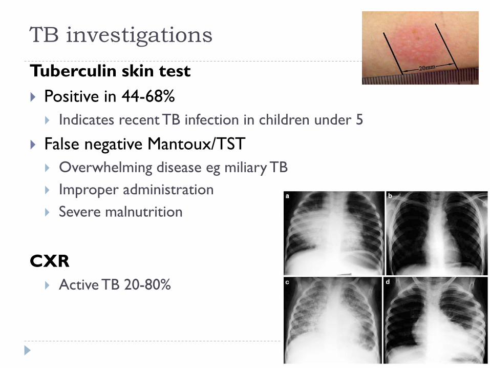
TB investigations
Tuberculin skin test
Positive in 44-68%
Indicates recent TB infection in children under 5
False negative Mantoux/TST
Overwhelming disease eg miliary TB
Improper administration
Severe malnutrition
CXR
Active TB 20-80%
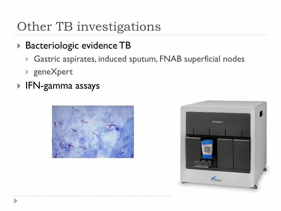
Other TB investigations
Bacteriologic evidence TB
Gastric aspirates, induced sputum, FNAB superficial nodes
geneXpert
IFN-gamma assays
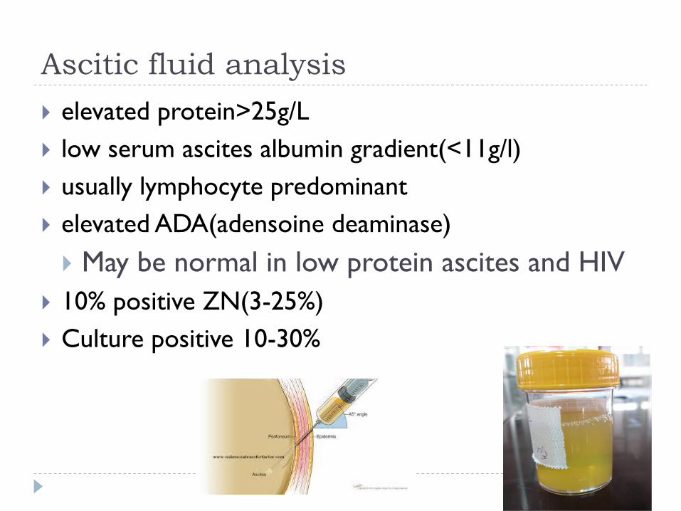
Ascitic fluid analysis
elevated protein>25g/L
low serum ascites albumin gradient(<11g/l)
usually lymphocyte predominant
elevated ADA(adensoine deaminase)
May be normal in low protein ascites and HIV
10% positive ZN(3-25%)
Culture positive 10-30%

Imaging
Abdominal Xray
Intestinal obstruction, calcification
Ultrasound
Lymph nodes: Central hypoechoic areas
Ascites: clear or strands, loculations, debris
Mass, liver/spleen
CT scan
Lymph nodes: central low attenuation(liquefaction), peripheral
enhancement(DDx: lymphoma, pyogenic), calcification
Intestinal wall thickening
Omental caking

Surgical diagnostic methods
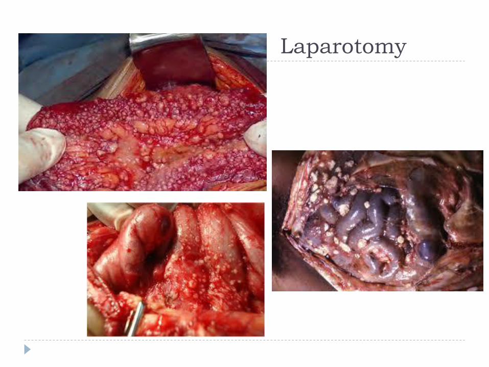
Laparoscopy
Laparotomy
Colonoscopy

Laparoscopy
Sensitivity 85-100%
Straw coloured ascites
Yellow-white nodules on peritoneum
Fibrous bands and adhesions
Hyperaemic oedematous bowel loops
Lymph node enlargement
Biopsy peritoneum and lymph nodes:
Caseating granulomas and/or AFBs, culture, PCR

Laparotomy
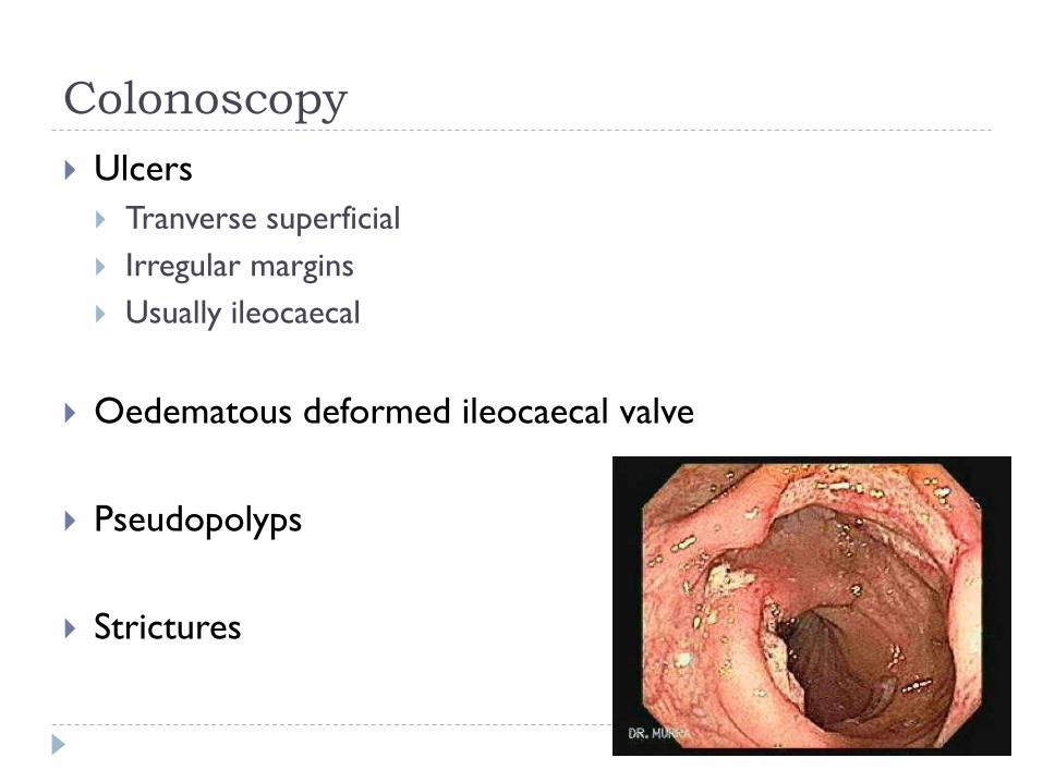
Colonoscopy
Ulcers
Tranverse superficial
Irregular margins
Usually ileocaecal
Oedematous deformed ileocaecal valve
Pseudopolyps
Strictures

Differential diagnosis
1. Ileocaecal
1. Crohns disease
2. Lymphoma
2. Differential for ascites
3. Mass/lymph nodes
1. MAC
2. Lymphoma
3. Yersinia
4. Round worm mass
5. Retroperitoneal tumours
4. Other surgical
1. Appendicitis, Hirschrungs, psoas abscess, urachus abn
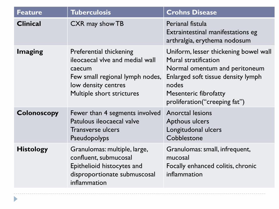
Feature Tuberculosis Crohns Disease
Clinical CXR may show TB Perianal fistula
Extraintestinal manifestations eg
arthralgia, erythema nodosum
Imaging Preferential thickening
ileocaecal vlve and medial wall
caecum
Few small regional lymph nodes,
low density centres
Multiple short strictures
Uniform, lesser thickening bowel wall
Mural stratification
Normal omentum and peritoneum
Enlarged soft tissue density lymph
nodes
Mesenteric fibrofatty
proliferation(“creeping fat”)
Colonoscopy Fewer than 4 segments involved
Patulous ileocaecal valve
Transverse ulcers
Pseudopolyps
Anorctal lesions
Apthous ulcers
Longitudonal ulcers
Cobblestone
Histology Granulomas: multiple, large,
confluent, submucosal
Epithelioid histocytes and
disproportionate submuscosal
inflammation
Granulomas: small, infrequent,
mucosal
Focally enhanced colitis, chronic
inflammation

Effect of HIV co-infection on TB abdomen
Increased number with
extrapulmonary TB
Younger
Fever, LOW, night sweats
more common
Ascites and jaundice less
common
Intra-abdominal
lymphadenopathy more
common with CT
Disseminated TB more
common
AFB positive lymph node
aspirates more common
Decreased absorption
drugs
Atypical mycobacteria
more common
Increased mortality
NB causes of acute
abdomen
Confirming the diagnosis
Actively pursue microbiologic diagnosis
Trial of therapy frequently supports a presumptive
diagnosis
Especially in high incidence regions, classic presentation
Monitoring:
fever resolution
Improved nutritional status
resolution ascites and masses
Reduction size lymph nodes
Any uncertainty: laparoscopy/biopsy
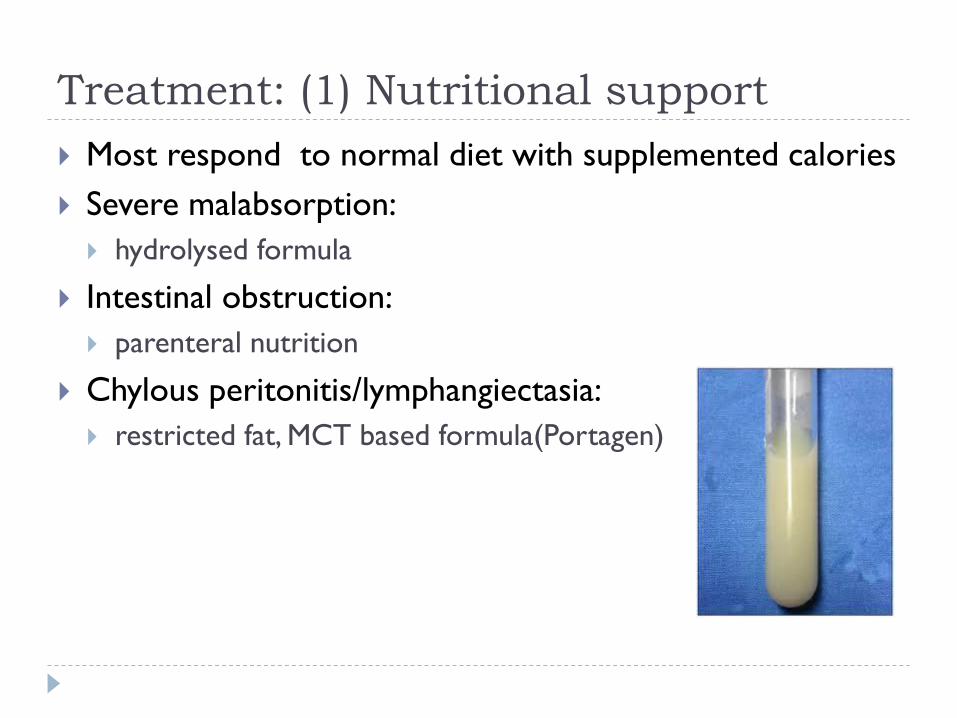
Treatment: (1) Nutritional support
Most respond to normal diet with supplemented calories
Severe malabsorption:
hydrolysed formula
Intestinal obstruction:
parenteral nutrition
Chylous peritonitis/lymphangiectasia:
restricted fat, MCT based formula(Portagen)
Treatment (2) Antituberculous Drugs
Local national/WHO guidelines
4 drug regimen
Isoniazid, rifapicin, pyrazinamide, ethambutol
WHO: INH, RIF only after first 2 months
Prescribe at upper range of recommended doses
Most respond to medical management
Including most strictures and fistulas and subtotal obstruction
?steroids – insufficient evidence

Treatment (3) Surgical
Avoid unless clear indication
High rate of complications and mortality
Perforations and formation enterocutaneous fistulae
Elective laparoscopy/laparotomy if diagnosis uncertain
Indications for surgery
Complete obstruction
Perforation
Persistent fistula (stricturoplasty)
Severe intestinal bleeding

Increased morbidity/mortality
Delayed diagnosis/initiation treatment
Co-morbidity
HIV
Cirrhosis
Renal failure
Severe malnutrition
Disseminated disease
Surgical intervention
Multiple strictures and perforations

SUGGESTIVE CLINICAL FEATURES
•abdominal pain, fever, LOW, +/-TB contact
•abdominal mass, doughy abdomen, ascites
Basic blood work +ultrasound
Suspect ATB Laparotomy/laparoscopy
if surgical complication TB INVESTIGATIONS:
CXR, Mantoux, sputum/gastric
aspirate, FNAB nodes
SUGGEST TB UNCERTAIN OR NEGATIVE FOR TB
Confirmatory investigations
•Ascitic tap: ADA levels, SAAG
•CT abdomen
Confirmatory investigations
•Ascitic tap: ADA levels, SAAG
•CT abdomen
Empiric Rx, evaluate response 2-4 weeks Contraindication surgery?
Laparoscopy and biopsy if poor response
or complication
Yes
No
Laparoscopy and biopsy
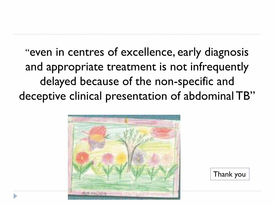
“even in centres of excellence, early diagnosis
and appropriate treatment is not infrequently
delayed because of the non-specific and
deceptive clinical presentation of abdominal TB”
Thank you