abcs of wound care-janna - iapasociety.org · • true depth unknown, but either stage iii or iv...
TRANSCRIPT

9/26/16
1
Janna Girardi, M.D. Mercy Healing Center Medical Director
• None
• Understand the basic assessment of wound and normal healing • Understand contributing factors to poor healing/problem
wounds • Understand the initial treatment regarding pressure ulcers,
venous ulcers, diabetic ulcers, surgical wounds
• A diabetic foot ulcer is more deadly than cancer • In a study of 20373 diabetic foot ulcers – 5% of people with new ulcers
died within 12 months of 1st ulcer visit, 42.2% of people with foot ulcers died within 5 years (versus cancer – 32% 5 year mortality)
• The population is aging and becoming sicker • More diabetic • More obese • Yet living longer (maybe…)
• Wounds are a source of infection (cellulitis, osteomyelitis, other infections) = hospitalization
• Chronic wounds affect 6.5 million Americans per year – cost is $25 billion per year
• When to intervene – depends on the wound and etiology • If a wound is due to diabetes, pressure, venous or arterial insufficiency etc, we need to
create an environment that permits the patient to heal…
• Wound care is an emerging specialty with improved outcomes over “standard of care” and lower cost
• Coordinating care and working together improves outcomes • There are many factors to consider that may be delaying ability to heal
• Malperfusion/hypoxia • Necrotic tissue, bioburden • Infection/inflammation • Edema • Microenvironment issues • Deficient tissue growth • Unrelieved pressure/trauma • Pain • Systemic issues
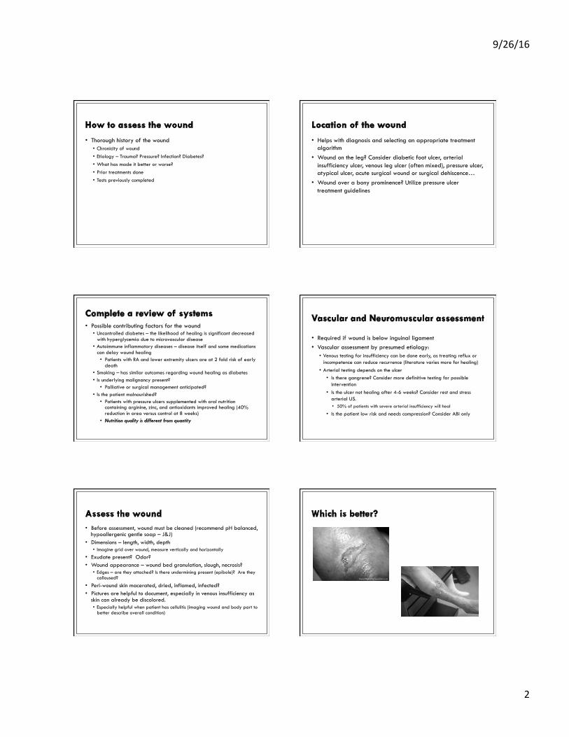
9/26/16
2
• Thorough history of the wound • Chronicity of wound • Etiology – Trauma? Pressure? Infection? Diabetes? • What has made it better or worse? • Prior treatments done • Tests previously completed
• Helps with diagnosis and selecting an appropriate treatment algorithm
• Wound on the leg? Consider diabetic foot ulcer, arterial insufficiency ulcer, venous leg ulcer (often mixed), pressure ulcer, atypical ulcer, acute surgical wound or surgical dehiscence…
• Wound over a bony prominence? Utilize pressure ulcer treatment guidelines
• Possible contributing factors for the wound • Uncontrolled diabetes – the likelihood of healing is significant decreased
with hyperglycemia due to microvascular disease • Autoimmune inflammatory diseases – disease itself and some medications
can delay wound healing • Patients with RA and lower extremity ulcers are at 2 fold risk of early
death • Smoking – has similar outcomes regarding wound healing as diabetes • Is underlying malignancy present? • Palliative or surgical management anticipated?
• Is the patient malnourished? • Patients with pressure ulcers supplemented with oral nutrition
containing arginine, zinc, and antioxidants improved healing (40% reduction in area versus control at 8 weeks)
• Nutrition quality is different from quantity
• Required if wound is below inguinal ligament • Vascular assessment by presumed etiology: • Venous testing for insufficiency can be done early, as treating reflux or
incompetence can reduce recurrence (literature varies more for healing) • Arterial testing depends on the ulcer • Is there gangrene? Consider more definitive testing for possible
intervention • Is the ulcer not healing after 4-6 weeks? Consider rest and stress
arterial US. • 50% of patients with severe arterial insufficiency will heal
• Is the patient low risk and needs compression? Consider ABI only
• Before assessment, wound must be cleaned (recommend pH balanced, hypoallergenic gentle soap – J&J)
• Dimensions – length, width, depth • Imagine grid over wound, measure vertically and horizontally
• Exudate present? Odor? • Wound appearance – wound bed granulation, slough, necrosis? • Edges – are they attached? Is there undermining present (epibole)? Are they
calloused?
• Peri-wound skin macerated, dried, inflamed, infected? • Pictures are helpful to document, especially in venous insufficiency as
skin can already be discolored. • Especially helpful when patient has cellulitis (imaging wound and body part to
better describe overall condition)

9/26/16
3
• Infection – usually see increase in erythema or exudate • Difference in contamination, colonization, infection
• Low O2 in tissue (hypoxia) • Cellular failure – wound bed has senescent cells that send
improper signals for tissue growth • Weekly debridement has been shown to be optimal interval for healing
• Unrelieved pressure or trauma – not off-loading ulcerated area or continued injury
• Pathologic inflammation – vasculitis, cancer – biopsy may be helpful
• Unfortunately there is no universal guideline for all wounds • Multiple societies & organizations exist that have evidence-
based guidelines • Remembering the basic assessment and first steps will get you
far, and working together with wound clinic gives the best possible outcomes (evidence-based) • Wound care center in diabetic foot ulcers – increased probability of
healing after 20 weeks compared with standard care, less costly for healed outcome
• Managing contributing conditions is most helpful (treating cellulitis, prescribing compression, controlling diabetes, etc)
• For supplies/prescriptions: • Pressure ulcers are staged 1-4, unstageable, deep tissue injury • Diabetic ulcers are graded 0-5 (Wagner) • Other ulcers are partial or full thickness
• For ICD-10 documentation/billing: • Diabetic and other ulcers: describe anatomic location, add limited to
breakdown of skin, with fat layer exposed, with necrosis of muscle, with necrosis of bone • Pressure ulcers: document stage 1-4, unstageable, suspect DTI, device
related • New nomenclature from NPUAP recommends injury instead of ulcer,
eliminated roman numerals, eliminated “suspect DTI”
• Recent hip surgery • BMI 19 • Developed pressure ulcer post-operatively • Difficulty eating
Stage 3 pressure ulcer over R ischium (bony prominence)

9/26/16
4
• 1: non-blanchable erythema, skin intact • 2: partial thickness – shallow ulcer with red pink wound bed, no
slough, exposed dermis • Intact or open/ruptured serum or serosanguineous-filled blister • No granulation tissue, slough, eschar are present • No bruising – bruising indicates deep tissue injury
• 3: full thickness – subcutaneous fat may be visible, no bone, tendon or muscle exposed • Slough may be present but does not obscure depth of tissue loss • Depth varies by anatomical location
• 4: full thickness with exposed bone, tendon or muscle • Unstageable: full thickness skin or tissue loss, depth unknown • True depth unknown, but either stage III or IV • Stable eschar on heels should not be removed
• Deep tissue injury – purple or maroon discolored intact skin • Additional definitions exist for medical device related and mucosal
membrane pressure injuries
• Clean the wound (soap & water) and advise the patient or caregiver to do so – gently removes debris and bioburden
• Most important for healing – nutrition and off-loading • Off-loading – depending on the stage of pressure ulcer, may
qualify for a group 1-3 surface, but need equal pressure distribution = redistribution • For CMS, need 30 days of comprehensive program & meet other criteria
for group 2 (low air loss, alternating mattresses) • Air cushions are available for purchase at DME (insurance does not cover)
• Protect skin from moisture and shear forces to reduce further damage (zinc-based formulation if not allergic)
Not this: This
This is a basic pressure redistribution cushion. Patients who are paraplegic may have a ROHO cushion. Patients can have their more expensive cushions mapped (in CR – done at St Lukes physical medicine & rehab).
• Lower leg ulcer for 5 months, weeping • BMI 48 • Diabetic, A1c 9 • Standard American diet
• Venous leg ulcer
Findings: Venous dermatitis Atrophie blanche Hemosiderin deposition Lipodermatosclerosis
• C0: no visible or palpable signs of venous disease • C1: telangiectasies or reticular veins • C2: varicose veins • C3: edema • C4a: pigmentation or eczema • C4b: lipodermatosclerosis or atrophie blanche • C5: healed venous ulcer • C6: active venous ulcer • This classification is not clinically useful in wound care (all are
C5 or C6), nor ICD-10 documentation • Classification of lymphedema also exists

9/26/16
5
• Based on history and appearance – most likely venous ulcers (but 30-40% of chronic ulcers have more than 1 cause…) – test both arteries & veins
• Primary treatment – compression, minimum 25-30 at ankle if normal ABI • Need to ensure good flow prior to high-grade compression as adding
compression can further stress compromised system • Patient must be compliant – use level of compression that patient can
tolerate!
• Treating venous disease can reduce recurrence, may help ulcer heal depending on findings
• More important – weight loss, diabetes control
• Stockings – reduced compliance due to difficulty donning & doffing (consider prescribing device to improve compliance); multiple classes • Class 1: 18-21 mmHg • Class 2: 23-32 mmHg • Class 3: 34-46 mmHg • Class 4: 49+ mmHg • Circular knit – off the shelf • Flat knit – custom made – esp for extreme body shapes
• Wraps – good investment for patient, not always covered • SOMETIMES covered if active ulcer present
• 3 layer wraps – done by OT or other trained personel • Pneumatic compression • CMS requires 4 week trial of conservative therapy – appropriate compression
garment, exercise, elevation • Calf muscle pump failure is one of largest negative prognostic
indicators – calf muscle atrophy, fixed ankle, limited ROM
• Fitted garments with (usually) velcro closure • Diabetic foot ulcer for 1 year • Neuropathy present bilaterally • Foot deformity present • A1c 10.3 • BMI 35
Diabetic foot ulcer
• Wagner more commonly used , UT system is complex • Both systems associated with amputation risk • UT system more predictive of healing time • UT system distinguishes ischemia and infection
• Wagner 0: foot at risk • Wagner 1: superficial ulcer – no deep exposed structure or
infection • Wagner 2: full thickness of skin with deeper structure exposed • Wagner 3: grade 2 with infection of deeper structure • Wagner 4: partial gangrene • Wagner 5: gangrene of entire foot • GRADES 4 & 5 indicative of ISCHEMIA

9/26/16
6
• Stress importance of off-loading – gold standard is total contact cast – 88% heal at mean time of 44 days
• Not well received until the healing is evident • Not always feasible, some refuse • Ensure no infection is present (esp osteomyelitis) • Perfusion is important for healing • Hyperbaric oxygen is an option in select cases
• No specific guidelines exist in US • NICE (UK) guidelines 2008 • Major recommendations postoperatively: • Wounds healing by secondary intention: do not use Eusol (chlorinated
lime and boric acid solution), moist cotton gauze, mercuric antiseptic solutions • Use appropriate interactive dressing to manage – refer to tissue viability
nurse for advice on appropriate dressings
• Surgical site infection noted – use antibiotic that covers likely causative agent considering local resistance, or based on results of microbiology
• Wet to dry??? • Started in 1930s – used wound coverings and fillings consisting of gauze,
cotton wool pads, impregnated gauze, absorbent cotton, adhesive pads • Despite evidence, practice has not changed since • Problems: • Does not provide physical barrier to entry of bacteria • Frequent (3-4 times daily) dressing changes can reduce wound
temperature ! vasoconstriction ! decreased perfusion • Impairs leukocyte mobility, phagocytic efficiency • No significant impedance of evaporation – do not keep moist unless
continuously wet • Prolong inflammatory phase of wound healing • Causes substantial discomfort and wound bed disturbance • Nonselective mechanical debridement (causes tissue destruction)
• Ovington, 2001 • Over 4 weeks – advanced dressings is cost effective – reduced nursing time, fewer dressing
changes, less time to closure • Colwell et al, 1993
• Semiocclusive dressing had higher hard dollar costs, less frequent changes, provided faster healing outcomes, less expensive then wet-to-dry
• Moffat et al, 2002 • European Wound Management Association no longer recommended gauze as best practice
– newer products (hydrogels, hydrofibers, alginates, soft silicones) less likely to cause pain • Coyne, 2003
• Cost-benefit of wet-to-dry versus polyacrylate moist wound dressing – 26% savings annual for home care agencies, wet-to-dry caused more pain, slower healing, increased infection rate
• Vermeulen et al, 2004 • Cochrane review – dressings and topical agents for surgical wounds healing by secondary
intention • Most evidence low quality • Gauze used more nursing time, associated with more pain, patients less satisfied
• Typically, products are compared to moist gauze prior to FDA approval. While these studies are biased, they typically require efficacy to be approved. In contrast, wet-to-dry dressings are not evidence based – they are done because of history…
• Almost never the primary treatment in wound care - control contributing factors first
• Help control exudate, reduce bioburden, promote healing • Evaluate the etiology of the wound, rule out infection, if high
bioburden is present, choose an appropriate dressing
• Studies indicate that weekly debridement improves healing versus less frequent debridement • Most of literature is in DFU, some in VLU • Typically also evaluating advanced wound care product as primary
endpoint
• SO… much of the data about benefits of debridement for all wounds is based on weaker evidence that shows a small effect, and is extrapolated from DFU and VLU
• Debridement should not be done in a stable eschar on heel or ischemic limb

9/26/16
7
CLEAN WOUND! Wound exudate
Moderate High
Foam • Mepilex • Mepilex AG
Alginate • Melgisorb • Melgisorb AG • Aquacel
Disclaimer: this is NOT good care – this leaves out all the important evaluation and gold standards to help heal wounds. This list is not inclusive.
• Please consider early on in wound presence, especially for diabetic foot ulcers and venous/mixed ulcers. Our clinic goal is to schedule initial visit within 3-5 business days of referral.
• Many traumatic wounds (skin tears) and acute wounds can be handled by PCP if comfortable. We typically have a full schedule, so add-on’s are not usually possible. If significant pain or infection, ER may be more appropriate. • Burns, especially extensive or 3rd degree, are better served at a burn
clinic (UIHC)
• We do a thorough intake, so new appointments can take 60+ minutes.
• Goal is to work together to heal the patient – weight loss, diabetes control, compression, smoking cessation should be encouraged by everyone involved in patient’s care. Education is important!
• There are many guidelines from different societies. The Wound Healing Society publishes their guidelines for free on-line, and National Pressure Ulcer Advisory Panel has their 75 page “quick reference guide” online.
• If a patient is admitted to the hospital for another reason, the wound should be undressed, cleaned and evaluated. It may be a contributing factor. The WOC team is available for consults during “business hours.”
• Our clinic is heavily evidence-based. We review new recommendations and literature frequently. We started a nursing CME program to ensure our practice is most effective and up-to-date in helping heal wounds.
1. Walsh JW, Hoffstad OJ, Sulivan MO, Margolis DJ. Association of diabetic foot ulcer and death in a population-based cohort from the United Kingdom. Diabet Med 2015 Dec. doi: 10.1111/dme.13054 [Epub ahead of print]
2. Cancer. Yesterday, today & tomorrow: NIH research timelines. Accessed 1/19/16 from URL https://report.nih.gov/NIHfactsheets/ViewFactSheet.aspx?csid=75
3. Slow-Healing Wounds Common in Rheumatoid Arthritis. Arthritis Today Magazine. Accessed 1/19/16 from URL http://blog.arthritis.org/rheumatoid-arthritis/slow-healing-wound/
4. Cereda E, Klersy C, Serioli M, Crespi A, D’Andrea F, OligoElement Store Trial Study Group. A nutritional formula enriched with arginine, zinc, and antioxidants for the healing of pressure ulcers: a randomized trial. Ann Intern Med 2015 Feb 3; 162(3): 167-74.
5. Kantor J, Margolis DJ. Treatment options for diabetic neuropathic foot ulcers: a cost-effectiveness analysis. Dermatol Surg 2001; 27(4):347-351.
6. Bergqvist D, Lindholm C, Nelzen O. Chronic leg ulcers: The impact of venous disease. J of Vasc Surg 1999 Apr; 29(4):752-755.
7. Oyibo SO, Jude EB, Tarawhen I, Nguyen HC, Harkless LB, Boulton AJM. A comparison of tow diabetic foot ulcer classification systems. Diabetes Care 2001 Jan; 24(1): 84-88.
8. Brodsky JW. An improved method for staging and classification of foot lesions in diabetic patients. In: Bowker JH, Pfeifer MA, editors. Levin and O’neal’s the diabetic foot. 6th ed. Mosby; 2001. p 273-282.
9. Ovington L.G. Hanging wet-to-dry dressings out to dry. Home Health Nurse. 2001;19(8):1–11. 10. Colwell J.C., Foreman M.D., Trotter J.P. A comparison of the efficacy and cost effectiveness of two methods of
managing pressure ulcers. Decubitus. 1993;6(4):28–36. 11. Moffat C.J., Franks P.J., Hollinworth H. Medical Education Partnership Ltd.; London, UK: 2002. Pain at wound
dressing changes, European Wound Management Association Position Document. 2. 12. Fleck CA. Why “wet to dry”? J Am Col Certif Wound Spec. 2009 Dec; 1(4): 109–113. 13. Wilcox JR, Carter MJ, Covington S. Frequency of debridement and time to heal: a retrospective cohort study of
312744 wounds. JAMA Dermatol 2013; 149(9): 1050-1058.