aalborg universitet pigments from microalgae a …...functional food development using aqueous...
TRANSCRIPT

Aalborg Universitet
Pigments from microalgae
a new perspective with emphasis on phycocyanin
Eriksen, Niels T.
Published in:Book of Abstracts and proceedings of the 7th International Congress on Pigments in Foods
Publication date:2013
Document VersionEarly version, also known as pre-print
Link to publication from Aalborg University
Citation for published version (APA):Eriksen, N. T. (2013). Pigments from microalgae: a new perspective with emphasis on phycocyanin. In M. Arlorio(Ed.), Book of Abstracts and proceedings of the 7th International Congress on Pigments in Foods (pp. 37)
General rightsCopyright and moral rights for the publications made accessible in the public portal are retained by the authors and/or other copyright ownersand it is a condition of accessing publications that users recognise and abide by the legal requirements associated with these rights.
? Users may download and print one copy of any publication from the public portal for the purpose of private study or research. ? You may not further distribute the material or use it for any profit-making activity or commercial gain ? You may freely distribute the URL identifying the publication in the public portal ?
Take down policyIf you believe that this document breaches copyright please contact us at [email protected] providing details, and we will remove access tothe work immediately and investigate your claim.
Downloaded from vbn.aau.dk on: March 25, 2020
Book of Abstracts and Proceedings
of the
7th International Congress on Pigments in Food
18-21 June 2013, Novara, Italy

Pigments in Food VII 2013 2
Book of Abstracts and Proceedings of the 7th International Congress on Pigments in Food Edited by: Jean Daniel Coïsson
Fabiano Travaglia
Marco Arlorio
Università degli Studi del Piemonte Orientale “A. Avogadro”, Department of Pharmaceutical Sciences, Novara, Italy
ISBN: 978-88-903360-3-4 Realizzazione a cura di Booksystem srl, Novara www.booksystem.it - [email protected] Finito di stampare nel mese di Giugno 2013 da Terra Promessa Novara
The Scientific and Organising Secretariats have the right to change this programme if they deem it necessary

Pigments in Food VII 2013 3
Preface On behalf of the Organizers of the Seventh International Congress PIGMENTS IN FOOD, it is a pleasure to welcome all of you at the Dipartimento di Scienze del Farmaco, Novara (Università degli Studi del Piemonte Orientale “A. Avogadro”). After six successfully organized congresses, starting in Sevilla, Spain (1999) and passing through Lisbon, Portugal (2002), Quimper, France (2004), Stuttgart-Hohenheim, Germany (2006), Helsinki, Finland (2008), Budapest, Hungary (2010) the seventh event be held in Novara (2013), a beautiful town located in northern Italy, beside the Lake District of Piedmont. The most important aim of “Pigments in Food” is to offer a possibility for meeting and discussion for scientists dealing with different aspects of food pigments, such as pigment chemists, food chemists, food technologists, agriculturists, nutritionists, but also industry people from all over the world. The “natural pigments” science is developing worldwide, particularly concerning technological novel solutions for foods and food supplements, and under the meaning of the “healthy functional properties”. A “comprehensive” scientific approach is particularly strategic, in order to discover, characterize and design new performing and functional pigments from natural food sources. Cool and charming topics like isolation of pigments from sustainable sources using sustainable “mild” techniques, novel technologies development for pigments stabilization, pigments stability bioactivity and functionality, regulatory affairs are the object of this edition of the Conference. The capacity to exploit new technological strategies and alternative food sources (also considering new promising microorganisms, like microalgae) increases more and more the interest towards this field of food science. Beside the scientific aspect of the Congress, hoping to share this Event with a significant number of Scientists from Academia and technicians from Industry, we really hope to host our guests in our beautiful Italian Region, offering nice coloured (and tasty) food … Pigments in Food VII 2013: a coloured vision on coloured food, quality and safety, for new functional foods with healthy profiles.
On behalf of the Organizing Committee
Marco Arlorio, Chair
Pigments in Food VII 2013 4
Scientific Committee
Øyvind M. Andersen (Norway)
Marco Arlorio (Italy - Chair)
George Britton (United Kingdom)
Reinhold Carle (Germany)
Laurent Dufossé (Réunion Island)
José Empis (Portugal)
Vincenzo Fogliano (Italy)
Nicola Galaffu (Switzerland)
Vural Gökmen (Turkey)
Marina Heinonen (Finland)
Adriana Mercadante (Brazil)
Maria Roca Lopez Cepero (Spain)
Steven Schwartz (USA)
Livia Simon-Sarkadi (Hungary)
Carmen Socaciu (Romania)
Organizing Committee
Marco Arlorio (Chair)
Jean Daniel Coϊsson
Vincenzo Fogliano
Daniele Giuffrida
Aldo Martelli
Fabiano Travaglia
Secretariat
Matteo Bordiga
Elisabetta Cereti
Cristiano Garino
Monica Locatelli
e-mail: [email protected] Phone: +39 0321 375873
Pigments in Food VII 2013 5
Contents
Session 1: Chemistry and Biochemistry
Natural carotenoids: a study in oils and water colours Britton G.
13
Differences in anthocyanin content of food and natural sources correlated with differences in anthocyanin chemistry and properties Andersen Ø.M., Jordheim M.
14
Carotenoid ester profiles in Solanum tuberosum and Solanum phureja cultivars Burmeister A., Bondiek S., Jerz G., Fleischmann P.
15
Intramolecular and intermolecular factors affecting the degradation kinetics of xanthophyll esters Jarén-Galán M., Hornero-Méndez D., Pérez-Gálvez A.
16
Analytical and technological aspect of carotenoids from red-bell peppers Daood H.G., Palotás Gábor, Palotás Gábriella, Pék Z., Helyes L.
17
Anthocyanin-synthesizing tomato genotype “Sun BlackTM
” as principal ingredient for a new functional tomato sauce Blando F., Albano C., Gerardi C., Mita G., Mazzucato A.
18
Studies on coupling reactions of proanthocyanidins and malvidin-3-O-glucoside in a wine model solution system Nickolaus P., Weber F., Durner D.
19
Post-harvest modifications enhance the zeaxanthin content in vegetables Esteban R., Fleta-Soriano E., Buezo J., Miguez F., Becerril J.M., García-Plazaola J.I.
20
Description of a new chlorophyll catabolite in ripened fruits of quince (Cydonia oblonga Mill.) Roca M., Ríos J.J., Pérez-Gálvez A.
21
Relationships among flag leaf chlorophyll content, agronomical traits, and some physiological traits of winter bread wheat genotypes Bahar B., Sirat A.,Kilic R., Aydin I.
22
Oxidation routes for betacyanins Wybraniec S., Szot D., Nemzer B., Pietrzkowski Z.
23
Session 2: Technology, Biotechnology and Processing
Artificial intelligence: improving the color measurement
Gökmen V.
27
Microwawe and ultrasound assisted food pigments extraction: highly efficient reactors for green, sustainable processes Cravotto G., Binello A., Mantegna S., Boffa L., Alexandru L.
28
Influence of some oak wood components on stability of malvidin-3-glucoside and chromatic characteristics in model wine solutions Correia A.C., Jordão A.M.
29
Stabilization of anthocyanin-metal chelates with hydrocolloids for their application as blue food colorants Buchweitz M., Kammerer D.R., Carle R.
30
Stabilisation of beetroot derived betanin through interaction with an extract from Barbados cherry Kendrick A.
31
Natural hydroxyanthraquinoid pigments: current situation and future opportunities in food Caro Y., Fouillaud M., Laurent P., Dufossé L.
32
Degradation of anthocyanins in processed strawberry fruit Kermasha S., Borgomano S.
33

Pigments in Food VII 2013 6
Session 3: Pigments from microalgae
Pigments from microalgae: a new perspective with emphasis on phycocyanin
Eriksen N.T.
37
Algal carotenoids as novel pigments in nutrition Christaki E.
38
Functional food development using aqueous extract of Artrospira (Spirulina) maxima rich in phycobiliproteins Langellotti A.L., Buono S., Vargas I., Martello A., Fogliano V.
39
Session 4: Health and Nutrition
Enhanced bioavailability of carotenoids: the influence of chromoplast morphology, dietary lipid, and thermal processing Schweiggert R.M., Kopec R.E., Cooperstone J.L., Villalobos-Gutierrez M.G., Högel J., Young G.S., Francis D.M., Quesada S., Esquivel P., Schwartz S.J., Carle R.
43
Bioaccessibility and changes in the carotenoid profile from murici fruit after in vitro gastrointestinal digestion Mariutti L., Rodrigues E., Mandelli F., Mercadante A.
44
A mini review on the colourless carotenoids phytoene and phytofluene. Are they invisible bioactive compounds? Meléndez-Martinez A.J., Mapelli Brahm P., Stinco C.M., Wang X.-D.
45
Dissecting the pharmacophore of curcumin: two case studies Minassi A., Appendino G.
46
Poster Session
P 01: Synthesis of water-soluble carotenoids via click-reaction Agócs A., Háda M., Nagy V., Deli J.
49
P 02: Thermal and light stability of β-cryptoxanthin esters Bunea A., Andrei S., Rugină D., Pintea A.
50
P 03: Effect of esterification on thermal stability and antioxidant activity of zeaxanthin Pintea A., Bunea A., Socaciu C.
51
P 04: Measurement of enzymatic hydrolysis of lutein esters from dairy products during in vitro digestion Xavier A.A.O., Garrido-Fernández J., Mercadante A.Z., Pérez-Gálvez A..
52
P 05: Oil bodies as a potential microencapsulation carrier for astaxanthin stabilization and safe delivery Acevedo F., Rubilar M., Villarroel M., Navarrete P., Jofré I., Romero F., Acevedo V., Shene C..
53
P 06: Microencapsulation of astaxanthin oleoresin from Phaffia rhodozyma Villalobos-Castillejos F., Yáñez-Fernández J., Barragán-Huerta B.E.
55
P 07: Effect of genotype and growing conditions on lutein and β-carotene content of green leafy Brassica species Arrigoni E., Reif C., Berger F., Baumgartner D., Nyström L.
56
P 08: Effect of processing on content of vital carotenoids in new vegetable puree Palotás Gábor, Palotás Gábriella, Daood H., Pék Z., Helyes L.
57
P 09: Effect of addition of sodium erythorbate and urucum on the lipid oxidation in pork meat Figueiredo B., Bragagnolo N.
58
P 10: Identification of Cionosicyos macranthus carotenoids Murillo E., Watts M., Reyna G.
59
P 11: Bioactive compounds in supercritical CO2-extracted pumpkin oil Durante M., Lenucci M.S., D’Amico L., Dalessandro G., Mita G.
60
Pigments in Food VII 2013 7
P 12: Evaluation of carotenoids and capsaicinoids content in powder of chilli peppers during one year of shelf-life Giuffrida D., Cavazza A., Dugo P., Torre G., Corradini C., Bignardi C., Dugo G.mo
61
P 13: Carotenoids in red fleshed sweet oranges Merussi G.D., Latado R.R., Rossi E.A., Sylos C.M.
63
P 14: Colour changes in heat-treated orange juice during ambient storage Wibowo S., Vervoort L., Lemmens L., Hendrickx M., Van Loey A.
64
P 15: Carotenoid deposition and profiles in peach palm (Bactris gasipaes Kunth) fruits, and their implication on its nutritional potential Hempel J., Esquivel P., Carle R., Schweiggert R.M.
65
P 16: Deposition of lycopene, β-carotene, and β-cryptoxanthin in different chromoplast substructures in papaya fruits Schweiggert R.M., Steingass C.B., Heller A., Esquivel P., Carle R.
66
P 17: Evaluation of quality parameters and carotenoid content of three cultivars of mango (Mangifera indica L.) from Réunion island Rosalie R., Chillet M., Joas J., Lechaudel M., Payet B., Vulcain E., Dufossé L.
67
P 18: Genuine profiles and bioaccessibilities of carotenoids from red- and yellow-fleshed Mamey sapote (Pouteria sapota) fruits Chacón-Ordóñez T., Jiménez V.M., Esquivel P., Carle R., Schweiggert R.M.
69
P 19: Trasgenic tomatoes and their carotenoid and flavour profiles Höfelmeier H., Burmeister A., Schwab W., Fleischmann P.
70
P 20: Study of the time-course cis/trans isomerisation of lycopene, phytoene and phytofluene from tomato Meléndez-Martinez A.J., Paulino M., Stinco C.M., Wang X.-D.
71
P 21: Carotenoid composition of three Hungarian algae species Deli J., Vasas G., Parizsa P., Hajdú G., Szabó I., Lambert N.
86
P 22: HPLC method validation for the determination of fucoxanthin Travaglia F., Bordiga M., Locatelli M., Coïsson J.D., Arlorio M.
72
P 23: Carotenoids stabilisation for use in beverages: two different approaches Mesnier X., Boukobza F., Bily A., Roller M.
74
P 24: Effect of heat processing on the profile of pigments and antioxidant capacity of Jalapeño peppers at intermediate ripening stages Cervantes-Paz B., Ornelas-Paz J. de J., Yahia E.M.
75
P 25: Micellarization and digestive stability of pigments from Jalapeño peppers at intermediate ripening stages Victoria-Campos C.I., Ornelas-Paz J. de J.
76
P 26: Changes in lutein, chlorophylls and chlorophyll degradation products in pistachio kernels (Pistacia vera) during roasting Pumilia G., Schwartz S.J., Cichon M.J., Giuffrida D., Dugo G.mo
77
P 27: Decolouration processes under non-oxygen thermal auto-oxidation of chlorophyll and carotenoids fractions in virgin olive oils Aparicio-Ruiz R., Gandul-Rojas B.
78
P 28: Pigment changes during processing of green table olive specialities treated with alkali and without fermentation Gallardo-Guerrero L., Gandul-Rojas B.
79
P 29: Polyphenols and volatile compounds in Ogliarola and Cellina olive Romani A., Banelli L., Fierini E., Mancuso S., Masi E., Haimler D.
80
P 30: Chlorophyllian pigments in extra virgin olive oils Rovellini P., Venturini S., Fusari P.
81
P 31: Subcellular distribution in olive fruit of peroxidise activity on chlorophyll substrate Vergara-Dominguez H., Roca M., Gandul-Rojas B.
82
P 32: Chlorophyll and carotenoid pigments in a survey of marketed apple varieties Delgado-Pelayo R., Gallardo-Guerrero L., Hornero-Mendez D.
83
P 33: Quantitation of polyphenols in different apple varieties cultivated in Aosta valley Valentini S., Sado A., Chasseur M., Thedy L., Lale Murix H., Barrel I., Chatel A.
84
Pigments in Food VII 2013 8
P 34: Anthocyanins, phenolic acids and antioxidant activity in yellow, red and purple-fleshed potatoes after steam cooking Bellumori M., Innocenti M., Cerretani L., Mulinacci N.
85
P 35: Chemical characterization and antioxidant activity of six rice cultivars grown in Piedmont (pigmented and non-pigmented)
Bordiga M., Coïsson J.D., Locatelli M., Travaglia F., Arlorio M.
86
P 36: Effect of the use of enzymatic preparations on extraction of phenolic compounds from blue maize (Zea mays L.), from the region of Tlaxcala, Mexico Martínez de Santos M.L., Conteras-Llano L.E., Lozada-Ramírez J.D., Ortega-Regules A.E.
87
P 37: Techno-functional properties of tomato sauce fortified with anthocyanin pigments Blando F., Biasco M., Albano C., Gerardi C., Dal Porto L., Mita G.
88
P 38: Effect of post-harvest treatment on anthocyanin content and total phenolics in mango (Mangifera indica L.) peels Geerkens C.H., Müller-Maatsch J.T.L., Geissler M., Carle R.
89
P 39: Maqui (Aristotelia chilensis (Mol.) Stuntz) – Detailed analysis of the highly pigmented “superfruit” Brauch J., Buchweitz M., Carle R.
90
P 40: Prunus mahaleb L. fruit extracts: a novel source for natural food pigments Gerardi C., Albano C., Blando F., Pinthus E., Rescio L., Mita G.
91
P 41: Red orange anthocyanins in colored juices and drinks: analytical method validation Scordino M., Sabatino L., Gargano M., Lazzaro F., Borzì M.A., Traulo P., Gagliano G.
92
P 42: Anthocyanins extraction from mulberry by a combination of high hydrostatic pressure and enzymatic hydrolysis as emerging technology Kim C.-T., Maeng J.-S., Kim C.-J., Cho Y.-J., Kim N., Oh H.-J., Kwon S.-J., Sung G.B.
93
P 43: Anthocyanins and bioactives content in healthy red fruit drinks Castellar M.R., Díaz-García M.C., Obón J.M., Vicente-Castillo A.
94
P 44: Bioactive compounds and antioxidant activity in fruits from Atlantic rainforest, Southeast Brazil Azevedo-Silva N., Rodrigues E., Mercadante A.Z., Oyama L.M., De Rosso V.V.
95
P 45: Phenolic composition of Nebbiolo grapes from Piedmont: changes during ripening and identification of geographic origin Locatelli M., Travaglia F., Bordiga M., Coïsson J.D., Arlorio M.
97
P 46: Antioxidant pigments in red grape juices (Vitis vinifera L. cv Aglianico N.): in vitro bioaccessibility, bioavailability and plasma protein interaction Tenore G.C., Ritieni A., Campiglia P., Manfra M., Coppola L., Novellino E.
98
P 47: Stability of naturally coloured food plant extracts Papetti A., Gazzani G.
99
P 48: Color diversity and antioxidant activity in cactus pear fruits from Southern Italy genotypes Albano C., Aprile A., Negro C., Gerardi C., Mita G., Miceli A., De Bellis L., Blando F.
100
P 49: Betanin stability in selected aqueous-organic solutions influenced by heavy metals Wybraniec S., Szot D., Nemzer B., Pietrzkowski Z.
101
P 50: Teaching food biotechnology in secondary schools using riboflavin as example Pietzner V., Zorn H.
102
P 51: Application and stability of the natural pigment neocandenatone in candy products in comparison with a commercial anthocyanin
Gutierrez Zúñiga C., Yáñez-Fernández J., Barragán-Huerta B.E.
103
P 52: Characterization and genetic fingerprint of saffron Vignolini P., Pinelli P., Albertini E., Romani R.
104
P 53: Extraction methods of natual pigments from stamen of saffron flower Einafshar S., Rohani R., Khorsand Beheshti H.
105

Pigments in Food VII 2013 9
P 54: Effect of salt-stress on the production of pigments by Chlorella vulgaris under heterotrophic culture Benavente-Valdés J.R., Montañez J.C., Aguilar C.N., Méndez-Zavala A.
106
P 55: Carotenoids profile of ultrasound-assisted extract Phormidium sp. Rodrigues D.B., Weis G.C.C., Schio K.L., Jacob-Lopes E., Zepka L.Q.
107
P 56: Microorganisms used ad pigment source Sariçoban C., Battal S.
108
P 57: Pigmented filamentous fungi isolated from tropical marine environments around Réunion island Fouillaud M., Boyer E., Fel A., Caro Y., Dufossé L.
109
P 58: Valorisation of vinasse, a rum distillery effluent, by the production of carotenoid pigments using filamentous fungi Dorla E., Caro Y., Fouillaud M., Dufossé L., Laurent P.
110
P 59: Pigments produced by the bacteria belonging to the genus Arthrobacter Sutthiwong N., Caro Y., Fouillaud M., Laurent P., Valla A., Dufossé L.
111
P 60: Characterization of Arthrobacter arilaitensis pigmentation using spectrocolorimetry Sutthiwong N., Caro Y., Fouillaud M., Dufossé L.
112
P 61: Modeling thermal stability of red pigments produced by Penicillium purpurogenum GH2 Morales-Oyervides L., Oliveira J., Souza-Gallagher M.J., Méndez-Zavala A., Montanez J.C.
113
P 62: pH stability of red pigments produced by Penicillium purpurogenum GH2 Morales-Oyervides L., Oliveira J., Souza-Gallagher M.J., Méndez-Zavala A., Montanez J.C.
114
P 63: Preparation of brown-coloured submicron-sized hazelnut skin fiber with high antioxidant capacity using high shear homogenization Özdemir K.S., Yilmaz C., Gökmen V.
115
P 64: Survey on occurrence of aminocarminic acid in E120 (carmine)-labeled food additives and beverages Sabatino L., Scordino M., Gargano M., Lazzaro F., Borzì M.A., Traulo P., Gagliano G.
116
Author Index 117

Session 1: Chemistry and Biochemistry
Pigments in Food VII 2013 10

Session 1: Chemistry and Biochemistry
Pigments in Food VII 2013 11
Session 1
Chemistry and Biochemistry

Session 1: Chemistry and Biochemistry
Pigments in Food VII 2013 12
Session 1: Chemistry and Biochemistry
Pigments in Food VII 2013 13
Plenary A
NATURAL CAROTENOIDS: A STUDY IN OILS AND WATER COLOURS
Britton G. University of Liverpool, School of Biological Sciences, Crown Street, Liverpool L69 7ZB, U.K. e-mail: [email protected]
Colour has always been important in human life and culture. Brightly coloured foods are
attractive to the eye and the bright colour is considered a sign of quality and freshness.
This colour is provided by many different classes of chemical substances, pigments,
among them the carotenoids, which are widespread and familiar in yellow, orange and
red vegetables and fruits such as carrots, oranges and tomatoes, and in seafood.
We know the structures, we know the chemical and physical properties of the molecules
and how these properties may be modified and the carotenoid protected and stabilised in
natural foods in vivo by the molecular environment and interactions.
An important aspect of the large-scale production of manufactured foods now is to
reproduce the colours of natural food by adding colouring materials during manufacture
and processing, in the form of natural extracts, isolated compounds, synthetic dyes, or
nature-identical colorants produced by chemical synthesis. The use of carotenoids for
this poses particular challenges: they are, insoluble in water, not very soluble in
vegetable oils, and susceptible to oxidative degradation, aggregation and crystallisation,
leading to colour instability. Understanding the properties of the carotenoid molecules
allows these difficulties to be overcome so that carotenoids are now used extensively as
colorants, not only in oil-based applications but also in forms that allow dispersion in
water so that they can be used for colouring drinks and other water-based products. The
physical state or formulation also influences bioavailability of the carotenoids and their
efficiency as health-promoting substances.
Session 1: Chemistry and Biochemistry
Pigments in Food VII 2013 14
Plenary B
DIFFERENCES IN ANTHOCYANIN CONTENT OF FOOD AND NATURAL SOURCES CORRELATED WITH DIFFERENCES IN
ANTHOCYANIN CHEMISTRY AND PROPERTIES Andersen Ø.M., Jordheim M.
Department of Chemistry, University of Bergen, N-5007 Bergen, Norway. email: [email protected]
If intake of anthocyanins has positive health effect(s) and if the various anthocyanins or
their derivatives in the human body have different properties, then of course both the
qualitative and quantitative anthocyanin content of our food as well as the individual
chemistry of these compounds should be more closely correlated.
The major aim of this presentation is to show how the anthocyanins in fruits, vegetables
and products thereof vary substantially with respect to structures and quantities, with
serious impact on differences with respect to anthocyanin reactivity, stability and
bioavailability, including formation of anthocyanin degradation products and phase II
metabolites. We will show accurately that there exist a distinct difference between the
anthocyanin content in vegetables and fruits of our diet, at least with respect to aromatic
acylation and number of monosaccharide units. Thus, if anthocyanins or their derivatives
have impact on our health, we have to design our diet with respect to choice of fruits and
vegetables in a far more precise way than ‘5 A Day’ to obtain optimum effects!
References
[1] Andersen, Ø.M., Jordheim, M. Basic anthocyanin chemistry and dietary sources. In: Anthocyanins in
Health and Disease Prevention (M.M Giusti, T.C. Wallace, Eds.) Taylor & Francis Group, New York.
2013, pp 13–90, In Press.
Session 1: Chemistry and Biochemistry
Pigments in Food VII 2013 15
CAROTENOID ESTER PROFILES IN SOLANUM TUBEROSUM AND SOLANUM PHUREJA CULTIVARS
Burmeister A., Bondiek S., Jerz G., Fleischmann P.
Institute of Food Chemistry, TU Braunschweig, Schleinitzstrasse 20, 38106 Braunschweig, Germany e-mail: [email protected]
Tubers of Solanum species are important stable foods and a continuous source of
antioxidant pigments like carotenoids[1]
.
We will present several profiles of free and esterified carotenoids found in the old
Solanum tuberosum variety Shetland Black, the new breed Red Laura, and the Solanum
phureja cultivars Mayan Gold and Mayan Twilight. Breithaupt and Bamedi [²] elucidated
carotenoid ester profiles of potatoes available in Germany, showing the absence of
carotenoid monesters. Our results, however, proof the occurrence of carotenoid
monoesters and diesters in our cultivars. We identified lutein and zeaxanthin esters as
well as their respective fatty acids in the all investigated varieties. Neoxanthin and
violaxanthin predominantly occur in esterified forms as well. Lutein, however, is
predominantly present in non-esterified form in our tubers.
The carotenoid ester patterns are different and typical for each Solanum species and
variety investigated. Thus our results may also be used for authenticity considerations of
raw and processed food and foodstuff based on these Solanum tubers.
References
[1] Burmeister, A et al., Comparison of carotenoid and anthocyanin profiles of raw and boiled Solanum
tuberosum and Solanum phureja tubers. Journal of Food Composition and Analysis, 2011,24, 865 – 872
[2] Breithaupt, D et al. Carotenoids and carotenoid esters in potatoes (Solanum tuberosum L.): new
insights into an ancientvegetable. J Agric Food Chem. 2002, 50, 7175 - 7181
Session 1: Chemistry and Biochemistry
Pigments in Food VII 2013 16
INTRAMOLECULAR AND INTERMOLECULAR FACTORS AFFECTING THE DEGRADATION KINETICS OF
XANTHOPHYLL ESTERS Jarén-Galán M., Hornero-Méndez D., Pérez-Gálvez A.
Food Biotechnology Department, Instituto de la Grasa (CSIC), Avenida Padre García Tejero, 4, 41012, Sevilla, Spain. e-mail: [email protected]
The kinetics of esterified xanthophylls degradation can depend on the structural features
of the pigment, its intramolecular environment composed by fatty acids of different
nature, and the intermolecular surrounding where the pigment is dissolved. In this study
degradation of either free or esterified xanthophylls (β-cryptoxanthin, zeaxanthin,
capsanthin and capsorubin) was monitored at four temperatures in two different
environments (oil and oil-in-water emulsion) and considering that reaction can follow
either zero or first order model to obtain kinetic and thermodynamic parameters. Results
show that zero order model describes data from the oily environment while data from the
oil-in-water emulsion fit to a first order kinetics. Free capsanthin and capsorubin are
more stable than β-cryptoxanthin and zeaxanthin in oily media but when xanthophylls are
emulsified in water differences in stability among pigments are vanished. This scenario
changed for xanthophyll esters because there were not found significant differences in
the stability of a pigment just changing the nature of the esterifying fatty acid. Thus, in
the oily environment, capsanthin and capsorubin esters showed a degradation pattern
not related to their esterification nature while kinetic constants of β-cryptoxanthin and
zeaxanthin esters were different. However in the oil-in-water emulsion the intramolecular
environment of any xanthophyll esters reached a higher significance in the kinetics,
being responsible for an increased degradation rate. Considering the length of carbon
chain of the fatty acid(s) esterifying the xanthophylls it was possible to establish it as an
influencing factor, with negative consequences on the degradation profile of xanthophyll
esters.
References
Pérez-Gálvez, A. et al. Structure-reactivity relationship in the oxidation of carotenoid pigments of the
pepper (Capsicum annuum L.). J. Agric. Food Chem. 2001, 49, 4864-4869.
Session 1: Chemistry and Biochemistry
Pigments in Food VII 2013 17
ANALYTICAL AND TECHNOLOGICAL ASPECT OF CAROTENOIDS FROM RED-BELL PEPPERS
Daood H.G.
1, Palotás G.
2, Palotás G.
2, Pék Z.
1, Helyes L.
1
1: Szent István University, Páter K. u. 1, H-2103 Gödöllő, Hungary 2: Univer Product PSI, Szolnoki út 35, 6000 Kecskemét, Hungary e-mail: [email protected]; [email protected]
Ripe fruits of either vegetable or spice red-bell pepper are a good source of nutritionally
important carotenoids. The attractive red colour of red-bell peppers is due to a diverse
composition of several yellow-and red-coloured carotenoids, which occur esterified with fatty
acids in form of mono- and di-esters. The content, composition and stability of carotenoids
determine to a high extent, the quality of red pepper products and the acceptance of
consumers towards them. Recent in-vivo or in-vitro epidemiological and chemotherapeutic
studies confirmed cancer chemo-preventive activity of carotenoids from red-bell peppers.
Many chromatographic methods have been worked out and developed for the separation and
determination of Carotenoids from red-bell peppers. In some of those methods the extract of
fruits is simplified by alkaline hydrolysis of fatty acid esters and applied to a separation on
reversed-phase adsorbent with gradient elution. In the other methods un-hydrolysed extracts
were fractionated to their individual carotenoids by separation on reversed-phase column with
analytical dimensions using gradient elution.
In the present work the recent advances in the analysis of carotenoids and carotenoid esters
are described. Some methods were developed to ensure simultaneous, one-run analysis of
naturally occurring and added carotenoids or synthetic dyes. In such methods analytical
columns having adsorbents of 3 µm particle size were used with optimised gradient elution
conditions that provided excellent separation of free xanthophylls, mono-esters, carotenes, di-
esters and contaminating pigments or dyes.
The most recent development was in the application of reversed-phase material having cross-
linked end-capping with high steric activity and stability to separate hydrolysed carotenoid
extract of red–bell peppers under specific conditions similar to those of ultra-performance
liquid chromatography. The run time of complete analysis was decreased from 40 min with
conventional RP column to 16 min with cross-linked column.
Also included in this work is content of carotenoids in spice red pepper hybrids cultivated
under plastic house conditions and their stability to different drying temperatures. In addition
to many agronomic characteristics of some new hybrids, their carotenoid content and stability
were significantly higher than those of the parent varieties.
This work is a part of research supported by the National Development Agency (NFÜ) under project
grant no. TECH-09-A3- 2009-0230, USOK2009 and TÁMOP-4.2.1. B-11/2/KMR-2011
Session 1: Chemistry and Biochemistry
Pigments in Food VII 2013 18
ANTHOCYANIN-SYNTHESIZING TOMATO GENOTYPE ‘SUN BLACKTM’ AS PRINCIPAL INGREDIENT FOR A NEW
FUNCTIONAL TOMATO SAUCE Blando F.
1, Albano C.
1, Gerardi C.
1, Mita G.
1, Mazzucato A.
2
1 Institute of Sciences of Food Production, CNR, Lecce Unit, Lecce, Italy 2 Department of Sciences and Technologies for Agriculture, Forestry, Nature and Energy, Tuscia University, Viterbo, Italy e-mail: [email protected]
‘Sun Black’TM
is a trademark protected tomato line characterized by a remarkable
phenotype with deep purple pigmentation in the pericarp, due to an increased level of
anthocyanins on the peel [1, 2]. Such line has been obtained by the combination of the
Anthocyanin fruit (Aft) allele from Solanum chilense (a gene increasing the anthocyanin
content of the fruit) with atroviolaceum (atv) from S. chesmaniae (an allele enhancing the
anthocyanin presence on stem and leaves). ‘Sun Black’ is therefore a breeding product,
not a GMO product as in [3].
The anthocyanin pigments accumulate in the fruit epidermis, particularly the side much
exposed to the sun. Anthocyanins have been extracted from the peel of ripe ‘Sun black’
tomatoes by ethanol acidified with 2% formic acid. At the HPLC analysis, while in the
control tomato line there is no anthocyanin presence, in the ‘Sun black’ extract there are
several peaks corresponding to delphinidin, petunidin and malvidin aglycones, differently
glycosilated and acylated.
In order to increase the beneficial effect of consuming tomato sauce, we have planned to
produce tomato sauce from ‘Sun Black’ tomatoes, obtaining thus a ‘functional tomato
sauce’ with added nutraceutical value due to the anthocyanin presence.
The HPLC analysis of ‘Sun Black’ tomato sauce revealed the presence of anthocyanin
molecules, even after pasteurization process.
The ORAC value of ‘Sun Black’ extract (peel or whole fruit) and ORAC value of ‘Sun
Black’ tomato sauce extract is reported.
References
[1] Mes P.J. et al. Characterization of tomatoes expressing anthocyanin in the fruit. J. Am. Soc. Horti.
Sci., 2008, 133, 262-269.
[2] Gonzali et al. Purple as a tomato: towards high anthocyanin tomatoes. Trends Plant Sci., 2009, 14,
237, 241.
[3] Butelli E. et al., Enrichment of tomato fruit with health-promoting anthocyanins by expression of
selected transcription factors. 2008, Nat. Biotech., 26, 1301-1308.

Session 1: Chemistry and Biochemistry
Pigments in Food VII 2013 19
STUDIES ON COUPLING REACTIONS OF PROANTHOCYANIDINS AND MALVIDIN-3-O-GLUCOSIDE IN A WINE-LIKE MODEL
SOLUTION SYSTEM Nickolaus P., Weber F., Durner D.
Competence Center for Viticulture & Enology, Breitenweg 71, 67435 Neustadt an der Weinstraße, Germany. e-mail: [email protected]
Reactions involving wine polyphenols are seen as the key reactions changing the
chromatic characteristics of red wines over storage time. Acetaldehyde, a reaction
product occurring in course of wine oxidation, is in this context known to change color
intensity and color hue, as it accelerates coupling reactions between anthocyanins and
tannic structures to form polymeric pigments such as acetaldehyde-bridged anthocyanin-
flavanol-adducts. Many of these pigments have been characterized by their absorption
spectra. However, most studies focused solely on the products resulting from the
reaction between one specific flavanol and one specific anthocyanin. Moreover, none of
the studies hitherto have focused on the formation and decay of those products over
time. In the presented study, a wine-like model solution containing different tannic
structures extracted from grape skins or grape seeds and malvidin-3-O-glucoside was
monitored over a time period of 45 days and analyzed by different chromatographic and
spectrophotometric methods. The model wines were either spiked with acetaldehyde or
not and were stored at different pH values. UV/Vis-spectroscopy was applied to measure
the time-dependent changes in color intensity, color hue and color contribution of
polymeric pigments. HPLC-DAD analysis was carried out to monitor the decreasing
precursor compounds. LC-QToF-MS analysis was used to screen for the reaction
products over time including potentially unknown anthocyanin-flavanol-adducts such as
the 8-6-acetaldehyde-bridged malvidin-3-O-glucoside-catechin-dimer. Time elapsed
regression fits were applied suggesting that the commonly known acetaldehyde-bridged
anthocyanin-flavanol-adducts were not stable under wine conditions. However,
subsequent derivatization reactions may finally lead to products with a higher stability, as
seen for some hydroxyethyl derivatives.

Session 1: Chemistry and Biochemistry
Pigments in Food VII 2013 20
POST-HARVEST MODIFICATIONS ENHANCE THE ZEAXANTHIN CONTENT IN VEGETABLES
Esteban R., Fleta-Soriano E., Buezo J., Miguez F., Becerril J.M., García-Plazaola J.I.
Department of Plant Biology and Ecology, University of Basque Country, UPV/EHU, Bilbao, Spain e-mail: [email protected]
Humans are subjected to oxidative stress and in order to counteract it, they are obliged
to consume antioxidants and carotenoids of plant origin. Indeed, numerous studies show
the need to include high amounts of the carotenoid zeaxanthin (Z) in the diet due to the
fact that it has been negatively correlated to the development of age related macular
degeneration [1] which leads to irreversible loss of vision. Enhancement of Z content
would thus be a desirable trait to incorporate into crops in order to improve the nutrient
intake. However, in green edible vegetables after harvest, Z is usually found in low
amounts. In order to improve this, the aim of the present study was to increase the Z
content in green vegetables by post harvest modifications. We found that light
exposition before cooking and vinegar dressing in spinach and rocket respectively
increased more than 3-fold the initial content of Z. On the other hand, dehydration speed
could be critical for Z content in plants destined for storage as dry material such as
parsley. Findings from this study revealed several post harvest treatments that can
increase the nutritional value of food. Easy recommendations for improving food both to
industry and to the common household quality can be derived from this finding.
References
[1] Gale CR et al. Lutein and zeaxanthin status and risk of age-related macular degeneration. Invest
Ophthalmol Vis Sci 2003, 44, 2461–2465.
Session 1: Chemistry and Biochemistry
Pigments in Food VII 2013 21
DESCRIPTION OF A NEW CHLOROPHYLL CATABOLITE IN RIPENED FRUITS OF QUINCE (Cydonia oblonga, Mill.)
Roca M., Ríos J.J., Pérez-Gálvez A.
Food Biotechnology Department, Instituto de la Grasa (CSIC), Avenida Padre García Tejero 4, 41012, Sevilla, Spain. e-mail: [email protected]
Senescence means structural modifications on chlorophyll molecule producing terminal
chlorophyll catabolites that accumulate in vegetal vacuoles. Since they were first
described in 1991, only 13 different structures have been described to date. In this work
we describe a new chlorophyll catabolite in ripened fruits of quince (Cydonia oblonga,
Mill.) named de-Zm-NCC1. This was accomplished with an easy, rapid and reliable
characterization of chlorophyll catabolites by a HPLC/ESI-TOF-MS method. High
selectivity is achieved with the use of data post-processing software tools considering
accurate mass, isotopic pattern and MS-MS fragmentation profile. We exploited the high
selectivity of the instrument and software algorithm capabilities to show that a
heterogeneous profile arose from the chlorophyll breakdown pathway, with different
possible re-functionalizations reactions of a common structural precursor of chlorophyll
catabolites. Screening was not the solely strategy for chlorophyll catabolites
determination as we included in the target database elemental composition of either de-
esterified or esterified chlorophyll catabolites with a methyl group at the C132 position for
those structures in which such possibility has not been described to date. Consequently,
our method proved to be a straightforward tool for screening of chlorophyll catabolites in
vegetal tissues and for searching new structures expanding knowledge of chlorophyll
catabolism routes.
References
Kräutler, B. et al. On the enigma of chlorophyll degradation: The constitution of a secoporphinoid
catabolite. Angew. Chem. Int. Ed Engl. 1991, 30, 1315–1318.
Session 1: Chemistry and Biochemistry
Pigments in Food VII 2013 22
RELATIONSHIPS AMONG FLAG LEAF CHLOROPHYLL CONTENT, AGRONOMICAL TRAITS, AND SOME
PHYSIOLOGICAL TRAITS OF WINTER BREAD WHEAT GENOTYPES
Bahar B., Sirat A., Kilic, R., Aydin, I.
Siran Vocational School, Gumushane University, Gumushane, Turkey. e-mail: [email protected]
In this study, relationships among flag leaf chlorophyll contents of some winter wheat
genotypes, agronomical traits, and some physiological characters such as canopy
temperature, membrane thermostability, membrane injury, and relative water content of
flag leaf were evaluated. The study was conducted in the Application and Research Area
of Siran Vocational School of Gumushane University in the growth season of 2010-2011.
Chlorophyll content of genotypes were measured by a portable chlorophyll meter at the
start of anthesis (ZGS 60) and the early milky stage (ZGS 73). The mean chlorophyll
content of the tested genotypes at ZGS 60 was 45.6 as SPAD unit, and ranged from 39.1
for line 51 to 54.0 for line 42. Chlorophyll content as the mean of all genotypes at ZGS 73
was 41.8 as SPAD unit. Mean chlorophyll content of the genotypes at this growth stage
ranged between 35.2 for line 27 and 50.9 for line 44. The mean pigment loss was the
percent of 8.3 as an average of all genotypes. The chlorophyll loss ranged between 1.7
% for line 75 and 19.2 % for line 32. The statistically significant correlations between
chlorophyll contents and main yield components like grain number per spike and spike
yield were obtained at both measuring stages. The significant correlation between
chlorophyll loss and chlorophyll content was positive at ZGS 60, but negative at ZGS 73.
These results show that determination of flag leaf chlorophyll content in winter wheat is
important selection criteria for yield components in breeding programs.
References
[1] Markwell, J. et al. Calibration of the Minolta SPAD-502 leaf chlorophyll meter. Photosynth. Res. 1995,
46, 467-472
[2] Monje, O.A. and Bugbee, B. Inherent limitations of nondestructive chlorophyll meters: A comparison of
two types of meters. Hortic. Sci. 1992, 27, 69-71
[3] Uddling, J. et al. Evaluating the relationship between leaf chlorophyll concentration and SPAD-502
chlorophyll meter readings. Photosynth Res. 2007, 91, 37-46
Session 1: Chemistry and Biochemistry
Pigments in Food VII 2013 23
OXIDATION ROUTES FOR BETACYANINS Wybraniec S.1, Szot D.1, Nemzer B.2, Pietrzkowski Z.3
1 Department of Analytical Chemistry, Institute C-1, Faculty of Chemical Engineering and Technology,
Cracow University of Technology, ul. Warszawska 24, Cracow 31-155, Poland. 2 Chemistry Research, FutureCeuticals, Inc., 2692 N. State Rt. 1-17, Momence, IL 60954 USA.
3 Applied BioClinical Inc., 16259 Laguna Canyon Rd, Irvine, CA 92618, USA.
e-mail: [email protected]
Betanin (5-O-glucosylated betanidin) is one of betacyanins, which are a group of natural,
water soluble and non-toxic red-violet plant pigments. Because of the presence of a few
functional groups in betanin structure, it is highly reactive and very sensitive to oxidation
and factors such as increased temperature, presence of organic solvents and metal ions
as well as low and high pH. Recent studies have shown importance of research on
betacyanins oxidation paths, because of their high natural, antiradical and antioxidant
activity and potential benefits to human health.
In this contribution, the identification of the products of betanin and its decarboxylated
derivatives oxidation by ABTS cation radicals and horseradish peroxidase (HRP) in
aqueous solutions at pH 3-8 is presented. The effects induced by these two different
oxidizing agents were monitored by spectrophotometry and LC-DAD-MS/MS.
In general, the oxidation most probably results in a generation of quinonoid derivatives of
the pigments at the first stage [1]. If oxidation of a betacyanin results in a formation of a
semiquinone radical, it should undergo a subsequent oxidation resulting in a formation of
its quinone methide intermediate and rearrangement to 2,3-dehydrogenated betacyanin
as the most probable product since the formation of the aminochrome intermediate is
impossible, because of the blocking hydroxyl at C-5 [1]. Therefore, one of the most
frequently detected betanin and 2-decarboxy-betanin oxidation products is 2-decarboxy-
2,3-dehydro-betanin.
References
[1] Wybraniec, S. and Michałowski, T. New pathways of betanidin and betanin enzymatic oxidation. J.
Agric. Food Chem. 2011, 59, 9612–9622.

Session 1: Chemistry and Biochemistry
Pigments in Food VII 2013 24
Session 2: Technology, Biotechnology and Processing
Pigments in Food VII 2013 25
Session 2
Technology, Biotechnology
and Processing

Session 2: Technology, Biotechnology and Processing
Pigments in Food VII 2013 26
Session 2: Technology, Biotechnology and Processing
Pigments in Food VII 2013 27
Plenary C
ARTIFICIAL INTELLIGENCE: IMPROVING THE COLOR MEASUREMENT
Gökmen V.
Food Engineering Department, Hacettepe University, 06800 Beytepe, Ankara, Turkey email: [email protected]
The overall appearance of any object is a combination of its chromatic and geometric
attributes. The color is the first sensation that the consumer perceives and uses as a tool
to accept or reject, because the color observation allows the detection of certain
anomalies or defects of a product. Commercially available color-measuring devices are
designed to contact the material for measurement. Since they measure a small area with
a fixed geometry, repetitive measurements are necessary to increase accuracy for the
mean color information of a heterogeneous food product. Computer vision based image
analysis is a non-contact alternative technique taking the whole surface into account
while measuring color. It does not only give mean information in any color space (Lab,
RGB, XYZ), but also provide featured information such browning ratio for a product. In
this presentation, basic principles of digital image analysis to obtain mean and featured
color information from an object are discussed. Using custom-designed algorithms,
potential applications of computer vision are exemplified for color measurements in
various raw and processed foods.
Keywords: Color measurement, image analysis, artificial intelligence, mean color, featured color

Session 2: Technology, Biotechnology and Processing
Pigments in Food VII 2013 28
Plenary D
MICROWAVE AND ULTRASOUND ASSISTED FOOD PIGMENTS EXTRACTION: HIGHLY EFFICIENT REACTORS FOR GREEN,
SUSTAINABLE PROCESSES Cravotto G., Binello A., Mantegna S., Boffa L., Alexandru L.
Dipartimento di Scienza e Tecnologia del Farmaco, University of Turin, Italy. e-mail: [email protected]
The design of efficient and sustainable extraction methods for vegetal matrices has been
a hot research topic over the last decade. In spite of the scanty efficiency, maceration
still remain the most common extraction technique also in industrialized countries.
Conventional extraction processes are quite laborious, time consuming, involve large
amounts of solvents and, ultimately, may cause some target molecule degradation.
Great improvements can be achieved with the use of non-conventional techniques such
as microwave [1] and ultrasound-assisted extraction [2]. The industrial production of
natural colouring pigments, requires a technological innovation to improve extraction
yield and minimize fading and degradation. With the aim to obtain extracts and pigments
in high yield and outstanding quality, we developed several methods and equipments
suitable for scaling up. These green extraction techniques applied to medium and large
scale, possibly in flow-reactors, may lead to effective process intensification and also a
carbon footprint reduction [3].
References
[1] Microwave-assisted extraction for bioactive compounds: Theory and practice. Editors: Chemat F. and
Cravotto G. (2013), XII, 238 pp. Series: Food Engineering Series, Vol. 4 Springer Science, U.S.A.
[2] Cravotto G. et al. Improved extraction of natural matrices under high-intensity ultrasound and
microwave, alone or combined. Ultrason. Sonochem. 2008, 15, 898-902.
[3] Chemat F., Abert-Vian M., Cravotto, G. Review: Green Extraction of Natural Products: Concept and
Principles. Int. J. Mol. Sci. 2012, 13, 8615-8627.

Session 2: Technology, Biotechnology and Processing
Pigments in Food VII 2013 29
INFLUENCE OF SOME OAK WOOD COMPONENTS ON STABILITY OF MALVIDIN-3-GLUCOSIDE AND CHROMATIC
CHARACTERISTICS IN MODEL WINE SOLUTIONS Correia A.C., Jordão A.M. Agrarian Higher School, Polytechnic Institute of Viseu (CI&DETS), Viseu, Portugal. e-mail: [email protected]
Many constituents can be extracted from staves during wine aging in barrels. The
evolving environment inside oak barrels during the maturation of wines provides
conditions for further reactions involving wood compounds and wine phenolic
compounds.
The aim of the current study was to use model red wine solutions (12% ethanol and
adjusted to pH 3.5) to evaluate the influence of furfural, eugenol, guaiacol, vanillin,
ellagic acid and oak wood extracts on the changes in the levels of malvidin-3-glucoside
and chromatic characteristics over a period of 64 days. The compounds were used in
concentrations which are similar to the levels that occur in wine during aging in barrels.
Malvidin-3-glucoside and ellagic acid were quantified by HPLC [1,2]. Furfural, eugenol,
guaiacol and vanillin were quantified by GC [3]. Chromatic characteristics were
calculated using CIELAB parameters.
The results showed that the decrease in malvidin-3-glucoside was more pronounced in
the presence of ellagic acid and oak wood chip extracts. After 64 days, when incubated
alone, the malvidin-3-glucoside content was 30.0 mg/L, and this fell to 21.0 mg/L in the
presence of the oak extract and 19.0 mg/L when incubated with ellagic acid. Breakdown
of malvidin-3-glucoside was also slightly more pronounced in the presence of guaiacol,
furfural, vanillin and eugenol. Changes in the levels of furfural, guaiacol, eugenol and
vanillin are characterized by a continuous steep decline throughout the storage period
with no significant influence of malvidin-3-glucoside. For chromatic parameters, a* values
showed a more evident decrease in solutions containing malvidin-3-glucoside and oak
wood extracts.
References
[1] Dallas, C. et al. Interactions of oligomeric procyanidins in model wine solutions containing malvidin-3-
glucoside and acetaldehyde. J. Sci. Food Agric. 1996, 70, 493-500.
[2] Viriot, C. et al. Ellagitannins in woods of sessile oak and sweet chestnut dimerization and hydrolysis
during wood ageing. Phytochemistry 1994, 36, 1253-1260.
[3] Jordão, A.M. et al. Comparison of volatile composition of cooperage oak wood of different origins
(Quercus pyrenaica vs. Quercus alba and Quercus petraea). Mitt. Klosterneuburg 2005, 55, 31-40.
Session 2: Technology, Biotechnology and Processing
Pigments in Food VII 2013 30
STABILIZATION OF ANTHOCYANIN–METAL CHELATES WITH HYDROCOLLOIDS FOR THEIR APPLICATION
AS BLUE FOOD COLORANTS Buchweitz M., Kammerer D. R., Carle R.
Institute of Food Science and Biotechnology, Chair Plant Foodstuff Technology, Hohenheim University, Stuttgart, Germany. e-mail: [email protected]
Recently, artificial dyes commonly applied in uncolored food and pharmaceutical
products were suspected to be detrimental to human health. Consequently, replacement
of these colorants by their natural counterparts is a major challenge for the industry. In
contrast to red, orange and yellow hues, for blue tints only few and expensive natural
colorants such as Spirulina and Gardenia Blue are commercially available, and cheaper
ferric anthocyanin chelates revealing intense blue colors would be an interesting option
[1].
For this purpose, optimal conditions for stabilizing these complexes with pectins were
identified in a screening at a micro scale using juices and phenolic extracts of different
pigment sources being frequently used as coloring foodstuffs [2]. Blue tints were limited
to model systems consisting of amidated and high methoxylated pectins, and pH values
≥4.0. Blue color hues and their thermal and storage stabilities markedly differed between
the pigment sources. While model systems containing red cabbage extract and juice
displayed appealing gentian blue hues, stabilities of these chelates were poor. Purple
carrot extract proved to be the most promising pigment source, producing intense and
stable cobalt blue colors during storage and heat treatment.
To elucidate the potential of ferric anthocyanin chelates in food matrices, such dyes were
added to protein and polysaccharide based gels to evaluate the impact of storage
conditions on color stability [3]. The trends regarding color hues and stability observed in
the gels were consistent with previous findings, providing clear evidence that the basic
knowledge gained in a small scale screening may easily be transferred to complex food
matrices.
References
[1] Castañeda-Ovando, A. et al. Chemical studies of anthocyanins: A review. Food Chem. 2009, 113,
859–871
[2] Buchweitz, M. et al. Colour and stability assessment of blue ferric anthocyanin chelates in liquid
pectin-stabilised model systems. Food Chem. 2013, 138, 2026–2035
[3] Buchweitz, M. et al. Application of ferric anthocyanin chelates as natural blue food colourants in
polysaccharide and gelatin based gels. Food Res. Int. 2013, 51, 274–282

Session 2: Technology, Biotechnology and Processing
Pigments in Food VII 2013 31
STABILISATION OF BEETROOT DERIVED BETANIN THROUGH INTERACTION WITH AN EXTRACT FROM BARBADOS CHERRY
Kendrick A.
Diana Food Division, 7 Allée Ermengarde d’Anjou - ZAC Atalante Champeaux, CS 41137 - 35011 Rennes Cedex - France e-mail: [email protected]
Objective:
To develop a natural stabilisation system for betanin to increase its use as a food colour.
A concentrate of beetroot can be used to provide a red colour for a wide variety of food
and beverages. The colouring pigment is betanin and a limiting factor to its increased
use as a food colour is its heat stability.
Anti-oxidants have been used to slow the colour loss during processing and one of the
most effective is ascorbic acid, often used in combination with citric acid.
A limiting factor with ascorbic acid is that at higher concentrations a pro-oxidant effect is
observed and colour loss is promoted.
Barbados Cherry or Acerola extract is rich in Vitamin C and specifically a formulation
was used from Diana Food Division containing a high level of naturally occurring
Vitamin C.
Ascorbic acid, when delivered in the form of Barbados Cherry extract does not show the
pro-oxidant effect and betanin colour loss exhibited by ascorbic acid in the pure form.
The use of ascorbic acid from Barbados Cherry allowed retention of 67% of betanin
compared to an unstabilised beetroot which retained only 32% betanin under controlled
heating conditions.
An additional advantage of this is that the final colour formulation has the current market
requirement of a ‘Clean label’ ingredient.
Session 2: Technology, Biotechnology and Processing
Pigments in Food VII 2013 32
NATURAL HYDROXYANTHRAQUINOID PIGMENTS: CURRENT SITUATION AND FUTURE OPPORTUNITIES IN FOOD
Caro Y., Fouillaud M., Laurent P., Dufossé L. Laboratoire de Chimie des Substances Naturelles et des Sciences des Aliments, Université de la Réunion, Sainte-Clotilde, Ile de la Réunion, France e-mail: [email protected]
Natural pigments and colorants are widely used in the world in many industries such as
textile dying, food processing or cosmetic manufacturing. Among the natural products of
interest are various compounds belonging to carotenoids, anthocyanins, chlorophylls,
melanins, betalains… This article emphasizes pigments with anthraquinoid skeleton and
gives an overview on hydroxyanthraquinoids described in Nature. Main natural sources
of such pigments are summarized, followed by discussion about toxicity and
carcinogenicity observed in some cases. Current industrial applications of natural
hydroxyanthraquinoids are described with two examples, carminic acid from an insect
and Arpink red™ from a filamentous fungus. As a conclusion, it focuses on the
description of the hydroxyanthraquinoid colouring compounds produced by filamentous
fungi. The conclusions indicate that, even if the toxicological investigations of a new
additive are not financially negligible, non-mycotoxigenic filamentous fungi such as
strains of Drechslera spp., Herpotrichia spp., Paecilomyces spp. and Isaria spp. at least,
could be used for the production of dyestuffs rich in hydroxyanthraquinoid pigments as
potent natural food grade colorants, with different shades according to the biomass
composition: such as red, reddish brown, bronze, maroon and orange-yellow.
Keywords: anthraquinone, hydroxyanthraquinone, natural colorant, food colorant,
microbial pigment
References
[1] Caro, Y. et al. Natural hydroxyanthraquinoid pigments as potent food grade colorants: an overview.
Nat. Prod. Bioprospect. 2012, 2, 174–193
[2] Nagia, F.A. and EL-Mohamedy, R.S.R. Dyeing of wool with natural anthraquinone dyes from Fusarium
oxysporum. Dyes Pigments. 2007, 75, 550–555
[3] Sutthiwong, N. et al. Production of biocolours. (Chapter 12) In : “Biotechnology in Agriculture and Food
Processing: Opportunites and Challenges”, 1st edition, Panesar P.S. and Marwaha S.S. (Eds.), Francis &
Taylor, CRC Press, Boca Raton, Florida, USA (planned for April 2013)

Session 2: Technology, Biotechnology and Processing
Pigments in Food VII 2013 33
DEGRADATION OF ANTHOCYANINS IN PROCESSED STRAWBERRY FRUIT
Kermasha S., Borgomano S.
Department of Food Science and Agricultural Chemistry, McGill University, 21,111 Lakeshore, Ste-Anne
de Bellevue, Qc, Canada H9X 3V9
e-mail: [email protected]
The aim of the research work was to investigate selected enzymatic activities, including
β-glucosidases (GOXD), polyphenol oxidases (PPO) and peroxidases (POXD), involved
in the degradation and stability of anthocyanins in processed red fruits, as well as certain
processing condition, using substrate models and in vitro strawberry fruit. Using the
substrate models, catechin and guaiacol, the results showed the presence of PPO and
POXD activities in the fruit; however, there was an absence of these activities when
catechol and 4-methoxy-α-naphthol were used as substrates. The determination of
glucose, resulted from the hydrolysis of endogenous anthocyanins by β-glucosidase
activity, indicated its important role in the degradation of anthocyanins during the
strawberry fruits processing. The use of selected carboxylic acids as inhibitors of the
degradation of anthocyanins showed that 0.045% of δ-gluconic acid lactone resulted in
the highest rate of increase (26.1%) in total anthocyanins, followed by that of 17.9% by
the combination (1:1, v/v) of 0.1% citric acid and 0.045% δ-gluconic acid lactone, and
then 14.6% by 0.1% citric acid. The results also indicated that the use of collupulin HC
effectively inhibited the rate of oxidation of induced catechol and catechin in the fruit
matrix. On the other hand, there was a gradual decrease in the anthocyanins content of
26.7 and 38.9%, when the fruit was incubated for 1 h at 60 and 80oC, respectively. The
experimental findings showed that the increase in anthocyanins content of the thermal
treated fruit (25oC, 3 h) decreased gradually as the pH value increased, with an optimum
increase at pH 4.0.

Session 2: Technology, Biotechnology and Processing
Pigments in Food VII 2013 34

Session 3: Pigments from microalgae
Pigments in Food VII 2013 35
Session 3
Pigments from microalgae

Session 3: Pigments from microalgae
Pigments in Food VII 2013 36

Session 3: Pigments from microalgae
Pigments in Food VII 2013 37
Plenary E
PIGMENTS FROM MICROALGAE: A NEW PERSPECTIVE WITH EMPHASIS ON PHYCOCYANIN
Eriksen N.T.
Department of Biotechnology, Chemistry and Environmental Engineering. Aalborg University, Denmark e-mail: [email protected]
C-phycocyanin (C-PC) is a blue light harvesting phycobiliprotein in cyanobacteria and
microalgae, used as dye in cosmetics, in diagnostic applications, and in foods [1]. The
chromophore phycocyanobilin, which structurally and chemically resembles biliverdin
and shows similar physiological effects also makes C-PC a potential biopharmaceutical.
C-PC is nowadays produced in the cyanobacterium Arthrospira platensis grown
phototrophically in open ponds. The unicellular rhodophyte Galdieria sulphuraria is an
alternative host for production of C-PC that offers a number of advantages because
selected isolates of this alga maintain their pigments when grown heterotrophically. G.
sulphuraria grows well in ordinary bioreactors where hygienic standards are higher than
in open ponds. Light limitation is of no concern and biomass productivities have been up
to 20-50 g L-1
day-1
in fed-batch and continuous flow cultures of G. sulphuraria [2].
Phototrophic cultures have biomass productivities around only 1 g L-1
day-1
. Therefore
has also the productivity of C-PC been more than 10 times higher in G. sulphuraria than
in A. platensis cultures; highest C-PC productivities have been 0.5-0.9 g L-1
day-1
[2]. C-
PC from G. sulphuraria can be extracted and purified to similar standards as C-PC from
A. platensis by combinations of ammonium sulphate precipitation, aqueous two-phase
extraction, ultrafiltration, and anion exchange chromatography [3]. Although microalgae
contain many different pigments, only a few are produced in significant amounts, largely
because of low productivities. G. sulphuraria provides an excellent example of the larger
productivity potential of heterotrophic compared to phototrophic microalgal cultures, even
with respect to the production of a photosynthetic pigment.
References
[1] Eriksen, N.T. Production of phycocyanin - a pigment with applications in biology, biotechnology, foods,
and medicine. Appl Microbiol Biotechnol. 2008, 80, 1-14
[2] Graverholt, O.S., Eriksen, N.T. Heterotrophic high cell-density fed-batch and continuous flow cultures
of Galdieria sulphuraria and production of phycocyanin. Appl Microbiol Biotechnol. 2007, 77, 69-75
[3] Sørensen, L. et al. Purification of the photosynthetic pigment C-phycocyanin from heterotrophic
Galdieria sulphuraria. J Sci Food Agri. 2013, In press, DOI: 10.1002/jsfa.6116

Session 3: Pigments from microalgae
Pigments in Food VII 2013 38
ALGAL CAROTENOIDS AS NOVEL PIGMENTS IN NUTRITION Christaki E.
Laboratory of Nutrition, Faculty of Veterinary Medicine, Aristotle University of Thessaloniki, Thessaloniki, Greece. e-mail: [email protected]
Carotenoids are isoprenoid molecules which may be the first naturally occurring
pigments. They are synthesized de novo by photosynthetic plants, fungi and algae and
are responsible for the bright colors of various fruits and vegetables. They are lipid-
soluble compounds which can be chemically classified into xanthophylls (oxygenated
molecules) and carotenes (hydrocarbons lacking oxygen). Microalgae seem to be very
promising sources of carotenoids and other novel functional ingredients. For example,
Spirulina spp. is rich in β-carotene and Hematococcus pluvialis is rich in astaxanthin.
Industrially, many carotenoids find application as food pigments in dairy products and
beverages, as well as in salmonid and poultry feeds. Nowadays, this application has
great importance due to the increased consumer demands for natural products. Besides,
there has been considerable interest in dietary carotenoids with respect to their
antioxidant properties and their ability to reduce the appearance of some chronic
diseases involving free radicals, i.e. aging, atherosclerosis, cancer and
neurodegenerative diseases. Consequently, carotenoid production appears to be one of
the most successful cases of blue biotechnology and further increase is expected in the
near future.
References
[1] Christaki E, et al. Functional properties of carotenoids originating from algae. J. Sci. Food Agric. 2013,
93, 5-11.
[2] Lorenz RT, et al. Commercial potential for Haematococcus microalgae as a natural source of
astaxanthin. Trends Biotechnol. 2000, 18, 160-167.
[3] Takaichi S. Carotenoids in algae: Distributions, biosyntheses and functions. Mar. Drugs, 2011, 9,
1101-1118 .
Session 3: Pigments from microalgae
Pigments in Food VII 2013 39
FUNCTIONAL FOOD DEVELOPMENT USING AQUEOUS EXTRACT OF ARTROSPIRA (SPIRULINA) MAXIMA RICH IN
PHYCOBILIPROTEINS Langellotti A.L., Buono S., Vargas I., Martello A., Fogliano V. *
CRIAcq Research Centre, University of Naples “Federico II”, Portici, Italy e-mail: [email protected]
Phycobiliproteins are antenna pigments present in cyanobacteria and some algae (eg.
rhodophytes, cryptomonads) that capture light energy and pass it to chlorophylls during
photosynthesis. Phycobiliproteins are constituents of the phycobilisomes and are
complex between proteins and covalently bound phycobilins that act as chromophores.
C-phycocyanin (C-PC), A-phycocyanin and phycoerythrin are the major phycobiliproteins
and in Spirulina C-PC is the prevalent one and represent sometimes the main protein in
terms of percentage. The aqueous extract of A. maxima is dominated by the blue colour
of C-PC and can be used directly as dye in cosmetics and in foods.
Although phycobiliproteins, in particular C-PC, have demonstrated remarkable functional
activities (eg. free radical-scavenging, anti-inflammatory) [1] they are very sensitive to
thermal processing especially in presence of water [2,3]; the incorporation of this
ingredient in food must be optimized to avoid loss of activities.
The present work shows the results of a new functional pasta enriched with aqueous
extract of Spirulina. Stability of colour and biological activities (eg. antioxidant, ACE
inhibitory activities) of the final product were assessed after different methodologies of
incorporation of the functional ingredient and using the Spirulina extract in form of
solution or spry dried in presence of some protective agents.
References
[1] C.H. Romay, R. Gonzalez, N. Ledon, D. Remirez, V. Rimbau. Phycocyanin: A biliprotein with
antioxidant, anti-inflammatory and neuroprotective effects. Current Protein and Peptide Science, 4 (3)
(2003), pp. 207–216
[2] R. Sarada, Manoj G. Pillai, G.A. Ravishankar. Phycocyanin from Spirulina sp: influence of processing
of biomass on phycocyanin yield, analysis of efficacy of extraction methods and stability studies on
phycocyanin. Process Biochemistry 34 (1999) 795–801
[3] Francine S. Antelo, Jorge A.V. Costa, Susana J. Kalil. Thermal degradation kinetics of the
phycocyanin from Spirulina platensis, Biochemical Engineering Journal, Volume 41, Issue 1, 1 August
2008, Pages 43-47

Session 4 Technology and Biotechnology
Pigments in Food VII 2013 40
Session 4: Health and Nutrition
Pigments in Food VII 2013 41
Session 4
Health and Nutrition

Session 4: Health and Nutrition
Pigments in Food VII 2013 42
Session 4: Health and Nutrition
Pigments in Food VII 2013 43
ENHANCED BIOAVAILABILITY OF CAROTENOIDS: THE INFLUENCE OF CHROMOPLAST MORPHOLOGY, DIETARY LIPID,
AND THERMAL PROCESSING Schweiggert R.M.
1,2, Kopec R.E.
2,6, Cooperstone J.L.
2, Villalobos-Gutierrez
M.G.3, Högel J.
4, Young G.S.
5, Francis D.M.
7, Quesada S.
8, Esquivel P.
3,
Schwartz S.J.2, Carle R.
1
1Institute of Food Science and Biotechnology, Hohenheim University, Germany,
2Department of Food
Science and Technology, The Ohio State University, USA, 3School of Food Technology, University of
Costa Rica, Costa Rica, 4Institute of Human Genetics, Ulm University, Germany,
5Center for Biostatistics,
The Ohio State University, Columbus, OH, USA, 6Department of Human Nutrition, The Ohio State
University, Columbus, OH, USA, 7Department of Horticulture and Crop Science, The Ohio State
University, Wooster, OH, USA, 8Department of Biochemistry, University of Costa Rica, Costa Rica.
email: [email protected]
A sufficient dietary supply of vitamin A is a prerequisite for human health, ensuring a
functional immune system as well as normal growth and development. To date, a high
prevalence of vitamin A deficiency is observed among children and pregnant women,
especially in developing countries. Animal products rich in preformed vitamin A are often
expensive or simply unavailable to poorer populations, thus highlighting the dietary
importance of fruits and vegetables rich in provitamin A carotenoids. Although humans
can convert these carotenoids (e.g. β-carotene) to vitamin A, their absorption and
conversion is generally found to be low from plant foods [1]
. In this presentation, three
different strategies for enhancing carotenoid absorption were investigated in three
clinical trials. First, the consumption of a liquid-crystalline form of β-carotene from
papaya was shown to provide significantly higher levels of carotenoids than the
consumption of solid-crystalline β-carotene from raw carrots and tomatoes. Secondly,
the addition of dietary lipid resulted in an increase of the initially poor carotenoid
absorption from carrots. Third, commercial processing of a novel tomato containing solid-
crystalline β-carotene delivered a surprisingly high level of β-carotene, an effect which
was further enhanced by dietary lipid. For diminishing vitamin A deficiencies worldwide,
the education of affected populations to consume more dietary provitamin A was strongly
recommended [1]
. Co-consumption of dietary lipid should be heavily considered,
particularly when carrots or other fruits and vegetables containing provitamin A
carotenoids in solid-crystalline form are consumed. Furthermore, nutritional education
programs should highlight local provitamin A carotenoid sources with highest
bioavailability, like papaya fruits, in order to help diminishing this most prevalent but
avoidable deficiency.
References [1] Word Health Organisation (WHO). Global prevalence of vitamin A deficiency in populations at risk 1995-2005. WHO Global Database on Vitamin A Deficiency 2009, 1-19.

Session 4: Health and Nutrition
Pigments in Food VII 2013 44
BIOACCESSIBILITY AND CHANGES IN THE CAROTENOID PROFILE FROM MURICI FRUIT AFTER IN VITRO
GASTROINTESTINAL DIGESTION Mariutti L., Rodrigues E., Mandelli F., Mercadante A.
Department of Food Science, University of Campinas, Campinas, Brazil. e-mail: [email protected]
Murici (Byrsonima crassifolia) is a tropical tree native from North and Northeast regions
of Brazil. Its spherical fruit (1-2 cm diameter) is appreciated by the local population due
to its typical rancid cheese-like aroma and it is usually consumed in natura, and also as
ice cream, juice, jelly and liquor. Considering that the bioactive compounds, such as
carotenoids, must be bioaccessible to exert their biological functions, we determined the
in vitro bioaccessibility1 of the carotenoids from murici and the changes in the carotenoid
profile2
after in vitro digestion by HPLC-DAD-MS/MS. Moreover, the major free and
esterified carotenoids were identified. The freeze-dried fruit was rehydrated to its original
moisture content (75 g/100g) before analyses. In the freeze-dried fruit, the total
carotenoid content was 101±10 g/g (dry weight) and 9.0±1.8% of the carotenoids was
bioaccessible after in vitro digestion. This value is in the range of the bioaccessibility of
carotenoids from fruits and vegetables, which usually varies from 5 to 100% depending
on the matrix.3 Free lutein (38%), free zeaxanthin (14%), lutein-monoesters (10%) and
lutein-diesters (37%) were found in the freeze-dried murici. No qualitative changes were
noticed in the carotenoid profile after in vitro digestion. In addition, xanthophyll ester
hydrolysis was incomplete, and both free and ester forms were incorporated into the
micelles. The relative proportion of free zeaxanthin (14%) after digestion did not change;
however, the relative proportions of lutein (52%) and lutein-monoesters (23%) increased
and of lutein-diesters (13%) decreased.
Acknowledgments: CNPq and FAPESP.
References
[1] Xavier, A.A.O., Mercadante, A.Z., Pérez-Gálvez, A. In vitro bioaccessibility of lutein added to milk and
yogurt with different fat contents. J. Agric. Food Chem. 2013, in preparation.
[2] Breithaupt, D.E. et al. Differentiation between lutein monoester regioisomers and detection of lutein
diesters from Marigold Flowers (Tagetes erecta L.) and several fruits by liquid chromatography-mass
spectrometry. J. Agric. Food Chem. 2002, 50, 66–70.
[3] Gõni, I. et al. Bioaccessibility of -carotene, lutein, and lycopene from fruits and vegetables. J. Agric.
Food Chem. 2006, 54, 5382–5387.
Session 4: Health and Nutrition
Pigments in Food VII 2013 45
A MINI REVIEW ON THE COLOURLESS CAROTENOIDS PHYTOENE AND PHYTOFLUENE. ARE THEY INVISIBLE
BIOACTIVE COMPOUNDS?
Meléndez-Martínez A. J.1,2
, Mapelli Brahm P.2, Stinco C.M.
2, Wang X-D.
1,3
1J. Mayer USDA Human Nutrition Research Center on Aging at Tufts University, Boston, MA.
2 Food Colour & Quality Laboratory, Department of Nutrition and Food Science, Universidad de Sevilla,
Sevilla, Spain. 3
Department of Nutritional Science, Gerald J. and Dorothy R. Friedman School of Nutrition Science and Policy, Tufts University, Boston, MA. e-mail: [email protected]
The colourless carotenoids phytoene (PT) and phytofluene (PTF) have not been studied
thoroughly within the Food Science and Technology and Nutrition fields yet. This is
surprising as they are the precursors of all the remaining carotenoids and they are
readily bioaccessible and bioavailable in humans.
With the exception of tomato and tomato products, little quantitative information
concerning food sources of PT and PTF have been reported, although their presence
has also been described in pitanga, grapefruit, watermelon, orange and other citrus
fruits, peach and apricot.
There are studies that report the presence of PT and PTF in animals and human plasma
and tissues, above all, after the intake of tomato products. These studies indicate that
they may have different metabolism and be more bioavailable and efficiently
accumulated in some organs than LYC. However, almost all the studies dealing with the
bioaccessibility (release from the food structure and processing into a potentially
absorbable form) of carotenoids that have been carried out in the last years have not
considered PT and PTF. A high-throughput digestion method was recently used to
screen the release of carotenoids from their biological matrix in 69 introgression lines of
tomato to single out those leading with the highest potential carotenoid bioavailabilities.
As a result of this study it was observed that, on average, the release of PT wa higher
(ca. 46%) than that of LYC (ca. 40%), while that of PTF was ca. 33%.
Recent reviews indicate that some of the putative biological actions of LYC are actually
observed when tomato carotenoid extracts (containing high levels of PT and PTF) are
used. Taken into consideration these and other observations on the involvement of these
colourless carotenoids in the promotion of health it appears necessary to expand our
current knowledge on them. In this contribution we summarize important aspects of the
current knowledge on PT and PTF related to their presence in foods, animals and
possible involvement in biological actions.
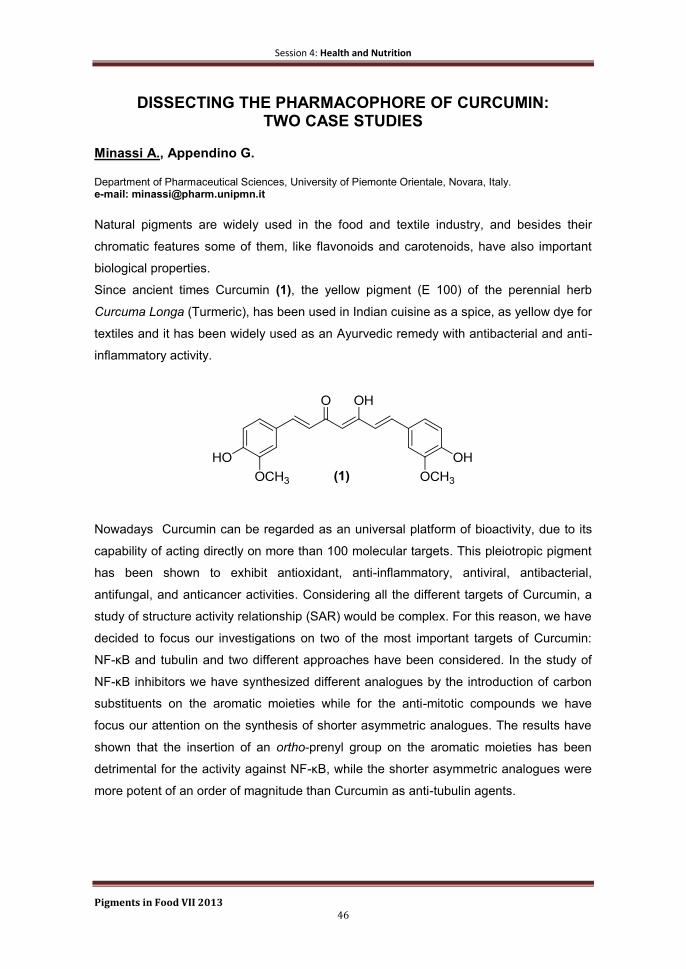
Session 4: Health and Nutrition
Pigments in Food VII 2013 46
DISSECTING THE PHARMACOPHORE OF CURCUMIN: TWO CASE STUDIES
Minassi A., Appendino G.
Department of Pharmaceutical Sciences, University of Piemonte Orientale, Novara, Italy. e-mail: [email protected]
Natural pigments are widely used in the food and textile industry, and besides their
chromatic features some of them, like flavonoids and carotenoids, have also important
biological properties.
Since ancient times Curcumin (1), the yellow pigment (E 100) of the perennial herb
Curcuma Longa (Turmeric), has been used in Indian cuisine as a spice, as yellow dye for
textiles and it has been widely used as an Ayurvedic remedy with antibacterial and anti-
inflammatory activity.
Nowadays Curcumin can be regarded as an universal platform of bioactivity, due to its
capability of acting directly on more than 100 molecular targets. This pleiotropic pigment
has been shown to exhibit antioxidant, anti-inflammatory, antiviral, antibacterial,
antifungal, and anticancer activities. Considering all the different targets of Curcumin, a
study of structure activity relationship (SAR) would be complex. For this reason, we have
decided to focus our investigations on two of the most important targets of Curcumin:
NF-κB and tubulin and two different approaches have been considered. In the study of
NF-κB inhibitors we have synthesized different analogues by the introduction of carbon
substituents on the aromatic moieties while for the anti-mitotic compounds we have
focus our attention on the synthesis of shorter asymmetric analogues. The results have
shown that the insertion of an ortho-prenyl group on the aromatic moieties has been
detrimental for the activity against NF-κB, while the shorter asymmetric analogues were
more potent of an order of magnitude than Curcumin as anti-tubulin agents.

Poster Session
Pigments in Food VII 2013 47
Poster Session

Poster Session
Pigments in Food VII 2013 48
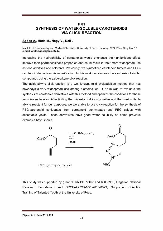
Poster Session
Pigments in Food VII 2013 49
P 01
SYNTHESIS OF WATER-SOLUBLE CAROTENOIDS VIA CLICK-REACTION
Agócs A., Háda M., Nagy V., Deli J.
Institute of Biochemistry and Medical Chemistry, University of Pécs, Hungary, 7624 Pécs, Szigeti u. 12 e-mail: [email protected]
Increasing the hydrophilicity of carotenoids would enchance their antioxidant effect,
improve their pharmacokinetic properties and could result in their more widespread use
as food additives and colorants. Previously, we synthetized carotenoid trimers and PEG-
carotenoid derivatives via esterification. In this work our aim was the synthesis of similar
compounds using the azide-alkyne click reaction.
The azide-alkyne click-reaction is a well-known, mild cycloaddition method that has
nowadays a very widespread use among biomolecules. Our aim was to evaluate the
synthesis of carotenoid derivatives with this method and optimize the conditions for these
sensitive molecules. After finding the mildest conditions possible and the most suitable
alkyne reactant for our purposes, we were able to use click-reaction for the synthesis of
PEG-carotenoid conjugates from carotenoid pentynoates and PEG azides with
acceptable yields. These derivatives have good water solubility as some previous
examples have shown.
This study was supported by grant OTKA PD 77467 and K 83898 (Hungarian National
Research Foundation) and SROP-4.2.2/B-10/1-2010-0029, Supporting Scientific
Training of Talented Youth at the University of Pécs.
Poster Session
Pigments in Food VII 2013 50
P 02
THERMAL AND LIGHT STABILITY OF β-CRYPTOXANTHIN ESTERS
Bunea A.,
Andrei S., Rugină D., Pintea A.
Department of Chemistry and Biochemistry, University of Agricultural Sciences and Veterinary Medicine, Cluj-Napoca, Romania e-mail: [email protected]
β-Cryptoxanthin esters are major components in orange fruits but also present in some
vegetables [1]. Comparable bioavailability of free and esterified β-cryptoxanthin was
found after supplementation in humans [2]. It was also showed that esterification with
saturated fatty acids increases the thermal stability of β-cryptoxanthin [3]. The aim of the
present study was the evaluation of thermal and light stability of β-cryptoxanthin esters
with saturated and unsaturated fatty acids.
β-Cryptoxanthin myristate, palmitate, oleate and linoleate were obtained by acylation with
the corresponding acyl chlorides in dry pyridine and purified by HPLC. The compounds
were dissolved in benzene and the initial concentration was adjusted at 3 +/- 0.2
micrograms/ml. Samples were kept at 40, 55 and 70ºC for up to 130 hours. For the light
stability test the samples were exposed to white light in the absence or in the presence of
oxygen for up to 300 hours. Residual concentration was determined by HPLC-PDA on a
C30 column. The rate constant of thermal and photoxidative degradation (k) and the half-
life periods (t½) were calculated by the integral method.
Esterification with both saturated and unsaturated fatty acids increased the thermal
stability of β-cryptoxanthin compared to the free form. However, the thermal stability of
unsaturated esters was lower than that of saturated ones. The light stability was not
significantly influenced by esterification. Cis-trans isomerisation products significantly
increased in samples exposed simultaneously to light and oxygen
Acknowledgement: This work was supported by PN-II-ID-PCE-2011-3-0721 research grant.
References
[1] Breithaupt, D.E. and Bamedi, A. Carotenoid esters in vegetables and fruits: A screening with
emphasis on β-cryptoxanthin esters. J Agric Food Chem. 2001, 49, 2064-2070
[2] Breithaupt, D.E. et al. Plasma response to a single dose of dietary β-cryptoxanthin esters from papaya
(Carica papaya L.) or non esterified β-cryptoxanthin in adult human subjects: a comparative study. British
J Nutr. 2003, 90, 795-801
[3] Fu, H.F. et al. Effect of esterification with fatty acid of β-cryptoxanthin on its thermal stability and
antioxidant activity by chemiluminescence method. Food Chem. 2010, 122, 602-609
Poster Session
Pigments in Food VII 2013 51
P 03
EFFECT OF ESTERIFICATION ON THERMAL STABILITY AND ANTIOXIDANT ACTIVITY OF ZEAXANTHIN
Pintea A., Bunea A., Socaciu C.
Department of Chemistry and Biochemistry, University of Agricultural Sciences and Veterinary Medicine, Cluj-Napoca, Romania e-mail: [email protected]
Hydroxycarotenoids often occur in plants in the form of esters with various fatty acids.
Carotenoid esters are major components in fruits and their thermal and photochemical
stability, as well as the antioxidant capacity are important attributes. The studies pointing
out the stability of xanthophylls esters are relatively scarce and the approaches used are
very different [1, 2]. The aim of the present study was the evaluation of thermal stability
and antioxidant capacity of different zeaxanthin esters compared to free zeaxanthin.
Zeaxanthin myristate, palmitate, oleate and linoleate were obtained by acylation with the
corresponding acyl chlorides in dry pyridine and purified by HPLC [3]. The compounds
were dissolved in benzene and the initial concentration was adjusted at 3 +/- 0.2
micrograms/ml. Samples were kept at 40, 55 and 70ºC for up to 130 hours. Residual
concentration was determined by HPLC-PDA on a C30 column. The rate constant of
thermal degradation (k) and the half-life periods (t½) were calculated by the integral
method. The antioxidant activities were determined by TEAC and FRAP methods
adapted for lipophilic compounds.
The zeaxanthin esters with saturated fatty acids had a better stability than free
zeaxanthin. The ester with unsaturated fatty acids have a lower stability than those with
saturated fatty acids but higher than free zeaxanthin.
Esterification with both saturated and unsaturated fatty acids did not significantly modify
the antioxidant activity of zeaxanthin, similar values being obtained by both methods for
free and esterified forms.
Acknowledgement: This work was supported by PN-II-ID-PCE-2011-3-0721 research grant.
References
[1] Perez-Galvez, A. and Minguez-Mosquera, M.I. Degradation of non-esterified and esterified
xanthophylls by free radicals. Biochim Biophys Acta 2002, 1569, 31-34
[2] Fu, H.F. et al. Effect of esterification with fatty acid of β-cryptoxanthin on its thermal stability and
antioxidant activity by chemiluminescence method. Food Chem. 2010, 122, 602-609
[3] Breithaupt, D.E and Bamedi, A. Carotenoid esters in vegetables and fruits: A screening with emphasis
on β-cryptoxanthin esters. J Agric Food Chem. 2001, 49, 2064-2070

Poster Session
Pigments in Food VII 2013 52
P 04
MEASUREMENT OF ENZYMATIC HYDROLYSIS OF LUTEIN ESTERS FROM DAIRY PRODUCTS DURING IN VITRO DIGESTION Xavier A.A.O.
1, Garrido-Fernández J.
1, Mercadante A.Z.
2, Pérez-Gálvez A.
1
1 Food Biotechnology Department, Instituto de la Grasa, CSIC, Avenida Padre García Tejero 4, 41012,
Sevilla, Spain. 2 Food Science Department, Faculty of Food Engineering, UNICAMP, Campinas, Brazil.
e-mail: [email protected]
Xanthophylls are commonly present as fatty acid esters in fruits and vegetables so that
dietary contribution of esterified xanthophylls is of marked value. As only free forms but
not esters are found in human serum and peripheral tissues, bioaccessibility of esterified
xanthophyll depends on the efficiency of the enzymatic hydrolysis during digestion1. In
this study we evaluated the bioaccessibility of lutein esters added to dairy products
(skimmed, semi-skimmed and whole milk or yogurt) using an in vitro digestion model.
Lutein content in the micellar fraction was measured by HPLC to determine
bioaccessibility, amount of lutein from the food matrix that is solubilised in mixed
micelles, and the hydrolysis efficiency reached during digestion, ratio of free lutein to
lutein esters in micelles. Milk and yogurt offer the same lutein bioaccessibility degree, but
depending on the fat content the efficiency was different. Thus, skimmed milk and yogurt
present a bioaccessibility efficiency lower than 15%, while the percentage transferred to
micelles in semi-skimmed or whole products was higher than 35%. Although lutein esters
were added to the products, lutein present in micelles was mainly in its free form either in
milk and yogurt with any fat content, and hydrolysis efficiency reached a 2:1 ratio.
Therefore, lutein esters are substrates for pancreatic lipase although previous work has
shown a different result2, and hydrolysis efficiency is not influenced by food structure and
amount of fat in the digesta.
References
[1] Pérez-Gálvez, A. et al. Esterification of xanthophylls and its effect on chemical behavior and
bioavailability of carotenoids in the human. Nutr. Res. 2005, 25, 631-640.
[2] Breithaupt, DE. et al. Carotenol fatty acid esters: easy substrates for digestive enzymes? Comp.
Biochem. Physiol. B Biochem. Mol. Biol. 2002, 132, 721–728.

Poster Session
Pigments in Food VII 2013 53
P 05
OIL BODIES AS A POTENTIAL MICROENCAPSULATION CARRIER FOR ASTAXANTHIN STABILIZATION AND SAFE
DELIVERY Acevedo F.
1,2, Rubilar M.
1,2, Villarroel M.
1, Navarrete P.
3,4, Jofré I.
3, Romero F.
3,
Acevedo V.2,5
, Shene C.1,2
1Agriaquaculture Nutritional Genomic Center, CGNA (R10C1001), Technology and Processes Unit,
Universidad de La Frontera, Casilla 54-D, Temuco, Chile 2Scientific and Technological Bioresource Nucleus, BIOREN, Universidad de La Frontera, Casilla 54-D,
Temuco, Chile 3Center of Neurosciences and Peptides Biology- CEBIOR-BIOREN-CEGIN, Universidad de La Frontera,
Temuco, Chile. 4Laboratorio Neurobiología Molecular y Celular II, Departamento de Neurobiología Molecular y Celular,
Instituto de Neurobiología, Campus Juriquilla-Querétaro, UNAM, México. 5Programa de Doctorado en Ciencias de Recursos Naturales, Universidad de La Frontera, Casilla 54-D,
Temuco, Chile e-mail: [email protected]
Natural astaxanthin (AST) is a particularly valued molecule because of its high
antioxidant properties. However, AST is extremely sensitive to oxidation and thus its
bioactive properties decrease during processing and storage. Oleosomes isolated from
plant seeds are oil bodies (OB) consisting of a triacylglycerol core bound by a
phospholipid monolayer embedded with proteins known as oleosins [1,2,3]. The
purposes of this study were to define conditions for microencapsulating astaxanthin in oil
bodies (OB) from Brassica napus for the enhancement of its oxidative stability, and to
test the bioactivity of the microencapsulated AST (AST-M) in CRL1730 endothelial cells.
Conditions for maximizing microencapsulation efficiency (ME) [AST/OB, stirring speed
and contact time] were determined using the Response Surface Methodology. The
highest ME (> 99 %) was obtained with an AST/OB ratio of 0.14, 200 rpm, 5.3 h. OB
loaded with AST showed a strong electrostatic repulsion in a wide range of pH (5.5 to
9.5) and ionic strength (0 – 150 mM NaCl). It was found that the half-life of AST-M
exposed to air and light was twice as long as free AST, which indicates the protective
role of OB. In addition, AST-M at a non-cytotoxic concentration (1 µg/mL) had a
protective effect against reactive oxygen species. These results suggest that OB isolated
from oil seeds offer a novel option for stabilizing and delivering AST with bioactive
properties in industrial applications in the food and pharmaceutical fields.
Acknowledgements
This research was supported by funding from Conicyt through Fondecyt project 3120022, and project
DI10-7006 from the Universidad de La Frontera. Patricia Navarrete thanks the support provided by the
postdoctoral fellowship from CTIC-UNAM and BECAS Chile.
Poster Session
Pigments in Food VII 2013 54
References
[1] Huang, A.H. Oil bodies and oelosins in seeds. Annu. Rev. Plant Physiol. Plant Mol. Biol. 1992, 43,
177-200
[2] Moloney, M.M., Sembiosys Genetics Inc. Oil-body proteins as carriers of high-value peptides in plants.
1997, United States patent US 5650554.
[3] Boucher, J. et al. Sorption of hydrophobic organic compounds (HOC) in rapeseed oil bodies.
Chemosphere. 2008, 70, 1452–1458
Poster Session
Pigments in Food VII 2013 55
P 06
MICROENCAPSULATION OF ASTAXANTHIN OLEORESIN FROM Phaffia rhodozyma
Villalobos-Castillejos F.
1, Yáñez-Fernández J.
2, Barragán-Huerta B.E.
1
1Departmento de Ingeniería en Sistemas Ambientales. Escuela Nacional de Ciencias Biológicas. Instituto
Politécnico Nacional. Av. Wilfrido Massieu S/N UP Adolfo López Mateos. México, D.F. 07738. Mexico. 2Departamento de Ingeniería en Alimentos. Unidad Profesional Interdisciplinaria de Biotecnología.
Instituto Politécnico Nacional. Av. Luis Enrique Erro S/N, UP Adolfo López Mateos, México D.F. CP 07738, Mexico. e-mail: [email protected]; [email protected]
Astaxanthin is a carotenoid used in functional foods for humans because of its
antioxidant activity higher than Vitamin E, however its use in foods is limited due to their
low solubility in water, in addition it has a low stability because is affected by the
oxidation and could be easily isomerized due to heat, acids or light [1,2]. In this study,
the astaxanthin was extracted from the yeast cells using the microwave as pretreatment
at low power (105 W) for 1 minute, followed by extraction with ethyl acetate. The
extraction was optimized using a design Box-Behnken, resulting as the best extraction
conditions, a temperature of 65 ºC, for 24 minutes, and a solid-solvent ratio of 1:19,
obtaining an astaxanthin oleoresin. The astaxanthin oleoresin was dispersed using
propylene glycol. Water-dispersible oleoresin encapsulated were obtained using a spray
dryer, with inulin and maltodextrin as wall materials with inlet/outlet temperatures of
150/100 and 105/67 °C respectively. The encapsulated with inulin were more stable in
solution (t½=15.33±0.686 h) than maltodextrin (t½=1.13±0.141 h). The shelf life of
maltodextrin (t½=34.48 ±1.57 h) was higher than inulin (t½=12.16±0.622h) at 25 °C. The
encapsulated presented a simple form and a diameter between 1 – 10 μm. The
antioxidant capacity of the oleoresin (mM Trolox / g sample) was of 1.135 ± 0.048 and
for microcapsules the values of 10.105 ± 0.502 and 10.509 ± and 0.281 was obtained for
inulin and maltodextrin respectively.
References
[1] Chen, X. et al. The preparation and stability of the inclusion complex of astaxanthin with β-
cyclodextrin. Food Chemistry. 2006, 101, 1580-1584.
[2] Fratianni, A. et al. Degradation in orange juice during microwave heating. LWT- Food Science and
Technology, 2010, 43, 867-871.
Poster Session
Pigments in Food VII 2013 56
P 07 EFFECT OF GENOTYPE AND GROWING CONDITIONS ON LUTEIN AND β-CAROTENE CONTENT OF GREEN LEAFY
BRASSICA SPECIES
Arrigoni E.1, Reif C.
1, Berger F.
1, Baumgartner D.
1, Nyström L.
2
1Research Station Agroscope Changins-Wädenswil ACW, Wädenswil, Switzerland.
2 ETH Zurich, Institute of Food, Nutrition and Health, Zurich, Switzerland.
e-mail: [email protected]
Brassicaceae vegetables are rich in nutritionally important constituents such as vitamins
and minerals and they contain a broad spectrum of various secondary plant metabolites
[1]. Their composition and content depends on genus and species, but also on genotype
and agricultural factors. While Brassicaceae are particularly known for their high
glucosinolate content, green leafy species are also rich in lutein and β-carotene.
Moreover, their carotenoid content correlates with chlorophyll concentration.
The aim of the present study was to evaluate the impact of genotype and growing
method on lutein and β-carotene contents of Japanese Greens and to establish
correlations between carotenoid and chlorophyll contents. Twelve green leafy Brassica
cultivars were produced in two different series. For spring production seeds were grown
in the greenhouse either in beds or in pots, whereas summer cultivations took place
either on open fields or under tunnels. Lutein, β-carotene as well as chlorophyll a and b
contents of Japanese Greens were determined by HPLC-DAD [2].
Overall, lutein concentrations ranged from 3.8 to 10.5 mg/100 g FM and β-carotene
contents from 2.1 to 6.8 mg/100 g FM. Botanical classification showed the strongest
impact on carotenoid contents with B. rapa subsp. chinensis producing significantly more
carotenoids than B. rapa subsp. nipposinica and B. juncea. The effect of growing
conditions was less pronounced, but slightly higher β-carotene contents were observed
in summer. In conclusion, choosing optimal genotypes and to some extent the
manipulation of growing conditions offer strategies to increase lutein and β-carotene
concentrations.
References
[1] Jahangir, M.et al. Health-affecting compounds in Brassicaceae. Compr. Rev. Food Sci. F. 2009, 8, 31-
43
[2] Reif, C. et al. Carotenoid database of commonly eaten Swiss vegetables and their estimated
contribution to carotenoid intake. J. Food Compos. Anal. 2013, 29, 64-72

Poster Session
Pigments in Food VII 2013 57
P 08
EFFECT OF PROCESSING ON CONTENT OF VITAL CAROTENOIDS IN NEW VEGETABLE PUREE
Palotás Gabor
1, Palotás Gabriella
1, Daood H.
2, Pék Z.
2, Helyes L.
2
1: Univer Product Plc, Szolnoki út 35, H-6000 Kecskemét, Hungary 2: Szent István University, Páter K.u.1, H-2301 Gödöllő, Hungary email: [email protected]
Vital carotenoids such as lycopene, β-carotene and their biologically active derivatives
have received an increasing attention during the last decades due to their association
with the reduced risk of heart diseases and cancer. Recent investigations and
researches suggested and recommended consumption of considerable amount of plant-
derived products rich in bioactive nutrients such as carotenoids, poly-phenols and other
bio-antioxidant compounds daily. The objective of the present work was to study the
changes in content and composition of carotenoids as a function of processing of new
vegetables puree (cream) prepared from red bell pepper, eggyplant, tomato paste and
onion under pilot plant conditions including washing, cutting, mixing in a double-jacket
mixer, heating at 95°C for 2 min. The puree was packaged in plastic dishes, cooled and
stored at 2-6°C.
Carotenoids were extracted by a conventional procedure using methanol first followed by
1,2-dichloroethane. The individual carotenoids were separated and determined by HPLC
method using analytical RP-column and stepwise gradient elution starting with water-
methanol and ending with methanol-acetonitrile-isopropanol. Identification was based on
comparison of spectral characteristics and retention behaviour of sample carotenoids
with those of standard materials or with literature data.
The red-bell pepper was the main source of vital zeaxanthin, β-cryptoxanthin and β-
carotene and non-vital xanthiophylls, while sufficient amounts of bio-active lycopene
were derived from tomato paste in the puree. It was found that processing caused
substantial loss of carotenoids of red pepper, while lycopene was markedly protected
against thermal degradation indicating that red pepper carotenoids (mainly xanthophylls
and their esters) were functioning as the first oxidation barrier under the chemical
conditions of the new vegetable puree. These results are of special importance in the
project since the aim is to produce vegetable puree rich in bio-active carotenoids beside
other antioxidants and has accentuated sensory properties that increase consumer
acceptance for the product.
This work is a part of research project supported financially by Hungarian Development Agency (NFÜ)
under grant no. TECH-09-A3-2009-0230 USOK2009.
Poster Session
Pigments in Food VII 2013 58
P 09
EFFECT OF ADDITION OF SODIUM ERYTHORBATE AND URUCUM ON THE LIPID OXIDATION IN PORK MEAT
Figueiredo B. , Bragagnolo N. Department of Food Science, University of Campinas, São Paulo, Brazil e-mail: [email protected]
Lipid oxidation is a major cause of food deterioration being responsible for the generation
of undesirable flavors, decrease of shelf life, loss of nutritional value and formation of
harmful compounds to human health, such as cholesterol oxides[1]
. This study
investigated the effect of sodium erythorbate (0.1%) and annatto powder (0.05%) on the
inhibition of fatty acid and cholesterol oxidation in pork loin over 120 days of storage and
subsequent heat treatment. Cholesterol and cholesterol oxides were identified and
quantified simultaneously by HPLC-PDA-RID[2]
and fatty acids by GC-FID[3]
. TBARS
values were determined by spectrophotometry. Five cholesterol oxides were identified:
7-ketocholesterol, α-epoxycholesterol, β-epoxycholesterol, 7α-hydroxycholesterol and
7β-hydroxycholesterol. The addition of annatto, sodium erythorbate and a mixture of
sodium erythorbate with annatto inhibited the formation of cholesterol oxides since these
compounds were found only in the control sample. The lowest TBARS values were
observed in the samples containing annatto, sodium erythorbate and a mixture of sodium
erythorbate and annato during the first 75 days of storage. During storage the control
sample showed a 18% reduction in PUFA´s levels while the samples containing annatto,
sodium erythorbate and a mixture of sodium erythorbate with annatto showed a
reduction of 9%. On the other hand, heat treatment decreased MUFA and PUFA content
despite the addition of annatto, sodium erythorbate and mixture of sodium erythorbate
with annatto. The results demonstrated that the addition of mixture of erythorbate and
annatto had higher antioxidant effect on lipid fraction of pork meat than the addition of
each antioxidant alone.
References
[1] Erickson, M. C Lipid Oxidation of muscle foods. In: Food Lipids. Akoh, C. C, Min, D. B., ed. Marcel
Dekker, Inc, 3rt ed, 2008, 321-363
[2] Saldanha, T. et al. HPLC separation and determination of 12 cholesterol oxidation products in fish:
comparative study of RI, UV and APCI-MS detectors. J. Agric. Food Chem. 2006, 54, 4107-4113
[3] Sancho, R. A. S. et al. Effect of annatto seed and coriander leaves as natural antioxidants in fish
meatballs during frozen storage. J. Agric Food Sci. 2011, 76, 838-845

Poster Session
Pigments in Food VII 2013 59
min5 10 15 20 25 30 35 40
mAU
0
200
400
600
800
1000
1200
DAD1 A, Sig=450,4 Ref=off (C30MA\MARACHIN1000003.D)
1
2
34
5 67 8
9
10
11
1213
14
15
16
17
18
19
20
21
22
P 10
IDENTIFICATION OF Cionosicyos macranthus CAROTENOIDS Murillo E., Watts M., Reyna G. Departamento de Bioquímica, Universidad de Panamá, Panamá e-mail: [email protected]
Cionosicyos macranthus (Chinese passion fruit) is a cucurbit fruit, native of Central
America and Mexico, which grows as the passion fruit. The pulp of this fruit is red by the
high content of carotenoids with termination keto-κ. We recently found that Pouteria
sapota, other fruit native to this region, is also characterized by the high content of
carotenoids with termination keto-κ. This is the first report on the separation and
identification of all Cionosicyos macranthus carotenoids. The samples used were
obtained directly from the tree and extracted fresh. Carotenoids were identified by their
spectra UV / Vis and molar mass, obtained by HPLC-DAD and HPLC-MS, respectively.
To minimize the interference caused by the overlapping of peaks, the saponified extract
was separated into four fractions by column chromatography packed with Al2O3 (ether:
hexane 10%, ether:hexane 50%, ether 100% and methanol:ether 5%) . The figure shows
the HPLC chromatogram of the original extract saponified, with the numbers
corresponding to carotenoids identified.
The cryptoxanthin (peak 20) is the main carotenoid of Cionosicyos macranthus, followed
cryptocapsin (peak 19). The composition of the extract indicates most carotenoids are
epoxides or derivatives thereof. It is known that κ ring carotenoids are formed by pinacol
arrangement of 5,6-epoxides. When comparing Cionosicyos macranthus with Pouteria
sapota carotenoids, we found that both are the same but in different proportions.
Poster Session
Pigments in Food VII 2013 60
P 11
BIOACTIVE COMPOUNDS IN SUPERCRITICAL CO2-EXTRACTED PUMPKIN OIL
Durante M.
1, Lenucci M.S.
2, D'Amico L.
1, Dalessandro G.
2, Mita G.
1
1 Istituto di Scienze delle Produzioni Alimentari-CNR, Via Prov.le Lecce-Monteroni, 73100 Lecce, Italy
2 Dipartimento di Scienze e Tecnologie Biologiche ed Ambientali (D ,
Via Prov.le Lecce-Monteroni, 73100 Lecce, Italy e-mail: [email protected]
Pumpkin (Cucurbita moscata Duch.) flesh has an intense yellow/orange color owing to
its high levels of carotenoids, mainly α- and β-carotene, β-cryptoxanthin, lutein and
zeaxanthin [1]. These pigments are largely used in food preparation to make color
appealing to consumers and improve food healthiness and stability because of their
antioxidant and pro-vitamin A activities. Supercritical carbon dioxide (SC-CO2) is a
“green” technology suitable for carotenoid extraction. It gives extracts totally free of toxic
organic solvents.
Few reports describe SC-CO2 carotenoid extraction from pumpkin [2,3]. The authors
used freeze-dried and milled flesh to feed the extractor, getting a maximum carotenoid
yield of ~10.9 mg/100 g dry weight (d.w.). In this work a process for obtaining a
considerably higher carotenoid yield by SC-CO2 extraction from dehydrated pumpkin
flesh is described. Our results show that dehydration method is a key factor to improve
carotenoid extraction from pumpkin. Oven-drying increased SC-CO2 extraction yield of
total carotenoids of ~8.5 folds with respect to freeze-drying (49.2 mg/100 g d.w. vs 5.8
mg/100 g d.w., respectively). Moreover, mixing the oven-dried flesh with milled pumpkin
seeds (1:1 by weight) further increased carotenoid yield of ~1.6 fold (79.2 mg/100 g d.w.
of fed pumpkin flesh). At the same time, it led to an enrichment of the extract with
tocopherols, tocotrienols and PUFA from seed oil.
These findings encourage further studies in order to scale up the process for possible
industrial production of high quality bioactive ingredients from pumpkin for functional food
or cosmeceutical preparations.
References
[1] Rodriguez-Amaya, D. B., Kimura, M., Godoy, H. T., Amaya-Farfan, J., Updated Brazilian database on
food carotenoids: Factors affecting carotenoids composition. J Food Comp Anal. 2008, 21, 445–463.
[2] Seo, J. S., Burri, B. J., Quan, Z., Neidlinger, T. R., Extraction and chromatography of carotenoids from
Pumpkin. J Chromatogr A 2005, 1073: 371−375.
[3] Shi, J., Chun, Y., Xingqian, Y., Xue, S., Jiang, Y., Ma, Y., Liu, D., Effects of supercritical CO2 fluid
parameters on chemical composition and yield of carotenoids extracted from pumpkin. LWT – Food Sci
and Technol 2010, 43: 39-44.

Poster Session
Pigments in Food VII 2013 61
P 12
EVALUATION OF CAROTENOIDS AND CAPSAICINOIDS CONTENT IN POWDER OF CHILLI PEPPERS DURING ONE YEAR
OF SHELF-LIFE Giuffrida D.
1, Cavazza A.
2, Dugo P.
3, Torre G.
3, Corradini C.
2, Bignardi C.
2,
Dugo G.mo1
1 Dipartimento di Scienze dell’Ambiente, della Sicurezza, del Territorio, degli Alimenti e della Salute
(S.A.S.T.A.S.). Università degli Studi di Messina, Viale Ferdinando Stagno d’Alcontres, 31, 98166 S. Agata. Messina, Italy 2 Dipartimento di Chimica, Parco Area delle Scienze 17/A, 43124, Università degli Studi di Parma, Italy
3 Università di Messina, Dipartimento farmaco-chimico, Viale Annunziata, 98168 Messina, Italy
e-mail: [email protected]
Red peppers are often employed as a source of pigment with the purpose of conferring
colour to food preparations. The intense red colour is conferred to the fruits by the
presence of almost 50 carotenoids, both free or esterified with fatty acids. The use of
powdered dried fruits has largely increased during the last years all over the world.
During storage of the powdered berries, a progressive loss of colour intensity is
observed, causing the product to change from brilliant red to dull brown. The oxidative
degradation of carotenoids seems to be the main reason for colour degradation. Factors
as high temperature, humidity, water activity, exposure to light and contact with oxygen
have been found to play an important role in causing deterioration.
Piquancy is the main tasty feature of red chilli peppers, and it is assured by the presence
of a group of molecules belonging to the family of capsaicinoids. In the present study, the
profiles of carotenoids and capsaicinoids in paprika samples stored along one year
under different conditions have been recorded. Liquid chromatography coupled to mass
spectrometry has been employed for carotenoid analysis, while DAD detector was used
for capsaicinoids detection. Samples of powdered dried berries were stored at room
temperature and in the dark in envelopes made of paper/PET. One more aliquot was
stored at refrigerating conditions (4°C). Aliquots of each sample were analysed along a
12 months period. Data obtained showed that carotenoids higher retention occurred in
the samples stored under refrigerating condition with a relative small percentage loss
going from 6 to 12 months of storage; whereas the carotenoid retention in sample stored
at R.T., significantly decreased going from 6 to 12 months of storage.
Poster Session
Pigments in Food VII 2013 62
As for capsaicinoids, no differences were found during the first six months-period in all
aliquots stored at the different conditions, while from nine months onwards a slight
decrease in capsaicinoids content was registered in the samples stored at room
temperature. On the contrary, the piquancy of the refrigerated samples was found
unaltered during the entire period.

Poster Session
Pigments in Food VII 2013 63
P 13
CAROTENOIDS IN RED FLESHED SWEET ORANGES Merussi G.D.
1, Latado R.R.
2, Rossi E.A.
1, Sylos C.M.
1
1 Department of Food and Nutrition, School of Pharmaceutical Sciences, UNESP-São Paulo State
University, Araraquara, SP, Brazil. 2Ctr APTA Citros Sylvio Moreira, Instituto Agronômico de Campinas, Cordeiropolis, SP, Brazil
e-mail: [email protected]
The orange carotenoids are responsible for the color of the peel and juice that are
important quality factor, influencing its acceptance for the consumers and have also
important functions for the human health. Orange is the fruit more consumed worldwide
both as fresh and juice. In Brazil are being cultivated varieties of red fleshed sweet
oranges, which have distinct red pulp due to the presence of the lycopene in their
carotenoid composition. The aim of this study was to compare three varieties of red
fleshed sweet oranges (Sanguínea de Mombuca, Baia Cara-Cara and Valência Puka
with a variety Valencia in relation to the major carotenoids composition and color
parameters. The carotenoids were separated and quantified by liquid chromatography
and with C18 column. The mobile phase was composed of acetonitrile:methanol:ethyl
acetate, in gradient elution: 0 min (99:1:0), 30 min (64:1:35), 35 min (64:1:35) and 58 min
(99:1:0)1. Flow rate was 0.6ml/min and the peaks were monitored to 450 nm. The results
showed that red fleshed sweet oranges when compared with Valencia had juices less
acid and a darker color with greater saturation of red coloration and higher vivacity. The
content of carotenoids showed no significant differences (p< 0.05) between the varieties
of red pulp oranges, but these proved to have 20-76% more total carotenoid that
Valencia oranges2, and the same behavior was observed for concentrations of -
carotene, -cryptoxanthin, and lutein which respectively showed an increase 230-375%,
36-65% and up to 37%.
References
[1] Gama J.J.T. and Sylos, C.M. Major carotenoid composition of Brazilian Valencia orange juice:
identification and quantification by HPLC. Food Res. Int. 2005,38, 899-903.
[2] Melendez-Martinez A.J., Vicario I.M., Heredia F.J. Review: Analysis of carotenoids in orange juice. J.
Food Compos. Anal. 2007, 20, 638-649.
Poster Session
Pigments in Food VII 2013 64
P 14
COLOUR CHANGES OF HEAT-TREATED ORANGE JUICE DURING AMBIENT STORAGE
Wibowo S., Vervoort L., Lemmens L., Hendrickx M., Van Loey A. Laboratory of Food Technology, Leuven Food Science and Nutrition Research Center (LFoRCe), Department of Microbial and Molecular Systems (M²S), Katholieke Universiteit Leuven, Kasteelpark Arenberg 22 box 2457, 3001 Heverlee, Belgium e-mail: [email protected]
Orange juice is one of the most consumed fruit juices and appreciated for its high
content of vitamin C. The attractive orange colour is known as an important sensory
attribute to evaluate juice quality. Changes of colour during processing and storage
indicate the occurrence of several chemical reactions, one of them is non-enzymatic
browning. Ascorbic acid degradation is considered as one of the main contributors to
browning in orange juice1. Meanwhile, Maillard reaction may also occur but is considered
to be of minor importance2. Several components such as ascorbic acid, sugars, citric
acid, and oxygen were identified as major factors affecting browning in orange based
juices3.
Studies have been undertaken to understand the cause of browning and identify the
main factors involved in colour changes in orange juice. Pasteurised single-strength
orange juice (11.2 °Brix) in PET bottles were stored at 20, 28, 35 and 42 °C. Change in
colour was assessed using the CIE-L*a*b* colour system. The L*value decreased during
storage which indicates a darker appearance of the juice. Higher losses of total vitamin C
and oxygen were found at higher storage temperature. After 8 weeks of storage, the
vitamin C retention was 25% at 42 °C meanwhile at 20 °C the retention was 68%. A
sharp decrease from the first to the second week of storage may imply oxidative
changes in the orange juice. Furthermore, kinetic modelling has been applied to describe
the evolution of several quality attributes of orange juice during storage.
References
[1] Roig, M. et al. Studies on the occurrence of non-enzymatic browning during storage of citrus juice.
Food Res. Int. 1999, 32, 609–619
[2] Clegg, K.M. Non-enzymic browning of lemon juice. J. Sci. Food Agric. 1964, 15, 878–885
[3] Shinoda, Y. et al. Browning and decomposed products of model orange juice. Biosci. Biotechnol.
Biochem. 2004, 68 (3), 529–536
Poster Session
Pigments in Food VII 2013 65
P 15
CAROTENOID DEPOSITION AND PROFILES IN PEACH PALM (BACTRIS GASIPAES KUNTH) FRUITS, AND THEIR IMPLICATION
ON ITS NUTRITIONAL POTENTIAL Hempel J.
1, Esquivel P.
2, Carle R.
1, Schweiggert R.M.
1
1 Institute of Food Science and Biotechnology, Hohenheim University, Germany,
2 School of Food Technology, University of Costa Rica, Costa Rica.
e-mail: [email protected]
The tropical peach palm develops 3-6 cm broad, edible drupes with a yellow, orange, or
red exocarp. The yellow to light orange fruit pulp has been reported to be rich in starch,
lipids and provitamin A carotenoids1.
In this study, major nutrients and the genuine carotenoid profile of highly diverse peach
palm fruits, purchased at local Costa Rican markets, were investigated. Selected
accessions showed widely ranging levels of (all-E)-β-carotene (0.4-5.4 mg/100 g on
fresh weight (FW)) and (Z)-γ-carotene isomers (total 0.2-5.3 mg/100 g on FW),
representing predominant carotenoids of the raw mesocarp. Total carotenoid levels in
the edible portion of the fruit ranged from 0.7 mg to 14.6 mg/100 g on FW. Orange-
fleshed fruits were shown to contain 9- and 6-fold higher amounts of total carotenoids
and (all-E)-β-carotene, respectively, than yellow-fleshed ones. Provitamin A equivalents,
i.e. retinol activity equivalents (RAE), of uncooked fruits ranged from 38 μg to 642 μg
RAE/100 g on FW.
Light microscopic studies of mesocarp tissue revealed the occurrence of globular
chromoplasts, where carotenoids are assumed to be lipid-dissolved. Solubility
estimations based on lipid contents of the fruit supported carotenoids to be deposited in
a lipid-dissolved state, since fruit-own lipid contents would suffice to dissolve total
carotenoids contained. Consequently, improved bioaccessibility of β-carotene and further
lipid-dissolved carotenoids from peach palm fruit is expected due to their deposition in
globular-tubular chromoplasts2. In summary, peach palm fruits represent a most
promising source for dietary provitamin A, potentially being highly bioavailable due to
their concomitant high lipid contents.
References
[1] Lieberei, R., Reisdorf, C. Nutzpflanzenkunde. 2007, Georg Thieme Verlag, Stuttgart, 243-244
[2] Schweiggert, R.M., Mezger, D., Schimpf, F., Steingass, C.B., Carle, R. Influence of chromoplast
morphology on carotenoid bioaccessibility of carrot, mango, papaya, and tomato. Food Chem. 2012, 135,
2736-2742
Poster Session
Pigments in Food VII 2013 66
P 16
DEPOSITION OF LYCOPENE, β-CAROTENE, AND β-CRYPTOXANTHIN IN DIFFERENT CHROMOPLAST
SUBSTRUCTURES IN PAPAYA FRUITS
Schweiggert R.M.1, Steingass C.B.
1, Heller A.
2, Esquivel P.
3, Carle R.
1
1Institute of Food Science and Biotechnology, Hohenheim University, Germany.
2Institute of Botany, Hohenheim University, Germany.
3School of Food Technology, University of Costa Rica, Costa Rica.
e-mail: [email protected]
Papaya fruits commonly are a rich source of β-carotene and esterified β-cryptoxanthin,
whereas the additional accumulation of lycopene was only observed in red-fleshed
papaya fruits[1]
. In this study, the role of different chromoplast substructures should be
elucidated, considering the deposition form of the respective carotenoids. For this
purpose, carotenoid profiles of one red- and one yellow-fleshed genotype were
compared using HPLC-PDA-MSn in order to confirm similarity of patterns regarding the
occurrence of β-carotene and β-cryptoxanthin esters. Both (all-E)-lycopene and some
minor (Z)-lycopene isomers were demonstrated to exclusively occur in the red-fleshed
genotype. Green-ripe as well as fully ripe yellow- and red-fleshed fruits were
subsequently investigated by light and electron microscopy, identifying the carotenoid-
bearing chromoplast substructures. Undifferentiated proplastids and plastids with
plastoglobules were observed in the uncoloured mesocarp of green-ripe fruit. In both ripe
fruits, tubular chromoplast structures were found being associated with the deposition of
β-carotene and β-cryptoxanthin. Crystalloid elements were exclusively identified in red-
fleshed genotypes and, therefore, hypothesized to be the deposition site of lycopene.
Electron micrographs from lycopene-rich tomato mesocarp revealed striking similarities
to the “lycopenic” substructures in red-fleshed papaya chromoplasts[2]
. While carotenoids
in chromoplast tubules were previously described to be deposited in a liquid-crystalline
physical state, crystalloid substructures were related to the solid crystalline accumulation
of the respective carotenoid[3]
.
References
1. Schweiggert RM, Steingass CB, Esquivel P, Carle R. Chemical and morphological characterization of
Costa Rican papaya (Carica papaya L.) hybrids and lines with particular focus on their genuine
carotenoid profiles. J Agric Food Chem 2012, 60, 2577-2585.
2. Schweiggert RM, Steingass CB, Heller A, Esquivel P, Carle R. Characterization of chromoplasts and
carotenoids of red and yellow fleshed papaya (Carica papaya L.). Planta 2011, 234, 1031-1044.
3. Sitte P, Falk H, Liedvogel B. Chromoplasts. In: Czygan, F, (ed.) Pigments in plants. Stuttgart: G
Fischer, 1980, 117-148.
Poster Session
Pigments in Food VII 2013 67
P 17
EVALUATION OF QUALITY PARAMETERS AND CAROTENOID CONTENT OF THREE CULTIVARS OF MANGO
(MANGIFERA INDICA L.) FROM REUNION ISLAND
Rosalie R.1, Chillet M.
2, Joas J.
3, Lechaudel M.
4, Payet B.
1, Vulcain E.
2,
Dufossé L.1
1 Laboratoire de Chimie des Substances Naturelles et des Sciences des Aliments, Université de La
Réunion, Sainte Clotilde, Ile de La Réunion, France. 2 CIRAD, UMR QUALISUD, Sainte Clotilde, Ile de La Réunion, France.
3 CIRAD, UMR QUALISUD, Montpellier, France.
4 CIRAD, HORTSYS, Saint Pierre, Ile de La Réunion, France.
e-mail: [email protected]
Mango (Mangifera indica L.) is a well-accepted fruit worldwide thanks to its qualities,
such as taste, aroma, color and health promoting content, making it one of the first
tropical fruit produced [1]
. Most imported mangoes in Europe are “American-type”
characterized by a blush on the fruit’s sun exposed side (ex: Kent). In this study, we
compared three cultivars produced in Reunion Island: Kent (American-type, well known
in international market), Cogshall (American-type) and José (Indian-type, imported by the
first Reunion Island inhabitants). Fruit mass, Total Titrable Acidity (TTA), Total Soluble
Solids (TSS) and carotenoid content of flesh were measured on ripe fruits harvested at a
commercial maturity stage and ripened in relative humidity and temperature controlled
chamber [2]
. Mango taste was estimated using TSS/TTA ratio indicating that José was
sweeter than the two other cultivars, with a ratio about 10. Cogshall and Kent had a lower
ratio, about 4 and 3, respectively. Total carotenoid content suggested that Kent mangoes
had the highest level followed by Cogshall and José, equal to 44.9, 35.9 and 34.3µg eq.
β-carotene/g Fresh Matter, respectively. A correlation was found between the hue angle
(h°) value and carotenoid content for American-type mangoes Kent and Cogshall
showing a direct link between the two cultivars. Subsequently quantification of Cogshall’s
carotenoid content was experimented using High Performance Liquid Chromatography.
Identification was obtained by comparison of retention times versus pure commercial
standards [3]
. This study allowed us to get a first identification of Cogshall’s carotenoids
and quantify the β-carotene.
References
[1] Tharanathan, R. N. et al. Mango (Mangifera indica L.), "The king of fruits" - an overview. Food Reviews
International 2006, 22, 95-123
[2] Joas, J. et al. "Comparison of postharvest changes in mango (cv Cogshall) using a Ripening class
index (Rci) for different carbon supplies and harvest dates." Postharvest Biol Technol 2009, 54, 25-31
Poster Session
Pigments in Food VII 2013 68
[3] Ornelas-Paz, J. d. J. et al. Identification and Quantification of Xanthophyll Esters, Carotenes, and
Tocopherols in the Fruit of Seven Mexican Mango Cultivars by Liquid Chromatography-Atmospheric
Pressure Chemical Ionization-Time-of-Flight Mass Spectrometry [LC-(APcI+)-MS]. J Agric Food Chem
2007, 55, 6628-6635

Poster Session
Pigments in Food VII 2013 69
P 18
GENUINE PROFILES AND BIOACCESSIBILITIES OF CAROTENOIDS FROM RED- AND YELLOW-FLESHED MAMEY
SAPOTE (Pouteria sapota) FRUITS Chacón-Ordóñez T.
1, Jiménez V.M.
2, Esquivel P.
3, Carle R.
4, Schweiggert R.M.
4
1 School of Biology, University of Costa Rica, Costa Rica
2 CIGRAS, Universidad de Costa Rica, 2060 San Pedro, Costa Rica.
3 School of Food Technology, University of Costa Rica, Costa Rica
4 Institute of Food Science and Biotechnology, Hohenheim University, Ger
many
e-mail: [email protected]
Mamey sapote (Pouteria sapota (Jacq.) H.E. Moore & Stearn) is an underutilized
carotenoid-rich fruit containing rare κ-ring carotenoids such as the newly identified
sapotexanthin [1]. Although different genotypes showing distinct pulp colors are
available, knowledge on differences in their carotenoid composition is still scarce.
Therefore, the goal of the present research work was to qualitatively and quantitatively
describe the carotenoid profiles of different genotypes using HPLC-DAD coupled to
mass spectrometry. Validation of the extraction method applied was performed for
violaxanthin, β-cryptoxanthin, and β-carotene. While high amounts of sapotexanthin and
cryptocapsin were observed in red-fleshed genotypes, high concentrations of free β-
cryptoxanthin and violaxanthin esters were found, for the first time in yellow-fleshed
fruits. However, β-carotene contents were comparatively low, resulting in low retinol
equivalent values for both red and yellow-fleshed sapote. An in vitro digestion model was
used to assess the liberation of sapote carotenoids during digestion, indicating low
carotenoid bioaccessibility from both the raw and cooked fruit matrix, possibly due to the
high contents of acid-labile violaxanthin esters.
References
[1] Murillo, E, McLean, E, Britton, G, Agócs, A, Nagy, V, Deli, J. Sapotexanthin, an A-provitamin
carotenoid from red mamey (Pouteria sapota). J. Nat. Prod. 2011, 74 (2), 283–285.

Poster Session
Pigments in Food VII 2013 70
P 19
TRANSGENIC TOMATOES AND THEIR CAROTENOID AND FLAVOUR PROFILES
Höfelmeier H.
1, Burmeister A.
1, Schwab W.
2, Fleischmann P.
1
1 Institute of Food Chemistry, TU Braunschweig, Schleinitzstrasse 20, 38106 Braunschweig, Germany 2 Program Biotechnology of Natural Products, TU Munich, Liesel-Beckmann-Str. 1, 85354 Freising, Germany e-mail: [email protected]
Transgenic tomatoes (Solanum lycopersicum) containing genes of the Ccd1 and Ccd4
type genes from Cucumis melo, Osmanthus fragrans, Malus domestica and Rosa
damascena were evaluated for differences in their carotenoid and flavour profiles.
We will present the carotenoid and respective norisoprenoid profiles of these transgenic
tomatoes. These results will provide insight into the flavour and carotenoid metabolism
with a special focus on the action of the transgenic CcdX genes used for our tomato
varieties.
All carotenoids and flavour compounds were extracted simultaneously from the same
tomatoes and subsequently analyzed by GC-MS (flavour compounds, after concentration
on SPME fibers) and by LC-MS-MS (carotenoids). The simultaneous extraction and
analysis of our samples provided excellent comparability between all transgenic tomato
strains while ensuring the direct correlation of carotenoids and their related flavour
compounds within the respective strains.
Whereas we could proof that carotenoids and directly derived flavour compounds are
closely linked to each other, the influence of transgenic Ccdx genes on flavour formation
and carotenoid composition in Solanum lycopersicum fruits was not evident in our data.

Poster Session
Pigments in Food VII 2013 71
P 20
STUDY OF THE TIME-COURSE CIS/TRANS ISOMERISATION OF LYCOPENE, PHYTOENE AND PHYTOFLUENE FROM TOMATO
Meléndez-Martínez A. J.
1,2, Paulino M.
3, Stinco C. M.
2 and Wang X-D.
1,4
1Jean Mayer USDA Human Nutrition Research Center on Aging at Tufts University, Boston, MA.
2 Food Colour & Quality Laboratory, Department of Nutrition and Food Science, Universidad de Sevilla,
Sevilla, Spain. 3 LaBioFarMol, DETEMA, Facultad de Química, Universidad de la República, Montevideo, Uruguay.
4Department of Nutritional Science, Gerald J. and Dorothy R. Friedman School of Nutrition Science and
Policy, Tufts University, Boston, MA. e-mail: [email protected]
Carotenoids are susceptible to geometrical isomerization, so they can exist as all-trans
(all-E) or cis (Z) isomers. The formation of the latter as analytical artifacts is well-known
although they can also be naturally found in many sources. The study of the geometrical
isomerism of carotenoids is interesting as there can be differences among isomers in
aspects so important to explain the levels and actions or functions of these compounds
like reactivity, specificity for cleavage enzymes, release from the matrix, bioavailability,
vitamin A activity, etc. The presence of certain geometrical isomers in foods can also
provide information about industrial processing and storage or cooking conditions. In this
study we have stereomutated lycopene (LYC), phytoene (PT) and phytofluene (PTF)
from a natural extract of lycopene-rich tomatoes following two different methodologies.
The main objectives were to compare the different isomeric sets at the equilibrium and to
obtain information about kinetically- and thermodynamically-favoured compounds by
evaluating their formation over time. Our results indicated that regardless of the
compound and the stereomutation procedure cis carotenoids clearly predominated over
their all-trans counterparts at the equilibrium. Except in the case of LYC, important
differences were observed in the isomeric sets at the equilibrium as a function of the
stereomutation method followed. The data on the relative stability of geometrical isomers
obtained from the isomerization reactions are being compared to those obtained from in
silico approaches.
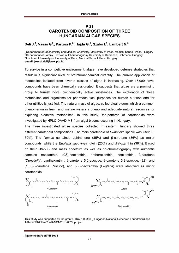
Poster Session
Pigments in Food VII 2013 72
P 21
CAROTENOID COMPOSITION OF THREE HUNGARIAN ALGAE SPECIES
Deli J.
1, Vasas G
2., Parizsa P.
2, Hajdú G.
2, Szabó I.
1, Lambert N.
3
1 Department of Biochemistry and Medical Chemistry, University of Pécs, Medical School, Pécs, Hungary
2 Department of Botany, Division of Pharmacognosy University of Debrecen, Debrecen, Hungary
3 Institute of Bioanalysis, University of Pécs, Medical School, Pécs, Hungary
e-mail: [email protected]
To survive in a competitive environment, algae have developed defense strategies that
result in a significant level of structural-chemical diversity. The current application of
metabolites isolated from diverse classes of algae is increasing. Over 15,000 novel
compounds have been chemically assignated. It suggests that algae are a promising
group to furnish novel biochemically active substances. The exploration of these
metabolites and organisms for pharmaceutical purposes for human nutrition and for
other utilities is justified. The natural mass of algae, called algal-bloom, which a common
phenomenon in fresh and marine waters a cheap and adequate natural resources for
exploring bioactive metabolites. In this study, the patterns of carotenoids were
investigated by HPLC-DAAD-MS from algal blooms occuring in Hungary.
The three investigated algae species collected in eastern Hungary showed three
different carotenoid compositions. The main carotenoid of Dunaliella specie was lutein (~
50%). The Nostoc contained echinenone (35%) and β-carotene (36%) as major
compounds, while the Euglena saugvinea lutein (23%) and diatoxanthin (39%). Based
on their UV-VIS and mass spectrum as well as co-chromatography with authentic
samples neoxanthin, (9Z)-neoxanthin, antheraxanthin, zeaxanthin, β-carotene
(Dunaliella), canthaxanthin, β-carotene 5,6-epoxide, β-carotene 5,8-epoxide, (9Z)- and
(13Z)-β-carotene (Nostoc), and (9Z)-neoxanthin (Euglena) were identified as minor
carotenoids.
O
HO
OH
H
OH
HO
-Carotene
Echinenone
Lutein
Diatoxanthin
This study was supported by the grant OTKA K 83898 (Hungarian National Research Foundation) and TÁMOP/SROP-4.2.2/B-10/1-2010-0029 project.

Poster Session
Pigments in Food VII 2013 73
P 22
HPLC METHOD VALIDATION FOR THE
DETERMINATION OF FUCOXANTHIN
Travaglia F., Bordiga M., Locatelli M., Coïsson J.D., Arlorio M.
Dipartimento di Scienze del Farmaco, Largo Donegani 2, 28100 Novara (Italy) e-mail: [email protected]
The Fucoxanthin is a carotenoid present primarily in brown seaweeds (Feoficee) of the
genus Undaria pinnatifida (commonly called Wakame), Sargassum fulvellum, Laminaria
japonica (better known as Kombu) and Hizikia fusiformis, which gives them the peculiar
dark color, overlapping the typical greenish color of the common algae.
Fucoxanthin is a characteristic xanthophyll present in brown seaweed and the most
abundant amongst aquatic carotenoids, accounting for more than 10% of estimated total
natural production of carotenoids (Prabhasankar et al., 2009).
The fucoxanthinol, primary active metabolite of this molecule, has been recently found to
have anti-obesity effects through the mobilization of adipose deposits for energy
purposes, as well in the production of DHA (docosahexaenoic acid) which is claimed to
provide a variety of different health benefits (Maeda et al., 2006).
In this work a HPLC-Vis validation method for determination and quantification of all-
trans-fucoxanthin (CAS number 3351-86-8) in dry extracts of Wakame, used as raw
material for the formulation of dietary supplements and in several commercial samples
(fucoxanthin-based), was reported.
The first step of this research was the optimization of the chromatographic conditions
and its validation in accordance with the international guidelines ICH (International
Conference on Harmonization), also applying common statistical tests suitable for
validation of a chromatographic method.
The second step included the optimization of a protocol for extraction of fucoxanthin and
its quantification in dry extracts of Wakame seaweed and several commercial products.
References
Prabhasankar, P., Ganesan, P., Bhaskar, N., Hirose, A., Stephen, N., Gowda, L. R., Hosokawa, M.,
Miyashita, K. 2009. Edible Japanese seaweed, wakame (Undaria pinnatifida) as ingredient in pasta:
chemical, functional and structural evaluation. Food Chem. 115: 501-508
Maeda, H., Hosokawa, M., Sashima, T., Takahashi, N., Kawada, T., Miyashita, K. 2006. Fucoxanthin and
its metabolite, fucoxanthinol, suppress adipocyte differentiation in 3T3-L1 cells. Int. J. Mol. Med. 18: 147-
152

Poster Session
Pigments in Food VII 2013 74
P 23
CAROTENOIDS STABILISATION FOR USE IN BEVERAGES: TWO DIFFERENT APPROACHES
Mesnier X.
1, Boukobza F.
1, Bily A.
2, Roller M.
2
1Naturex, Overseal, UK
2Naturex, Avignon, France
email: [email protected]
Over the last few years, Naturex has developed several techniques to extend the stability
of natural colours for applications in the beverage industry. The two following examples
illustrate some of the technical improvements achieved.
Oil soluble paprika is commonly incorporated in water based beverages as an emulsion
stabilised by non-ionic surfactants. The incorporation of Polysorbate 80 for example
leads to the production of clear acid stable products. However, high content of
Polysorbate 80 can cause foaming issues and impact the taste of beverages. Naturex
has developed a series of polysorbate 80 free emulsions presenting a similar acid
stability, transparency and shelf life. The droplet size distributions of both formulations
are presented together with the light stability results.
Carotenoid-based colours are generally prone to oxidation. There are several
combinations of oil soluble antioxidants that can be used to optimise the stability of the
colour through various food & beverages processes. Oil soluble Rosemary extracts
standardised in carnosic acid and carnosol added in the colour formulation significantly
improve the stability of carotenoids based colours. In addition to this oil soluble form of
Rosemary extracts, Naturex has developed a range of water soluble Rosemary extracts,
StabilEnhance® WSR that can prolong beverages stability when incorporated as an
antioxidant directly in the final application.
Poster Session
Pigments in Food VII 2013 75
P 24
EFFECT OF HEAT PROCESSING ON THE PROFILE OF PIGMENTS AND ANTIOXIDANT CAPACITY OF JALAPEÑO
PEPPERS AT INTERMEDIATE RIPENING STAGES
Cervantes-Paz B.1, Ornelas-Paz J. de. J.
1, Yahia E. M.
2
1Centro de Investigación en Alimentación y Desarrollo A.C.-Unidad Cuauhtémoc, Av. Río Conchos S/N,
Parque Industrial, C.P. 31570 Cd. Cuauhtémoc, Chihuahua, México 2Facultad de Ciencias Naturales, Universidad AutónomENTa de Querétaro, Avenida de las Ciencias S/N,
C.P. 76230 Juriquilla, Querétaro, México e-mail: [email protected]
Recent studies have demonstrated that household heat-treatments alter differentially the
content (qualitative and quantitative) and antioxidant capacity (AOC) of pigments in
green and red Jalapeño peppers.1,2
However, these attributes have not been evaluated
in Jalapeño peppers at intermediate ripening stages. In this study, profiles of carotenoids
(free and esterified), chlorophylls, and chlorophyll derivatives were evaluated by HPLC-
APCI-TOF-MS analysis in raw and heat-processed (boiled or grilled) Jalapeño peppers
at intermediate ripening stages (brown, 50% red, and 75% red). The pigment extracts
were also evaluated for AOC (DPPH and FRAP methods). The AOC values were higher
for 75% red peppers that for peppers at other ripening stages. The AOC was
differentially affected by heat-processing style. Sixty-four compounds were observed in
the pigment estracts, being the all-trans-capsanthin and the chlorophyll a the most
abundant. Chlorophyll b was not observed in 75% red peppers. Nine compounds were
generated by heat-processing, highlighting several pheophytins (a, a′, b and b′) and two
β-cryptoxanthin isomers. Finally, the concentration of pigments was decreased by boiling
and grilling. Our results reveal the importance of Jalapeño peppers at intermediate
ripening stages, which might provide a greater health effects that green or red peppers
since they contain a more complex mixture of pigments.
References
[1] Cervantes-Paz B. et al. Effect of Heat Processing on the Profile of Pigments and Antioxidant Capacity
of Green and Red Jalapeño Peppers. J. Agric. Food Chem. 2012, 60, 10822–10833
[2] Alvarez-Parrilla E. et al. Antioxidant Activity of Fresh and Processed Jalapeño and Serrano Peppers.
J. Agric. Food Chem. 2011, 59, 163–173.
Poster Session
Pigments in Food VII 2013 76
P 25
MICELLARIZATION AND DIGESTIVE STABILITY OF PIGMENTS FROM JALAPEÑO PEPPERS AT INTERMEDIATE RIPENING
STAGES Victoria-Campos C. I.
1, Ornelas-Paz J. de. J.
1
1 Centro de Investigación en Alimentación y Desarrollo A. C. (CIAD), Cd. Cuauhtémoc, Chihuahua,
Mexico. e-mail: [email protected]
The effect of the interaction between different factors on the micellarization of
carotenoids has been scarcely studied.[1,2]
We evaluated the effect of different fat
sources (soybean oil and beef tallow) and heat treatments (raw, boiling, and grilling) on
the in vitro digestive stability and micellarization and carotenoids (free and esterified),
chlorophylls, and chlorophyll derivatives from Jalapeño peppers at intermediate ripening
stages (brown, 50% red, and 75% red). Around of 76 pigments were identified by HPLC-
MS analysis in tested fruits. Fifty four pigments were micellarized, including free and
esterified pigments. The micellarization of free carotenoids was higher with brown and
50% red than with 75% red peppers. Chlorophyll derivatives were micellarized only with
brown and 50% red peppers. The interaction effect between dietary fat and heat
treatments determined the digestive stability and micellarization of pigments. Dietary fat,
mainly soybean oil, increased the micellarization of esterified carotenoids, pheophytins,
and the less polar free carotenoids. The micellarization of free and monoesterified
carotenoids was higher with raw than with processed peppers. Grilling increased the
micellarization of carotenoid diesters. Micellarization values of esterified carotenoids
were influenced by their fatty acid moiety. The results showed that the interaction
between the tested factors determined the micellarization of the pigments from Jalapeño
peppers. Peppers at intermediate ripening could show an advantage on the green or red
peppers, providing a higher variety of pigments.
References
[1] Cilla, A. et al. Bioaccessibility of tocopherols, carotenoids, and ascorbic acid from milk- and soy-based
fruit beverages: influence of food matrix and processing. J. Agric. Food Chem. 2012, 60, 7282–7290.
[2] Hornero-Méndez, D. and Mínguez-Mosquera, M. I. Bioaccessibility of carotenes from carrots: effect of
cooking and addition of oil. Innov. Food Sci. Emerg. Tech. 2007, 8, 407-412.
Poster Session
Pigments in Food VII 2013 77
P 26
CHANGES IN LUTEIN, CHLOROPHYLLS AND CHLOROPHYLL DEGRADATION PRODUCTS IN PISTACHIO KERNELS
(PISTACIA VERA L.) DURING ROASTING Pumilia G.
1, Schwartz S.J.
2, Cichon M.J.
2, Giuffrida D.
1 , Dugo G.mo
1.
1 Dipartimento di Scienze dell’Ambiente, della Sicurezza, del Territorio, degli Alimenti e della Salute
(S.A.S.T.A.S.). Università degli Studi di Messina, Viale Ferdinando Stagno d’Alcontres, 31, 98166 S. Agata. Messina, Italy 2
Department of Food Science & Technology, The Ohio State University, 110 Parker Food Science & Technology Building, 2015 Fyffe Court, Columbus, Ohio 43210, United States e-mail: [email protected]
Bright green pistachio kernels are desirable in the food industry and a loss of color can
be associated with a change in quality. Chlorophylls a and b, as well as lutein, are the
predominant pigments in pistachios. The heat treatment of chlorophyll containing foods
results in color degradation due to the conversion of chlorophylls (bright green color) to
pheophytins and pyropheophytins (yellow-brown olive color). In this work changes in
lutein, chlorophylls and chlorophyll degradation products in pistachios were monitored
during oven roasting for the first time. A high performance liquid chromatography method
with photodiode-array detection (HPLC-PDA) was developed to separate and quantitate
the pigments in pistachios. Interestingly, chlorophylls a and b were higher in pistachio
kernels roasted for 5 and 10 minutes than in the raw kernels. The most drastic losses
were observed with pheophytins a and b, which both decreased by approximately 85%
after 60 minutes of roasting. On the other hand, pyropheophytins a and b increased
significantly during roasting and were 10-12x higher in the pistachios roasted for 60
minutes than in the raw pistachios. Lutein concentration increased by 37% after 5
minutes, but did not change significantly after that. Color measurements of the pistachios
supported changes in chlorophyll pigments observed at the later roasting times. Initial
increases in chlorophylls a and b and lutein were likely due to enhanced extractability
with roasting.
Poster Session
Pigments in Food VII 2013 78
P 27
DECOLOURATION PROCESSES UNDER NON-OXYGEN THERMAL AUTO-OXIDATION OF CHLOROPHYLL AND
CAROTENOID FRACTIONS IN VIRGIN OLIVE OILS Aparicio-Ruiz R., Gandul-Rojas B*. Chemistry and Biochemistry Pigment Group, Department of Food Biotechnology. Instituto de la Grasa (CSIC), Av. Padre García Tejero, 4, 41012 Sevilla, Spain. e-mail: [email protected].
Kinetic models are capable of predicting shelf life in keeping with the different variables
that can affect the degradation of the food item. Numerous experimental works describe
virgin olive oils (VOOs) degradation, but until recently the kinetic performance in
oxidation parameters1 and individual pigment thermodegradation products have not been
reported2-3
. In this work, VOOs collected at three ripening stages with high, medium and
low pigment content respectively, were thermodegraded to characterize the kinetic and
thermodynamic parameters for the global degradation of chlorophyll (CHL) and
carotenoid (CAR) fractions. A first-order kinetic mechanism was appropriate for
describing the decolouration processes under non-oxygen thermal auto-oxidation. A
marked effect of temperature has been pointed out, with the CARs being the most
affected by heat. The kinetic constants for the CARs degradation were 3.6 times higher
than the respective for CHLs that showed a more stable structure to decolouration by
heat. As well, higher activation energy of CHLs (16.03±0.26 kcal/mol) as compared to
CARs (15.45±0.17 kcal/mol) implies that a smaller temperature change is needed to
increase the kinetic constant of CHLs. Neither isokinetic ratio and nor compensation
existed between the three VOO matrices and further, for each pigment fraction (CHLs or
CARs) all kinetic constants were explained by a single Arrhenius line. Consequently, the
oily medium did not significantly affect the decolouration mechanisms, and moreover, the
kinetic parameters obtained as temperature functions according to Arrhenius model, can
be used to develop a prediction mathematical model for CHL and CAR decolouration in
VOO over time and depending on temperature.
References
[1] Mancebo-Campos, V. et al. Kinetic study for the development of an accelerated oxidative stability test
to estimate virgin olive oil potential shelf life. Eur. J. Lipid Sci. Technol. 2008, 110, 969−976.
[2] Aparicio-Ruiz, R. et al. Thermal degradation kinetics of chlorophyll pigments in virgin olive oils. 1.
Compounds of series a. J. Agric. Food Chem. 2010, 58, 6200−6208.
[3] Aparicio-Ruiz, R. et al. Thermal degradation kinetics of lutein, β-carotene and β-cryptoxanthin in virgin
olive oils. J. Food Compos. Anal. 2011, 24, 811−820.
Poster Session
Pigments in Food VII 2013 79
P 28
PIGMENT CHANGES DURING PROCESSING OF GREEN TABLE OLIVE SPECIALITIES TREATED WITH ALKALI AND
WITHOUT FERMENTATION Gallardo-Guerrero L., Gandul-Rojas B.
Food Biotechnology Department, Instituto de la Grasa (CSIC), Avenida Padre García Tejero, 4, 41012, Sevilla, Spain. e-mail: [email protected]
Table olives are among the most important fermented vegetable foods produced in the
world. The main green table olive commercial preparation is the so-called Spanish or
Seville-style which comprises an alkaline treatment of the olives and subsequent natural
lactic fermentation in brine. Recently, other green table olive preparations, which include
an alkaline treatment but not preservation by natural fermentation, are gaining
importance in international trade, being included in the trade standard applying to table
olives as "Specialities"1. One of the main highly valued characteristic for this product is a
bright green colour, as similar as possible to the fresh fruit. Generally, colour is one of
the most important quality attributes for table olives and can be considered as a quality
index. The pigments of green table olives treated with alkali but not fermented (such as
Castelvetrano and Campo Real-styles) have been scarcely studied for different
commercial products and no detailed study about the pigment transformation during
processing has been carried out. Colour of green table olives is due to the presence of
two main families of natural pigments, chlorophylls and carotenoids2. In the present work,
changes in the chlorophyll and carotenoid composition of olive fruits during
Castelvetrano-style olive processing (lasting two weeks), and their involvement in the
colour of the final product, were investigated. During processing, the main
transformations of pigments were due to allomerization reactions of chlorophylls a and b,
what do not imply colour change of the green fruit. Certain degradation to uncoloured
compounds was also detected. With respect to the carotenoid fraction, any noticeable
change took place.
References
[1] IOC (International Olive Council). Trade standard applying to table olives. COI/OT/NC No.1. Madrid,
Spain. 2004.
[2] Gallardo-Guerrero, L. et al. Olives and olive oil. In Tropical and Subtropical Fruits. Postharvest
Physiology, Processing and Packaging. Siddiq M (Ed.). 1st ed.; John-Wiley & Sons, Inc. Publishing, Co,
Ames, Iowa, USA. 2012, 503-528

Poster Session
Pigments in Food VII 2013 80
P 29
POLYPHENOLS AND VOLATILE COMPOUNDS IN OGLIAROLA AND CELLINA OLIVE
Romani A.
1, Banelli L.
1, Fierini E.
1,
Mancuso S.
2, Masi E.
2 ,
Heimler D.
2
1 Dipartimento di Statistica, Informatica, Applicazioni “G. Parenti”, University of Florence, Italy.
2 Dipartimento di Scienze delle Produzioni Agroalimentari e dell'Ambiente (DISPAA), University of
Florence, Italy e-mail: [email protected]
The Italian region Apulia is characterized by a large virgin oil production; in Italy, about
80% of olive oil production is located in southern regions, where Apulia represents the
most important area. Owing to the difficult in modernizing the traditional olive growing
designs, from one side super high density models are proposed [1] and from the other
monovarietal oils from particular geographic areas were characterized [2]. In this study,
monocultivar olive (Ogliarola and Cellina di Nardò) at different ripening degree from
Apulia were analysed. The spectrophotometric and HPLC/DAD/MS quali-quantitative
analyses of CMP were performed. CMP play an important role in diet, because they
seem to prevent many diseases; several pharmacological studies demonstrated that the
phenolic fraction extracted from virgin olive oil, and from waste waters collected within
the production of olive oil, are able to prevent the LDL oxidation and can cause a
decrease in the platelet aggregation induced by collagen in conditions of oxidative
stress. Other aim of this study was the in vivo analyses of volatile compounds without a
previous pre-treatment of the sample with PTR-TOF-MS instrumentation, a non-invasive
technique which allows the achievement of whole mass spectra with a time of resolution
lesser than 1 second and the detection of high molecular weight molecules with a high
resolution power and to provide unambiguous determination of chemical formula leading
a better interpretation of mass spectra [3].
Further analyses are in progress to validate a non-destructive optical method (as
Multiplex sensor) to monitoring anthocyanins accumulation directly on olive fruits.
References
[1]Camposeo, S. et al. Preliminary observations about the performance of 13 varieties according to the
super high density oliveculture training system in Apulia (southern Italy). Advances in Horticultural
Science, 2010, 24, 16-20.
[2]Vlahov, G. et al. Determination of geographic origin of olive oil using 13C nuclear magnetic resonance
spectroscopy. Classification of olive oils of the Puglia region with denomination of protected origin. J.
Agric. and Food Chem., 2003, 51, 5612-5615.
[3]Cappellin, L. et al. Improved mass accuracy in PTR-TOF-MS: another step towards better compound
identification in PTR-MS. International Journal of Mass Spectrometry, 2010, 290, 60-63.

Poster Session
Pigments in Food VII 2013 81
P 30
CHLOROPHYLLIAN PIGMENTS IN EXTRA VIRGIN OLIVE OILS Rovellini P., Venturini S., Fusari P.
Innovhub-SSI Divisione SSOG, Milan, Italy. e-mail: [email protected], [email protected]
The chemical composition of the olive oils pigments (Olea europaea L.) varies on the
base of parameters such as variety, degree of ripeness, environmental conditions,
growing region, processing and storage.
The pigments, in particular the chlorophylls and pheophytins affects considerably the
preservation of this important diet mediterranean product as pro-oxidants in synergy with
metals trace eventually present enhancing the autooxidation process.
In virgin olive oils the principal compounds present in decreasing content order are
pheophytins A and A’ followed from pheophytins B and B’, pyropheophytin A and at last
chlorophyll A.
This type of analysis is conducted as quality parameter to point out high oxidized lipidic
status or detect eventually deodorization process or thermal treatment.
The official method to quantify these compounds is ISO 29841:2009 method, but on the
base of our big experience on a lot of samples of this lipidic matrix we have evidenced
that applying the over method the % content of pyropheophytin A respect to pheophytins
A+A+pyropheophytin A is increased until to 40 %, respect to the data obtained after
direct injection of a sample solution in acetone in a HPLC-UV system: the passage
through the silica column cause a partial modification of pigments profile, in particular
chlorophyll A, pheophytins B+B’, pheophytins A+A’ are partially detained on column in
particular these last.
References
[1] Gallardo, L. et al. Effect of storage on the original pigment profile of Spanish Virgin Olive Oil. JAOCS.
2005, 82 (1), 33-39
[2] Cicchelli, A. High performance liquid chromatography analysis of chlorophylls, pheophytins and
carotenoids in virgin olive oils: chemometric approach to variety classification. Journal of Chromatography
A., 2004, 1046, 141-146
Poster Session
Pigments in Food VII 2013 82
P 31
SUBCELLULAR DISTRIBUTION IN OLIVE FRUIT OF PEROXIDASE ACTIVITY ON CHLOROPHYLL SUBSTRATE
Vergara-Domínguez H., Roca M., Gandul-Rojas B*. Chemistry and Biochemistry Pigment Group, Department of Food Biotechnology. Instituto de la Grasa (CSIC), Av. Padre García Tejero, 4, 41012 Sevilla, Spain. e-mail: [email protected]
The catabolism of chlorophylls has been mainly studied on leaf senescence, assuming
that the degradation of these pigments should be similar in fruits. However, recent
research has revealed important differences on chlorophyll metabolism in leaves and
fruits. Even, it has been postulated the coexistence, only in certain fruits, of an
exclusively oxidative metabolism concomitant with the general chlorophyll degradation
pathway proposed by Kräutler and Hörtensteiner1 (2011). Type III peroxidase (EC
1.11.1.7) is the enzyme most clearly involved on chlorophyll oxidative metabolism. In this
work we studied the distribution of chlorophyll oxidative peroxidase activity (POD) in
different subcellular fractions from mesocarp cells of olive fruit (Olea europea L.):
Arbequina and Hojiblanca varieties. The specific methodology used for olive fruit it is
based on Salas et al2, which implies differential centrifugation techniques. The POD
activity in the cell fractions enriched in the thylakoid membranes was monitored
throughout the whole life cycle of the olive fruit and a positive correlation between the
levels of the maximum activity of POD and the presence of oxidized derivatives was
obtained in the fruits of the Arbequina variety. The study also showed varietal differences
in the distribution of POD activity present in different subcellular fractions that explain the
greater accumulation of oxidised derivatives of chlorophyll in fruits of the Arbequina than
in Hojiblanca variety3. This work supposes one step further on deciphering chlorophyll
degradation pathway on fruits.
References
[1] Hörtensteiner, S. et al. Chlorophyll breakdown in higher plants. Biochem et Biophysic Acta. 2011,
1807, 977–988.
[2] Salas, J. et al. Lipoxygenase activity in olive (Olea europaea) fruit. J. Am. Oil Chem. Soc. 1999, 76,
1163–1168.
[3] Roca et al. Involvement of chlorophyllase in chlorophyll metabolism in olive varieties with high and low
chlorophyll content. Physiol Plantarum. 2003, 117, 459-466.
Poster Session
Pigments in Food VII 2013 83
P 32
CHLOROPHYLL AND CAROTENOID PIGMENTS IN A SURVEY OF MARKETED APPLE VARIETIES
Delgado-Pelayo R., Gallardo-Guerrero L., Hornero-Méndez D.
Food Biotechnology Department, Instituto de la Grasa (CSIC), Avda. Padre García Tejero, 4, 41012, Seville, Spain. e-mail: [email protected]
Domesticated apple (Malus x domestica Borkh) varieties have been traditionally
consumed due to their sensory quality attributes and in relation to their health benefits
which are associated with their high content in phenolic compounds, vitamin C,
antioxidants and dietary fibre. Apart from the anthocyanins, responsible for the reddish
colour of the skin of many apple fruit, chloroplastic pigments (chlorophylls and
carotenoids) contribute also to the external (skin) and internal (flesh) fruit colouration.
Apple has always been considered a fruit with low chloroplastic pigment content,
especially carotenoids, when compared with other fruit species. In the last decade
increasing efforts are being dedicated to the selection of fruits with enhanced levels of
phytonutrients, including the carotenoids, involving multidisciplinary breeding
programmes. In the present study we have characterised the chlorophyll and carotenoid
composition of 13 marketed apple varieties, distinguishing between skin and flesh. All
the varieties where characterised by common pigment profile, composed by chlorophyll a
and b, neoxanthin (all-trans and 9-cis isomers), violaxanthin (all-trans and 9-cis), lutein
and β-carotene as major compounds. Several 5,8-epoxide derivatives (neochrome,
luteoxanthin and auroxanthin) were also found in certain varieties, whereas β-
cryptoxanthin and antheraxanthin were mostly detected as minor compounds. Especial
attention has been paid to determine the presence of carotenoid esters, being their
detailed identification under study. Total chlorophyll and carotenoid contents were
always higher in the skin, where lutein was the main carotenoid followed by violaxanthin,
neoxanthin and β-carotene, a characteristic chloroplastic profile. On the contrary,
neoxanthin and/or violaxanthin dominated the carotenoid composition in pulp, presenting
also a higher proportion of esterified xanthophylls (mainly violaxanthin and neoxanthin).
Poster Session
Pigments in Food VII 2013 84
P 33
QUANTITATION OF POLYPHENOLS IN DIFFERENT APPLE VARIETIES CULTIVATED IN AOSTA VALLEY
Valentini S., Sado A., Chasseur M., Thedy L., Lale Murix H., Barrel I., Chatel A. Institut Agricole Régional, 11100 Aosta-Italy e-mail: [email protected]
The aim of this study was to compare polyphenols content in different apple varieties
(Renetta Canada, Golden Delicious, Gala, Jonagold, Red Delicious, Fuji, Pinova,
Falstaff, Mairac, Topaz, Goldrush) cultivated in orchards situated in different areas of
Aosta Valley, at harvest time and after controlled atmosphere (CA) storage, over four
consecutive years. Harvest time was decided according to commonly used ripening
indices: flesh firmness, sugar content, titrable acidity and starch. Samples were extracted
from fresh fruit with a mixture of acetone/water to achieve a good extraction of
polyphenols, following the method of Mattivi et al. [1]. The total amount of polyphenols
was measured with an optimized Folin-Ciocalteu assay [2], according to which interfering
compounds were removed by cleanup on a C18 cartridge. Quantitation of ascorbic acid
was also performed in all the varieties in this study. The polyphenol content depends on
the year and on the apple variety; Renetta Canada has a much higher content of total
polyphenols than any other variety studied, according to other authors. Over four years,
the average content of total polyphenols detected in the apple at harvest time was
between 65.40 and 156.63 mg/100g of fresh fruit according to the variety in the following
increasing order: Fuji, Gala, Goldrush, Pinova, Golden Delicious, Mairac, Falstaff, Topaz,
Red Delicious, Jonagold and Renetta Canada, with some differences depending on the
harvest year. After storage no highly significant change in polyphenol content was
observed in all the years considered and in all the apple varieties collected.
References
[1] Mattivi F. et al. Gli antiossidanti polifenolici naturali. Laboratorio 2000 2002, 3, 46-56.
[2] Rigo A. et al. Contribution of proanthocianidins to the peroxy radical scavenging capacity of some
italian red wines. J. Agric. Food Chem. 2000, 6, 1996-2002.
Poster Session
Pigments in Food VII 2013 85
P 34
ANTHOCYANINS, PHENOLIC ACIDS AND ANTIOXIDANT ACTIVITY IN YELLOW, RED AND PURPLE-FLESHED POTATOES
AFTER STEAM COOKING Bellumori M.
1, Innocenti
M.
1, Cerretani L.
2, Mulinacci N.
1
1
Department of NEUROFARBA, Division of Pharmaceutical and Nutraceutic, University of Florence, Sesto Fiorentino, Florence, Italy 2 Pizzoli S.p.A., Budrio, Bologna, Italy
e-mail: [email protected]
Potatoes, belonging to the species Solanum tuberosum L., constitute a food consumed
worldwide prepared and served in a variety of ways. Coloured potatoes with high levels
of anthocyanins, total phenols and antioxidant activity, represent healthy food choice for
consumers and a potential source for natural food colorants. This study aimed to
determine anthocyanin and phenolic acid content in potato flesh with different colours,
and to evaluate the antioxidant activity of these cultivars.
Potatoes, after boiled steam cooking, were extracted with 70% EtOH adjusted to pH 2.0
by HCOOH, as already described in a previous study [1] and the compounds were
identified by HPLC/DAD/MS according to our previous work [2]. The ABTS method
described by Lachman et al. (2011) was used for the antioxidant activity determination.
Six yellow-, two purple- and three red-fleshed varieties were studied. For purple- and
red-fleshed cultivars, average total anthocyanin content values ranged from 64,1 to
437,2 μg/g of fresh material and a correlation with antioxidant activity was found. The
lowest antioxidant activity was achieved by the group of varieties with yellow flesh,
averaging 0,36 μg ascorbic acid equivalent/mL estract, instead in the group of red- and
purple-fleshed cultivars it was higher 3,25 times considering the variety with the highest
total anthocyanin content.
For these coloured potatoes, differences in antioxidant activity were found and the
analysis showed a strong correlation between antioxidant activity and total anthocyanin
content. Consequently, red and purple-fleshed cultivars could be a promising source of
important antioxidants in human nutrition.
References
[1] Mulinacci, N. et al. Effect of cooking on the anthocyanins, phenolic acids, glycoalkaloids, and resistant
starch content in two pigmented cultivars of Solanum tuberosum L. J. Agric. Food Chem. 2008, 56,
11830–11837.
[2] Ieri, F. et al. Rapid HPLC/DAD/MS method to determine phenolic acids, glycoalkaloids and
anthocyanins in pigmented potatoes (Solanum tuberosum L.) and correlations with variety and
geographical origin. Food Chem. 2011, 125, 750–759.
[3] Lachman, J. et al. Impact of selected factors – Cultivar, storage, cooking and baking on the content of
anthocyanins in coloured-flesh potatoes. Food Chem. 2012, 133, 1107–1116.

Poster Session
Pigments in Food VII 2013 86
P 35
CHEMICAL CHARACTERIZATION AND ANTIOXIDANT ACTIVITY OF SIX RICE CULTIVARS GROWN IN PIEDMONT
(PIGMENTED AND NON-PIGMENTED) Bordiga M., Coϊsson J.D., Locatelli M., Travaglia F. and Arlorio M.
Dipartimento di Scienze del Farmaco, Largo Donegani 2, 28100 Novara (Italy) e-mail: [email protected]
Different extracts from pigmented rice cultivars grown in Piedmont (Artemide, Venere,
Nerone (black); Ermes, Russ (red); Selenio (white)) were analyzed by high-performance
liquid chromatography coupled with diode array detection and electrospray ionization -
mass spectrometry (HPLC-DAD-ESI-MS/MS). The aim of this work was to identify the
anthocyanins, flavonols, and flavan-3-ols contents so characterizing the different
typologies of rice [1]. The stable radical 1,1-diphenyl-2-picrylhydrazyl (DPPH˚) has been
used to compare the antioxidant activities of the different rice cultivars and moreover
total phenolic content has been examined. Black rice cultivars contained eight
anthocyanins and all the three black varieties Artemide, Venere, and Nerone presented
the same qualitative composition of anthocyanin pigments [2, 3]. The flavonols that occur
in rice samples made up the series of 3-glc, and 3-rut of quercetin and isorhamnetin.
Derivatives of dihydroquercetin and dihydroisorhamnetin were detected only in the 3-glc
form. The total sum of flavan-3-ol monomers and dimers was substantially different
considering the six rice varieties; Artemide, Ermes, and Russ presented significantly
higher values (193.1, 101.8, and 63.7 mg/kg rice respectively) if compared to the others.
The free phenolic content of 6 rice varieties ranged from 1404 ± 322 (Selenio) to 11872 ±
1161 (Artemide) mg of gallic acid equiv/kg of dry weight. Considering the antioxidant
activity, the black rice cultivars showed the highest antiradical potential (DPPH˚
scavenging of 88.4%, 53.6% and 51.2% for Artemide, Nerone and Venere, respectively),
while the lowest activity was observed for the non-pigmented rice sample (Selenio:
5.36%).
References
[1] Abdel-Aal, E. S. M. et al. Anthocyanin composition in black, blue, pink, purple, and red cereal grains.
Journal of Agricultural and Food Chemistry. 2006, 54, 4696-4704.
[2] Cho, M. H. et al. Chemical structure of the major color component from a Korean pigmented rice
variety. Agricultural Chemistry and Biotechnology. 1996, 39, 304-308.
[3] Escribano-Bailón, M. T. et al. Anthocyanins in cereals. Journal of Chromatography A. 2004, 1054,
129-141.
Poster Session
Pigments in Food VII 2013 87
P 36
EFFECT OF THE USE OF ENZYMATIC PREPARATIONS ON EXTRACTION OF PHENOLIC COMPOUNDS FROM BLUE MAIZE
(ZEA MAYS L.), FROM THE REGION OF TLAXCALA, MEXICO
Martínez de Santos M.L.
1; Conteras-Llano L.E.
1; Lozada-Ramírez J.D.
2;
Ortega-Regules A.E.1
1 Universidad Politécnica de Tlaxcala, Tepeyanco. Tlaxcala.
2 Universidad de las Américas Puebla, Santa Catarina Mártir, Cholula. Puebla.
e-mail: [email protected]
The presence of anthocyanins on blue maize (Zea mays L.), converts it on a potential
supply of natural colorants and antioxidants1. Mexico has a diversity of maize
germplasm2, but with a very low exploitation, despite its nutritional and nutraceutical
properties, so its use has potential for the development of new products. While,
enzymatic degradation of vegetables cell walls is considered essential on the extraction
of phenolic compounds from fruits and vegetables3. In this work, it was evaluated the use
of commercial enzymatic preparations on the extraction of total phenolic compounds
(TPC), total monomeric anthocyanins (TMA), antioxidant capacity (AC) and color
intensity (CI) from two blue maize varieties. Anthocyanin extracts from pericarp and
aleurone from grains (1, 2, and 3 g) were obtained using phosphate buffer at four pH
intervals (2.5, 3.0, 3.5 y 6.5), adding three enzymatic preparations and a combination
between them (Pectinase®, Viscozyme® y Protease®). A MANOVA statistical analysis
was performed, and the data obtained shows differences (P≤0.05) between the studied
factors and variables. Treatments using Pectinase®, buffer pH 3.0 and 1 g of blue maize
from variety 1, and Viscozyme®, buffer pH 3.5, and 1 g of blue maize from variety 2,
showed the best interaction conditions with values of 17.11±0.77 and 35.77±0.81 of TPC
mg gallic acid/100g maize, 2.49±0.13 and 23.75 ± 0.69 of TMA mg cyanidine-3-
glucoside/100g maize, 0.1053±0.0002 and 0.1773± 0.0015 of AC mM Trolox/100 g
maize, and 0.2198±0.0035 and 2.4773±0.0022 of CI (Abs), respectively; evidencing a
favorable effect of enzymatic preparations on extraction.
References:
[1] Cuevas Montilla E., et al. (2008). Análisis y caracterización de antocianinas de diferentes variedades
de maíz (Zea mays L.). Memorias del encuentro Final Alfa Logrotech, Cartagena Colombia.
[2] López-Martínez, L. y García-Galindo, H. (2009). Actividad antioxidante de extractos metanólicos y
acuosos de distintas variedades de maíz mexicano. Revista Electrónica Nova Scientia, 3 (2). pp: 51–65
[3] Ovando Chacón S. L., Walieszewski K. N. (2005). Preparativos de celulosas comerciales y
aplicaciones en procesos extractivos. Universidad y Ciencia. Universidad Juárez Autónoma de Tabasco,
Villahermosa, México, 21(042): 113-122.
Poster Session
Pigments in Food VII 2013 88
P 37
TECHNO-FUNCTIONAL PROPERTIES OF TOMATO SAUCE FORTIFIED WITH ANTHOCYANIN PIGMENTS
Blando F.
1, Biasco M.
2, Albano C.
1, Gerardi C.
1, Dal Porto L.
2, Mita G.
1
1 Institute of Sciences of Food Production, CNR, Lecce Unit, Lecce, Italy
2 Pr.ali.na. Ltd, Melpignano, Lecce, Italy
e-mail: [email protected]
Tomato sauce can be considered a ‘staple food’ not only in Italy but all over the world,
because of its usage in many recipies. It can be also considered a ‘functional food’, due
to epidemiological evidence of reduced risk of certain types of cancers and CVD related
to its high content of antioxidant molecules, first of all lycopene, and also vitamins C and
E [1, 2].
The nutraceutical value of tomato sauce can be increased by the presence of added
antioxidant compound, obtaining thus a ‘fortified tomato sauce ‘ or a ‘functional tomato
sauce’.
With this aim, we added to control tomato sauce (Von Felten, Parma) (10-12° Brix)
different % of anthocyanin pigments, obtained from natural source (Prunus mahaleb L.
fruits, Daucus carota L. var. atrorubens, Vitis vinifera L. fruit skins). The addition of the
anthocyanin extracts at maximum % (1% w/w) has decreased the pH values (- 0.1
points) to acceptable values for processed products, whereas the soluble solids have
increased (+ 3° Brix). Since the addition of anthocyanin extracts is early in the
production flowchart, the evaluation of colour (made by colorimeter with CIELAB
parameter) has revealed a good colour retention even after pasteurization.
References
[1] Raffo A. et al. Nutritional value of cherry tomatoes (Lycopersicon esculentum cv Naomi F1) harvested
at different ripening stages. J. Agric. Food Chem. 2002, 50, 6550-6556.
[2] Canene-Adams K. et al. The tomato as a functional food. J. Nutr. 2005, 135, 1226-1230.
Poster Session
Pigments in Food VII 2013 89
P 38
EFFECT OF POST-HARVEST TREATMENT ON ANTHOCYANIN CONTENT AND TOTAL PHENOLICS IN MANGO
(MANGIFERA INDICA L.) PEELS
Geerkens C.H., Müller-Maatsch J.T.L., Geissler M., Carle R.
Institute of Food Science and Biotechnology, Hohenheim University, Stuttgart, Germany. e-mail: [email protected]
Mango (Mangifera indica L.) is one of the most important tropical fruit crops. Its
worldwide popularity and extensive processing led to a serious disposal problem of
perishable by-products. Peels and kernels were shown to be rich in health promoting
bioactive phenolic compounds such as quercetin, mangiferin, gallotannins, and
anthocyanins[1, 2]
. The effect of post-harvest treatment on mango peels was assessed
comparing drying of peels without and after previous steam blanching to prevent
phenolic compounds from enzymatic oxidation.
Peels of the mango cultivars Palmer, Nam Doc Mai, Tommy Atkins, and Kent were
analyzed for their total monomeric anthocyanin content. Individual anthocyanins were
characterized using HPLC-MSn and quantified by HPLC-DAD. Furthermore, total
phenolic contents and antioxidative capacities were determined using Folin Ciocalteu-
and FRAP-assay, respectively. Additionally, fruit ripeness was determined according to
previous work[3]
.
In accordance with literature, cyanidin 3-O-galactoside and 7-O-methylcyanidin 3-O-ß-D-
galactopyranoside were identified, showing the latter as predominant anthocyanin in the
heterogeneously pigmented cultivars[1]
. Cv. Palmer displayed the highest anthocyanin
contents as compared to the three other varieties. While cv. Nam Doc Mai was devoid of
anthocyanins, it was the richest source of total phenolics (107 mg gallic acid
equivalents/g DM) exerting the highest antioxidant capacity (78 mg trolox equivalents/g
DM). Blanching was shown to reduce anthocyanin contents, total phenolics, and
antioxidative capacities as compared to peels dried without previous blanching. Peel
preservation by blanching lowered the contents of bioactive compounds, supposedly due
to thermal degradation. Additionally, spectrophotometrical measurement was shown to
underestimate anthocyanin values as compared to HPLC-DAD determination.
References [1] Berardini N, Fezer R, Conrad J, Beifuss U, Carle R. Screening of mango (Mangifera indica L.) cultivars for their contents of flavonol O- and xanthone C-glycosides, anthocyanins, and pectin. J Agric Food Chem. 2005, 53, 1563-1570 [2] Engels C, Knödler M, Zhao Y-Y, Carle R, Gänzle MG, Schieber A. Antimicrobial activity of gallotannins isolated from mango (Mangifera indica L.) kernels. J Agric Food Chem. 2009, 57, 7712-7718
Poster Session
Pigments in Food VII 2013 90
P 39
MAQUI (ARISTOTELIA CHILENSIS (MOL.) STUNTZ) – DETAILED ANALYSIS OF THE HIGHLY PIGMENTED “SUPERFRUIT”
Brauch J., Buchweitz M., Carle R. Institute of Food Science and Biotechnology, Chair Plant Foodstuff Technology, Hohenheim University, Stuttgart, Germany. e-mail: [email protected]
Maqui (Aristotelia chilensis (Mol.) Stuntz), a South American evergreen shrub belongs to
the Elaeocarpaceae family [1]. Due to its high phenolic levels maqui fruits have been
suggested to exhibit health-promoting properties. In particular its high anthocyanin
content, being responsible for the intense blackish color, has aroused increasing interest
in maqui fruits as a healthy coloring food additive. Although maqui has previously been
classified as novel food, its consumption in Europe is well documented since the end of
the 19th century [2].
Detailed analyses of maqui fruit constituents are still scarce. Hence, lipids of dried and
ground maqui berries were characterized by GC-MSn after accelerated solvent
extraction (ASE). Further fruit constituents were analyzed by HPLC-MSn. Antioxidant
activities and total phenolics were determined using FRAP-, TEAC- and the Folin-
Ciocalteu-assay, respectively. In addition, total monomeric pigment content and the
browning index were assessed spectrophotometrically. Sugar contents and edible acids
were quantitated using enzymatic assays.
The lipid content of maqui was higher than usually observed in berries, predominantly
consisting of linoleic and oleic acids contained in the seeds. High amounts of flavonols,
ellagic acid and hydroxycinnamic acid derivatives and anthocyanins were found in the
pulp. In accordance with literature, eight pigments were characterized, representing
mono- and di-glycosides of cyanidin and delphinidin [3]. Total phenolic contents and
exceptionally high antioxidant activities confirmed the high phenolic content as
determined by HPLC-DAD. The berries contained moderate amounts of D-glucose and
D-fructose, and citric acid was found to be the major acid.
The results confirm the potential of maqui as a so far under-estimated pigment-rich
“superfruit” revealing promising properties as functional food additive.
References
[1] Hoffmann, A. et al. Plantas Medicinales de Uso Común en Chile. Fundación C. Gay: Santiago, 1992
[2] Benn Brothers. The chemist and the druggist: the newsweekly pharmacy. London, 1890, 36, 417
[3] Escribano-Bailón, M. T. et al. Anthocyanins in berries of maqui [Aristotelia chilensis (Mol.) Stuntz].
Phytochemical Analysis, 2006, 17, 8–14
Poster Session
Pigments in Food VII 2013 91
P 40
PRUNUS MAHALEB, L. FRUIT EXTRACTS: A NOVEL SOURCE FOR NATURAL FOOD PIGMENTS
Gerardi C.
1, Albano C
1, Blando F.
1, Pinthus E
2., Rescio L.
3, Mita G
1.
1Institute of Food Production, CNR, Territorial Unit Lecce, Italy.
2 FRUTAROM Ltd, Haifa, Israel.
3 Pierre s.r.l, Galatina, Lecce, Italy
e-mail: [email protected]
Prunus mahaleb is a deciduous tree native in Mediterranean region, Iran and central
Asia. The tree prefers warm and dry climate, well-drained soil and is robust and resistant
to diseases. For these characteristics, Mahaleb cherry trees are used as rootstock in
grafting cherries, especially in Italy and above all in Apulia region. The plant is used also
for a spice obtained from the seeds in Turkey, Armenia and Greece. P. mahaleb fruits
are small thin-fleshed cherry-like drupe, green at first, turning red than purple to black
when mature. They have no importance for crop production due to their astringent and
sour taste. These fruits had been preliminary characterized [1], revealing a high content
in anthocyanins, whose main qualities are an attractive vivid pink-red colour and
numerous bioactive effects like inhibition of growth of human colon cancer cells [2] and
high antioxidant and anti-inflammatory activity [3]. The aim of this study is the
identification of the better extraction conditions to obtain from P.mahaleb fruits an extract
rich in bio-functional compounds that can be used as a natural pigment for food industry.
With this aim we extracted chemically untreated fruits and used food grade chemicals.
The better total extract and anthocyanin yield were obtained using ethanol + 1% citric
acid 1M as extraction solvent. Successively the ethanol was removed and we obtained a
concentrate fruit juice with high anthocyanin content (9,852 ± 0,49 mg/ml) and
antioxidant activity. A more detailed chemical characterization of the concentrated
P.mahaleb fruit juice is in progress.
References
[1] Blando, F. et al. Anthocyanins and proanthocyanidins of P. mahaleb L. Fruits as novel potential source
of functional pigments. Proceedings of the sixth International Congress on Pigments in Food, Budapest,
20-24 June 2010.
[2] Zhao, C. et al. Effects of commercial anthocyanin-rich extracts on colonic cell growth. J. Agric. Food
Chem. 2004, 52, 6122-6128.
[3] Kelley, D.S. et al. Consumption of Bing sweet cherries lowers circulating concentration of inflammation
markers in healthy men and women. J. Nutr. 2006, 136, 981-986.
Poster Session
Pigments in Food VII 2013 92
P 41
RED ORANGE ANTHOCYANINS IN COLORED JUICES AND DRINKS: ANALYTICAL METHOD VALIDATION
Scordino M., Sabatino L., Gargano M., Lazzaro F., Borzì M. A., Traulo P., Gagliano G.
Ministero delle Politiche Agricole Alimentari e Forestali – Dipartimento dell’Ispettorato Centrale della Tutela della Qualità e Repressione Frodi dei Prodotti Agroalimentari (ICQRF). Laboratorio di Catania. – Via A. Volta 19, 95122 Catania e-mail: [email protected]
An increasing diffusion of drinks including red orange juice as ingredient is spreading on
the market. Because of the red orange juice high cost, the red color is often achieved by
the addition of natural and synthetic dyes (black carrot, red berries, carminic acid, allura
red, etc.), reducing the amount of red orange juice in the final product even up to not use
it as ingredient.
In order to assess the occurrence of red orange juice in colored drinks, a qualitative
HPLC/PDA/ESI-MS/MS analytical method was developed to detect red orange typical
anthocyanins (cyanidin 3-glucoside and cyanidin 3-(6''-malonyl) glucoside) [1-2]. The
method was statistically validated in terms of accordance (100%, n=10),
specificity/selectivity, repeatability of retention times (<0.5%), instrumental limit of
detection (0.05 mg/L), method limit of decision (CCα, 0.36 mg/L) and limit of detection
(CCβ, 0.40 mg/L). The chromatograms were evaluated by photo diode array and by
tandem mass spectra of the quasi-molecular ions [M+H]+ at m/z 449u and m/z 536u,
limiting the UV-Vis interference due to other dyes eventually added. Because the
cyanidin 3-(6''-malonyl) glucoside in processed and stored juices easily hydrolyses to
cyanidin 3-glucoside, the judgment of conformity is expressed according to the following
criteria:
- the presence of red orange juice in the analyzed sample is confirmed if the amount of
cyanidin 3-glucoside>CCα;
- the absence of red orange juice in the analyzed sample is established if the amount of
cyanidin 3-glucoside<CCα.
References
[1] Dugo, P. et al. Characterization of the Anthocyan Fraction of Sicilian Blood Orange Juice by Micro-
HPLC-ESI/MS. J. Agric. Food Chem. 2003, 51, 1173-1176.
[2] Mondello, L. et al. Determination of anthocyanins in blood orange juices by HPLC analysis. J. Pharm.
Biomed. An., 2000, 23, 191-195.
Poster Session
Pigments in Food VII 2013 93
P 42
ANTHOCYANINS EXTRACTION FROM MULBERRY BY A COMBINATION OF HIGH HYDROSTATIC PRESSURE AND ENZYMATIC HYDROLYSIS AS EMERGING TECHNOLOGY
Kim C.-T.
1, Maeng J.-S.
1, Kim C.-J.
1, Cho Y.-J.
1, Kim N.
1, Oh H.-J.
1, Kwon S.-J.
1,
Sung G. B.2
1 Functional Materials Research Group, Korea Food Research Institute, Seongnam, Korea.
2 National Academic of Agricultural Science, Rural Development Administration, Suwon, Korea.
e-mail: [email protected]
Natural colours such as anthocyanins, carotenoids, chlorophylls and betalains have used
in as alternative source in food industry. Most natural colours commonly extracted by
aqueous mixtures of organic solvent such as ethanol, methanol or acetone which contain
a small amount of hydrochloric acids or others. Traditional extraction methods for natural
colours have several disadvantages, such as involving high energy consumption, much
time, low selectivity and low extraction efficiency. A combination of high hydrostatic
pressure and enzymatic hydrolysis (HHP-EH) is a relatively new extraction technique
that is programmable and thus is very reproducible with respect to product development.
The aim of this research was to develop the practical application of HHP technology to
extract anthocyanins from fresh mulberry in combination with enzymes Termamyl,
Celluclast and Viscozyme L. This HHP-EH technique may provide a safe, simple, highly
efficient and cost-effective method of extraction without using solvent or excessive heat.
Fresh mulberry was chopped, homogenized and liquefied by a colloid mill. The
suspension was mixed with enzymes Termamyl, Celluclast and Viscozyme L. The
mixture was poured into a plastic bag, excess air carefully removed and then transferred
to the programmable high pressure treatment apparatus set at pressures of 0.1 and
100 MPa for 12 h at 50 °C. Under the HHP-EH condition, there are significantly higher
yields of anthocyanins, total phenolics and total flavonoids than those obtained with no
enzymatic or high pressure treatment.
References
[1] Hyun-Sun Lee et al. A comparison between high hydrostatic pressure extraction and heat extraction of
ginsenosides from ginseng (Panax ginseng CA Meyer). J.SCI. Food Agric. 2011, 91, 1466–1473
[2] Hoon Sunwoo and Chong-Tai Kim. High hydrostatic pressure and enzymatic hydrolysis for extraction
of ginsenosides from fresh ginseng root (Panax ginseng C. A. Myer). Submitted to Food Bioprocess
Technology
Poster Session
Pigments in Food VII 2013 94
P 43
ANTHOCYANINS AND BIOACTIVES CONTENT IN HEALTHY RED FRUIT DRINKS
Castellar M.R., Diaz-Garcia M.C., Obón J.M., Vicente-Castillo A. Departamento de Ingeniería Química y Ambiental, Universidad Politécnica de Cartagena, Murcia, Spain. e-mail: [email protected]
Nowadays one of the interesting topics for the juice industry is the investigation in new
healthy red beverages. Anthocyanins are natural red pigments present especially in fruits
as black grapes, strawberries and raspberries. These kinds of polyphenols are of interest
because of its potential effect in reducing the incidence of cardiovascular disease,
cancer and other chronic diseases through the intake of anthocyanin-rich foods (1).
Evaluation of anthocyanins content and other bioactive polyphenols as flavonoids
(anthocyanins, flavonols, flavanols, flavones, flavanones, isoflavones), phenolic acids
(hydroxybenzoics and hydroxycinnamics), stilbenes and lignans, may be related to a
beneficial effect on consumers’ health.
Codex Stand 247-2005 includes the analysis methods for verification of composition,
quality and authenticity of fruit juices and nectars. IFU (International Federation of Fruit-
Juice Producers) method nº 71 (1998) determine the anthocyanins profile by HPLC with
visible detection. This method has been validated, type I, by the IFU analytical
commission (2).
The present work has studied anthocyanins and bioactives content of four commercial
antiox red fruit juices and three smoothies. A modification of IFU method nº 71 was used
for analyses (3). This method allow in a single analysis, without any sample pre-
treatment, to identify and quantify not only anthocyanins but also other bioactive
polyphenols. Polyphenols from vegetal residues were extracted with methanol and
water-HCl for an accurate total individual polyphenol determination. From 25 to 30
different polyphenols such as flavonoids, phenolic acids and stilbenes were identified in
every studied sample. Total polyphenol values were around 90 mgGAE/100 ml in antiox
juices and around 130 mgGAE/100 ml in smoothies.
References
[1] De Pascual-Teresa et al. Anthocyanins: from plant to health. Phytochem. 2008, 7, 281-299.
[2] IFU method nº 71. Anthocyanins by HPLC.
[3] Díaz-García, M.C. et al. Quantification by UHPLC of total individual polyphenols in fruit juices. Food
Chem. 2013, 138, 938-949
Poster Session
Pigments in Food VII 2013 95
P 44
BIOACTIVE COMPOUNDS AND ANTIOXIDANT ACTIVITY IN FRUITS FROM ATLANTIC RAINFOREST, SOUTHEAST BRAZIL.
Azevedo-Silva N.
1, Rodrigues E.
2, Mercadante A.Z.
2, Oyama L.M.
3,
De Rosso V.V.¹ 1 Department of Biosciences, Federal University of São Paulo (UNIFESP) – Campus Baixada Santista,
São Paulo, Brazil. 2 Department of Food Sciences, University of Campinas (UNICAMP), Campinas, São Paulo, Brazil.
3 Department of Physiology, Federal University of São Paulo (UNIFESP) – São Paulo, Brazil.
e-mail: [email protected]
This study aimed to quantify the bioactive compounds, identify anthocyanins and
carotenoids and determine antioxidant capacity by chemical and cellular methods in
fruits from Atlantic Rainforest. The identification was performed using HPLC-PDA-
MS/MS. To determine total phenolic compounds was used Follin-Ciocalteau method and
the antioxidant activity was established by radical ABTS°+ and peroxil (ORAC and
cellular methods). In relation to total carotenoids the fruits showed values varying
between 76.82±21.82μg/100g in araçá and 1331.96±21.40μg/100g in uvaia. The
grumixama and juçara had concentrations of 483.25±170.46 and 737.59±75.80μg/100g,
respectively. The majority carotenoids in all fruits were all-trans-lutein, all-trans-β-
cryptoxanthin and all-trans-β-carotene. Anthocyanins were identified and quantified only
in grumixama and juçara. The first presented monomeric anthocyanins level of
2.90±0.1mg/100g and the majority was identified as cyanidin-3-glucoside. In juçara
majority anthocyanin was cyanidin 3-rutinoside. The monomeric anthocyanin value was
found to be 201.4±6.4mg/100g. In the analysis of total phenolics compounds, the araçá,
grumixama and uvaia showed similar values (138.1 to 156.9mgGAE/100g). Juçara
showed 553.7±3.6mgGAE/100g, the higher content of phenolic compounds of the all
fruits. In determining the chemical antioxidant activity by ABTS method, the juçara
(67.5±1.4μmoltrolox/g) showed the highest activity followed by grumixama, araçá and
uvaia. The same order was maintained for testing peroxyl radical deactivation in
hydrophilic extracts. In the cellular antioxidant activity assay showed the highest activity
juçara (108.9µmolQE/100g), followed by grumixama (99.3µmolQE/100g), araçá
(66.9µmolQE/100g) and uvaia (19.1µmolQE/100g). Taking into account the levels of
bioactive and antioxidant activity analyzes already conducted, it is recommended the
insertion of juçara and grumixama in the brazilian diet.

Poster Session
Pigments in Food VII 2013 96
References
[1] De Rosso, V.V.; Mercadante, A.Z. Identification and quantification of carotenoides, by HPLC-PDA-MS,
from Amazonian fruits. J. Agric. Food Chem. 2007, 55, 5062-5072
[2] Rodrigues, E. et al. Microcapsules containing antioxidant molecules as scavengers of reactive oxygen
and nitrogen species. Food Chemistry. 2012, 134, 704-711
[3] Wolfe, K.L.; Liu, R.H. Cellular antioxidant activity (CAA) assay for assessing antioxidant, foods, and
dietary supplements. J. Agric. Food Chem. 2007, 55, 8896-8907.
Poster Session
Pigments in Food VII 2013 97
P 45
PHENOLIC COMPOSITION OF NEBBIOLO GRAPES FROM PIEDMONT: CHANGES DURING RIPENING AND IDENTIFICATION
OF GEOGRAPHIC ORIGIN Locatelli M., Travaglia F., Bordiga M., Coïsson J.D., Arlorio M.
Dipartimento di Scienze del Farmaco, Università degli Studi del Piemonte Orientale, Novara, Italy.
e-mail: [email protected]
Polyphenols are widely distributed in the plant kingdom and involve a very
heterogeneous group of molecules. They are responsible of several properties of
vegetables (fruits, flowers, leaves), such as colour, bitterness and astringency and are
directly involved in the defence mechanisms of the plants. Moreover, polyphenols
possess a variety of recognized beneficial effects on human health, such as anti-
inflammatory, antimicrobial and antiaging effects, and are also considered as key
compounds for their antioxidant potential.
Grapes (Vitis vinifera L.) are rich in polyphenols; the phenolic composition of grape is
very complex, including both monomeric and/or polymeric molecules as anthocyanins,
flavan-3-ols, flavonols, dihydroflavonols, various phenolic acids and hydroxystilbenes.
Grape polyphenols are also important from the taxonomical point of view; in fact, the
patterns of some classes of flavonoids are under strict genetic control and their
distribution varies considerably among different grape cultivars, even if their absolute
concentrations can vary widely depending on both environmental and agronomical
factors.
Since polyphenols are important compounds determining the grape characteristics (and
consequently the wine quality), and could be also potentially used to discriminate
different cultivars, the principal aim of this work was to characterize the phenolic
composition of Nebbiolo grapes. The amount of total polyphenols, anthocyanins and
tannins, the antioxidant activity (DPPH method) and the chromatic characteristics of
phenolic extracts obtained from Nebbiolo grapes (from Piedmont, four different
geographic origin) were determined. Three different ripening stages were considered:
before veraison, at the veraison and at the physiological ripeness (harvest), in order to
follow the changes of phenolic fraction during berry development. Liquid chromatography
(RP-HPLC/DAD) has been also applied for the characterization of individual polyphenols.
Finally, the different Nebbiolo samples at maturity stage were compared with other
Piedmont grape cultivars (Uva rara, Vespolina) and analysed in order to recognize their
geographic origin.
Poster Session
Pigments in Food VII 2013 98
P 46
ANTIOXIDANT PIGMENTS IN RED GRAPE JUICES (VITIS VINIFERA L. CV. AGLIANICO N.): IN VITRO BIOACCESSIBILITY,
BIOAVAILABILITY AND PLASMA PROTEIN INTERACTION Tenore
G.C.
1, Ritieni A.
1, Campiglia P.
2, Manfra M.
3, Coppola L.
4, Novellino E.
1
1Department of Pharmacy, University of Naples “Federico II”, Naples, Italy
2Department of Pharmaceutical Sciences, University of Salerno, Salerno, Italy
3Department of Science, University of Basilicata, Potenza, Italy
4Coppola S.p.a., Scafati (Salerno), Italy
e-mail: [email protected]
Positive health benefits related to the consumption of red grape juice and the in vitro
pharmaceutical potential of its polyphenols have been previously reported. 1-3
To achieve
any effect in a specific tissue or organ, polyphenols must be available, which refers to
the compound tendency to be extracted from the food matrix and then be absorbed from
the gut via the intestinal cells. The aim of the present work was to evaluate the in vitro
bioaccessibility and bioavailability of red grape juice polyphenols, by simulating an in
vitro gastrointestinal (GI) digestion model that could reproduce the parameters at the
base of the bioaccessibility and bioavailability of bioactive compounds in the human
organism. GI digestion was distinguished into salivary, gastric and duodenal digestive
steps; Caco2 cell lines were adopted to mimic the intestinal transit; the interaction of
polyphenolic compounds with plasma albumins and lipoproteins after intestinal
absorption was evaluated. Red grape juice provided a good intestinal bioaccessibility
and bioavailability of oligomeric procyanidins. Since 49.4% of native procyanidins were
not absorbed, they are expected to accumulate in the intestinal lumen where an
antioxidative protection and a potential inhibition capacity of cellular cholesterol uptake
could be assumed. The permeated procyanidins (6.7% of their native pattern, 12.0% of
intestinal procyanidins) significantly bound (58.7%) to plasma HDLs, suggesting a major
role in cholesterol metabolism. Our results would indicate red grape juice and its
potential nutraceuticals as potent systemic antioxidants and effective tools in the
regulation of plasma cholesterol levels.
References
Dávalos A. et al. Antioxidant properties of commercial grape juices and vinegars. Food Chem. 2005,
93:325–333
Felice F. et al. Red grape skin and seeds polyphenols: Evidence of their protective effects on endothelial
progenitor cells and improvement of their intestinal absorption. Eur. J. Pharm. Biopharm. 2012, 80:176–
184
Tenore, G. et al. Antioxidant profile and in vitro cardiac radical-scavenging versus pro-oxidant effects of
commercial red grape juices (Vitis vinifera L. cv. Aglianico). J. Agric. Food Chem. 2012, 60:9680−9687
Poster Session
Pigments in Food VII 2013 99
P 47
STABILITY OF NATURALLY COLOURED FOOD PLANT EXTRACTS
Papetti A., Gazzani G. 1Department of Drug Sciences, University of Pavia, Pavia, Italy.
e-mail: [email protected]
The use of food colorants as additives in the food industry is an important factor for both
food companies and consumers in determining the acceptability of processed food. In
recent years, due to the potential harmful effects (i.e. allergies and intolerance) of
synthetic pigments, the use of natural pigments in foodstuff has some marketing
advantages and therefore it has been increasing. The use of natural pigments (main
groups are anthocyanins, chlorophylls, carotenoids, curcuminoids, and betacyanins) can
cause problem due to their stability, so detailed knowledge about their behaviour at
different pH, temperature, and light conditions are needed in order to optimize the
industrial production and storage of the final products.
In the present investigation the stability of different aqueous and methanolic colored
plant extracts to 1) thermal treatment at three different temperatures for different time, 2)
different pH conditions, 3) sunlight and 4) UV radiation, were detected. Furthermore the
antioxidant and antiradical activity of the plant extracts were evaluated.
References
[1] Wrolstad, R.E. et al. Alternatives to those artificial FD&C food colorants. Annu. Rev. Food Sci. 2012, 3,
59–77
[2] Lech, K. et al. Novel methodology for the extraction and identification of natural dyestuffs in historical
textiles by HPLC-UV-Vis-ESI-MS. Case study:chasubles from the Wawel Cathedral collection.Anal.
Bioanal. Chem. 2011, 399, 3241–3251
[3] Fernandez-Lopez, J.A.. et al. Thermal stability of selected natural red extracts used as food
colorants.Plant Foods Hum. Nutr. 2013, in press.
Poster Session
Pigments in Food VII 2013 100
P 48
COLOR DIVERSITY AND ANTIOXIDANT ACTIVITY IN CACTUS PEAR FRUITS FROM SOUTHERN ITALY GENOTYPES
Albano C.
1, Aprile A.
2, Negro C.
2, Gerardi C.
1, Mita G.
1, Miceli A.
2, De Bellis L.
2,
Blando F.1
1 Institute of Sciences of Food Production, CNR, Lecce Unit, Lecce, Italy 2 Department of Biological and Environmental Sciences and Technologies, Salento University, Lecce, Italy e-mail: [email protected]
Betalains are a water-soluble nitrogen-containing pigments that are responsible for the
bright red or yellow coloration of fruits, flowers, roots and leaves of plant belonging to the
order of Caryophyllales. One of this plant, Opuntia ficus-indica (L.) Mill. (cactus pear)
contains in fact betalains in the fruits, particularly betacyanin in the red variety and
betaxanthin in the yellow variety. Cactus pear has been considered as good source for
red and yellow food colorants for use at neutral pH [1]. Recently, cactus pear has
attracted attention due to the nutritional and health-promoting benefits [2].
With the aim to characterize the betalain composition and antiradical activity of Opuntia
ficus-indica grown in South Apulia, we extracted with 10 ml ethanol:formic acid:water
(50:5:45 V/V) 1g of liophylized fruits of different colours: the purple, the yellow and the
green one. The lack of commercially available standards make HPLC betalain analysis
and quantification much difficult [3]. For this purpose we identified the unique betacyanin
of Amaranthus retroflexus L. (betanin) and used it as standard retention time. We
developed different HPLC analytical method in order to separate betacyanin (DAD at 535
nm) and betaxanthin peacks (DAD at 484 nm). As an average for all the methods, in
purple variety the betacyanin peacks account for nearly 65% of the total, instead
betaxanthin nearly 35% of the total. In the yellow variety, betacyanin account for 4% of
the total, instead the betaxanthin for the 96% of the total. We report here also the TEAC
(Trolox Equivalent Antioxidant Capacity) of fruit extract of different varieties.
References
[1] Stintzing C. F. et al. Identification of betalains from yellow beet (Beta vulgaris L.) and cactus pear
[Opuntia ficus-indica (L.) Mill.] by high-performance liquid chromatography-electrospray ionization mass
spectrometry. J. Agric. Food Chem. 2002, 50, 2302-2307.
[2] Butera D. et al. Antioxidant activities of Sicilian prickly pear (Opuntia ficus-indica) fruit extracts and
reducing properties of its betalains: betanin and indicaxanthin. J Agric. Food Chem. 2002, 50, 6895-6901.
[3] Cai Y. et al. Identification and distribution of simple and acylated betacyanins in the Amaranthaceae. J.
Agric. Food Chem. 2001, 49, 1971-1978.
Poster Session
Pigments in Food VII 2013 101
P 49
BETANIN STABILITY IN SELECTED AQUEOUS-ORGANIC SOLUTIONS INFLUENCED BY HEAVY METALS
Wybraniec S.
1, Szot D.
1, Nemzer B.
2, Pietrzkowski Z.
3
1
Department of Analytical Chemistry, Institute C-1, Faculty of Chemical Engineering and Technology, Cracow University of Technology, ul. Warszawska 24, Cracow 31-155, Poland. 2 Chemistry Research, FutureCeuticals, Inc., 2692 N. State Rt. 1-17, Momence, IL 60954 USA.
3 Applied BioClinical Inc., 16259 Laguna Canyon Rd, Irvine, CA 92618, USA.
e-mail: [email protected]
An influence of several heavy metal cations on betanin stability was investigated at
different physico-chemical conditions in selected aqueous-organic solutions. In general,
heavy metal ions lead to betalainic pigments discoloration and decomposition [1] which
is accelerated by high or very low pH. A possibility of a formation of complexes with
metal cations is also considered, because of a presence of new maxima of absorption in
betanin Vis spectra in tested solutions.
The study proved very low betanin stability under the presence of heavy metal ions with
high organic solvent concentration. The decomposition of betanin catalyzed by heavy
metal ions most probably proceeds by oxidation of the pigment. The highest influence on
betanin was noticed for Cu(II), Fe(II), Fe(III) and Ni(II) ions.
The fastest changes in betanin spectra effected by Cu(II) are observed at pH 6-8 within
first minutes of reaction. The presence of new absorption maxima could be an effect of a
complex formation or betanin oxidative degradation. For the alcoholic and acetonitrilic
solutions containing Fe(II) ions, there is no shift of the absorption band observed even
during a few hours of reaction. The fastest degradation proceeds mainly at pH 3,
especially in ethanolic solutions. In the case of Fe(III), the pigment degradation proceeds
much faster than for Fe(II). Moreover, during the reaction, a bulk precipitation is
observed. A possibility of a complex formation between betanin and Ni(II) is stated for pH
7-8 when its relatively high stability in aqueous samples is observed.
References
[1] Attoe, E.L. and von Elbe J.H., Oxygen involvement in betanin degradation: oxygen uptake and
influence of metal ions. Z. Lebensm. Unters.–Forsch. A 1984, 179, 232-236.

Poster Session
Pigments in Food VII 2013 102
P 50
TEACHING FOOD BIOTECHNOLOGY IN SECONDARY SCHOOLS USING RIBOFLAVIN AS EXAMPLE
Pietzner V.
1, Zorn H.
2
1 Institute of Biology and Chemistry, University of Hildesheim, Hildesheim, Germany.
2 Department of Chemistry, Justus Liebig University Gießen, Gießen, Germany.
e-mail: [email protected]
Food Biotechnology accompanies mankind since thousands of years by using micro-
organisms to produce food. Typical biotechnological products such as beer, bread, wine
and vinegar are established topics in chemistry classes of upper secondary level. A lot of
publications can be found dealing with the usage of food in chemistry classes. But here,
food is used to teach different analytical methods, to analyse different food additives or
to talk about good nutrition. Modern ways of food production and processing are usually
not taught in German Secondary Schools.
Modern Food Biotechnology, however, includes more than just the production of food
itself. Optimizing production technologies is also a vital part of this field, but this is not
well known. Another example is that basic knowledge on the biotechnological production
of vitamins is not widespread in public. Using the example of riboflavin [1] [2], which is
also an important food colour, it is possible to teach the basic biotechnological
manufacturing processes used for food production as well as consequences of using
food biotechnology for the environment. We will present some ideas about how to
include this topic into the chemistry curriculum of upper secondary level.
References
[1] Bacher, A. et al. Biosynthesis of Vitamin B2 (Riboflavin). Annu. Rev. Nutr. 2000, 20, 153–67
[2] Stahmann, K.-P., Revuelta, J. L., Seulberger, H. Three biotechnical processes using Ashbya gossypii,
Candida famata, or Bacillus subtilis compete with chemical riboflavin production. Appl Microbiol.
Biotechnol. 2000, 53, 509-516
Poster Session
Pigments in Food VII 2013 103
P 51
APPLICATION AND STABILITY OF THE NATURAL PIGMENT NEOCANDENATONE IN CANDY PRODUCTS IN COMPARISON
WITH A COMMERCIAL ANTHOCYANIN Gutiérrez Zúñiga C.
(1) , Yáñez-Fernández J.
(2), Barragán Huerta B.E.
(1)*
1
Departamento de Ingeniería en Sistemas Ambientales. Escuela Nacional de Ciencias Biológicas. Instituto Politécnico Nacional. Av. Wilfrido Massieu S/N UP Adolfo López Mateos. México, D.F. 07738. Mexico
2Departamento de Ingeniería en Alimentos. Unidad Profesional Interdisciplinaria de Biotecnología.
Instituto Politécnico Nacional. Av. Luis Enrique Erro S/N, UP Adolfo López Mateos, México D.F. CP 07738, Mexico. e-mail: [email protected]; [email protected]
Neocandenatone is a natural pigment from Dalbergia congestiflora heartwood [1]. This
pigment displays red-purple colours similar to anthocyanin and it has shown excellent
solution stability [2], therefore its addition to food products could meet the needs of these
compounds, due to the growing trend in the market for the use of natural products.
In this study application tests in model systems of candy products were performed,
various doses of pigment and citric acid were used in a candy with protein (gummies)
and without protein (hard candy). To match commercial products 0.007, 0.014, 0.028
and 0.031% of neocandenatone and 0.025, 0.050, 0.062 0.1% of anthocyanin at 3% was
used. Additionally, colour changes were obtained when percentage of citric acid was
modified from 0.3 to 1.0 % in the formulation. L, a, b * CIELAB parameters and Chroma
and H angle values were determinate for products pigmented with neocandenatone and
compared with those obtained with the commercial anthocyanin. The H values obtained
for Neocandenatona were from 1.78 ± 0.92 to 79.42 ± 0.06) and for anthocyanin from
7.19 ± 1.36 to 45.54 ± 1.0, indicating that the neocandenatone obtained with a wider
range of colours in the final product. Accelerated shelf life assays were performed for all
products at 40 °C for 192 h showing not colour changes in the pigmented products with
both pigments in that conditions. These results showed that both pigments could be used
for colouring candy products and they are stable in these matrices.
References
[1] Barragán-Huerta B.E. et al. Neocandenatone an isoflavan cinnamylphenol quinone methide from
Dalbergia congestiflora. Phytochemistry. 2004. 65, 925-928.
[2] Barragán-Huerta B.E., et al., A novel anthocyanin type pigment from Dalbergia congestiflora
heartwood as a potential food colorant. In: Empis J. (Ed.) Functionalities of pigments in food–Conference
Proceedings, 209-211, Sociedade Portuguesa de Quimica Publ., Portugal, 456 pp. (ISBN 972-960-65-8-
7) [Second International Congress on Pigments in Food], Lisbon (Portugal), June 11-14, 2002.
Poster Session
Pigments in Food VII 2013 104
P 52
CHARACTERIZATION AND GENETIC FINGERPRINT OF SAFFRON
Vignolini P.
1, Pinelli P.
1, Albertini E.
2, Romani R.
1
1 Department of Informatics, Statistics and Applications, University of Florence, Italy.
2 Department of Department of Applied Biology, University of Perugia, Italy.
e-mail: [email protected]
The dried stigmas of Crocus sativus L. are a very expensive spice known as saffron
used as food flavouring and colouring agent and as traditional herbal medicine. Crocus is
cultivated in India, Iran, Spain, Greece and Italy. The production process involves a big
manual work and cannot be completely mechanized. In Italy from a 1000 m2 area about
120000-150000 flowers can be obtained (4000-5000 kg) which give rise to 5-7 kg of
fresh stigma, i.e. 1.0-1.3 kg of dried product.
Many papers deal with analytical aspects to set up methods for the separation and
determination of the biological active components [1, 2], and aroma components [3].
The purpose of this paper is the analysis of stigmas from Crocus sativus cultivated in
Italy, Iran and Marocco to characterize secondary metabolites and the quality of
commercial saffron.
The major biologically active components of saffron are crocin analogues which are all
glycosides of trans-crocetin, a carotenoid derivative which are responsible for colour.
Safranal which is responsible for the characteristic aroma of saffron, is formed during the
storage by dehydration of picocrocin which is responsible for its bitter taste.
Stigmas of Crocus sativus L. samples were analysed for their crocins and flavonols
content.
Identification of crocins, safranal, picrocrocin, and flavonols (kaempferol derivatives) was
carried out by HPLC/DAD/MS analysis.
Other aim of this study will be the definition of a genetic fingerprint useful for the
characterization of italian germplasm.
References
[1] Alonso, G.L. et al. Composition of crocins and picocrocin from Spanish saffron (Crocus sativus L.).
Food Quality, 2001, 24, 219-233.
[2] Tarantilis, P.A. et al. Determination of saffron (Crocus sativus L.) components in crude plant extract
using high-performance liquid chromatography-UV-visible photodiode-array detection-mass spectrometry.
J. Chromatogr. A, 1995, 699, 107-118
[3] Straubinger, M. et al. Identification of novel glycosidic aroma precursors in saffron (Crocus sativus L.).
J. Agric. Food Chem., 1998, 46, 3238-3243.
Poster Session
Pigments in Food VII 2013 105
P 53
EXTRACTION METHODS OF NATURAL PIGMENTS FROM STAMEN OF SAFFRON FLOWER
Einafshar S.
1, Rohani R.
2, Khorsand Beheshti H.
3
1.Assistant Professor, Department of Agricultural Engineering Institute, Khorasan Agricultural and Natural
resources Research Center, Mashhad/IRAN, P.O.Box: 91735-488. 2. Msc student of Agricultural Engineering, Food science & technology, Ghoochan azad university young
researchers club, Ghoochan,Iran. 3. Bsc agricultural and plant breeding, Fatty acid factory, Mashhad, IRAN.
e-mail: [email protected]
In Iran annually produce high amounts of petal and stamen of saffron flower as by
product. Stamen contains many kinds of valuable components such as antioxidants,
phenolic compounds, carotenoeids and anthocyanins. The possibility of extraction and
applying of natural pigments from stamen is the purpose of this paper. The extraction of
pigments was done by different solvents in an ultrasound apparatus at different times (5-
15 minutes) and intensities (20-100%). Results showed the stamen contain three
components of natural pigment: soluble and insoluble water carotenoeids and
anthocyanin. The best time and intensity of ultrasound with the highest yield and
antioxidant activity were introduced.
References
[1] Barton. A.M. et al. Use of CART analysis to differentiate pollen pf red pine (Pinus resinosa) and jack
pine (P. banksiana) in New England. Journal homepage. 2011,18-23
[2] Bell.R, et al. Composition and Protein Quality of Honeybee-Collected Pollen of Eucalyptus marginata
and Eucalyptus calophylla. Western Australian Institute of Technologyment of Botany 2011. 2479-2483.
[3] Vatai, T. et al. Extraction of phenolic compounds from elder berry and different grape marc varieties
using organic solvent and/or supercritical carbon dioxide, Journal of Supercritical Fluids. 2008. 45, 32–36
Poster Session
Pigments in Food VII 2013 106
P 54
EFFECT OF SALT-STRESS ON THE PRODUCTION OF PIGMENTS BY CHLORELLA VULGARIS UNDER HETEROTROPHIC CULTURE
Benavente-Valdés J.R.
1, Montañez J.C.
2, Aguilar C.N.
1, Méndez-Zavala A
2.
1 Department of Food Research, Universidad Autónoma de Coahuila, Saltillo, México.
2 Department of Chemical Engineering, Universidad Autónoma de Coahuila, Saltillo, México.
e-mail: [email protected]
Nowadays microalgae represent a potential, economic and ecological option for the
development of new products in the alimentary industries [1]. These photosynthetic
microorganisms are an exclusive biological source of pigments such as carotenoids and
chlorophylls [2]. Nitrogen and phosphate deprivation, high light intensity and salinity are
factors than can improve the pigments and lipid content [3]. The effect of salt
concentrations (0, 1, 2 and 3% NaCl w/v) was evaluated under two light regimens
(constant illumination and dark). Cultivation was carried out in heterotrophic conditions
using glucose (2 g L-1
) as a carbon source during 216 h at 30 °C and 145 rpm. Growth
was determined gravimetrically and pigments (total chlorophyll and carotenoids) were
quantified spectrophotometrically. The microalga Chlorella vulgaris was able to grow in
all the NaCl concentrations tested under both conditions. The biomass yields was higher
using 1% of NaCl in comparison with basal medium (without NaCl), however, at higher
concentrations of NaCl (2 and 3%) the growth decreased for both light conditions.
Microscopic observation revealed an increment in cell volume with green-orange content
under NaCl concentrations of 2 and 3% in dark and light conditions. The salt-stress
treatment under darkness increased the accumulation of the total carotenoids per dry
biomass to 1.45 mg g-1
in comparison with non-stressed cells (0.77 mg g-1
). These
findings indicate that salt-stress may have a potential commercial importance; simplifying
the algae culture in open system in which light availability is low and the saline
environment reduce the risk of contamination.
References
[1] Griffiths et al. Interference by pigment in the estimation of microalgal biomass concentration by optical
density. J Microbiol Methods 2011, 85, 119–123.
[2] Del Campo et al. Outdoor cultivation of microalgae for carotenoid production: current state and
perspectives. Appl Microbiol Biotechnol. 2007, 74, 1163–1174.
[3] Ruangsomboon et al. Effect of light, nutrient, cultivation time and salinity on lipid production of newly
isolated strain of the green microalga, Botryococcus braunii KMITL2. Bioresourse Technol. 2012, 190,
261-265.

Poster Session
Pigments in Food VII 2013 107
P 55
CAROTENOIDS PROFILE OF ULTRASOUND-ASSISTED EXTRACT PHORMIDIUM SP.
Rodrigues D.B., Weis G.C.C., Schio K.L., Jacob-Lopes E., Zepka L.Q. Department of Food Science and Technology, Federal University of Santa Maria, Brazil. e-mail: [email protected]; [email protected]
Microalgae are recognized as a source of natural colorants such as carotenoids.
Ultrasound-assisted extraction (UAE) is an environmentally friendly technique that cut
down working times and a solvent consumption. In this study, the carotenoid profile from
microalgae Phormidium sp. used in agroindustrial wastewater treatment, extracted by
conventional and UAE techniques, was investigated. The experiments were carried out
in a bubble column bioreactor, initial cell concentration of 0.1 g.L−1, and absence of
light. The biomass was separated from the culture medium by centrifugation.
Carotenoids were extracted exhaustively with acetone in a homogenizer, in conventional
method. For the UAE extract obtaining, the instrument was operated with an amplitude
wave of 20% for 20min. The carotenoids were determined by high performance liquid
chromatography coupled to photodiode array detector (HPLC-PDA, Shimadzu) on a C30
column. The identification was performed according to the information: elution order, co-
chromatography with authentic standards, UV-visible spectrum, compared with literature
(Britton, 2004; Zepka & Mercadante, 2009). A total of 19 different carotenoids were
separated in both extracts. Although the same carotenoids have been found in the
different extracts analyzed, the proportion among they wasn’t equivalent. While all-trans-
-carotene was the major carotenoid found in the extract from conventional method
(31%), all-trans-zeaxanthin was the major in UAE extract (32%). Echinenone, all-trans-
lutein, and 9-cis- -carotene also was present in representative amounts in both extracts.
In summary, compared with conventional extraction, the time used in UAE is shorter,
with higher extraction yield. UAE, therefore, is a rapid, competitive method for extraction
of carotenoid from microalgae.
References
[1] Zepka, L. Q.; Mercadante, A. Z. Degradation compounds of carotenoids formed during heating of a
simulated cashew apple. Food Chem. 2009, 117, 28–34.
[2] Britton, G.; Liaaen-Jensen, S.; Pfander, H. Carotenoids. Handbook. Birkhauser, Basilea, Suiza,
2004.

Poster Session
Pigments in Food VII 2013 108
P 56
MICROORGANISMS USED AS PIGMENT SOURCE Sarıçoban C.
1, Battal S.
2
1Department of Food Engineering, Selçuk University, Konya, Turkey
2Department of Food Engineering, Karamanoğlu Mehmetbey University, Karaman, Turkey
e-mail: [email protected]
Production food coloring agent from microbial source is an important issue. The color is
very important for the acceptance of foods. Pigment producing microorganisms exist
commonly in nature and various pigments are produced from these microorganisms.
One of these microorganisms is microalgae. Microalgae are microbial sources that they
show saving feature on some valuable commercial metabolite. β-carotene, lining,
pycocyanin, xanthophyll and phycoerythrine examples of pigments that are produced by
microalgae. Five different pigment producing microorganism are Monascus, Penicillium,
Dunaliella, Haematococcus and Parohyridium. Carotenoid, melanine, flavine, quinone,
monascin, violacein, phycocyonin and indigo are examples of pigments that produced by
these microorganisms. These pigments are used in various field including foods. Food
coloring agents producing by fungus take researchers attention. Pigments producing by
microorganism and microalgae are explained with examples in this review.
Keywords; microorganism, microalgae, fungus, pigment References
[1] Dufosse, L. et al. Microorganisms and microalgae as sources of pigments for food use: a scientific
oddity or an industrial reality? Food Science and Technology, 16 (2005), 389–406.
[2] Çelikel, N. et al. Mikroalglerin Gıdalarda Renk Verici Madde (Pigment ) Kaynagı Olarak Kullanımı.
Turkey 9. Food Congress; 24-26 May 2006, Bolu.
[3] Tokatlı, M. and Özçelik, F., Mikrobiyal Kaynaklardan Gıda Renklendiricisi Üretimi. Turkey 9. Food
Congress; 21-23 May 2008, Erzurum.
Poster Session
Pigments in Food VII 2013 109
P 57
PIGMENTED FILAMENTOUS FUNGI ISOLATED FROM TROPICAL MARINE ENVIRONMENTS AROUND REUNION ISLAND
Fouillaud M., Boyer E., Fel A., Caro Y., Dufossé L.
LCSNSA, University of La Réunion, Reunion Island e-mail : [email protected]
Filamentous fungi are ubiquitous organisms, whose role is often overlooked in marine
ecosystems [1]. They are otherwise potential producers of natural substances of interest
to many industries [2, 3]. This study initiated the search for filamentous fungi in some
marine biotopes of La Reunion island’s reef flat. The focus was on fungal populations
with low culture requirements which would facilitate the expression of potentially
workable strains in an industrial context, especially with regard to biomass or pigments
production. Samples were collected in the back reef-flat and on the external slope of the
coral reef, on the west coast. Sediments, open water, hard substrate and coral’s
fragments from different sites were analysed for fungal contents. The fungi were
recovered through cultivation under oxygenic conditions on synthetic media prepared
with sea water. About 40 different species were identified. Amongst the more frequent
species recovered, the Aspergillus and Penicillium genera are very represented. Twenty
five isolates at least produced pigments during their growth. The shades observed range
from pale yellow to dark brown, through red colors, according to the strains. Some
species such as Monascus ruber, Penicillium citrinum, Penicillium rubrum or Penicillium
purpurogenum are already known as pigments producers. The study confirms that La
Reunion island’s back reef flat and coral reef shelter many revivable fungi. The isolated
species, usually considered as terrestrial could just survive or could have been
accustomed to the marine environment. Whatever, the strains which could be new
sources of pigments for food or dyeing industries shall be further investigated.
References
[1] Newell, S.Y. Established and potential impacts of eukaryotic mycelial decomposers in
marine/terrestrial ecotones. Journal of Experimental Marine Biology and Ecology. 1996, 200 (1-2), 187-
206
[2] Dufossé, L., Galaup, P., Yaron, A., Arad, S.M., Blanc, P., Chidambara Murthy, K.N., Ravishankar, G.
A.. Microorganisms and microalgae as sources of pigments for food use: a scientific oddity or an
industrial reality? Trends in Food Science Technology. 2005, 16 (9), 389-406
[3] . D., Berlinck R. G.S., Fusetani N. Marine pharmacology in 2007–8: Marine
compounds with antibacterial, anticoagulant, antifungal, anti-inflammatory, antimalarial, antiprotozoal,
antituberculosis, and antiviral activities; affecting the immune and nervous system, and other
miscellaneous mechanisms of action. Review Article. Comparative Biochemistry and Physiology Part C:
Toxicology & Pharmacology. 2011, 153 (2), 191-222

Poster Session
Pigments in Food VII 2013 110
P 58
VALORISATION OF VINASSE, A RUM DISTILLERY EFFLUENT, BY THE PRODUCTION OF CAROTENOID PIGMENTS USING
FILAMENTOUS FUNGI Dorla E., Caro Y., Fouillaud M., Dufossé L., Laurent P. Laboratoire de Chimie des Substances Naturelles et des Sciences des Aliments, Université de La Réunion, 15 avenue René Cassin, BP 7151, 97715 Saint‐Denis Messag Cedex 9, Ile de La Réunion e-mail: [email protected]
Among natural pigments, carotenoids are lipophilic molecules that are commonly found
in fruits and vegetables1. These molecules are known for their antioxidant properties and
provitamin A activities. Filamentous fungi constitute an alternative source for the
production of carotenoids. The originality of our work lies in the use of vinasse as a
growth substrate for those fungi. Vinasse is a dark brown effluent obtained by the
distillation of rum. It is poorly valorised and pollutant for the environment (high Biological
Oxygen Demand and Chemical Oxygen Demand). Having an acidic pH, vinasse
contains organic molecules and can be a potential good source of carbon and nitrogen
for the growth of filamentous fungi2.
We worked with 10 strains from 6 different species which were chosen for their ability to
produce carotenoids. Once the fungus was cultivated, the biomass was lyophilized then
crushed and carotenoids were extracted with methanol and methyl tertiary butyl ether.
The absorbance of samples was measured by a spectrophotometer (450nm) and the
total carotenoids concentration was calculated by the Beer-Lambert law. HPLC was used
to evaluate the qualitative production of carotenoids.
Among the 10 strains only Mucor circinelloides and Phycomyces blakesleanus were able
to grow on vinasse and to produce significant amounts of pigments. HPLC experiments
showed that β-carotene was the major product for all strains but chromatographic
profiles were different depending on the culture medium and the lighting conditions.
Those results showed that carotenoid production using fungi could be an interesting and
alternative way to valorize vinasse.
References
[1] Rao, A.V, and Rao L.G. Carotenoids and human health, Pharmacological Research. 2007, 55, 207–
216.
[2] Pant, D et al. Biological approaches for treatment of distillery wastewater: A review, Bioresource
Technology. 2007, 98, 2321–2334
Poster Session
Pigments in Food VII 2013 111
P 59
PIGMENTS PRODUCED BY THE BACTERIA BELONGING TO THE GENUS ARTHROBACTER
Sutthiwong N.
1,2, Caro Y.
2, Fouillaud M.
2, Laurent P.
3, Valla A.
4, Dufossé L.
2,5
1
Agricultural Technology Department, Thailand Institute of Scientific and Technological Research, Pathum Thani, Thailand 2 Laboratoire de Chimie des Substances Naturelles et des Sciences des Aliments, ESIROI, Université de
La Réunion, Sainte-Clotide, Ile de La Réunion, France 3 Département Génie Biologique, IUT, Université de La Réunion, Saint-Pierre, Ile de La Réunion, France
4 FRE 2125 CNRS, Chimie et Biologie des Substances Naturelles, Thiverval-Grignon, France
5 Laboratoire ANTiOX, Université de Bretange Occidentale, Pôle Université Pierre-Jakez Hélias,
Quimper, France e-mail: [email protected]
Since several decades, pigments have been used as a taxonomic tool for the
identification and classification of bacteria. Nowadays, pigment producing
microorganisms have been also widely interested in scientific disciplines because of their
biotechnological potential. With the growing interest in microbial pigments because of
factors such as production regardless of season and geographical conditions, novel
microorganisms which their pigments can be extracted are being evaluated. In the
nature, a numerous number of microorganisms e.g. yeast, fungi, algae and bacteria
produce pigments. The genus Arthrobacter is one among diverse microorganisms which
has been found to produce pigments. Most of bacteria in this genus produce a range of
pigments. Several previous studies show that pigments produced by bacteria belonging
to the genus Arthrobacter have various hues depending on the chromophore which is
present, e.g. yellow by carotenoid and riboflavin, green and blue by indigoidine and
indochrome, and red by porphyrins and carotenoids. Since long time numerous strains in
this genus have been reported that their colonies are colored; however, the purification
and characterization of their pigments were not frequently conducted until well know
chemical structures and role in these strains. Consequently, a study of pigments
produced by the genus Arthrobacter may be worthy to play attention for discovering a
novel source of natural colourants.
References
[1] Dufossé, L. Microbial production of food grade pigments. Food Technol Biotechnol. 2006, 44(3), 313-
321.
[2] Fong, N.J.C. et al. Carotenoid accumulation in the psychrotrophic bacteria Arthrobacter agilis in
response to thermal and salt stress. Appl Microbiol Biotechnol. 2001, 56, 750-756.
[3] http://www.bacterio.cict.fr/a/arthrobacter.html
Poster Session
Pigments in Food VII 2013 112
P 60
CHARACTERIZATION OF ARTHROBACTER ARILAITENSIS PIGMENTATION USING SPECTROCOLORIMETRY
Sutthiwong N.
1,3, Caro Y.
1, Fouillaud M.
1, Dufossé L.
1,2
1 Laboratoire de Chimie des Substances Naturelles et des Sciences des Aliments, ESIROI, Université de
La Réunion, Sainte-Clotide, Ile de La Réunion, France
2 Laboratoire ANTiOX, Université de Bretagne Occidentale, Pôle Université Pierre-Jakez Hélias, Quimper,
France 3
Agricultural Technology Department, Thailand Institute of Scientific and Technological Research, Pathum Thani, Thailand e-mail: [email protected]
Spectrocolorimetry was used for the evaluation of Arthrobacter arilaitensis pigmentation.
A total of 14 strains of Arthrobacter arilaitensis isolated from smeared cheeses were
cultivated on milk ingredients-based agar. After 7 days, the bacterial biofilms were
measured and expressed by the CIE L*a*b colorimetric system. Alignments of hue value
from each experimental (a*b) pair ranged from 72.39 to 240.83o. The effect of light
exposure against storage in the dark was also investigated using this approach. The
color intensity (function of chroma, C*) of 8 strains have been significantly decreased
under darkness. Three groups with distinct behaviors by hue angle were demonstrated
for the 14 A. arilaitensis strains.
References
[1] Guyomarc’h, F. et al. Characterization of Brevibacterium linens pigmentation using spectrocolorimetry.
Int J Food Microbiol. 2000, 57, 201-210.
[2] Dufossé, L. et al. Spectrocolorimetry in the CIE L*a*b* color space as useful tool for monitoring the
ripening process and the quality of PDO red-smear soft cheeses. Food Res Int. 2005, 38(8-9), 919-924.
[3] Galaup, P. et al. First pigment fingerprints from the rind of French PDO red-smear ripened soft
cheeses Epoisses, Mont d’Or and Maroilles. Innov Food Sci Emerg Technol. 2007, 8(3), 373-378.

Poster Session
Pigments in Food VII 2013 113
P 61
MODELING THERMAL STABILITY OF RED PIGMENTS PRODUCED BY PENICILLIUM PURPUROGENUM GH2
Morales-Oyervides L.
1, Oliveira J.
1, M.J. Souza-Gallagher M.J.
1, Méndez-Zavala
A2, Montanez JC.
2
1 Department of Process and Chemical Engineering, University College Cork, Cork, Ireland.
2 Department of Chemical Engineering, Universidad Autónoma de Coahuila, Saltillo, Coahuila, México.
e-mail: [email protected]
In recent years studies focused on the production of natural pigments have received
much attention due to their potential applications as an alternative to synthetic food
colorants. It has been reported that some microorganisms have the ability to produce
pigments in high quantities [1]. Microorganism are more feasible sources of pigments in
comparison to pigments extracted from plants and animals because they do not have
seasonal impediments and could be produced in high yields [2]. The knowledge of how
the process conditions affect the stability of natural pigments is important to allow their
promising use as food additives in the food industry [3]. Optimization of food processes
relies on adequate degradation kinetic models to reduce nutrients damage and to
increase product quality. This study aimed to evaluate the influence of temperature on
the degradation kinetics of a red pigment extract (RPE) produced by Penicillium
purpurogenum GH2 in order to predict its impact on thermal processing. RPE was
treated at different time-temperature combinations in the range of 0-270 min and 30-80
oC. Results showed that a fractional conversion model was the best describing the time-
dependent pigment degradation (R2=0.95-0.98) with k-values ranged between 0.210 min
-
1 and 0.235 min
-1 and D values between 182.7 h and 18.83 h. Results suggested that
RPE is a relatively thermostable pigment with a z-value of 50.66 °C and Ea of 43.67 kJ
mol-1
.
References
[1] Dufossé, L. Microbial production of food grade pigments. Food Technol Biotech. 2006, 44, 313-321.
[2] Carvalho, JC. et al. Biopigments from Monascus: strains selection, citrinin production and color
stability. Braz Arch Biol Techn. 2005, 48, 885-894.
[3] Mapari, SAS. et al. Photostability of natural orange-red and yellow fungal pigments in liquid food
model systems. J Agr Food Chem, 2009, 57, 6253–6261.
Poster Session
Pigments in Food VII 2013 114
P 62
pH STABILITY OF RED PIGMENTS PRODUCED BY PENICILLIUM PURPUROGENUM GH2
Morales-Oyervides L.
1, Oliveira J.
1, Souza-Gallagher M.J.
1, Méndez-Zavala A
2,
Montanez J.C.2
1 Department of Process and Chemical Engineering, University College Cork, Cork, Ireland.
2 Department of Chemical Engineering, Universidad Autónoma de Coahuila, Saltillo, Coahuila, México.
e-mail: [email protected]
Stability of natural pigments and bioactive content in thermal and non-thermal processing
technologies has been a major challenge in food processing [1]. To date, the application
of heat is the most common method for processing food. However, pigments stability is
not only a function of the processing temperature, it is also influenced by other properties
such as pH, chemical structure, enzymes, proteins and metallic ions and other storage
conditions like light, and oxygen [2]. There are many studies in degradation of food
compounds such as anthocyanins, carotenes, enzymes [3], but there is scarce
information in literature about stability of natural pigments produced by microorganisms.
The aim of this study was to evaluate the influence of pH on the stability of red pigments
produced by Penicillium purpurogenum GH2. Red pigment solutions were adjusted to
different pH values (4.0, 5.0, 6.0, 7.0 and 8.0) and incubated at 80 °C from 0 to 270 min.
Results showed that red pigments were less stable at acidic pH values (4 and 5), whilst
at pH values rinsing to neutrality (6 and 7) and alkaline (8) the pigments were more
stable. The red pigments were capable to maintain 48%, 50%, 61%, 63% and 65% of the
initial colour at pH values of 4, 5, 6, 7 and 8 after 270 min, respectively. Despite the low
pH stability determined in this study, Penicillium purpurogenum GH2 pigments compares
well with other natural pigments, so that these pigments are still a promising colour food
additive.
References
[1] Rawson, A. et al. Effect of thermal and non-thermal processing technologies on the bioactive content
of exotic fruits and their products: Review of recent advances. Food Res Int, 2011,44, 1875-1887.
[2] Jing, P. et al. Anthocyanin and glucosinolate occurrences in the roots of chinese red radish (Raphanus
sativus L.), and their stability to heat and pH. Food Chem, 2012, 133, 1569-1576.
[3] Patras, A. et al. Effect of thermal processing on anthocyanin stability in foods; mechanisms and
kinetics of degradation. Trends Food Sci and Tech, 2011, 21, 3–11.
Poster Session
Pigments in Food VII 2013 115
P 63
PREPARATION OF BROWN-COLOURED SUBMICRON-SIZED HAZELNUT SKIN FIBER WITH HIGH ANTIOXIDANT CAPACITY
USING HIGH SHEAR HOMOGENIZATION Özdemir K.S., Yılmaz C., Gökmen V. Food Engineering Department, Hacettepe University, 06800 Beytepe, Ankara, Turkey. e-mail: [email protected]
With its natural brown color, hazelnut skin that is a by-product of roasted hazelnuts offer
potential for utilization as a coloring agent in foods. However, its use for this purpose
needs further modification of this fiber-rich material. Ground hazelnut skin contains
approximately 15% of lipids that should be removed prior to use. Large particles of
ground hazelnut skin is not useful for direct utilization in food formulation.
In this study, sub-micron sized particles from hazelnut skins defatted by hexane were
obtained by means of high shear homogenizer. Half grams of defatted ground skin
material was suspended in 100 ml of water. The suspension was pre-homogenized using
a low shear mixer (Heidolph, Silent M Crusher) at 25000 rpm for 8 minutes. Then, pre-
homogenized suspension was passed through a high shear microfluidizier (M110P,
Microfluidics, Newton, MA, USA). High shear homogenization treatment was performed
under various processing conditions applying different pressures (10000 or 30000 psi)
and cycle times (1, 3, 5 and 10). Homogenized suspensions were then lyophilized to
obtain sub-micron sized solid particles. Obtained materials were characterized by
measuring particle size distribution, color, phenolic compounds profile and total phenolic
compounds , and total antioxidant capacity. The results indicated that high shear
homogenization process significantly improves physical and chemical properties of
hazelnut skin as a potential coloring agent and bioactive material. Brown color intensity
of hazelnut skin could be improved by high shear homogenization process. Decreasing
the particle size significantly increased availabe concentration of bioactive phenolic
compounds.
Poster Session
Pigments in Food VII 2013 116
P 64
SURVEY ON OCCURRENCE OF AMINOCARMINIC ACID IN E120 (CARMINE)-LABELED FOOD ADDITIVES AND BEVERAGES
Sabatino L., Scordino M., Gargano M., Lazzaro F., Borzì M. A., Traulo P., Gagliano G. Ministero delle Politiche Agricole Alimentari e Forestali – Dipartimento dell’Ispettorato Centrale della Tutela della Qualità e Repressione Frodi dei Prodotti Agroalimentari (ICQRF). Laboratorio di Catania. – Via A. Volta 19, 95122 Catania e-mail: [email protected]
With the aim of gaining a greater understanding on the conformity and legal compliance
of additives used for the preparation of commercial red juice-based beverages, our
research focused on an in-depth study of the chemical structure of an unknown colouring
stated on the label as E120 (Carminic acid). This unknown red-purple colorant presented
spectroscopic, chromatographic and mass spectrometric properties different from
carminic acid [1]:
- retention time of the unknown molecule, under the chromatographic analytical
conditions, eluted about 7 minutes later from carminic acid;
- UV-Vis spectrum at pH 1 showed maximum of absorptions at 530 and 564 nm,
differently, carminic acid showed only a maximum at 490 nm;
- ESI/MSn studies, NMR and elemental composition confirmed that the unknown
molecule is a semi-synthetic amino derivate of carminic acid, namely the 4-
aminocarminic acid [2, 3].
Analyses of about 30 samples of commercial E120-labeled red-colored-beverages and
E120 additives, collected in the Italy during Ministry quality control investigation,
demonstrated that more than 50% of the investigated samples contained aminocarminic
acid, evidencing the alarming illicit employ of this semi-synthetic carmine acid derivate
instead of carminic acid [1].
Aminocarminic acid is not present in the lists of authorized additives (EC Reg.
1333/2008) and there are no current studies on its toxicity.
References
[1] Sabatino, L. et al. Aminocarminic acid in E120-labelled food additives and beverages. Food Addit.
Contam. B. 2012, 5, 295-300.
[2] Schul, J. Colorant based on carminic acid, method of preparation, and method of coloring a foodstuff.
U.S. Patent 5147673, 1992.
[3] Sugimoto, N. et al. Structure of acid-stable carmine. J. Food Hyg. Soc. Japan. 2002, 43, 118-123.
Author Index
Pigments in Food VII 2013 117
Author Index
Acevedo F. 53
Acevedo V. 53
Agócs A. 49
Aguilar C.N. 106
Albano C. 18, 88, 91,100
Albertini E. 104
Alexandru L. 28
Andersen O.M. 14
Andrei S. 50
Aparicio-Ruiz R. 78
Appendino G. 46
Aprile A. 100
Arlorio M. 73, 86, 97
Arrigoni E. 56
Aydin I. 22
Azevedo-Silva N. 95
Bahar B. 22
Banelli L. 80
Barragán-Huerta B.E. 55, 103
Barrel I. 84
Battal S. 108
Baumgartner D. 56
Becerril J.M. 20
Bellumori M. 85
Benavente-Valdés J.R. 106
Berger F. 53
Biasco M. 88
Bignardi C. 61
Bily A. 74
Binello A. 28
Blando F. 18, 88, 91, 100
Boffa L. 28
Bondiek S. 15
Bordiga M. 73, 86, 97
Borgomano S. 33
Borzì M.A. 92, 116
Boukobza F. 74
Boyer E. 109
Bragagnolo N. 58
Brauch J. 90
Britton G. 13
Buchweitz M. 30, 90
Buezo J. 20
Bunea A. 50, 51
Buono S. 39
Burmeister A. 15, 70
Campiglia P. 98
Carle R. 30, 43, 65, 66, 69, 89, 90
Caro Y. 32, 109, 110, 111, 112
Castellar M.R. 94
Cavazza A. 61
Cerretani L. 85
Cervantes-Paz B. 75
Chacón-Ordóñez T. 69
Chasseur M. 84
Chatel A. 84
Chillet M. 67
Cho Y.-J. 93
Christaki E. 38
Cichon M.J. 77
Coïsson J.D. 73, 86, 97
Conteras-Llano L.E. 87
Cooperstone J.L. 43
Coppola L. 98
Corradini C. 61
Correia A.C. 29
Cravotto G. 28
Dalessandro G. 60
Dal Porto L. 88
D’Amico L. 60
Daood H.G. 17, 57
De Bellis L. 100
Delgado-Pelayo R. 83
Deli J. 49, 72
De Rosso V.V. 95
Díaz-García M.C. 94
Dorla E. 110
Dufossé L. 32, 67, 109, 110, 111, 112
Dugo G.mo 61, 77
Dugo P. 61
Durante M. 60
Durner D. 19
Einafshar S. 105
Eriksen N.T. 37
Esquivel P. 43, 65, 66, 69
Esteban R. 20
Fel A. 109
Fierini E. 80
Figueiredo B. 58
Fleischmann P. 15, 70
Fleta-Soriano E. 20
Fogliano V. 39
Fouillaud M. 32, 109, 110 ,111, 112
Francis D.M. 43
Fusari P. 81
Gagliano G. 92, 116
Gallardo-Guerrero L. 79, 83
Gandul-Rojas B. 78, 79, 82
Garcia-Plazaola J.I. 20
Gargano M. 92, 116
Garrido-Fernández J. 52
Gazzani G. 99
Author Index
Pigments in Food VII 2013 118
Geerkens C.H. 89
Geissler M. 89
Gerardi C. 18, 88, 91, 100
Giuffrida D. 61, 77
Gökmen V. 27, 115
Gutiérrez Zúñiga C. 103
Háda M. 49
Hajdú G. 72
Heimler D. 80
Heller A. 66
Helyes L. 17, 57
Hempel J. 65
Hendrickx M. 64
Höfelmeier H. 70
Hornero-Méndez D. 16, 83
Innocenti M. 85
Jacob-Lopes E. 107
Jaren-Galan M. 16
Jerz G. 15
Jiménez V.M. 69
Joas J. 67
Jofré I. 53
Jordão A.M. 29
Jordheim M. 14
Kammerer D.R. 30
Kendrick A. 31
Kermasha S. 33
Khorsand Beheshti H. 105
Kilic R. 22
Kim C.-J.. 93
Kim C.-T. 93
Kim N. 93
Kopec R.E. 43
Kwon S.-J. 93
Lale-Murix H. 84
Lambert N. 72
Langellotti A.L. 39
Latado R.R. 63
Laurent P. 32, 110, 111
Lazzaro F.. 116
Lechaudel M. 67
Lemmens L. 64
Lenucci M.S. 60
Locatelli M. 73, 86, 97
Lozada-Ramírez J.D. 87
Maeng J.-S. 93
Mancuso S. 80
Mandelli F. 44
Manfra M. 98
Mantegna S. 28
Mapelli Brahm P. 45
Mariutti L. 44
Martello A. 39
Martínez de Santos M.L. 87
Masi E. 80
Mazzucato A. 18
Meléndez-Martinez A.J. 45, 71
Méndez-Zavala A. 106, 113, 114
Mercadante A.Z. 44, 52, 95
Merussi G.D. 63
Mesnier X. 74
Miceli A. 100
Miguez F. 20
Minassi A. 46
Mita G. 18, 60, 88, 91, 100
Montañez J.C. 106, 113, 114
Morales-Oyervides L. 113, 114
Müller-Maatsch J.T.L. 89
Mulinacci N. 85
Murillo E. 59
Nagy V. 49
Navarrete P. 53
Negro C. 100
Nemzer B. 23, 101
Nickolaus P. 19
Novellino E. 98
Nyström L. 56
Obón J.M. 94
Oh H.-J. 93
Oliveira J. 113, 114
Ornelas-Paz J. de J. 75, 76
Ortega-Regules A.E. 87
Oyama L.M. 95
Özdemir K.S. 115
Palotás Gábriella 17, 57
Palotás Gabor 17, 57
Papetti A. 99
Parizsa P. 72
Paulino M. 71
Payet B. 67
Pék Z. 17, 57
Pérez-Gálvez A. 16, 21, 52
Pietrzkowski Z. 23, 101
Pietzner V. 102
Pinelli P. 104
Pintea A. 50, 51
Pinthus E. 91
Pumilia G. 77
Quesada s. 43
Reif C. 53
Rescio L. 91
Reyna G 59
Rios J.J. 21
Ritieni A. 98
Roca M. 21, 82
Rodrigues D.B. 107

Author Index
Pigments in Food VII 2013 119
Rodrigues E. 44, 95
Rohani R. 105
Roller M. 74
Romani A. 80
Romani R.. 104
Romero F. 53
Rosalie R. 67
Rossi E.A. 63
Rovellini P. 81
Rubilar M. 53
Rugină D. 50
Sabatino L. 92, 116
Sado A. 84
Sariçoban C. 108
Schio K.L. 107
Schwab W. 70
Schwartz S.J. 43, 77
Schweiggert R.M. 43, 65, 66, 69
Scordino M. 92, 116
Shene C. 53
Sirat A. 22
Socaciu C. 51
Sousa-Gallagher M.J. 113, 114
Steingass C.B. 66
Stinco C.M. 45, 71
Sung G. B. 93
Sutthiwong N. 111, 112
Sylos C.M. 63
Szabó I. 72
Szot D. 23, 101
Tenore G.C. 98
Thedy L. 84
Torre G. 61
Traulo P. 92, 116
Travaglia F. 73, 86, 97
Valentini S. 84
Valla A. 111
Van Loey A. 64
Vargas I. 39
Vasas G. 72
Venturini S. 81
Vergara-Domínguez H. 82
Vervoort L. 64
Vicente-Castillo A. 94
Victoria-Campos C.I. 76
Vignolini P. 104
Villalobos-Castillejos F. 43, 55
Villarroel M. 53
Vulcain E. 67
Wang X.-D. 45, 71
Watts M. 59
Weber F. 19
Weis G.C.C. 107
Wibowo S. 64
Wybraniec S. 23, 101
Xavier A.A.O. 52
Yahia E.M. 75
Yáñez-Fernández J. 55, 103
Yilmaz C. 115
Young G.S. 43
Zepka L.Q. 107
Zorn H. 102

Acknowledgements
Pigments in Foods VII 2013 120
The Organizing Committee expresses sincere thanks to: