aalborg universitet genetic risk factors of inflammatory...
TRANSCRIPT

Aalborg Universitet
Genetic risk factors of inflammatory bowel disease
with special emphasis on CARD15 and the biotransformation enzymes: GSTM1, GSTT1,GSTP1, NAT2 and mEHErnst, Anja
Publication date:2011
Document VersionAccepted author manuscript, peer reviewed version
Link to publication from Aalborg University
Citation for published version (APA):Ernst, A. (2011). Genetic risk factors of inflammatory bowel disease: with special emphasis on CARD15 and thebiotransformation enzymes: GSTM1, GSTT1, GSTP1, NAT2 and mEH. Center for Sensory-Motor Interaction(SMI), Department of Health Science and Technology, Aalborg University.
General rightsCopyright and moral rights for the publications made accessible in the public portal are retained by the authors and/or other copyright ownersand it is a condition of accessing publications that users recognise and abide by the legal requirements associated with these rights.
? Users may download and print one copy of any publication from the public portal for the purpose of private study or research. ? You may not further distribute the material or use it for any profit-making activity or commercial gain ? You may freely distribute the URL identifying the publication in the public portal ?
Take down policyIf you believe that this document breaches copyright please contact us at [email protected] providing details, and we will remove access tothe work immediately and investigate your claim.

1
GENETIC RISK FACTORS OF
INFLAMMATORY BOWEL DISEASE
WITH SPECIAL EMPHASIS ON CARD15 AND THE BIOTRANSFORMATION ENZYMES:
GSTM1, GSTT1, GSTP1, NAT2 AND mEH
Anja Ernst
Department of Clinical Biochemistry, Section of Molecular Diagnostics, Aalborg Hospital, Aarhus University Hospital, Department of Gastroenterology, Aalborg Hospital, Aarhus University Hospital & Center for Sensory-Motor Interactions
(SMI), Department of Health Science and Technology, Aalborg University

2
ISBN 978-87-7094-089-4

3
ACKNOWLEDGEMENTS..............................................................................................................................6
ABBREVIATIONS ...........................................................................................................................................7
INTRODUCTION ..............................................................................................................................................8
Inflammatory bowel diseases..........................................................................................................................................8 Incidence and prevalence............................................................................................................................................8 Symptoms and diagnosis ............................................................................................................................................8 Treatment and prognosis ............................................................................................................................................9
Genes in complex diseases .............................................................................................................................................9 Common variants vs. rare variants in common diseases ................................................................................... 10
Genes in inflammatory bowel diseases.......................................................................................................................10 Disease associated genes .........................................................................................................................................11 CARD15.........................................................................................................................................................................11
Gene environment interactions.....................................................................................................................................14 The xenobiotica metabolising enzyme system......................................................................................................14 Glutathione S-transferases........................................................................................................................................15 microsomal Epoxide Hydrolase................................................................................................................................ 16 N-acetyl transferase 2................................................................................................................................................. 17 P-glycoprotein and Breast Cancer Resistance Protein........................................................................................ 17
Hypothesis ........................................................................................................................................................................17
Aims ...................................................................................................................................................................................18
METHODS ...................................................................................................................................................... 20
Study design.....................................................................................................................................................................20 Strength and limitations of the study ...................................................................................................................... 20
Genotyping assays..........................................................................................................................................................21 PCR gel based assays................................................................................................................................................ 21 Real-time PCR based assays ....................................................................................................................................21
Allelic discrimination assay..................................................................................................................................21 Relative quantification assay ............................................................................................................................... 23
Genotyping assays used in the thesis.........................................................................................................................23 Genotyping of CARD15 .............................................................................................................................................. 23 Genotyping of GSTT1 ................................................................................................................................................. 24 Genotyping of GSTM1 ................................................................................................................................................ 24 Genotyping of GSTP1................................................................................................................................................. 25 Genotyping of mEH..................................................................................................................................................... 25 Genotyping of NAT2 ................................................................................................................................................... 25 Genotyping of MDR1 and BCRP............................................................................................................................... 26
4
RESULTS - RISK FACTORS OF IBD ..................................................................................................... 28
Smoking ............................................................................................................................................................................28
CARD15 .............................................................................................................................................................................28
Xenobiotica metabolising enzymes..............................................................................................................................30 Glutathione S-transferase family.............................................................................................................................. 30 N-acetyltransferase 2.................................................................................................................................................. 32 microsomal Epoxide Hydrolase................................................................................................................................ 33 Xenobiotic transporters MDR1 and BCRP.............................................................................................................. 33
CONCLUSION AND PERSPECTIVES ................................................................................................... 35
Perspectives .....................................................................................................................................................................35
SUMMARY IN DANISH ............................................................................................................................... 37
REFERENCES .............................................................................................................................................. 39
PAPERS I-III .................................................................................................................................................. 58
5
The present thesis is partly based on three studies, which are referred to in the text by Roman numerals I-III. The studies have been carried out in the period from 2005-2010 at the Department of Clinical Biochemistry, Section of Molecular Diagnostics, Aalborg Hospital, and Mech-Sense, Department of Gastroenterology, Aalborg Hospital in collaboration with Centre for Sensory-Motor Interactions (SMI), Aalborg University.
I: Ernst A, Jacobsen B, Østergaard M, Okkels H, Andersen V, Dagiliene E, Pedersen IS,
Thorsgaard N, Drewes AM, Krarup HB. Mutations in CARD15 and smoking confer
susceptibility to Crohn's disease in the Danish population. Scand J Gastroenterol. 2007
Dec;42(12):1445-51. (doi:10.1080/00365520701427102)
II: Ernst A, Andersen V, Østergaard M, Jacobsen BA, Dagiliene E, Pedersen IS,
Drewes AM, Okkels H, Krarup HB. Genetic variants of glutathione S-transferases mu,
theta, and pi display no susceptibility to inflammatory bowel disease in the Danish
population. Scand J Gastroenterol. 2010 Sep;45(9):1068-75.
(doi:10.3109/00365521.2010.490594)
III: Ernst A, Andersen V, Østergaard M, Jacobsen BA, Pedersen IS, Drewes AM, Okkels
H, Krarup HB. Common polymorphisms in the microsomal epoxide hydrolase and N-
acetyltransferase 2 genes in association with inflammatory bowel disease in the Danish
population. Eur J Gastroenterol Hepatol. 2011;23:269-74.
(doi: 10.1097/MEG.0b013e3283438a44)

6
ACKNOWLEDGEMENTS
I would like to thank my supervisors, PhD, MD Henrik Krarup for making research possible and
for his never failing encouraging and optimistic support, PhD, MSc Henrik Okkels for introducing
me to the subject of biotransformation and for constructive discussion of manuscripts, and
Professor, PhD, DMSc Asbjørn Mohr Drewes for tremendous help with scientific writing and for
adding perspective to the process of writing a PhD thesis.
I want to express my gratitude to our collaboration partners from Viborg, PhD, MSc Mette
Østergaard for great collaboration in structuring our cohort data and for constructive writings of
manuscripts, and PhD, MD Vibeke Andersen for great cooperation with our joined projects, and
for expanding our projects into new areas of research with new collaboration partners.
I would like to thank our collaboration partners at the Department of Gastroenterology
Aalborg Hospital, Aarhus University Hospital MD Bent A. Jacobsen for access to the patient
database, and for help with sub-grouping the patients according to disease phenotype, and MD
Enrika Dagiliene for collecting samples from the participating patients. I want to thank all my co-
authors who contributed to the manuscripts and PhD, MSc Claus Dethlefsen for competent
assistance with the logistic regression analyses.
I am very grateful towards my colleagues, PhD MSc Tina Vasehus Madsen for the
comments on this thesis and for her encouraging behaviour during the writing of this thesis, and
PhD, MSc Inge Søkilde Pedersen for assistance with realtime PCR methods and for help and
support of whatsoever at all times. I would also like to thank my colleagues at the Section of
Molecular Diagnostics for making our lab a wonderful workplace which I am very fortunate to be
a part of. A special thanks goes to the administration at the Department of Clinical Biochemistry
for giving me the opportunity to perform research.
A special thanks goes to the participants for their willingness to participate in this study. I
also want to express my gratitude to the fundings received from Aalborg Hospital, The County
of Northern Jutland, the Research Initiative at Aarhus University Hospital, the Augustinus
Foundation, the Obel Family Foundation, the Danish Crohn and Colitis Foundation, and the
Peder Kristian Toefting and Dagmar Toefting Foundation.
Finally, I would like to thank my family, my parents Helmut and Maibritt Ernst, and my
husband Michael Bai Ernst for always believing in me and for supporting me at all times. A
special thanks to my children My and Sejr; you give me perspective as to what is important in
life.
Anja Ernst, March 2011

7
ABBREVIATIONS
BCRP: Breast cancer resistance protein
CARD: CAspase Recruitment Domain
CARD15: CAspase Recruitment Domain 15
CD: Crohn's Disease
GST: Glutathione S-transferase
GSTM1: Glutathione S-transferase µ
GSTM1*0: Glutathione S-transferase µ null genotype
GSTP1: Glutathione S-transferase π
GSTP1 105 low: Glutathione S-transferase π 105 low activity genotype
GSTP1 114 low: Glutathione S-transferase π 114 low activity genotype
GSTT1: Glutathione S-transferase θ
GSTT1*0: Glutathione S-transferase θ null genotype
GWA: Genome Wide Association
IBD: Inflammatory Bowel Disease
MDR1/ABCG1: MultiDrug Resistance p-glycoprotein
mEH: microsomal Epoxide Hydrolase
MLPA: Multiplex Ligation dependent Probe Amplification
NAT2: N-acetyltransferase 2
NFkB: Nuclear Factor kappa Beta
NLRs: Nucleotide oligorimisation Like Receptors
OR: Odds Ratio
PCR: Polymerase Chain Reaction
SNP: Single Nucleotide Polymorphism
TLRs: Toll-Like Receptors
UC: Ulcerative Colitis

8
INTRODUCTION
Crohn's disease (CD) and ulcerative colitis (UC) are two related diseases that belong to a larger
group of illnesses called chronic inflammatory bowel diseases (IBD). Studies indicate that the
inflammation in IBD involves a complex interaction of several factors, among these inherited
genetic susceptibility, the immune system, and environmental factors. However, relatively little
is known about genetic risk factors and the interaction between genetic and environmental
factors.
Inflammatory bowel diseases IBD is mainly used to describe CD and UC but other forms such as indeterminate colitis also
exist. CD and UC are characterised by an abnormal immune response. CD and UC differ by
both the localisation of and the nature of the disease1. CD usually involves the colon and ileum,
but may involve any part of the gastrointestinal tract. All layers of the intestine may be involved,
and there can be normal healthy bowel in between patches of diseased gut. In UC the
gastrointestinal involvement is limited to the colon and the rectum. The rectum is nearly always
involved and the lesion extends proximally in a continuous pattern1.
Incidence and prevalence The prevalence and incidence of IBD have historically been higher in developed
countries2;3.The incidence of CD and UC has recently been investigated in different
geographical regions of Denmark. For women the incidence of CD was approximately 10 pr
100.000 and for men 8 pr 100.000 in the two regions4;5. The incidence of UC was in the range of
13-17 pr 100.000 for both men and women in the two distinct regions of Denmark4;5. The
prevalence of CD and UC was 151 and 294 pr 100.000 inhabitants in Northern Jutland on the
31st of December 2002, which gave an estimate of 25000 Danish IBD patients5.
Symptoms and diagnosis A diagnosis of IBD is usually made in young adults. Two recent Danish studies revealed that the
majority of IBD diagnoses is made in the period between the late teenage years and into the
thirties4;5. The diagnosis of CD or UC is established by finding characteristic intestinal
ulcerations and excluding alternative diagnoses, such as enteric infections. CD patients typically
present with diarrhoea, abdominal pain, fever and weight loss. UC patients typically present with
rectal bleeding, diarrhoea, and abdominal pain. Active disease in UC is characterized by the
endoscopic appearance of superficial ulcerations and bloody stools are common. The diagnosis

9
of IBD can be made only when other reasonable alternatives in the differential diagnosis have
been excluded6;7.
Treatment and prognosis IBD is a chronic disease, but the activity will fluctuate between disease flare-up and times of
remission. The quality of the social life of a patient may be tremendously affected. Symptoms
may range from mild to severe, and therapy is dependent on both severity of disease, location
of disease, and disease associated complications. The medical therapy includes anti-
inflammatory and immunosuppressive drugs and biological drugs6;7. The response to medical
treatment and tolerance of the medicaments vary greatly between individuals. The main goal of
disease treatment is to induce remission of disease, and for the patient to remain in remission.
Colorectal cancer risk is an important concern for patients with UC or CD8. In general, the
longer a person has had IBD, the greater is the risk of developing colorectal cancer.
Surveillance using colonoscopy is used to detect early dysplasia in IBD patients.
Genes in complex diseases Where monogenic diseases have recognisable inheritance patterns for recessive, autosomal
dominant and gender-linked diseases which allows for exact calculation of risk of disease, the
inheritance pattern of complex diseases are less evident. Complex diseases are associated with
the effects of multiple genes and also in combination with environmental factors such as lifestyle
factors.
Genetic risk factors of complex diseases are usually found by association studies.
Hypothesis generated studies have been used for decades to investigate whether candidate
genes are associated with disease. The completion of the Human Genome Project revealed the
human genetic profile to consist of more than 3 billion base pairs. An estimate of between
20.000-25.000 genes was found which means that only a small percentage (1.0-1.4%) of the
genome sequence encodes proteins9. The genomes of human individuals are more than 99%
identical, leaving approximately 1% of the human genome responsible for both normal genetic
variation and genetic predisposition of diseases. The International HapMap Project, a project
investigating the genetic variation between populations of different ethnic origin, was conducted
in parallel with the Human Genome Project9. The information from these studies is used in
search of genetic predisposition of diseases. The HapMap Project has made an estimate that
more than 10 million Single Nucleotide Polymorphisms (SNPs) are present in the genome10.
Many of the polymorphisms are common functional polymorphisms influencing the phenotype of
the individual. Hence, the polymorphisms are responsible for variations in the population.

10
Common variants vs. rare variants in common diseases According to the common disease - common variant hypothesis, the risk of contracting common
diseases is influenced by genetic polymorphisms that are relatively common in the
population11;12. Genome Wide Association (GWA) studies has made the search for susceptibility
genes without any prior assumptions of the genes possible. GWA studies use a large number of
well-spaced SNPs to provide almost complete coverage of the human genome. The function of
the disease associated variant is often unknown. The causative gene variant may then be found
by scanning nearby genes that could possibly be related to the disease of interest. There is also
the possibility that the common variants may act as modifiers of the effect of other rare
variants11;12. However, some precautions may be taken. When using the GWA approach very
large study populations are needed because the variants in general contribute with only a
modest disease risk. The significance levels must take into account the large number of multiple
testing to avoid large number of false positives. Replication studies are needed to eliminate
false positives found in preliminary studies.
The common disease – rare variant hypothesis argues that rare relatively high penetrant
genetic variants contribute to common diseases13. For rare variants the functional effect lies
within the rare variant. The rare variants are often population specific due to the founder effect,
and replication studies of rare variant associations are difficult because of the rarity of the
variant. Rare variants are found by sequencing regions in functionally relevant genes. Hence,
selection of genes to investigate is extremely essential when searching for rare disease causing
variants.
GWA studies uncovering common variants vs. trying to identify rare disease associated
variants are two different ways to identify genetic susceptibility of disease. Pros and cons have
been made concerning both approaches and the discussion will probably continue in upcoming
years12;13.
Genes in inflammatory bowel diseases A positive family history of IBD is the greatest independent risk factor of developing disease.
Twin studies have shown a higher concordance rate among monozygotic than among dizygotic
twin pairs proving a genetic influence on occurrence of IBD. The monozygotic disease
concordance was approximately 50% for CD and 18% for UC14-18. Thus, the genetic contribution
to disease seems to be more pronounced with regard to CD than to UC.
11
Disease associated genes As IBD is characterized by altered epithelial barrier function and defects in the immune
response, genes involved in the immune response, especially the innate immune response, are
likely candidates as risk factors of IBD. Genes that directly or indirectly affect the epithelial
barrier are also possibly candidates when searching for genetic risk factors of IBD.
Huge progress has been made in unraveling the genetic background of IBD. The recent
year’s GWA studies have made an immense contribution to the number of known genetic risk
factors of IBD, but the contribution to disease risk is in general low. More than 100 loci are now
known for both UC and CD, where some of them are shared19-21. Many of the identified
susceptibility genes cluster into known cellular processes of immunity and autophagy22-24. It is
intriguingly exciting when more than one gene in a pathway is associated with disease
susceptibility. Several genes involved in the differentiation of the T helper cells Th17 have
shown to be associated with IBD. Genes such as IL12B, JAK2, STAT3, CCR6 and
IL23R20;22;25;26. Most of these are shared for CD and UC, but CCR6 are specific to CD23.
Specific genetic variants in ATG16L1 and IRGM involved in autophagy have shown disease
susceptibility to CD 22;27;28. A possible interaction between ATG16L1 and the first identified CD
susceptibility gene CARD15 strengthen the importance of autophagy related genes in CD29-31.
Genes involved in epithelial barrier function such as ECM1, LAMB1 have shown susceptibility
specific to UC and not to CD22;23;26.
A tremendous job is still ahead unraveling the causal genes and genetic variants within
susceptibility loci identified by GWA studies.
CARD15 The innate immune system serves to immediately recognize pathogen associated molecular
patterns and control infection by inducing pro-inflammatory cytokines and chemokines that
recruit inflammatory cells32. Pattern recognition receptors are proteins expressed by epithelial
cells and cells of the innate immune system. Pattern recognition receptors have a key role in
maintaining the integrity of the epithelial barrier33;34. Two distinct systems of pattern recognition
receptors have been investigated in susceptibility to IBD; the membrane bound Toll-Like
Receptors (TLRs) and the cytoplasmic Nucleotide-binding oligomerisation domain Like
Receptors (NLRs)35. The receptors initiate immunity by recognizing different molecular patterns
such as bacteria cell wall components34.
The TLRs are important initiators of immunity by recognizing different pathogen associated
molecular patterns shared by bacteria36. The action of TLRs upon recognition of microbial
patterns is an initiation of a signaling cascade that triggers immunity by a pro-inflammatory

12
pathway37. Several TLRs, TLR2, TLR4, TLR5 and TLR9, have been associated with IBD32;36;38.
TLR4 is the most intensively investigated TLR with respect to IBD and a large meta-analysis
have reported two polymorphisms in the gene to cause susceptibility to both UC and IBD in
Caucasians36. The data suggest that TLRs are crucial for initiation and progression of IBD, but
mutations in a single TLR gene are insufficient to explain the complex pathogenesis of IBD.
The NLRs are like the TLRs important in regulation of pro-inflammatory pathways in
response to bacteria by inducing signaling pathways initiating an immune response39-41. The
NLRs consist of three domains; a C-terminal leucine rich repeat, a central domain and a
variable N-terminal domain which is responsible for the diversity of the NLRs42.
A caspase recruitment domain (CARD) is the N-terminal domain in the NLRs nod1 and
nod2. These receptors are alternatively named CARD4 and CARD15 due to changes in the
nomenclature. CARD15 was the first IBD susceptibility gene identified, and it has been found
only to be a risk factor of CD43;44. Few variations have been reported in African and Asian
populations, but at least 30 variations may be seen in Caucasians45;46. The majority of variations
are specific for each individual but three variations occur more frequently and may in some
populations be considered rather as polymorphisms and not mutations. The three common
variants are two SNPs and one frame-shift mutation which account for up to 82% of all CARD15
mutations found46. The three variations are located within the C-terminal region of the protein
responsible for ligand recognition42. Recognition of bacterial components by CARD15 proteins
activate the Nuclear Factor kappa Beta (NFkB) pathway which initiates transcription of
proinflammatory cytokines and antimicrobial peptides such as defensins35;40;47 This is illustrated
in figure 1 on the following page.

13
Figure 1. Action of CARD15
CARD15 is activated by binding of degradation products of peptidoglycans (PGNs) derived from
bacterial cells walls. Their presence triggers CARD15 oligomerisation and recruitment of RIP-like
interacting CLARP kinase (RICK) via CARD-CARD interaction. RICK then activates
the nuclear factor kB inhibitor (IkB) kinase complex (IKK) via phosphorylation of IKKc. The IKK complex
next phosphorylates IkB resulting in nuclear factor kB (NFkB) translocation to the nucleus and
transcriptional activation of NFkB responsive genes such as proinflammatory cytokines or
defensins. Modified from Gasche et al47.
The exact mechanism of how CARD15 variations contribute to CD is not fully understood. It has
been debated whether the contribution to CD pathogenesis happens either through a loss of
function or a gain of function mechanism. The loss of function explanation relies on a
diminished response upon recognition of microbial patterns leading to a less effective
recruitment and function of innate cells. Based on this hypothesis otherwise harmless
commensal bacteria will increasingly translocate into the intestinal mucosa leading to an
activation of the adaptive immune system and ultimately a chronic inflammation34. The gain of
function explanation states initiation of a hyper-response upon recognition of microbial patterns
leading to an excessive innate immune response, despite of normal levels of microbial patterns
present34;48.
Prevalence of the three CARD15 variants have shown great ethnic differences, indicating
that genetic susceptibility differ between populations of different ethnic origin49;50. Heterogeneity
exist even between the European countries, i.e. CARD15 variants are less frequent in Northern

14
Europe49;51. Regional diversity of CARD15 variants in Europe indicates the existence of
selection pressure within a region. Natural selection within a certain geographical region along
with genetic drift can ultimately lead to elimination, or in this particular case fixation, of a specific
variant52.
Gene environment interactions More than genetic susceptibility is responsible for developing IBD. The environmental
contribution to IBD is evident by the fact that incidence and prevalence of IBD are increasing in
areas which have historically been low incident areas20. Environmental factors play a crucial
role in disease pathogenesis and the interaction between susceptibility genes and different
environmental factors has been shown to influence the risk of developing IBD53;54.
All sorts of environmental factors such as breastfeeding, childhood infections, use of oral
contraceptives, appendectomy, smoking and hygiene have been investigated in search for
association with IBD53;55-57. Most research has resulted in contradictory results. Appendectomy
has shown some protective effect against UC53, but the most intensively investigated factor is
tobacco smoking. Smoking has consistently proven to be a risk factor for CD, while on the
contrary current smoking has shown to be a protective factor against UC53;55;58;59. Differences in
associated genes between smoking and non-smoking CD patients point towards complex gene
environment interactions 53;54;60.
The xenobiotica metabolising enzyme system The human body is exposed to a wide array of xenobiotics; from environmental components
and pharmaceuticals to endogenously produced reactive substances. The body comprises a
complicated enzymatic biotransformation system which detoxifies these substances61;62. The
majority of the detoxification reactions take place in the liver but a great amount of detoxification
occurs in the gastrointestinal tract as well63. The enzymes are highly polymorphic displaying
wide phenotypic variation. Impaired ability to remove reactive substances from the body may
play a role in the aetiology of chronic conditions i.e. autoimmune diseases by gene environment
interactions61.
The intestinal epithelial barrier constitutes the largest and most important barrier against the
external environment. The crucial function of the epithelial barrier is to allow absorption of
nutrients and water, while maintaining an effective defence against luminal toxins and antigens.
The permeability of the epithelial barrier is regulated by multiple factors such as cytokines,
immune cells, apoptosis and exogenous factors such as xenobiotics61;64;65. Increased intestinal
permeability have been shown not only in IBD patients, but also in healthy first degree

15
relatives66-68. Xenobiotica metabolising enzymes and cellular efflux transporters are critical
components in maintaining intestinal barrier integrity by removing or detoxifying reactive
metabolites of xenobiotics which make these enzymes candidates as risk factors69. The consequence of biotransformation is in most cases detoxification; however, metabolism of
some xenobiotics generates metabolites that are more reactive than their substrate compound.
The biotransformation system involves several enzyme systems that are commonly divided into
two phases; phase I and phase II. The phase I enzymes are responsible for oxidation, reduction
or hydrolysis and can be either detoxifying or activating63. The phase II enzymes exert primarily
detoxifying potential by conjugation61. The export of xenobiotics and conjugates out of the cell
may be considered phase III biotransformation61. The efflux transporters (phase III) mediates
the transport of xenobiotics and conjugated compounds back into the gut lumen or into the
lymph for transport back to the liver70;71. The transporters play a pivotal role in drug resistance
but are also involved in protecting tissue from xenobiotic accumulation and toxicity. Two
members of this group of transporters were included in the present study; p-glycoprotein
encoded by MDR1/ABCB1 (MDR1), and the breast cancer resistance protein encoded by
BCRP/ABCG2 (BCRP)72. Figure 2 depicts the route towards excretion for xenobiotics with
different characteristics.
Figure 2. The road towards excretion of xenobiotics. * denotes enzymes covered in this thesis.
Glutathione S-transferases Substrates of the Glutathione S-transferase (GST) enzyme family could be by-products of either
free radical damage generated during oxidative stress such as fatty acid hydro peroxides or
diol-epoxide by-products derived from polyaromatic hydrocarbons originating from incomplete
combustion of tobacco smoking73-77. The GST enzymes share several substrates derived from
tobacco smoke. Hence, GST genotype may have a modifying effect on smoking. A previous
study found genetic GST variants encoding low (Glutathione S-transferase π (GSTP1 105)) and
missing activity (Glutathione S-transferase µ (GSTM1*0)) to have a modifying effect on smoking
increasing the level of inflammation78.
Hydrophilic metabolites Excretion
CYP mEH*
GSTs* NATs* SULT
MDR1* BCRP*
Product of oxidation, reduction and hydrolysis CYP
Lipophilic xenobiotica
Phase II Phase I Phase III
16
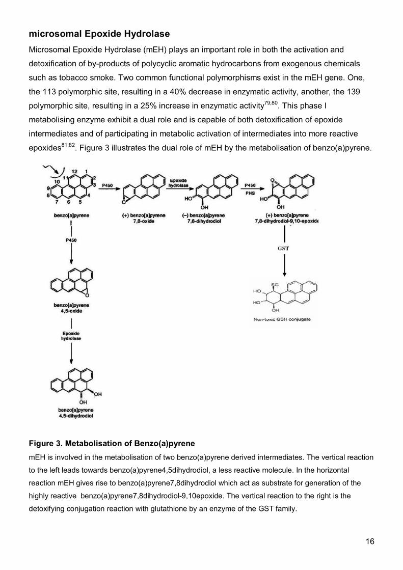
microsomal Epoxide Hydrolase Microsomal Epoxide Hydrolase (mEH) plays an important role in both the activation and
detoxification of by-products of polycyclic aromatic hydrocarbons from exogenous chemicals
such as tobacco smoke. Two common functional polymorphisms exist in the mEH gene. One,
the 113 polymorphic site, resulting in a 40% decrease in enzymatic activity, another, the 139
polymorphic site, resulting in a 25% increase in enzymatic activity79;80. This phase I
metabolising enzyme exhibit a dual role and is capable of both detoxification of epoxide
intermediates and of participating in metabolic activation of intermediates into more reactive
epoxides81;82. Figure 3 illustrates the dual role of mEH by the metabolisation of benzo(a)pyrene.
Figure 3. Metabolisation of Benzo(a)pyrene mEH is involved in the metabolisation of two benzo(a)pyrene derived intermediates. The vertical reaction
to the left leads towards benzo(a)pyrene4,5dihydrodiol, a less reactive molecule. In the horizontal
reaction mEH gives rise to benzo(a)pyrene7,8dihydrodiol which act as substrate for generation of the
highly reactive benzo(a)pyrene7,8dihydrodiol-9,10epoxide. The vertical reaction to the right is the
detoxifying conjugation reaction with glutathione by an enzyme of the GST family.

17
N-acetyl transferase 2 The N-acetyltransferase 2 (NAT2) enzyme is important in the biotransformation of a number of
aryl amines and heterocyclic amines derived from foods, tobacco smoking or other
environmental substances83. It is possible that individuals with slow NAT2 acetylator status have
a decreased ability to metabolise xenobiotics leading to accumulation which leads to increased
permeability in the gastrointestinal tract. The accumulation of xenobiotics are believed to induce
an autoimmune mechanism and NAT2 slow acetylator status has been shown to be associated
with autoimmune diseases such as systemic lupus erythematosus, rheumatoid arthritis and
diabetes mellitus84-86. A Japanese study has shown an association between NAT2 slow
acetylator status and CD87.
P-glycoprotein and Breast Cancer Resistance Protein The enzymes P-glycoprotein and Breast cancer Resistance Protein (BCRP) function as
intestinal transporter of xenobiotics and are highly expressed in the gut88;89. P-glycoprotein
encoded by the Multi Drug Resistance protein (MDR1) gene has shown decreased levels of
expression in inflamed tissue in active UC89-91. Genetic variants of the MDR1 gene exist and the
main focus has been on two polymorphic sites in the gene. The two functional genetic variants
have been shown to correlate with the activity and expression of the enzyme, and the two
polymorphisms have been associated with IBD in different populations but with conflicting
results90;92-94.
BCRP has shown decreased levels of expression in the inflamed mucosa of patients with
active UC as was the case for p-glycoprotein89;90. BCRP as a possible susceptibility gene of IBD
has not been as intensively investigated as MDR1. A Hungarian study showed no association
between two variants of the BCRP gene and IBD95.
Hypothesis CARD15 was the first gene found to confer susceptibility to CD43;44. The variations in CARD15
have shown to display wide ethnic variation, and even display heterogeneity within Western
populations and between European countries49;51. The CARD15 variants have consistently
shown to display susceptibility only to CD and not UC28;96. Hence, the hypothesis was that
CARD15 variations were associated with CD and not with UC in our population, and that
phenotypic associations would be likely.

18
High levels of oxidative stress along with increased intestinal permeability have been
observed in IBD97-99. Reactive molecules play a central role in altering the intestinal
permeability. Hence, impairment in the biotransformation system responsible for detoxification
of reactive substances might be important in the aetiology of IBD. This led to the hypothesis that
genetic polymorphisms responsible for low or missing activity of detoxification enzymes are risk
factors of IBD. This hypothesis is supported by the common disease – common variant
hypothesis11;12. Polymorphisms in detoxification enzymes have been associated with
autoimmune disease i.e. systemic lupus erythematosus and rheumatoid arthritis84;86. However,
only a few contradictory studies have focused on polymorphisms in detoxification enzymes as
possible risk factors of IBD.
Aims The overall objective of this study was to clarify the extent of three relatively common variations
in the CARD15 gene in susceptibility to IBD in our cohort. It was also intended to make a
comparison with other Western populations and to determine whether the CARD15 mutations
were important regarding disease phenotype (study I). The other main objective of this study
was to estimate the significance of the highly polymorphic xenobiotica metabolising enzymes of
the GST family (study II), the mEH (study III) and the NAT2 (study III) in susceptibility to IBD in
our cohort. Whether the polymorphic enzymes were important for disease phenotype and
whether an interaction with smoking was present was also assessed (I, II, III).
The aims were:
1. To assess the significance of three common variations in the CARD15 gene in
susceptibility to IBD, and to determine whether any genotype-phenotype correlations
were present.
2. To assess the significance of having a genotype encoding low or no enzymatic activity of
Glutathione S-transferase µ (GSTM1), Glutathione S-transferase θ (GSTT1) and
Glutathione S-transferase π (GSTP1) in susceptibility to IBD, and to determine whether
any genotype-phenotype correlations were present.
3. To estimate whether interactions were present between the GST enzymes in
susceptibility to IBD.
4. To assess the significance of having the slow acetylator genotype of NAT2 in
susceptibility to IBD, and to determine whether any genotype-phenotype correlations
were present.
5. To assess the significance of low activity genotype of mEH in susceptibility to IBD, and to
determine whether any genotype-phenotype correlations were present.

19
6. To determine the role of smoking behaviour in susceptibility to CD and UC, and to
investigate a possible modifying effect of smoking on GST genotypes, NAT2 acetylator
status and mEH genotypes in susceptibility to CD and UC.

20
METHODS
Study design The present epidemiologic study was conducted as an observational study of the case-control
type. This was concerned with the frequency of exposure (genotype or smoking) in IBD patients
(cases) and people without the disease (controls). This type of study is the predominant design
when searching for aetiology and causal associations in diseases. Association between
exposure and disease was expressed as odds ratios (OR) which represent the odds of
exposure in cases divided by the odds of exposure in controls.
Strength and limitations of the study A great advantage when choosing the case-control design was the possibility to examine many
exposures in the same study. A great strength of this study design was that of being able to
recruit cases fast. Three hundred and eighty-eight patients with CD and 565 patients with UC
were included in the study. The patients were recruited from the outpatient clinics between
January 2004 and March 2005 from three Danish hospitals in the cities of Aalborg, Viborg and
Herning. A senior registrar reviewed the case record and only patients fulfilling the diagnostic
criteria of CD and UC as proposed by Binder were included100. Patients with indeterminate IBD
were excluded, and patients under the age of 18 were also excluded from the study.
The collection of patients was biased by the fact that colectomised patients were not
included in the study group because these patients no longer attended routine follow-up
consultations. These patients are estimated to represent 10-15% of the patients. A control
group of 796 healthy blood donors representative of the general Danish population were
recruited from Viborg County during the same period. All participating patients and controls
gave written consent and the local Ethical Committee at Aalborg and Viborg County approved
the protocol (VN2003/5).
The purpose of the control group was to provide an estimate of the frequency of genetic
polymorphisms and smoking status in subjects in the population without the disease. A major
challenge in the case-control study design was the selection of a representative control group.
The controls in this study were blood donors recruited at Viborg Hospital. Blood donors tend to
be healthier than the general population which may be reflected in their genetic profile
(discussed in paper III). Recall bias is common regarding exposure in case-control studies. In
the present study genetic polymorphisms represent the exposure, thus recall bias was not an
issue. Uncertainty of whether the former smokers were smokers at the time of diagnosis, or
whether they quit smoking prior to being diagnosed with IBD, may have biased the results when

21
separating smoking into three groups of current smokers, former smokers, and never smokers.
To avoid bias the current smokers and former smokers were grouped together. For a few sub-
analyses the ever smoker group were separated into current and former smokers.
Genotyping assays Two different Polymerase Chain Reaction (PCR) based methods were used to determine the
genotype of the different genes in the individual papers I, II and III.
PCR gel based assays
Two steps are included in the PCR gel based assays: PCR amplification by use of specifically
designed primers, followed by visualisation using gel electrophoresis. A housekeeping gene is
usually used as an internal control of amplification. Primers with similar annealing temperatures
are designed for the gene of interest as well as the control gene and are run for 35-45 cycles
depending on the assay. Real-time PCR based assays
The real-time PCR assay visualises the exponential PCR amplification as it progresses,
whereas in traditional PCR, results are collected after the reaction is complete. In real-time PCR
the quantity of the PCR product is directly proportional to the amount of template.
Allelic discrimination assay
The real-time allelic discrimination assay was used to genotype SNPs (I, II, III). As for the PCR
gel based assays a specific set of primers was designed. Besides the primers, two specific
probes which recognised the two possibilities at the polymorphic site of the gene of interest
were designed. TaqMan technology from Applied Biosystems was used for genotyping in the
present studies (I, II, III). The two probes specific for each of the two possible alleles of the
polymorphic site was labelled with two different fluorescent dyes at the 5´ end (V and F fig. 4).
The 3´end was labelled with a quencher (Q, fig. 4), which absorbs the fluorescent emission. The
probe anneals to the DNA sequence complementary to its sequence and it is incorporated into
the DNA strand. This separates the fluorophore from the quencher and fluorescent emission
occurs.

22
Figure 4. Taq-Man based allelic discrimination
Top: Vic-labelled probe recognises its target allele 1 and is incorporated during the amplification of DNA.
The FAM-labelled probe does not recognise allele 1. Bottom: The FAM-labelled probe recognises its
target allele 2 and is incorporated during the amplification of DNA. The Vic-labelled probe does not
recognise allele 2. Figure from manual supplied with the HT7900 Real-time apparatus from Applied
Biosystems.
This emission represents the amplification of products in real-time. Figure 5 shows a signal for
both fluorescent dyes. Hence, this sample is heterozygous for the polymorphic site. If the
sample had been homozygous for either the wild type or the variant, only one curve would
show. For allelic discrimination assays a scatter plot is produced for each run, which allows for a
fast check of the results. Figure 6 (on the following page) represents the genotype callings
made by the system for an allelic discrimination run from the genotyping of NAT2 (III).
Figure 5. Amplification curves of real-time assay
Amplification curves for a sample being heterozygous for the polymorphic site investigated. Each curve
represents a signal for one specific fluorescent dye.

23
Figure 6. Allelic discrimination scatter plot
Scatter plot of allelic discrimination run. Circles in upper left corner represent the wild type genotype,
circles in the lower right corner represent the homozygous variant genotype, and circles along the
diagonal represent the heterozygous genotype. The black square is a negative control. The x is a
genotype not called by the system.
Relative quantification assay
In the relative quantification real-time assay a target gene and a reference gene are amplified in
the same tube. For the method to be valid, the efficiency of the target gene amplification and the
efficiency of the reference gene amplification must be approximately equal. This method
compares the threshold cycle of one target gene to a reference housekeeping gene in a single
sample.
Genotyping assays used in the thesis
Genotyping of CARD15
The allelic discrimination design was chosen for genotyping of the three common CARD15
variants (I). The primers and probes used as well as the concentrations and the run parameters
are described in detail in paper I. Direct sequencing of the three common variants was used for
validation of the assay using primers from King et al Human Mutation Supll. Online Nov. 2005.
Another approach for genotyping CARD15 on the entire cohort could be direct sequencing. This
would have been the method of genotyping if searching for rare private for the genotyping of the
entire cohort, which would have been the preferable technique if we were searching for rare
private mutations in our cohort.
Wild type
Heterozygous
Homozygous
24
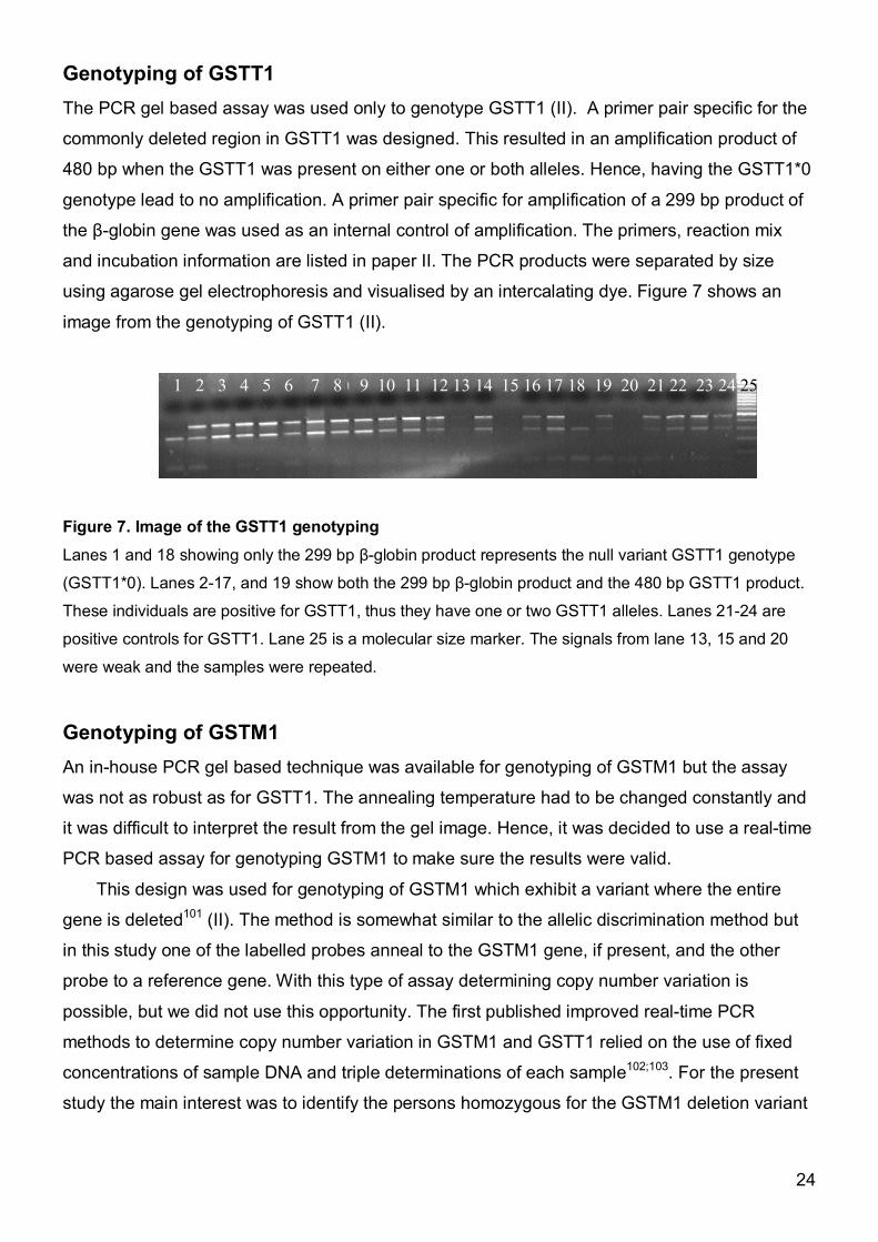
Genotyping of GSTT1 The PCR gel based assay was used only to genotype GSTT1 (II). A primer pair specific for the
commonly deleted region in GSTT1 was designed. This resulted in an amplification product of
480 bp when the GSTT1 was present on either one or both alleles. Hence, having the GSTT1*0
genotype lead to no amplification. A primer pair specific for amplification of a 299 bp product of
the β-globin gene was used as an internal control of amplification. The primers, reaction mix
and incubation information are listed in paper II. The PCR products were separated by size
using agarose gel electrophoresis and visualised by an intercalating dye. Figure 7 shows an
image from the genotyping of GSTT1 (II).
Figure 7. Image of the GSTT1 genotyping
Lanes 1 and 18 showing only the 299 bp β-globin product represents the null variant GSTT1 genotype
(GSTT1*0). Lanes 2-17, and 19 show both the 299 bp β-globin product and the 480 bp GSTT1 product.
These individuals are positive for GSTT1, thus they have one or two GSTT1 alleles. Lanes 21-24 are
positive controls for GSTT1. Lane 25 is a molecular size marker. The signals from lane 13, 15 and 20
were weak and the samples were repeated.
Genotyping of GSTM1 An in-house PCR gel based technique was available for genotyping of GSTM1 but the assay
was not as robust as for GSTT1. The annealing temperature had to be changed constantly and
it was difficult to interpret the result from the gel image. Hence, it was decided to use a real-time
PCR based assay for genotyping GSTM1 to make sure the results were valid.
This design was used for genotyping of GSTM1 which exhibit a variant where the entire
gene is deleted101 (II). The method is somewhat similar to the allelic discrimination method but
in this study one of the labelled probes anneal to the GSTM1 gene, if present, and the other
probe to a reference gene. With this type of assay determining copy number variation is
possible, but we did not use this opportunity. The first published improved real-time PCR
methods to determine copy number variation in GSTM1 and GSTT1 relied on the use of fixed
concentrations of sample DNA and triple determinations of each sample102;103. For the present
study the main interest was to identify the persons homozygous for the GSTM1 deletion variant
1 2 3 4 5 6 7 8 9 10 11 12 13 14 15 16 17 18 19 20 21 22 23 24 25

25
(GSTM1*0) and no discrimination were made between copy number variation of GSTM1 (being
homozygous or heterozygous for the GSTM1 allele) (II). Thus, only single determination was
necessary. Recently a high-throughput single determination genotyping method of copy number
variation was published104. The determination of copy number variation in GSTM1 could
become relevant for future studies examining the role of GSTM1 genotype in relation to adverse
effects of azathioprine treatment in IBD patients105;106. Another approach to determine the copy
number variation of GSTM1 is to use the Multiplex Ligation dependent Probe Amplification
(MLPA) by MRC-Holland which offers a panel containing several xenobiotica metabolising
enzymes107.
Genotyping of GSTP1 The allelic discrimination design was chosen for genotyping of the two specific GSTP1
polymorphic sites (II). The primers and probe sequences are listed in paper II. The reaction set-
up and run parameters were identical to those for the CARD15 genotyping and are also
described in paper II.
Genotyping of mEH For genotyping of the two mEH polymorphic sites the allelic discrimination design was chosen
(III). Commercially available assays from Applied Biosystems specific for the two polymorphic
sites were used and the run parameters are listed in paper III. The allelic discrimination assay
was chosen in preference to a conventional PCR gel based assay due to a report of genotyping
errors from a PCR based assay in a previous publication108. This publication showed an
association between low activity of mEH and CD. Due to a questioning of their results the group
came up with a reanalysis of their cohort using a real-time PCR based assay and was not able
to reproduce the positive association between low activity of mEH and CD109;110.
Genotyping of NAT2 The NAT2 genotypes can be divided into three phenotypic categories of rapid, intermediate and
slow acetylators. The NAT2 acetylator status is determined by a number of SNPs. A
combination of six relatively common SNPs in NAT2 was used to decide the acetylator status in
study III. Haplotype determination for the large study group used in this thesis was laborious.
The haplotype determination was performed by hand, but in many cases several haplotypes
were possible. The NAT2PRED web server is an extremely useful tool in assigning individual
acetylator status from the SNPs without having to determine the haplotypes111. The NAT2PRED

26
is based on a dataset of 1377 individuals (94% Caucasians). The performance was high with
sensitivity and specificities ranging between 99.6 and 100% for determining the three acetylator
phenotypes111. The ability of the NAT2PRED web server has been assessed by a study
including 8489 individuals from 56 populations with different geographic origin112. The
conclusion from this study was that the server correctly identified the slow acetylator phenotype
with more than 99% sensitivity in all populations outside Sub-Saharan Africa where another
variant, the 191 G>A SNP, plays an important role113. The classification error rate found in the
evaluation study implied that the NAT2PRED is poor at distinguishing between fast and
intermediate acetylators. The classification error rate was though not high in European
populations112. The data were submitted for each of the six SNPs for all the participants in this
study and the results were returned by email. The results listed the probability of all three
acetylation phenotypes for each individual. The intermediate and fast acetylators were grouped
for data analysis in our study as suggested by the NAT2PRED evaluation study112.
Table 1 depicts the acetylation calls (prediction) for 10 samples from our cohort. The prediction
for index 10 was not very secure. In total 7 of 1716 calls were in the range of 0.5-0.6 all
resulting in a rapid genotype call. (See Table 1 footnotes for further information).
Table 1. Acetylation callings from the NAT2PRED webserver
index genotype p(R) p(I) p(S) prediction 1 1,1,1,1,1,1 0.97995 0.01221 0.00784 R 2 3,1,1,3,1,1 0.00116 0.00126 0.99758 S 3 3,1,1,3,1,1 0.00116 0.00126 0.99758 S 4 1,3,3,1,3,1 0.00164 0.00118 0.99718 S 5 1,3,2,1,3,1 0.00164 0.00118 0.99718 S 6 1,3,3,1,3,1 0.00164 0.00118 0.99718 S 7 1,2,2,1,2,1 0.00100 0.99696 0.00203 I 8 3,2,2,1,2,2 0.00001 0.00126 0.99874 S 9 1,3,3,1,3,1 0.00164 0.00118 0.99718 S 10 1,1,2,1,1,1 0.61518 0.14525 0.23957 R
Index is the number of the individual. Genotype cover the genotypes for each of the six polymorphic sites separated by a
comma: 1 is wildtype homozygous, 2 is heterozygous, 3 is homozygous for the variant. p(R), p(I) and p(S) are the probabilities
for the three callings Rapid, Intermediate and Slow acetylator status. Prediction is the actual call made by the server: R=Rapid,
I=Intermediate and S=Slow. The results were returned by email.
Genotyping of MDR1 and BCRP Genotyping of MDR1 and BCRP on our study population was performed by one of our
collaborators, MSc. PhD, Mette Østergaard at Viborg Hospital. An allelic discrimination assay
was used to genotype the polymorphic sites G2677T/A and C3435T and intron variant G-
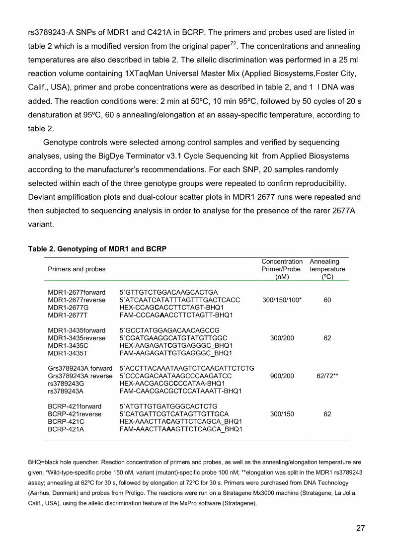
27
rs3789243-A SNPs of MDR1 and C421A in BCRP. The primers and probes used are listed in
table 2 which is a modified version from the original paper72. The concentrations and annealing
temperatures are also described in table 2. The allelic discrimination was performed in a 25 ml
reaction volume containing 1XTaqMan Universal Master Mix (Applied Biosystems,Foster City,
Calif., USA), primer and probe concentrations were as described in table 2, and 1µl DNA was
added. The reaction conditions were: 2 min at 50ºC, 10 min 95ºC, followed by 50 cycles of 20 s
denaturation at 95ºC, 60 s annealing/elongation at an assay-specific temperature, according to
table 2.
Genotype controls were selected among control samples and verified by sequencing
analyses, using the BigDye Terminator v3.1 Cycle Sequencing kit from Applied Biosystems
according to the manufacturer’s recommendations. For each SNP, 20 samples randomly
selected within each of the three genotype groups were repeated to confirm reproducibility.
Deviant amplification plots and dual-colour scatter plots in MDR1 2677 runs were repeated and
then subjected to sequencing analysis in order to analyse for the presence of the rarer 2677A
variant.
Table 2. Genotyping of MDR1 and BCRP
Primers and probes Concentration Primer/Probe
(nM)
Annealing temperature
(ºC) MDR1-2677forward 5´GTTGTCTGGACAAGCACTGA MDR1-2677reverse 5´ATCAATCATATTTAGTTTGACTCACC 300/150/100* 60 MDR1-2677G HEX-CCAGCACCTTCTAGT-BHQ1 MDR1-2677T FAM-CCCAGAACCTTCTAGTT-BHQ1 MDR1-3435forward 5´GCCTATGGAGACAACAGCCG MDR1-3435reverse 5´CGATGAAGGCATGTATGTTGGC 300/200 62 MDR1-3435C HEX-AAGAGATCGTGAGGGC_BHQ1 MDR1-3435T FAM-AAGAGATTGTGAGGGC_BHQ1 Grs3789243A forward 5´ACCTTACAAATAAGTCTCAACATTCTCTG Grs3789243A reverse 5´CCCAGACAATAAGCCCAAGATCC 900/200 62/72** rs3789243G HEX-AACGACGCCCCATAA-BHQ1 rs3789243A FAM-CAACGACGCTCCATAAATT-BHQ1 BCRP-421forward 5´ATGTTGTGATGGGCACTCTG BCRP-421reverse 5´CATGATTCGTCATAGTTGTTGCA 300/150 62 BCRP-421C HEX-AAACTTACAGTTCTCAGCA_BHQ1 BCRP-421A FAM-AAACTTAAAGTTCTCAGCA_BHQ1
BHQ=black hole quencher. Reaction concentration of primers and probes, as well as the annealing/elongation temperature are
given. *Wild-type-specific probe 150 nM, variant (mutant)-specific probe 100 nM; **elongation was split in the MDR1 rs3789243
assay; annealing at 62ºC for 30 s, followed by elongation at 72ºC for 30 s. Primers were purchased from DNA Technology
(Aarhus, Denmark) and probes from Proligo. The reactions were run on a Stratagene Mx3000 machine (Stratagene, La Jolla,
Calif., USA), using the allelic discrimination feature of the MxPro software (Stratagene).

28
RESULTS - RISK FACTORS OF IBD
Smoking Smoking has been found to be an independent risk factor of CD and current smoking to have a
protective effect against UC, which was also true for this study population58;59(I). Ever smoking
was associated with CD with an OR of 1.8 (1.4-2.3), (P<0.001), whereas current smoking had a
protective effect against UC with an OR of 0.7 (0.5-0.9), (P=0.015) (Table 3). The score of the
ORs were similar to what has been found in a large number of studies114. Extensive research
has been made concerning the dual effect of smoking on IBD. A recent publication shared some
thoughts as to why current smoking has a protective effect against UC55. Intestinal permeability
is known to be high in UC patients67. The mucus thickness has shown to be shallower in UC
compared with CD55. Nicotine has shown to enhance mucosal production, thus this could
possibly strengthen the epithelial barrier in UC hereby decreasing the intestinal permeability55.
Smoking reduces rectal blood flow, which may cause less recruitment of pro-inflammatory
mediators to the rectum, thus protecting against sustained inflammation.
Table 3. Association between smoking behaviour and IBD in the cohort.
Smoking status OR (95%CI) P-value CD UC Ever smoker 1.8 (1.4-2.3) P<0.001 1.1 (0.9-1.4) P=0.28 Current smoker 2.6 (2.0-3.5) P<0.001 0.7 (0.5-0.9) P=0.015
OR=Odds ratio, 95%CI=95% confidence interval, P =P value. CD=Crohn´s disease, UC=Ulcerative colitis.
CARD15 We found that harboring at least one CARD15 variant to be associated with CD but not UC in
the Danish population (I). The OR for a CD patient carrying one CARD15 variant was 1.9 (1.3-
2.8), P<0.001 (I). A gene-dosage effect was observed in the population raising the OR to 21.1
(4.9-91.2), P<0.001 for carrying two CARD15 variants (I). In comparison the ORs were 2.4 (2.8-
8.0) and 6.7 (4.1-10.9) respectively in a large meta-analysis of 79 studies (in which our study
was also included)115. The confidence interval for carrying two variants was wide in our study
compared with that of the meta-analysis. These numbers suggest that the OR of 6.7 for CD
patients carrying two variants is more likely than our OR of 21.1 (Table 4).
The susceptibility to CD is inherent in the R702W SNP and in the 1007insC frame-shift
mutation, whereas the rare G908R SNP was not associated with CD in our population (I).

29
The frequencies of the three variants in the control group of our population was similar to those
of a recent publication of more than 38 000 healthy Danes (Table 4)116. When comparing the
frequencies with a meta-analysis of 3500 healthy Caucasians our findings are similar to the
findings in other Northern European countries, which in general has a lower CARD15 variant
frequency than other European countries49.
Table 4. Distribution of CARD15 variants in the Danish population
CARD15 variants n, (%) CD UC HC Ernst et al (I) R702W alleles *40 (5.5) 36 (3.3) 44 (2.8) G908R alleles 11 (1.5) 9 (0.8) 16 (1.0) 1007insC alleles **46 (6.4) 12 (1.1) 19 (1.2) 0 variant 365 (79.4) 504 (89.8) 718 (90.3) 1 variant ^61 (15.9) 56 (10.0) 75 (9.4) 2 variants ^^18 (4.6) 1 (0.2) 2 (0.3) Least 1 variant ^^^79 (20.6) 57 (10.2) 77 (9.7) Yazdanyar et al 2009116 0 variants 38592 (87.0) 1 variant 4838 (12.5) 2 variants 164 (0.4) Least 1 variant 5002 (13.0)
*R702W allele frequency in CD patients versus controls: OR=1.9 (1.3-3.0) P=0.02.
**1007insC allele frequency in CD patients versus controls: OR=5.3 (3.1-9.1) P<0.001.
^CARD15 1 variant genotype frequency in CD patients versus controls OR=1.9 (1.3-2.8) P<0.001.
^^CARD15 2 variants genotype frequency in CD patients versus controls OR=21.1 (4.9- 91.2) P<0.001.
^^^CARD15 at least 1 variant genotype frequency in CD patients versus controls OR=2.4 (95% confidence
interval (1.7-3.4) P<0.001). OR=Odds ratio, 95%CI=95% confidence interval, P =P value.
CD=Crohn´s disease, UC=Ulcerative colitis.
Compatible with the high expression of CARD15 in the Paneth cells of the ileum, the
CARD15 variants were associated with ileal involvement in CD. The OR was 2.6 (1.5-4.5), P=
0.001, for carrying at least one CARD15 variant (I). When considering the three CARD15
variants separately, the association with ileal involvement of CD reached only statistical
significance for the 1007insC frame-shift mutation (I). This association between CARD15
variants and disease location in CD patients have been demonstrated in the majority of
association studies117-120. A weak association was found between CARD15 variants and less
than 40 years of age at disease onset with an OR of 2.0 (1.0-4.0), P=0.038. Smoking was found
to confer risk of CD and to display a protective effect of UC, but no modifying effect of smoking
on CARD15 genotype was found for either CD or UC (I). Summarised study I found an
association between two relatively common CARD15 variants (R702W and 1007insC) and CD.

30
The association was strongest for the 1007insC mutation and a gene-dosage effect was
observed. CARD15 seemed to influence disease phenotype by affecting disease onset and the
1007insC variant was associated with ileal involvement in CD. No direct association or
phenotypic associations was found between CARD15 variants and UC (I).
Xenobiotica metabolising enzymes
Glutathione S-transferase family Genotyping of GSTM1 resulted in very similar frequencies within the three groups of CD
patients, UC patients and healthy controls in our population (II) (Table 5). The findings were
similar to the findings in three previous European studies108;121;122 (Table 5). An Indian study
found a significant association between GSTM1*0 and UC123. The frequency of the GSTM1*0
genotype in the Indian UC patients (61%) was similar our study group (53%), but the difference
appear to reside in the control populations where the GSTM1*0 genotype has shown to be less
frequent in the general Indian population (30%)124;125. (Table 5).
The frequencies of the Glutathione S-transeferase θ null variant genotype (GSTT1*0) were
also similar between the three groups of CD, UC and healthy controls in our population (II)
(Table 5). Higher frequencies of GSTT1*0 were found in two previous European study
populations (II). The GSTT1*0 frequency is generally lower in Scandinavian populations, thus,
the outcome of the studies were the same, with no difference in GSTT1*0 frequency between
IBD patients and healthy controls108;121;126. In the previously mentioned Indian study a strong
association was found between GSTT1*0 and both UC and IBD123. Ethnic differences are
expected in susceptibility genes, but the fact that very few IBD patients were recruited in the
Indian study could very likely have biased the results. Further research into whether GSTM1*0
and GSTT1*0 genotypes are truly associated with IBD in the Indian population are needed.
The distribution of GSTP1 low activity genotypes (GSTP1 105 low and GSTP1 114 low)
found in our cohort, were in agreement with previous findings in Caucasian populations108;126
(Table 5). Research regarding other diseases has shown GST genotypes to be risk factors of
disease only when present in combination and not as a single gene73. Hence, it could be
expected that combinations of several of the GST genotypes might be necessary to induce
susceptibility to IBD. However, no association was found between any combination of having
GSTM1*0, GSTT1*0 and GSTP1 low activity genotypes and IBD in our study (II).
Early onset disease has been associated with high familial prevalence of CD, hence,
suggesting a stronger genetic contribution in this group of patients15;51;127. We were not able to
replicate a Swedish finding of an association of GSTM1*0 and early onset of UC122. With regard
31
to phenotypic association in general we found no association between GST genotypes and the
phenotypic behaviours of early onset of disease, localisation of disease and severity of disease
in our study population (II). Neither did we find any indication of GST single gene or a
combination of several of the GST genotypes to cause susceptibility to IBD in our study
population (II). Hence, GST genotypes do not seem to play an important role in susceptibility to
IBD. Table 5. GST genotypes among IBD patients and healthy controls in different populations
CD=Crohn´s disease. UC=ulcerative colitis. HC=healthy controls. GSTM1*0:GSTM1 null genotype. GSTT1*0: GSTT1 null genotype. GSTM1*0 & GSTT1*0: GSTM1 null and GSTT1 null genotype. *only percentages available in paper.
By-products from tobacco smoke are likely substrates of the GST enzymes and current smoking
seems to have a protective effect against UC. This was supported in the current study where an
interaction was found between GSTM1*0 and smoking for UC patients, with GSTM1*0 genotype
strengthening the protective effect of smoking (II). One might speculate the GSTM1 active
genotype could eliminate or reduce the protective effect of smoking in UC, whereas the
GSTM*0 genotype does not interrupt the protective effect of tobacco smoking.
Genotype Distribution of GST genotypes n, (%) CD UC HC GSTM1*0 Ernst et al (II) 215 (56) 296 (53) 417 (53) Hertervig122 65 (60) 101 (56) 219 (49) Duncan et al121 68 (62) 112 (49) 203 (54) De Jong et al108 82 (54) 74 (50) Mittal et al123 9 (45) 52 (61) 49 (30) GSTT1*0 Ernst et al (II) 66 (17) 82 (15) 104 (13) Duncan et al121 17 (16) 52 (24) 47 (18) De Jong et al108 23 (15) 30 (20) Mittal et al123 18 (90) 77 (91) 26 (16) GSTM1*0 &GSTT1*0
Ernst et al (II) 36 (9) 44 (8) 50 (6) Duncan et al121 10 (13) De Jong et al108 *(7) *(13) Mittal et al123 4 (20) 28 (33) 8 (5)

32
N-acetyltransferase 2 We were not able to replicate a Japanese finding of a positive association between NAT2 slow
acetylator status and CD(III)87. In our study the frequency of NAT2 slow, intermediate and rapid
acetylators was in agreement with the findings from another Caucasian study population (II)128.
Highly different frequencies of NAT2 acetylator status are found between different ethnic
groups. The NAT2 rapid acetylator genotype is present in less than 10% in Caucasian
populations and in populations of African descent129;130. The NAT2 slow acetylator genotype
varies between 50-65% in these populations131(III). In Asian populations the NAT2 slow
acetylator genotype is less frequent ranging between 10-25% but with great differences
between countries131 (Table 6). An overrepresentation of NAT2 slow metabolisers have been
shown among aryl-amine exposed bladder cancer patients compared with healthy controls in
Caucasian populations131. In contrast, the NAT2 slow metabolisers have been shown to be
underrepresented among aryl-amine exposed bladder cancer patients in a Chinese population.
This indicates that pathways other than NAT2 could be involved in the metabolism of aromatic
amines132. This example demonstrates that genetic risk factors may only be risk factors in some
ethnic populations not only because of interaction with environmental exposure differences, but
possibly also depending on the general prevalence of the genetic variant in the given
population.
Table 6. NAT2 acetylator status in healthy controls of different populations
NAT2 acetylator status in healthy controls of different populations n, (%) (Origin of population) Rapid Intermediate Slow Ernst et al (II) (Caucasian) 44 (6) 300 (38) 443 (56) Kiyohara et al84 (Japanese) ^(30) ^(49) ^(21) Inatomi et al131 (Japanese)* 10 (7) Su et al131 (Taiwan)* 13 (13) Kim et al131 (Korean)* 24 (11)
* Taken from Golka et al131 ^only percentages avaliable in paper
We found no evidence of NAT2 playing a key role in phenotypic characteristics of disease
regarding either UC or CD (III). Nor was any interaction between NAT2 and smoking found in
susceptibility to IBD (III). In conclusion, NAT2 does not seem to be important in susceptibility to
IBD in the Danish population (III).

33
microsomal Epoxide Hydrolase We found no association between either of the two mEH polymorphic sites 113 or 139 or a
combination of the polymorphisms with IBD (III) (Table 7). The findings are in agreement with
two previous European studies using the same genotyping procedure109;110.
Table 7. Distribution of mEH genotypes
CD=Crohn´s disease. UC=Ulcerative colitis. HC=Healthy controls. Percentages may not add up to exactly 100% due to rounding.
The dual role of mEH meant that we had to consider the possibility of both low and high activity
mEH genotypes as possible risk factors. Analysing the mEH high activity genotype against low
and intermediate activity genotypes an association with diagnosis of CD before age 40 was
found with an OR of 2.2 (1.1-4.2), P=0.02. The association was expected to be stronger among
smokers, because of the dual role of mEH towards benzo(a)pyrene of tobacco smoke, but when
analysing ever smokers isolated the association did not reach statistical significance (II). This
finding could possibly be biased by the small numbers in this sub-grouping of patients. Thus,
the results indicate that mEH may influence the age at disease onset among CD patients but
further research on a larger population is needed to clarify this. No other phenotypic
associations were found.
Dealing with mEH a trend towards a modifying effect of smoking on low mEH activity
genotype was found for both CD and UC patients. Thus, smokers with a low activity mEH
genotype may have a higher risk of developing IBD compared with never smokers (III). Hence,
mEH may be important in susceptibility of IBD in combination with environmental factors.
Xenobiotic transporters MDR1 and BCRP In the xenobiotica transporter gene MDR1 two functional polymorphic sites were genotyped, the
G2667T/A and C3435T variants, and an intron-variant G-rs3789243-A72. No association was
mEH polymorphic sites (n, (%)) mEH 113 genotypes mEH 139 genotypes High Intermediate Low High Intermediate Low CD 177 (47) 167 (44) 36 (9) CD 21 (6) 118 (32) 235 (63) UC 286 (52) 225 (41) 42 (8) UC 25 (5) 190 (35) 335 (61) HC 373 (47) 359 (45) 61 (8) HC 42 (5) 284 (36) 466 (59) mEH combined genotypes High Intermediate Low CD 67 (18) 166 (44) 142 (38) UC 101 (19) 278 (51) 167 (31) HC 156 (20) 363 (46) 274 (35)
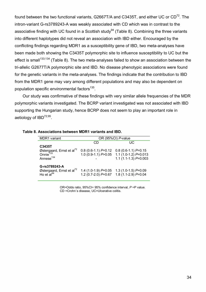
34
found between the two functional variants, G2667T/A and C3435T, and either UC or CD72. The
intron-variant G-rs3789243-A was weakly associated with CD which was in contrast to the
associative finding with UC found in a Scottish study94 (Table 8). Combining the three variants
into different haplotypes did not reveal an association with IBD either. Encouraged by the
conflicting findings regarding MDR1 as a susceptibility gene of IBD, two meta-analyses have
been made both showing the C3435T polymorphic site to influence susceptibility to UC but the
effect is small133;134 (Table 8). The two meta-analyses failed to show an association between the
tri-allelic G2677T/A polymorphic site and IBD. No disease phenotypic associations were found
for the genetic variants in the meta-analyses. The findings indicate that the contribution to IBD
from the MDR1 gene may vary among different populations and may also be dependent on
population specific environmental factors135.
Our study was confirmative of these findings with very similar allele frequencies of the MDR
polymorphic variants investigated. The BCRP variant investigated was not associated with IBD
supporting the Hungarian study, hence BCRP does not seem to play an important role in
aetiology of IBD72;95.
Table 8. Associations between MDR1 variants and IBD.
MDR1 variant OR (95%CI) P-value CD UC C3435T Østergaard, Ernst et al72 0.8 (0.6-1.1) P=0.12 0.8 (0.6-1.1) P=0.15 Onnie133 1.0 (0.9-1.1) P>0.05 1.1 (1.0-1.2) P=0.013 Annese134 - 1.1 (1.1-1.3) P=0.003 G-rs3789243-A Østergaard, Ernst et al72 1.4 (1.0-1.9) P=0.05 1.3 (1.0-1.5) P=0.09 Ho et al94 1.2 (0.7-2.0) P=0.67 1.8 (1.1-2.9) P=0.04
OR=Odds ratio, 95%CI= 95% confidence interval, P =P value. CD =Crohn´s disease, UC=Ulcerative colitis.
35
CONCLUSION AND PERSPECTIVES The frequencies of the CARD15 variants were similar to the findings from other Northern
European countries. Harbouring at least one of three common CARD15 variants was
associated with CD but not UC in the Danish population, and a stronger association was found
for being homozygous for the CARD15 variants compared with being heterozygous (I). The
susceptibility was inherent in two of the three common CARD15 variants, the R702W and the
1007insC, the latter displaying the strongest susceptibility to CD. CD patients carrying at least
one CARD15 variant were more likely to have ileal disease than CD patients with no CARD15
variants (I). A weak phenotypic association was found between carrying at least one CARD15
variant and early onset CD (I). No disease susceptibility and no phenotypic associations were
found between CARD15 and UC (I). Smoking was found to confer risk of CD and current
smoking to be a protective factor of UC, but no modifying effect of smoking on CARD15
genotype was found for either CD or UC (I).
GSTM1, GSTT1 and GSTP1 did not confer susceptibility to either CD or UC, and no
disease phenotypic associations were found (II). Combining members of the GST family did not
reveal any association with either CD or UC (II). A modifying effect of smoking was found on
GSTM1 genotype. Harbouring the GSTM1*0 genotype seemed to strengthen the protective
effect of current smoking with regard to UC (II).
NAT2 did not confer susceptibility to either CD or UC and no disease phenotypic
associations were found (III). No modifying effect of smoking was found on NAT2 acetylator
status, thus NAT2 does not seem to play an independent role in IBD (III).
No association was found between either of the two polymorphisms in mEH or the
combined mEH genotype and IBD (III). One possible phenotypic association was found, that of
mEH high activity genotype and early onset CD (III). An indication of a possible modifying effect
of smoking on mEH genotype was found for both CD and for UC (III).
Perspectives It is a general opinion that IBD is based on a dysfunctional immune response, but the exact
mechanism is unknown. Increased intestinal permeability also plays an important role in IBD but
one might speculate what comes first; intestinal permeability leading to enhanced bacterial
sensing which induces an immune response, or an exaggerated immune response which
results in a great production of reactive by-products which may alter the intestinal barrier? The
extremely complicated nature of the immune system makes it very difficult to find isolated
genetic variants which contribute independently to disease pathogenesis. This particular thesis

36
focused on genetic susceptibility from variants in enzymes of the xenobiotica metabolising
system, which is also a very complicated system comparable with the immune system. A
combination of various genetic variants is possibly responsible for the genetic susceptibility to
IBD and individual genetic variants confer only a modest disease risk which may be very difficult
or impossible to demonstrate. Environmental factors also have a substantial role in disease
susceptibility to complex diseases. Exposure to environmental factors might be very difficult to
measure which further complicates the finding of true risk factors of IBD. IBD is currently
emerging in Asia which leaves us with a great opportunity to investigate the shift in
environmental exposures in that part of the world. Hopefully this will add knowledge to how
environmental factors influence disease aetiology. The CARD15 variants have shown
susceptibility to CD only in the Western world and not in the Asian world. This indicates that the
genetic susceptibility to IBD differ in different populations. Identification of specific genetic
susceptibility genes is important and may help decode the pathways essential to disease
pathogenesis. The many susceptibility genes that have been uncovered from the GWA studies
have shown to participate in specific pathways, which may lead to a better understanding of the
disease and may also improve the treatment. The xenobiotica metabolising enzymes
investigated in this thesis did not prove to cause susceptibility to IBD. This may however, be
important in predicting which patients might develop adverse effects in response to treatment
with certain pharmaceuticals. GSTM1 has demonstrated its importance in relation to adverse
effects of treatment with pharmaceuticals containing azathioprine105;106. Looking for copy
number variation in GSTM1 in relation to adverse effects of azathioprine treatment is something
we plan to investigate in the near future. NAT2 slow acetylator status has shown to be
associated with adverse effects of sulfasalazine treatment of IBD in Asian populations136;137. We
already have the NAT2 acetylator status of the large group of IBD patients included in this
thesis and we will probably look into this also.

37
SUMMARY IN DANISH Kronisk inflammatorisk tarmsygdom dækker over sygdommene colitis ulcerosa og morbus
Crohn. Studier har vist, at en række faktorer er involveret i inflammationsprocessen ved kronisk
inflammatorisk tarmsygdom. Medfødt genetisk prædisposition, immunsystemet og
miljømæssige elementer er alle medvirkende faktorer i udvikling og forløb af sygdommene.
Sygdommene har historisk set været en byrde overvejende i den vestlige verden, men de
seneste årtier har incidensen været stigende i Asien. Den hurtige stigning tydeliggør
betydningen af miljømæssige påvirkninger i ætiologien af kronisk inflammatorisk tarmsygdom.
Variationer i CARD15 genet, der koder for et protein der genkender bakteriedele, var det
første gen der viste sig at prædisponere til morbus Crohn, men ikke til colitis ulcerosa. De
seneste års intensive genom associations studier har bidraget betydeligt med nye genetiske
risikofaktorer. Nogle af disse gener er fælles for sygdommene, men flere er specifikke for enten
colitis ulcerosa eller morbus Crohn. Desuden viser flere studier, at der er store etniske forskelle
i udbredningen af de fundne genetiske risikomarkører. Tre varianter i CARD15 genet der
prædisponerer for morbus Crohn er eksempelvis ikke nær så hyppige i Skandinavien
sammenlignet med andre europæiske lande, og er ikke til stede i Asien. Der er også store
etniske forskelle i miljøpåvirkninger, hvilket gør det kompliceret at finde sande genetiske og
miljømæssige risikofaktorer. Tobaksrygning er den bedst undersøgte miljømæssige faktor. Det
har vist sig at rygning forøger risikoen for at udvikle morbus Crohn, men at det har en
beskyttende effekt mod udvikling af colitis ulcerosa.
I et case-control studie undersøgte vi hyppigheden af tre CARD15 variationer i den danske
befolkning. To af de tre hyppige variationer var associeret med morbus Crohn, men var ret
sjældne i Danmark sammenlignet med andre europæiske lande.
Patienter med kronisk inflammatorisk tarmsygdom har en øget permeabilitet over tarmvæggen.
Det ledte til vores hypotese om at en lav aktivitet af enzymer, der omsætter fremmedstoffer
lokalt i tarmen, kunne forårsage skade på tarmepitelet. Dette var en mulig risikofaktor for
udvikling af kronisk inflammatorisk tarmsygdom. Flere af disse enzymer (samlet under
betegnelsen xenobiotika metaboliserende enzymer), har hyppige gen polymorfier der påvirker
enzymaktiviteten. Vi undersøgte om nogle af disse polymorfier var associeret med kronisk
inflammatorisk tarmsygdom i Danmark, men vi fandt ingen evidens for at det var tilfældet. Vi
undersøgte også for en mulig interaktion mellem gen polymorfierne og rygning. Vi fandt enkelte
genvariationer, hvor der var en risikoforskel for rygere og ikke rygere. Eksempelvis fandt vi en
genvariation kodende for manglende aktivitet af et enzym (GSTM1), der forstærkede den
beskyttende effekt af rygning på udvikling af colitis ulcerosa.

38
CARD15 variationer er altså ikke særligt udbredte i Danmark, men de prædisponerer til
udvikling af morbus Crohn. Generelt set kunne vi konkludere, at genetiske polymorfier i
xenobitotika metaboliserende enzymer ikke ser ud til at spille en afgørende rolle for udvikling af
kronisk inflammatorisk tarmsygdom. Enzymerne er vigtige i omsætningen af medikamenter
brugt til behandling af kronisk inflammatorisk tarmsygdomme. Det kan betyde at bestemmelse
af genetiske polymorfier i xenobitika metaboliserende enzymer er relevant i forbindelse med
medicinsk behandling af sygdommene og fremtidige studier er planlagt for at afdække dette.

39
REFERENCES
1. www.ccfa.org. 2010.
Ref Type: Internet Communication
2. Logan I, Bowlus CL. The geoepidemiology of autoimmune intestinal diseases.
Autoimmun Rev 2010;9:A372-A378.
3. Economou M, Pappas G. New global map of Crohn's disease: Genetic,
environmental, and socioeconomic correlations. Inflamm Bowel Dis 2008;14:709-
20.
4. Vind I, Riis L, Jess T et al. Increasing incidences of inflammatory bowel disease
and decreasing surgery rates in Copenhagen City and County, 2003-2005: a
population-based study from the Danish Crohn colitis database. Am J
Gastroenterol 2006;101:1274-82.
5. Jacobsen BA, Fallingborg J, Rasmussen HH et al. Increase in incidence and
prevalence of inflammatory bowel disease in northern Denmark: a population-
based study, 1978-2002. Eur J Gastroenterol Hepatol 2006;18:601-6.
6. Lichtenstein GR, Hanauer SB, Sandborn WJ. Management of Crohn's disease in
adults. Am J Gastroenterol 2009;104:465-83.
7. Kornbluth A, Sachar DB. Ulcerative colitis practice guidelines in adults: American
College Of Gastroenterology, Practice Parameters Committee. Am J Gastroenterol
2010;105:501-23.
40
8. Xie J, Itzkowitz SH. Cancer in inflammatory bowel disease. World J Gastroenterol
2008;14:378-89.
9. Finishing the euchromatic sequence of the human genome Nature 2004;431:931-
45.
10. A haplotype map of the human genome Nature 2005;437:1299-320.
11. Becker KG. The common variants/multiple disease hypothesis of common
complex genetic disorders. Med Hypotheses 2004;62:309-17.
12. Bodmer W, Bonilla C. Common and rare variants in multifactorial susceptibility to
common diseases. Nat Genet 2008;40:695-701.
13. Schork NJ, Murray SS, Frazer KA, Topol EJ. Common vs. rare allele hypotheses for
complex diseases. Curr Opin Genet Dev 2009;19:212-19.
14. Halfvarson J, Bodin L, Tysk C, Lindberg E, Jarnerot G. Inflammatory bowel disease
in a Swedish twin cohort: a long-term follow-up of concordance and clinical
characteristics. Gastroenterology 2003;124:1767-73.
15. Jess T, Riis L, Jespersgaard C et al. Disease concordance, zygosity, and
NOD2/CARD15 status: follow-up of a population-based cohort of Danish twins with
inflammatory bowel disease. Am J Gastroenterol 2005;100:2486-92.

41
16. Orholm M, Binder V, Sorensen TI, Rasmussen LP, Kyvik KO. Concordance of
inflammatory bowel disease among Danish twins. Results of a nationwide study.
Scand J Gastroenterol 2000;35:1075-81.
17. Spehlmann ME, Begun AZ, Burghardt J, Lepage P, Raedler A, Schreiber S.
Epidemiology of inflammatory bowel disease in a German twin cohort: results of a
nationwide study. Inflamm Bowel Dis 2008;14:968-76.
18. Halme L, Paavola-Sakki P, Turunen U, Lappalainen M, Farkkila M, Kontula K.
Family and twin studies in inflammatory bowel disease. World J Gastroenterol
2006;12:3668-72.
19. Franke A, McGovern DP, Barrett JC et al. Genome-wide meta-analysis increases to
71 the number of confirmed Crohn's disease susceptibility loci. Nat Genet
2010;42:1118-25.
20. Latella G, Fiocchi C, Caprili R. News from the "5th International Meeting on
Inflammatory Bowel Diseases" CAPRI 2010. J Crohns Colitis 2010;4:690-702.
21. McGovern DP, Gardet A, Torkvist L et al. Genome-wide association identifies
multiple ulcerative colitis susceptibility loci. Nat Genet 2010;42:332-37.
22. Jung C, Hugot JP. Inflammatory Bowel Diseases: the genetic revolution.
Gastroenterol Clin Biol 2009;33 Suppl 3:S123-S130.
23. Thompson AI, Lees CW. Genetics of ulcerative colitis. Inflamm Bowel Dis 2010.

42
24. Xavier RJ, Podolsky DK. Unravelling the pathogenesis of inflammatory bowel
disease. Nature 2007;448:427-34.
25. Sarra M, Pallone F, Macdonald TT, Monteleone G. IL-23/IL-17 axis in IBD. Inflamm
Bowel Dis 2010;16:1808-13.
26. Zhang H, Massey D, Tremelling M, Parkes M. Genetics of inflammatory bowel
disease: clues to pathogenesis. Br Med Bull 2008;87:17-30.
27. Hampe J, Franke A, Rosenstiel P et al. A genome-wide association scan of
nonsynonymous SNPs identifies a susceptibility variant for Crohn disease in
ATG16L1. Nat Genet 2007;39:207-11.
28. Noomen CG, Hommes DW, Fidder HH. Update on genetics in inflammatory
disease. Best Pract Res Clin Gastroenterol 2009;23:233-43.
29. Homer CR, Richmond AL, Rebert NA, Achkar JP, McDonald C. ATG16L1 and NOD2
interact in an autophagy-dependent antibacterial pathway implicated in Crohn's
disease pathogenesis. Gastroenterology 2010;139:1630-41, 1641.
30. Lecat A, Piette J, Legrand-Poels S. The protein Nod2: an innate receptor more
complex than previously assumed. Biochem Pharmacol 2010;80:2021-31.
31. Billmann-Born S, Lipinski S, Bock J, Till A, Rosenstiel P, Schreiber S. The complex
interplay of NOD-like receptors and the autophagy machinery in the
pathophysiology of Crohn disease. Eur J Cell Biol 2010.

43
32. Fukata M, Abreu MT. What are toll-like receptors and what role may they have in
IBD? Inflamm Bowel Dis 2008;14 Suppl 2:S90-S92.
33. Lavelle EC, Murphy C, O'Neill LA, Creagh EM. The role of TLRs, NLRs, and RLRs in
mucosal innate immunity and homeostasis. Mucosal Immunol 2010;3:17-28.
34. van Lierop PP, Samsom JN, Escher JC, Nieuwenhuis EE. Role of the innate
immune system in the pathogenesis of inflammatory bowel disease. J Pediatr
Gastroenterol Nutr 2009;48:142-51.
35. Kumagai Y, Akira S. Identification and functions of pattern-recognition receptors. J
Allergy Clin Immunol 2010;125:985-92.
36. Shen X, Shi R, Zhang H, Li K, Zhao Y, Zhang R. The Toll-like receptor 4 D299G and
T399I polymorphisms are associated with Crohn's disease and ulcerative colitis: a
meta-analysis. Digestion 2010;81:69-77.
37. Doyle SL, O'Neill LA. Toll-like receptors: from the discovery of NFkappaB to new
insights into transcriptional regulations in innate immunity. Biochem Pharmacol
2006;72:1102-13.
38. Levin A, Shibolet O. Toll-like receptors in inflammatory bowel disease-stepping
into uncharted territory. World J Gastroenterol 2008;14:5149-53.
39. Rosenstiel P, Till A, Schreiber S. NOD-like receptors and human diseases.
Microbes Infect 2007;9:648-57.
44
40. Elphick DA, Mahida YR. Paneth cells: their role in innate immunity and
inflammatory disease. Gut 2005;54:1802-9.
41. Vignal C, Singer E, Peyrin-Biroulet L, Desreumaux P, Chamaillard M. How NOD2
mutations predispose to Crohn's disease? Microbes Infect 2007;9:658-63.
42. Le Bourhis L, Benko S, Girardin SE. Nod1 and Nod2 in innate immunity and human
inflammatory disorders. Biochem Soc Trans 2007;35:1479-84.
43. Hugot JP, Chamaillard M, Zouali H et al. Association of NOD2 leucine-rich repeat
variants with susceptibility to Crohn's disease. Nature 2001;411:599-603.
44. Ogura Y, Bonen DK, Inohara N et al. A frameshift mutation in NOD2 associated
with susceptibility to Crohn's disease. Nature 2001;411:603-6.
45. King K, Sheikh MF, Cuthbert AP et al. Mutation, selection, and evolution of the
Crohn disease susceptibility gene CARD15. Hum Mutat 2006;27:44-54.
46. Lesage S, Zouali H, Cezard JP et al. CARD15/NOD2 mutational analysis and
genotype-phenotype correlation in 612 patients with inflammatory bowel disease.
Am J Hum Genet 2002;70:845-57.
47. Gasche C, Grundtner P. Genotypes and phenotypes in Crohn's disease: do they
help in clinical management? Gut 2005;54:162-67.
48. Schreiber S. Slipping the barrier: how variants in CARD15 could alter permeability
of the intestinal wall and population health. Gut 2006;55:308-9.

45
49. Hugot JP, Zaccaria I, Cavanaugh J et al. Prevalence of CARD15/NOD2 mutations in
Caucasian healthy people. Am J Gastroenterol 2007;102:1259-67.
50. Thia KT, Loftus EV, Jr., Sandborn WJ, Yang SK. An update on the epidemiology of
inflammatory bowel disease in Asia. Am J Gastroenterol 2008;103:3167-82.
51. Gaya DR, Russell RK, Nimmo ER, Satsangi J. New genes in inflammatory bowel
disease: lessons for complex diseases? Lancet 2006;367:1271-84.
52. Gasche C, Nemeth M, Grundtner P, Willheim-Polli C, Ferenci P, Schwarzenbacher
R. Evolution of Crohn's disease-associated Nod2 mutations. Immunogenetics
2008;60:115-20.
53. Krishnan A, Korzenik JR. Inflammatory bowel disease and environmental
influences. Gastroenterol Clin North Am 2002;31:21-39.
54. Danese S, Sans M, Fiocchi C. Inflammatory bowel disease: the role of
environmental factors. Autoimmun Rev 2004;3:394-400.
55. El Tawil AM. Smoking and inflammatory bowel diseases: what in smoking alters
the course? Int J Colorectal Dis 2010;25:671-80.
56. Carbonnel F, Jantchou P, Monnet E, Cosnes J. Environmental risk factors in
Crohn's disease and ulcerative colitis: an update. Gastroenterol Clin Biol 2009;33
Suppl 3:S145-S157.

46
57. Baumgart DC, Carding SR. Inflammatory bowel disease: cause and
immunobiology. Lancet 2007;369:1627-40.
58. Brant SR, Picco MF, Achkar JP et al. Defining complex contributions of
NOD2/CARD15 gene mutations, age at onset, and tobacco use on Crohn's disease
phenotypes. Inflamm Bowel Dis 2003;9:281-89.
59. Halfvarson J, Jess T, Magnuson A et al. Environmental factors in inflammatory
bowel disease: a co-twin control study of a Swedish-Danish twin population.
Inflamm Bowel Dis 2006;12:925-33.
60. van der HF, Nolte IM, Kleibeuker JH, Wijmenga C, Dijkstra G, Weersma RK.
Differences in genetic background between active smokers, passive smokers, and
non-smokers with Crohn's disease. Am J Gastroenterol 2010;105:1165-72.
61. Liska D, Lyon M, Jones DS. Detoxification and biotransformational imbalances.
Explore (NY) 2006;2:122-40.
62. Dekant W. The role of biotransformation and bioactivation in toxicity. EXS
2009;99:57-86.
63. Liska DJ. The detoxification enzyme systems. Altern Med Rev 1998;3:187-98.
64. Groschwitz KR, Hogan SP. Intestinal barrier function: molecular regulation and
disease pathogenesis. J Allergy Clin Immunol 2009;124:3-20.

47
65. Meddings J. What role does intestinal permeability have in IBD pathogenesis?
Inflamm Bowel Dis 2008;14 Suppl 2:S138-S139.
66. Buhner S, Buning C, Genschel J et al. Genetic basis for increased intestinal
permeability in families with Crohn's disease: role of CARD15 3020insC mutation?
Gut 2006;55:342-47.
67. McGuckin MA, Eri R, Simms LA, Florin TH, Radford-Smith G. Intestinal barrier
dysfunction in inflammatory bowel diseases. Inflamm Bowel Dis 2009;15:100-113.
68. Shen L, Su L, Turner JR. Mechanisms and functional implications of intestinal
barrier defects. Dig Dis 2009;27:443-49.
69. Xu C, Li CY, Kong AN. Induction of phase I, II and III drug metabolism/transport by
xenobiotics. Arch Pharm Res 2005;28:249-68.
70. Schinkel AH, Jonker JW. Mammalian drug efflux transporters of the ATP binding
cassette (ABC) family: an overview. Adv Drug Deliv Rev 2003;55:3-29.
71. Leslie EM, Deeley RG, Cole SP. Multidrug resistance proteins: role of P-
glycoprotein, MRP1, MRP2, and BCRP (ABCG2) in tissue defense. Toxicol Appl
Pharmacol 2005;204:216-37.
72. Ostergaard M, Ernst A, Labouriau R et al. Cyclooxygenase-2, multidrug resistance
1, and breast cancer resistance protein gene polymorphisms and inflammatory
bowel disease in the Danish population. Scand J Gastroenterol 2009;44:65-73.

48
73. Bolt HM, Thier R. Relevance of the deletion polymorphisms of the glutathione S-
transferases GSTT1 and GSTM1 in pharmacology and toxicology. Curr Drug Metab
2006;7:613-28.
74. Hayes JD, Pulford DJ. The glutathione S-transferase supergene family: regulation
of GST and the contribution of the isoenzymes to cancer chemoprotection and
drug resistance. Crit Rev Biochem Mol Biol 1995;30:445-600.
75. Hayes JD, Strange RC. Potential contribution of the glutathione S-transferase
supergene family to resistance to oxidative stress. Free Radic Res 1995;22:193-
207.
76. Salinas AE, Wong MG. Glutathione S-transferases--a review. Curr Med Chem
1999;6:279-309.
77. Strange RC, Spiteri MA, Ramachandran S, Fryer AA. Glutathione-S-transferase
family of enzymes. Mutat Res 2001;482:21-26.
78. Hayek T, Stephens JW, Hubbart CS et al. A common variant in the glutathione S
transferase gene is associated with elevated markers of inflammation and lipid
peroxidation in subjects with diabetes mellitus. Atherosclerosis 2006;184:404-12.
79. Hassett C, Aicher L, Sidhu JS, Omiecinski CJ. Human microsomal epoxide
hydrolase: genetic polymorphism and functional expression in vitro of amino acid
variants. Hum Mol Genet 1994;3:421-28.

49
80. Omiecinski CJ, Hassett C, Hosagrahara V. Epoxide hydrolase--polymorphism and
role in toxicology. Toxicol Lett 2000;112-113:365-70.
81. Fretland AJ, Omiecinski CJ. Epoxide hydrolases: biochemistry and molecular
biology. Chem Biol Interact 2000;129:41-59.
82. Decker M, Arand M, Cronin A. Mammalian epoxide hydrolases in xenobiotic
metabolism and signalling. Arch Toxicol 2009;83:297-318.
83. Wormhoudt LW, Commandeur JN, Vermeulen NP. Genetic polymorphisms of
human N-acetyltransferase, cytochrome P450, glutathione-S-transferase, and
epoxide hydrolase enzymes: relevance to xenobiotic metabolism and toxicity. Crit
Rev Toxicol 1999;29:59-124.
84. Kiyohara C, Washio M, Horiuchi T et al. Cigarette smoking, N-acetyltransferase 2
polymorphisms and systemic lupus erythematosus in a Japanese population.
Lupus 2009;18:630-638.
85. Yalin S, Hatungil R, Tamer L et al. N-acetyltransferase 2 polymorphism in patients
with diabetes mellitus. Cell Biochem Funct 2007;25:407-11.
86. Pawlik A, Ostanek L, Brzosko I, Gawroska-Szklarz B, Brzosko M, Dabrowska-
Zamojcin E. Increased genotype frequency of N-acetyltransferase 2 slow
acetylation in patients with rheumatoid arthritis. Clin Pharmacol Ther 2002;72:319-
25.
50
87. Machida H, Tsukamoto K, Wen CY et al. Crohn's disease in Japanese is associated
with a SNP-haplotype of N-acetyltransferase 2 gene. World J Gastroenterol
2005;11:4833-37.
88. Albermann N, Schmitz-Winnenthal FH, Z'graggen K et al. Expression of the drug
transporters MDR1/ABCB1, MRP1/ABCC1, MRP2/ABCC2, BCRP/ABCG2, and PXR
in peripheral blood mononuclear cells and their relationship with the expression in
intestine and liver. Biochem Pharmacol 2005;70:949-58.
89. Gutmann H, Hruz P, Zimmermann C et al. Breast cancer resistance protein and P-
glycoprotein expression in patients with newly diagnosed and therapy-refractory
ulcerative colitis compared with healthy controls. Digestion 2008;78:154-62.
90. Englund G, Jacobson A, Rorsman F, Artursson P, Kindmark A, Ronnblom A. Efflux
transporters in ulcerative colitis: decreased expression of BCRP (ABCG2) and Pgp
(ABCB1). Inflamm Bowel Dis 2007;13:291-97.
91. Langmann T, Moehle C, Mauerer R et al. Loss of detoxification in inflammatory
bowel disease: dysregulation of pregnane X receptor target genes.
Gastroenterology 2004;127:26-40.
92. Ardizzone S, Maconi G, Bianchi V et al. Multidrug resistance 1 gene polymorphism
and susceptibility to inflammatory bowel disease. Inflamm Bowel Dis 2007;13:516-
23.
93. Urcelay E, Mendoza JL, Martin MC et al. MDR1 gene: susceptibility in Spanish
Crohn's disease and ulcerative colitis patients. Inflamm Bowel Dis 2006;12:33-37.

51
94. Ho GT, Soranzo N, Nimmo ER, Tenesa A, Goldstein DB, Satsangi J. ABCB1/MDR1
gene determines susceptibility and phenotype in ulcerative colitis: discrimination
of critical variants using a gene-wide haplotype tagging approach. Hum Mol Genet
2006;15:797-805.
95. Fischer S, Lakatos PL, Lakatos L et al. ATP-binding cassette transporter ABCG2
(BCRP) and ABCB1 (MDR1) variants are not associated with disease susceptibility,
disease phenotype response to medical therapy or need for surgeryin Hungarian
patients with inflammatory bowel diseases. Scand J Gastroenterol 2007;42:726-33.
96. Abraham C, Cho JH. Inflammatory bowel disease. N Engl J Med 2009;361:2066-78.
97. Arrieta MC, Bistritz L, Meddings JB. Alterations in intestinal permeability. Gut
2006;55:1512-20.
98. Karp SM, Koch TR. Oxidative stress and antioxidants in inflammatory bowel
disease. Dis Mon 2006;52:199-207.
99. Rezaie A, Parker RD, Abdollahi M. Oxidative stress and pathogenesis of
inflammatory bowel disease: an epiphenomenon or the cause? Dig Dis Sci
2007;52:2015-21.
100. Binder V, Both H, Hansen PK, Hendriksen C, Kreiner S, Torp-Pedersen K.
Incidence and prevalence of ulcerative colitis and Crohn's disease in the County of
Copenhagen, 1962 to 1978. Gastroenterology 1982;83:563-68.

52
101. Xu S, Wang Y, Roe B, Pearson WR. Characterization of the human class Mu
glutathione S-transferase gene cluster and the GSTM1 deletion. J Biol Chem
1998;273:3517-27.
102. Brasch-Andersen C, Christiansen L, Tan Q, Haagerup A, Vestbo J, Kruse TA.
Possible gene dosage effect of glutathione-S-transferases on atopic asthma: using
real-time PCR for quantification of GSTM1 and GSTT1 gene copy numbers. Hum
Mutat 2004;24:208-14.
103. Girault I, Lidereau R, Bieche I. Trimodal GSTT1 and GSTM1 genotyping assay by
real-time PCR. Int J Biol Markers 2005;20:81-86.
104. Norskov MS, Frikke-Schmidt R, Loft S, Tybjaerg-Hansen A. High-throughput
genotyping of copy number variation in glutathione S-transferases M1 and T1
using real-time PCR in 20,687 individuals. Clin Biochem 2009;42:201-9.
105. Eklund BI, Moberg M, Bergquist J, Mannervik B. Divergent activities of human
glutathione transferases in the bioactivation of azathioprine. Mol Pharmacol
2006;70:747-54.
106. Stocco G, Martelossi S, Barabino A et al. Glutathione-S-transferase genotypes and
the adverse effects of azathioprine in young patients with inflammatory bowel
disease. Inflamm Bowel Dis 2007;13:57-64.
107. Schouten JP, McElgunn CJ, Waaijer R, Zwijnenburg D, Diepvens F, Pals G. Relative
quantification of 40 nucleic acid sequences by multiplex ligation-dependent probe
amplification. Nucleic Acids Res 2002;30:e57.
53
108. de Jong DJ, van der Logt EM, van Schaik A, Roelofs HM, Peters WH, Naber TH.
Genetic polymorphisms in biotransformation enzymes in Crohn's disease:
association with microsomal epoxide hydrolase. Gut 2003;52:547-51.
109. Cuthbert AP, Fisher SA, Lewis CM, Mathew CG, Sanderson J, Forbes A. Genetic
association between EPHX1 and Crohn's disease: population stratification,
genotyping error, or random chance? Gut 2004;53:1386.
110. Peters WH, van der Logt EM, Te Morsche RH, Roelofs HM, de Jong DJ, Naber TH.
No genetic association between EPHX1 and Crohn's disease. Gut 2005;54:1659-60.
111. Kuznetsov IB, McDuffie M, Moslehi R. A web server for inferring the human N-
acetyltransferase-2 (NAT2) enzymatic phenotype from NAT2 genotype.
Bioinformatics 2009;25:1185-86.
112. Sabbagh A, Darlu P, Vidaud M. Evaluating NAT2PRED for inferring the individual
acetylation status from unphased genotype data. BMC Med Genet 2009;10:148.
113. Hein DW. N-acetyltransferase 2 genetic polymorphism: effects of carcinogen and
haplotype on urinary bladder cancer risk. Oncogene 2006;25:1649-58.
114. Mahid SS, Minor KS, Soto RE, Hornung CA, Galandiuk S. Smoking and
inflammatory bowel disease: a meta-analysis. Mayo Clin Proc 2006;81:1462-71.
115. Yazdanyar S, Weischer M, Nordestgaard BG. Genotyping for NOD2 genetic
variants and crohn disease: a metaanalysis. Clin Chem 2009;55:1950-1957.
54
116. Yazdanyar S, Nordestgaard BG. NOD2/CARD15 genotype and common
gastrointestinal diseases in 43,600 individuals. J Intern Med 2010;267:228-36.
117. Ahmad T, Armuzzi A, Bunce M et al. The molecular classification of the clinical
manifestations of Crohn's disease. Gastroenterology 2002;122:854-66.
118. Annese V, Lombardi G, Perri F et al. Variants of CARD15 are associated with an
aggressive clinical course of Crohn's disease--an IG-IBD study. Am J
Gastroenterol 2005;100:84-92.
119. Cuthbert AP, Fisher SA, Mirza MM et al. The contribution of NOD2 gene mutations
to the risk and site of disease in inflammatory bowel disease. Gastroenterology
2002;122:867-74.
120. Hampe J, Grebe J, Nikolaus S et al. Association of NOD2 (CARD 15) genotype with
clinical course of Crohn's disease: a cohort study. Lancet 2002;359:1661-65.
121. Duncan H, Swan C, Green J et al. Susceptibility to ulcerative colitis and Crohn's
disease: interactions between glutathione S-transferase GSTM1 and GSTT1
genotypes. Clin Chim Acta 1995;240:53-61.
122. Hertervig E, Nilsson A, Seidegard J. The expression of glutathione transferase mu
in patients with inflammatory bowel disease. Scand J Gastroenterol 1994;29:729-
35.
123. Mittal RD, Manchanda PK, Bid HK, Ghoshal UC. Analysis of polymorphisms of
tumor necrosis factor-alpha and polymorphic xenobiotic metabolizing enzymes in

55
inflammatory bowel disease: study from northern India. J Gastroenterol Hepatol
2007;22:920-924.
124. Saxena A, Dhillon VS, Raish M et al. Detection and relevance of germline genetic
polymorphisms in glutathione S-transferases (GSTs) in breast cancer patients
from northern Indian population. Breast Cancer Res Treat 2008.
125. Tripathi S, Ghoshal U, Ghoshal UC et al. Gastric carcinogenesis: Possible role of
polymorphisms of GSTM1, GSTT1, and GSTP1 genes. Scand J Gastroenterol
2008;43:431-39.
126. Garte S, Gaspari L, Alexandrie AK et al. Metabolic gene polymorphism frequencies
in control populations. Cancer Epidemiol Biomarkers Prev 2001;10:1239-48.
127. Polito JM, Childs B, Mellits ED, Tokayer AZ, Harris ML, Bayless TM. Crohn's
disease: influence of age at diagnosis on site and clinical type of disease.
Gastroenterology 1996;111:580-586.
128. Mahid SS, Colliver DW, Crawford NP et al. Characterization of N-acetyltransferase
1 and 2 polymorphisms and haplotype analysis for inflammatory bowel disease
and sporadic colorectal carcinoma. BMC Med Genet 2007;8:28.
129. Walker K, Ginsberg G, Hattis D, Johns DO, Guyton KZ, Sonawane B. Genetic
polymorphism in N-Acetyltransferase (NAT): Population distribution of NAT1 and
NAT2 activity. J Toxicol Environ Health B Crit Rev 2009;12:440-472.

56
130. Nothlings U, Yamamoto JF, Wilkens LR et al. Meat and heterocyclic amine intake,
smoking, NAT1 and NAT2 polymorphisms, and colorectal cancer risk in the
multiethnic cohort study. Cancer Epidemiol Biomarkers Prev 2009;18:2098-106.
131. Golka K, Prior V, Blaszkewicz M, Bolt HM. The enhanced bladder cancer
susceptibility of NAT2 slow acetylators towards aromatic amines: a review
considering ethnic differences. Toxicol Lett 2002;128:229-41.
132. Hayes RB, Bi W, Rothman N et al. N-acetylation phenotype and genotype and risk
of bladder cancer in benzidine-exposed workers. Carcinogenesis 1993;14:675-78.
133. Onnie CM, Fisher SA, Pattni R et al. Associations of allelic variants of the
multidrug resistance gene (ABCB1 or MDR1) and inflammatory bowel disease and
their effects on disease behavior: a case-control and meta-analysis study. Inflamm
Bowel Dis 2006;12:263-71.
134. Annese V, Valvano MR, Palmieri O, Latiano A, Bossa F, Andriulli A. Multidrug
resistance 1 gene in inflammatory bowel disease: a meta-analysis. World J
Gastroenterol 2006;12:3636-44.
135. Ho GT, Gaya DR, Satsangi J. Multidrug resistance (MDR1) gene in inflammatory
bowel disease: a key player? Inflamm Bowel Dis 2005;11:1013-19.
136. Chen M, Xia B, Chen B et al. N-acetyltransferase 2 slow acetylator genotype
associated with adverse effects of sulfasalazine in the treatment of inflammatory
bowel disease. Can J Gastroenterol 2007;21:155-58.

57
137. Tanigawara Y, Kita T, Aoyama N et al. N-acetyltransferase 2 genotype-related
sulfapyridine acetylation and its adverse events. Biol Pharm Bull 2002;25:1058-62.

58
PAPERS I-III