a virosomal formulated her-2/neu multi-peptide vaccine
TRANSCRIPT

HAL Id: hal-00535420https://hal.archives-ouvertes.fr/hal-00535420
Submitted on 11 Nov 2010
HAL is a multi-disciplinary open accessarchive for the deposit and dissemination of sci-entific research documents, whether they are pub-lished or not. The documents may come fromteaching and research institutions in France orabroad, or from public or private research centers.
L’archive ouverte pluridisciplinaire HAL, estdestinée au dépôt et à la diffusion de documentsscientifiques de niveau recherche, publiés ou non,émanant des établissements d’enseignement et derecherche français ou étrangers, des laboratoirespublics ou privés.
A virosomal formulated Her-2/neu multi-peptide vaccineinduces Her-2/neu-specific immune responses in patients
with metastatic breast cancer: a phase I studyUrsula Wiedermann, C. Wiltschke, J. Jasinska, M. Kundi, R. Zurbriggen, E.Garner-Spitzer, R. Bartsch, G. Steger, H. Pehamberger, O. Scheiner, et al.
To cite this version:Ursula Wiedermann, C. Wiltschke, J. Jasinska, M. Kundi, R. Zurbriggen, et al.. A virosomal formu-lated Her-2/neu multi-peptide vaccine induces Her-2/neu-specific immune responses in patients withmetastatic breast cancer: a phase I study. Breast Cancer Research and Treatment, Springer Verlag,2009, 119 (3), pp.673-683. �10.1007/s10549-009-0666-9�. �hal-00535420�

CLINICAL TRIAL
A virosomal formulated Her-2/neu multi-peptide vaccine inducesHer-2/neu-specific immune responses in patients with metastaticbreast cancer: a phase I study
Ursula Wiedermann • C. Wiltschke • J. Jasinska •
M. Kundi • R. Zurbriggen • E. Garner-Spitzer • R. Bartsch •
G. Steger • H. Pehamberger • O. Scheiner • C. C. Zielinski
Received: 17 July 2009 / Accepted: 21 November 2009 / Published online: 11 December 2009
� Springer Science+Business Media, LLC. 2009
Abstract We have previously shown in mice that vac-
cination with three Her-2-peptides representing B-cell
epitopes of the extracellular domain of Her-2/neu induces
Her-2/neu-specific IgG antibodies with strong anti-tumor
activity in vitro and in vivo. We have now finalized a phase
I clinical trial with an anti-Her-2/neu vaccine-construct of
immunopotentiating reconstituted influenza virosomes with
the three peptides in patients with metastatic breast cancer
(MBC). Ten MBC patients with low protein overexpres-
sion of Her-2/neu of MBC (? or ?? upon immunohisto-
chemistry, FISH negative) and positive hormone receptor
status were enrolled in a single center phase I study. The
virosomal formulated vaccine, consisting of 10 lg/peptide,
was intramuscularly applied three times on days 1, 28, and
56. The primary endpoint of the study, which lasted
12 weeks, was safety, the secondary endpoint immunoge-
nicity. Local erythema at the injection site was the only
vaccine-related side effect occurring in four patients. In 8 of
10 patients an increase in peptide-specific antibody titer
measured by ELISA was found. Importantly, the induced
antibodies were also directed against the native Her-2/neu
protein. Cellular immune responses, as measured by in vitro
production of IL-2, IFN-c, and TNF-a of PBMCs showed a
marked increase after vaccination in the majority of vacc-
inees. Notably, the number of CD4?CD25?Foxp3?T
regulatory cells, which were significantly increased com-
pared to healthy controls prior to vaccination, was markedly
reduced following vaccination. In all, the immunological
responses after vaccination indicated that the patients in
stage IV of disease were immunocompetent and susceptible
to vaccination. The Her-2/neu multipeptide vaccine was
safe, well tolerated and effective in overcoming immuno-
logical tolerance to Her-2/neu. The induction of anti-Her-2-
specific antibodies could result in clinical benefit compa-
rable to passive anti-Her-2 antibody therapy.
Keywords Her-2/neu � B-cell peptides � Vaccination �Virosomes � Antibodies � T cell � T regulatory cells �Phase I trial
Introduction
The Her-2/neu protein is overexpressed in 15–20% of
breast cancers. Its primarily detrimental prognostic poten-
tial including rapid progression from early to metastatic
breast cancer, the formation of metastases of mainly
U. Wiedermann and C. Wiltschke contributed equally to this work.
U. Wiedermann (&) � J. Jasinska � E. Garner-Spitzer �O. Scheiner
Institute of Specific Prophylaxis and Tropical Medicine,
Medical University of Vienna, Kinderspitalgasse 15,
1090 Vienna, Austria
e-mail: [email protected]
C. Wiltschke � R. Bartsch � G. Steger � C. C. Zielinski
Clinical Division of Oncology, Department of Medicine I,
Medical University of Vienna, Vienna, Austria
M. Kundi
Department of Environmental Medicine, Medical
University of Vienna, Vienna, Austria
R. Zurbriggen
Pevion Biotech LTD, Bern, Switzerland
H. Pehamberger
Department of Dermatology, Medical University of Vienna,
Vienna, Austria
J. Jasinska � H. Pehamberger � O. Scheiner � C. C. Zielinski
BioLife Science, Vienna, Austria
123
Breast Cancer Res Treat (2010) 119:673–683
DOI 10.1007/s10549-009-0666-9

visceral origin and rapid disease progression to death [1, 2]
has been reversed by the introduction of trastuzumab in the
late 1990’s and the first decade of the current century.
Trastuzumab is a monoclonal antibody directed against the
extracellular domain of the Her-2/neu protein. Its clinical
use has resulted not only in significant prolongation of
disease-free and overall survival in early breast cancer, but
also in the significant prolongation of overall survival in
patients with metastatic breast cancer overexpressing the
Her-2/neu protein. Trastuzumab toxicity mainly consists of
the induction of congestive heart failure which occurs only
in a very small percentage of patients [3].
The therapeutic administration of such monoclonal
antibodies as trastuzumab, cetuximab, and rituximab tar-
geting various proteins on the tumor cell membrane has
brought about a milestone in the treatment of certain
malignant diseases [4, 5]. Although of high therapeutic
efficacy, the passive antibody administration suffers from
some drawbacks including the need of frequent adminis-
tration of the drug in varying, but usually rather short time
intervals, the necessity for a prolonged duration of drug
delivery and the impossibility of application in a prophy-
lactic manner in high-risk patients. All these difficulties
could be circumvented by the generation of an active
immune response aimed at the identical target induced by
vaccination with an appropriate substance. However, clin-
ical studies on the efficacy of various vaccines used in
cancer patients have resulted in limited results due to can-
cer- or treatment-related anergy with low seroconversion
rates, HLA-restriction of vaccine-induced cytotoxic T cells
and, finally, inappropriate immunologic targeting [6, 7].
In the present article, we report on a phase I clinical
study in patients with metastatic breast cancer, using a
vaccine directed against three peptides derived from the
extracellular domain of the Her-2/neu protein. These pep-
tides were preselected by computer-aided prediction to
search for B-cell epitopes able to induce anti-Her-2/neu
antibody responses [8]. In previously published animal
studies, we have shown that these peptides, when coupled
to tetanus toxoid and used for immunization in mice,
induced anti-Her-2/neu antibodies that were able to inhibit
growth of Her-2/neu overexpressing SKBR-3 cells in vitro
via direct proliferation inhibition, antibody-dependent cel-
lular cytotoxicity as well as complement-dependent cell
lysis [8]. Using a c-neu transgenic mouse model we further
demonstrated that immunization with this multi-epitope
vaccine led to delayed tumor growth onset and reduced
tumor growth progression in vivo [9]. To enhance the
antigenicity against self-antigens for a human phase I
study, we have chosen to couple these peptides to immu-
nopotentiating reconstituted influenza virosomes (IRIV),
which have been shown to exert impressive adjuvant
characteristics in the context of vaccines directed at
different targets associated with infectious diseases [10, 11,
12]. Here, we report on the primary endpoint of this phase I
trial consisting of safety and tolerability of the used vac-
cine, but also on secondary endpoint which included
immunogenicity of the multi-peptide vaccine. It is shown
that the vaccine yielded a specific immune response in
eight out of ten individuals with metastatic breast cancer
and exerted only very mild, local side effects, thus making
it a good candidate for further clinical phase II and phase
III trials.
Patients and methods
Her-2/neu peptide-based vaccine
The peptide-based vaccine contains three immunogenic,
putative B-cell peptides derived from the extracellular
domain of Her-2/neu: P4 (378–394), P6 (545–560), and P7
(610–623) as previously reported [9]. Peptides were asso-
ciated in a defined fashion to immunopotentiating recon-
stituted influenza virosomes (IRIV) [10]. These are
spherical, unilamellar vesicles consisting of a mixture of
natural and synthetic phospholipids and membrane glyco-
proteins. Native inactivated viruses were first dissolved in
detergent and the viral nucleocapsids were removed. Her-2/
neu peptides coupled to phosphatidylethanolamine and
hemagglutinin, originating from the influenza A/Singapore/
86 strain, were then integrated into the virosome membrane
during reconstitution process. The final vaccine formula-
tion (PEV 6) contained 10 lg of each of the three peptides
incorporated into the virosomes. The vaccine was manu-
factured by Pevion Biotech Ltd (Bern, Switzerland)
according to the rules of Good Manufacturing Practices
including tests for sterility, pyrogenicity, and stability. The
vaccine was supplied in 1-ml ready to use syringes for each
vaccination.
Patients
Patients with metastatic breast cancer were eligible for the
study. Measurable disease was not required as study
inclusion criteria, because metastatic breast cancer patients
with bone only disease were also included in the study.
These patients had to present elevated tumor markers (CA
15-3), and levels of tumor markers were used for response
evaluation.
Furthermore, patients were required to have positive
hormone receptor (estrogen and/or progesterone receptor)
and low Her-2/neu overexpression (? or ?? upon immu-
nohistochemistry, IHC, by HercepTest; DAKO, Carpinteria,
CA—and negative by fluorescence in situ hybridization,
FISH). Previously administered chemotherapy was allowed,
674 Breast Cancer Res Treat (2010) 119:673–683
123
but no concomitant chemotherapy was permitted for inclu-
sion of patients into the study protocol. All patients received
endocrine therapy (tamoxifen or aromatase inhibitors) for
treatment of their disease. Patients with positive results of
Her-2/neu testing upon IHC (???) or FISH were excluded.
Ten patients were enrolled in the study. Characteristics of the
patients are shown in Table 1.
Study design
We report on a single center, non-randomized phase I
study. Primary endpoint was to demonstrate the safety and
tolerability of the virosomal formulated Her-2/neu multi-
peptide vaccine. Secondary endpoint was to determine the
immune response against the three virosomal formulated
Her-2/neu-derived peptides. The vaccine was the virosomal
formulation of 10 lg/peptide administered in patients
intramuscularly on days 1, 28, and 56. The total duration of
the study for each patient was 12 weeks. This phase I study
was approved by the Ethical Review Board of the Medical
University Vienna and the General Hospital Vienna and
was monitored by four independent outside reviewers who
were assembled as an independent data safety monitoring
board (DSMB).
Safety assessments
Clinical examination and laboratory assessment of blood
counts, clinical chemistry, and liver function were carried
out at screening, before each of the three vaccinations, at
final assessment, and ad hoc as clinically indicated. Left
ventricular ejection fraction (LVEF) was assessed by
echocardiography at screening and final assessment.
Assessment of health status, including local vaccination
reactions were furthermore performed 2 and 7 days after
each vaccination. Adverse events were assessed every
cycle for the duration of the trial and graded according to
the National Cancer Institute Common Toxicity Criteria
(NCI CTC), version 2.0. Data on serious adverse events
(SAEs) were collected throughout the study.
Evaluation of response
Although not defined as a goal in this phase I study,
patients had to undergo CT scans as part of their routine
clinical observance. CT scans were performed routinely
every three months or earlier in the case of a change in the
clinical course of the disease. Responses were defined
according to the Response Evaluation Criteria in Solid
Tumors (RECIST).
Sample collection for immunological assays
Blood samples (40 ml) were taken prior to vaccination and
4 weeks after the last immunization. Serum was stored at -
20�C until analysis. Sera from healthy volunteers (n = 16)
were used for background determination in ELISA.
PBMC were separated on Ficoll-Paque density gradients
as described elsewhere [13] and cryopreserved until eval-
uation. In addition, PBMC were isolated from eight age-
and gender-matched healthy volunteers prior and after
vaccination with a hepatitis A vaccine (HAVRIX 1440)
[13].
Peptide-specific responses
Ninety-six-well microtiter plates (Nunc-Immuno Plate,
Nalge Nunc International, Denmark) were coated with
5 lg/ml Her-2/neu peptides conjugated to KLH (synthes-
ised and coupled by PiChem, Austria) or 5 lg/ml KLH
(SIGMA, St. Louis, MO, USA) in 100 mM carbonate
buffer, pH 9.6 overnight at 4�C. Plates were washed with
PBS and non-specific binding sites were blocked for 5 h
with PBS containing 2% milk powder (Roth-Lactan, Aus-
tria). Serial dilutions of sera 1:100, 1:200, 1:400, 1:800,
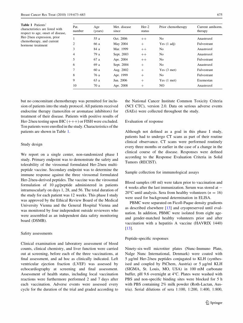
Table 1 Patients’
characteristics are listed with
respect to age, onset of disease,
Her-2/neu expression, prior
chemotherapy, and current
hormone treatment
Pat.
number
Age
(years)
Met. disease
since
Her-2
status
Prior chemotherapy Current antihorm.
therapy
1 55 a Oct. 2006 ?? No Anastrozol
2 66 a May 2004 ? Yes (1 adj) Fulvestrant
3 84 a Mar. 1999 ?? No Anastrozol
4 79 a Sept. 2003 ?? No Anastrozol
5 67 a Apr. 2004 ?? No Fulvestrant
6 69 a Sept. 2004 ? No Anastrozol
7 60 a Aug. 2002 ? Yes (3 met) Fulvestrant
8 76 a Apr. 1999 ? No Fulvestrant
9 63 a Jun. 2006 ? Yes (1 met) Exemestan
10 70 a Apr. 2008 ? NO Anastrozol
Breast Cancer Res Treat (2010) 119:673–683 675
123

1:1,600, and 1:3,200 were prepared in PBS-tw (0.05% v/v
Tween 20) containing 0.5% milk powder and incubated
overnight at 4�C. After washing with PBS-tw, HRP-labeled
anti-human IgG (SIGMA, St. Louis, MO, USA) diluted
1:60,000 in PBS-tw containing 0.5% milk powder were
added to the plates and incubated for 2 h at room tem-
perature. Color development was performed with tetram-
ethylbenzidin (TMB) substrate (Chemicon, Temecula, CA,
USA) and was stopped with H2SO4 after 10 min. Absor-
bance was measured with TECAN Genios reader. The
resulting OD of each serum dilution was calculated by
subtracting the OD obtained on KLH-coated wells from the
OD values of the peptide-KLH coated wells. Assay control
was performed with defined patient’s serum and defined
negative serum pool.
Titer was defined as serum dilution to obtain an
OD = 1.0 over the background derived from healthy
donors. Pre- and post-vaccination titers were calculated.
Her-2/neu-specific responses
Recombinant human ErbB2/Fc Chimera (R&D Systems,
MN, USA) consisting of the extracellular domain, six-
amino acid linkage, and the human IgG1 Fc fragment was
used as coating antigen. Plates were coated with 1 lg/ml
ErbB2/Fc Chimera in carbonate buffer overnight at 4�C.
Control plates were coated with the analogous dilution of
the reconstitution buffer for ErbB-2-Fc Chimera containing
0.1% BSA. Plates were blocked with 5% milk powder in
PBS for 5 h before serial dilutions of sera as listed above
were added and incubated overnight at 4�C. Bound Igj and
Igk antibodies were detected with HRP-labeled anti-human
j and anti k chain IgG (SIGMA, St. Louis, MO, USA),
respectively, diluted 1:10,000 in PBS-tw/0.5% milk. Her-
ceptin� (Roche, Switzerland) titration curve 0.05–
0.003125 lg/ml was used as control in assays with Igjdetection and serum obtained from patient with Her-2/
neu??? positive tumor for the Igk detection. ELISA was
developed with TMB substrate and stopped after 20 min.
Titer was calculated as described above. The ratio post-
vaccine titer/pre-vaccine titer represents titer increase.
Ratio greater than 1 was regarded as Her-2/neu-specific
titer increase.
Western blot
SK-BR-3 cells were lysed as described elsewhere [8]. The
protein content was quantified with BCA Protein assay kit
(Thermo scientific Rockford, IL USA). Her-2/neu was
immunoprecipitated from a total of 16 mg protein with
200 lg Herceptin� (Roche, Basel, Switzerland) and 150 ll
protein A ? G agarose (Merck-Calbiochem, Darmstadt,
Germany) in 0.1 M TBS over night at 4�C. Pellet was
washed twice in 0.1 M TBS pH 7.4 and twice in 0.1 M
TBS with 0.5% Nonidet P40. Precipitate was separated on
8% SDS PAGE and blotted on nitrocellulose membranes
(Schleicher & Schuell, Dassel, Germany). Strips were
blocked with 2% milk in 0.1 M TBS buffer and then
incubated over night with sera diluted 1:25 in 0.1 M TBS-
0.05% v/v Tween-20 containing 0.5% milk. Rabbit anti-
Her-2/neu antibody (Zymed, San Francisco, CA, USA) was
used as positive control and serum obtained from healthy
volunteers were as negative controls. After three washing
steps, bound human IgG was detected by AP-labeled anti-
human IgG (SIGMA, Saint Louis, Missouri, USA) and
subsequently stained with NBT/BCIP substrate mixture.
Rabbit IgG was detected by AP-swine anti-rabbit antibody
(DAKO A7S Denmark).
Hemagglutination-inhibition assay
The analysis of the serum samples for neutralizing anti-
body levels against influenza were performed according to
standard methodology [14]. Two serial twofold dilutions of
the sera were prepared. The diluted sera were incubated
with either the hemagglutinin antigen of the Influenza
strain A/Sing/86 or with the solvent. Thereafter a suspen-
sion of chicken erythrocytes was added. Hemagglutination
occurred with virus antigen in the absence of sufficient
inhibiting antibodies. The hemagglutination inhibition titer
is the reciprocal value of the highest dilution which inhibits
hemagglutination. The titer assigned to each sample is the
geometric mean of two independent determinations
(EMEA criteria in Appendix 13.3). A fourfold increase
(C4) in the titers after vaccination was taken as positive
vaccine response.
Cellular responses specific to virosomal antigens
Peripheral blood T cell responses were assessed by cyto-
kine production in vitro upon stimulation with virosomes.
PBMC were cultured at 1 9 106/well in medium (RPMI
with 10% heat inactivated human AB serum) containing
virosomes with total hemagglutinin content of 8 lg/ml or
1 lg/ml streptococcal enterotoxin B (SIGMA, Saint Louis,
Missouri, USA) as positive control antigen and in the
absence of any antigens (at least three wells per antigen).
The virosome formulation was kindly provided by Pevion
Biotech Ltd (Bern, Switzerland). The final culture volume
was 200 ll/well, culture conditions were 37�C and 5%
CO2. Supernatants were harvested after 48 h and stored
frozen until measurement of IFN-c, TNF-a, and IL-2 by
ELISA.
Commercially available ELISA kits were used for
measurement of TNF-a (R&D, MN, USA) and IL-2
676 Breast Cancer Res Treat (2010) 119:673–683
123

(ENDOGENE/Thermo Scientific, IL, USA). Assays were
carried out according to manufacturer’s instructions.
For IFN-c levels determination plates were coated with
0.5 lg/ml mouse anti-human IFN-c (Calbiochem, NJ,
USA) in carbonate buffer over night at 4�C. After blocking
with 4% BSA (bovine serum albumin) in PBS, 50 ll of
undiluted samples were added at room temperature for 1 h.
Recombinant human IFN-c (ENDOGENE/Thermo Scien-
tific, IL, USA) in concentration ranging from 8 to 500 pg/
ml was used to generate a calibration curve. Plates were
subsequently washed with PBS-tw. Biotin-labeled mouse
anti-human IFN-c (Thermo Scientific, IL, USA) at a con-
centration 0.9 lg/ml was applied, followed by peroxidase-
conjugated streptavidin (Thermo Scientific) 1:15,000 in
PBS-tw/4% BSA for 30 min. The assay was developed
with TMB substrate. Cytokine levels represent results after
subtraction of unstimulated medium values.
Flow cytometry
Immunofluorescent staining of cell surface markers on
PBMC was performed on patients and eight healthy age-
and gender-matched volunteers. Cryopreserved cells were
washed with PBS/0.5% BSA/0.5% sodium azide, diluted to
5 9 105 cells per micronic tube, blocked with 20% human
AB serum for 20 min and subsequently stained with
directly conjugated mAbs at predetermined optimal con-
centrations for 30 min at 4 �C in the dark. Antibodies used:
aCD3-PE-Cy5, aCD19-FITC (IgG1 k), aCD27-PE, aCD4-
FITC (IgG2 k), aCD4-PE-Cy5 (IgG1 k), aCD28-PE-Cy5
(IgG1 k), aCD8-APC (IgG1 k), aCD45R0-PE (IgG2a k),
aCD62L-FITC (IgG1 k), and aCD25-PE (IgG1 k). All
antibodies were purchased from BD Pharmingen, CA,
USA. Antibody binding to PBMC was analyzed on a FACS
CALIBUR flow cytometer (BD Biosciences) by gating on
cells with forward and side light scatter properties of
lymphocytes, using BD CellQuest software. Isotype mat-
ched negative control reagents were used to verify the
staining specificity of the experimental antibodies.
CD4?/CD25?/Foxp3? regulatory T cells were ana-
lyzed using Foxp3 staining kit (eBioscience, San Diego,
CA, USA) according to manufacturer’s instructions.
1 9 106 PBMC were incubated with anti-CD4/CD25
cocktail for 30 min. Cells were then washed with cold
staining buffer, resuspended in 1 ml fixation/permeabili-
zation buffer and incubated for 30 min. After another
washing step and blocking with 2% normal rat serum,
intracellular staining with anti human Foxp3-APC antibody
(PCH101) and isotype control Ab (rat IgG2a) was per-
formed for 30 min. Finally cells were washed with per-
meabilization buffer, resuspended in staining buffer, and
measured as described above. Results are depicted as
percent of CD25?Foxp3? of CD4? T cells.
Statistical analysis
As detailed above, anti-peptide- and anti-Her-2/neu anti-
bodies were measured by ELISA using six serum dilutions
in twofold steps. In simultaneous assays the pre-immuni-
zation serum and serum obtained 4 weeks after the third
vaccination were measured. Because there is no standard
antibody sample against which the obtained OD values can
be expressed, a parallel line procedure was chosen and the
post-vaccination antibody concentration was expressed as
fold-increase above the pre-vaccination serum. The pro-
cedure was as follows: in a simultaneous logistic regression
analysis all OD values from the pre- and post-vaccination
sera were related to the log dilution and a parameter for the
time of blood withdrawal (pre- or post-vaccination). The
anti-log of the shift from pre- to post-vaccination regres-
sion lines is the fold increase of antibody concentrations.
Confidence intervals were computed based on Fieller’s
theorem. The fit of the model was excellent with pseudo-R2
values above 0.98 in all cases.
Statistical comparison of Her2/neu peptide vaccinees
with the control group of age and sex matched anti-HAV
vaccinees was done by matched sample ANOVA with
repeated measurements. Differences between groups with
respect to the trend from pre- to post-immunization sera
were tested based on the interaction source of variance.
Fractions of lymphocytes from FACS analyses were arc-
sine transformed prior to analysis to obtain normality of
residuals that was assessed by Lilliefors’ corrected Kol-
mogorov–Smirnov tests. Differences between cytokine
concentrations in pre- and post-immunization sera were
tested by Wilcoxon’s matched-pairs tests. For all analyses a
P-value below 0.05 was considered significant. No cor-
rection for multiple endpoints was applied.
Results
Recruitment
Ten patients have been recruited. All participants have
successfully finished the schedule of three vaccinations.
Evaluation of safety
No local or systemic grade 3 or 4 side effects were
observed. Four patients showed a grade 1 local vaccination
reaction (Table 2). In particular serological evaluation
revealed no signs of hepatic or nephrologic toxicity. With
special regard to a possible cardiotoxicity comparable to
that seen in the treatment of breast cancer patients with
trastuzumab all patients had to undergo measurement of
LVEF at screening and at the end of study. No decrease in
Breast Cancer Res Treat (2010) 119:673–683 677
123

LVEF higher than 10% was documented in study patients
during the study duration.
Course of disease/clinical outcome
One patient died one month after the last vaccination due to
disease progression. An extensive evaluation of the course
of disease including the last episode of this patient by the
investigator as well as by an independent data safety
monitoring board (DSMB) did not suggest any vaccine-
related event, but disease progression to be responsible for
the patient’s death. Another patient died 6 months after the
last vaccination with progressive disease while receiving
concurrent chemotherapy. At time of study closure
(December 2008), the remaining 8 patients were alive, 5
were stable for a period of up to 12 months, 1 showed a
partial remission according to RECIST, and 2 had disease
progression after 5 and 7 months, respectively.
Immunogenicity of the virosomal-formulated
peptide-based vaccine
The vast majority of study participants (8 of 10) produced
peptide-specific antibodies to all peptides after the 3rd
vaccination, as shown in Fig. 1a. Two patients (patient 06
and 10) did not respond to any of the peptides. A moderate
titer increase was measured in one patient with existing
pre-immunization titer. Baseline levels obtained with
serum samples from healthy donors were significantly
below the post-immunization titers in the patients (data not
shown).
Immunization with Her-2/neu peptides generated
Her-2/neu-specific antibodies
Figure 1b demonstrates antibody titers specific to the
extracellular domain of the Her-2/neu protein determined
by ELISA. Her-2/neu-specific IgG, j and/or k-chain were
detected in 7 of 10 patients after immunization with the
multi-peptide vaccine. The ELISA results were verified by
Western blot analysis confirming the presence of Her-2/
neu-specific IgG in 5 of 10 patients. High pre-existing
antibody levels against the extracellular domain of Her-2/
Table 2 Safety evaluation
Pat.
number
Local vacc.
reaction grade
Systemic grade
3/4 toxicity
1 1 No
2 0 No
3 0 No
4 1 No
5 1 No
6 0 No
7 0 No
8 0 No
9 1 No
10 0 No
Occurrence of local reactions and systemic side reactions (grades 1–
4) were evaluated
A B
kappa IgG lambda IgG0
1
2
3
Her
-2/n
eu a
b t
iter
in
crea
se
C
185 kDa
1 2 3 4
pre- post- pre- post- pre- post-0
10
20
30
40
50
60
70
80
P4 P6 P7
pep
tid
e ab
tit
er
Fig. 1 Humoral responses after
immunizations with
virosomally formulated Her-2/
neu peptides. ELISA analysis
was performed with serum
samples taken at baseline and
4 weeks after the last
immunization. a IgG-specific
titers to peptides P4, P6, and P7.
b Her-2/neu-specific j- and k-
light chain IgG. Scatter graphs
represents the ratio post-/pre-
vaccination titer. c Western blot
analysis demonstrating increase
of Her-2/neu-specific IgG post-
vaccination in patient 04.
Serum prior (lane 3) and after
completed vaccinations (lane 4)
was analyzed. Polyclonal rabbit
anti Her-2/neu antibody was
used as positive control (lane 1),
serum of a healthy volunteer as
negative control (lane 2)
678 Breast Cancer Res Treat (2010) 119:673–683
123

neu was found in one patient; in this case, no increase of
Her-2/neu antibody level was achieved during vaccination
when measured by ELISA. Qualitative analysis of pre- and
post-vaccination serum by Western blot, as shown in
Fig. 1c, demonstrates that the Her-2/neu-specific responses
to linear epitopes significantly increased after immuniza-
tions with peptides.
In general, the j-light chain-IgG responses were found
in the majority of patients and the magnitude of this isotype
was higher than of the k-chain-isotype. Interestingly, only
k-chain light-IgG response was induced in one of the
patients (05).
Overall, in 8 of 10 patients induction/increase of Her-2/
neu-specific antibody levels was observed after the three-
dose vaccination schedule.
Influenza-specific responses/neutralizing antibodies
Hemagglutination-inhibition test (HI) was performed to
measure antibody levels against virosome-associated
hemagglutinin. Patients’ sera were analyzed at baseline and
4 weeks after the three-dose vaccination schedule. Neu-
tralizing antibodies against A Sing 86 specific-hemagglu-
tinin were detectable in 7 of 10 patients already prior to
vaccination. Vaccination with PEV6 elicited a significant
HI titer increase (C4-fold) in 6 of 10 patients. In four
patients the HI titer was not significantly influenced by the
multi-peptide vaccination (Table 3).
Cytokine release after in vitro stimulation with
virosomes
Cellular immune responses against the virosomal antigen
hemagglutinin were analyzed in PBMC obtained prior and
after completion of the vaccination schedule. In 7 of 10
patients an increase of cytokine production was observed
after completion of the vaccination schedule. IL-2 pro-
duction increased significantly (P = 0.023), also TNF-aproduction was markedly higher (P = 0.055) as well as
IFN-c production (n.s.). In one patient, high cytokine levels
already existed in cultures prior to vaccination and
remained at similar level after the third vaccination. In cell
cultures of another patient, cytokine concentrations before
and after vaccination were below detection limit. A
decrease of pre-vaccination cytokine secretion was
observed in PBMC of one patient. Of note, this patient
displayed also insufficient humoral responses to peptides
and influenza antigens (Fig. 2).
Analysis of surface marker expression on lymphocytes
We assessed various subpopulations of PBMC in all study
patients prior and after completion of vaccinations as well
as in eight age- and gender-matched healthy volunteers.
Cell surface marker expression on B cells (CD19), memory
B cells (CD19/CD27), T cells (CD4, CD8), memory sub-
populations (CD45R0), as well as CD25 (IL-2 receptor achain), and L-selectin (CD62L) expression on CD4 lym-
phocytes were evaluated (Table 4). A significant decrease
of CD3 T lymphocytes (P = 0.017) compared to healthy
controls was noticed after completion of the vaccination
series. The B cell lymphocyte population tended to
increase in the study group post-vaccination, similarly as in
the healthy donor group. CD4? and CD8? T cells did,
however, not significantly differ before and after vaccina-
tion between the study patients and the healthy donor
group. The L-selectin (CD62L) expressing subpopulation
declined in 6 of 10 study patients after vaccination
(Table 4).
The median value of CD4?/CD25?Foxp3? regulatory
T cells in circulating PBMC of the breast cancer patients
prior to vaccination was 4.86 [3.18–6.54] and significantly
declined after vaccination to 4.02 [2.74–5.95], P = 0.007
(Fig. 3). In the healthy control group the median values of
CD4?CD25?Foxp3? T reg cells prior (3.98 [2.90–3.66])
and after vaccination (2.69 [2.63–3.10]) were significantly
(P \ 0.033) lower than in the tumor patients (Fig. 3).
Discussion
Several studies in experimental models including our own
preclinical data [8, 9] have demonstrated that successful
reduction of Her-2/neu overexpressing tumors after vacci-
nation requires both humoral and cellular immune
responses. Here, we demonstrate the results of a single
center phase I clinical trial showing that a Her-2/neu multi-
peptide vaccine is safe and immunogenic concerning B-
Table 3 Neutralizing antibody titers against influenza associated
antigens
Patient Pre-immune Post-immune
01 80 160
02 40 640
03 640 320
04 20 160
05 20 160
06 60 60
07 \10 1280
08 \10 160
09 \10 160
10 80 80
HI titers prior and after the three dose vaccination schedule with
PEV6
Breast Cancer Res Treat (2010) 119:673–683 679
123

CBA
pre- post-0
100
200
300
400
500
600
700
800
IFN
-γ p
g/m
l
pre- post-0
100
200
300
400
500
600
700
800
TN
F-α
pg
/ml
pre- post-0
20
40
60
80
100
120
140
IL-2
pg
/ml
P<0.05
Fig. 2 Cellular responses to influenza antigens in vitro. PBMC
isolated at baseline and 4 weeks after the last vaccination were
stimulated with virosomes containing hemagglutinin. Cytokine
production in supernatants was measured by ELISA. Data represent
values after subtraction of unstimulated background
Table 4 Characterization of PBMC by surface marker expression analyzed by flow cytometry
Cell type CD
marker
Study patients n = 10 Normal donors n = 8 Pre-values* Trend�
Pre- Post- Pre- Post- P P
T lymphocytes CD3 66.5 [60.2–73.3] 62.1 [54.9–64.4] 73.6 [63.4–75.9] 73.6 [66.5–78.4] 0.332 0.017
B lymphocytes CD19 7.9 [5.9–12.5] 12.4 [10–14.5] 8.9 [6.6–14.8] 9.3 [8.2–13.1] 0.643 0.100
T cells/memory CD4 44.2 [38.7–52.3] 43.0 [37.0–46.0] 53.8 [38.5–57.0] 49.1 [37.0–55.3] 0.678 0.667
CD4/CD45R0 36.6 [30.6–41.6] 32.9 [27.4–37.6] 31.1 [29.3–32.9] 31.0 [28.0–32.0] 0.102 0.367
CD8 20.5 [18.6–26.0] 21.3 [18.2–24.5] 21.8 [17.8–31.3] 25.3 [17.8–28.2] 0.822 0.360
CD8/CD45R0 11.5 [10.2–13.0] 10.3 [8.1–10.7] 9.2 [6.8–15.7] 12.6 [6.8–16.0] 0.980 0.075
Peripheral lymph
node homing
receptor
CD4/CD62L 16.7 [6.4–22.9] 8.9 [5.0–14.6] 13.9 [11.5–27.6] 16.5 [12.8–22.3] 0.757 0.265
High affinity
IL-2 receptor
CD4/CD25 17.3 [11.3–21.4] 15.6 [7.7–19.3] 16.5 [10.7–21.5] 13.7 [10.2–23.3] 0.897 0.198
* P-values for comparison of preimmune cell populations in patients and normal donors� P-values for comparison of trend (pre-immune–post-immune cell populations) between patients and normal donors
Fig. 3 a Frequency of
regulatory T cells assessed by
flow cytometry in tumor patients
and healthy controls. Data
depict percentage of CD4? T
cells expressing CD25 and
Foxp3 before and after
vaccinations with the Her-2/neu
peptide vaccine in tumor
patients as well as in healthy
age- and gender-matched donors
prior and after vaccination with
a hepatitis A vaccine. b Scatter
plots of two representative
tumor patients and one healthy
control. Cells were gated on the
CD4? population of
lymphocytes, the numbers
represent percent of each
quadrant
680 Breast Cancer Res Treat (2010) 119:673–683
123

and T cell responses in patients with advanced breast
cancer. The current protocol foresaw vaccination in
patients with metastatic breast cancer without overexpres-
sion of the Her-2/neu protein (? and ?? upon immuno-
histochemistry, FISH negative), which was due to ethical
reasons in order not to withhold standard anti-Her-2/neu
immunotherapy with trastuzumab from patients with Her-
2/neu-driven disease.
According to the current trial conduct, this phase I study
had safety as primary endpoint. Additionally, immunoge-
nicity of the vaccine was chosen as secondary endpoint,
since the specificity of the evoked immune responses
toward Her-2/neu is of utmost importance as previously
demonstrated in our preclinical studies. Characterization of
the B- and T cell compartment was particularly interesting,
as immune responsiveness has been repeatedly shown to be
perturbed in cancer patients [15–17].
Unlike many peptide vaccines against cancers which
have been designed to induce cytotoxic T cell responses
using class I restricted peptides [18–23], we used putative
B cell epitopes from the extracellular domain of the Her-2/
neu protein being HLA type independent. In order to
induce CD4? T helper cell responses, important for the
induction of antibody isotype switching and memory
responses [24], these peptides were coupled to immuno-
potentiating reconstituted influenza virosomes (IRIV), an
adjuvant system currently used in two licensed vaccines, a
hepatitis A vaccine and an influenza vaccine, which proved
to cause minimal side effects while being very immuno-
genic [11, 25].
Indeed, the results from the primary study endpoint on
vaccine safety showed that only 4 of 10 vaccinees dis-
played minimal local reactions of grade 1, consisting of
mild redness and swelling or pain at the injection site,
while no fever or other systemic side effects (grade 4/5)
were reported. The deaths of two study participants, 1 and
6 months after vaccination, respectively, were not vaccine-
related but due to disease progression. On these occasions
an independent DSMB arrived at the same conclusion as
the investigators. Half of the patient collective (50%)
presented with stable disease up to 12 months after vac-
cination. Six months after initiation of vaccination one
patient showed a partial remission according to RECIST
and three developed tumor progression (Table 2). As all
patients received hormone treatment and did not show Her-
2/neu overexpression it is more probable to attribute any
clinical effects to the applied endocrine medication rather
than to the Her-2/neu vaccine.
The second endpoint of the study concentrated on the
immunogenicity of the vaccine. The majority of patients, 8
of 10, developed a significant increase in anti-peptide titers
to all three peptides (Fig. 1a), while only two participants
did not respond with peptide-specific titers. As all patients
received endocrine therapy by aromatase inhibitors during
this vaccination trial for ethical reasons, the question
whether aromatase inhibition might have influenced the
magnitude of the antibody responses remains unanswered
to date. This appears to be unlikely though, because recent
studies with other adjuvant therapies have shown not to
impact on immune response levels [26].
Importantly, in all but one of the peptide-responsive
vaccinees, the antibody levels were also directed against
the native Her-2/neu antigen. The Her-2/neu antibody
responses were measured by quantitative ELISA and ver-
ified by Western blot analysis. The discrepancy in the
detected number of Her-2/neu-specific IgG between the
two methods might be explained by the fact that only linear
epitopes can be recognized by Western blot analysis, while
also conformational epitopes can be measured by ELISA.
In contrast to other studies [27], the results from our trial
indicate that the B cell-derived peptide vaccine was potent
enough to overcome tolerance to the self-antigen Her-2/neu
(Fig. 1b, c). Whether the quality of the induced antibodies
is related to the kappa or lambda light chain IgG is
impossible to estimate at this stage. Studies on the antibody
avidity, as described by Goldblatt et al. [28], could give
further indications on the biological activity of the induced
antibodies. It must be stressed, however, that this phase I
study does not allow any evaluation of antibody/vaccine
efficacy, since the selection of patients with only weak
Her-2/neu expression, due to ethical reasons in order not to
withhold trastuzumab treatment when indicated, does not
represent the actual target population in which the anti-
tumor effects of the induced antibodies can be properly
evaluated. Along with further assessment of safety, eval-
uation of efficacy of the present vaccine would be the focus
of a subsequent phase II trial.
It has been proposed that successful vaccination to
generate Her-2/neu immunity is not only dependent on the
appropriate selection of epitopes but also on the use of a
potent adjuvant system. In this respect, we chose IRIV as
conjugation partner for two reasons: firstly, the presence of
the influenza hemagglutinin antigen from the influenza A
Singapore strain on the virosome surface to which the
majority of the population has previously been exposed
allows the induction of booster responses to this antigen.
Accordingly, the majority of the vaccinees displayed a
significant increase in the influenza neutralizing antibody
titers (Table 3). The two mentioned Her-2/neu non-
responder vaccinees also failed to develop influenza-spe-
cific antibody levels indicative for immunological incom-
petence. Secondly, the virosome system was used to
generate bystander CD4? T cell responses [24] necessary
to induce antibody isotype switching to IgG as well as
memory responses to the B cell peptides. The production of
IL-2 in particular, but also of TNF-a as well as IFN-c
Breast Cancer Res Treat (2010) 119:673–683 681
123

markedly increased in cultures of virosome-stimulated
PBMCs after completion of the vaccination series. These
results are in line with our preclinical data showing that the
selected B cell peptides from the ECD in conjunction with
a strong adjuvant system were able to induce both, humoral
and cellular responses. Noteworthy, the vaccine was
immunogenic (except for two) in patients across all HLA
types in stage IV disease. Thus, our study indicates that
patients with advanced metastatic breast cancer are
immunocompetent and fully susceptible to vaccination.
This is further supported by characterization of the lym-
phocyte population based on CD surface marker expression
in comparison to age- and gender-matched healthy controls
vaccinated with a hepatitis A vaccine [13] (Table 4).
Except for the number of CD3? T cells, no significant
differences in the distribution of lymphocyte subpopula-
tions (CD4, CD45RO, CD8, CD19) were detected between
the tumor patients and the healthy controls before and after
vaccination. Also the percentage of CD4 T cells expressing
CD62L, a homing receptor on naı̈ve T cells, did not sig-
nificantly differ between the two groups and declined after
vaccination, most probably as a result of immune respon-
siveness to the vaccine antigens, as also described for other
vaccines in healthy adults [13, 29].
Several studies have shown that T regulatory cells
(Tregs) are increased in patients with a variety of malig-
nancies and that higher numbers of Tregs are associated
with tumor progression [30–34]. Recently, it was shown
that tumor infiltration by Tregs is associated with a reduced
survival of breast cancer patients [35]. Treg cells may also
be responsible for the failure of an anticancer immune
response by hindering the generation and activity of anti-
tumor reactive T cells thereby maintaining self-tolerance to
the tumor antigens. Along these lines, also our data showed
that the patients had a significantly higher number of cir-
culating Treg cells, defined as CD4?CD25?Foxp3? T
cells, than the healthy matched control group prior to
vaccination. Of notice was the fact that the number of
Tregs significantly decreased after vaccination with the
multi-peptide vaccine (Fig. 3). The fact that a decline in
Treg cells was also observed in healthy donors after vac-
cination with a hepatitis A vaccine (Fig. 3) raises the
question whether each vaccination has the potential to
interfere with this cell population in order to guarantee
optimal immune responses. In tumor patients this sup-
pressive effect on Tregs might be particularly crucial for
the efficacy of a vaccine. Similar results have very recently
been described in breast cancer patients treated with
trastuzumab [30] as well as with an aromatase inhibitor
[35], thus suggesting that overcoming Treg cell reconsti-
tution might not only be seen in the present context, but
perhaps also as an important contributor to success in a
wider range of anti-tumor treatment strategies.
Taken together, the results from our phase I trial show
that the described Her-2/neu vaccine was safe and immu-
nogenic in 8 of 10 patients. As a next step it is of interest
whether an increased vaccine dose might lead to an ame-
liorated seroconversion rate. Current studies on the impact
of higher peptide vaccine doses on anti-tumor immunity
show controversial results [36, 37]. Thus, a second part of
this phase I study is on the way to be able to answer this
question. In future settings, a series of different options for
further phase II studies exist, such as the combination of
the current vaccine with passive antibody administration or
with other treatment modalities including Treg cell
inhibitors.
Acknowledgments The study was supported by a grant from Bio-
Life Science and by the Medical University of Vienna. The assistance
of Mrs Marika Rosner as study nurse during the phase I clinical trial is
very much appreciated.
References
1. Slamon DJ, Clark GM, Wong SG, Levin WJ, Ullrich A, McGuire
WL (1987) Human breast cancer: correlation of relapse and
survival with amplification of the HER-2/neu oncogene. Science
235(4785):177–182
2. Andrulis IL, Bull SB, Blackstein ME et al (1998) neu/erbB-2
amplification identifies a poor-prognosis group of women with
node-negative breast cancer. Toronto Breast Cancer Study Group.
J Clin Oncol 16(4):1340–1349
3. Suter TM, Procter M, van Veldhuisen DJ et al (2007) Trast-
uzumab-associated cardiac adverse effects in the herceptin
adjuvant trial. J Clin Oncol 25(25):3859–3865
4. Maloney DG, Grillo-Lopez AJ, Bodkin DJ et al (1997) IDEC-
C2B8: results of a phase I multiple-dose trial in patients with
relapsed non-Hodgkin’s lymphoma. J Clin Oncol 15(10):3266–
3274
5. Vogel CL, Cobleigh MA, Tripathy D et al (2002) Efficacy and
safety of trastuzumab as a single agent in first-line treatment of
HER2-overexpressing metastatic breast cancer. J Clin Oncol
20(3):719–726
6. Rosenberg SA, Yang JC, Restifo NP (2004) Cancer immuno-
therapy: moving beyond current vaccines. Nat Med 10(9):909–
915
7. Lollini PL, Cavallo F, Nanni P, Forni G (2006) Vaccines for
tumour prevention. Nat Rev Cancer 6(3):204–216
8. Jasinska J, Wagner S, Radauer C et al (2003) Inhibition of tumor
cell growth by antibodies induced after vaccination with peptides
derived from the extracellular domain of Her-2/neu. Int J Cancer
107(6):976–983
9. Wagner S, Jasinska J, Breiteneder H, Kundi M, Pehamberger H,
Scheiner O, Zielinski CC, Wiedermann U (2007) Delayed tumor
onset and reduced tumor growth progression after immunization
with a Her-2/neu multi-peptide vaccine and IL-12 in c-neu
transgenic mice. Breast Cancer Res Treat 106(1):29–38
10. Zurbriggen R (2003) Immunostimulating reconstituted influenza
virosomes. Vaccine 21(9–10):921–924
11. Bovier PA (2008) Epaxal: a virosomal vaccine to prevent hepa-
titis A infection. Expert Rev Vaccines 7(8):1141–1150
12. Thompson FM, Porter DW, Okitsu SL et al (2008) Evidence of
blood stage efficacy with a virosomal malaria vaccine in a phase
IIa clinical trial. PLoS ONE 3(1):e1493
682 Breast Cancer Res Treat (2010) 119:673–683
123
13. Garner-Spitzer E, Kundi M, Rendi-Wagner P et al (2009) Cor-
relation between humoral and cellular immune responses and the
expression of the hepatitis A receptor HAVcr-1 on T cells after
hepatitis A re-vaccination in high and low-responder vaccinees.
Vaccine 27(2):197–204
14. Phillips CA, Forsyth BR, Christmas WA, Gump DW, Whorton
EB, Rogers I, Rudin A (1970) Purified influenza vaccine: clinical
and serologic responses to varying doses and different routes of
immunization. J Infect Dis 122(1):26–32
15. Zielinski CC, Stuller I, Dorner F, Potzi P, Muller C, Eibl MM
(1986) Impaired primary, but not secondary, immune response in
breast cancer patients under adjuvant chemotherapy. Cancer
58(8):1648–1652
16. Molling JW, Kolgen W, van der Vliet HJ et al (2005) Peripheral
blood IFN-gamma-secreting Valpha24?Vbeta11?NKT cell
numbers are decreased in cancer patients independent of tumor
type or tumor load. Int J Cancer 116(1):87–93
17. Liyanage UK, Moore TT, Joo HG et al (2002) Prevalence of
regulatory T cells is increased in peripheral blood and tumor
microenvironment of patients with pancreas or breast adenocar-
cinoma. J Immunol 169(5):2756–2761
18. Fisk B, Blevins TL, Wharton JT, Ioannides CG (1995) Identifi-
cation of an immunodominant peptide of HER-2/neu protoon-
cogene recognized by ovarian tumor-specific cytotoxic T
lymphocyte lines. J Exp Med 181(6):2109–2117
19. Disis ML, Grabstein KH, Sleath PR, Cheever MA (1999) Gen-
eration of immunity to the HER-2/neu oncogenic protein in
patients with breast and ovarian cancer using a peptide-based
vaccine. Clin Cancer Res 5(6):1289–1297
20. Disis ML, Gralow JR, Bernhard H, Hand SL, Rubin WD, Che-
ever MA (1996) Peptide-based, but not whole protein, vaccines
elicit immunity to HER-2/neu, oncogenic self-protein. J Immunol
156(9):3151–3158
21. Matsui S, Ahlers JD, Vortmeyer AO, Terabe M, Tsukui T, Car-
bone DP, Liotta LA, Berzofsky JA (1999) A model for CD8?
CTL tumor immunosurveillance and regulation of tumor escape
by CD4 T cells through an effect on quality of CTL. J Immunol
163(1):184–193
22. Welters MJ, Kenter GG, Piersma SJ et al (2008) Induction of
tumor-specific CD4? and CD8? T-cell immunity in cervical
cancer patients by a human papillomavirus type 16 E6 and E7
long peptides vaccine. Clin Cancer Res 14(1):178–187
23. Peoples GE, Holmes JP, Hueman MT et al (2008) Combined
clinical trial results of a HER2/neu (E75) vaccine for the pre-
vention of recurrence in high-risk breast cancer patients: U.S.
Military Cancer Institute Clinical Trials Group Study I-01 and I-
02. Clin Cancer Res 14(3):797–803
24. Hung K, Hayashi R, Lafond-Walker A, Lowenstein C, Pardoll D,
Levitsky H (1998) The central role of CD4(?) T cells in the
antitumor immune response. J Exp Med 188(12):2357–2368
25. Kunzi V, Dornseiff M, Horwath J, Hartmann K (2009) Safe
vaccination of children with a virosomal adjuvanted influenza
vaccine. Vaccine 27(8):1261–1265
26. Coveler AL, Goodell V, Webster DJ, Salazar LG, Fintak PA,
Childs JS, Higgins DM, Disis ML (2009) Common adjuvant
breast cancer therapies do not inhibit cancer vaccine induced T
cell immunity. Breast Cancer Res Treat 113(1):95–100
27. Disis ML, Goodell V, Schiffman K, Knutson KL (2004) Humoral
epitope-spreading following immunization with a HER-2/neu
peptide based vaccine in cancer patients. J Clin Immunol
24(5):571–578
28. Goldblatt D, Vaz AR, Miller E (1998) Antibody avidity as a
surrogate marker of successful priming by Haemophilus influ-enzae type b conjugate vaccines following infant immunization. J
Infect Dis 177(4):1112–1115
29. Pfister G, Weiskopf D, Lazuardi L et al (2006) Naive T cells in
the elderly: are they still there? Ann N Y Acad Sci 1067:152–157
30. Horlock C, Stott B, Dyson PJ, Morishita M, Coombes RC,
Savage P, Stebbing J (2009) The effects of trastuzumab on the
CD4?CD25?FoxP3? and CD4?IL17A? T-cell axis in patients
with breast cancer. Br J Cancer 100(7):1061–1067
31. Mizukami Y, Kono K, Kawaguchi Y, Akaike H, Kamimura K,
Sugai H, Fujii H (2008) Localisation pattern of Foxp3? regula-
tory T cells is associated with clinical behaviour in gastric cancer.
Br J Cancer 98(1):148–153
32. Kono K, Kawaida H, Takahashi A, Sugai H, Mimura K, Miyagawa
N, Omata H, Fujii H (2006) CD4(?)CD25 high regulatory T cells
increase with tumor stage in patients with gastric and esophageal
cancers. Cancer Immunol Immunother 55(9):1064–1071
33. Vence L, Palucka AK, Fay JW, Ito T, Liu YJ, Banchereau J,
Ueno H (2007) Circulating tumor antigen-specific regulatory T
cells in patients with metastatic melanoma. Proc Natl Acad Sci
USA 104(52):20884–20889
34. Enarsson K, Lundgren A, Kindlund B, Hermansson M, Roncador
G, Banham AH, Lundin BS, Quiding-Jarbrink M (2006) Function
and recruitment of mucosal regulatory T cells in human chronic
Helicobacter pylori infection and gastric adenocarcinoma. Clin
Immunol 121(3):358–368
35. Generali D, Bates G, Berruti A et al (2009) Immunomodulation of
FOXP3? regulatory T cells by the aromatase inhibitor letrozole in
breast cancer patients. Clin Cancer Res 15(3):1046–1051
36. Disis ML, Schiffman K, Guthrie K, Salazar LG, Knutson KL,
Goodell V, dela Rosa C, Cheever MA (2004) Effect of dose on
immune response in patients vaccinated with an her-2/neu
intracellular domain protein-based vaccine. J Clin Oncol
22(10):1916–1925
37. Holmes JP, Gates JD, Benavides LC et al (2008) Optimal dose and
schedule of an HER-2/neu (E75) peptide vaccine to prevent breast
cancer recurrence: from US Military Cancer Institute Clinical
Trials Group Study I-01 and I-02. Cancer 113(7):1666–1675
Breast Cancer Res Treat (2010) 119:673–683 683
123