a tropical phytoplankton community development in ... · härnström et al.: tropical plankton...
TRANSCRIPT
MARINE ECOLOGY PROGRESS SERIESMar Ecol Prog Ser
Vol. 346: 75–88, 2007doi: 10.3354/meps07026
Published September 27
INTRODUCTION
Many phytoplankton groups have benthic restingstages, formed asexually or sexually. These stages arecommonly known to provide short- or long-term sur-vival, and may also contribute to natural dispersal bycurrents or when transported in ballast water tanks(Hallegraeff & Bolch 1992). The recruitment of vegeta-tive cells into the water column from resting stages ismost probably preceded by resuspension of bottomsediment. The potential of bloom initiation from restingstages is poorly examined, but a growing number of sci-entists have highlighted the importance of benthic lifestages in terms of function and influence on water col-umn dynamics (e.g. Raffaelli et al. 2003). After the ger-
mination, growth of cells is controlled by a number ofdifferent factors, such as temperature, light intensity,nutrient concentration, inter- and intraspecific competi-tion, and predation (Ishikawa & Furuya 2004). Supple-mentary understanding of algal dynamics and life cy-cles, as well as knowledge of different environmentalfactors regulating algal growth and blooms, is neces-sary for more precise predictions of algal bloom events.
Some diatom species form benthic resting stagesduring their life cycle. Formation of diatom restingstages is usually initiated by adverse conditions, likenutrient and light depletion, changes in salinity, or me-chanical stress (Hargraves & French 1983, Sicko-Goadet al. 1989). Hence, to avoid the poor conditions in thewater column, they sink to the bottom of the sea. There
© Inter-Research 2007 · www.int-res.com*Email: [email protected]
Tropical phytoplankton community development inmesocosms inoculated with different life stages
Karolina Härnström1,*, Anna Godhe1, V. Saravanan2, Indrani Karunasagar2, Iddya Karunasagar2, Ann-Sofi Rehnstam-Holm3
1Department of Marine Ecology, Marine Botany, Göteborg University, PO Box 461, 405 30 Göteborg, Sweden2Department of Fishery Microbiology, College of Fisheries, Karnataka Veterinary Animal and Fisheries Sciences University,
PO Box 527, Mangalore 575 002, India3Institution of Mathematics and Natural Sciences, Kristianstad University, 291 88 Kristianstad, Sweden
ABSTRACT: Many diatom species have the ability to form benthic resting stages, but the importanceof these stages as a supply for planktonic blooms is uncertain. A mesocosm study was carried out inDecember 2005 to January 2006 in Mangalore, India. Mesocosms were inoculated with various com-binations of benthic and/or planktonic cells, sampled from the coastal SE Arabian Sea, and the devel-opment of the planktonic community was followed. Diatoms dominated the phytoplankton commu-nity in all mesocosms, irrespective of inoculum. The most significant differences among inoculumtypes were altered species composition, and the timings of the maximum cell abundances, whichlagged behind in the sediment mesocosms. Populations of Thalassiosira were initiated by both plank-ton and benthic propagules. Taxa known from temperate coastal areas to seed bloom by benthicpropagules, such as Chaetoceros and Skeletonema, were predominantly seeded by planktonic cellsin this experiment; this implies differential seeding strategy within the same species at differentlatitudes. The species assemblage encountered in the plankton and sediment was similar, whichindicates that the benthic resting stages seed an autochthonous phytoplankton flora in the area. Highspecies diversity in all inoculated mesocosms was maintained throughout the experimental period,although the actual number of species was fewer at the end. The hydrographic conditions and timingof formation, survival, and germination of diatom resting stages in SE Arabian Sea are discussed.
KEY WORDS: Benthic resting stages · Mesocosm · Arabian Sea · Phytoplankton · Communitydevelopment
Resale or republication not permitted without written consent of the publisher
OPENPEN ACCESSCCESS
Mar Ecol Prog Ser 346: 75–88, 2007
they can remain viable for long periods (McQuoid et al.2002), and can occasionally seed blooms (McQuoid &Godhe 2004). Diatoms have different kinds of restingstages, such as spores (hypnospores), resting cells, andwinter stages, but the ecological significance of the dif-ferent stages is uncertain (Garrison 1981). The restingstages are formed by mitosis, with or without cytokine-sis and attendant valves involved. The process is usu-ally asexual, but there are exceptions with spores orig-inating from sexual auxospores (McQuoid & Hobson1996). Resting spores differ from vegetative cells inphysiology and morphology, while resting cells aremorphologically similar to the vegetative cells, butwith physiological and cytoplasmic differences (Mc-Quoid & Hobson 1996). We hereafter refer to thesevarious types as resting stages.
A record of a specific species in a given area is pre-ceded by any of the following: advection of the speciesfrom a neighboring area, growth of hidden flora, orresuspension and subsequent germination of restingstages. Frontal systems provide the physical facilitiesfor vegetative cells to settle and function as potentialpropagules (Smayda 2002). It is possible that exponen-tially growing cells transported into the surface watersof an area immediately result in a large population(Godhe et al. 2002a). Some vegetative cells may sur-vive stress in the given area, and these ‘fugitive cells’(Kilham & Kilham 1980) can grow and accumulateonce favorable conditions are re-established (Back-haus et al. 1999, 2003). Benthic resting stages may alsoact as an important inoculation source for initiatingplanktonic blooms. This phenomenon is known fromneritic regions, in which accumulation areas of restingstages are present. From there they can be brought upto the surface water triggered by currents or upwelling(Tommasa et al. 2000).
In India, little work has focused on resting stages.Various dinoflagellate cysts have been detected fromthe SW Indian coast (Godhe et al. 2000, 2002b). In gen-eral, the tropics have a low density of dinoflagellateresting cysts compared to temperate waters, but on theother hand the diversity is higher (Godhe et al. 2000).Previously published information on the quantity anddiversity of other phytoplankton resting stages in thecoastal sediment of the SE coastal Arabian Sea is, as faras we know, not available.
The aim of this mesocosm study, carried out in Man-galore, India, was to observe and compare the growthand species composition of tropical diatom phyto-plankton assemblages seeded with different lifestages. In previous studies, mesocosms have proven tobe well suited for large-scale studies in many differentenvironments (e.g. Brussaard et al. 2005). They enablereplication, allow control of the examined system, andpreserve natural conditions relatively well.
MATERIALS AND METHODS
Study site. Water surface temperature in the coastalArabian Sea ranges from 26 to 30°C all year round, andsalinity from 34 to 37 (Ramana et al. 1991). Fluctuationsin phytoplankton biomass are small, and dense algalblooms are unusual (Segar & Hariharan 1989). Ingeneral, nitrogen constitutes the limiting nutrientfor phytoplankton growth. The average annual ratioof major nutrients (N:Si:P) is 7:20:1 (Subrahmanyan &Sarma 1960). The SW monsoon, which prevails duringJune to September, induces coastal upwelling, and hasa direct influence on the hydrography and productivityalong the SW Indian coast. During the SW monsoon,nutrient concentrations rise, and as a consequence,rapid increases in chl a concentrations are recorded(Shenoy & Patil 2002). Monospecies blooms are onlyrecorded during this time of year (Subrahmanyan &Sarma 1960). The upwelling generated during theinitial stages of the SW monsoon can trigger diatomblooms by resuspension and reintroduce diatom restingstages back into the euphotic zone (Rixen et al. 2000).During post-monsoon, the hydrographic conditions sig-nify overall reduced nutrient concentrations, and a de-crease in primary production. Chl a concentrations inthe coastal zone normally reach a maximum of 2 µg l–1
during December to February. Diatoms dominate thephytoplankton community and sometimes constitute>95% of the total flora (Subrahmanyan 1958, 1959,1960, Subrahmanyan & Sarma 1965). Skeletonemacostatum and Thalassiosira sp. are the most abundantdiatoms during this period (Shenoy & Patil 2002). In thestate of Goa (400 km north of Mangalore), periodicblooms of diatoms have been reported during Decem-ber and January (D’Souza et al. 2003).
Mesocosms. The mesocosm experiment was carriedout at the College of Fisheries, Mangalore, India(12° 52’ N, 74° 53’ E). The experiment ran from Decem-ber 28, 2005, to January 13, 2006, inside a covered hall.Sediment cores were collected on December 7, 2005,with a gravity corer 35 nautical miles offshore Manga-lore, at a water depth of 30 m, and the cores wereinstantly sampled on the ship. Overlying water wascarefully drained and the top 0 to 1 cm sediment layerwas sub-sampled into a plastic jar. The sampled sedi-ment was thereafter kept dark and cool (4°C) for 3 wkin order to reduce the numbers of viable vegetativecells. Sea surface phytoplankton were collected fromthe same location using several net tows (10 µm meshsize), pooled into a 20 l can, and kept in the dark.The chl a concentration of the pooled net samples wasestimated (Parsons et al. 1984) immediately uponreturn to the laboratory and used as a reference valuefor calculating the volume of inoculum for the meso-cosms.
76

Härnström et al.: Tropical plankton community development
Seawater (36 PSU) from 10 km south of Mangalorewas ozonated (Ozone Generator-alt 11, Coren) and900 l were added to each of the 12 light blue cylindri-cal mesocosms (total volume 2000 l). Three mesocosmsserved as controls to exclude artefacts. The remainingmesocosms were inoculated with either 2.65 l of theplankton concentrate, 150 ml of the collected sedi-ment, or both, with 3 replicates of each treatment asdescribed below. The inocula were added to the meso-cosms in a random order. The mesocosm water wasconstantly aerated using plastic hoses connected to apump. Light intensity was 50 µmol photons m–2 s–1 atthe water surface, and lamps were switched on duringdaytime to keep a 12:12 h light:dark cycle. To investi-gate if suspended matter influenced the light condi-tion, the light penetration at half maximum depth(30 cm) was compared in plankton versus sedi-ment mesocosms after mixing by stirring every 30 minfor 5 h, by immersing a spherical light meter (Biospher-ical Instruments, Model QSL2101). A mesocosm pilotstudy carried out from December 10 to 20, 2005, withidentical set-up, revealed low phosphate concentra-tions (0.02 µM). Since phosphate is normally not alimiting factor for natural phytoplankton growth in thearea (Krishnakumar & Bhat 2007), and we wanted tosimulate the natural conditions as closely as possible,phosphate was added daily to all 12 mesocosmsuntil normal concentration was reached (i.e. 0.5 µM,reached on experiment Day 7).
The mesocosms were sampled every 2 to 3 d, untilthe chl a concentrations were stationary or had de-clined. Every day the mesocosms were mixed by stir-ring. During sampling, 2.5 l were taken from the meso-cosms using a ladle, transferred to black, light-protected bottles, and immediately processed in thelaboratory.
Inocula. The net tows were examined using aninverted light microscope (Zeiss Axiovert 25) to ensurethat cells were in a vegetative stage and in good condi-tion. No resting stages were noted in the planktoninoculum. The sediment was analyzed microscopicallyfor resting stages in 2 different ways, using standardprocedures for the detection and identification ofdiatom resting stages (Method 1), and dinoflagellatecysts (Method 2). Method 1: an aliquot of knownweight was added to filtered seawater, and subse-quently settled in a 10 ml sediment chamber (Utermöhl1958). Method 2: an aliquot of known weight was son-icated for 5 min in a Branson 1510 ultrasonic cleaner.Thereafter, the sediment was sieved through a 100 µmand a 25 µm Retsch stainless steel sieve (diameter10 cm), using filtered seawater. Filtered seawater wasadded to the sediment captured on the 25 µm sieve andsettled in a 10 ml sediment chamber. The counts fromthe 2 methods were pooled together to obtain a total
concentration of phytoplankton resting stages (cells g–1
wet weight) in the sediment inoculum.The quantity of plankton inoculum was added to
simulate a pre-bloom concentration (ca. 1.0 µg l–1) ofactively growing vegetative cells. After addition of theplankton concentrate, the chl a concentration was esti-mated to 0.9 µg l–1, which corresponds to a low-to-moderate concentration in the area offshore Manga-lore (Krishnakumar & Bhat 2007).
The sediment inoculum was designed to simulate theresuspension of 5 mm sediment from a seafloor area of1 m2 and its even distribution through an averagewater column depth (ca. 30 m). To all the mesocosmswith sediment inocula, i.e. 3 plankton plus sedimentand 3 sediment mesocosms, 150 ml of surface sedimentwas added.
Parameters analyzed in the mesocosms. Water sam-ples for analyzing environmental variables were col-lected at each sampling occasion in all 12 mesocosms,i.e. every second or third day. Inorganic nitrate, ammo-nia, and silicate were analyzed 4 times during the ex-perimental period (December 28, January 2, 6, and 13).
The concentration of inorganic nitrate, ammonia, andsilicate were monitored with Merck Spectroquant® kitsand quantified in a spectrophotometer (Spectroquant®NOVA 60, Merck). Phosphate concentrations werequantified using the ascorbic acid method, and oxygenby Winkler’s titration, according to the Swedish Stan-dardization (SS 028126 Determination of phosphate inwater, SS-EN 25 831 Determination of dissolved oxy-gen–Iodometric method, both in Swedish). Tempera-ture, salinity, and pH were measured using a thermo-meter, refractometer (Erma), and pH meter (MKVISystronics), respectively. Total viable bacterial counts(total plate count, TPC) were estimated by the spreadplate method (Cook et al. 2000). Total bacterial countswere estimated by DAPI staining of formalin fixed sam-ples (3%), following the standard protocols (Porter &Feig 1980). From each sample, 200 ml was filtered ontoGF/F filters for estimations of chl a concentrations.Chlorophyll was extracted in 7 ml of 90% acetone, andkept in 10 ml tubes overnight at 4°C. After extraction,1.5 ml was centrifuged at 4000 rcf (2000 × g) for 10 min,and thereafter the concentrations were measured on aspectrophotometer (Shimadzu). Chl a calculations werebased on equations from Parsons et al. (1984).
A 200 ml subsample was preserved with Lugol’ssolution for microscopic analysis of phytoplankton andpotential grazers in an inverted light microscope at200 to 400× magnifications. Samples of 10 or 50 mlwere settled in sedimentation chambers (Utermöhl1958). A minimum of 300 cells was counted in eachchamber. Samples from sediment-inoculated meso-cosms contained a large amount of inorganic particles,and could therefore only be settled in 10 ml chambers
77
Mar Ecol Prog Ser 346: 75–88, 2007
in order not to interfere with the phytoplankton speciesidentification and quantification. At least 100 cellswere counted in samples with very low cell densities.In cases when this number was not obtained with onesingle chamber, an additional 10 ml chamber wascounted. Only physiologically active vegetative cellswere recorded for the total cell count. Especially in thesediment-inoculated mesocosms, empty diatom frus-tules, or thecae and dinoflagellate cysts were encoun-tered. These were not included in the total cell count.
Species identification was based on Subrahmanyan(1958), Subrahmanyan & Sarma (1960), Thomsen(1992), Tomas (1997), and Throndsen et al. (2003).Some species belonging to diatom genera such asPseudo-nitzschia and Skeletonema can be correctlyidentified only by electron microscopy (Lundholm etal. 2002, Sarno et al. 2005). Identification by lightmicroscopy of some species in other genera, e.g. Tha-lassiosira, has to rely on examination of acid-cleanedvalves mounted in a medium of high refractive index(Hasle & Syvertsen 1996). Due to the large number ofsamples and species handled in this study, analysis byelectron microscopy, with its specific cleaning andmounting of individual specimens, was unmanage-able. Specimens attributed to a particular genus usinglight microscopy, and belonging to the same morpho-species, were denoted as, e.g., Skeletonema sp., Tha-lassiosira sp. When 2 unidentified morphospecies ofthe same genus were detected, they were numericallydenoted as, e.g., Pseudo-nitzschia sp. 1 or Pseudo-nitzschia sp. 2.
Dimensions of all recorded taxa were measured, andbiovolumes were calculated using formulas for thegeometric shapes closely approximating the taxa (Sun& Liu 2003).
Calculations and statistics. Comparisons of phyto-plankton development among inocula were made withsingle-factor ANOVA. Time was not included as a fac-tor in the analysis. The comparisons were based onchl a concentrations, biovolumes, or diatom abun-dances from Days 1 to 17 among the types of inocula.To determine significant comparisons between pairs ofinocula, t-tests were used. Significance level for thetests was defined as p < 0.05. To examine if any effectsof artefacts existed, single-factor ANOVA, includingthe controls and all inocula, and pair-wise t-tests,including controls and any of the inocula, were per-formed based on chl a concentration.
Internal gradient analysis was used to trace commu-nity changes in the different treatments and to exam-ine the relationships between composition of speciesand environmental factors. Canonical correspondenceanalysis (CCA) (CANOCO version 4.5, ter Braak &Smilauer 2002) was selected as the unimodal responsemodel, since the gradient was more than 2 SDs.
The CCA integrated 13 environmental variables and47 taxa having over 2% relative abundance. The typeof inoculum for each mesocosm can be represented bynominal variables, but these are not recognized byCANOCO; hence inoculum types were included as2 dummy variables, ‘plankton’ and ‘sediment’, whichwere coded by 0 for no addition or 1 for addition. Noneof the data were weighted. CCA was run constrainedto each environmental variable in turn to determinethe amount of variation that can be accounted for bythat variable. Samples for which all environmentaldata were available (48) were treated as active cases.The remaining samples (48) were treated as supple-mentary samples. Since CANOCO cannot determinethe ordination axes significance, a Monte Carlo testwas used to evaluate the significant relationshipbetween environmental and species data. Significanceof the CCA axes was tested with 199 permutations.
RESULTS
Inocula
Diatoms dominated the plankton inoculum, and themajor taxa were Chaetoceros socialis, Cylindrothecaclosterium, Dactyliosolen phuketensis, Pseudo-nitzschiasp. 2, Skeletonema sp., Leptocylindrus danicus, Thalas-siosira pseudonana, and Thalassionema nitzschioides.
The sediment inoculum contained various diatomresting stages and dinoflagellate cysts, as well as veg-etative cells. The major taxa in the sediment inoculumwere resting stages of Chaetoceros brevis (6%),C. didymus (6%) and Chaetoceros sp. (16%), Fragi-laria sp. (20%), Thalassiosira pseudonana (2%), andThalassiosira sp. (2%). Seemingly alive vegetativecells of Actinocyclus ehrenbergii, Odontella mobil-iensis, Coscinodiscus radiatus, Coscinodiscus sp. 3,and Pleurosigma sp. were recorded in small numbers(17 to 67 cells of each taxa g–1 wet weight). Dinoflagel-late cysts of Diplopsalis sp. (1%), Oblea sp. (1%),Protoperidinium avellana (1%), P. claudicans (1%),Protoperidinium spp. (1%), Scripsiella trochoidea(25%), and Spiniferites sp. (11%) were recorded. Thetotal concentration of diatom resting stages anddinoflagellate cysts was 1050 cells g–1 wet weight.
Environmental conditions
The light condition of the water column of the meso-cosms inoculated with only plankton was 88% of thelight provided at the water surface of the mesocosms.Immediately after stirring the sediment-inoculatedmesocosm, the light conditions in the water column of
78
Härnström et al.: Tropical plankton community development
the mesocosms were 55%. After 1 and 2 h, the lightconditions in the water column were 74 and 82%,respectively. By adding sediment, the bottom of thesediment-inoculated mesocosms was darkened. Thespherical light-measuring device that we used detectslight from all directions. Consequently, the recordedlight penetration in the sediment-inoculated meso-cosms was, even after 24 h, somewhat lower.
Minor changes in environmental conditions werenoticed throughout the experiment, but no significantdifferences were recorded between the differentmesocosms (Table 1). Minimal changes in tempera-ture, pH, O2, and salinity were observed in all meso-cosms. Nitrate and silicate concentrations in the meso-cosms fluctuated moderately during the experimentalperiod. The ammonium concentration of the pre-steril-ized water increased from 11 µM to ca. 40 µM in theplankton and plankton-plus-sediment mesocosms, andto ca. 60 µM in the sediment and control mesocosms.Phosphate was the fastest consumed inorganic nutri-ent, and was therefore monitored daily. A steadyphosphate concentration of 0.5 µM was attained onexperiment Day 7 after daily addition. Thereafter nophosphate was added. The increase of the total viablebacteria (TPC) was comparable in all mesocosms. Thetotal bacteria was initially 5- to 7-fold higher in thesediment-inoculated mesocosms. At the end of theexperiment, the sediment-inoculated mesocosms dis-played a 2- to 4-fold higher concentration of bacteria(cells ml–1) compared to the control and the planktonmesocosms (Table 1).
Chl a, biovolumes, and cell abundances inmesocosms
The initial chl a concentrations (experiment Day 1)were measured to 0.32–0.82 µg l–1 (Fig. 1A). In the
79
Variable Plankton P +S Sediment Control
Initial End Initial End Initial End Initial End
Salinity (PSU) 37 38 35 38 37 37 36 38Temperature (°C) 23 22 23 22 23 22 23 22pH 7.87 8.00 7.86 7.96 7.89 8.00 7.92 7.98O2 (ml l–1) 6.94 7.41 7.34 7.21 7.01 7.28 6.94 7.34PO4 (µM) 0.02 0.47 0.02 0.48 0.02 0.47 0.02 0.46NH4 (µM) 11.11 42.59 11.11 44.44 11.11 66.67 11.11 62.96NO3 (µM) 12.37 15.05 9.68 13.98 13.44 12.90 14.57 10.22SiO2 (µM) 13.82 13.60 14.78 13.68 13.55 14.74 15.18 13.90TPC (CFU ml–1) 1425 18 000 1528 12 000 2477 17 667 520 16 000Total bacteria (cells ml–1) 35 636 106 909 243 514 291 027 207 878 261 260 39 595 77 210Chl a (µg l–1) 0.40 0.04 0.67 1.06 0.66 0.86 0.14 0.06
Table 1. Mean of 3 replicates of environmental variables in the different mesocosms. TPC: total plate count; CFU: colony forming units; P+S: plankton plus sediment
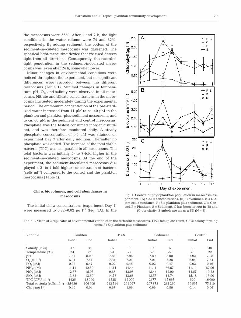
Fig. 1. Growth of phytoplankton population in mesocosm ex-periment. (A) Chl a concentrations. (B) Biovolumes. (C) Dia-tom cell abundance. P+S = plankton plus sediment, C = Con-trol, P = Plankton, S = Sediment. C has been left out in (B) and
(C) for clarity. Symbols are mean ± SD (N = 3)

Mar Ecol Prog Ser 346: 75–88, 2007
plankton mesocosms, initial chl a concentrations werelow, but increased until a maximum was reached onDay 8. Thereafter the chl a concentrations declinedrapidly. The mesocosms with plankton plus sedimentattained a chl a maximum concentration on Day 8.After Day 8, a moderate decline in chl a concentrationfollowed. In the sediment mesocosms, the maximumchl a concentration was recorded on Day 6. Algalgrowth was detected in the control mesocosms(Fig. 1A). Contamination could have been introducedby incomplete ozonation of experimental water, insuf-ficient cleaning of the empty mesocosms before thestart of the experiment, or cells from the surroundinginoculated mesocosms being transported via aerosolsto the controls. Entomoneis sp. was the dominatingtaxon in the controls, and specimen of this taxon in theinoculated mesocosms might have constituted a conta-mination. Abundances of Entomoneis sp. were there-fore excluded from all statistics.
Since cellular chl a content can vary with light condi-tions and cell physiology, and cell concentration isoften dependent on cell size, i.e. small cells reachhigher concentrations than larger cells, we also calcu-lated the changes in biovolumes with time (Fig. 1B).Diatoms dominated the phytoplankton communities inall mesocosms, irrespective of inocula. The graph dis-playing biovolumes is similar to the diagram of diatomcell abundances (Fig. 1C). The onset of phytoplanktongrowth in the sediment-inoculated mesocosms laggedbehind compared to the mesocosms inoculated withplankton (Fig. 1B,C). The highest biovolumes anddiatom abundances in the plankton mesocosms wereobserved early during the experiment (Day 6), com-pared to a week later (Day 13) in the sediment meso-cosms. Diatom cell abundances in the plankton meso-cosms reached a maximum on Day 6 and declinedthereafter (Fig. 1C). In the plankton-plus-sedimentmesocosms, 2 distinct biovolume and diatom abun-dance maxima were observed (Fig. 1B,C). The maxi-mum abundance on Day 6 was followed by a smallerpeak on Day 13. The sediment mesocosms displayedlow biovolumes and diatom abundances during theinitial part of the experiment, and did not reach themaximum cell abundance until Day 13.
Control mesocosms were significantly differentfrom any of the inoculated mesocosms based on chl aconcentrations (p < 0.01, single-factor ANOVA andpair-wise t-tests), thus any artefacts could beexcluded. Phytoplankton population development inthe mesocosms among the types of inocula based onchl a concentrations and biovolumes were signifi-cantly different (p < 0.05, single-factor ANOVA). Thedevelopment of diatom abundances was not signifi-cantly different among the 3 inocula types (p = 0.11,single-factor ANOVA). Based on diatom abundances,
pair-wise t-tests were significantly different (p < 0.01)between plankton, and plankton-plus-sediment meso-cosms, versus sediment mesocosms during the firsthalf of the experiment (Days 1 to 8). Plankton meso-cosms were significantly different from plankton-plus-sediment and sediment mesocosms (p < 0.05,pair-wise t-tests) during the second half of the experi-ment (Days 10 to 17).
Species proportions
High species diversity was noticed throughout theexperiment. The 12 most frequently occurring taxawere present in all the mesocosms, but proportionschanged over time depending on type of inocula.
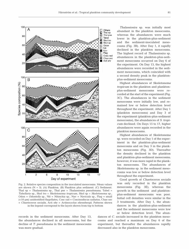
Mesocosms inoculated with plankton only had ini-tially similar proportions of the most common taxa(Fig. 2A). Towards the end of the experiment, Skele-tonema tropicum was the dominating species. AfterDay 6, proportions of Thalassiosira pseudonana andChaetoceros socialis declined. Thalassiosira sp., Skele-tonema sp., and Odontella sp. disappeared on, orshortly after, Day 10.
Plankton-plus-sediment mesocosms had initially ahigh diversity of species, but were less diverse anddominated by small flagellates at the end of the experi-ment (Fig. 2B). Skeletonema tropicum, Skeleto-nema sp., Odontella sp., and Chaetoceros socialisvanished after, or shortly after Day 6. Thalassiosirapseudonana and Thalassiosira sp. maintained a con-stant proportion of the population throughout the study.
Mesocosms inoculated with sediment only displayeda different community structure. Few of the dominat-ing taxa had large initial relative abundance (Fig. 2C).Thalassiosira pseudonana and Thalassiosira sp. main-tained continuous proportions of the species assem-blages throughout the experiment, while Chaetocerossocialis disappeared after Day 13. The proportion ofOdontella sp. declined during the second half of thestudy.
Abundances of major diatom species
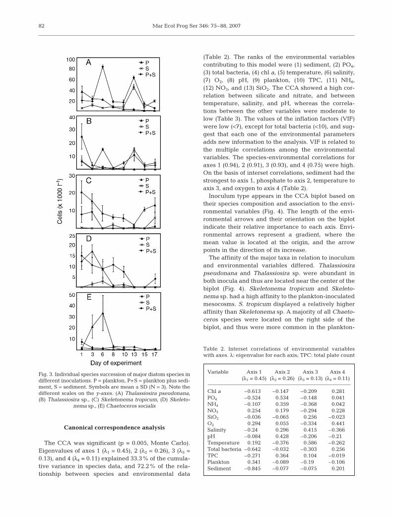
Thalassiosira pseudonana was initially most abun-dant in the plankton mesocosms (Fig. 3A), whereasthe abundances were lower in the plankton-plus-sediment and the sediment mesocosms. After Day 1,it gradually declined in the plankton mesocosms.The highest record of T. pseudonana abundanceoccurred on Day 6 of the experiment in the mesocosmsinoculated with plankton plus sediment. A later den-sity peak on Day 13 in the plankton-plus-sedimentmesocosms coincided with the highest abundance
80
Härnström et al.: Tropical plankton community development
records in the sediment mesocosms. After Day 13,the abundances declined in all mesocosms, but thedecline of T. pseudonana in the sediment mesocosmswas more gradual.
Thalassiosira sp. was initially mostabundant in the plankton mesocosms,whereas the abundances were muchlower in the plankton-plus-sedimentand the sediment-inoculated meso-cosms (Fig. 3B). After Day 1, it rapidlydeclined in the plankton mesocosms.The highest record of Thalassiosira sp.abundances in the plankton-plus-sedi-ment mesocosms occurred on Day 6 ofthe experiment. On Day 13, the highestabundances were recorded in the sedi-ment mesocosms, which coincided witha second density peak in the plankton-plus-sediment mesocosms.
Highest abundances of Skeletonematropicum in the plankton and plankton-plus-sediment mesocosms were re-corded at the start of the experiment (Fig.3C). The abundances in the sedimentmesocosms were initially low, and re-mained low or below detection levelthroughout the experiment. After Day 1(plankton mesocosms) and Day 3 ofthe experiment (plankton-plus-sedimentmesocosms), the abundances of S. tropi-cum declined. On Days 15 to 17, higherabundances were again recorded in theplankton mesocosms.
Highest abundances of Skeletonemasp. were recorded on Day 1 of the exper-iment in the plankton-plus-sedimentmesocosms and on Day 3 in the plank-ton mesocosms (Fig. 3D). Thereafterthe density declined in the planktonand plankton-plus-sediment mesocosms;however, it was more rapid in the plank-ton mesocosms. The abundances ofSkeletonema sp. in the sediment meso-cosms was low or below detection levelthroughout the experiment.
Good growth of Chaetoceros socialiswas only recorded in the planktonmesocosms (Fig. 3E), whereas thegrowth in the sediment and plankton-plus-sediment mesocosms was poor.Initial cell densities were similar in the3 treatments. After Day 1, the abun-dances in the plankton-plus-sedimentand the sediment mesocosms were lowor below detection level. The abun-
dances of C. socialis increased in the plankton meso-cosms and reached a maximum on Day 6 of theexperiment, but thereafter the abundances rapidlydecreased also in the plankton mesocosms.
81
Fig. 2. Relative species composition in the inoculated mesocosms. Mean valuesare shown (N = 3). (A) Plankton. (B) Plankton plus sediment. (C) Sediment.Thal sp = Thalassiosira sp., Thal pse = Thalassiosira pseudonana, Tabel =Tabellaria sp., Skel tro = Skeletonema tropicum, Skel sp = Skeletonema sp.,Odon = Odontella sp., Nit = Nitzschia sp., Nav = Navicula sp., Flag = small(<10 µm) unidentified flagellates, Cosc rad = Coscinodiscus radiatus, Chae soc= Chaetoceros socialis, Acti ehr = Actinocyclus ehrenbergii. Patterns shown
in the legend correspond to the layers shown from top to bottom
Mar Ecol Prog Ser 346: 75–88, 2007
Canonical correspondence analysis
The CCA was significant (p = 0.005, Monte Carlo).Eigenvalues of axes 1 (λ1 = 0.45), 2 (λ2 = 0.26), 3 (λ3 =0.13), and 4 (λ4 = 0.11) explained 33.3% of the cumula-tive variance in species data, and 72.2% of the rela-tionship between species and environmental data
(Table 2). The ranks of the environmental variablescontributing to this model were (1) sediment, (2) PO4,(3) total bacteria, (4) chl a, (5) temperature, (6) salinity,(7) O2, (8) pH, (9) plankton, (10) TPC, (11) NH4,(12) NO3, and (13) SiO2. The CCA showed a high cor-relation between silicate and nitrate, and betweentemperature, salinity, and pH, whereas the correla-tions between the other variables were moderate tolow (Table 3). The values of the inflation factors (VIF)were low (<7), except for total bacteria (<10), and sug-gest that each one of the environmental parametersadds new information to the analysis. VIF is related tothe multiple correlations among the environmentalvariables. The species-environmental correlations foraxes 1 (0.94), 2 (0.91), 3 (0.93), and 4 (0.75) were high.On the basis of interset correlations, sediment had thestrongest to axis 1, phosphate to axis 2, temperature toaxis 3, and oxygen to axis 4 (Table 2).
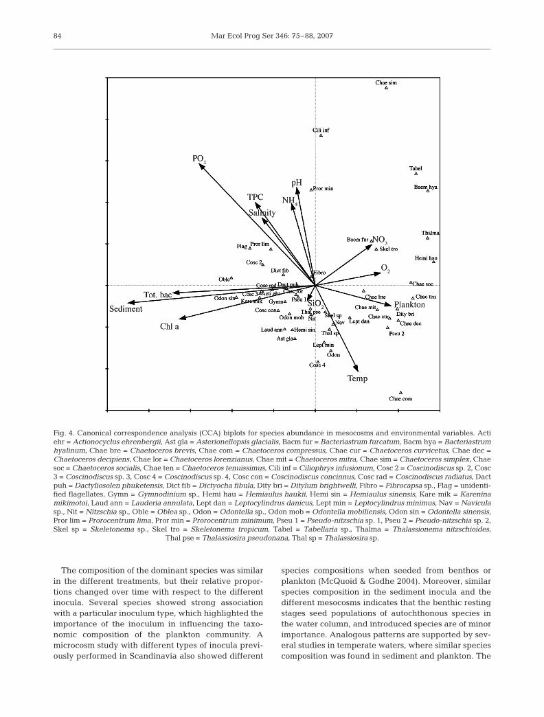
Inoculum type appears in the CCA biplot based ontheir species composition and association to the envi-ronmental variables (Fig. 4). The length of the envi-ronmental arrows and their orientation on the biplotindicate their relative importance to each axis. Envi-ronmental arrows represent a gradient, where themean value is located at the origin, and the arrowpoints in the direction of its increase.
The affinity of the major taxa in relation to inoculumand environmental variables differed. Thalassiosirapseudonana and Thalassiosira sp. were abundant inboth inocula and thus are located near the center of thebiplot (Fig. 4). Skeletonema tropicum and Skeleto-nema sp. had a high affinity to the plankton-inoculatedmesocosms. S. tropicum displayed a relatively higheraffinity than Skeletonema sp. A majority of all Chaeto-ceros species were located on the right side of thebiplot, and thus were more common in the plankton-
82
Variable Axis 1 Axis 2 Axis 3 Axis 4 (λ1 = 0.45) (λ2 = 0.26) (λ3 = 0.13) (λ4 = 0.11)
Chl a –0.613 –0.147 –0.209 0.281PO4 –0.524 0.534 –0.148 0.041NH4 –0.107 0.359 –0.368 0.042NO3 0.254 0.179 –0.294 0.228SiO2 –0.036 –0.065 0.256 –0.023O2 0.294 0.055 –0.334 0.441Salinity –0.24 0.296 0.415 –0.366pH –0.084 0.428 –0.206 –0.21Temperature 0.192 –0.376 0.586 –0.262Total bacteria –0.642 –0.032 –0.303 0.256TPC –0.271 0.364 0.104 –0.019Plankton 0.341 –0.089 –0.19 –0.106Sediment –0.845 –0.077 –0.075 0.201
Table 2. Interset correlations of environmental variableswith axes. λ: eigenvalue for each axis; TPC: total plate count
Fig. 3. Individual species succession of major diatom species indifferent inoculations. P = plankton, P+S = plankton plus sedi-ment, S = sediment. Symbols are mean ± SD (N = 3). Note thedifferent scales on the y-axes. (A) Thalassiosira pseudonana,(B) Thalassiosira sp., (C) Skeletonema tropicum, (D) Skeleto-
nema sp., (E) Chaetoceros socialis

Härnström et al.: Tropical plankton community development
inoculated mesocosms, even though many species ofthis genus have resting stages, and some of these wereabundant in the sediment used as inoculum. Chaeto-ceros lorenzianus was an exception and appeared onthe left side of the biplot, and was thus associated tothe sediment inocula. Pseudo-nitzschia spp., a genusnot known to form resting stages, was common in theplankton-inoculated mesocosms. Bacteriastrum furca-tum, B. hyalinum, Ditylum brightwelli, Leptocylindrusdanicus, and L. minimus were located on the right sideof the biplot and had a high affinity to plankton-inoculated mesocosms, in spite of having previouslydocumented resting stages. Several dinoflagellatespecies were found in the sediment mesocosms but allhad low abundances. Other environmental variablesappeared not to influence the frequency of the majorspecies groups.
The arrow representing chl a concentration wasclosely associated with the arrow representing sedi-ment inocula (Fig. 4). This is also illustrated in Fig. 1A,where chl a concentrations in the sediment mesocosmswere high early in the experiment. The location on theleft side of the biplot of large cells with many chloro-plasts, such as Coscinodiscus spp., is a consequence ofthe environmental variable chl a (Fig. 4).
DISCUSSION
The present study demonstrated the importanceof benthic resting stages in seeding the planktonicpopulation and showed that the type of inoculum influ-ences the development and the taxonomic compositionof the phytoplankton community. The most significanteffects of the inoculum types were the timing of thebloom and differences in species proportions. To our
knowledge, no mesocosm studies on naturally occur-ring phytoplankton communities and their develop-ment have been performed along the SW coast of Indiabefore. The contribution of resting stages to phyto-plankton blooms in the tropics is poorly investigated,but this study showed that resuspended resting stagesare important for phytoplankton bloom dynamics inthe area.
The onset of phytoplankton growth lagged behind inthe sediment mesocosms compared to the plankton-inoculated mesocosms. This was a consequence of 2different seeding strategies: (1) proliferation of vegeta-tive cells originating from the plankton attaining expo-nential growth soon after inoculation, and (2) later ger-minated resting stages and subsequent growth. This isin accordance with a previous microcosm experimentin Scandinavia, where bloom development also wasfaster in plankton-inoculated microcosms (McQuoid &Godhe 2004). Diatom resting stages have no dormancyperiod, as is common among dinoflagellate cysts. How-ever, they may show a 1 to 8 d lag period of growthcompared to vegetative cells, which can grow almostimmediately (Itakura et al. 1997, Kuwata & Takahashi1999), which may then influence the timing of bloomdevelopment. Previous mesocosm experiments haveshown that the size of the inoculum is also an impor-tant aspect in time required for bloom development(Pitcher et al. 1993, McQuoid & Godhe 2004). Diatomresting stages are generally less common in the tropics(Hargraves & French 1983), and moreover, the lowdinoflagellate cyst abundances found in the study area(Godhe et al. 2000, 2002b) indicate that benthic restingstages in general may be less abundant compared totemperate waters. From this, it follows that the sedi-ment inoculum contains fewer resting stages, and thusthe lag period is prolonged.
83
Variable Chl a PO4 NH4 NO3 SiO2 O2 Salinity pH Temp. Total TPC Plankton Sedimentbacteria
Chl a 1.00PO4 0.32 1.00NH4 –0.09 0.07 1.00NO3 –0.09 –0.26 0.62 1.00SiO2 –0.10 –0.32 0.55 0.90 1.00O2 0.16 –0.38 0.07 0.29 0.23 1.00Salinity 0.01 –0.53 0.34 0.51 0.52 0.37 1.00pH 0.03 –0.54 0.34 0.52 0.53 0.41 1.00 1.00Temp. 0.02 –0.56 0.32 0.50 0.52 0.40 1.00 1.00 1.00Total bacteria 0.06 0.33 0.00 0.07 0.07 0.27 –0.02 0.00 –0.01 1.00TPC 0.01 0.39 0.19 –0.13 –0.17 –0.37 –0.12 –0.15 –0.16 –0.13 1.00Plankton –0.02 –0.31 0.05 0.22 0.19 0.33 0.17 0.18 0.19 –0.05 –0.47 1.00Sediment 0.49 0.46 –0.07 –0.13 –0.06 –0.11 –0.21 –0.21 –0.22 0.78 0.12 –0.37 1.00
Table 3. Correlation among environmental and nominal variables (plankton and sediment) used in canonical correspondence analysis (CCA)
Mar Ecol Prog Ser 346: 75–88, 2007
The composition of the dominant species was similarin the different treatments, but their relative propor-tions changed over time with respect to the differentinocula. Several species showed strong associationwith a particular inoculum type, which highlighted theimportance of the inoculum in influencing the taxo-nomic composition of the plankton community. Amicrocosm study with different types of inocula previ-ously performed in Scandinavia also showed different
species compositions when seeded from benthos orplankton (McQuoid & Godhe 2004). Moreover, similarspecies composition in the sediment inocula and thedifferent mesocosms indicates that the benthic restingstages seed populations of autochthonous species inthe water column, and introduced species are of minorimportance. Analogous patterns are supported by sev-eral studies in temperate waters, where similar speciescomposition was found in sediment and plankton. The
84
Fig. 4. Canonical correspondence analysis (CCA) biplots for species abundance in mesocosms and environmental variables. Actiehr = Actionocyclus ehrenbergii, Ast gla = Asterionellopsis glacialis, Bacm fur = Bacteriastrum furcatum, Bacm hya = Bacteriastrumhyalinum, Chae bre = Chaetoceros brevis, Chae com = Chaetoceros compressus, Chae cur = Chaetoceros curvicetus, Chae dec =Chaetoceros decipiens, Chae lor = Chaetoceros lorenzianus, Chae mit = Chaetoceros mitra, Chae sim = Chaetoceros simplex, Chaesoc = Chaetoceros socialis, Chae ten = Chaetoceros tenuissimus, Cili inf = Ciliophrys infusionum, Cosc 2 = Coscinodiscus sp. 2, Cosc3 = Coscinodiscus sp. 3, Cosc 4 = Coscinodiscus sp. 4, Cosc con = Coscinodiscus concinnus, Cosc rad = Coscinodiscus radiatus, Dactpuh = Dactyliosolen phuketensis, Dict fib = Dictyocha fibula, Dity bri = Ditylum brightwelli, Fibro = Fibrocapsa sp., Flag = unidenti-fied flagellates, Gymn = Gymnodinium sp., Hemi hau = Hemiaulus haukii, Hemi sin = Hemiaulus sinensis, Kare mik = Kareninamikimotoi, Laud ann = Lauderia annulata, Lept dan = Leptocylindrus danicus, Lept min = Leptocylindrus minimus, Nav = Naviculasp., Nit = Nitzschia sp., Oble = Oblea sp., Odon = Odontella sp., Odon mob = Odontella mobiliensis, Odon sin = Odontella sinensis,Pror lim = Prorocentrum lima, Pror min = Prorocentrum minimum, Pseu 1 = Pseudo-nitzschia sp. 1, Pseu 2 = Pseudo-nitzschia sp. 2,Skel sp = Skeletonema sp., Skel tro = Skeletonema tropicum, Tabel = Tabellaria sp., Thalma = Thalassionema nitzschioides,
Thal pse = Thalassiosira pseudonana, Thal sp = Thalassiosira sp.

Härnström et al.: Tropical plankton community development
diatom resting stages in the sediment were recordedthroughout the year, and seasonal cycles of diatomspecies in the plankton indicated autochthonous seed-ing (Pitcher 1990, Itakura et al. 1997).
Most mesocosm studies focus on one or just a fewspecies (e.g. Brussaard et al. 2005, Patel et al. 2005),and how varying parameters affect their growth. Stud-ies focusing on bloom development and successionwithin the natural phytoplankton community are lesscommon, but these multi-species studies show that oneor just a few species become dominant shortly after theinitiation of the experiments (e.g. Alldrege et al. 1995,McQuoid & Godhe 2004). In the present study, the spe-cies diversity was maintained throughout the experi-ment, although the diversity in all inocula typesdecreased towards the end. This is probably a conse-quence of the fact that the natural phytoplankton com-munity in the tropics generally displays high speciesdiversity, and that single-species phytoplanktonblooms rarely occurs (Subrahmanyan & Sarma 1960,Irigoien et al. 2004). Generally the chl a concentrationsof all the mesocosms were low (<3 µg l–1) in our study,which are normal conditions in the coastal Arabian Seaduring December and January (Chaturvedi 2005). Lowchl a values can be a result of high diversity sinceexclusive competition will be less (Duarte et al. 2006),which is in accordance with the overall results from ourstudy.
Diatoms may employ a variety of seeding strategiesfor initiation of planktonic growth. Our results suggestthat the strategies differ among species, which isreflected in the strong association of specific species toinocula type in the CCA. Moreover, our results furtherindicate that the one species may use the same, e.g.Thalassiosira pseudonana, or different, e.g. Chaeto-ceros socialis and Skeletonema sp., strategies in differ-ent parts of the world. Species belonging to the genusThalassiosira are adapted to varying environments,and are dominant in both water column and surfacesediment (Pedersen et al. 2005, Kasim & Mukai 2006).The resting stages contribute extensively by seedingthe plankton in temperate waters (Itakura et al. 1997,McQuoid et al. 2002, Ishikawa & Furuya 2004). InIndian waters, Mitbavkar & Anil (2002) recorded vege-tative cells of Thalassiosira in cultures established fromsediment slurries. In our study, Thalassiosira declinedin the plankton mesocosms, while in the plankton-plus-sediment mesocosms, 2 cell density peaks wereobserved. The last peak coincided with the highestrecorded abundances in the sediment mesocosms,which indicates recruitment of resting stages from thesediment. Resting stages of Skeletonema are very suc-cessful in seeding blooms in temperate water (Itakuraet al. 1997, McQuoid & Godhe 2004), but our experi-ment indicates that in coastal SW India, propagation
by resting stages is not important. We recorded noresting stages in the sediment inoculum, and observedvirtually no growth in the sediment mesoscosms.Blooms of Thalassiosira have been recorded to be suc-ceeded by blooms of Chaetoceros socialis (Booth et al.2002). A similar scenario was recorded in the planktonmesocosms in our study. Resting stages of C. brevis, C.didymus, and Chaetoceros sp. were recorded in thesediment inoculum, but had no noticeable contributionto the vegetative community in the sediment meso-cosms. The suggested explanation for these contradict-ing results is that germination of Chaetoceros spp.spores in the tropics might be triggered by some sea-sonal cue (McQuoid & Hobson 1996), although contin-uous germination of spores from the same genus havebeen recorded from temperate waters (Itakura et al.1997).
In the present study, the steady concentration of sili-cate and nitrate despite diatom dominance in themesocosms is puzzling. This can be explained by thelow bacterial concentrations and the increased growthof flagellates with time. The low bacterial abundancessuggest a significant predation by bacterivorous flagel-lates, which leads to remineralization of major nutri-ents (Azam et al. 1983). In addition, the nutrient con-centrations can probably support much higherphytoplankton cell abundances, but the great speciesdiversity recorded in all mesocosms will prevent anymonospecific bloom, which is required to attain highcell abundance and subsequent nutrient depletion.
Unidentified small (<10 µm) flagellates increasedtowards the end of the experiment. Ciliates were theonly potential predators on diatoms recorded in themesocosms, but the proportions of these were low allthrough the study, ranging from 0.3% of the totalabundance at the onset of the experiment to a maxi-mum of 1.7%. This phenomenon, i.e. a shift from anautotrophic- to a heterotrophic-dominated community,is commonly observed in enclosures with small volumerelative to its large surfaces. This is related to a varietyof ‘wall-effects’ that can change the species composi-tion (Chen & Kemp 2004). Another commonly ob-served artefact in mesocosms is the proliferation ofbenthic diatoms, mainly pennates, which are notregular members of the phytoplankton community(M. R. McQuoid & A. Godhe unpubl.). In that experi-ment, Nitzschia sp. and Navicula sp. were common butin similar proportions irrespective of mesocosm types.Limiting the mesocosm experimental time can mini-mize these type of artefacts. In our experimentalsystem, we could see a shift from an autotrophic-dominated to a heterotrophic community, and there-fore the experiment was terminated.
Chl a in the sediment mesocosms reached its highestconcentrations early during our experiment, whereas
85
Mar Ecol Prog Ser 346: 75–88, 2007
the biovolume and the abundances of diatom cells inthe same mesocosms were relatively low. Presence oflarge cells such as Coscinodiscus spp. were recordedin the Lugol’s fixed samples from the sediment meso-cosms, and might have contributed to the observed risein chl a concentrations. Further, since many of theselarge cells were partially damaged and only activelygrowing cells were counted, they had no contributionto the estimated biovolume or total abundance of livingdiatom cells, but probably influenced the high initialchl a concentrations (Fig. 1A). Also, the sedimentmesocosms had in general higher abundance of cyano-bacteria, which also can be a factor in raising the initialconcentration of chl a. However, due to the smallvolume of cyanobacteria, their effect on the total bio-volume was minor.
Total cell abundances were comparable in all inocu-lated mesocosms, despite lower light penetration in thesediment-inoculated mesocosms. Accordingly, thephytoplankton growth in the plankton mesocosms wasnot assumed to be favored by better light conditions.Light conditions in standard operating protocols fordiatom germination and subsequent growth are set to40–50 µmol photons m–2 s–1 (Itakura et al. 1997,Kuwata & Takahashi 1999), comparable to the lightprovided in all mesocosms.
Although diatom resting stage formation is less com-mon in tropical upwelling waters where nutrients arenot often depleted (Hargraves & French 1983), ourstudy has shown that resting stages may contributesignificantly to the phytoplankton community of theSW coast of India. Formation of resting stages is oftenseasonal, and a rapid change of hydrographic condi-tions (nutrients, temperature, pH, salinity, or light) isrequired for the induction of diatom resting stages(McQuoid & Hobson 1996). Moreover, resting stagesare, under natural conditions, often formed during orfollowing periods of maximum vegetative division(Garrison 1981). Considering the annual variation inhydrographic parameters and phytoplankton biomassof the area we studied, the post-bloom conditions inAugust with reduced inorganic nutrients of the watercolumn (Subrahmanyan & Sarma 1960) would consti-tute the most plausible time for diatom resting stageformation in the SE coastal Arabian Sea.
The resting stage is thought to mainly serve as long-term survival, and therefore might be scarce in thetropics. Laboratory experiments have documentedprolonged survival of diatom resting stages in cool (5 to15°C) conditions (Itakura et al. 1997). Bottom sea watertemperature in the SE coastal Arabian Sea is >25°C(Subrahmanyan & Sarma 1960), and perhaps the rest-ing stages do not survive as long as in temperatewaters. However, functions other than long-term sur-vival have also been suggested. Resting stages could
serve as a short-term survival strategy between inter-mittent blooms (Garrison 1981), and germination ofdistinct genetic clones adapted to different environ-mental settings could ensure species survival in un-favorable environments (Rynearson et al. 2006).
The germination of diatom resting stages is believedto be most affected by light (French & Hargraves 1985).The Secchi depth at coastal Mangalore is approxi-mately 5 m, due to high turbidity, and therefore resus-pension is required for germination. Tidal force isstrong and is felt up to the 10 m depth contour, whichcorresponds to ca. 2 km from the shore (Segar & Hari-haran 1989). The tidal currents could thus continuouslyseed the water column with resuspended restingstages, at least in the shallower parts. Most probablythe strong SW monsoon, which induces upwellingalong the coast, is the most important factor for resus-pension, germination, and subsequent vegetative dia-tom growth from deeper areas. Primary productionand diatom abundances are also known to be highestduring this period of the year (Subrahmanyan & Sarma1960). Benthic germination can be regulated by sea-sonal factors (McQuoid & Hobson 1996, Eilertsen &Wyatt 2000), and thus, diatoms in the region could betuned for germination during SW monsoon upwelling.
The results from this study further confirm thatbenthic resting stages contribute by seeding the watercolumn. Despite a presumed low density of benthicresting stages in tropical sediments, they can influencethe phytoplankton community in the water column.Our results indicate that species composition can bealtered if a population is seeded by resting stages or byplanktonic cells.
Acknowledgements. This study was financed by Sida (SWE2004-129) and Formas-Sida (2005-255). It was also supportedby the Kapten Carl Stenholms donationsfond, MagnusBergvalls Stiftelse (MBS), and the Oscar and Lili Lamms Foun-dation. We thank Å. Lindskog, who helped with the laboratorywork as part of her Master thesis, and Dr. P. K. Krishnakumarat the CMFRI in Mangalore for logistic support. Three anony-mous reviewers are acknowledged for their constructivecomments on the manuscript.
LITERATURE CITED
Alldrege AL, Gotschalk C, Passow U, Riebesell U (1995) Massaggregation of diatom blooms: insights from a mesocosmstudy. Deep-Sea Res II 42:9–27
Azam F, Fenchel T, Field JG, Gray JS, Meyer-Reil LA,Thingstad F (1983) The ecological role of water-columnmicrobes in the sea. Mar Ecol Prog Ser 10:257–263
Backhaus JO, Wehde EN, Hegseth EN, Kämpf J (1999) Phyto-plankton convection: the role of oceanic convection in pri-mary production. Mar Ecol Prog Ser 189:77–92
Backhaus JO, Hegseth EN, Wehde H, Irigoien X, Hatten K,Logemann K (2003) Convection and primary production inwinter. Mar Ecol Prog Ser 251:1–14
86
Härnström et al.: Tropical plankton community development
Booth BC, Larouche P, Bélanger S, Klein B, Amiel D, Mei ZP(2002) Dynamics of Chaetoceros socialis blooms in theNorth Water. Deep-Sea Res II 49:5003–5025
Brussaard CPD, Kuipers B, Veldhuis MJW (2005) A mesocosmstudy of Phaeocystis globosa population dynamics I. Reg-ulatory role of viruses in bloom control. Harmful Algae 4:859–874
Chaturvedi N (2005) Variability of chlorophyll concentrationin the Arabian Sea and Bay of Bengal as observed fromSeaWiFS data from 1997–2000 and its interrelationshipwith Sea Surface Temperature (SST) derived from NOAAAVHRR. Int J Remote Sens 26:3695–3706
Chen CC, Kemp WM (2004) Periphyton communities inexperimental marine ecosystems: scaling the effect ofremoval from container walls. Mar Ecol Prog Ser 271:27–41
Cook D, DePaola A, McCarthy S (2000) Direct plating proce-dure for the enumeration of total and pathogenic Vibrioparahaemolyticus in oyster meats. US Food and DrugAdministration (FDA), Office of Seafood, Gulf SeafoodLaboratory, Dauphin Island, AL
D’Souza F, Garg A, Bhosle NB (2003) Biogeochemical charac-teristics of sedimenting particles in Dona Paula Bay, India.Estuar Coast Shelf Sci 58:311–320
Duarte P, Macedo MF, da Fonseca LC (2006) The relationshipbetween phytoplankton diversity and community functionin a coastal lagoon. Hydrobiologia 555:3–18
Eilertsen H, Wyatt T (2000) Phytoplankton models and lifehistory strategies. S Afr J Mar Sci 22:323–338
French FW, Hargraves P (1985) Spore formation in the lifecycles of the diatoms Chaetoceros diadema and Lepto-cylindrus danicus. J Phycol 21:477-483
Garrison DL (1981) Monterey Bay phytoplankton I. Restingspore cycles of phytoplankton assemblages. J Plankton Res3:137–156
Godhe A, Karunasagar I, Karunasagar I, Karlson B (2000)Dinoflagellate cysts in recent marine sediments from SWIndia. Bot Mar 43:39–48
Godhe A, Svensson S, Rehnstam-Holm AS (2002a) Oceano-graphic settings explain fluctuations in Dinophysis spp.and concentrations of diarrhetic shellfish toxin in theplankton community within a mussel farm area on theSwedish west coast. Mar Ecol Prog Ser 240: 71–83
Godhe A, Rehnstam-Holm AS, Karunasagar I, Karunasagar I(2002b) PCR detection of dinoflagellate cysts in field sedi-ment samples from tropic and temperate environments.Harmful Algae 1:361–373
Hallegraeff GM, Bolch CJ (1992) Transport of diatom anddinoflagellate resting spores in ships’ ballast water:implications for plankton biogeography and aquaculture.J Plankton Res 14:1067–1084
Hargraves PE, French FW (1983) Diatom resting spores: signif-icance and strategies. In: Fryxell GA (ed) Survival strate-gies of the algae. Cambridge University Press, Cambridge,p 49–68
Hasle G, Syvertsen E (1996) Marine diatoms. In: Tomas C (ed)Identifying marine diatoms and dinoflagellates. AcademicPress, San Diego, CA, p 5–385
Irigoien X, Huisman J, Harris RP (2004) Global biodiversitypatterns of marine phytoplankton and zooplankton.Nature 429:863–867
Ishikawa A, Furuya K (2004) The role of diatom resting stagesin the onset of the spring bloom in the East China Sea.Mar Biol 145:633–639
Itakura S, Imai I, Itoh K (1997) ‘Seed bank’ of coastal plank-tonic diatoms in bottom sediments of Hiroshima Bay, SetoInland Sea, Japan. Mar Biol 128:497–508
Kasim M, Mukai H (2006) Contribution of benthic and epi-phytic diatoms to clam and oyster production in theAkkeshi-ko estuary. J Oceanogr 62:267–281
Kilham P, Kilham SS (1980) The evolutionary ecology ofphytoplankton. In: Morris I (ed) The physiological ecologyof phytoplankton. Blackwell, Boston, MA, p 571–597
Krishnakumar PK, Bhat GS (2007) Seasonal and interannualvariations of oceanographic conditions off Mangalorecoast (Karnataka, India) in the Malabar upwelling systemduring 1995-2004 and their influences on pelagic fishery.Fish Oceanogr (in press)
Kuwata A, Takahashi M (1999) Survival of resting spores andresting cells of the marine planktonic diatom Chaetocerospseudocurvicetus under fluctuating nitrate conditions.Mar Biol 134:471–478
Lundholm N, Daubjerg N, Moestrup Ø (2002) Phylogeny ofthe Bacillariaceae with emphasis on the genus Pseudo-nitzschia (Bacillariophyceae) based on partial LSU rDNA.Eur J Phycol 37:115–134
McQuoid M, Godhe A (2004) Recruitment of coastal plank-tonic diatoms from benthic versus pelagic cells: variationin bloom development and species composition. LimnolOceanogr 49:1123–1133
McQuoid M, Hobson L (1996) Diatom resting stages. J Phycol32:889–902
McQuoid MR, Godhe A, Nordberg K (2002) Viability of phy-toplankton resting stages in the sediments of a coastalSwedish fjord. Eur J Phycol 37:191–201
Mitbavkar S, Anil AC (2002) Diatoms of the microphytoben-thic community: population structure in a tropical inter-tidal sand flat. Mar Biol 140:41–57
Parsons T, Maita Y, Lalli C (1984) A manual of chemical andbiological methods for seawater analysis. Pergamon,Oxford
Patel D, Thake B, Thornton DCO (2005) Effect of light andturbulent mixing on the growth of Skeletonema costatum(Bacillariophyceae). Mar Biol 146:633–644
Pedersen SA, Ribergaard MH, Simonsen CS (2005) Micro-and mesozooplankton in southwest Greenland waters inrelation to environmental factors. J Mar Syst 56:85–112
Pitcher G (1990) Phytoplankton seed populations of the CapePeninsula upwelling plume, with particular reference toresting spores of Chaetoceros (Bacillariophyceae) and theirrole in seeding upwelling waters. Estuar Coast Shelf Sci 31:283–301
Pitcher G, Bolton J, Brown P, Hutchings L (1993) The deve-lopment of phytoplankton blooms in upwelled waters ofthe southern Benguela upwelling system as determinedby microcosm experiment. J Exp Mar Biol Ecol 165:171–189
Porter K, Feig Y (1980) The use of DAPI for identifying andcounting aquatic microflora. Limnol Oceanogr 25:943–948
Raffaelli D, Bell E, Weithoff G, Matsumoto A and 5 others (2003)The ups and downs of benthic ecology: considerations ofscale, heterogeneity and surveillance for benthic–pelagiccoupling. J Exp Mar Biol Ecol 285-286: 191–203
Ramana TV, Nathaniel DE, Reddy PM (1991) Distribution ofsome oceanographic factors in the Arabian Sea off Man-jeswar and theoretic possible effect on oilsardine and mack-erel fisheries in the area. J Mar Biol Assoc India 33:9–18
Rixen T, Haake B, Ittekkot V (2000) Sedimentation in thewestern Arabian Sea: the role of coastal and open-oceanupwelling. Deep-Sea Res II 47:2155-2178
Rynearson T, Newton J, Armbrust A (2006) Spring bloomdevelopment, genetic variation, and population succes-sion in the planktonic diatom Ditylum brightwelli. LimnolOceanogr 51:1249–1261
87

Mar Ecol Prog Ser 346: 75–88, 2007
Sarno D, Kooistra W, Medlin L, Percopo I, Zingone A (2005)Diversity in the genus Skeletonema (Bacillariophyceae). II.An assessment of the taxonomy of S. costatum-like specieswith the description of four new species. J Phycol 41:151–176
Segar K, Hariharan V (1989) Seasonal distribution of nitrate, ni-trite, ammonium and plankton in effluent discharge area offMangalore, west coast of India. Indian J Mar Sci 18: 170–173
Shenoy DM, Patil JS (2002) Temporal variations in dimethyl-sulphoniopropionate and dimethyl sulphide in the Zuariestuary, Goa (India). Mar Environ Res 56:387–402
Sicko-Goad L, Stoermer E, Kociolek J (1989) Diatom restingcell rejuventaion and formation: time course, speciesrecords and distribution. J Plankton Res 11:375–389
Smayda TJ (2002) Turbulence, watermass stratification andharmful algal blooms: an alternative view and frontalzones as ‘pelagic seed banks’. Harmful Algae 1:95–112
Subrahmanyan R (1958) Ecological studies on the marinephytoplankton on the west coast of India. Mem Indian BotSoc 1:145–151
Subrahmanyan R (1959) Studies on the phytoplankton of thewest coast of India. Proc Indian Acad Sci 50:113–252
Subrahmanyan R (1960) Observation on the effect of the mon-soon in the production of phytoplankton. J Indian Bot Soc39:78–89
Subrahmanyan R, Sarma AHV (1960) Studies on the phyto-plankton of the west coast of India. III. Seasonal variation
of the phytoplankton and the environmental factors.Indian J Fish 7:307–336
Subrahmanyan R, Sarma AHV (1965) Studies on the phyto-plankton of the west coast of India. IV. Magnitude of thestanding crop for 1955–1962, with observations onnanoplankton and its significance to fisheries. J Mar BiolAssoc India 7:406–419
Sun J, Liu D (2003) Geometric models for calculating cell bio-volume and surface area for phytoplankton. J PlanktonRes 25:1331–1346
ter Braak CJF, Smilauer P (2002) CANOCO reference man-ual and CanoDraw for Windows user’s guide: softwarefor canonical community ordination (version 4.5). Micro-computer Power, Ithaca, NY
Thomsen HA (ed) (1992) Plankton i de inre danske farvande,Vol 11. Dansk Miljøstyrelse, Copenhagen (in Danish)
Throndsen J, Hasle G, Tangen K (2003) Norsk kystplankton-flora. Almater Forlag, Oslo (in Norwegian)
Tomas C (1997) Identifying marine phytoplankton. AcademicPress, San Diego, CA
Tommasa LD, Belmonte G, Palanques A, Pugi P (2000)Resting stages in a submarine canyon: a comparisonof shallow-deep-sea coupling? Hydrobiologia 440:249–260
Utermöhl H (1958) Zur Vervollkommnung der quantitativenPhytoplankton-Methodik. Mitt Int Ver Limnol 9:1–38 (inGerman)
88
Editorial responsibility: Alain Vézina (Contributing Editor),Dartmouth, Nova Scotia, Canada
Submitted: October 27, 2006; Accepted: May 9, 2007Proofs received from author(s): September 11, 2007