a study on dermatoglyphics pattern in patients with
TRANSCRIPT

Dissertation on
A STUDY ON DERMATOGLYPHICS PATTERN IN PATIENTS WITH MYOCARDIAL INFARCTION
Submitted in partial fulfillment for
M.S. DEGREE EXAMINATION BRANCH‐V ANATOMY
Upgraded Institute of Anatomy Madras Medical College & Research Institute,
Chennai‐ 600 003
THE TAMILNADU Dr.M.G.R. MEDICAL UNIVERSITY CHENNAI – 600 032
TAMILNADU
APRIL 2011

CERTIFICATE
This is to certify that this dissertation entitled “A STUDY ON
DERMATOGLYPHICS PATTERN IN PATIENTS WITH
MYOCARDIAL INFARCTION” is a bonafide record of the research work
done by Dr.K.R.ARUNKUMAR., Post graduate in the Institute of Anatomy,
Madras Medical College and Research Institute, Government General
Hospital,Chennai-03, in partial fulfillment of the regulations laid down by
The Tamil Nadu Dr.M.G.R. Medical University for the award of M.S. Degree
Branch V- Anatomy, under my guidance and supervision during the academic
years 2008-2011.
Dr.R.AMUDHA. M.B.B.S.,M.S
Dean, Director and Professor,
Madras Medical College & Institute of Anatomy,
Research Institute, Madras Medical College &
Govt. General Hospital, Research Institute,
Chennai – 600 003 Chennai - 600003

ACKNOWLEDGEMENT
It is with great pleasure and deep sense of gratitude that I acknowledge
my debt to my guide Prof.Dr.R.Amudha M.S,
Professor and Director,
Institute of Anatomy, Madras Medical College, Chennai – 3,for her
affectionate guidance, meticulous attention, keen interest with which she has
provided me the suggestions, knowledge and support to construct this work.
I wish to take this opportunity to express my profound gratitude to my
respected teacher who suggested choosing this topic, Prof.Dr.Mrs.Christilda
Felicia Jebakani M.S, Former Director, Institute of Anatomy, Madras Medical
College, Chennai - 3 for her guidance, valuable suggestions and constant
encouragement throughout the study which facilitated the completion of my
dissertation.
I am thankful to Dr.J.Mohanasundaram M.D., Ph.D., Dean, Madras
Medical College, Chennai – 3 for permitting to avail the facilities in this college
for performing this study.
I express my sincere gratitude to Dr.I.Jeyaraj, Associate professor,
Dr.B.Chezhian, Tmt.M.S.Thenmozhi, Dr.V.Loganayaki,
Dr.V.Sathialakshmi, Dr.M.Vijayalakshmi and Dr.P.Murugesan, Assistant
professors, Institute of Anatomy, Madras Medical College, Chennai – 3, for
their valuable suggestions and encouragement throughout the study.
I owe my obligation to Dr.R.Subramaniyam M.D.D.M., Director,
Institute of Cardiology, Govt. General Hospital, Chennai – 3 for permitting to
take the palmar prints from the patients in his institute.

I am thankful to my seniors, Dr.V.Rajapriya, Dr.K.Sangameswaran,
Dr.R.Vijaya, Dr.K.Sujatha for their support and encouragement throughout
the study.
I express my sincere thanks to my colleagues Dr.V.Dhanalakshmi,
Dr.T.Sureshkumar and Dr.S.Satishkumar for their invaluable support and
constant encouragement throughout the study.
I am thankful to my juniors Dr.Kalai Anbusudar, Dr.P.Kanagavalli,
Dr.S.Kousalya, Dr.S.Suganya for their support and help in carrying out this
study.
I am very thankful to Dr.Jagadh Janani for helping me in statistical
analysis.
I am grateful to my parents Mr.K.Rathinasamy and Mrs.R.Aruna and
my sister R.Abinaya who have helped me making this study a reality.
Last but not the least, I would like to thank all my patients and subjects
without whom the study would not have been possible.
Above all, I thank the ALMIGHTY GOD for providing all the necessary
help through his lovely creations in this endeavour of mine.

CONTENTS
Sl. No.
Title Page no.
1. INTRODUCTION 1
2. AIM OF THE STUDY 5
3. REVIEW OF LITERATURE 7
4. EMBRYOLOGY 23
5. MATERIALS AND METHOD 30
6. OBSERVATION 44
7. DISCUSSION 56
8. CONCLUSION 64
9. BIBLIOGRAPHY

1
INTRODUCTION
The entire human body is clothed with the skin which happens to
be the largest and most important organ of the body. It performs many
vital functions in the life of an individual, viz, it protects and safeguards
the body from the vagaries of the weather, maintains the body
temperature and saves the internal organs of body from injuries.
However, the skin on the ventral sides of hands and plantar sides of feet
is exclusively designed and is corrugated with the ridges and
configurations which are functionally useful as they help in grasping
without which the objects would easily slip away from hands. (Sir
Charles Bell, 1833)15
The scientific study of these ridges and configurations on the
palmar region of hands and fingers and plantar region of feet and toes is
termed as Dermatoglyphics. The term Dermatoglyphics was coined by
Cummins and Midlo in 1926. Etymologically this term is harmonious
blend of two words Derma, skin; Glyphe, carve. The synonyms in use for
epidermal ridges are papillary ridges, friction ridges and finger prints.
Papillary ridges form narrow parallel or curved arrays separated by
narrow furrows (Fig. 1).
They depend upon the large size and peculiar arrangements of the
papillae upon which the epidermis is placed (Fig. 5). The direction of the
ridges is at right angles with the force that tends to produce slipping or to
the resultant of such forces when these forces vary in direction. In each
individual, the lines on the tips of the fingers and thumbs form distinct
patterns unlike those of any other person. A method of determining the
identity of a criminal is based on this fact, impressions (“finger-prints”)

2
of these lines being made on paper covered with soot or on white paper
after first covering the fingers with ink.
The papillae are minute conical eminences, having rounded or
blunted extremities, occasionally divided into two or more parts, and are
received into corresponding pits on the under surface of the cuticle. On
the general surface of the body, more especially in parts endowed with
slight sensibility, they are few in number, and exceedingly minute; but in
some situations, as upon the palmar surfaces of the hands and fingers, and
upon the plantar surfaces of the feet and toes, they are long, of large size,
closely aggregated together, and arranged in parallel curved lines,
forming the elevated ridges seen on the free surface of the epidermis.
Each ridge contains two rows of papillae, between which the ducts of the
sudoriferous glands pass outward to open on the summit of the ridge
(Fig. 2). Each papilla consists of very small and closely interlacing
bundles of finely fibrillated tissue, with a few elastic fibers; within this
tissue is a capillary loop, and in some papillae, especially in the palms of
the hands and the fingers, there are tactile corpuscles. (Fig. 6)
Generally the pattern of fingerprint is divided into three types,
namely arch, loop and whorl. The arch type is further divided into two
subgroups: simple and tented and the loop type is divided to two
subgroups: radial and ulnar. The whorl type is divided to five groups as
simple, central pocket loop, twinned loop, lateral pocket loop and
accidental. In general population, the line pattern is consisted of 4%, 55%
and 41% of arch, loop and whorls respectively97 (Fig. 3).
The papillary ridges which are formed in their definitive forms
during third and fourth month of foetal life are under genetic control and

3
their configurations are determined by the morphologic events in the
embryonic hands and feet. From the time the primary ridges have been
formed, no further change is apparent either in the structure of the ridges
or the configurations they form. Once formed, the dermal configurations
are resistant to environmental effects and they can reflect the growth
disturbances that occur before or after their development.
Genes in their optimal state are nearly symmetrical. Asymmetry
will be illustrated in various human bilateral structures like eyes, teeth,
hands etc where genes have been damaged.55 Thus, as the genetic damage
can also be reflected in the hands through the dermatoglyphic patterns
(Walker, 1964)106, dermatoglyphic analysis can be an extremely useful
diagnostic tool for the preliminary investigation into conditions with a
suspected genetic base. When combined with other clinical features of a
particular disease, dermatoglyphics can serve to strengthen a diagnostic
impression and may be useful in screening selected individuals for
additional diagnostic studies. (Shiono H. 1986)98.
Ischemic Heart Disease (IHD) is the leading cause of death in
economically developed countries and is rapidly assuming serious
dimensions in developing countries. It is expected to be the single most
important cause of death in India by the year 2015 A.D.96 Ischemic heart
disease is defined as myocardial impairment due to imbalance between
coronary blood flow and myocardial requirement. The commonest cause
of IHD is atherosclerotic Coronary Artery Disease (CAD).96
The manifestations of coronary artery disease are influenced by a
complex interplay of numerous genetic and environmental factors
(Fischer et al. 200529). Taking into consideration of genetic

4
predisposition of dermatoglyphics and coronary artery disease, this study
was undertaken to find out correlation between them so that
dermatoglyphics may be helpful in the diagnosis of predisposition
towards this disease at an earlier age.

5
AIM OF THE STUDY
Coronary Artery Disease (CAD) is the most important cause of
mortality and morbidity in the world. Their diagnosis is often difficult
due to sparsity of physical signs, especially in rural areas of developing
countries where diagnostic facilities are lacking. It is, therefore, all the
more important to pay attention to the preventive aspects of the disease.
The knowledge of dermatoglyphics pattern in patients with
myocardial infarction is an interesting matter and little information is
available about this relation. Thus, with regard to high incidence of
Myocardial Infarction (MI) in the World, the existence of such relation
might be important in the screening program for prevention of
Myocardial Infarction. If an individual with special pattern of
dermatoglyphics is susceptible to MI, he or she can be screened for
prevention by controlling other risk factors in early detection programs.
Hence this study is conducted with the following aims and
objectives:
1. To study the finger and palmar dermatoglyphic pattern in MI
patients.
2. To establish sexual and digital differences in dermatoglyphic
patterns of MI patients.
3. To compare the dermatoglyphic configurations of MI with the
controls.
4. To find out whether a specific dermatoglyphic trait/feature exists in
MI and whether it is significant.

6
5. To observe the usefulness of dermatoglyphic patterns in serving as
predictor of MI.
The present study is done with the following parameters:
1. Qualitative analysis of Fingerprints
a. Loops
b. Arches
c. Whorls
2. Quantitative analysis of Fingerprint
a. Total Finger Ridge Count (TFRC)
b. Absolute Finger Ridge Count (AFRC)
3. Position of Axial Triradii (t, t’, t’’)
4. Total Number of Palmar Triradii
5. a b Ridge Count
6. atd Angle

7
REVIEW OF LITERATURE
The literature is reviewed in the following aspects;
1) Dermatoglyphics and its historical aspects
2) Dermatoglyphics in medical disorders
3) Dermatoglyphics in myocardial infarction and
4) Coronary artery disease and myocardial infarction.
DERMATOGLYPHICS AND ITS HISTORICAL ASPECTS
The Pattern of ridges of palms and soles has aroused curiosity and
interest of mankind since time unmemorable. There are records that show
acquaintance with dermatoglyphics long ago, prior to its scientific study.
The most telling fragment of this unwritten history is an aboriginal Indian
carving found on rock at the edge of Kejimkoojik Lake in Nova Scotia.
What can be stated with certainty is that as early as 500 BC
Babylonian business transactions are recorded in clay tablets that include
fingerprints, and at approximately the same time, Chinese documents are
found having clay seals imprinted with the fingerprints of the author. The
most bizarre use of fingerprints in recorded history dates to sixteenth
century China where the sale of children is concluded by placing their
hand and foot prints on the bill of sale (Cummins H and Midlo, 1961).19
Nehemiah Grew (1684)69 lectured in the Royal College of
Physicians of London about the interesting markings found on human
fingertips. He described them as composed of numerous ‘ridges of equal
bigness and distance and everywhere running parallel with one another’.

8
He pointed out that, in certain places, ‘triangles’ and ‘ellipticks’ were
formed and that there were pores, which excreted sweat, situated along
the tops of the ridges.
Govard Bidloo (1685)9 described the fingerprints with detailed
drawings in his book on Human Anatomy.
Marcello Malphigi (1686)57, professor of Anatomy at the
University of Barcelona observed the fingerprints under microscope. A
layer of skin was named after him, Malphigi layer.
J.C.A.Mayer (1788)63 described about the basic tenets of
fingerprint analysis that the arrangement of skin ridge is never duplicated
in two patterns, nevertheless, the similarities are closer among some
individuals.
John.E.Purkinjie (1823)83 proposed the rules for classification of
fingerprints and classified them into nine categories: 1.Transverse curve,
2.Central longititudinal stria, 3.Oblique strip, 4.Oblique loop, 5.Almond
whorl, 6.Spiral whorl, 7.Ellipse, 8.Circle and 9.Double whorl.
Sir William James Herschel (1858),44 chief Magistrate of
Hooghly district in Bengal, India first used the fingerprints on native
contracts to prevent the impersonation of signature.
Henry Faulds (1880)28 discussed fingerprints as a means of
personal identification, and the use of printer`s ink as a method for
obtaining fingerprints in his article in the Scientific Journal, Nature.
Gilbert Thompson (1882)104 of the US Geological Survey in New
Mexico used his thumb print on a document to prevent forgery.

9
Mark Twain (1883)60 in his book “Life on the Mississippi” had
written that a murderer was identified by use of fingerprint identification.
Sir Francis Galton (1892)31 published a book `Fingerprints’ in
which he established the individuality and permanence of fingerprints and
included the first classification system for fingerprints. According to his
calculations, the odds of two individual fingerprints being the same were
1 in 64 billion.
Galton identified the characteristics by which fingerprints can be
identified. These characteristics (minutia) are still in use today and
sometimes referred to as Galton Details (Fig. 9).
Juan Vucetich (1892)52 made the first criminal fingerprint
identification. He identified a woman named Francis Rojas, who
murdered her two sons and cut her own throat in an attempt to place
blame on another. Her bloody print was left on a door post, proving her
identity as murderer.
Wilder HH (1897)112 studied the details of palmar and plantar
dermatoglyphics and their racial differences and their inheritance.
Kristine Bonnievie (1924)13 studied the palmar dermatoglyphics
of Norwegian criminals in Oslo and her frequency of the patterns was in
close agreement with earlier results of Galton in England. She proposed
the qualitative genetic method to study the inheritance of fingerprint
characteristics. She also illustrated the embryological process leading to
expression of particular pattern.
Harold Cummins (1926)20 professor of Anatomy in the Tulane
University, was the first person to show that palm and fingerprints could

10
be of use in clinical medicine. He published a book “An introduction to
Dermatoglyphics” with the help of Midlo which became an indispensable
in dermatoglyphics and got worldwide recognition.
Grunberg (1928)35 developed an elaborate hypothesis of
fingerprint inheritance.
Sarah.B.Holt (1961)48 devised a method of ridge counting and
introduced quantitative element in the study of fingerprints.
L.S.Penrose (1968)77 brought out Memorandum on
dermatoglyphics nomenclature.
Scheimann M.D (1969)94 discussed a number of fingerprint
features as well as features of dermal ridges on the palm. He observed
that loops and whorls were the most common fingerprints and tented
types were the most common palmar patterns.
DERMATOGLYPHICS IN MEDICAL DISORDERS
Harold Cummins (1936)22 was the first person to show the
possible use of dermatoglyphics in clinical medicine. He noted
characteristic dermatoglyphic features in Mongolism. There is decrease in
frequency of whorls and increase in ulnar loops, a single transverse
palmar crease, wide atd angle, significant deviation of axial triradii,
increased frequency of patterns in hypothenar, second and their third
interdigital areas and more common Simian line as compared to non-
Mongols.

11
Ludy.J.B (1944)56 reported the hereditary absence of epidermal
ridges in some of the cases.
Cherrill.F.R (1950)17 revealed that the atrophy of epidermal
ridges is associated with osseous tuberculosis.
Hale.A.R (1952)39 also reported the absence of fingerprints in
some persons in whom the tips of the fingers were found to be smooth.
Uchida et al (1962)105 noted increased frequency of arches,
absence of digital flexion crease, maximum atd angle and higher position
of axial triradii in trisomy 18 and trisomy 21.
Hodges and Simon (1962)46 reported an increased incidence of
fingertip whorls particularly on the right thumb in patients with Wilson`s
disease
Penrose.L.S (1963)75 found that trisomy 13 is associated with
distal axial triradius, 108° atd angle and extra pattern in thenar region.
Holt.S.B and Lindstein.J (1964)49 found atd angle increased by
10 degrees than normal and high ab ridge count in Turners`s syndrome.
Alter M (1966)3 had given the detailed account of important
dermatoglyphic analysis as a diagnostic tool for some of the pathological
conditions and chromosomal disorders.
Penrose.L.S (1968)78 found low ridge count in klinefilter`s
syndrome.

12
T.J.David (1972)23 found decrease in ab ridge count in patients
with tuberous sclerosis and also suggested that single gene disorders do
not affect the dermatoglyphic patterns.
Goodman et al (1976)89 observed whorls in seven or more digits,
vertically oriented A-D variants with numerous palmar creases in the
patients suffering from Tel Hashomer Camptodactyly syndrome which is
thought to be transmitted as an autosomal recessive trait.
Woolf and Gianas (1976)115 noted increased fluctuating
asymmetry for the atd angle in familial cases of cleft lip and cleft palate.
Masakatsu Goto et al (1977)61 studied the fingerprints in children
with congenital heart disease and found lower total finger ridge count in
both the affected children and their mothers. They also stated that
abnormal fingerprints were inherited from their mothers.
Rodewald et al (1980)90 found excess of ulnar loops on the
fingertips, symmetrical high terminations of the A line, symmetrical ulnar
loops on the hypothenar areas, distally placed axial triradii and Sydney
lines in carriers of balanced 15;21 translocation.
Padma T et al (1980)70 reported an increase in whorls (44.9%) and
decrease in ulnar loops (51.8%) in the patients of corneal dystrophy. They
also found out that the patterns occurred with high frequencies on thenar,
a-b, b-c and c-d regions and with low frequency on hypothenar region in
the palm prints of corneal dystrophy patients.
T.J.David (1981)23 studied the dermatoglyphics in congenital heart
disease and noticed overall increase in incidence of hypothenar pattern
with increased atd angle.

13
Weinreb HJ (1986)110 reported more digital ulnar loops, bilateral
simian creases and excess of palmar hypothenar pattern in patients with
Alzheimer`s disease.
Gupta CM and Tutakne MA (1986)36 found out significant high
frequency (p<0.001) of palmar pattern in thenar and 1st interdigital area
on left palm of “Multibacillary Leprosy” patients with slight increase in
frequency of distal axial triradii.
Godfrey et al (1993)34 after studying the relation of fingerprints
and shape of the palm to fetal growth concluded that fingertip whorls and
a narrow palmar angle are indelible markers of impaired foetal
development and whorls on the right hand were associated with high
blood pressure, mean systolic pressure rising by 2.2 mmHg for each
additional whorls on right hand.
Mattos-Fiore and Saldanha (1996)62 found significant difference
in frequencies of the loops in male patients of epilepsy and suggested an
epigenetic connection between the embryonic regions I-III and normal
physiology of CNS.
Ravindranath et al (2003)87 emphasized that dermatoglyphics
could be used as a diagnostic tool to patients with rheumatoid arthritis.
They noted increased arches in male patients and whorls in females and
significant increase in partial simian crease.
Kumar and Manou (2003)54 found the peculiar pattern of palmar
dermatoglyphics in patients of Mayer-Rokitansky-Kuster-Hauser
syndrome. They found a rare type of hypothenar pattern of open fields
with straight ridge pattern on both hands which is classified as type ‘O’

14
palm print and increased frequency of whorls on fingertips. They
concluded that this syndrome could be diagnosed through its
characteristic dermatoglyphic pattern.
Gupta UK and Prakash S (2003)37 observed higher frequency of
whorls on the first digit and reduced frequency of arches in two
successive generations of the bronchial asthma patients.
M.K.Tabhane and K.G. Palikundwar (2003)102 found an
increased percentage of whorl patterns on Ist and 2nd digits, increased
percentage of loops on 2nd digit, increase in the Absolute Finger Ridge
Count (AFRC) and Total Finger Ridge Count and significant decrease in
the “atd” angle in Vitiligo cases.
Bharadwaja.A et al (2004)5 studied the fingertip patterns in
different ABO blood groups and found that the blood group A had more
loops and blood group AB had more whorls.
Sayee Rajangam et al (2008)92 suggested, that ‘a-d’ ridge count
could be considered as marker for male as well as female patients as a
diagnostic tool in linking the rheumatoid arthritis to dermatoglyphics
after studying the dermatoglyphics pattern in patients of Rheumatoid
arthritis.
Polovina-Proloscic et al (2009)81 observed increased total finger
ridge counts in the children with cerebral palsy.
15
DERMATOGLYPHICS IN MYOCARDIAL INFARCTION
Takashina T et al (1966)103 studied the palmar dermatoglyphic
patterns on 44 patients with congenital heart disease and compared with
patterns on 362 patients with acquired heart disease. Distal displacement
(t” or multiple axial triradii) of the palmar axial triradii occurred
significantly with greater frequency in the patients with congenital heart
disease (64%) as compared to acquired heart disease (17%). Significant
increase was seen in the loop pattern in hypothenar area in acquired heart
disease (33%) as compared to congenital heart disease (21%). However
there was increase in frequency of palmar tented arches in congenital
heart disease (79%) as compared to acquired heart disease (65%).
Rashad and Mi (1975)84 carried out dermatoglyphic studies on
800 Japanese patients. Individuals with MI had a significantly higher
figure of true whorls and a correspondingly lower frequency of ulnar
loops than the control group. Total and absolute finger ridge counts were
also significantly higher in MI. However individuals with HT were not
significantly different in most dermatoglyphic traits from the controls.
Rashad et al (1978)85 observed that individuals who had MI were
significantly higher in total and absolute finger ridge counts than the
controls. There was also an increased frequency of true whorls with a
proportional decrease in the frequency of ulnar loops. The MI patients
had significantly higher frequency of true whorls, double loops and less
ulnar loops and tented arches. Total and absolute ridge counts were
significantly higher (p<0.05) in all digits in favour of MI patients.
Anderson MW et al (1981)4 studied an association between
dermatoglyphic features and MI in the Caucasian males. It was found that

16
there was no statistically significant difference in finger pattern type
frequencies between MI and control subjects nor did the analysis of the
total and absolute ridge count distribution in MI patient.
Shamsadini et al (1997)97 concluded that loop type
dermatoglyphic of fingerprint compared with whorl and arch types were
more associated with MI (P<0.001).
Dhall V et al (2000)25 reported that the total number of whorls
was significantly higher in patients with MI as compared to control group
(P<0.0001), while there was significant less number of loops in MI
(P<0.0001). There was also decrease in the percentage of arches in MI
but not statistically significant. All the digits in MI patients showed
higher percentage of whorls with statistically significant in right thumb
(P<0.05), right little (P<0.01) and left ring finger (P<0.05). Also there
was decreased frequency of loops in all digits with significant in right
thumb and left ring finger in MI patient. The distribution of fingertip
pattern in MI was 67%, 23% and 9.7% of loops, whorls and arches
respectively.
Jalali F et al (2002)50 studied cross sectional study of 900 patients
of MI and 900 control group. They noticed that in MI patients, the
distribution of dermatoglyphic pattern was 7.2% arch type, 46.8% loop
type and 46% whorl type of fingertip patterns in contrast to 3.7%, 50.7%
and 45.5% respectively in control group. Thus the arch type was
significantly decreased in MI as compared to the control (P<0.001) and
particularly in left thumb, left index and left ring finger (P<0.0001). They
had also grouped MI cases into Q-wave MI and non-Q-wave MI on the
basis of electrocardiography and noticed that the percentage of arch type

17
was significantly increased in both Q-wave and non-Q wave MI when
compared to the control (P<0.0001), however the percentage of arch was
greater in non-Q-wave MI as compared to Q-wave MI. There was
roughly two times increase in the rate of arch patterns in MI patients.
Hemlata Dhanraj (2010)41 studied the palmar dermatoglyphics
in coronary artery disease. She found that there was decrease in loops and
increase in whorls in all digits of CAD in both sexes and both hands and
significant decrease was seen in loops in thumb in males with significant
increase in whorls in little finger in males, and thumb and little finger in
left hand in CAD patients. There was also significant decrease in arches
in little finger in males and thumb in females with significant increase in
arches in little finger in females in CAD patients.
She also noted the increase in the mean value of TFRC and AFRC
in CAD in both sexes and decrease in the percentage of axial triradii near
wrist (t) with increase in distal displacement (t’, tt”, t’+tt’) of axial triradii
in both CAD in both sexes and both hands but not significant. The ‘4’
palmar triradii was significantly increased in CAD males, CAD (M+F)
and CAD right hand; and ‘5’ palmar triradii in CAD females with
significant decrease in ‘6’ palmar triradii in CAD in both sexes and both
sides as compared to the controls.
There were no significant differences in the mean value of ab ridge
count in CAD. She also found significant increase in the mean value of
atd angle in CAD males, CAD (M+F) and CAD left hand.

18
ISCHEMIC HEART DISEASE
Ischemic Heart Disease (IHD) is a condition in which there is an
inadequate supply of blood and oxygen to a portion of myocardium. It
typically occurs when there is an imbalance between myocardial oxygen
supply and demand. IHD is defined by a Joint International Society and
Federation of Cardiology and WHO task force as ‘myocardial impairment
due to an imbalance between coronary blood flow and myocardial
requirements caused by changes in coronary circulation107. The most
common cause of myocardial ischemia is atherosclerotic disease of an
epicardial coronary artery (or arteries) sufficient to cause a regional
reduction in myocardial blood flow and inadequate perfusion of the
myocardium supplied by the involved coronary artery (Fauci A S et al.
2008)27.
Patients with ischemic heart disease fall into two large groups:
patients with chronic coronary artery disease (CAD) who most commonly
present with Stable Angina and patients with Acute Coronary Syndromes
(ACSs). The latter group, in turn, is composed of patients with acute
Myocardial Infarction (MI) with ST-segment elevation on their
presenting electrocardiogram (STEMI) and those with Unstable Angina
and non ST-segment elevation MI (UA/NSTEMI).
Alexander R W et al. (1966)2 classified the atherosclerotic
coronary heart diseases into stable and unstable subsets. The most
common stable syndrome is stable angina pectoris. The unstable
syndromes consist of unstable angina pectoris, myocardial infarction with
and without complications and sudden death.

19
William Heberden (1768)40 was the first to introduce the word
‘angina pectoris’ in a lecture before the Royal college of Physicians. But
he had no idea that the symptoms he described were due to coronary heart
disease.
Jenner (1772)51 concluded that angina pectoris was caused by
disease of the coronary arteries.
Samuel Black (1819)10 described many cases of angina pectoris
and produced a list of factors associated with increased and decreased
susceptibility to angina.
J.B.Herrick (1912)43 described about the coronary artery disease
in his book ‘clinical features of sudden obstruction of the coronary
arteries’.
White et al (1943)111 reported that average life expectancy from
the onset of angina pectoris is 9 to 10 years.
Parker et al (1946)72 observed that women had a better prognosis
than men with coronary heart disease and that patients over the age of 40
years fared better than patients under that age.
Paul Wood (1956)114 described the natural history of Myocardial
Infarction and concluded from his observations that about one – quarter
of patients with an acute coronary event died suddenly.
Sones (1958)99 performed the first coronary arteriogram which
was a giant step forward in diagnostic work in that very useful objective
data could be obtained in a living human being to understand the
pathophysiology of ischemic heart disease.

20
Epidemiology
Myocardial Infarction (MI) is a common presentation of ischemic
heart disease. Ischemic heart disease in its various forms is the leading
cause of death for both men and women in the United States and other
industrialized nations The WHO estimated in 2002, that 12.6 percent of
worldwide deaths were from ischemic heart disease91. Previously
considered a disease of the affluent, the past three decades have seen
decline in incidence and prevalence of atherosclerotic coronary heart
disease in the industrialized western world; whereas at the same time this
problem is assuming epidemic proportions in the developing world.
The Asian Indians whether living in their own country or
elsewhere have much higher incidence of CAD as compared to all other
ethnic groups. In ethnic Asian Indians the insulin resistance syndrome,
metabolic syndrome X, lipoprotein (a), atherogenic dyslipidemic
phenotype and some newer emerging risk factors (homocysteine, tPA,
PAI-1,fibrinogen, factor VII, infections and inflammations) may be more
relevant. An underlying genetic susceptibility associated with a modest
abnormality in lipid and lifestyle factors makes CAD assume a malignant
course in Asian Indians96.
Coronary heart disease is responsible for 1 in 5 deaths in the
United States. It is becoming more common in the developing world such
that in India, cardiovascular disease (CVD) is the leading cause of death
(Mukherjee, 1995)68. The deaths due to CVD in India were 32% of all
deaths in 2007 and are expected to rise from 1.17 million in 1990 and
1.59 million in 2000 to 2.03 million in 2010 (A.Ghaffar et al,2004)32.
The WHO estimated that 60% of the world’s cardiac patients will be

21
Indian by 2010.Although a relatively new epidemic in India, it has
quickly become a major health issue with deaths due to CVD expected to
double during 1985-2015.Mortality estimates due to CVD vary widely by
state, ranging from 10% in Meghalaya to 49% in Punjab (percentage of
all deaths). Punjab (49%), Goa (42%), Tamil Nadu (36%) and Andhra
Pradesh (31%) have the highest CVD related mortality estimates
(Gupta.R et al, 2006)38. State-wise differences are correlated with
prevalence of specific dietary risk factors in the states. Moderate physical
exercise is associated with reduced incidence of CVD in India (those who
exercise have less than half the risk of those who don't) (Rastogi et al,
2004)86.
From the 1960’s to the 1990’s the CAD prevalence increased two
fold (from 2% to 4%) in rural India and threefold (3.45% to 9.45%) in
Urban India. The prevalence is even higher in South India (13% urban
and 7% rural).96
Coronary artery disease and its genetic correlation
The manifestation of coronary artery disease is influenced by a
complex interplay of numerous environmental and genetic factors.
Heredity exerts a major influence on the development of some of the
established risk factors, such as hypertension, diabetes mellitus and
hypercholesterolemia.
Stone PH et al (1981)100 reported a statistically significant
association between HLA BW38 and presence of CAD, found in 21% of
cases as compared to 4% in control population.

22
At younger ages, death from CAD is influenced by genetic factors
in both sexes. The death from CAD at an early age in one’s twin was a
strong predictor of the risk of death from this disorder. The risk was
greater in monozygotic twins than in dizygotic twins and was largely
independent of other personal risk factors for CAD. (Marenberg ME et
al, 1994)59.
Fischer et al (2005)29 demonstrated that proximal localizations of
stenoses at the left main coronary artery display a heritable component.

23
EMBRYOLOGY OF DERMATOGLYPHICS
The skin over most of the body is relatively smooth except in
palms and soles where it is specialized with friction ridges or epidermal
ridges. The projection of dermal papillae into the stratum basale of
epidermis produces more pronounced undulations and patterning known
as ridges and furrows which produce fingerprints. The apertures of the
sweat ducts (pore openings) are present along the summit of friction
ridges. They are fairly evenly placed for the fact that one pore opening
along with one sweat gland exists for each ridge ‘unit’. Each ridge ‘unit’
corresponds to one primary epidermal ridge formed directly beneath each
pore opening.
There are approximately 2700 ridges per square inch of friction
skin. The width of a fingerprint ridge is approximately 1/50 of an inch for
men and slightly less in women. Men tend to have coarser patterns of
fingerprint ridges and there is general relationship between the height of
an individual and width of the fingerprint ridges.
The pattern of these ridges is unique to an individual and no two
identical sets of fingerprints have been found. The pattern formed by the
ridge structure of the skin never changes in life span of a person. The
ridges are in their form on the fetus before birth. It does not change
except for injury, disease or decomposition after death.
DEVELOPMENT OF EPIDERMAL RIDGES
The development of epidermal ridge pattern takes place in first
four months of gestation, according to Eugiene Scheimann94 and fully
formed in 25 weeks according to Penrose and Ohara80. The ridge

24
configurations are genetically determined and are influenced by
environmental factors as exemplified to Rubella and Thalidomide.
Bonnevie.K (1929)12 demonstrated that the development of
epidermal ridges is preceded by the formation of volar pads whose size
and positions are responsible for the configurations of papillary ridge
patterns.
Fetal Volar pads
Fetal volar pads are mount shaped formation of mesenchymal
tissue elevated over the end of most distal metacarpal bone on each
finger, in each interdigital area, in thenar and hypothenar areas of the
palms. Secondary pads are found in the centre of the palm and on the
proximal phalanges. The fingertip formations of volar pads are first
visible in the sixth to seventh week of embryonic development. During
the twelfth and thirteenth weeks, while the pads begin to regress in
relative size, the ridges begin to develop at the dermal-epidermal junction
while the surface remains smooth. These primary dermal ridges subdivide
to form more parallel ridges during the seventeenth week. During the
twentieth week, the underlying patterns become reflected by identical
configurations on the skin surface (Mullvhill and Smith,1969)67.
Harold Cummins (1929)21 described that, growth of epidermal
ridges is dependent on the variations in the histology of different regions
and differential growth incident to the production of irregular reliefs of
the volar surfaces. This is simplified as, friction ridge patterns are not just
the result of genetic factors but also random physical stresses and
tensions.

25
Bonnevie.K (1929)12 also speculated that fingerprint patterns were
dependent upon the underlying arrangement of peripheral nerves. She
described that a nerve developing on the ulnar side would produce a
triradius and a radial loop and, correspondingly, a radially placed nerve
would lead on to ulnar loop and two equally powerful nerves could make
a whorl (Fig. 8).
Abel (1936)1 emphasized that changes of pressure in the fingertip
may be sufficient to burst the embryonic epidermis. This causes
disruptions of the patterns already laid down, the formation of embryonic
pattern taking place at two and half to four and a half months. The central
loop, whorl or arch is generally not distorted but other lines are deranged
in patches of varying size, producing changes in the direction of the
ridges. The part of pattern which is most frequently abnormal is that
which appears last in development.
Bradley.M.Patten (1946)14 commented that in the early stages of
formation of skin, the line of union between the epithelium and the
dermal connective tissue is smooth, but during the fourth month, as the
epithelium thickens, its lower surface becomes irregular, exhibiting ridges
and hollows into which the connective tissue pushes. In the palms and
soles, by the sixth month, these irregularities begin to show on the surface
in the extraordinarily intricate and highly individualistic patterns recorded
in fingerprinting.
L.S.Penrose (1968)78 suggested that the alignment of ridges is
produced by the compression forces that have acted at right-angles to the
ridges and tensions along them. Consequently, the ridge directions tend to

26
take the shortest routes on the embryonic surface, encircling the fingers
and also the pads which are present in the limb buds.
He further added that fetal edema, a typical feature in Turner’s
syndrome if occurred during formation of fingertips would lead to
increase in finger ridge count since it requires large pattern to cover them.
And where there are too many sex chromosomes, there may be
dehydration of the finger-tips at the same developmental stage, which
results in diminution in pattern size and ridge-count65.
L.S.Penrose and P.T.Ohara (1973)80 suggested that the ridges
followed the lines of greatest convexity in the embryonic epidermis. They
thought that the cells in the lower part of the epidermis were, at a very
early period, sensitive to curvature and that the slight pressure produced
by the concavity in the primitive basal layer induced cells to multiply and
to form folds, with rows of papillae, along lines of pressure than in other
directions. They observed that, in the absence of pattern, ridges tend to
run around the limb or digit at right angles to the long axis and in the
direction of greatest curvature.
W.Hirsch and J.U Schweichel (1973)45 pointed out that the
regularity in the arrangement of blood vessels and nerve pairs under the
smooth epidermis that exists shortly before the glandular folds. They
speculated that the folds were induced by blood vessel – nerve pairs and
the pattern of papillary ridges is set after the development of the glandular
folds i.e. after four months. They emphasized the importance of
neuroepithelium in the development of dermatoglyphic patterns after
recording the aberrations of these patterns in cases where the nervous
tissue has been damaged during embryological development.

27
Schaumann and Alter (1976)93 claimed that, besides nerves and
blood vessels, many factors such as inadequate supply of oxygen to the
tissue, deviations in the formation and distribution of sweat glands,
disturbances in the proliferation of the epithelial basal layer and
disturbances in the keratinisation of epithelium may influence the
epidermal ridge pattern. They also stated that environmental factors such
as external pressure on the fetal pads and embryonic fetal finger
movement could influence ridge formation.
William.J.Babler (1978)6 illustrated that the epidermal ridges first
appear in the form of localized cell proliferations around 10-11 weeks of
gestation. These proliferations form shallow corrugations that project into
the superficial layer of dermis. The number of ridges continue to increase,
being formed either between or adjacent to existing ridges and the
characteristic patterns are formed during this period of primary ridge
formation. At about 14 weeks, the primary ridge formation ceases and
secondary ridges begin to develop along the apices of primary ridges at
uniform intervals. At this time, the epidermal ridges first begin to appear
on volar surfaces. The dermal papillae are reported to develop in the
valleys between ridges on the deep surfaces of epidermis around the
twenty fourth week. Until then the morphology of primary and secondary
ridges appears as a smooth ridge of tissue and thereafter peg like
structures, the dermal papillae, characteristic of the definitive dermal
ridges are progressively formed.
He also reported that there is a relationship between volar pad
shape and epidermal ridge configuration. He suggested the association
between the shape of distal phalanx and the pattern type and time of
ossification might be a key factor in ridge patterning.

28
Moore.S.J and Munger.B.L (1989)66 concluded that the
developing afferent nerve fibers provide a grid which influences the
temporal or spatial factors involved in the sequential onset and cessation
of formation of papillary ridges. They suggested that the dermatoglyph
can reflect the ontogeny of the afferent nervous system that occurred
prior to papillary ridge development.
DERMATOGLYPHICS AND GENETICS
Francis Galton(1892)31 and Wilder.H.H(1902)113 were the first
to study the hereditary basis of dermal patterns, suggesting that these
ridge patterns are under genetic influence.
Bonnevie.K (1924)13 concluded that the inheritance of the double
– cored pattern is due to a dominant gene. She considered the elliptical
form dominant to the circular.
Karl (1934)53 pointed out that the inheritance of the
double – cored pattern is due to recessive gene and also found out that the
pattern direction is inherited.
Weinand (1937)109 using qualitative methods on palm concluded
that inheritance of palmar patterns is due to a pair of recessive genes.
Cummins and Midlo (1943)18 gave a full account of genetic
researches on the dermatoglyphic patterns.
Fang (1950)26 made a study on inheritance of ab ridge count and
genetics of third interdigital pattern.

29
Mohr (1953)65 found some indication of linkage of finger ridge
count with the Lewis blood group factor.
Pons (1964)82 used the method of correlation in his genetical study
on palmar main lines. Using Cummin’s main–line index as a measure of
transverseness, he obtained parent-child, sib-sib and monozygotic twin
correlations similar to those found for total finger ridge count.
T.Reed and R.S.Young (1982)88 found the evidence for maternal
influences on palmar patterning after conducting a study on monozygotic
twins.
Sengupta and Karmakar (2006)95 suggested that dermatoglyphic
asymmetry has a genetic basis and are influenced by intrauterine
environment.
Recently it has been found that Sonic hedgehog (shh), retinoic
acid and its receptors and the homeobox genes are implicated in the
establishment of skin fields and also related to the pattern formation108.

30
MATERIALS AND METHODS
Study materials
The present study was conducted in the Institute of Anatomy,
Madras Medical College, Chennai-3. The palm prints of the patients
admitted in the Institute of Cardiology, Madras Medical College,
Chennai-3 with myocardial infarction were taken. Of them 120 were
Males and 30 were females. Similarly the palm prints of equal number
from the normal healthy individuals in and around Chennai were taken as
controls. The individuals with systemic hypertension, dyslipidemia and
previous history of coronary heart disease were excluded from controls.
The study group (Cases) included the persons admitted in the Institute of
Cardiology with chest pain, in whom the diagnosis of myocardial
infarction was confirmed by E.C.G and some of them by Coronary
Angiography.
Patients and controls were informed about the procedure in detail
and their consent was obtained to conduct the study.
Method Used
Among the various number of methods used for recording
dermatoglyphics like ink method, inkless method, transparent adhesive
tape method, photographic method etc., the most routinely used one i.e.
the “INK METHOD” described by Cummins (1936)22 and Cummins
and Midlo (1961)19 was used for this study

31
Materials required
1. Black duplicating ink (Kores, Bombay) (Pic. 1)
2. Roller
3. Glass or metal inking slab
4. Sponge rubber
5. White paper
6. Protractor
7. Scale
8. Magnifying Lens
9. Pencil
10. Needle for ridge counting.
Steps involved in the method:
1. The subjects were asked to clean their hands with soap and water.
They were also asked to dry their hands but to leave some
moisture.
2. The requisite amount of ink daub was placed on the glass slab. It
was uniformly spread by the rubber roller to get a thin even ink
film on the glass slab (Pic. 2).
3. The thin film of ink was applied on the palm by passing the inked
rubber roller uniformly over the palm and digits taking care that the

32
hollow of the palm and the flexor creases of the wrist were
uniformly inked.
4. The palm was examined for the uniformity of the ink, and if found
otherwise, ink was also applied to the hollow of the palm with the
help of cotton puffs (Pic. 3).
5. Left hand of the subject was then placed on the sheet of paper (kept
over the pressure pad) from proximal to distal end. The palm was
gently pressed between inter-metacarpal grooves at the root of
fingers, and on the dorsal side corresponding to thenar and
hypothenar regions. The palm was then lifted from the paper in
reverse order, from the distal to proximal end. The fingers were
also printed below the palmar print by rolled finger print method.
The tips of the fingers were rolled from radial to ulnar side to
include all the patterns (Pic. 4).
6. The same procedure was repeated for right hand on separate paper.
7. The printed sheets were coded with name, age, sex for case group
(MI) and control group.
8. The prints were then subjected for detail dermatoglyphic analysis
with the help of magnifying hand lens and ridge counting was done
with the help of a sharp needle. The details were noted on the same
paper with the pencil.

33
MORPHOLOGY:
1. Dermatoglyphics Configurations
Ridge detail (Minutiae)
Intricate details of structure of epidermal ridges are termed as
minutiae by Galton F (1892)31. They are highly variable but unique to an
individual. They are valuable and reliable for personal identification.
They do not have other medical value (Fig. 9).
Penrose LS (1968)77 classified the minutiae in 7 groups.
Nomenclature of minutiae (Fig. 9):
Island or Point: It is a very short, approximately independent circular
ridge bearing only one sweat pore.
Short Ridge: It contains 2-5 pores of sweat glands.
Fork or Y Formation: It represents bifurcation of a ridge.
Enclosures: It is formed by reunion of 2 branches of bifurcated ridge.
End: It is an abrupt termination of the ridge.
Interstitial Line: It is a narrow subsidiary ridge in the furrows between
individual ridges. It is inconstant and omitted in ridge counting.
2. Pattern Configurations:
A. Fingers: 1. Fingertip pattern configurations.
2. Dermatoglyphic landmarks

34
3. Patterns of middle and proximal phalanges.
B. Palms: Palmar pattern configuration.
1.Hypothenar (Hyp)
2. Thenar (Th)
3. interdigital area–1st, 2nd, 3rd,and 4th (ID1, ID2, ID3, ID4).
QUALITATIVE ANALYSIS
Fingertip pattern configuration (Fig. 10)
Galton F (1982)31 divided fingertip patterns into 3 groups- Loops,
Arches and Whorls. Henry ER (1900)42 added 4th group ‘Composites’ to
demarcate more complex patterns. The composited form heterogeneous
group and include four chief types: 1.Central pocket loops, 2.Lateral
pocket loops, 3.Twin loops and 4. Accidental (Fig. 4).
Penrose LS (1954)73 and Holt SB (1961)44 classified the fingertip
ridge patterns into three main types based on the presence of number of
triradii. Thus, there is no triradius in the arch, one in a loop and typically
two triradii in a whorl.
Fingertip Pattern
1.ARCH (A)
An arch is the simplest pattern. It consists of more or less parallel
ridges. The ridges curve the pattern area. The curve is proximally
concave. The curve is gentle in low arch and sharp in high arch.

35
a) Simple or Plain Arch(Ap): ridges cross fingertip from one side
to the other without recurving. It is not a true pattern (Fig.11).
b) Tented Arch (At): ridges meet at a point. So their smooth
sweep is interrupted. The triradius is located near the midline
axis and distal phalanx (Fig.12).
The distal radiant of the triradius usually points towards the apex of
the fingertip. The ridges passing over this radiant are abruptly elevated
and form a tent like pattern.
Triradius: Triradius is the point of confluence of ridges. The ridges
usually radiate from this point in three different directions. (Penrose LS,
1965)75
2. LOOP (L)
It is the most frequent pattern on fingertip. In this configuration
series of ridges enter and leave the pattern area on same side.
a) Ulnar Loop (Lu): In Ulnar Loop ridges opens on the ulnar side
(Fig.13).
b) Radial Loop (Lr): In Radial Loop ridges open on the radial side
(Fig.14).
Triradius: the Triradius is located on the fingertip and on the same side
where the loop is crossed.
3. WHORLS (W)
According to Galton’s classification, whorl is any ridge
configuration with two or more triradii. According to Henry’s
classification whorl is a ridge configuration in which ridges actually

36
encircles core and more complex patterns are called as ‘composite’.
Whorls are usually classified into Simple/Plain Whorls (spiral or
concentric) and Double Loop Whorls (Twin loop or Lateral pocket loop).
Types:
a. Concentric Whorl (Wc): the ridges are arranged as concentric
rings or ellipse (around the core).
b. Spiral Whorl (Ws): the ridges spiral around the core in
clockwise or anti-clockwise direction (Fig.15).
c. Mixed Whorl (Wmix): it contains circles and ellipse or spirals
in the same pattern.
d. Central Pocket Whorl (Wcp): it contains a smaller whorl
within a loop. It is sub-classified as ulnar or radial according to
the side on which outer loop opens (Fig.16).
e. Lateral Pocket Whorl (Wlp) or Twin Loop (Wtl): these types
are morphologically similar, have 2 triradii. In lateral pocket
whorl both ridges emanating from each core emerge on the same
side of the pattern. In twin loop whorl the ridges emanating from
each core open towards the opposite margin of the finger.
f. Accidents (Wacc): complex patterns, which cannot be
classified as one of the above patterns, are called accidentals.
They represent a combination of two or more configurations
(Fig.17).

37
Dermatoglyphic Landmarks on the fingertip pattern
1.Triradius: A triradius is formed by confluence of three ridges systems.
Triradius point is the geometric centre of the triradius. Ideally it is the
meeting point of three ridges, if they fail to meet, the triradial point can
be represented by very short, dot like ridge called as island or by a ridge
ending or it may lie on the ridge at the point near the centre of the
divergence of the three innermost ridges. Triradius in such cases is
described as extralimital and commonly observed in the hypothenar area
of the palm (Fig.18).
2. Core: It is the approximate center of palm. The core may be of
different shapes. In ridge counting the point of the core (not the whole
core) is used (Fig.19).
3. Radiants: Radiants are ridges that emanate from the triradius and
enclose the pattern area.
QUANTITATIVE ANALYSIS
Many dermatoglyphic characteristics can be described quantitatively.
1. Pattern Intensity:
Pattern intensity refers to the complexity of ridge configuration. It
can be expressed by counting the number of triradii present. A digit may
have pattern intensity 0-3 according to the number of triradii. The simple
arch which lacks triradius is assigned number 0, whereas the tented arch
and the loop have intensity one. Similarly, pattern intensity of the palm
can be expressed as the sum of all triradii present.

38
2. Ridge Counting
Ridge counting indicates the pattern size. It is primarily utilized in
fingertips as a way of expressing the distance between digital triradii to
the ridge density in a given area.
The counting is done along the straight lines connecting the core
and the triradius. Ridges containing triradial point and point of core are
excluded. In case of whorl with two triradii and at least one point of core,
two different counts are made, one from each radius. Each count is made
along a line drawn between the triradial point and the nearer point of
core. The two counts are specified as first radial and ulnar counts (Fig.
20).
Usually the symbols and ridge counts are recorded in order,
beginning with the little finger of the left hand continuing to the thumb.
While digits of right hand are started with thumb and continued up to
little finger. Because the ridge counts are used to express the size, only
the largest count is scored in a pattern with more than one possible count.
Both simple and tented arches have ‘0’ count.
To some extent, ridge count reflects the pattern type (Holt SB
1961)48.The ridge count includes Total finger ridge count and Absolute
finger ridge count.
a. Total Finger Ridge Count (TRFC):
TRFC represents the sum of ridge counts of all ten digits, where
only the larger count is used on those digits with more than one ridge
count. It expresses the size of pattern.

39
b. Absolute Finger Ridge Count (AFRC):
AFRC is the sum of the ridge count from all the separate triradii on
fingers. It reflects the pattern size as well as pattern intensity, which
depends on the pattern type. The total score of all ten fingers averages
about 145 in males and 127 in females. This sum has been shown to be
completely under genetic control. (Gibbs R C 1967)33
Palmar pattern configuration:
The palm has been divided into several anatomically well-divided
areas to carry out dermatoglyphic analysis. These areas approximate the
sites of embryonic volar pads. They include the thenar area, interdigital
areas and hypothenar area (Fig.21).
Hypothenar (Hypo): Hypothenar area is situated along the lower part of
ulnar border of the hand and labeled as ‘Hypo’.
Thenar (Th): Thenar area is situated at the base of the thumb and
labelled as ‘Th’.
First, Second, Third, Fourth Interdigital Areas (ID1, ID2, ID3, and
ID4): The first, second, third, fourth interdigital areas are found in the
distal palm in region of heads of metacarpal bones. Each is bordered
laterally by a digital triradii. The digital triradii are located proximal to
the base of digits II-V.
Digital Triradii are labeled as a, b, c and d starting from digits II-V.
The interdigital area ID1 lies between ‘Th’ and ‘a’, ID2 between ‘a’ and
‘b’, ID3 between ‘b’ and ‘c’ and ID4 lies between ‘c’ and ‘d’.

40
If a (digital) triradius is absent, the midpoint of the base of the
corresponding digits can be used to separate interdigital areas.
AXIAL TRIRADIUS (t)
The triradius or triradii close to palmar axis are termed as Axial
triradius (t). Symbol t, t’, t” are used to designate the position of these
triradii in the proximal distal direction on the palm (Fig. 22).
t - triradius situated near the wrist.
t” – triradius situated near the centre of palm.
t’ – intermediate triradius situated between t” and t.
Palmar Landmarks:
Digital and axial triradii are traced in the distal portion of the palm.
They are found in the metacarpal regions at the base of digits 2, 3, 4 and
5. They are labeled as a, b, c and d from radial to ulnar direction. The two
distal radiant of each triradius run laterally to the nearest interdigital area
(ID) subtending the digit concerned.
ab RIDGE COUNT
It is the number of ridges between triradii ‘a’ and ‘b’.
atd ANGLE
atd angle is an indication of the degree of distal displacement of
axial triradius; and the angle increases as the triradius is more distally
located. It has been extensively used in dermatoglyphic examinations
since it was originally introduced by Penrose in 1949. (Berg JM, 1968)8

41
It is formed by lines drawn from digital triradius ‘a’ to the axial
triradius ‘t’ and from axial triradius ‘t’ to the digital triradius ‘d’. The
more distal the position of t, the larger the atd angle and it is the most
widely used method in interpreting the position of triradius ‘t’(Fig. 22).
Though a valuable and rapid measurement, the atd angle has the
disadvantage of altering with age, because of the growth of the hand. It
also varies a little, with the amount of pressure applied in producing a
palm print. (Berg JM, 1968).8
STATISTICAL ANALYSIS:
The following statistical tests are chosen for the research project:
1). Arithmetic Mean (X): It is the most commonly used measure of
central tendency. It is a simple expression showing the net result of a
series or group.
Formula:-
a. For ungrouped data: X = xi n Where, X = Mean
Xi = ith observation
n = total number of observation
b. For grouped data: X = fixi fi
Where, xi = mid value of ith class interval
fi = frequency of ith class interval.

42
2. Standard Deviation (SD): It is the most frequently used measure of
deviation. In simple terms, it is defined as “Root – Means – Square -
Deviation”.
Formula:- For n>30
a. For Ungrouped data: SD = √(x-X)2 n Where, X =Mean
n = total number of observation
b. For Grouped data: SD = √ fd2 n Where, d = deviation of items in series from mean
f = frequency of a particular class interval
3. Standard Error of Mean (SE): It is a measure which enables us to
judge whether the mean of a given sample is within the set of confidence
limits or not.
Formula :- SE = SD n
4. Coefficient of variation (C.V): It helps us in finding which of the
given groups is more stable or show less variations.
CV = SD × 100
n
5. ‘t’ test for siginificance: It is used to test significance of the difference
between two sample means.

43
Formula:
X1 - X2
t =√(S.D)12 + (S.D)2
2
n1 n2
6. X2 – chi Square test of significance:
Formula : X2 = (Oi- Ei)2
Ei
And df = (r-1)(c-1)
Ei = ith expected frequency
r = number of rows
c = number of columns
r × c = size of contingency table.
7. Furuhata’s Index:
Formula : Furuhata’s Index = Whorls × 100
Loops
8. Dankmejer’s Index:
Formula : Dankmejer’s Index = Arches × 100
Whorls

44
OBSERVATION
The dermatoglyphic patterns on right and left hand of the
myocardial infarction patients are analyzed with controls according to
sex and are subjected to statistical tests to evaluate significant pattern of
identifiable difference between patients and controls.
The dermatoglyphic patterns are analysed under following headings:
1. Qualitative analysis of Fingerprints
a. Loops
b. Arches
c. Whorls
2. Quantitative analysis of Fingerprint
a. Total Finger Ridge Count (TFRC)
b. Absolute Finger Ridge Count (AFRC)
3. Position of Axial Triradii (t, t’, t’’)
4. Total Number of Palmar Triradii
5. ab Ridge Count
6. atd Angle

45
In the present study, 150 cases of Myocardial infarction patients
admitted in the Institute of Cardiology, Madras Medical College, Chennai
who were diagnosed by ECG and some by coronary angiography and 150
healthy individuals (controls) were included for comparison of various
parameters in the palmar dermatoglyphics. There were 120 males and 30
females in each group. The age ranges from 27-76 with the mean age of
male and female being 50.25 years and 53.50 years respectively in
myocardial infarction. The age ranges from 25-75 years with mean age of
male and female being 44.67 years and 52.80 years respectively in
controls. (Table 1)
1. QUALITATIVE ANALYSIS OF FINGERTIP PATTERNS
a. Percentage wise distribution of Finger Tip Patterns in total
myocardial infarction patients and controls. (Table 2)
(Pic. 5, 6, 7, 8, 9 & 10)
The percentage of loops is 54.4% and 56.7% in MI males and MI
females as compared to 62.3% and 47.7% in control males and control
females respectively.
The percentage of arches is 8.0% and 9.3% in MI males and MI
females as compared to 10.1% and 25.7% in control males and control
females respectively.
The percentage of whorls is 37.6% and 34.0% in MI males and MI
females as compared to 27.6% and 26.6% in control males and control
females respectively.

46
The percentage of loop, arch and whorls is 54.9%, 8.3% and
36.8% respectively in MI (M+F) as compared to 59.4%, 13.2% and
27.4% respectively in control (M+F) (Chart. 1, 2 & 3).
Thus, there is decrease in the percentage of loops/arches in both
sexes with corresponding increase in the percentage of whorl patterns in
MI as compared to the controls.
Furuhata’s index is 69.06 in MI males and 60.0 in MI females,
whereas it is 44.25 in control males and 55.94 in control females. The
Furuhata’s index is 67.19 in MI (M+F) and 46.12 in controls (M+F).
The Dankmejer’s index is 21.28 in MI males and 27.45 in MI
females, whereas it is 36.55 in control males and 96.25 in control
females. The Dankmejer’s index is 22.42 in MI patients and 48.17 in
controls.
b. Digit wise frequency distribution of fingertip patterns of both
hands in MI cases and controls
In MI, there is increase in the frequency of loops in D2 and D3,
arches in D1 and D4 and whorls in D1 and D5. The maximum percentage
of loops is 67.3% in D2 of right hand and 65.3% in D2 of left hand. The
maximum percentage of arches is 12% in D4 of right hand and 8% in
D1and D2 of left hand. The maximum percentage of whorls is 48.7%
seen in D5 of right hand and 43.3% in D1 of left hand. (Table 3)
In controls, there is an increase in the frequency of loops in D3 and
D5 and whorls in D2 and D4 and arches in D1 and D2. The maximum
percentage of loops is 81.3% in D3 of right hand and 70.0% in D3 of left

47
hand. The maximum percentage of arches is 24% in D1 of right hand and
23% in D1 of left hand. The maximum percentage of whorls is 44.0%
seen in D4 of left hand and 36.67% in D4 of right hand (Table 4).
c. Digit wise frequency distribution of Fingertip patterns in MI and
controls in males and females – chi-square test analysis (Table 5(a)).
The frequency of loops decreases in most of the digits of the male
patients with statistically significant difference in all the digits except D4
(p<0.05). But the frequency of loops increases in females as compared to
controls and the statistically significant difference is seen only in D2
(p<0.05).
The frequency of arches is decreased in first two digits of male
patients with statistical significant difference (p<0.05) as compared to
controls and in all digits of female patients with statistically significant
difference in all digits (p<0.01) except D4 as compared to controls.
The frequency of whorls in male patients is increased in D1, D3
and D5 with statistically significant difference (p<0.0001) as compared to
controls. In females also, the frequency is increased in D1, D3 and D5
with statistically significant difference (p<0.05) as compared to controls.
d. Digit wise frequency distribution of Finger tip patterns in right
and left hand – chi-square test analysis (Table 5(b)).
There is decrease in frequency of loops in most of the digits of both
hands in MI patients with statistically significant difference (p<0.05) in

48
all the digits except D1 of both hands and D5 of left hand when compared
with controls.
There is decrease in the frequency of arches in all the digits of left
hand and D1 and D2 of right hand in MI as compared to controls. The
statistical significant difference (p<0.05) between cases and controls is
seen in D1 of right hand and D1 and D4 of left hand.
The frequency of whorls increases in D1, D3 and D5 of both hands
of the MI patients when compared with controls with statistically
significant difference (p<0.005) in the above mentioned digits except D3
of left hand.
e. Statistical comparison of different Fingertip patterns between MI
cases and controls in (a) Males and Females and (b) Right hand and
Left hand. (Table 6(a) and Table 6(b)).
In MI males, the loops are seen in 54.4%, arches in 8.0% and
whorls in 37.6% whereas in controls, the loops are seen in 62.3%, arches
in 10.1% and whorls in 27.6%. Thus, there is decrease in the percentage
of both loop and arch patterns and increase in the percentage of whorl
pattern with statistically significant difference seen in loop pattern
(p<0.0001) and whorl pattern (p<0.000001).
In MI females, loops are seen in 56.7%, arches in 9.3% and whorls
in 34% but in controls, the loops are seen in 47.7%, arches in 25.7% and
whorls in 26.6%. Thus there is increase in the percentage of both loop

49
and whorl pattern and decrease in the arch pattern with statistically
significant difference in loop (p<0.05) and arch pattern (p<0.000001).
In both sexes of MI, the percentage of loops, arches and whorls is
54.9%, 8.3% and 36.8% respectively while in controls of both sexes, it is
59.4%, 13.2% and 27.4%. Thus, there is overall decrease in the frequency
of loops and arches and significant increase in the frequency of whorls in
MI patients (M+F). The statistical significance is seen in all the patterns;
loop (p<0.05), arch (p<0.0001) and whorl (p<0.000001).
In Right hand and Left hand also, there is decrease in the frequency
of loop and arch patterns and increase in the frequency of whorl pattern.
The statistical significant difference is seen in whorl pattern of both hands
(p<0.01) and in loop pattern of right hand (p<0.001) and in arch pattern
of left hand (p<0.00001).
2. QUANTITATIVE CHARACTERISTICS OF FINGER
PATTERNS: RIDGE COUNTS
The ridge counts, which are size related numerical representatives
of pattern types are considered to be of greatest significance in genetic
terms. The absolute and total ridge counts effectively summarize the
quantitative characteristics of all digits of either hands (Holt SB, 1961)48
(Gibbs, 1967).33

50
a. Total Finger Ridge Count (TFRC)
Frequency distribution of TFRC in total cases and total controls
In the present study, the maximum percentage (39%) of TFRC in
MI males is seen in the class interval of 151-175 and is the same in
females (11%). (Table 7) (Chart. 4).
In controls, the maximum percentage (39%) of TFRC in males is
seen in the class interval of 126-150 and in females, the maximum
percentage (15%) is seen in the class interval of 101-125. (Table 8)
(Chart. 4).
There is increase in the TFRC in MI as compared to controls. In MI
cases, the maximum percentage of TFRC is seen in the class interval of
151-175 (49%) as compared to the controls, where it is seen in the class
interval of 126-150 (42%) (Table 9).
Statistical calculation for TFRC in total MI and controls.
There is increase in the mean value of TFRC in MI males and
females and also in MI (M+F) when compared with the control
(Table 10)
Table 11 shows ‘t’ value of different comparison groups with their
statistical significance for TFRC in MI and controls. There is a statistical
significant difference in the mean value of TFRC in all comparison
groups (p<0.001).
51
b. Absolute Finger Ridge Count
Frequency distribution of AFRC in total cases and total controls
There is decrease in the AFRC in MI as compared to the controls.
In MI, the maximum percentage of AFRC is seen in the class interval of
176-200 (14.7%) as compared to controls where it is seen in the interval
of 226-250(14.7%) (Table 12 and Table 13) (Chart. 5).
Statistical calculation for AFRC in total MI and controls.
There is increase in the mean value of AFRC in MI males, MI
females and also in MI (M+F) when compared with the controls
(Table 14).
Table 15 shows ‘t’ value of different comparison groups in MI and
controls with their statistical significance for AFRC. The statistically
significant difference in the mean value is seen between MI females and
control females but not in other groups.
3. POSITION OF AXIAL TRIRADII
Frequency distribution of different position and distal displacement
of axial triradii in MI cases and controls. (Table 16 and Table 17).
The axial triradii is situated near the wrist (t) maximally in both
cases and controls (Pic.12 & 13). The distal displacement of axial triradii
in MI (M+F) is 26.7% and 28% in controls (M+F). Hence the percentage

52
of distal displacement of axial triradii is decreased in MI cases when
compared to controls (Chart. 6).
chi-square values for statistical comparison of different position of
axial triradii between MI and controls in males and females & right
and left hand (Table 18 and Table 19).
There is increase in the frequency of position of axial triradii at t’,
t”, tt” in both MI males and females when compared to control males and
females with decrease in the frequency of axial triradii near wrist in
males. However, there is no statistical significant difference in most of
the positions of axial triradii except in the position of tt’.
In MI (M+F) combined series also, there is increase in the
frequency of position of axial triradii in at t’, t”, tt” and DDA with
decrease in the frequency of position of axial triradii near the wrist (t).
There is also no statistically significant difference in any position of axial
triradii when compared to controls.
Similarly, there is increase in the frequency of position of axial
triradii in t’, t” and tt” in both right and left hands in MI when compared
to controls, with decrease in the position of axial triradii near wrist in left
hand in MI cases. There is also increase in the frequency of DDA in left
hand of MI cases. There is no statistical significant difference in most of
the positions of axial triradii except in the position of tt’.

53
4. NUMBER OF PALMAR TRIRADII (Pic. 14 & 15)
In MI males, there is increase in the frequency of ‘4’, ‘5’ and ‘6’
with decrease in ‘7’, ‘8’ and ‘9’ when compared to controls with
statistically significant difference seen in ‘4’ palmar triradii (p<0.01)
(Table 20, Table 21 & Table 22).
In MI females, there is increase in the frequency of ‘4’ and ‘5’ with
decrease in ‘6’ ‘7’, ‘8’ and ‘9’ when compared to controls with no
statistically significant difference between them (Table 20, Table 21 &
Table 22).
In MI (M+F) combined series, there is increase in the frequency of
‘4’ and ‘6’ and decrease in the frequency of ‘5’, ‘7’, ‘8’ and ‘9’ as
compared to the controls (M+F) with statistically significant difference
seen in ‘4’ palmar triradii (p<0.001), ‘8’ palmar triradii (p<0.05) and ‘9’
palmar triradii (p<0.05) (Table 20, Table 21 & Table 22). (Chart. 7)
In the right hand, there is increase in the frequency of ‘4’, ‘5’ and
‘6’ and decrease in ‘7’, ‘8’ and ‘9’ with statistically significant difference
seen in ‘4’ palmar triradii (p<0.05), ‘7’ palmar triradii (p<0.01) and ‘8’
palmar triradii (p<0.05) (Table 23).
In the left hand, there is increase in the frequency of ‘4’, ‘6’ and ‘7’
and decrease in ‘5’, ‘8’ and ‘9’ with statistically significant difference
seen in ‘4’ palmar triradii (p<0.05) (Table 23).

54
5. ab RIDGE COUNT (Pic. 16)
Frequency distribution of a-b ridge count in total cases and controls.
In MI males, the maximum percentage of a-b ridge count is seen
between 36 – 40 (49.5%) whereas in control males, it is seen between
41- 45(41.2%). In MI females also, the maximum percentage of a-b ridge
count is seen between 36 - 40 (48.3%) whereas in control females, it is
seen between 41- 45(43.3%) (Table 24 & Table 25) (Chart. 8)
Statistical calculation of a-b ridge count in MI cases and controls.
There is slight decrease in the mean value of a-b ridge count in MI
males and MI (M+F) when compared with controls and slight increase in
mean value of a-b ridge count in MI females when compared to control
females (Table 26).
Table 27 shows the t-value for a-b ridge count for comparison
between total MI and controls. There is statistically significant difference
in the mean value of a-b ridge count of MI males in both right and left
hands and in combined series (R+L) of MI females when compared to
controls. Also, there is statistically significant difference seen in the mean
value of a-b ridge count in right hand when compared to controls.
6. atd ANGLE (Pic. 17 & 18)
Frequency distribution of atd angle in total MI cases and controls.
In MI males, the maximum percentage of atd angle is seen between
41-45 (42.5%) as compared to control males, where it is seen between 36-
40 (52.0%). In MI females, the maximum percentage of atd angle is seen

55
between 41- 45 (38.3%) which is same as in control females (30%)
(Table 28 & Table 29) (Chart. 9).
Statistical calculation of atd angle in MI cases and controls.
There is increase in the mean value of atd angle in MI males and
females when compared with the controls. The increase is also seen in
both right and left hands in MI when compared between two groups
(Table 30)
Table 31 shows the t-value for atd angle for comparison between
total MI cases and controls. There is statistically significant difference in
the mean value of atd angle in MI males (p<0.001) & in both right and
left hands of total MI cases (p<0.05). There is no statistically significant
difference in atd angle in MI females.

56
DISCUSSION
Dermatoglyphics as a diagnostic tool is now well established in a
number of diseases which have strong hereditary basis. As the hereditary
factors play a major role in the pathogenesis of Myocardial Infarction,
certain dermatoglyphic variation is to be expected in it.
The present study is carried out in the Institute of Anatomy,
Madras Medical College, Chennai. The study consists of 150 patients of
Myocardial Infarction admitted in the Institute of Cardiology, Madras
Medical College, who were diagnosed by E.C.G and some by coronary
angiography and 150 normal healthy individuals included as controls for
comparison. The prints were obtained by “ink method” and analysed to
find out variations in dermatogyphic features among MI and control
group.
The observed values in the current study were first subjected to the
test of statistical significance and the findings were then compared the
available literature of previous workers.
In the present study, there are 120 males and 30 females in both MI
and control groups. The mean age of male and female is 50.25 years and
53.5 years respectively in MI as compared to 44.26 years and 52.8 years
respectively in controls.

57
The dermatoglyphic patterns are analysed as follows :
I. Qualitative analysis of finger prints
Loops
In the present study, there is decrease in the percentage of loops in
MI males and MI (M+F) but increased percentage is observed in MI
females. The loop pattern is significantly decreased in the right hand of
MI cases. The frequency of loop is significantly decreased in the middle
finger (p<0.01), ring finger (p<0.05) and little finger (p<0.00001) of right
hand and middle finger of left hand (p<0.05).
Rashad and Mi (1975)84 observed significantly lower frequency of
ulnar loops in myocardial infarction patients. Rashad et al (1978)85
reported less frequency of ulnar loops in MI patients. Anderson et al
(1981)4 found decrease in the loop pattern in MI but not statistically
significant difference when compared with controls. Dhall et al (2000)25
revealed that the loop pattern was significantly lower in MI patients as
compared to the control group (p<0.001). Jalali et al (2002)50 also
reported significant decrease in the percentage of loops in MI. Hemlata
Dhanraj (2010)41 also found out the significant decrease in loop pattern
in all digits of CAD patients (Chart.1D).
Thus the finding of decreased percentage of loops in the present
study coincides with finding of above workers. But Shamsadini S et al
(1997)97 noted significant increase in the frequency of loops in MI
patients (p<0.001). Shamsadini et al (1997)97 and Jalali et al (2002)50
had classified the myocardial infarction patients into Q-wave MI and Non

58
Q-wave MI. and Jalali et al (2002)50 found out that the percentage of
loops was greater in Q-wave MI as compared to Non Q-wave.
Arches
In the present study, the percentage of arches is decreased in both
sexes [with statistical significant difference seen in females (p<0.000001)
and M+F combined (p<0.0001)] and in both hands of MI patients [with
statistical significant difference seen in left hand (p<0.00001)]. The
frequency of arches is significantly decreased in thumb (p<0.05) and
index finger (p<0.05) of both hands in male and female patients of
myocardial infarction.
Rashad et al (1978)85 reported lower frequency of tented arches in
MI patients. Anderson et al (1981)4 studied the association of
dermatoglyphics with myocardial infarction but found no statistically
significant difference in the fingertip pattern when compared with the
controls. Dhall et al (2000)25 noted decrease in the frequency of arches in
MI patients but not statistically significant. But Jalali et al (2002)50
reported two fold increase in the frequency of arch pattern in MI patients.
They also noticed the increased arch pattern in all the digits of MI
patients with significant increase in left thumb, left index and left ring
finger. Hemlata Dhanraj (2010)41 documented the decrease in the
percentage of arches in CAD patients of both sexes and in both hands but
not significant (Chart. 1D).
Thus the finding of decreased frequency of arches in MI patients
coincides with the findings of above workers except Jalali et al. (2002)
who found two fold increases in the frequency of arch pattern in MI
patients.

59
Whorls
In the present study, the percentage of whorl is increased in MI
patients with significant increase in males (p<0.000001) and M+F
combined (P<0.000001) and significantly increased in both hands
(p<0.01). The frequency of whorls is significantly increased in the thumb
(p<0.0001) and middle finger (p<0.01) of both hands and index finger
(p<0.005) and little finger (p<0.00001) of right hand and ring finger
(p<0.05) of left hand. In males, it is increased in thumb (P<0.05), middle
finger (p<0.000001) and little finger (P<0.0001) and in females it is
increased in thumb (p<0.05), index finger (p<0.05) and little finger
(p<0.05).
Rashad and MI (1975)84 observed significantly higher frequency
of whorls in myocardial infarction patients. Rashad et al (1978)85 also
noted significantly higher frequency of true whorls in MI patients.
Anderson MW et al (1981)4 reported increase in the whorl pattern in MI
but had not significantly differed from controls. Dhall et al (2000)25
reported significant increase in the whorl pattern (p<0.001) in MI when
compared to controls. Jalali et al (2002)50 also revealed slight increase in
the frequency of whorls in MI but not statistically significant. Hemlata
Dhanraj (2010)41 also found out that percentage of whorls is increased in
all digits of CAD in both sexes (except D4 in females) and in both hands
with significant increase in D5 in males (p<0.01) and D1 and D5 of left
hand (p<0.05) (Chart. 1D).
Thus the finding of increased frequency of whorls in the present
study is similar with the finding of above workers. Dhall et al (2000)25
also reported significant increase in the whorls in right thumb, right little

60
finger and left ring finger. Hemlata Dhanraj (2010)41 too observed
significant increase in thumb and little finger of left hand in CAD. These
findings are correlated with the present study finding with significant
increase in thumb of both hands, right little finger and left ring finger of
MI patients.
ii. Quantitative analysis of finger print
a. Total Finger Ridge Count (TFRC)
In the present study, there is increase in the mean value of TFRC
in MI patients. The statistically significant increase is seen in MI males
(p<0.0005), MI females (p<0.005) and MI (M+F) (p<0.0005) when
compared to the controls.
b. Absolute Finger Ridge Count (AFRC)
In the present study, there is increase in the mean value of AFRC
in MI males, MI females and MI (M+F) when compared to the controls.
But the statistically significant increase is seen only in the MI females
(p<0.01).
Rashad and MI (1975)84 noted significant increase in TFRC and
AFRC in myocardial infarction patients. Rashad et al (1978)85 observed
significant increase in TFRC and AFRC in MI patients. Anderson MW
et al (1981)4 also noted the increase in TFRC and AFRC in MI patients
but not statistically significant. Hemlata Dhanraj (2010)41 too revealed
the increase in TFRC and AFRC in CAD patients but couldn’t establish
the statistically significant difference when compared with controls.

61
Thus the finding of increased TFRC and AFRC in MI patients is
correlated with the findings of above workers.
iii. Position of axial triradii (t, t’,tt”)
In the present study, there is decrease in the frequency of axial
triradii near wrist (t) with increase in the frequency of t’, t” and tt” in both
sexes and in both hands and distal displacement of axial triradii in M+F
and in left hand but not statistically significant when compared with the
controls.
Takashina et al (1966)103 noted significantly greater frequency of
distal displacement (t’, t” and other position) of axial triradii in patients
with congenital heart disease (64%) as compared to the acquired heart
disease (17%).
Hemlata Dhanraj (2010)41 observed decrease in the frequency of
axial triradii near wrist with increase in the frequency of t’, tt”, t’+ tt” and
distal displacement of axial triradii in both sexes and in both hands but
not statistically significant when compared with the controls.
Thus the finding of decrease in the frequency of axial triradii near
wrist (t) with increase in the frequency of t’, t” and tt” in both sexes and
in both hands and distal displacement of axial triradii in M+F and in left
hand is correlated with the finding of above worker.
iv. Total number of palmar triradii
In the present study, there is increase in the frequency of ‘4’ and
‘6’ palmar triradii in MI males, ‘4’ and ‘5’ in MI females and ‘4’and ‘6’
palmar triradii in MI (M+F) combined and ‘4’ and ‘6’ in both hands. The

62
statistically significant increase in ‘4’ palmar triradii is seen in MI males
(p<0.005), MI (M+F) (p<0.001) and in both hands (p<0.05). Also there is
significant decrease in ‘8’ palmar triradii in MI (M+F) (p<0.05) and ‘7’
and ‘8’ in right hand (p<0.05).
Hemlata Dhanraj (2010)41 observed significant increase in the
frequency of ‘4’ palmar triradii in CAD males (p<0.01), CAD (M+F)
(p<0.01) and CAD right hand (p<0.05) and ‘5’ palmar triradii in CAD
female.
Thus, the finding of statistically significant increase in ‘4’ palmar
in MI males, MI (M+F), in both hands of MI patients is similar to the
finding of above author. Moreover, there is also increase in frequency of
‘5’ palmar triradii in MI females which can be compared with the result
of the above mentioned author but it is not statistically significant in this
study.
v. ab ridge count
In the present study, there is statistically significant decrease in
the mean value of ab ridge count in MI males (p<0.05) and in right hands
of (M+F) combined (p<0.05). The statistically significant increase is seen
in MI females (p<0.05).
But Hemlata Dhanraj (2010)41 noted increase in the mean value
of ab ridge count in CAD males and decrease in CAD females, CAD
(M+F) and in both hands as compared to controls but not statistically
significant.

63
vi. atd angle
In the present study, the mean value of atd angle is increased in
both sexes and in both hands with significant increase in MI males
(p<0.001), MI (M+F) (p<0.001) and in both hands (p<0.05).
Hemlata Dhanraj (2010)41 noted increase in the mean value of
atd angle in both sexes and in both hands as compared to controls with
statistically significant increase in CAD males (p<0.001), CAD (M+F)
(p<0.001) and CAD left hand (p<0.01) which is similar to the finding of
present study.

64
CONCLUSION
The dermatoglyphic features of the patients with Myocardial
Infarction has been studied from the dermatoglyphic prints taken by “Ink
Method” and subjected to analysis to find out variations between patients
and controls.
The following conclusions are derived from the present study:
• The percentage of loop, arch and whorls is 54.9%, 8.3% and
36.8% respectively in MI and 59.4%, 13.2% and 27.4%
respectively in controls. Thus, there is decrease in loops and
arches with increase in whorls in most of the digits of both
hands in MI.
• There is significant decrease in loops in middle finger of both
hands (p<0.05) and arches in thumb and index finger of both
hands (p<0.05) in MI.
• There is significant increase in whorls in thumb (p<0.05) and
little finger (p<0.05) of both hands in MI.
• The mean value of TFRC is 127.5 and 103 in MI and controls
respectively. The increase in the mean value of total finger ridge
count (TFRC) in MI patients as compared to controls is
statistically significant in the present study (p< 0.01).
• The mean value of AFRC is 185 in MI and 170 in controls.
Thus, there is increase in the mean value of absolute finger
ridge count (AFRC) with statistically significant increase in
female patients of MI (p<0.005).

65
• There is decrease in the frequency of axial triradii near wrist (t)
with increase in the frequency of t’, t” and tt” in both sexes and
in both hands and distal displacement of axial triradii in M+F
and in left hand but not statistically significant.
• There is significant increase in the frequency of ‘4’ palmar
triradii (p<0.05) with significant decrease in the frequency of
‘8’ palmar triradii (p<0.05).
• The mean value of ab ridge count in MI is 39 and in controls it
is 40 and there is no significant decrease in the mean value of ab
ridge count in male patients of MI, but significant decrease is
observed in female patients (p<0.05) when compared to
controls.
• There is significant increase in the mean value of atd angle in
both hands of MI patients (p<0.01) and the mean value of atd
angle is 41 in MI and 39 in controls.
Thus from the present study, it appears that there do exist
variations in the dermatoglyphic patterns in Myocardial Infarction. The
specific features of dermatoglyphic patterns present in it raise the
possibility of using dermatoglyphic study as a mass screening program
for prevention of MI.

66
LEGEND
MI - Myocardial Infarction
CAD - Coronary Artery Disease
IHD - Ischemic Heart Disease
Shh - Sonic hedgehog Gene
AFRC - Absolute Finger Ridge Count
TFRC - Total Finger Ridge Count
M - Male
F - Female
M+F - Male + Female
Ws - Whorl Spiral
Wc - Whorl Concentric
Wtl - Twin Loop Whorl
Wlp - Lateral Pocket Whorl
Wcp - Central Pocket Whorl
D1 - First Digit/ Thumb
D2 - Second Digit/ Index Finger
D3 - Third Digit/ Middle Finger
D4 - Fourth Digit/ Ring Finger
D5 - Fifth Digit/ Little Finger
t - Triradius near wrist crease
t" - Triradius near centre of palm
t' - Triradius between t and t"
WHO - World Health Organisation
CVD - Cardio Vascular Disease

67
SA node - Sinu atrial node
AV node - Atrio ventricular node
E.C.G - Electrocardiogram
Hypo - Hypothenar
Th - Thenar
ID1 - First Inter-digital area
ID2 - Second Inter-digital area
ID3 - Third Inter-digital area
ID4 - Fourth Inter-digital area
HLA - Human Leucocyte Antigen
X - Mean
SD - Standard Deviation
SE-M - Standard Error of Mean CV - Coefficient of Variation in %
HT - Systemic Hypertension

BIBLIOGRAPHY
1. Abel (1936) : Human Genetics, cited by Reginald Ruggles Gates 1946.
The Macmillan company, New York, Volume 2: page 1423.
2. Alexander R W, Schlant R C, Fuster V. Hurst’s The Heart, Ninth
Edition, 1998. McGraw-Hill. Vol-1; page 1127-1136.
3. Alter M. Dermatoglyphic analysis as diagnostic tool. Medicine Update,
1966; 46(1): 35-36.
4. Anderson MW, Haug PJ, Critchfield G. Dermatoglyphic features of
Myocardial Infarction patients. Amer J Physl Anthropol, 1981; 55(4):
523-27.
5. Bharadwaja A, Saraswat P.K., Aggarwal V, Banerji P, Bharadwaja
S. Pattern of finger-prints in different ABO blood groups. JIAFM, 2004;
26(1): page 6-9.
6. Babler W J. Prenatal selection and dermatoglyphic patterns. Am J. Phys
Anthropol, 1978; 46: 21-25.
7. Babler W J, Prenatal Communalities in Epidermal Ridge Development,
cited in, Trends in Dermatoglyphic Research, Norris M. Durham and
Chris C. Plato, Kluwer Academic Publishers, Dordrecht/London/Boston
1990, Studies in Human Biology, Vol 1: pp. 54-68.
8. Berg JM. The study of td dermal ridge count on the human palm. Hum
Biol, 1968; 40: 375-85.

9. Bidloo.G (1685), as cited by Cummins H and Midlo. Finger prints of
palms and soles. An introduction to dermatoglyphics. 1943; Dovar Pub.
INC, New York.
10. Black S, as quoted in Evans A, Dr Black's favourite disease, Br Heart J
1995;74:696-697.
11. Bonnevie K. Studies on papillary patterns of human fingers. J.
Genet,1924;15:1-11.
12. Bonnevie K. Was lehrt die Embryologic der papillarmuster iiber ihre.
Bedeulung als Rassenund Familien charater Ztchr. F. induct. Abst.
U.Verebungstehre, 1929; 50: 219-272.
13. Bonnevie K(1924), cited by L.S.Penrose. Recent Advances in Human
Genetics. J. & A. Churchill Ltd, 1961; page -102.
14. Bradley M.Pattern. Human Embryology, 1946. The Blakiston
Company, Philadelphia : page 233-235.
15. Charles Bell. The Hand, its Mechanism and vital endowments as
evincing design, 1833 Carey, Lea & Blanchard, Philadelphia: page 100-
115.
16. Charles W M, Gianas Ann. D. Congenital Cleft Lip and Fluctuating
Dermatoglyphic Asymmetry. Am J Hum Genet. 1976; 28:400-403.
17. Cherril.F.R. Finger prints and Disease. Nature, (Oct 7) 1950; 166: 581.

18. Cummins H and Midlo. Finger prints of palms and soles. An
introduction to dermatoglyphics. 1943; Dovar Pub. INC, New York.
19. Cummins H and Midlo. Finger prints of palms and soles: An
introduction to dermatoglyphics. 1961; Dovar pub. INC, New York.
20. Cummins H and Midlo. Palmar and plantar epidermal configurations
(dermatoglyphics) in European Americans. Am.J. Phys Anthopol,
1926; 9:471-502.
21. Cummins H: The topographic history of the volar pads (walking pad,
Tastballen) in the human embryo. Contrib Embryol Carnegie Inst
Wash,1929; 20:103-126.
22. Cummins H. Dermataglyphics stigmata in Mongolism. Anat. Record,
1936;64 (suppl.2):11.
23. David TJ. Palmar Dermatoglyphs in Tuberous Sclerosis. Journal of
Medical Genetics (1972). 9, 443-447.
24. David TJ. Dermatoglyphics in congenital heart disease. J
Med. Genet, 1981;18 (5): 344-349.
25. Dhall V, Rathee SK, Dhall A. Utility of finger prints in myocardial
infarction patients. J Anat Soc India, 2000; 49 (2): 153-154.
26. Fang.T.C The third interdigital patterns on the palms of the general
British Population of Mongoloid and Non- Mongoloid mental defectives.
J. Ment. Sci, 1950; 96: 780-787.

27. Fauci A S, Kasper D L, Longo D L, Braunwald E, Hauser S L,
Jameson J L, Loscalzo J. Harrison’s Principles of Internal Medicine.
2008, 17th edition, Vol-2, McGraw Hill: page 1514-1532.
28. Faulds H. On the skin furrows of the land. J Nature, 1880; 22:605.
29. Fischer M, Broeckel U, Holmer S, Baessler A, Hengstenberg C,
Mayer B,Erdmann J, Klein G, Riegger G, Jacob JH, Schunkert H.
Distinct Heritable patterns of angiographic Coronary Artery Disease in
families with Myocardial Infarction. Circulation, 2005; 111: 855-862.
30. Galton F. (1889). Natural Inheritance. Macmillan, London.
31. Galton F. (1892). Fingerprints. MacMillan, London.
32. Ghaffar A, Reddy KS and Singhi M. Burden of non-communicable
diseases in South Asia. BMJ, 2004. 328 (7443): 807–810.
33. Gibbs RC. Fundamentals of Dermatoglyphics. Arch Derm, (Dec)
1967;96:721-725.
34. Godfrey KM, Barker D J P, Peace J, Cloke J, Osmond C. Relation of
fingerprints and shape of the palm to fetal growth and adult blood
pressure. BMJ 1993; 307: 405-9.

35. Gruneberg (1928): Human Genetics, cited by Reginald Ruggles Gates
1946. The Macmillan Company, New York, Volume 2: page 1423.
36. Gupta CM and Tutakne MA. An evaluation of palmar flexion creases
and dermatoglyphics in leprosy. Indian J. Lepr., 1986; 58: 263-275.
37. Gupta UK and Prakash S. Dermatoglyphics: a study of finger tip
patterns in bronchial asthma and its genetic disposition. Kathmandu
University Medical Journal (2003) Vol. 1, No. 4, Issue 4, 267-271.
38. Gupta R, Misra A, Pais P, Rastogi P and Gupta VP.
Correlation of regional cardiovascular disease mortality in
India with lifestyle and nutritional factors. International Journal of
Cardiology 2006, 108 (3): 291–300.
39. Hale.A.R. Morphogenesis of Volar skin in the Human Fetus.
Amer.J.Anat, (July) 1952; 91: 147-173.
40. Heberden W (1768). as cited from Friesinger G.C. and Hurst J.W.
Hurst’s The Heart, Ninth Edition, 1998. McGraw-Hill. Vol-1: page 1127-
1136.
41. Hemlata Dhanraj. A study on palmar dermatoglyphics in coronary
artery disease. Thesis for MD Anatomy. Maharashtra University of
Health Sciences, Nashik. 2010.

42. Henry ER. Classification and uses of finger prints. 1900, Routlege and
Sons, London.
43. Herrick J.B. Clinical Features of Sudden Obstruction of the Coronary
Arteries. JAMA, 1912; 59: 2015-2020.
44. Herschel W J (1858): The encyclopedia of palmistry-A perigee Book.
1996; Berkley Publishing Group, New York, pages: 98-124.
45. Hirsch. W and Schweichel. J.W, Morphological evidence concerning
the problem of skin ridge formation. J. Ment. Defic. Res. 1973, 17:58.
46. Hodges R. E., and J. R. Simon: Relationship between fingerprint
patterns and Wilson's disease. J. Lab. clin. Med, 1962; 60: 629–640.
47. Holt S.B (1968).The Genetics of Dermal Ridges. Thomas, Springfield.
48. Holt S.B. Dermatoglyphic patterns (Ed) Genetical variation in human
population. 1961; Oxford, Pregamon, pp: 791.
49. Holt SB and Lindsten J. Dermatoglyphic anomalies in Turner’s
syndrome. Ann Hum Genet London 1964; 28: 87-100.
50. Jalali F, KO Hajian-Tilaki. A Comparative Study of Dermatoglyphic
Pattern in Patients with Myocardial Infarction and Control Group. Acta
Medica Iranica.2002, 40(3): 187-91.

51. Jenner (1772) as cited from, Friesinger G.C. and Hurst J.W. Hurst’s The
Heart, 9th Edition, 1998. McGraw-Hill, Vol-1; page 1127-1136.
52. Juan Vucetich (1892): The encyclopedia of palmistry 1996. A
perigee Book, Berkley Publishing Group, New York; pp: 98-124.
53. Karl (1934) cited by L.S.Penrose, Inheritance of Dermal Ridge Patterns
in Recent Advances in Human Genetics, J. & A. Churchill Ltd, 1961;
page -102.
54. Kumar A, Manou SJ. Palmar dermatoglyphics as diagnostic tool:
Mayer-rokintansky-kuster-hauser syndrome. Indian J Dermatol Venereol
Lepro , 2003;69:95-96.
55. Livshits G, Kobyliansky E. Dermatoglyphic traits as possible markers of
development processes in humans. Am J Med Genet 1987;26:111-122.
56. Ludy.J.B. Congenital absence of finger-prints. Arch Derm Syph, (May)
1944; 49: page 373.
57. Malphigi Marcello (1686): De extermotaetus organo London.
Dermatology times: IX (No. 1): 1-3.
58. Marcus Fischer, MD; Ulrich Broeckel, MD; Stephen Holmer, MD;
Andrea Baesssler, MD; Christian Hengstenberg, MD; Bjoern Mayer,
MD; Jeanette Erdmann, PhD; Gernot Kiein, MD; Guenter Riegger,
MD; Howard J. Jacob, Phd; Heribert Schunkert, MD. Distinct
Heritable Patterns of Angiographic Coronary Artery Disease in Families
with Myocardial Infarction. Circulation. 2005;111:855-862.

59. Marenberg ME, Risch N, Berkman LF, Floderus B, Faire U. Genetic
susceptibility to death from Coronary Heart Disease in a study
of twins.New Engl J Med., 1994; 330(15): 1041-46.
60. Mark Twain’s: Life of Mississippi (1883): The encyclopedia of
palmistry 1996. A perigee Book, Berkley Publishing Group, New York;
pp: 98-124.
61. Masakatsu Goto, M.D., zenshiro Onouchi, M.D., Munehiko
Tomisawa, M.D.,Kazayasu Nakata, M.D., Motoko Goto, M.D.
Tomoichi Kusunoki, M.D. Quantitative analysis of dermatoglyphics.
Japanese circulation Journal. Dec 1977; Vol-41:page 1353-1356.
62. Mattos-Fiore M.A.B, Saldanha P H. Dermatoglyphics in Juvenile
Epilapsy. Brazilian Journal Of Genetics (1996), 19(1) :151-163 (Revista
Brasileira de Genetica).
63. Mayer.J.C.A (1788). Cited by Mark R. Hawthorne. Fingerprints
Analysis and Understanding CRC Press, 2009. Pages 3–13.
64. Mohan V, Deepa R, Rani SS, Premlatha G. Chennai Urban population
study (CUPS No-5): Prevalence of Coronary Artery Disease and its
relationship to lipids in a selected population in South India. J. Amer.
Coll. Cardiol., 2001; 38:682-7.
65. Mohr (1953) cited by L.S.Penrose , Inheritance of Dermal Ridge Patterns
in Recent Advances in Human Genetics, J. & A. Churchill Ltd, 1961;
page -118.

66. Moore.S.J and Munger.B.L. The early ontogeny of the afferent nerves
and papillary ridges in human digital glabrous skin. Developmental Brain
Research, 1989;48(1):119-141.
67. Mulvihill JJ, Smith DW. The genesis of dermatoglyphics. J Pediatr
1969; 75: 579–589.
68. Mukherjee AK. Prediction of coronary heart disease using risk factor
categories. J Indian Med Assoc, 1995, 93 (8): 312–315.
69. Nehemiah G (1684): cited by Penrose L.S. Medical Significance of
Finger-prints and Related Phenomena. Brit. Med.J, 1968, 2: 321-325.
70. Padma T, Murty JS, Reddy PR. Palmar dermatoglyphics in corneal
dystrophy. Indian J Ophthalmol 1980 ; 28: 63-66.
71. Park K. Park’s Textbook of Preventive and Social Medicine. 19th ed.,
2007, Bhanot Publishers, Jabalpur, pp: 302-13.
72. Parker RL, Dry TJ, Willius FA, Gage RP. Life expectancy in angina
pectoris. JAMA 1946; 131: 95-100.
73. Penrose LS. Finger prints, palms and chromosomes. Ann Hum Genet,
1954;19: 10-14.
74. Penrose L.S. Recent advances in Human Genetics, J & A
Churchill Ltd. London, 1961, pg no: 302-313.
75. Penrose L.S. Finger prints, palms and chromosomes. Nature, 1963; 197:
page 933- 938.

76. Penrose L.S. Topology of dermatoglyphics. Nature. 1965; 205: page
544-546.
77. Penrose L.S. Memorandum on dermatoglyphics nomenclature. Birth
Defects. Original Article Series. 1968; 4(3): page 1-13.
78. Penrose L.S. Medical Significance of Finger-prints and Related
Phenomena. Brit. med. J, 1968; 2: page 321-325.
79. Penrose L.S. Dermatoglyphics. J. Scientific. American,1969; 221: page
71-82.
80. Penrose L.S., Ohara P.T. The Development of Epidermal Ridges.
Journal of Medical Genetics (1973); 10: 201-208.
81. Polovina-Proloscić T, Milicić J, Cvjeticanin M, Polovina A, Polovina
S. Comparison of digito-palmar dermatoglyphic traits in children with
cerebral palsy and their close family members. Coll Antropol. 2009
Sep;33(3):925-931.
82. Pons (1954) cited by L.S.Penrose. Inheritance of Dermal Ridge Patterns
in Recent Advances in Human Genetics , J. & A. Churchill Ltd, 1961;
page -118.
83. Purkinjee J E (1823): Commentiatio de examines physiologico organi
visus ef systematis cutanei, Breslau (translated into English by Cummins
H. And Kennedy RW) Ad. J. Crim Law Criminol, 1940; 31: 343-356.

84. Rashad MN, Mi MP. Dermatoglyphic traits in patients with
cardiovascular disorders. Amer J Physl Anthropol, 1975; 42(2): 281-283.
85. Rashad MN, Mi MP, Rhoads G. Dermatoglyphic studies of myocardial
infarction patients. Hum Hered 1978; 28:1–6.
86. Rastogi T, Vaz M, Spiegelman D, Reddy KS, Bharathi AV, Stampfer
MJ, Willett WC and Ascherio1 A. Physical activity and risk of
coronary heart disease in India. Int. J. Epidemiol 2004, 33 (4): 1–9.
87. Ravindranath R. Shubha R and Nagesh HV. Dermatoglyphics in
rheumatoid arthritis. Indian J. Med Sci., 2003; 57: 437-444.
88. Reed T and Young R S. Maternal Effects in Dermatoglyphics:
Similarities from Twin Studies among Palmar, Plantar, and Fingertip
Variables. Am J Hum Genet, 1982; 34:349-352.
89. Richard M.Goodman, Mariassa Bat-Miriam Katznelson, Marjorie
Hertz, and Alexander Katznelson. Camptodactyly, with
muscular hypoplasia, skeletal dysplasia, and abnormal palmar
creases: Tel Hashomer camptodactyly syndrome. Journal of Medical
Genectics (1976). 13, 136-141.
90. Rodewald A, Zankl M, Zankl H, and Zang K D. Dermatoglyphs in
carriers of a balanced 15;21 translocation. Journal of Medical Genetics,
1980; 17: 301-30.

91. Robert Beaglehole(2004). The World Health Report 2004 - Changing
History. World Health Organization: page 120–124.
92. Sayee Rajangam, Roopa Ravindranth, Shubha R, Nagesh H V and
Job Johnson. Dermatoglyphics-Quantitative Analysis in Rheumatoid
Arthritis. Anthropologist (2008), 10(3): 233-235.
93. Schaumann and Alter. Dermatoglyphics in medical disorders Springer
Verlag New York, 1976; page: 187-189.
94. Scheimann E. The Doctors's Guide to Better Health Through Palmistry,
Parker Publishing, 1969; page: 59-76.
95. Sengupta M, Karmakar B Genetics of dermatoglyphic asymmetry in
Vaidyas of West Bengal, India. Hum Biol. 2006 Apr;78(2):199-214.
96. Shah N H and Sethi K.K. API Textbook of Medicine. 2006, 7th edition,
revised reprint, The Associations of Physicians of India: page
432 – 442.
97. Shamsadini S, Masomi M, Nagad-Hossini M. Relation between the
lines on the fingers of hand and the incidence of disease in human.
Scientific Journal of Kerman Medical Science University 1997; 4(3):136-
142.
98. Shiono. H. Dermatoglyphics in medicine. Am J Forensic Pathol. 1986
June; 7(2):120-126

99. Sones FM Jr, Shirey EK. Cine coronary Angiography. Mod Concepts in
Cardiovasc Dis 1962; 31: page 735-738.
100. Stone PH, Sherrid MV, Cohn KE. Correlation of HLA Types in
premature Coronary Artery Disease: An attempt to define independent
genetic risk factors. Chest, 1981; 79: 381-385.
101. Susan Standring. Gray’s Anatomy. 39th edition, Elsevier Limited. 2005
reprinted 2006: pages 157-174.
102. Tabhane M.K. and Palikundwar K.G. Study of Palmar
Dermatoglyphyics in Vitiligo. J. Anat. Soc. India, 2003; 52 (1): 82-115.
103. Takashina T, Yorifuji S. Palmar dermatoglyphics in heart disease.
Differential studies in Japanese and American populations with
congenital and acquired heart diseases. JAMA. 1966; 197(9):689-692.
104. Thompson G. (1892): The encyclopedia of palmistry. 1996. A perigee
Book, Berkley Publishing Group, New York, pp. 98-124.
105. Uchida IA, Patau K, Smith DW. Dermal pattern of 18 and D1
trisomes. Am J of Hum Genetics, 1962; 14(4): 345-352.
106. Walker JFA. Sex linked recessive finger print pattern. J. Hered., 1964;
32:279-280.
107. Warrell D A, Cox T M, Firth J D, Benz E J Jr. Oxford Textbook of
Medicine. 2003, 4th edition, Oxford University Press, New York; vol- 2:
page 906.

108. Waseem A, Leigh I M. Embryos, Genes and Birth defects. 2006, 2nd
edition, John Wiley & Sons Ltd. Pages 373-410.
109. Weinand (1937) cited by L.S.Penrose. Inheritance of Dermal Ridge
Patterns in Recent Advances in Human Genetics. J. & A. Churchill Ltd,
1961; page -118.
110. Weinreb HJ. Dermatoglyphic patterns in Alzheimer's disease. J
Neurogenet. 1986 Jul;3(4):233-46.
111. White PD, Bland EF, Miskall EW. The prognosis of angina pectoris:
A long time follow-up of 497 cases, including note on 75 additional cases
of angina pectoris decubitus. JAMA 1943; 123: 801-804.
112. Wilder HH. On the disposition of the epidermal folds upon the palms
and soles of primates. Anat Ann 1897; 13: 250-256.
113. Wilder HH. Palm and sole. Amer J Anat., 1902; 1: 423-
441.
114. Wood P. Diseases of the Heart and Circulation. 1956, 2nd edition,
Lippincott, Philadelphia: page 725.
115. Woolf CM, Gianas AD. Congenital cleft lip and fluctuating
dermatoglyphic asymmetry.American Journal of Human Genetics,1976;
28:400-403


PATIENT’S INFORMATION SHEET
Title of Research Project: Study of Dermatoglyphic patterns in Patients with Myocardial Infarction. Name of the Investigator: Dr. Aim of this research project - to study the palmar dermatoglyphic pattern in the patients of MI & compare it with the dermatoglyphic pattern of the non-affected general population. Method used - ‘Ink Method’. Palmer & finger prints will be taken on white paper by ink method. Biological samples are not required for this project. Expected duration required to take palmer prints by this method is about 10-15 minutes. By participating in the study there is no risk to the patients. All these records will be kept confidential. The patient can withdraw from research at any time without penalty.
INFORMATION CONSENT FORM (ICF) (CONFIDENTIAL)
Title of Research Project: Study of Dermatoglyphic Patterns in Patients with Myocardial Infarction. I_________________________________ resident of ____________________ _____________________________ aged ____ years, excercising my free will/choice,without any pressure/lure of incentive in any form hereby give my consent. I acknowledge the receipt of “ Patient’s Information Sheet” and also the doctors have informed me about this research project suitably & sufficiently to my satisfaction. I am ready do give my palmar & finger prints by using ink. I shall cooperate with doctors & paramedical staff on all participation in this study. I shall not be given any reimbursment or compensation. I have been informed of my right to opt out of this research project at any time without giving any reason for doing so.I hereby record my consent for participation in the research project.
1. _________________________ _____________ _________ _________ Patient’s Name Signature/Thumb Print Date Time 2. __________________________ _____________ ________ _________ Attenders Name Sign Date Time 3. __________________________ _____________ ________ _________ Investigator’s Name Sign Date Time

PROFORMA
Name : Age : Sex: Address : Occupation : Date: OP/IP No. Admitted for Complaints of : Past History:
Systemic Hypertension Diabetes mellitus Dyslipidemia CAD
Personal history : Smoking Alcohol
Family history : Diagnosis :

Fig. 1: Palmar aspect of the terminal phalanx to show fingerprint ridges
(From Gray’s Anatomy, 39th edition)
Fig. 2: Scanning electron micrograph of volar surface of human digit showing papillary ridges, with opening of sweat duct (arrows)
(From Gray’s Anatomy, 39th edition)

Fig. 3: Photographs of fingertips showing three basic digital patterns, from left to right: arch, loop and whorl
( From Miller.J.R and Giroux.J.)

ARCHES
Plain arch Tented arch
LOOPS
Composite loop Twinned loop
Ulnar loop radial loop
WHORLS
Simple whorl Accidental whorl
Fig. 4: Different finger tip patterns ( From Henry.E.R )

Fig. 5: Hairless skin (palmar and plantar) and hairy skin. The epidermis has been peeled off to show interdigitating epidermal and dermal papillae.
(From Gray’s Anatomy, 39th edition)

Fig. 6: The dermis & papillary dermis of thick skin stained with acidic dyes to show different layers of skin (Arrow mark lies within dermal papillae). (From Gray’s Anatomy, 39th edition)

Fig.7.The three main types of finger pattern: A. Whorl. B. Loop. C. Arch (From Cummins and Midlo. Fingerprints, Palms and Soles., 1943)

Fig. 8. Diagrammatic cross sections through embryonic fingertips showing the relation between finger nerves and finger pattern. A. Development of arches, B. Of whorls, C. Of a double centre. N. dig. = digital nerves; Pn. lat. = lateral nerve branches, correlated with Lat. = lateral pattern; Pn. med. = median nerve branches, correlated with Centr. = central pattern (From Bonnevie, 1929)

Fig.9: Showing Ridge Details (Minutiae)

Fig.10. Fingertip pattern configuration
(AP – Plain Arch, AT – Tinted Arch, LU – Ulnar Loop, LR – Radial loop, WC – Concentric Whorl, WS – Spiral Whorl, LPU – Lateral pocket ulnar loop, LPR – Lateral Pocket Radial Loop, CPU – Central Pocket Ulnar loop, CPR – Central Pocket Radial loop, TL – Twinned Loop, Acc – Accidentals)

Fig. 11. Simple Arch
Fig. 12. Tented arch
Fig. 13. Ulnar loop
Fig. 14. Radial loop

Fig. 15. Spiral whorl
Fig. 16. Central pocket whorl
Fig. 17. Accidental whorl

Fig. 18. Delta or outer triradius (X)
Fig.19. Core

Loop Whorl (symmetrical)
Spiral Whorl Double loop Whorl
Tented Arch Plain Arch
Fig. 20. Ridge counting in different finger patterns

Fig. 21. Palmar pattern configuration

Fig.22. atd angle and different positions of axial triradii.

Pic 1. Ink used for taking print

Pic 2: Glass slab inked for taking print

Pic.3: Hands inked with rubber roller for taking print

Pic. 4: Inked hand placed on white paper for taking prints.

Pic.5: Volar aspect of thumb showing loop pattern
Pic.6: Loop pattern of fingertip from palm prin

Pic.7: Volar aspect of fingertip showing Arch pattern
Pic.8: Arch pattern of fingertip from palmprint

Pic. 9: Volar aspect of fingertip showing whorl pattern
Pic.10: Whorl pattern of fingertip from palm print

Pic. 11: Palmar dermatoglyphics of a patient with myocardial infarction

Pic.12: Palmar aspect of the hand showing the axial triradius

Pic. 13: Axial triradii ( ‘t’ position ) in the palmar dermatoglyphic

Pic. 14: Palmar aspect of hand showing the palmar triradii

Pic.15: Counting number of palmar triradii in palmar dermatoglyphic of a patient with myocardial infarction

Pic.16. Calculating ab Ridge Count

Pic. 17: atd angle measurement in the palmar dermatoglyphic

Pic. 18: Measurement of atd angle in the palmar dermatoglyphic

Chart. 1. Percentage distribution of Fingertip pattern
in MI cases and Controls

LOOPS55%
ARCHES8%
WHORLS37%
PERCENTAGE OF FINGERTIP PATTERNS IN MI CASES
Chart. 2. Percentage of Fingertip patterns in Myocardial Infarction Cases
LOOP60%
ARCHES13%
WHORLS27%
PERCENTAGE OF FINGERTIP PATTERNS IN CONTROLS
Chart. 3. Percentage of Fingertip patterns in Controls

0
5
10
15
20
25
30
35
40
45
50
0‐25 26‐50 51‐75 76‐100 101‐125 126‐150 151‐175 176‐200
0
9
1513
2826
49
10
0
10
22
9
34
42
0 0
FREQ
UEN
CY OF TFTRC
CLASS INTERVALS
FREQUENCY DISTRIBUTION OF TFRC
MI CASES CONTROLS
Chart. 4 Frequency distribution of TFRC in MI cases and Controls.

Chart. 5 Frequency distribution of AFRC in MI cases and Controls

0
50
100
150
200
250
t t' t'' tt' tt'' t't''
freq
uency
Different positions of Axial triradii
Position of axial triradii
MI cases Controls
Chart. 6. Position of axial triradii

0
20
40
60
80
100
120
140
160
180
4 5 6 7 8 9
FREQUENCY DISTRIBUTION OF NUMBER OF PALMAR TRIRADII
MI CASES CONTROLS
Chart. 7. Number of Palmar triadii

Chart. 8. Frequency distribution of ab ridge count

0
20
40
60
80
100
120
140
160
26‐30 31‐35 36‐40 41‐45 46‐50 51‐55 56‐60
FREQ
UEN
CY
CLASS INTERVAL
FREQUENCY DISTRIBUTION OF atd ANGLE
MI CASES
CONTROLS
Chart. 9. Frequency distribution of atd angle

Chart. 1D. Percentage distribution of Fingertip patterns

Table 1
Age and sex wise distribution in MI cases and controls
Age group in
yrs
MI CASES CONTROLS
MALES FEMALES TOTAL MALES FEMALES TOTAL
NO % NO % NO % NO % NO % NO %
21-30
31-40
41-50
51-60
61-70
>70
2
27
45
16
24
6
1.6
22.5
37.5
13.3
20.0
5.0
0
6
6
12
3
3
0.0
20.0
20.0
40.0
10.0
10.0
2
33
5
28
27
9
1.3
22.0
34.0
18.7
18.0
6.0
10
40
40
10
20
0
8.3
33.3
33.3
8.3
16.7
0.0
0
5
7
13
3
2
0.0
16.7
23.3
43.3
10.0
6.7
10
45
47
23
23
2
6.7
30.0
31.3
15.3
15.3
1.3
TOTAL 120 80.0 30 20.0 150 100.0 120 80.0 30 20 150 100.0

Table 2. Percentage wise distribution of total fingertip patterns
in MI cases and controls
SUBJECT SEX SIDE
Total Loops Total Arches Total whorls FH index DH
index No % No % No %
MI
M
R 311 57.8 57 9.5 232 38.7 74.5 24.5 L 342 57.0 39 6.5 219 36.5 64.0 17.8
R+L 653 54.4 96 8.0 451 37.6 69.0 21.2
F
R 76 50.7 16 10.7 58 38.6 76.3 27.5 L 94 62.7 12 8 44 29.3 46.8 27.2
R+L 170 56.7 28 9.3 102 34 60.0 27.4
M+F
R 387 51.6 73 9.7 290 38.7 74.9 25.1 L 436 58 51 6.8 263 35 60.3 19.3
R+L 823 54.9 124 8.3 553 36.8 67.1 22.4
Controls
M
R 388 64.7 55 9.2 157 26.1 40.4 35.0 L 360 60.0 66 11.0 174 29.0 48.3 37.9
R+L 748 62.3 121 10.1 331 27.6 44.2 36.5
F
R 74 49.3 36 24 40 26.7 54.0 90.0 L 69 46 41 27.3 40 26.7 57.9 102.5
R+L 143 47.7 77 25.7 80 26.6 55.9 96.2
M+F
R 462 61.6 91 12.1 197 26.3 42.6 46.1 L 429 57.2 107 14.3 214 28.5 49.8 50.0
R+L 891 59.4 198 13.2 411 27.4 46.12 48.1 M- Male F - Female
R- Right L- Left

Table 3
Digit wise frequency distribution of finger tip patterns of both hands in MI cases
Dig
its
Side
Loops Arches Whorls Total Loops
Total Arch
es
Total Whor
ls M F M F M F
No % No % No % No % No % No % No % No % No %
D1
R
L
52 43.3
58 48.3
13 43.3
15 50.0
12 10.0
06 5.0
5 16.7
6 20.0
56 46.7
56 46.7
12 40.0
09 30.0
65 43.3
73 48.67
17 11.3
12 8.0
68 45.3
65 43.3
D2
R
L
76 63.3
80 66.7
25 83.3
18 60.0
12 10.0
9 7.5
0 0
03 10
32 26.6
31 25.8
5 16.7
9 30.0
101 67.3
98 65.3
12 8.0
12 8.0
37 24.6
40 26.7
D3
R
L
75 62.5
66 55.0
22 73.3
17 56.7
10 8.3
8 6.7
0 0
0 0
35 29.2
46 35.8
08 26.7
13 43.3
97 64.7
83 55.3
10 6.7
8 5.3
43 28.7
59 39.3
D4
R
L
53 44.2
73 60.8
10 33.3
20 66.7
10 8.3
6 5.0
8 26.7
3 10.0
57 47.5
41 34.2
12 40.0
7 23.3
63 42.0
93 62.0
18 12.0
9 6.0
69 46.0
48 32.0
D5
R
L
55 45.8
65 54.2
6 20.0
24 80.0
13 10.8
10 8.3
3 10.0
0 0
52 43.4
45 37.5
21 70.0
06 20.0
61 40.7
89 59.3
16 10.7
10 6.7
73 48.7
51 34.0
M- Males F – Females R- Right L- Left D1- Thumb D2- Index finger D3 – Middle finger D4- Ring finger D5- Little finger

Table 4
Digit wise frequency distribution of finger tip patterns of both hands in controls
Dig
its
Side
Loops Arches Whorls Total Loop
s
Total Arch
es
Total Whorls M F M F M F
No % No % No % No % No % No % No % No % No %
D1
R
L
69 52.5
72 60.0
12 40.0
15 50.0
24 20.0
21 17.5
12 40.0
12 40.0
27 22.5
27 22.5
06 20.0
03 10.0
81 54.0
87 56.0
36 24.0
33 23.0
33 22.0
30 21.0
D2
R
L
56 46.7
66 55.0
12 40.0
12 40.0
18 15.0
21 17.5
3 10.0
12 40.0
46 38.3
27 22.5
15 50.0
03 10.0
68 45.3
87 56.0
24 16.0
33 23.0
59 39.3
30 21.0
D3
R
L
101 84.1
84 70.0
21 70.0
21 70.0
4 3.3
12 10.0
03 10.0
05 16.7
15 12.5
24 20.0
06 20.0
04 13.3
122 81.3
105 75.7
07 4.6
17 08.0
21 14.0
28 16.3
D4
R
L
72 60.0
54 45.0
14 46.7
09 30.0
0 0
12 10.0
9 30.0
9 30.0
48 40.0
54 45.0
07 23.3
12 40.0
86 57.3
63 49.7
09 06.0
21 10.0
55 36.7
66 40.3
D5
R
L
90 75.0
84 70.0
15 50.0
12 40.0
9 7.5
3 2.5
9 30.0
9 30.0
21 7.5
33 27.5
06 20.0
09 30.0
105 70.0
96 67.0
18 12.0
12 10.0
27 18.0
42 23.0
M- Males F – Females R- Right L- Left D1- Thumb D2- Index finger D3 – Middle finger D4- Ring finger D5- Little finger
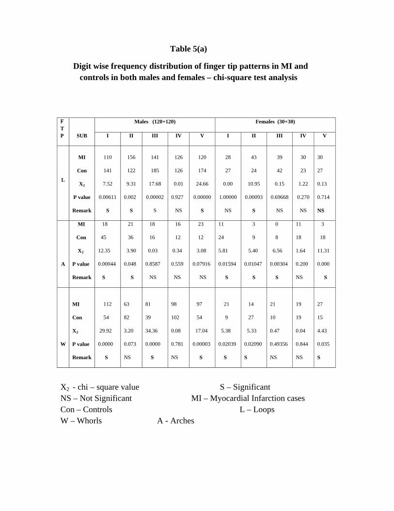
Table 5(a)
Digit wise frequency distribution of finger tip patterns in MI and controls in both males and females – chi-square test analysis
FTP SUB
Males (120+120) Females (30+30)
I II III IV V I II III IV V
L
MI
Con
X2
P value
Remark
110
141
7.52
0.00611
S
156
122
9.31
0.002
S
141
185
17.68
0.00002
S
126
126
0.01
0.927
NS
120
174
24.66
0.00000
S
28
27
0.00
1.00000
NS
43
24
10.95
0.00093
S
39
42
0.15
0.69668
NS
30
23
1.22
0.270
NS
30
27
0.13
0.714
NS
A
MI
Con
X2
P value
Remark
18
45
12.35
0.00044
S
21
36
3.90
0.048
S
18
16
0.03
0.8587
NS
16
12
0.34
0.559
NS
23
12
3.08
0.07916
NS
11
24
5.81
0.01594
S
3
9
5.40
0.01047
S
0
8
6.56
0.00304
S
11
18
1.64
0.200
NS
3
18
11.31
0.000
S
W
MI
Con
X2
P value
Remark
112
54
29.92
0.0000
S
63
82
3.20
0.073
NS
81
39
34.36
0.0000
S
98
102
0.08
0.781
NS
97
54
17.04
0.00003
S
21
9
5.38
0.02039
S
14
27
5.33
0.02090
S
21
10
0.47
0.49356
NS
19
19
0.04
0.844
NS
27
15
4.43
0.035
S
X2 - chi – square value S – Significant NS – Not Significant MI – Myocardial Infarction cases Con – Controls L – Loops W – Whorls A - Arches

Table 5(b)
Digit wise frequency distribution of finger tip patterns in MI and controls in both hands – chi-square test analysis
FTP
SUB RIGHT HAND LEFT HAND
I II III IV V I II III IV V
L MI
Con
X2
P value
Remark
65
81
3.00
0.8315
NS
101
68
13.88
0.000195
S
97
122
9.74
0.00180
S
63
86
6.45
0.01107
S
61
105
22.74
0.000
S
73
87
2.26
0.1324
NS
98
78
4.96
0.0259
S
83
105
6.28
0.0121
S
93
63
11.23
0.000804
S
89
96
0.51
0.4761
NS
A MI
Con
X2
P value
Remark
17
36
7.42
0.006
S
12
21
2.18
0.13989
NS
10
7
0.25
0.61747
NS
18
9
2.60
0.10654
NS
16
18
0.03
0.855
NS
12
33
10.46
0.0012
S
12
24
3.82
0.5066
NS
8
17
2.79
0.0946
NS
9
21
4.48
0.034264
S
10
12
0.05
0.8247
NS
W MI
Con
X2
P value
Remark
68
33
17.25
0.0000
S
37
61
8.02
0.00463
S
43
21
8.76
0.00308
S
69
55
2.32
0.12746
NS
73
27
30.08
0.00
S
65
30
17.81
0.0000
S
40
48
0.79
0.3747
NS
59
28
14.57
0.0001
S
48
66
4.09
0.043167
S
51
42
1.00
0.3179
NS
X2 - chi – square value S – Significant NS – Not Significant MI – Myocardial Infarction cases Con – Controls L – Loops W – whorls A - Arches

Table 6(a)
Statistical comparison of different finger tip pattern between MI and controls in males and females
Sex SUB
Finger Tip Patterns
Loops Arches Whorls
M
MI 653 96 451
Control 748 121 331
chi-sq 15.15 2.92 26.86
p-value 0.0000992 0.0875 0.0000002
Remark S NS S
F
MI 170 28 102
Control 143 77 80
chi-sq 4.52 26.60 3.48
p-value 0.03359 0.0000003 0.06218
Remark S S NS
M+F
MI 823 124 553
Control 891 198 411
chi-sq 6.11 18.54 30.39
p-value 0.013 0.0000166 0.00000
Remark S S S
chi – Sq = chi – Square Value S = Significant
NS = Not Significant SUB = Subject
M = Males F = Females

Table 6(b)
Statistical comparison of different finger tip pattern between MI and controls in both hands
SIDE SUB
Finger Tip Patterns
Loops Arches Whorls
Rt Hand
MI 387 73 290
Control 462 91 197
chi-sq 14.86 1.98 25.74
p-value 0.0001157 0.1595 0.0000004
Remark S NS S
Lt Hand
MI 436 53 263
Control 429 107 214
chi-sq 0.10 19.65 7.08
p-value 0.75386 0.0000093 0.0077845
Remark NS S S
chi – sq = chi – Square Value S = Significant NS = Not Significant SUB = Subject

Table 7. Frequency distribution of TFRC in MI
Class intervals
Males Females Total
No % No % No %
0-25 0 0 0 0 0 0
26-50 8 6.6 1 3.3 9 6.0
51-75 10 8.3 5 16.7 13 8.7
76-100 11 9.1 2 6.7 13 8.7
101-125 8 15.0 9 30.0 28 18.7
126-150 24 20.0 2 6.7 26 17.3
151-175 39 32.5 11 36.7 49 32.7
176-200 10 8.3 0 0 10 6.7
Total 120 100 30 100 150 100
Mean 129 - 121 - 127.5 -
Table 8. Frequency distribution of TFRC in controls
Class intervals
Males Females Total
No % No % No %
0-25 0 0 0 0 0 0
26-50 10 8.3 0 0 10 6.7
51-75 16 13.3 6 20.0 22 14.7
76-100 24 20.0 15 50.0 39 26.0
101-125 28 23.3 6 20.0 34 22.7
126-150 39 32.5 3 10.0 42 28.0
151-175 3 2.5 0 0 03 2.0
176-200 0 0 0 0 0 0
Total 120 100 30 100 150 100
Mean 104.9 - 93.5 - 102.7 -

Table 9
Frequency distribution of Total finger Ridge count (TFRC) in both MI and controls
Class intervals
MI cases Controls
No % No %
0-25 0 0 0 0
26-50 9 6.0 10 6.7
51-75 13 8.7 22 14.7
76-100 13 8.7 39 26.0
101-125 28 18.7 34 22.7
126-150 26 17.3 42 28.0
157-175 49 32.7 03 2.0
176-200 10 6.7 0 0
Total 150 100 150 100
Mean 127.5 - 102.7 -

Table 10
Statistical calculation of TFRC in total MI and controls
Sub Sex Mean SD SE-M CV
Cases
M 129.04 42.31 3.86 35.25
F 121 39.34 7.19 131.13
M+F 127.5 41.78 3.41 27.85
Controls
M 104.95 33.46 3.05 27.88
F 93.5 21.79 3.98 72.63
M+F 102.67 31.81 2.59 21.20
SD – Standard Deviation SE – M – Standard Error of Mean CV – Coefficient of Variation
Table 11
Test of Significance of TFRC for comparison between total MI cases and controls
Comparison t-value p value Remark
NM x CM 4.8923 0.0001 S
NF x CF 3.3495 0.0014 S
N(M+F) x C(M+F) 5.7912 0.0001 S
NM - Normal Male CM - Case Male CF - Case Female NF - Normal Female N(M+F) – Normal Males + Females C(M+F) - Case Male + Female S- Significant NS – Not Significant
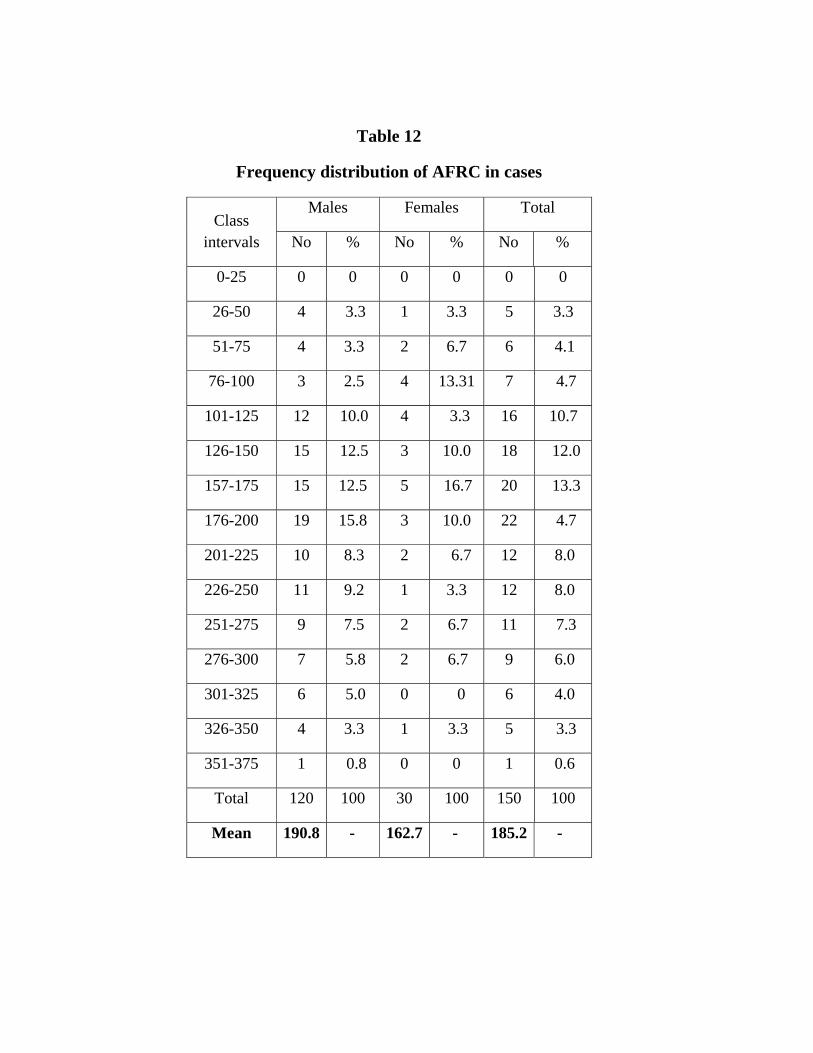
Table 12
Frequency distribution of AFRC in cases
Class intervals
Males Females Total
No % No % No %
0-25 0 0 0 0 0 0
26-50 4 3.3 1 3.3 5 3.3
51-75 4 3.3 2 6.7 6 4.1
76-100 3 2.5 4 13.31 7 4.7
101-125 12 10.0 4 3.3 16 10.7
126-150 15 12.5 3 10.0 18 12.0
157-175 15 12.5 5 16.7 20 13.3
176-200 19 15.8 3 10.0 22 4.7
201-225 10 8.3 2 6.7 12 8.0
226-250 11 9.2 1 3.3 12 8.0
251-275 9 7.5 2 6.7 11 7.3
276-300 7 5.8 2 6.7 9 6.0
301-325 6 5.0 0 0 6 4.0
326-350 4 3.3 1 3.3 5 3.3
351-375 1 0.8 0 0 1 0.6
Total 120 100 30 100 150 100
Mean 190.8 - 162.7 - 185.2 -

Table 13
Frequency distribution of AFRC in controls
Class intervals
Males Females Total
No % No % N0 %
0-25 0 0 0 0 0 0
26-50 4 3.3 0 0 4 2.7
51-75 9 7.5 3 10.0 12 8.0
76-100 7 5.8 9 30.0 16 10.7
101-125 8 6.6 13 43.3 21 14.0
126-150 10 8.3 2 6.7 12 8.0
157-175 10 8.3 3 10.0 13 8.7
176-200 17 14.2 1 3.3 18 12.0
201-225 15 12.5 1 3.3 16 10.7
226-250 22 18.4 0 0 22 14.7
251-275 12 10.0 0 0 12 8.0
276-300 6 5.0 0 0 6 4.1
Total 120 100 30 100 150 100
Mean 182.3 - 121.0 - 169.7 -

Table 14
Statistical calculation of AFRC in total MI and controls
Sub Sex Mean SD SE-M CV
MI
M 190.8 74.44 6.79 62.03
F 162.7 74.152 13.556 247.17
M+F 185.2 75.17 6.137 50.11
Controls
M 182.3 68.59 6.26 57.15
F 121.05 36.19 6.61 120.63
M+F 169.7 69.27 5.656 46.18
SD – Standard Deviation SE – M – Standard Error of Mean CV – Coefficient of Variation
Table 15
Test of Significance of AFRC for comparison between total cases and controls
Comparison t-value P value Remark
NM x CM 0.9199 0.3586 NS
NF x CF 2.764 0.0076 S
N(M+F) x C(M+F) 1.857 0.0643 NS
NM - Normal Male CM - Case Male CF - Case Female NF - Normal Female N(M+F) – Normal Males + Females C(M+F) - Case Male +Female S- Significant NS – Not Significant

Table 16. Frequency distribution of position of Axial triradii in MI cases
Sex Side t t' t'’ tt' tt’’ t’t’’ DDA
No % No % No % No % No % No % No %
M
R 9.8 81.7 16 13.3 2 1.7 3 2.5 1 0.8 0 0 24 20.0
L 79 65.8 20 16.7 5 4.1 8 6.7 7 5.8 1 0.8 33 27.5
R+L 177 13.7 36 15.0 7 2.9 11 4.5 8 3.3 1 0.4 57 23.7
F
R 16 53.3 5 16.7 3 2.5 0 0 6 20.0 0 0 22 36.7
L 22 73.3 5 16.7 0 0 0 0 3 2.5 0 0 8 26.7
R+L 38 63.3 10 16.7 3 5.0 0 0 9 15.0 0 0 22 36.7
M+F
R 114 76.0 21 14.0 5 3.3 3 2.0 7 4.7 0 0 36 24.0
L 101 67.3 25 16.7 5 3.3 8 5.3 10 6.7 1 0.6 48 32.0
R+L 215 71.7 46 15.3 10 3.3 11 3.7 17 5.7 1 0.3 84 28.0
Table 17. Frequency distribution of position of Axial triradii in total controls
Sex Side t t' t'’ tt' tt’’ t’t’’ DDA
No % No % No % No % No % No % No %
M
R 91 75.8 15 12.5 3 2.5 8 6.7 2 1.7 1 1.8 38 31.7
L 99 82.5 9 7.5 2 1.7 4 3.4 5 4.1 1 0.5 20 16.7
R+L 190 99.1 24 10.0 5 2.0 12 5.0 7 2.9 2 0.8 58 24.2
F
R 22 73.3 3 10.0 0 0 4 13.3 1 3.3 0 0 8 26.6
L 16 53.3 9 30.0 0 0 4 13.3 1 3.3 0 0 14 46.7
R+L 38 63.3 12 20.0 0 0 8 13.3 2 3.3 0 0 22 36.7
M+F
R 113 75.3 18 12.0 3 2.0 12 8.0 3 2.0 1 0.6 46 30.7
L 115 76.7 18 12.0 2 1.2 8 5.3 6 4.0 1 0.6 34 22.7
R+L 228 76.0 36 72.0 5 1.7 13 4.3 9 3.0 2 0.6 80 26.7
R – Right L – Left M – Males F – Females t, t’, t” – Different Positions of axial triradii DDA – Distal displacement of axial triradii

Table 18
Statistical comparison of position of axial triradii between MI and controls in Males and Females
Sex Sub t t' t'’ tt' tt’’ t’t’’ DDA
M
MI 177 36 7 11 8 1 57
Control 190 24 5 12 7 2 58
chi-sq 1.67 2.30 0.07 0.00 0.00 0.00 0.00
p-value 0.196700 0.128978 0.79228 1.000 1.000 1.000 1.000
Remark NS NS NS NS NS NS NS
F
MI 38 10 3 0 9 0 22
Control 38 12 0 8 2 0 22
chi-sq 0.04 0.06 1.37 6.56 3.60 0 0.04
p-value 0.849752 0.81349 0.12848 0.00304 0.05767 - 0.84975
Remark NS NS NS S NS NS NS
M
+
F
MI 215 46 10 11 17 1 84
Control 228 36 5 13 9 2 80
chi-sq 1.24 1.14 1.09 0.04 1.97 0.06 0.08
p-value 0.265037 0.284771 0.295582 0.834968 0.160449 0.50000 0.783462
Remark NS NS NS NS NS NS NS
R – Right L – Left S – Significant NS – Not Significant S = Significant NS = Not Significant t, t’, t” – Different Positions Of Axial Triradii chi – sq = chi – square Value DDA – Distal displacement of axial triradii SUB = Subject

Table 19
Statistical comparison of position of axial triradii between MI and controls in both hands
Side Subject t t' t'’ tt' tt’’ t’t’’ DDA
R
MI 114 21 5 3 7 0 36
Control 113 18 3 12 3 1 46
chi-sq 0.00 0.12 0.13 4.49 0.93 0.00 1.36
p-value 1.0000 0.731334 0.36142 0.03406 0.33459 0.5000 0.24364
Remark NS NS NS S NS NS NS
L
MI 101 25 5 8 10 1 48
Control 115 18 2 8 6 1 34
chi-sq 2.79 0.98 0.59 0.07 0.59 0.50 2.84
p-value 0.0945 0.322869 0.22397 0.79722 0.44080 0.750836 0.092161
Remark NS NS NS NS NS NS NS
R – Right L – Left S – Significant NS – Not Significant t, t’, t” – Different Positions Of Axial Triradii DDA – Distal displacement of axial triradii

Table 20
Frequency distribution of number of palmar triradii in MI
Sex Side 4 5 6 7 8
No % No % No % No % No %
M
R 15 12.5 62 51.7 37 30.8 4 3.3 2 1.7
L 11 9.2 54 45.0 43 35.8 9 7.5 3 2.5
R+L 26 10.8 116 48.3 80 33.3 13 5.4 5 2.0
F
R 4 13.0 19 63.3 6 20.0 1 3.3 0 0
L 2 6.7 23 76.7 4 13.0 1 3.3 0 0
R+L 6 10.0 42 70.0 10 16.7 2 3.3 0 0
M+F
R 19 12.7 81 54.0 43 28.7 5 3.3 2 1.3
L 13 8.7 77 51.3 47 31.3 10 6.7 3 2.0
R+L 32 10.7 158 52.7 90 30.0 15 5.0 05 1.5
M- Males F - Females
R- Right L- Left

Table 21
Frequency distribution of number of palmar triradii in controls
Sex Side 4 5 6 7 8 9
No % No % No % No % No % No %
M
R 5 4.1 59 49.1 29 24.2 15 12.5 8 6.7 4 3.3
L 3 2.5 70 58.3 40 33.3 2 1.7 4 3.3 1 0.8
R+L 8 3.3 129 53.7 69 28.7 17 7.0 12 5.0 5 2.0
F
R 1 3.3 15 30.0 8 26.7 3 10.0 3 10.0 0 0
L 1 3.3 17 56.7 6 20.0 2 6.7 1 3.3 1 3.2
R+L 2 3.3 32 53.3 14 23.3 5 8.3 4 6.7 1 1.7
M+F
R 6 4.0 74 40.3 37 24.7 18 12.0 11 7.3 4 2.7
L 4 2.7 87 58.0 46 30.7 4 2.4 5 3.3 2 1.3
R+L 10 3.3 161 53.7 8 27.7 22 7.3 16 5.3 6 2.0
M- Males F - Females
R- Right L- Left

Table 22
Statistical comparison of number of palmar triradii between MI and controls in males and females
SEX SUB 4 5 6 7 8 9
M
Cases 26 116 80 13 5 0
Control 8 129 69 17 12 5
chi-sq 9.15 1.20 0.97 0.32 2.20 3.23
p-value 0.00248 0.27321 0.32386 0.57160 0.138423 0.030599
Remark S NS NS NS NS NS
F
Cases 6 42 10 2 0 0
Control 2 32 14 5 4 1
chi-sq 1.21 2.86 0.47 0.61 2.33 0.00
p-value 0.1360 0.09106 0.49356 0.21948 0.05936 0.5000
Remark NS NS NS NS NS NS
M+F
Cases 32 158 90 15 5 0
Control 10 161 83 22 16 6
chi-sq 11.29 0.03 0.29 1.04 4.93 4.21
p-value 0.000779 0.8700 0.58868 0.3085 0.02632 0.01523
Remark S NS NS NS S S
chi – sq = chi – square Value S = Significant M = Males NS = Not Significant SUB = Subject F = Females

Table 23
Statistical comparison of number of palmar triradii between cases and controls in both hands
SIDE SUBJECT 4 5 6 7 8 9
R
Cases 19 81 43 5 2 0
Control 6 74 37 18 11 4
chi-sq 6.28 0.48 0.43 6.78 5.15 2.28
p-value 0.01218 0.4881 0.51389 0.00921 0.02329 0.06124
Remark S NS NS S S NS
L
Cases 13 77 47 10 3 0
Control 4 87 46 4 5 2
chi-sq 3.99 1.09 0.00 1.87 0.13 0.50
p-value 0.045749 0.2965 01.000 0.17111 0.72007 0.24916
Remark S NS NS NS NS NS
S = Significant NS = Not Significant R = Right L = Left chi – sq = chi – Square Value

Table 24. Frequency distribution of a-b ridge count in MI cases
ab ridge count
Male Female
R L T % R L T %
26-30 5 2 7 2.9 1 1 2 3.3
31-35 22 19 41 17.8 5 7 12 20
36-40 68 51 119 49.58 17 12 29 48.3
41-45 20 22 42 17.5 4 5 9 15
46-50 3 21 24 10 2 4 6 10
51-55 2 4 6 2.5 1 4 5 8.3
56-60 0 2 2 0.8 0 1 1 1.7
Mean 37.5 39.4 38.9 - 38.2 45.9 42.0 -
Table 25. Frequency distribution of a-b ridge count in controls
R- Right L – Left T - Total
ab ridge count
Male Female
R L T % R L T %
26-30 6 2 8 3.3 2 4 6 10
31-35 25 13 38 15.8 3 7 10 16.7
36-40 29 31 60 25.0 7 3 10 16.7
41-45 51 48 99 41.25 15 11 26 43.3
46-50 6 20 26 10.83 2 3 5 8.3
51-55 3 4 7 2.91 1 2 3 5
56-60 0 2 2 0.8 0 0 0 0
Mean 38.9 41.3 40.1 - 40.0 38.9 39.4 -

Table 26
Statistical calculation for a-b ridge count in total cases and controls
Subject Sex Side Mean SD SE-M CV
MI
M
R 37.5 4.377 0.3997 3.647
L 39.39 5.987 0.5467 4.989
R+L 38.92 5.378 0.347 2.240
F
R 38.17 4.956 0.9048 16.52
L 45.83 9.629 1.7580 32.09
R+L 42.0 7.148 0.9229 11.91
M+F
R 37.63 4.969 0.3207 3.3126
L 41.45 6.513 0.4204 4.342
R+L 39.54 5.627 0.3248 1.8756
Controls
M
R 38.95 5.415 0.4945 4.572
L 41.29 5.552 0.5070 4.626
R+L 40.125 5.595 0.3611 2.331
F
R 40.0 5.437 0.9926 18.123
L 38.83 7.1805 1.3110 23.935
R+L 39.42 6.399 0.826 10.665
M+F
R 39.166 5.436 0.3509 3.624
L 40.80 5.994 0.3869 3.996
R+L 39.98 5.781 0.3337 1.927
M- Males F – Females R- Right L- Left SD – Standard Deviation SE – M – Standard error of Mean CV – Co efficient of Variation

Table 27
Test of significance for a-b ridge count for comparison between MI and controls
Comparison t-value p value Remark
NMR x CMR 2.2813 0.0234 S
NML x CML 2.5491 0.0114 S
NM(R+L) x CM(R+L)
1.7009 0.0903 NS
NFR x CFR 1.3625 0.1783 NS
NFL x CFL 3.1913 0.0023 S
NF(R+L) x CF(R+L)
2.0831 0.0394 S
NTR x CTR 2.5543 0.0111 S
NTL x CTL 0.8994 0.3692 NS
NT(R+L) x CT(R+L)
0.9447 0.3452 NS
NMR - Normal Male Right CMR - Case Male Right NFR - Normal Female Right NTR - Normal Total Right (Total =M+F) CTR - Case Total Right (Total =M+F) NML – Normal Male Left CTL – Case Total Left (Total = M+F) CFL – Case Female Left S- Significant NS – Not Significant CFR - Case Female Right

Table 28. Frequency distribution of atd angle in MI
Table 29. Frequency distribution of atd angle in controls
atd Angle
Male Female
R L T % R L T %
26-30 2 2 4 1.6 0 0 0 0
31-35 19 17 36 15 5 4 9 15
36-40 68 57 125 52.08 11 14 25 10.4
41-45 24 28 52 21.7 10 8 18 30
46-50 4 10 14 5.8 2 3 5 8.3
51-55 3 5 8 3.3 2 1 3 5
56-60 0 1 1 0.4 0 0 0 0
Mean 38.2 39.4 38.8 - 40.0 39.7 37.8 -
atd Angle Male Female
R L T % R L T %
26-30 0 2 2 0.08 0 0 0 0
31-35 10 13 23 9.58 7 2 9 15
36-40 32 40 72 30.0 11 8 19 31.67
41-45 57 45 102 42.5 9 14 23 38.33
46-50 11 15 26 10.8 2 3 5 8.33
51-55 7 3 10 4.17 1 2 3 5
56-60 3 2 5 2.08 0 1 1 1.67
Mean 38.2 40.0 41.2 - 39.0 42.2 40.6 -

Table 30
Statistical calculation for atd angle in total MI and controls
Subject Sex Side Mean SD SE-M CV
MI
M
R 38.25 6.386 0.5831 5.321
L 40.08 5.456 0.498 4.546
R+L 41.19 5.431 0.3505 2.262
F
R 39.0 5.530 1.0096 18.433
L 42.17 5.467 0.9981 18.223
R+L 40.58 5.552 0.7168 9.253
M+F
R 42.37 5.500 0.355 3.666
L 40.93 5.420 0.349 3.613
R+L 41.06 5.432 0.313 1.810
Controls
M
R 38.24 4.361 0.398 3.634
L 39.40 4.949 0.4579 4.124
R+L 38.83 5.457 0.3522 2.273
F
R 40.0 5.283 0.9645 19.41
L 39.67 4.775 0.8718 15.916
R+L 39.83 5.039 0.6506 8.398
M+F
R 38.23 4.631 0.2989 3.087
L 39.47 5.224 0.3372 3.482
R+L 38.84 4.969 0.2868 1.656
M- Males F - Females R- Right L- Left SD – Standard deviation SE- M – Standard error of Mean CV – Coefficient of Variation.

Table 31
Test of Significance for atd angle for comparison between MI and controls
Comparison t-value p value Remark
NMR x CMR 0.0142 0.9887 NS
NML x CML 1.0112 0.3129 NS
NM(R+L) x CM(R+L) 4.7488 0.0001 S
NFR x CFR 0.7162 0.4768 NS
NFL x CFL 1.8664 0.0642 NS
NF(R+L) x CF(R+L) 0.7748 0.4400 NS
NTR x CTR 7.0521 0.0001 S
NTL x CTL 2.3754 0.0182 S
NT(R+L) x CT(R+L) 5.2230 0.00001 S
NMR - Normal Male Right CFR - Case Female Right NFR - Normal Female Right NTR - Normal Total Right (Total =M+F) CTR - Case Total Right (Total =M+F) NML – Normal Male Left CTL – Case Total Left (Total = M+F) CFL – Case Female Left S- Significant NS – Not Significant CMR - Case Male Right