a study on acid – base status among intensive medical …
TRANSCRIPT

DISSERTATION ON
A STUDY ON
ACID – BASE STATUS AMONG INTENSIVE
MEDICAL CARE UNIT PATIENTS
Submitted in partial fulfillment of
Requirements for
M.D.DEGREE BRANCH I INTERNAL MEDICINE
Of
THE TAMILNADU DR.M.G.R. MEDICAL UNIVERSITY
CHENNAI
MADRAS MEDICAL COLLEGE
CHENNAI – 600 003
MARCH – 2008
brought to you by COREView metadata, citation and similar papers at core.ac.uk
provided by ePrints@TNMGRM (Tamil Nadu Dr. M.G.R. Medical University)

CERTIFICATE
This is to certify that this dissertation entitled “ A STUDY ON
ACID BASE STATUS AMONG INTENSIVE MEDICAL
CARE UNIT PATIENTS ” submitted by Dr. SASIREKHA K
appearing for Part I & II M.D Branch I Internal Medicine Degree
examination in March 2008 is a bonafide record of work done by her under
by direct guidance and supervision in partial fulfillment of regulations of the
TamilNadu Dr.M.G.R. Medical University, Chennai. I forward this to the
TamilNadu Dr.M.G.R. Medical University, Chennai, TamilNadu, India.
Additional Professor of Medicine
Institute of Internal Medicine
Madras Medical College
Government General Hospital
Chennai – 600 003
Director Dean
Institute of Internal Medicine Madras Medical College
Government General Hospital Government General Hospital
Chennai – 600 003 Chennai – 600 003

DECLARATION
I solemnly declare that dissertation entitled “ A STUDY ON
ACID BASE STATUS AMONG INTENSIVE MEDICAL
CARE UNIT PATIENTS ” is done by me at Madras Medical
College & Government General Hospital, Chennai, during 2006 –
2007 under the guidance and supervision of Prof.V.K.Rajamani,
M.D.
The dissertation is submitted to The TamilNadu Dr.M.G.R.
Medical University towards the partial fulfillment of requirements
for the award of M.D. Degree (Branch I) in Internal Medicine.
Place : Chennai Dr. SASIREKHA K.
Date : Postgraduate Student
M.D. Internal Medicine
Institute of Internal Medicine
Madras Medical College
Chennai

ACKNOWLEDGEMENT
I owe my thanks to the Dean, Madras Medical College and Govt.
General Hospital, Prof. Dr. T.P.KALANIDHI M.D. for allowing
me to avail the facilities needed for my dissertation work.
I am grateful to Prof. Dr. P. THIRUMALAI KOLUNDU
SUBRAMANIAN M.D. , Professor and Head of Department of
Medicine, Madras Medical College for permitting me to do the study
and for his constant encouragement.
I am extremely thankful to my unit chief Prof.Dr.
V.K.RAJAMANI M.D for his guidance and encouragement.
I sincerely thank Prof. Dr. C. RAJENDRAN M.D. , Chief
IMCU & Toxicology units for his valuable guidance.

I am thankful to Prof.Dr. A. MANAMALLI M.D. , Head of
the Department, Department of BioChemistry for allowing me to
utilize the services of her department for the purpose of my study
I sincerely thank Assistant Professors for the cooperation and
guidance.
I am thankful to all my Postgraduate colleagues for their
constant support and sharp constructive criticism.
I should thank each and every patient for their whole-hearted
cooperation despite the morbidity they suffered.
I should thank each and every member of my family for their
constant support and encouragement.

CONTENTS
SL.NO. TITLE PAGE NO.
1. INTRODUCTION 1
2. AIMS & OBJECTIVES 5
3. REVIEW OF LITERATURE 6
4. MATERIALS AND METHODS 21
5. OBSERVATIONS AND RESULTS 26
6. DISCUSSION 50
7. CONCLUSION 62
8. BIBLIOGRAPHY 65
9. PROFORMA 70
10. MASTER CHART 71

1
Introduction

2
INTRODUCTION
A little learning is a dangerous thing.
Drink deep, or taste not the pyrean spring
• Alexander Pope
Acid Base abnormalities are common in critically ill patients. Our ability to
describe acid base disorders must be precise. Small differences in
corrections for anion gap, different types of analytical processes, and the
basic approach used to diagnose acid base aberrations can lead to markedly
different interpretation and treatment strategies for the same disorder (1).
Life is a struggle;
Not against sin;
Not against money power;
Not against malicious animal magnetism;
But against [H+] ions
• H.L. Mencken
Mencken was neither a physiologist nor a physician but he knew the
importance of [H+] ions.
To maintain homeostasis the body has to keep [H+] ions concentration
at 40 nanomoles/L (2). A blood pH less than normal (normal range 7.35 –
7.45) is called acidemia; the underlying process causing acidemia is called

3
acidosis. Similarly alkalemia and alkalosis refer to the pH and underlying
process respectively. While an acidosis and an alkalosis may coexist, there
can be only one resulting pH. Therefore acidemia and alkalemia are
mutually exclusive conditions (3).
The metabolic and respiratory that regulate systemic pH are described
by the Henderson – Hasselbach equation;
pH = 6.1 + log (HCO3 / PaCO2 x 0.03)
Alternatively H+ ions can be expressed directly as (4).
H+ = 24 x (PCO2 / HCO3-)
Primary change in PaCO2 can cause acidosis or alkalosis, depending
on whether PaCo2 is above or below the normal value of 40 mm Hg. Primary
alkalosis of PCo2 evokes cellular buffering and renal adaptation. A primary
change in the plasma HCO3 – as a result of metabolic or renal factors results
in compensatory changes in ventilation that blunt the changes in blood pH.
Such respiratory alterations are referred as secondary or compensatory
changes, since they are occurring in response to primary metabolic changes.
Simple and mixed acid base disorders are commonly encountered in
clinical practice and are particularly frequent in critically ill patients (5).
Acid base disorders contribute importantly to patient morbidity and
mortality, especially in critically ill (6). Therefore it is essential to recognize

4
and properly diagnose acid base disorders and understand their impact on
organ function.

5
AIMS AND OBJECTIVES
� To analyze the acid base status among intensive care unit patients.
� To elicit various acid base disturbance in IMCU and toxicology units.
� To study the pattern of acid base disturbances.
� To study the effect of pH on the prognosis of the diseases.
� To elicit the causes for acid base disturbances.

6
REVIEW OF LITERATURE
Primary respiratory acid base disturbances invoke secondary metabolic
response and primary metabolic acid base disturbance invoke secondary
respiratory response (7).
SIMPLE ACID BASE DISORDERS
Metabolic Acidosis
Metabolic Alkalosis
Respiratory Acidosis
Respiratory Alkalosis
Primary Changes HCo3- HCo3
- PCo2- PCo2
-
Compensation PCo2- PCo2
- HCo3- HCo3
-
Effect on pH pH pH pH pH
Since the definition of simple disturbances includes both the initial
process producing a change in HCo3- and PCo2
- , all the compensatory
mechanisms affecting these substances, lack of appropriate compensation for
a simple disturbance is an evidence for a mixed disturbance (8).
So it is critical to know both magnitude and the course of
compensatory responses to simple disorders to identify the presence of
mixed acid base disorders.

7
COMPENSATORY RESPONSE IN SIMPLE ACID BASE DISTURBANCES Acid base disorders
Prediction of Compensation
Limits
Metabolic acidosis
PCo2 = 1.5 x HCo3
- + 8 Or PCo2 will decrease by 1.25 mm Hg per mmol/L decrease in HCo3
-
10 mm Hg
Metabolic alkalosis
PCo2 = 0.9 x HCo3
- + 16 Or PCo2 will increase by 0.75 mm Hg per mmol/L increase in HCo3
-
55 mm Hg
Acute
HCo3
- will decrease by 2 mmol/L per 10 mm Hg decrease in PCo2
30 mEq/L
Respiratory alkalosis
Chronic
HCo3
- will decrease by 4 mmol/L per 10 mm Hg decrease in PCo2
45 mEq/L
Acute
HCo3
- will increase by 1 mmol/L per 10 mm Hg increase in PCo2
18 mEq/L
Respiratory acidosis
Chronic
HCo3
- will increase by 4 mmol/L per 10 mm Hg increase in PCo2
15 mEq/L
A mixed acid base disturbance is defined as the simultaneous coexistence of
two or more simple disorder in a same patient (9). Mixed acid base
disturbances are more commonly predictable on the basis of clinical setting

8
and physical examination. Lab data mainly serve to confirm the clinical
impression.
McCurdy et al (1981) suggested a systematic method to analyze acid
base disorders by clinical approach (10).
I. Suspect the disturbances from history
II. Suspect the disturbances from physical examination
III. Evaluate routine lab data
1. HCo3-
a) If increased think of metabolic alkalosis or compensated
respiratory acidosis.
b) If decreased think of metabolic acidosis or compensated
respiratory alkalosis.
2. K+
a) If increased think of acidemia.
b) If decreased think of alkalemia.
3. Cl-
c) If increased think of hyperchloremic metabolic acidosis
d) If decreased think of metabolic alkalosis.
4. Anion Gap.
IV. Evaluate blood gas values to check appropriate compensation.

9
In the setting of known primary disorders the presence of normal pH
implies a mixed disturbance, since compensation rarely corrects pH to
normal. Generally more marked the primary disturbance, the less likely the
pH will be normal, unless there is a mixed disorder.
Important Causes of mixed acid base disturbances :
1. Respiratory acidosis with metabolic acidosis
Ex : Cardio pulmonary arrest
Severe Pulmonary edema
2. Respiratory alkalosis with metabolic alkalosis
Ex : Hepatic failure treated with diuretics
3. Respiratory alkalosis with metabolic acidosis
Ex : Septic shock
4. Respiratory acidosis with metabolic alkalosis
Ex : Corpulmonale treated with diuretics
5. Metabolic acidosis with metabolic alkalosis with respiratory alkalosis
Ex : Diabetic keto acidosis with vomiting
6. Metabolic acidosis with metabolic alkalosis with respiratory acidosis

10
1) RESPIRATORY ACIDOSIS + METABOLIC ACIDOSIS :
This combination occurs in a variety of clinical situations including
cardio pulmonary arrest, severe pulmonary edema, drug ingestion with
severe central nervous system depression and hypo ventilation, metabolic
acidosis with potassium depletion producing paralysis of the respiratory
muscles (11).
In these cases, serum HCo3- is usually low, PCo2 is normal or elevated
and the resultant pH is usually low. The CO2 retention prevents respiratory
compensation for the metabolic acidosis and metabolic process prevents
compensation for respiratory acidosis. Significant acidemia can seriously
impair cardiac function and leads to cardio vascular collapse (12).
Specific therapy must be initiated aggressively to correct the acidemia
by simultaneously treating the metabolic acidosis with bicarbonate and the
respiratory acidosis with measures to improve ventilation in this setting,
hyperkalemia is a serious problem.
2) RESPIRATORY ALKALOSIS + METABOLIC ALKALOSIS :
This mixed disorder is commonly present in patients with hepatic
failure, who are placed on diuretics or nasogastric suction. It is also
commonly in critically ill patients who require ventilator support and or

11
given diuretics or nasogastric suction. In this cases, serum HCo3- is usually
elevated. PCo2 is normal or low and pH is extreme alkalemia, adversely
affect both cerebral and peripheral haemodynamics (13).
Here metabolic process prevents compensation for respiratory
alkalosis and hyper ventilation prevents compensation for metabolic
alkalosis. In order to return the pH toward normal, therapy theoretically
should again be directed at alleviating both disorders simultaneously.
Treatment of metabolic alkalosis with volume, chloride and potassium
replacement should be initiated.
3) RESPIRATORY ALKALOSIS + METABOLIC ACIDOSIS
Clinical settings in which this combination can be found include
septic shock, pulmonary embolism, and renal failure with sepsis and
salicylate ingestion. The metabolic acidosis in these cases is frequently of
the high anion gap variety (14).
Serum HCo3- is markedly diminished, PCo2 is also low. pH may be
normal or mildly deviated depending upon individual disturbances.
Examining the respiratory compensation for the metabolic acidosis is critical
in this situation, since the respiratory alkalosis will be missed until it is
realized that ventilation is greater than predicted value. Specific treatment

12
aimed at correcting the pH is not necessary. In fact bicarbonate therapy is
contraindicated in severe respiratory alkalosis. Therefore the main value in
recognizing this disturbance is more for its diagnostic potential rather than
treatment purpose.
4) RESPIRATORY ACIDOSIS + METABOLIC ALKALOSIS
It is present most commonly in patients with chronic lung disease and
CO2 retention. In these patients metabolic alkalosis arises because of
vomiting or treatment with diuretics under low salt diet (15).
Serum HCo3- is raised, PCo2 is also raised, pH may be normal. So it is
important to recognize and treat primary metabolic alkalosis with volume,
chloride and potassium replacement. Since the elevated bicarbonate may
itself depress respiration (16).
pH AND SURVIVAL :
Severe acidemia is defined as pH less than 7.20 and severe alkalemia
as pH more than 7.60. Adverse consequences can occur independent of
whether the acidemia is of metabolic, respiratory or mixed origin (17).
13
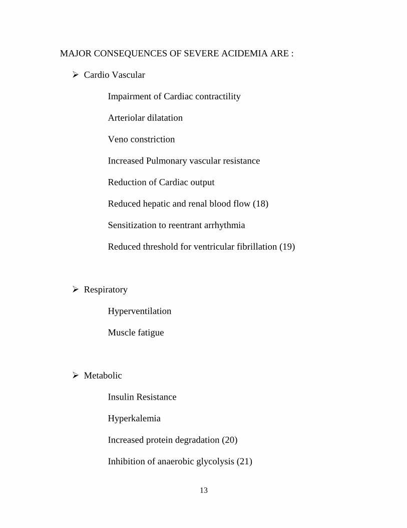
MAJOR CONSEQUENCES OF SEVERE ACIDEMIA ARE :
� Cardio Vascular
Impairment of Cardiac contractility
Arteriolar dilatation
Veno constriction
Increased Pulmonary vascular resistance
Reduction of Cardiac output
Reduced hepatic and renal blood flow (18)
Sensitization to reentrant arrhythmia
Reduced threshold for ventricular fibrillation (19)
� Respiratory
Hyperventilation
Muscle fatigue
� Metabolic
Insulin Resistance
Hyperkalemia
Increased protein degradation (20)
Inhibition of anaerobic glycolysis (21)

14
� Cerebral
Inhibition of metabolism and cell volume regulation
Obtundation and coma
ADVERSE CONSEQUENCES OF SEVERE ALKALEMIA
� Cardio Vascular
Arteriolar constriction (22)
Reduction in coronary blood flow
Reduced anginal threshold
Predisposes to SVT/VT
� Respiratory
Hypoventilation
Hypercapnia and hypoxia.
� Metabolic
Stimulation of anaerobic glycolysis
Hypokalemia (23)
Reduced plasma ionized calcium
� Cerebral
Reduced blood flow
Tetany, Seizure, Lethargy, stupor

15
RESPIRATORY FAILURE:
It is a condition in which respiratory system fails in one or both of its
function namely, gas exchange, oxygen delivery and carbon di oxide
elimination. Respiratory failure may be acute or chronic, the clinical
presentation of patients with acute and chronic respiratory failure are quite
different. While acute respiratory failure is characterized by life threatening
derangements in arterial blood gases and acid base status, chronic failure are
more indolent and clinically in apparent (24).
In the healthy adult at C level (760 mm atm. Pressure), breathing
room air (FiO2 0.21), the normal PaO2 is stated to be 97 mm Hg
(25).Hypoxemia is defined as an arterial PO2 of less than 80 mm Hg,
hypercapnia when arterial PCO2 more than 45 mm Hg (26)
ADULT VALUES FOR PaO2 AND SaO2
PaO2 SaO2
Normal 97 97
Normal Range >= 80 >= 95
Hypoxia < 80 < 95
Mild 60 – 79 90 – 94
Moderate 40 – 59 75 – 89
Severe < 40 < 75

16
Hypoxia and Hypercapnia stimulate chemoreceptors in arterial circulation
(peripheral receptor) and ventro lateral medulla ( central ), which increases
the motor activity of the respiratory skeletal muscle of chest wall and upper
airways.
Under isocapnic conditions ventilation increases in curvilinear fashion
as PO2 falls (27). However hypoxic response depends on prevailing level of
PCO2 . Hypercapnia increase the hypoxic response by shifting the PO2
threshold to higher level (28). In contrast to hypoxic response, the response
to hypercapnia under isotoxic condition is on linear fashion (29).
Hypoxic and hypercapnic stimulous acts multiplicatively to enhance
the motor activity of the respiratory muscle. Chemo sensitivity to hypoxia
and hypercapnia are heredito-familial and vary individually (30). The chemo
sensitivity response decreases with age, which explains predilection of
respiratory failure in elderly.
Small changes in PO2 (5 – 15 mm Hg) occurring gradually over days
or week leaving pH at 7.25 – 7.30 are well tolerated but rapid PO2 changes
and pH less than 7.25 are life threatening and conveys the needs for
ventilatory support.
17
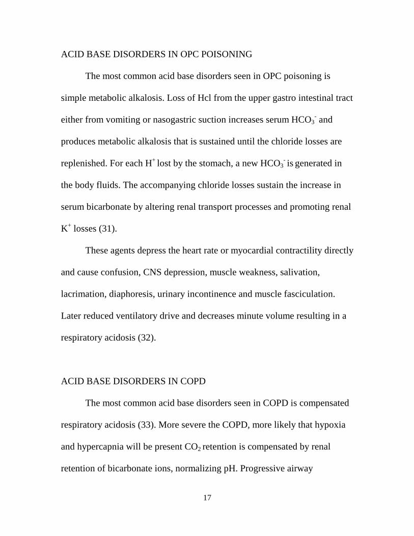
ACID BASE DISORDERS IN OPC POISONING
The most common acid base disorders seen in OPC poisoning is
simple metabolic alkalosis. Loss of Hcl from the upper gastro intestinal tract
either from vomiting or nasogastric suction increases serum HCO3- and
produces metabolic alkalosis that is sustained until the chloride losses are
replenished. For each H+ lost by the stomach, a new HCO3- is generated in
the body fluids. The accompanying chloride losses sustain the increase in
serum bicarbonate by altering renal transport processes and promoting renal
K+ losses (31).
These agents depress the heart rate or myocardial contractility directly
and cause confusion, CNS depression, muscle weakness, salivation,
lacrimation, diaphoresis, urinary incontinence and muscle fasciculation.
Later reduced ventilatory drive and decreases minute volume resulting in a
respiratory acidosis (32).
ACID BASE DISORDERS IN COPD
The most common acid base disorders seen in COPD is compensated
respiratory acidosis (33). More severe the COPD, more likely that hypoxia
and hypercapnia will be present CO2 retention is compensated by renal
retention of bicarbonate ions, normalizing pH. Progressive airway

18
obstruction leads to compensatory metabolic alkalosis (34). The presence of
elevated bicarbonate level in a patient with hypercapnic failure indicates
indulging chronic hypercapnia.
Respiratory alkalosis is the most common acid base disorders in acute
asthma (35). Respiratory alkalosis causes broncho constriction, reduced
cerebral blood flow and neuro excitatory symptoms. Acute respiratory
acidosis can give rise to CO2 narcosis.
ACID BASE DISORDERS IN DIABETIC KETO ACIDOSIS
During the development of ketosis in a diabetic individual, the keto
acids released into the extra cellular fluid are titrated by bicarbonate ions.
This buffering results in increased plasma unmeasured anions and cause
high anion gap acidosis (36). In DKA each increase in anion gap from
retained keto acid should be identical to decrease in plasma HCO3- . Thus in
uncomplicated DKA, the increase in anion gap above its normal value
should be equal to decrease in HCO3- (37). Plasma HCO3
- must be reduced
in DKA, unless it is complicated by coexisting respiratory acidosis or
metabolic alkalosis. Acidemia is the rule in DKA, unless coexisting
metabolic alkalosis. As the renal threshold of plasma keto acids is low, its
products can reach 1000 – 2000 meq per day. Urinary loss of keto acids may

19
be enormous which is associated with sodium and potassium excretion
which is replenished by chloride ions absorption causing net effect of hyper
chloremic acidosis.
ACIDBASE DISORDERS IN SEPSIS
American college of chest physician and critical care medicine
formulated a working definition of sepsis as a clinical evidence of infection
with temperature of > 38o c or < 36o C, respiratory rate > 20 / mt, heart rate
> 90 / mt, WBC > 12000 or < 4000 with > 10 % immature band forms (38).
Severe sepsis is defined if sepsis associated with organ dysfunction like
hypotension, hypoxia, oliguria, confusion, metabolic acidosis and DIC. The
hallmark of sepsis is wide spread peripheral vaso dilatation due to nitric
oxide production in response to cytokines, causing loss of homeostatic
regulation of tissue blood flow (39). Thus much of circulating blood volume
is shunted through capillary beds bypassing deep tissue and reducing the
opportunity for O2 extraction. This will in turn exacerbate tissue hypoxia
and cause metabolic acidosis. Lung is the most vulnerable organ in sepsis.
TNF alpha, platelet aggregating factor, IL-8 play prominent role in
development of ARDS. Neutrophils degranulation causing loss of
endothelial integrity and accumulation of fluid producing impaired gas

20
exchange and hypoxia. Typically respiratory alkalosis occurs yearly and
metabolic acidosis late. The degree of acidosis is a marker of severity of
illness. The onset of hypoxia indicates the severe disease and high risk for
ARDS.
ACID BASE DISORDERS IN CHRONIC RENAL FAILURE
An individual ingeting a normal diet produces about 1 meq of H+ ions
/ kg body weight (40). Kidney is responsible for excretion of these metabolic
H+ ions. A normal kidney excretes 60% H+ ions as ammonium and
remaining 40% as titrable acid. As GFR falls, metabolic H+ ions balance is
maintained for as long as residual nephrons are able to increase H+ ions
excretion is proportional to fall in GFR. As GFR falls less than 30 ml / min,
decrease in ammonium excretion causes metabolic acidosis. Both
hyperchloremic metabolic acidosis and anion gap acidosis can complicate
renal failure. Hyperchloremic metabolic acidosis can occur only if positive
H+ ions balance develops, before GFR has fallen sufficiently.
Here fall in serum HCO3- is matched by increased in chloride ion
concentration. Individual with interstitial renal disease are particularly likely
to develop this form of metabolic acidosis. In more advanced renal failure,
organic acids are retained and an anion gap metabolic acidosis supervenes.

21
MATERIALS AND METHODS
This study is descriptive study conducted in 100 patients admitted in
the intensive care and toxicology units, Government General Hospital,
Chennai.
The study was conducted between September 2006 – August 2007 for
a period of one year.
INCLUSION CRITERIA
Acid base abnormalities is evaluated by using a five step approach.
Step 1: Validity is checked by using formula H+ = 24 x PCO2 / HCO3-
Step 2: Minimum diagnosis is obtained using pH.
Step 3: To find out simple or mixed acid base disorder.
Step 4: To calculate anion gap ( AG = Na+ - (HCO3- + Cl-) ).
Step 5: To Identify triple acid base disorder.
If a primary acidosis or alkalosis is present, the expected degree of
compensation can be predicted using following equations.

22
SIMPLE ACID BASE DISORDER :
Metabolic Acidosis :
Expected PCO2 = 1.5 x (HCO3- + 8 + 2)
Metabolic Alkalosis :
Expected PCO2 = 0.9 x (HCO3- + 16 + 2)
If measured PCO2 is less than expected PCO2 then respiratory alkalosis is
present. If measured PCO2 is greater than expected PCO2 then respiratory
acidosis is present.
Respiratory Acidosis :
Plasma HCO3 will increase by 1 meq / L for each 10 mm Hg increase PCO2
in acute cases and 4 meq / L in chronic cases.
Respiratory Alkalosis :
Plasma HCO3 will increase by 2 meq / L for each 10 mm Hg decrease PCO2
in acute cases and 4 meq / L in chronic cases.

23
MIXED ACID BASE DISORDERS
Lack of appropriate compensation for a single acid base disturbance
suggests mixed acid base disorder.
EXCLUSION CRITERIA
1. All Surgical patients
2. All obstetrics and Gynaecological patients
METHODS:
OBTAINING A ARTERIAL BLOOD SAMPLE :
Based on safety, accessibility and patients comfort, site for obtaining
arterial blood samples is chosen. Radial, femoral and brachial arteries are the
ones from which blood samples most commonly taken. The radial artery is
used most often, because it is superficially located and well supported by
collateral circulation by ulnar artery. If radial artery is inaccessible, brachial
or femoral artery is punctured.
The syringe is adequately heparinised to prevent the sample clotting.
About 0.25 ml of heparin is drawn up in to the syringe. The plunger is
withdrawn to allow the heparin to coat the wall of the syringe and then the
heparin is completely expelled.

24
EQUIPMENTS REQUIRED :
1. Skin preparation fluid – alcohol or iodine
2. Syringe of size 21 G – 2 ml containing 0.5 % plain lignocaine.
3. A needle size 23 G is attached to the heparinised syringe.
4. A cap to seal the syringe.
5. Ice packs if transferred to the lab take more than 5 minutes.
PROCEDURE :
After explaining to the patient and obtaining their consent, pulse in the
desired area is identified. At the maximum point of pulsation the lignocaine
is infiltrated subcutaneously.
Using 23 G needle the heparinised syringe is inserted at an angle of 20
to 30 degrees towards radial artery. About 2 ml of blood is aspirated and the
syringe is sealed with cap without any air bubbles. Tight pressure is applied
over the punctured side for two minutes.
25
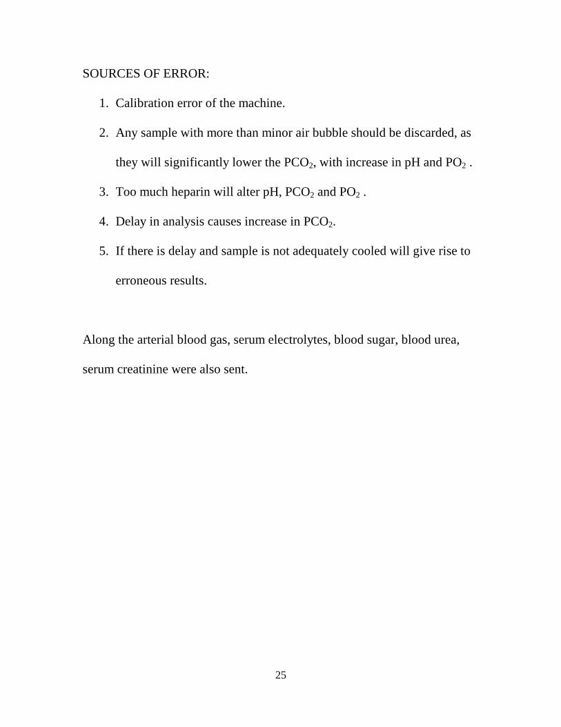
SOURCES OF ERROR:
1. Calibration error of the machine.
2. Any sample with more than minor air bubble should be discarded, as
they will significantly lower the PCO2, with increase in pH and PO2 .
3. Too much heparin will alter pH, PCO2 and PO2 .
4. Delay in analysis causes increase in PCO2.
5. If there is delay and sample is not adequately cooled will give rise to
erroneous results.
Along the arterial blood gas, serum electrolytes, blood sugar, blood urea,
serum creatinine were also sent.

26
OBSERVATIONS AND RESULTS
Table – 1
CATEGORY OF CASES INCLUDED FOR THE STUDY & PREVALENCE OF ACIDBASE DISORDERS
Sex Distribution in the study group
100 patients were taken up to study 63 patients were male 37 patients were female Mean age of all patients was 39.89

27
Sl no SUB CATEGORY OF CASES NUMBER PERCENTAGE 1 2 3 4 5
Toxicology : Organo phosphorus compound poisoning Alcoholic intoxication Infection : Sepsis Diseases of metabolic derangements Diabetic Keto acidosis Chromic Kidney disease Chromic obstractive pulmonary disease Decompensated Liver disease BITES AND STINGS Scorpion sting Snake bite Miscellaneous
17 6
13
10 10 9 4 6 5
20
17% 6%
13%
10% 10% 9% 4%
6% 5%
20%
Total Cases
100

28
Table 2
ACID BASE DISORDER IDENTIFIED
Total 100
Simple 40
Mixed 60
Mixed acid base disorder accounts for 60%
Table 3
SIMPLE ACID BASE DISORDER
TOTAL 40
Metabolic acidosis 19
Metabolic alkalosis 14
Respiratory alkalosis 4
Respiratory acidosis 3
Metabolic acidosis is the most common simple acid base disorder.
Table 4
MIXED ACID BASE DISORDER
Metabolic alkalosis + Respiratory alkalosis 22
Metabolic acidosis + Respiratory acidosis 19
Metabolic alkalosis + Respiratory acidosis 9
Metabolic acidosis + Respiratory alkalosis 6
Metabolic acidosis + Met.alkalosis + Resp.alkalosis 3
Met.acidosis + Met.alkalosis + Resp.acidosis 1
Total 60
The most common mixed acid base disorder is combination of
Metabolic alkalosis + Respiratory alkalosis

29
Table 5 Acid base disorder
Opc
poi
soni
ng
Sep
sis
DK
A
CK
D
CO
PD
Alc
holic
in
toxi
catio
n
Sco
rpio
n S
ting
Sna
ke B
ite
DC
LD
Mis
cella
niou
s
Tot
al
Simple Met. Acidosis
3 4 4 3 2 1 2 19
Simple Met. Alkalosis
6 3 5 14
Simple resp. Acidosis
2 1 3
Simple resp. Alkalosis
1 2 1 4
M. alkalosis + R.alkalosis
3 5 2 2 2 3 5 22
M. acidosis + R. acidosis
3 1 1 4 4 2 4 19
M.alkalosis + R. acidosis
2 3 1 1 2 9
M. acidosis + R. alkalosis
2 3 1 6
M. acd + M.alk + resp.alkalaosis
3 3
M. acd + M.alk + resp.acidosis
1 1
Total
17 13 10 10 9 6 6 5 4 20 100
Expired
3 8 - 7 4 - 2 - 2 6 32
Organo phosphorus compound poisoning is the most common acid base disturbance in our study followed sepsis , DKA and CKD.

30
Table 6
PREVALENCE OF INDIVIDVAL ACID BASE DISTURBANCES :
METABOLIC ACIDOSIS
Simple metabolic acidosis
Mixed Metabolic Acidosis Total
+ Res. Alkalosis
+ Res. Alkalosis
+ Met. alkalosis
19
19 6 4
48
48% of patients had metabolic acidosis either in simple or mixed form
Table 7
RESPIRATORY ALKALOSIS
35% of patients had respiratory alkalosis either in simple or mixed form.
Table 8
METBOLIC ALKALOSIS
Simple Metabolic Alkalosis
Mixed Metabolic Alkalosis Total
+ Res. acidosis +Res. alkalosis +Met. Acidosis 14 9 22 4
49
49% of patients had metabolic either in simple or mixed form
Simple Respiratory Alkalosis
Mixed Respiratory Alkalosis Total
+ met. acidosis + met. alkalosis + met. acidosis + met. alkalosis
4
6 22 3
35

31
Table 9
RESPIRATORY ACIDOSIS Simple respiratory acidosis
Mixed respiratory acidosis Total
+ Met . alkalosis
+ Met . acidosis + Met. Alkalosis + Met. Acidosis
3 9
19
1
32
Respiratory acidosis is the least common disturbance accounting for 32 %
Table 10
pH AND SURVIVAL:
7.2 - 7. 6 pH < 7.2 7.2 – 7.35 7.35 – 7.45 7.45 – 7.6
> 7.6 Total
Total
13
27
22
37
1
100
Expired
10
7
3
11
1
32
%
76
25
13
29
100
32
The mortality rate is higher in extreme acidemia and alkalemia

32
Table 11 ACID BASE DISORDER AND RESPIRATORY FAILURE
ABG
Respiratory Failure
Yes No 42% 58% Type 1 Type II 14% 28% Total Cases
Simple 9 8 23 40 Mixed 5 20 35 60
COPD Sepsis OPC Poisoning CKD DKA Snake Bite Scorpion Sting Alcoholic Intoxication DCLD Miscellaneous
- 4 2 1 - 1 1 - 1 4
9 2 7 - - 2 2 1 1 4
- 7 8 9
10 2 3 5 2
12
9
13
17
10
10 5 6 6 4
20
14 28 58 100

33
Table 12
ACID BASE DISORDER AND COPD TOTAL
9
Expired
4
Respiratory Failure
9
Type 1 - Type 2 9 < 7 . 2 2 7.2 - 7.6 7
PH
> 7 . 6 0 Simple 2
Met . acidosis 0 Met . alkalosis 0 Res . acidosis 2
Res . alkalosis 0 Mixed 7
Met . alkalosis + Res. Alkalosis 0 Met . acidosis + Res. acidosis 4 Met. alkalosis + Res . acidosis 3 Met.acidosis + Res. Alkalosis 0 Met.acid + M.alk + Res.alkalosis 0
M.acd + M.alk+Res.acidosis 0 All patients with COPD had Respiratory Failure (Type 2)

34
Table 13
ACID BASE DISORDERS IN COPD
No Pt Acid Base disturbance PH Resp . failure
Survial
1 1 Simple Resp. acidosis 7.42 Type II - 2 26 Mixed Resp+ Met. acidosis 7.22 Type II - 3 27 Mixed Resp+ Met. acidosis 7.18 Type II Expired 4 52 Mixed Resp+ Met. acidosis 7.05 Type II Expired 5 53 Mixed Resp acidosis + M.alkalosis 7.44 Type II - 6 67 Mixed Resp acid + Met.alkalosis 7.34 Type II - 7 77 Mixed Resp acid + Met.alkalosis 7.51 Type II Expired 8 78 Mixed Res+ Met acidosis 7.20 Type II Expired 9 89 Simple Resp.acidosis 7.31 Type II -
Out of 9 patients, 4 had expired accounting for a mortality of 44%

35
Table – 14
SEPSIS AND ACID BASE DISTURBANCE TOTAL
13
Expired
8
Respiratory Failure
6
Type 1 4 Type 2 2 < 7 . 2 0 7.2 - 7.6 12
PH
> 7 . 6 1 Simple 8
Met . acidosis 4 Met . alkalosis 3 Res . acidosis 0
Res . alkalosis 1 Mixed 5
Met . alkalosis + Res. Alkalosis 5 Met . acidosis + Res. Acidosis 0 Met. alkalosis + Res . acidosis 0 Met.acidosis + Res. Alkalosis 0 Met.acid + M.alkalosis + Res.alkalosis 0
M.acid + M.alkalosis+Res.acidosis 0 6 patients had Respiratory failure ( Type 1 – 4 , Type 2 -2)

36
Table – 15
ACID BASE DISORDERS IN SEPSIS S.No Pt Acid Base Disorder pH Respiratory
Failure Survival
1 2 Simple Respiratory Alkalosis 7.54 Type 1 Expired 2 28 Mixed Met + Resp Alkalosis 7.58 - Expired 3 29 Simple Met. acidosis 7.31 - 4 54 Simple met . acidosis 7.32 Type 1 5 68 Mixed met + Res Alkalosis 7.48 - 6 69 Simple metabolic alkalosis 7.43 Type 2 7 79 Mixed met + Resp alkalosis 7.62 Type 1 Expired 8 80 Mixed met + Res alkalosis 7.52 - Expired 9 81 Simple met. alkalosis 7.46 Type 2 Expired 10 90 Simple met. acidosis 7.32 - Expired 11 91 Mixed met. + resp. alkalosis 7.51 Type 1 12 93 Simple met alkalosis 7.54 - Expired 13 97 Simple met acidosis 7.38 - Expired 8 Expired out of 13 patients with mortality rate of 61%.
Table – 16 ACID BASE DISORDERS IN SEPSIS
S.No Pt Acid Base Disorder pH Respiratory Failure
Survival
1 2 Simple Respiratory Alkalosis 7.54 Type 1 Expired 4 54 Simple met . acidosis 7.32 Type 1 6 69 Simple metabolic alkalosis 7.43 Type 2 7 79 Mixed met + Resp alkalosis 7.62 Type 1 Expired 9 81 Simple met. alkalosis 7.46 Type 2 Expired 11 91 Mixed met. + resp. alkalosis 7.51 Type 1
Table – 17 ACID BASE DISORDERS IN SEPSIS
S.No Pt Acid Base Disorder pH Respiratory Failure
Survival
2 28 Mixed Met + Resp Alkalosis 7.58 - Expired 3 29 Simple Met. acidosis 7.31 - 5 68 Mixed met + Res Alkalosis 7.48 - 8 80 Mixed met + Res alkalosis 7.52 - Expired 10 90 Simple met. acidosis 7.32 - Expired 12 93 Simple met alkalosis 7.54 - Expired 13 97 Simple met acidosis 7.38 - Expired

37
Table – 18
ACID BASE DISTURBANCE IN ORGANO PHOSPHORUS COMPOUND POISONING
TOTAL
17
Expired
3
Respiratory Failure
9
Type 1 2 Type 2 7 < 7 . 2 3 7.2 - 7.6 14
PH
> 7 . 6 0 Simple 9
Met . acidosis 3 Met . alkalosis 6 Res . acidosis 0
Res . alkalosis 0 Mixed 8
Met . alkalosis + Res. Alkalosis 3 Met . acidosis + Res. Acidosis 3 Met. alkalosis + Res . acidosis 2 Met.acidosis + Res. Alkalosis 0 Met.acid + M.alkalosis + Res.alkalosis 0
M.acid + M.alkalosis+Res.acidosis 0 Simple metabolic alkalosis is most common in OPC poisoning accounting for 35% of cases

38
Table 19
ACID BASE DISORDERS IN OPC POISONING
No Pt Acid Base disturbance PH Resp . failure
Survival
1 15 Mixed Res acid + Met alkalosis 7.33 Type II - 2 16 Simple Metabolic alkalosis 7.47 Type II - 3 17 Mixed Met alkalosis+Resp acidosis 7.38 Type II - 4 38 Mixed Met + Res acidosis 6.70 Type II Expired 5 39 Simple metabolic alkalosis 7.48 Type II - 6 40 Mixed Met + Resp. alkalosis 7.49 - - 7 41 Mixed Met + Resp.alkalosis 7.58 - - 8 57 Simple Met alkalosis 7.54 Type II - 9 71 Simple Met acidosis 7.01 Type I Expired 10 72 Simple Met alkalosis 7.46 - - 11 73 Mixed Met + Resp.alkalosis 7.53 - - 12 84 Simple Met alkalosis 7.49 - - 13 85 Simple Met alkalosis 7.46 - -
14 95 Simple Met acidosis 7.30 Type I -
15 96 Mixed Met + Res acidosis 7.09 - -
16 99 Mixed Met + Res acidosis 6.93 Type II Expired
17 100 Simple Met acidosis 7.34 - -
3 out of 17 patients expired with mortality rate of 17%
Table 20
ACID BASE DISORDERS IN OPC POISONING
No Pt Acid Base disturbance PH Resp .
failure Survival
1 15 Mixed Res acid + Met alkalosis 7.33 Type II - 2 16 Simple Metabolic alkalosis 7.47 Type II - 3 17 Mixed Met alkalosis+Resp acidosis 7.38 Type II - 4 38 Mixed Met + Res acidosis 6.70 Type II Expired 5 39 Simple metabolic alkalosis 7.48 Type II - 8 57 Simple Met alkalosis 7.54 Type II - 9 71 Simple Met acidosis 7.01 Type I Expired
14 95 Simple Met acidosis 7.30 Type I -
16 99 Mixed Met + Res acidosis 6.93 Type II Expired

39
Table 21
ACID BASE DISORDERS IN OPC POISONING
No Pt Acid Base disturbance PH Resp . failure
Survival
6 40 Mixed Met + Resp. alkalosis 7.49 - - 7 41 Mixed Met + Resp.alkalosis 7.58 - - 10 72 Simple Met alkalosis 7.46 - - 11 73 Mixed Met + Resp.alkalosis 7.53 - - 12 84 Simple Met alkalosis 7.49 - - 13 85 Simple Met alkalosis 7.46 - -
15 96 Mixed Met + Res acidosis 7.09 - -
17 100 Simple Met acidosis 7.34 - -
Table – 22 ACID BASE DISTURBANCE AND CKD
TOTAL
10
Expired
7
Respiratory Failure
1
Type 1 1 Type 2 0 < 7 . 2 2 7.2 - 7.6 8
PH
> 7 . 6 0 Simple 3
Met . acidosis 3 Met . alkalosis 0 Res . acidosis 0
Res . alkalosis 0 Mixed 7
Met . alkalosis + Res. Alkalosis 2 Met . acidosis + Res. Acidosis 1 Met. alkalosis + Res . acidosis 0 Met.acidosis + Res. Alkalosis 3 Met.acid + M.alkalosis + Res.alkalosis 0
M.acid + M.alkalosis+Res.acidosis 1 Metabolic acidosis is the most common acid base disturbance in CKD

40
Table – 23
ACID BASE DISORDERS IN CKD S.No Pt Acid Base Disorder pH Respiratory
Failure Survival
1 3 Mixed Met.Acd + Res.Alk 7.39 - - 2 30 Mixed Met.Acd + Res.Alk 7.52 - - 3 31 Simp Met.Acd 7.23 - - 4 55 Mixed Met.Acd + Res.Alk 7.5 - Expired 5 70 Mixed Met.Alk + Res.Alk 7.58 - Expired 6 82 Mixed Met.Acd + Res.Alk 7.08 - Expired 7 83 Triple Met.Acd + Met.Alk + Res.Acd 7.3 - Expired 8 92 Simp Met.Acd 7.32 TYPE I Expired 9 94 Mixed Met.Alk + Res.Alk 7.44 - - 10 98 Simple met. acidosis 7.1 - Expired 7 out of 10 patients expired with mortality rate of 70%.

41
Table – 24
ACID BASE DISTURBANCES AND DKA TOTAL
10
Expired
0
Respiratory Failure
0
Type 1 0 Type 2 0 < 7 . 2 0 7.2 - 7.6 10
PH
> 7 . 6 0 Simple 4
Met . acidosis 4 Met . alkalosis 0 Res . acidosis 0
Res . alkalosis 0 Mixed 6
Met . alkalosis + Res. Alkalosis 0 Met . acidosis + Res. Acidosis 1 Met. alkalosis + Res . acidosis 0 Met.acidosis + Res. Alkalosis 2 Met.acid + M.alkalosis + Res.alkalosis 3
M.acid + M.alkalosis+Res.acidosis 0 Mixed acid base disorder is the most common disturbance in DKA accounting for 60% of cases.

42
Table – 25
ACID BASE DISORDERS IN DKA S.No Pt Acid Base Disorder pH Respiratory
Failure Survival
1 12 Simp Met.Acd 7.30 - - 2 13 Simp Met.Acd 7.27 - - 3 36 Simp Met.Acd 7.31 - - 4 37 Simp Met.Acd 7.3 - - 5 50 Mixed Met.Acd + Res.Acd 7.23 - - 6 51 Mixed Met.Alk + Res.Alk 7.38 - - 7 65 Mixed Met.Acd + Met.Alk + Res.Alk 7.42 - - 8 66 Mixed Met.Acd + Met.Alk + Res.Alk 7.42 - - 9 76 Mixed Met.Acd + Res.Alk 7.43 - - 10 88 Mixed Met.Acd + Res.Alk 7.4 - - All patients survived without any mortality

43
Table – 26
ACID BASE DISTURBANCES AND SCORPION STING TOTAL
6
Expired
2
Respiratory Failure
3
Type 1 1 Type 2 2 < 7 . 2 3 7.2 - 7.6 3
PH
> 7 . 6 0 Simple 2
Met . acidosis 2 Met . alkalosis 0 Res . acidosis 0
Res . alkalosis 0 Mixed 4
Met . alkalosis + Res. Alkalosis 0 Met . acidosis + Res. Acidosis 4 Met. alkalosis + Res . acidosis 0 Met.acidosis + Res. Alkalosis 0 Met.acid + M.alkalosis + Res.alkalosis 0
M.acid + M.alkalosis+Res.acidosis 0 3 out of 6 patients had respiratory failure in scorpion sting. The combination of metabolic acidosis and respiratory acidosis is the most common disturbance in scorpion sting

44
Table – 27
ACID BASE DISTURBANCES AND SNAKE BITE TOTAL
5
Expired
0
Respiratory Failure
3
Type 1 1 Type 2 2 < 7 . 2 1 7.2 - 7.6 4
PH
> 7 . 6 0 Simple 1
Met . acidosis 1 Met . alkalosis 0 Res . acidosis 0
Res . alkalosis 0 Mixed 4
Met . alkalosis + Res. Alkalosis 2 Met . acidosis + Res. Acidosis 2 Met. alkalosis + Res . acidosis 0 Met.acidosis + Res. Alkalosis 0 Met.acid + M.alkalosis + Res.alkalosis 0
M.acid + M.alkalosis+Res.acidosis 0 3 out of 5 patients had respiratory failure in snake bite. Type II failure is the most common respiratory failure in snake bite accounting for 66% of cases.

45
Table – 28
ACID BASE DISTURBANCES AND ALCOHOLIC INTOXICATION TOTAL
6
Expired
0
Respiratory Failure
1
Type 1 0 Type 2 1 < 7 . 2 0 7.2 - 7.6 6
PH
> 7 . 6 0 Simple 2
Met . acidosis 0 Met . alkalosis 0 Res . acidosis 0
Res . alkalosis 2 Mixed 4
Met . alkalosis + Res. Alkalosis 2 Met . acidosis + Res. Acidosis 0 Met. alkalosis + Res . acidosis 1 Met.acidosis + Res. Alkalosis 1 Met.acid + M.alkalosis + Res.alkalosis 0
M.acid + M.alkalosis+Res.acidosis 0 Respiratory alkalosis is the most common disturbance in alcoholic intoxication.

46
Table – 29
ACID BASE DISORDERS IN SCORPION STING
S.No Pt Acid Base Disorder pH Respiratory Failure
Survival
1 21 Mixed Met.Acd + Res.Acd 6.94 - Expired 2 46 Simp Met.Acd 7.22 - - 3 61 Simp Met.Acd 7.4 Type I - 4 74 Met.Acd + Res.Acd 6.95 Type II Expired 5 86 Met.Acd + Res.Acd 7.17 Type II - 6 87 Met.Acd + Res.Acd 7.23 - -
Table – 30
ACID BASE DISORDERS IN SNAKE BITE
S.No Pt Acid Base Disorder pH Respiratory Failure
Survival
1 20 Mixed Met.Acd + Res.Acd 7.20 Type II - 2 44 Met.Alk + Res.Alk 7.50 - - 3 45 Simp Met.Acd 7.41 Type I - 4 60 Met.Acd + Res.Acd 7.17 Type II - 5 75 Met.Alk + Res.Alk 7.59 - -
Table – 31
ACID BASE DISORDERS IN ALCOHOLIC INTOXICATION
S.No Pt Acid Base Disorder pH Respiratory Failure
Survival
1 18 Simp Res.Alk 7.50 - - 2 19 Simp Res.Alk 7.50 - - 3 42 Mixed Met.Alk + Res.Alk 7.54 - - 4 43 Mixed Met.Alk + Res.Alk 7.54 - - 5 58 Mixed Met.Acd + Res.Alk 7.24 - - 6 59 Mixed Met.Alk + Res.Alk 7.44 Type II -

47
Table – 32
ACID BASE DISTURBANCES AND DCLD TOTAL
4
Expired
2
Respiratory Failure
2
Type 1 1 Type 2 1 < 7 . 2 0 7.2 - 7.6 4
PH
> 7 . 6 0 Simple 0
Met . acidosis 0 Met . alkalosis 0 Res . acidosis 0
Res . alkalosis 0 Mixed 4
Met . alkalosis + Res. Alkalosis 3 Met . acidosis + Res. Acidosis 0 Met. alkalosis + Res . acidosis 1 Met.acidosis + Res. Alkalosis 0 Met.acid + M.alkalosis + Res.alkalosis 0
M.acid + M.alkalosis+Res.acidosis 0 Combination of metabolic alkalosis and respiratory alkalosis is the most common disturbance in DCLD

48
Table– 33
ACID BASE DISTURBANCES IN MISCELLANEOUS GROUP
TOTAL
20
Expired
6
Respiratory Failure
8
Type 1 4 Type 2 4 < 7 . 2 1 7.2 - 7.6 19
PH
> 7 . 6 0 Simple 8
Met . acidosis 2 Met . alkalosis 4 Res . acidosis 1
Res . alkalosis 1 Mixed 12
Met . alkalosis + Res. Alkalosis 5 Met . acidosis + Res. Acidosis 4 Met. alkalosis + Res . acidosis 2 Met.acidosis + Res. Alkalosis 0 Met.acid + M.alkalosis + Res.alkalosis 0
M.acid + M.alkalosis+Res.acidosis 0 Combination of metabolic alkalosis and respiratory alkalosis is the most common disturbance in this group
Table – 34 ACID BASE DISORDERS IN DCLD
S.No Pt Acid Base Disorder pH Respiratory
Failure Survival
1 4 Mixed Met.Alk + Res.Alk 7.52 - Expired 2 32 Met.Alk + Res.Acd 7.41 Type II - 3 33 Met.Alk + Res.Alk 7.54 - Expired 4 56 Met.Alk + Res.Alk 7.54 Type I - Out of 4 patients 2 patients had expired

49
Table – 35
ACID BASE DISORDERS IN MISCELLANEOUS GROUP
S.No Pt Diagnosis Acid Base Disorder pH Respiratory
Failure Survival
1 5 CVA Met.Alk + Res.Alk 7.54 Type I - 2 6 CVA Met.Alk + Res.Alk 7.44 - Expired 3 34 CVA Met.Alk + Res.Alk 7.54 Type I - 4 7 Myasthenic
Crisis Met.Alk + Res.Acd 7.24 - -
5 8 Antiphospholipid antibody syndrome
Met.Alk + Res.Alk 7.50 - -
6 9 Pneumothorax Met.Acd + Res.Acd 7.26 - Expired 7 35 Pneumothorax Simp Met.Alk 7.50 - Expired 8 10 Cardiac failure Met.Alk + Res.Alk 7.40 Type II Expired 9 11 Pneumonia Simp Res.Alk 7.56 - - 10 14 Hypokalemic
paralysis Met.Alk + Res.Acd 7.38 Type II -
11 22 Corrosive poisoning
Simp Met.Alk 7.49 Type I -
12 47 Corrosive poisoning
Simp Res.Acd 7.29 Type II -
13 48 Corrosive poisoning
Simp Met.Acd 7.33 = Expired
14 62 Corrosive poisoning
Simp Met.Alk 7.49 Type I -
15 23 Oduvanthalai poisoning
Met.Acd + Res.Acd 7.00 Type II Expired
16 24 Attempted hanging
Met.Acd + Res.Acd 7.28 - -
17 49 Attempted hanging
Simp Met.Alk 7.49 - -
18 63 Attempted hanging
Met.Alk + Res.Alk 7.58 - -
19 64 Attempted hanging
Simp Met.Acd 7.38 - -
20 25 Sedative poisoning
Met.Acd + Res.Alk 7.43 - -
6 out of 20 patients expired with mortality rate of 30%

50
Discussion

51
DISCUSSION Disturbances of the acid base equilibrium occur in a wide variety of
critical illnesses and are among the most commonly encountered
disorders in the ICU. In addition to reflecting the seriousness of the
underlying disease, these disorders have their own morbidity and
mortality. This study has been undertaken to focus on acid base
disturbance in critically ill patient, admitted in Government General
Hospital, Chennai.
ORGANOPHOSPHOROUS COMPOUND POISONING,
SEPSIS, DIABETIC KETOACIDOSIS, CHRONIC KIDNEY
DISEASE AND CHRONIC OBSTRUCTIVE PULMONARY
DISEASE constitute 59% of acid base disorder in patients admitted in
IMCU and toxicology units.
OPC poisoning is the most common cause of acid base
disturbance in our study, which leads the tally, with 17 %. Sepsis
accounts for 13%. DKA and CKD accounts for 10% each. COPD
accounts for 9%. Alcoholic intoxication and scorpion sting account
for 6% each. Snake bite accounts for 6%. DCLD accounts for 5 %.
Corrosive poisoning and attempted hanging account for 4% each.

52
CVA accounts for 3%. Pneumothorax and cardiac failure account for
2% and 1% respectively.
Respiratory failure present in 42% of cases. Of which 14% are
type I and 28% are type II.
The most common acid base disorder observed in our study is
of mixed variety, with 60% of patients and only 40% of the patients
had simple acid base disorder. This observation is consistent with the
study of Anderson et al (1987) (41). His prospective study of over
thousand consecutive ABG samples obtained from patients in
intensive medical unit showed 51% of patients with mixed acid base
disorder. Among the simple acid base disorder the most common is
metabolic acidosis (19%). The next common disorder is metabolic
alkalosis (14%) and respiratory alkalosis (4%). The least common
disorder is respiratory acidosis is ( 3%). Our findings can be matched
with studies by Madias N E et al (1982) (42). According to the study
of 13,000 ABG samples, simple respiratory is the least common,
which accounts for only 3%.
Among the mixed acid base disorder, the combination of
metabolic alkalosis and respiratory alkalosis is the most common,
which is seen in 22% of patients. Closely followed by the combination

53
of metabolic acidosis and respiratory acidosis which constitutes 19%.
9% of the patients had metabolic alkalosis and respiratory acidosis,
while 6% had metabolic acidosis and respiratory alkalosis. Triple acid
base disorder is seen in 4% of patients.
In OPC poisoning, 47% had mixed acid base disorder and 52%
had simple disorder. The common acid base disorder seen in OPC
poisoning is simple metabolic alkalosis. The next common disorders
are simple metabolic acidosis and mixed metabolic and respiratory
alkalosis and mixed metabolic and respiratory acidosis. The least
common disorder is mixed metabolic alkalosis and respiratory
acidosis. In all 64% of the patients had metabolic alkalosis either
simple or mixed. Metabolic alkalosis occurs early and mixed
metabolic and respiratory acidosis late. The degree of acidosis is a
marker of severity of illness. 3 patients who had pH less than 7.2
expired, thus providing acidosis is a marker of severity of illness and
prognosis. The mortality late was 17%. This correlates with the study
done by Dhadke V N (45). According to the study of 50 patients of
OPC poisoning the mortality rate was 16%. (p less than 0.001).
Respiratory failure is seen in 9 out of 17 cases. Out of which 2
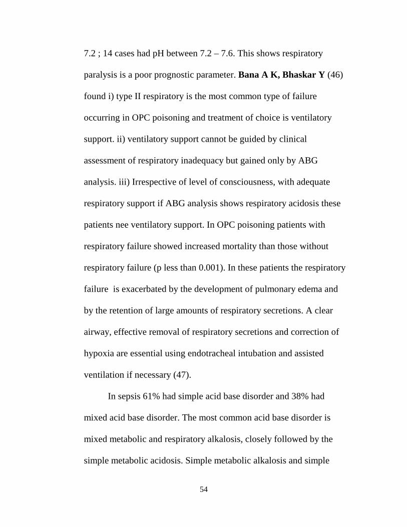
had type I failure and 7 had type II failure. 3 cases had pH less than
54
7.2 ; 14 cases had pH between 7.2 – 7.6. This shows respiratory
paralysis is a poor prognostic parameter. Bana A K, Bhaskar Y (46)
found i) type II respiratory is the most common type of failure
occurring in OPC poisoning and treatment of choice is ventilatory
support. ii) ventilatory support cannot be guided by clinical
assessment of respiratory inadequacy but gained only by ABG
analysis. iii) Irrespective of level of consciousness, with adequate
respiratory support if ABG analysis shows respiratory acidosis these
patients nee ventilatory support. In OPC poisoning patients with
respiratory failure showed increased mortality than those without
respiratory failure (p less than 0.001). In these patients the respiratory
failure is exacerbated by the development of pulmonary edema and
by the retention of large amounts of respiratory secretions. A clear
airway, effective removal of respiratory secretions and correction of
hypoxia are essential using endotracheal intubation and assisted
ventilation if necessary (47).
In sepsis 61% had simple acid base disorder and 38% had
mixed acid base disorder. The most common acid base disorder is
mixed metabolic and respiratory alkalosis, closely followed by the
simple metabolic acidosis. Simple metabolic alkalosis and simple

55
respiratory alkalosis account for 3 and 1 case respectively. Out of 13
cases 8 had expired, accounting for a mortality rate of 61%.
Respiratory failure is seen 6 out 13 cases. Out of which 4 cases had
type I failure and 2 had type II failure. 1patient had pH more than 7.6
and the rest in between 7.2 – 7.6. In all, 46% patients had respiratory
alkalosis either simple or mixed. Respiratory alkalosis occurs early
and metabolic acidosis late. The degree of acidosis is a marker of
severity of illness and prognosis.
The onset of hypoxia indicates severe disease and high risk for
ARDS. This has been proved in our study which shows major acid
base disturbance in sepsis is respiratory alkalosis; 4 patients who had
type I respiratory failure expired thus providing hypoxia is a marker
of severity of illness. Statistical analysis of survival in response to pH,
the mean pH in survived patients is 7.42; The mean pH in expired
patients is 7.54. The alkalemic pH is associated with high mortality.
Several studies have shown that alkalemia is associated with high
mortality in medical ward patients. Studies by Anderson L E,
Henrich W L (41) have shown the death rate is higher among the
medical patients with alkalemia and mixed metabolic and respiratory
alkalosis appears to be associated with a particularly poor prognosis.

56
Blood gas analysis may often reveal hypoxaemia due to intra
pulmonary shunting before the classical radiological appearance of
ARDS develops.it is evident that patient donot usually die of
hypoxaemia but from the complex disturbances that result from
multiple organ system failure. Thus the aim in the management of
ARDS is to support all body systems until the integrity of the alveolar
capillary membrane is restored. Early recognition with appropriate
pharmacological and supportive therapy favorably influence the
prognosis (48). Patients treated with antimicrobials to which the
organisms are sensitive do survive better than those in whom the
treatment was not appropriate. However the general condition and the
presence or absence of shock are powerful independent variables.
DKA is seen in 10 patients, none had respiratory failure in our
study. Mixed acid base disorder is the most common abnormality seen
in 60% of the cases. All of them had pH in between 7.2 – 7.6. All the
patients with DKA had an element of metabolic acidosis either simple
or mixed form. The most common triple acid base disorder in DKA is
combination of metabolic acidosis, metabolic alkalosis and respiratory
alkalosis. The underlying ketosis causes high anion gap acidosis.
Vomiting or nasogastric suction can cause metabolic alkalosis in

57
DKA. Underlying sepsis precipitating DKA can cause respiratory
alkalosis apart from neurogenic hyper ventilation.
CKD is seen in 10 patients; of which 7 patients had expired ; 1
patient had type I respiratoryfailure. The simple metabolic acidosis is
seen in 3 cases and mixed acid base disorder in 7 cases.
COPD is seen in 9 patients. All of the had type II respiratory
failure. Out of which 4 had expired and 7 had mixed acid base
disorders (77%). The most common acid base disorder in COPD is
mixed metabolic acidosis and respiratory acidosis. The next is
metabolic alkalosis and respiratory acidosis. Only 2 patients had pH
less than 7.2 in all respiratory acidosis seen in 100% of the cases,
either simple or mixed. Respiratory in COPD is due to respiratory
failure. Non-invasive ventilation is an important recent advance in the
management of patients with acute respiratory failure complicating
COPD. A number of large , well conducted, randomized studies have
shown that non-invasive ventilation improves survival in COPD
patients with an acute respiratory acidosis and reduces the need for
intubation (50). The achievement of adequate alveolar ventilation, as
indicated by falling PaCo2, improving pH and satisfactory inspiratory
chest wall movement, is the target. Patients who are likely to improve

58
with non-invasive ventilation will normally show both clinical and
biochemical improvement within the first few hours of treatment. A
falling respiratory rate, accompanied by improvement in PaCo2 and
pH are associated with a good outcome (51).
Alcoholic intoxication is seen in 6 patients. 66% of patients had
mixed disorder of metabolic alkalosis and respiratory alkalosis.
Respiratory alkalosis is seen in 2 patients. Respiratory alkalosis either
simple or mixed accounts for 83% of cases. This observation is
consistent with Dobes M (1993) (43) who did a prospective study of
77 alcoholics with delirium tremens and in 62 patients (80.5%)
respiratory alkalosis was detected. Only 1 patient had type II failure .
The main cause of respiratory alkalosis is rebound phenomenon of the
respiratory center which causes hyper ventilation. whatever the acid
base disturbance in alcoholic ketosis, administration of glucose,
thiamine and rehydration is usually adequate to deal with the
metabolic disturbance (52).
In scorpion sting, all six patients had an element of metabolic
acidosis either simple or mixed. 3 out of 6 patients had respiratory
failure (type I -1 , type II – 2). Out of the 6 cases 2 patients expired

59
accounting for a mortality rate of 33%. 3 of them had pH less than 7.2
and 3 had pH between 7.2 – 7.6.
In DCLD 3 out of 4 patients had an element of respiratory
alkalosis. The most common acid base disorder was mixed metabolic
and respiratory alkalosis. 2 patients had respiratory failure. The
mortality rate was 50%.
In the miscellaneous group out of 20 patients 3 had CVA ; 4
had corrosive poisoning ; 4 cases were attempted hanging ; 1 had
myasthenic crisis; 2 had pneumothorax ; 1 had pneumonia ; 1 had
antiphospholipid antibody syndrome; 1 had cardiac failure ; 1 had
hypokalemic paralysis ; 1 had oduvanthalai poisoning and 1 was
sedative tablet poisoning.
The most common acid base disorder observed in this group is
metabolic alkalosis and respiratory alkalosis. 8 patients had
respiratory failure (type I – 4, type II – 4). 1 patient had pH less than
7.2 ; 19 patients had pH between 7.2 – 7.6. Out of 20 patients 6
patients had expired accounting for a mortality rate of 30%.
CKD is the most common cause of metabolic acidosis as well
as mixed metabolic acidosis and respiratory alkalosis. COPD is the
most common causes of mixed respiratory acidosis and metabolic

60
acidosis. DKA is the most common cause for triple acid base disorder.
DCLD is the most common cause of mixed metabolic and respiratory
alkalosis.
While analyzing individual acid base disorders in combination
with both simple as well as mixed, metabolic alkalosis is the most
common. It accounts for 49% of cases ( 14% -simple and 35%
mixed). The next common is metabolic acidosis which is seen in 48%
( 19% simple, 29% mixed). Respiratory alkalosis is seen in 35% of
cases (4% simple and 31 % mixed). Respiratory acidosis is the least
common acid base disorder seen in 32% (3% simple and 29% mixed).
While analyzing pH and survival rate our study is well
correlated with any other studies. Of the total 100 patients 32 patients
had expired accounting for a mortality rate of 32%. Of which 10 of
them had pH less than 7.2; 1 had pH more than 7.6 and 21 were
between 7.2 and 7.6. Of the total cases of 13 who fall under pH less
than 7.2 , the mortality rate was 76%. 1 patient had pH more than 7.6
and expired, accounting for the mortality rate of 100%. Among the 86
cases, pH between 7.2 and 7.6, 21 patients expired with mortality rate
of 24%.

61
Irrespective of primary pathology it is the severe acidemia and
alkalemia that largely determines the patient status and prognosis (44).
Our study is consistent with the statement.
Among the respiratory failure COPD leads the tally with 9 out
of 9 patients presented with type II respiratory failure. 46% of patients
with sepsis had respiratory failure with type I being 4 and type II
being 2. 9 out of 17 patients admitted with OPC poisoning had
respiratory failure with majority being type II. 60% of patients with
snake bite envenomation had respiratory failure with type I being 1
and type II being 2. 50% of the patients with scorpion sting had
respiratory failure with type I being 1, type II being 2. In the
miscellaneous group 4 patient had respiratory failure with type I being
4 and type II being 4. Of 14 cases with type I failure 9 had simple and
5 had mixed acid base disorder. Of 28 cases with type II respiratory
failure 20 had mixed and 8 had simple acid base disorder. Among the
58 cases of non respiratory failure 35 had mixed and 23 had simple
acid base disorder.

62
Conclusion

63
CONCLUSION
1. OPC POISONING, SEPSIS, DKA, CKD, COPD are the most
common causes of acid base disorders in intensive medical care unit
and toxicology unit together accounting for 59% of cases.
2. The most common acid base disorder observed was mixed type,
which accounted for 60%, while simple acid base disorder was
observed in 40 % of patients.
3. The most common mixed acid base disorder was metabolic alkalosis +
respiratory alkalosis which was seen in 22% of cases. 19% of cases
had simple metabolic acidosis.
4. Respiratory failure was present in 42% of cases, Type I – 14% and
Type II – 28%.
5. OPC poisoning was the most common cause for acid base disturbance
in our study (17 %). The most common acid base disorder in OPC
poisoning was simple metabolic alkalosis. Respiratory failure was
present in 52 % (Type I – 2, Type II – 7 )
6. Sepsis accounts for 13 % of cases. The most common acid base
disorder in sepsis was mixed metabolic alkalosis and respiratory
alkalosis. Respiratory failure was present in 46 % (Type I – 4 , Type II
– 2).

64
7. DKA accounts for 10 % of acid base disturbance. The most common
single acid base disorder in DKA was metabolic acidosis.
8. CKD accounts for 10 % of acid base disturbance. The most common
acid base disorder in CKD was metabolic acidosis. 7 out of 10
patients expired with mortality rate of 70 %.
9. COPD accounts for 9 % of acid base disturbance. The most common
acid base disorder in COPD was mixed metabolic acidosis and
respiratory acidosis. All the patients with COPD were admitted with
Type II respiratory failure.
10. Among the individual acid base disturbance either in single or mixed
form, the metabolic alkalosis is most commonly seen in 49% of cases.
11. 86 % of patients had pH between 7.2 to 7.6, 13 % of patients had pH
less than 7.2, 1% of patients had pH more than 7.6 .
12. The mortality and morbidity was more severe in extreme acidemia
and alkalemia. In those with pH less than 7.2, the mortality rate was
76 % and in those with pH more than 7.6 mortality rate was 100%,
those between 7.2 to 7.6 the mortality rate was 24%.

65
Bibliography

66
BIBLIOGRAPHY
1. Gunnerson Kyle J, Critical Care 2005; 9 : 508-516.
2. Thomas D.DuBose, Jr. Acidosis and Alkalosis. Harrison’s principles of Internal Medicine. Vol 1, 16th edn, 2005; 42:263.
3. Eduardo Benchimol Saad, Acid Base Disorders in critical care.
Intensive Care, 2007;6: 1 – 6.
4. Kassiver et al, Rapid Estimate of Plasma Carbondioxide from pH, NEJM 1965; 272:1067.
5. Brady Nilux, Therapy in Nephrology and Hypertension. W & B
Saundas 1999 ; 1: 292.
6. Kellum JA, Determinants of blood pH in health and disease. Critical Care 2000; 4 : 6 – 14.
7. William Kachny, Manual for Nephrology , 2003 ; 15 : 49
8. Statement on Acid Base Terminology , Ann.Inte.med 1965; 63 : 885.
9. Margaret Bia, North American Clinics on Acid Base Disease. 1981; 65 : 347
10. McCurdy D. Mixed Acid Base Disturbance, Diagnosis and Treatment. Chest 1971; 62:353
11. Epstein sk et al, Respiratory Acidosis. Respir Care 2001 ; 46 : 363.
12. Mitchell et al. The effect of Acid Base on Cardio vascular findings. Kidney Internat. 1972 ; 1 :375
13. Kilbun K.H. , Seizures, coma with Alkalosis. Ann.Inter.Med.1966 ; 65 : 979.

67
14. Madias NE, Adrogue HJ, Acid Base Disturbance in Pulmonary Medicine. Fluid Electrolyte Acid Base Disorders 2nd edn, Churchill Livingston 1995 ; 223 :53.
15. Beer R, Goldstein, Effect of Metabolic Alkalosis in Respiratory function. Can.Med.Ass.J 1977 ; 117 : 900.
16. Miller PD. Acute Met.Alkalosis precipitating hypercarbia. JAMA 1997 ; 238 : 2400.
17. Adrogue HJ, Madias, Management of life threatening Acid Base Disorders. NEJM 1998 : 338 :107 – 112.
18. Orchard CH et al , Effects of pH on Contractile function of Cardiac mucscle. Am.J.Physio 1990 ; 258 :81 – 90
19. Orchard CH et al, Acidosis and Arrythmias in cardiac muscle. Cardivas.Res 1994 ; 28 : 1312 – 19.
20. England BK et al. Abnormalities in protein synthesis and degradation in pH changes. Am.J.Physio. 1991 ; 260 : 277 – 282.
21. Hood VL et al. Acid Base homeostasis in keto and lactic acidosis. Diabetes Rev. 1994 ; 2 : 177 – 94.
22. Harrington JT, Metabolic alaklosis, Acid Base , Boston - little brown, 1987 ; 227 : 306.
23. Rimmer JM, Metabolic Alkalosis. J. Int.Car.Med 1987 ; 2 : 137 - 150
24. Fishman, Pulmonary Diseases and Acid Base Disorders, Manual of Respiratory Medicine. 3rd edn 1998 ; 2 : 2525 – 27.
25. Ebistein SK et al. Overview of respiratory muscle function. Clin.Chest.Med 1994 ; 15 : 619 – 29.
26. Kook W. ABG and pH during sleep in COPD. Am.J.Med 1975 ; 58 : 633 – 670.

68
27. Rebuck AS. Measurement of Ventilatory response to hypercapnia and hypoxia. Lung Biopsy in Health and Disease. 1981 ; 2 : 745 – 94.
28. Lane BJ et al. Relation between airway obstruction and CO2 retention in Obstructive airway disease. BMJ 1968 ; 3 : 707 – 9.
29. Patrick JM, Influence of Age, Sex, Body, Size and Lung size on control and pattern of breathing in Caucasians. Resp.Physiol. 1972 ; 69 : 337 – 50.
30. Schiffman. Acid Base Disorders in COPD. Vol 1, 1988 ; 20 :181.
31. Jacobson HR , Seldin DW. On the generation , maintenance and correction of metabolic alkalosis. Am J Physiol 1983 ; 245 : 425 – 32.
32. Vernon DD, Gleich MC, Poisoning and Drug overdose. Critical care clinics 1997 ; 3 : 647 – 67.
33. Filley GF et al. Acid Regulation and CO2 retention. Chest 1970 ; 58 : 417 – 22.
34. Madias et al Hypoxia and Hypercapnia in Asthma. NEJM 1968 ; 278 : 1068.
35. Schrier et al. Diseases of Kidney 6th edn, Churchill Livingston 2007 ; 3: 2525 -39.
36. Adro et al. Change in Acid Base Pattern in DKA. NEJM 1982 ; 307 :1603 – 10.
37. Levovitz HE. Diabetic ketoacidosis. Lancet 1995 ; 345 : 767 – 72.
38. Jonathan Cohen, Infectious Diseases 2nd edn Mosby 2004 ; 2 : 619 – 22.
39. Lunburg JS et al Septic shock. Analysis of outcome for patients in ward vs Intensive care. Crit.Care.Med 1998 ; 26 : 1020 – 24.
40. Narins et al. The Renal Acidosis issues in Nephrology. Churchill Livingston 1978 : 30 : 64.

69
41. Anderson et al. Alkalemia associated morbidity and mortality in
medical ward. South Med.J 1987 ; 80 : 729 -33.
42. Madias NE et al, Acid Base Disturbance in pulmonary medicine. Fluid and Electrolyte Acid Base Disorders 2nd edn Churchill Livingston 1995 ; 223 : 53.
43. Dobes M. Disorders of Acid Base Equillibrium in delirium tremens. Cas Lek cesk Mar 8 1993 ; 132 : 142 – 5.
44. Adrogue et al. Life Threatening Acid Base Disorders, NEJM 1998 ; 338 : 107.
45. Dhadke V N , Kulkarni P M. The Clinical profile of organophophorous compound poisoning. JAPI 2001 ; 49 : 177
46. Bana A K, Bhaskar Y, Clinical correlation of Arterial blood gas Analysis in Organophosphorous compound poisoning. JAPI 2001 ; 49 : 180
47. Proudfoot A T, Vale J A Pesticides. Oxford Text book of Medicine, vol 1, 3rd edn, 1996 ; 8.3.8 : 1122
48. Garrard C, Foex P Adult respiratory distress syndrome Oxford Text book of Medicine, vol 2, 3rd edn, 1996 ; 17.10.20 : 2852 - 60
49. Murphy P A, Septicemia. Oxford Text book of Medicine, vol 1, 3rd edn, 1996 ; 7.19.2 : 1020 – 25.
50. Brochard L, Mancebo J et al, Non Invasive ventilation for acute exacerbation of COPD. NEJM 1995 ; 333 :817 – 22.
51. Bott J , Caroll M P et al, Randomised controlled trial of nasal ventilation in acute ventilatory failure due to COPD. Lancet 1993 ; 341 : 1555 – 7
52. Cohen R D, Woods H F, Disturbances of acid base homeostasis. Oxford Text book of Medicine, vol 2, 3rd edn, 1996 ; 11.4 : 1542 - 43

70
PROFORMA
Sl.No.
Name : Age: Sex :
Clinical Features :
Past H/o : DM / HT / COPD / IHD / CKD / DCLD
Drug H/o :
Diagnosis :
ABG :
pH PCO2 PO2 HCO3- Na+ K+ Cl-
Blood Sugar :
RFT :
Urea S.Creatinine
LFT :
S.Bilirubin SGOT SGPT S.Proteins SAP
ECG :
Chest X Ray :
Comments :

71
MASTER CHART
SL. NO.
NAME
AGE
SEX
DIAGNOSIS
pH
PCO2
PO2
HCO3
-
Na+
K+
CL- FAILURE
1. SANCHIAH 50 M COPD 7.42 98.3 22.1 30 131 3.2 96 TYPE 22. MAHENDRAN 25 M SEPSIS 7.54 28.2 64.4 23.8 134 2.53 98 TYPE 13. THIRUNIRAISELVAM 58 M CKD 7.39 26.3 87.5 15.6 139 5.8 140 4. RENUKA 20 F DCLD 7.52 39 83 31.5 136 2.7 116 5. GANAPATHY 77 M CVA 7.54 40.5 71.6 33.1 133 2.3 99 TYPE 16. MURALI 29 M CVA 7.44 39.9 99.7 26.7 142 3.9 100 7. RADHA KRISHNAN 60 M MYASTHENIA 7.24 29.2 100 12.4 138 3.5 104 8. MOOBINA 20 F APS 7.50 41.4 100 31.8 137 3.6 84 9. PREETHI 20 F PNEUMOTHOR 7.26 39.3 98 17.5 140 3.5 98 10. SARALA 38 F CCF 7.40 57 63 34 134 3.4 98 TYPE 211. SHANMUGAM 46 M PNEUMONIA 7.56 19.1 100 17.0 137 3.8 108 12. MUTHU 34 M DKA 7.30 34.9 102 17.1 132 3.4 117 13. FAZIL 39 M DKA 7.27 15.6 100 7.0 134 3.0 123 14. SUNDARI 25 F HYPOKALEMIA 7.38 50 92 29.2 134 2.1 100 TYPE 215. GANANASIDDHAN 23 M OPC POISON 7.33 58 100 30.5 137 4.2 98 TYPE 216. PERUMAL 40 M OPC POISON 7.47 48.1 97.8 34.8 137 3.6 119 TYPE 217. VINESH 42 M OPC POISON 7.38 55.3 94.7 32.2 132 3.6 103 TYPE 218. SATISH KUMAR 27 M ALCOHOLIC 7.50 28.0 95 22.6 132 3.9 104

72
SL. NO.
NAME
AGE
SEX
DIAGNOSIS
pH
PCO2
PO2
HCO3
-
Na+
K+
CL- RESP.FAILURE
19 VELU 52 M ALCOHOLIC 7.50 28.6 90 21.7 136 3.8 110 20 ETHIRAJ 55 M SNAKE
BITE(NEURO) 7.20 45.1 100 17.4 132 3.4 102 TYPE 2
21 RAJA 25 M SCORPION 6.94 44.3 100 9.5 131 3.6 104 22 SATYA 22 F CORROSIVE 7.49 30.5 68.9 23.1 131 2.3 98 TYPE 123 ANGAIAH 37 M ODVANTHAL 7.0 55.7 100 13.7 141 1.8 106 TYPE 224 ANAND 24 M ATT.HANGING 7.28 43.1 95 20.2 128 34 98 25 KALPANA 28 F SEDATIVE 7.43 45 84.3 29.3 140 3.9 96 26 KANNAYARAM 55 M COPD 7.22 87.7 27.1 30 135 3.6 98 TYPE 227 SUBRAMANI 54 M COPD 7.18 169.4 47.5 32 136 3.8 99 TYPE 228 NATARAJAN 84 M SEPSIS 7.58 38.8 100 36.3 134 4.8 98 29 SEKAR 57 M SEPSIS 7.31 22.1 100 11.0 147 36 77 30. FASHIYA 15 F CKD 7.52 21.7 100 17.3 138 3.9 112 31. ANANDHAN 35 M CKD 7.23 19.3 91.3 7.9 133 4.5 107 32. JOSEPH 45 M DCLD 7.41 51.1 80 31.8 133 3.2 104 33. DAYAKUMAR 45 M DCLD 7.54 31.6 92.4 26.9 136 4.0 88 34. SEKAR 52 M CVA 7.54 38.6 67.1 32.6 130 3.2 112 TYPE 1
35. THIRUPATHIAH 16 M PNEUMOTHER 7.50 42.8 100 33 134 3.1 115 36. SEKAR 57 M DKA 7.31 22.1 100 11.0 147 3.6 77

73
SL. NO.
NAME
AGE
SEX
DIAGNOSIS
pH
PCO2
PO2
HCO3
-
Na+
K+
CL-
RESP. FAILURE
37. NOORJAHAN 55 F DKA 7.32 32 100 16.4 137 3.9 107 38. LATHA 32 F OPC POISON 6.70 84.9 99 10.5 131 4.2 120 TYPE 2 39. KARUNAN 40 M OPC POISON 7.48 47.4 100 35.1 134 3.9 106 TYPE 2 40. VIJAYA 45 F OPC POISON 7.49 38.7 92 28.9 139 3.6 100 41. KANNAN 41 M OPC POISON 7.58 22 100 32 136 2.9 111 42. SUNDARA
RAJAN 45 M ALCOHOLIC 7.54 15.8 90 30.1 132 3.1 106
43. SUNDARAM 40 M ALCOHOLIC 7.54 35.8 100 31.1 130 3.1 100 44. ANNA DURAI 34 M SNAKE BITE
(VASCULO TOXIC)
7.50 35 94 26 124 3.8 106
45. DORAIKANNAN 52 M SNAKE BITE 7.41 31 73.4 19.6 144 3.6 109 TYPE 1 46. SELVI 35 F CORROSIVE 7.22 23.2 96.2 10.9 150 3.8 105 47. SWAPNA 23 F CORROSIVE 7.29 56.6 51.7 26.9 123 3.6 107 TYPE 2 48.. VENGATESH 25 M CORROSIVE 7.33 40.7 90 21.3 145 3.1 106 49. PRABHU 23 M HANGING 7.49 42.1 90.6 31.5 139 3.3 105 50. KAJA
MOHIDEEN 65 M DKA 7.23 28 95.9 7 146 5.2 106
51. AMMU 20 F DKA 7.38 22.5 90 13.3 136 3.6 98 52. KASI 40 M COPD 7.05 126.1 86.9 24.2 138 3.9 100 TYPE 2 53. KOUSALYA 63 F COPD 7.44 86.5 46.6 58 135 3.1 89 TYPE 2 54. JAMUNA 19 F SEPSIS 7.32 32.1 52.3 14.2 134 3.2 116 TYPE 1

74
SL. NO.
NAME
AGE
SEX
DIAGNOSIS
pH
PCO2
PO2
HCO3
-
Na+
K+
CL-
RESP. FAILURE
55. SUBRAMANI 39 M CKD 7.50 15.7 93 12.0 119 4.6 104 TYPE 2 56. KATHIRVEL 16 M DCLD 7.54 38.5 77.3 32.7 130 2.9 1.9 TYPE 1 57. VIYAY 42 M OPC
POISON 7.54 60.2 76 36.5 134 3.6 111 TYPE 2
58. SUNDARA RAJA 46 M ALCOHOLIC 7.24 34.4 94.4 14.7 137 3.9 124 59. ATHANKARAIYAN 60 M ALCOHOLIC 7.44 60.1 100 40.8 131 2.6 89 TYPE 12 60. AKTCHAYA 24 M SNAKEBITE 7.17 80 100 17.9 137 3.6 105 TYPE 2 61. RAMA 25 F SCORPION 7.40 22.2 58.9 13.5 132 3.2 98 TYPE 1 62. SUMATHI 26 F CORROSIVE 7.49 3.5 67.7 23.1 140 4 98 TYPE 1 63. NITHIYA 20 F HANGING 7.58 34.2 90 31.4 143 3.1 111 64. VINCENT 23 M HANGING 7.38 36.1 98 21.3 135 3.4 105 TYPE 2 65. VELU 28 M DKA 7.42 28.8 92.6 15 136 3.7 98 66. KANDAVEL 17 M DKA 7.42 28.8 98 15 135 3.6 978 67. SHANTHA 62 F COPD 7.34 138.9 79.2 59.1 123 3.2 95 TYPE 2 68. VENKADESHAN 34 M SEPSIS 7.48 37.8 93.6 27.6 140 3.6 111 69. LEELA 55 F SEPSIS 7.43 50.9 92 33.2 134 3.1 108 TYPE 2 70. MOHANA 58 F CKD 7.58 33.2 86.8 30.4 125 4.6 73 TYPE 2 71. SATHIYANARAYAN 72 M OPC
POISON 7.1 15.0 60 12 136 3.8 96 TYPE 1
72. CHINNAKUTTI 32 M OPC POISON
7.46 34 100 23 139 3.2 108

75
SL. NO.
NAME
AGE
SEX
DIAGNOSIS
pH
PCO2
PO2
HCO3
-
Na+
K+
CL-
RESP. FAILURE
73 KASTURI 50 F OPC POISON
7.53 35.7 100 29.7 135 3.6 91
74 MARTIN RAJ 24 M SCORPION 6.95 70.7 35.8 15.3 154 2.9 126 TYPE 2 75 RENUKA 32 F SNAKE
BITE 7.59 28.3 100 26.7 135 4.4 101
76 THANGAM 30 F DKA 7.43 24.3 98 15.9 136 3.5 100 77 CHELLIAH 77 M COPD 7.51 49.4 88.9 58.7 137 4.1 98 TYPE2 78 PERIASAMY 60 M COPD 7.20 97.2 100 37.9 133 4 108 TYPE2 79 SIDHIKA 45 F SEPSIS 7.62 38.1 49.5 36.8 121 2.7 101 TYPE1 80 KANTHAMANI 45 F SEPSIS 7.52 38 83.2 30.9 132 3.3 109 81 MOHANA 55 F SEPSIS 7.46 50.9 83 35.5 134 3.7 105 TYPE2 82 INDHUMATHI 29 F CKD 7.08 31.1 100 9.2 137 5.8 100 83 DHANASHEKAR 50 M CKD 7.30 49.3 100 24.0 150 6.46 115 84 SUMATHI 22 F OPC
POISON 7.49 30.5 87.7 23.1 137 3.28 103
85 RAMKUMAR 45 M OPC POISON
7.46 34 100 24 144 3.3 101
86 LAKSHMI 20 F SCORPION 7.17 45.2 90 16.2 136 3.2 98 TYPE2 87 SHANKAR 24 M SCORPION 7.23 28 94 7 138 3.6 99 88 RAMU 35 M DKA 7.40 28.6 95 17.5 147 3.5 98 89 RAJAN 60 M COPD 7.31 69.7 95 28.5 132 3.2 85 TYPE2 90 NOORJAHAN 55 F SEPSIS 7.32 32.3 100 16.4 137 3.9 107

76
SL. NO.
NAME
AGE
SEX
DIAGNOSIS
pH
PCO2
PO2
HCO3
-
Na+
K+
CL- FAILURE
91. AMALANATHAN 45 M SEPSIS 7.51 41.4 79 23 133 3.4 102 TYPE 192. SUNDARI 60 F CKD 7.32 24.0 67 12.1 139 4.6 105 TY93. PAGALESWARI 32 F SEPSIS 7.54 44.2 96 37.6 137 4.0 112 94. RAFIQ AHMED 62 M CKD 7.44 31.3 100 20.8 134 4.5 94 95. PARVATHI 23 F OPCPOISON 7.30 32.5 85 15.8 130 4.12 109 TYPE 196. VINAYAGAM 30 M OPCPOISON 7.09 39 100 11.6 135 3.8 102 97. SUBRAMANI 50 M SEPSIS 3.38 34.9 90.6 20.3 134 3.8 108 98. DEVAMMAL 40 F CKD 7.10 7.0 98 2.1 136 4.7 103 99. KARUNAKARAN 41 M OPCPOISON 6.93 106 58 22.1 142 4.5 104 TYPE 2100 RATHINAMMAL 55 F OPCPOISON 7.34 30.8 100 16.5 140 3.6 105