a study of using manganese chloride (mncl2) solution as an
TRANSCRIPT

Romanian JouRnal of neuRology – Volume XX, No. 1, 202172
Hiba Karim Abd1, Muhammed Mizher Radhi1, Hassan Jafar Hassan2
AbstrActIodine solution is the only contrast medium currently used in the computed tomography scan (CT scan) examina-tion. In the present study manganese chloride (MnCl2) solution has been chosen as alternative contrast medium in computed tomography scanning (CT scan). It was found that using MnCl2 solution as an alternative contrast medi-um in rabbits which enhanced the CT scan imaging in the resolution and increasing the Hounsfield unit (HU) values of heart and kidney organs in comparison with the iodine compound at the same doses. It was chosen the heart and kidney of rabbits to study the effect of using the iodine and MnCl2 solution as alternative contrast medium in CT scan imaging, the following results: the native has 45 HU for heart and 50.1 for kidney organ. While the results of using iodine solution at dose of 3 ml has 83 HU for heart and 164 HU for kidney organ. In the MnCl2 solution which used as alternative of contrast medium has 83 HU for heart and 70.3 HU for kidney at 2.5 ml of 0.5 molar of solution. From these results the resolution of CT scan image has well and clears when using the alternative contrast medium (MnCl2). We can concluded that the alternative contrast medium of MnCl2 solution has good HU values of both heart and kidney comparison with iodine solution.
Keywords: MnCl2 solution, contrast medium of CT scan, rabbits, Hounsfield unit, iodine
Corresponding author: Muhammed Mizher Radhi E-mail: [email protected]
ORiginal aRticlesRef: Ro J Neurol. 2021;20(1)DOI: 10.37897/RJN.2021.1.10
A study of using manganese chloride (MnCl2) solution as an alternative of contrast medium for iodine in
computed tomography (CT) imaging
INtrODUctION
through the different studies of alternatives to the contrast media which used in different radio-logical techniques that have important effects on human health and to avoiding the side effects from the use of current contrasts media such as iodine and gadolinium harmful to the health [1-4]. Para-magnetic and hypo magnetic metals are used as contrast materials for magnetic resonance (MR) techniques. the metal gadolinium lanthanide (gd) was the predominant and most prevalent semi-mag-netic contrast agent until the discovery and associ-ation of the mineral with nephrogenic systemic fi-brosis (nsF), a rare but serious side effect in patients with kidney or kidney problems. Manga-
nese was one of the earliest reported examples of paramagnetic contrast materials for magnetic reso-nance imaging due to its positive active contrast enhancement [5]. Manganese-enhanced magnetic resonance imaging (MRi) is a well-established neuroimaging method for signal enhancement, and functional studies in the rat. Further, with the in-creasing availability of positron tomography (Pet) and magnetic resonance imaging devices, interest in using Mn2 + as a contrast agent. in this work, we differentiate and compare the radioactive Mn2 + up-take in the brain of mice for MRi and Pet, respec-tively. additionally, we examined the Mn2 + in bio-assay of mice [6]. Two of manganese polysulfide (ii) complexes, Mn-DtPa cystamine polymersand Mn-eDta cystamine polymers were synthe-
Article History:Received: 28 January 2021
Accepted: 20 February 2021
1Radiology Department, Health and Medical Technology College, Middle Technical University (MTU), Baghdad, Iraq
2Alhussien teaching hospital, Alnasyria, Iraq

Romanian JouRnal of neuRology – Volume XX, No. 1, 2021 73
sized and labeled as novel molecular contrast agents that could be degradable by MRi. contrast enhancement of manganese-based contrast agents was evaluated in mice carrying with human breast cancer xenografts, compared with Mncl2 [7]. clinical applications of manganese-based MRi contrast agents are intravenous (intravenous) and oral formulations. the preparation is a commer-cially available manganese-dimeridoxyl diphos-phate chelate; whereas the oral formulation is a blend of Mncl2, alanine and vitamin D3, which is currently undergoing clinical trials. the formula-tions and preclinical and pharmacokinetic studies of both formulations are discussed. the main re-ported clinical difference between the two formu-las is that intravenously. administration exposes all organs, while oral ingestion only exposes the enterohepatic circulation [8]. Magnetic resonance imaging (MRi) is in increasing demand by re-searchers in many biological disciplines. not only is the use of high field magnets to obtain satisfac-tory spatial resolution, but the achievement of ade-quate contrast between tissues also requires deter-mination of applicable imaging parameters by cost and time- depreciation procedures. systematically ingested manganese can act as an effective con-trast agent in rapid imaging MRi. Due to the ten-dency of manganese ions to differentially accumu-late in most soft tissues, higher overall signal intensity and strongly improved contrast between structures yield data well suited to digital post-pro-cessing in 3D models [9].
in this work, the enhancement of computed to-mography imaging by chemical contrast media was studied by using an alternative of iodine con-trast agent of a manganese compound.
MAtErIALs AND MEtHODs
Materials
Bayer Pharma ag company from the german company (Berlin germany) iodine contrast as io-promide (Ultravist 370) was used as contrast me-dia in ct scan. Manganese chloride (Mncl2) was used from chinese scRc (china). anesthesia ma-terials used to anesthetize animals such as keta-mine 10% from alfasan company (Holland), xyla-zine 2% from alfasan (Holland). Blood samples of rabbits, and other chemicals and solvents were of
annular grade and were used as received by the manufacturers. Deionized water was used to pre-pare aqueous solutions.
Preparation manganese chloride 0.5 M Mncl2
a 0.5 molar solution of pure manganese chlo-ride (chinese scRc) was prepared in a 10 ml vol-umetric flask, and the crystals were dissolved in deionized water to obtain a 0.5 molar solution of manganese chloride which used as alternative con-trast medium.
ct scan apparatuses
the ct scan screw type general electric (ge) model tc Revolution eVO 128 slices, ge Health-care.
after preparing the rabbit for examination and in the case of anesthesia with the specified dose of the contrast, the rabbit was lying on the examina-tion table to perform the spiral ct scan as shown in Figure 1.
FIGURE 1. Preparation of the rabbit in CT scan
cyclic voltammetric apparatuse
ezstat series (potentiostat / glvanostat) nuVant systems inc. pioneering electrochemical technolo-gies Usa.
Pyrex cell measuring 10 milliliters and three electrodes was used:
a. Working electrode: where the glass carbon electrode (gce) was used
b. Reference electrode: where ag / agcl silver electrode (3M Kcl) was used

Romanian JouRnal of neuRology – Volume XX, No. 1, 202174
c. counter electrode: Where to use a platinum wire (1 mm diameter)
all three electrodes were dipped in the solution under study and linked to the potentio- stat, which in turn was connected with the personal computer to identify the properties of the materials studied in the blood medium as shown in Figure 2. the glassy carbon electrode (gce) was used in this study af-ter cleaning with alumina grand and sonic tech-nique for 10-15 min [10].
FIGURE 2. Cyclic voltammetry parts
rEsULts AND DIscUssION
study the rabbits by ct scan
the rabbits were chosen for a ct scan to check different organs of the rabbits’ abdomen, especial-ly the kidneys and the heart. The first group is the group that studied rabbits with ct scans without using (pre) contrast media. the second group, in which the spatial survey of rabbits was studied us-ing an alternative contrast medium for manganese chloride solution, where the kidneys and heart of rabbits were studied via intravenous manganese chloride alternate contrast at a concentration of 0.5 mol at different doses (1, 1.5, 2, and 2.5 ml) and the tests were performed following using ct scan-ning.
ct scan examination of heart organ
this examination was taken for all members of the abdominal area of the body of the rabbits such as kidney and heart without using any contrast me-dia, then the iodine contrast and the alternative contrast (Mncl2 solution) were used for the exam-ination was taken for the heart and kidney organs in the rabbits. The Hounsfield unit (HU) factor val-ues can be used to determine the clarity of the ct scan image.
Hounsfield unit (HU): Absorption coefficient unit of radiolucency of a substance; HU is normal-ized to water, where water = 0 HU, air = -1000 HU and bone = 1000 HU, the HU values in the ct scan are reported for each case taken for the studied rab-bits as in the following [3]:
1. the ct scan imaging of the heart tests were taken to turn the rabbits into the following three cases: a. checking the heart without using the contrast medium (native). it was found from the results as in Figure 3 of the rabbit’s heart without using any contrast medium, and the value of the clarity of the heart of the HU value was 45 as illus-trated in table 1.
B. the ct scan of cardiac examination using an iodine contrast medium which illustrated in Figure 4 with HU value of heart clearness have 53, 62, and 83 when using 1, 2, and 3 ml dose of iodine respectively, where diagnosis is possible.
c. cardiac examination using alternative con-trast medium of manganese chloride solution which illustrated in Figure 5, it was found an en-hancement of the heart ct scan imaging by higher HU values compering with the HU values at iodine and native cases. table 1 discuss the HU values when using alternative contrast agent (Mncl2 solu-tion) of 49.6, 65.1, 69.6, 70, and 83.3 at the doses of Mncl2 solution of 0.5, 1, 1.5, 2, and 2.5 ml re-spectively, it is a good enhancement of ct scan-ning when using the alternative contrast medium comparison with iodine contrast, moreover the safety of using Mncl2 solution.
TABLE 1. HU values of heart and kidney of rabbits at different contrast media
Kidney (HU)Heart (HU)Dose (ml)Contrast medium
50.145-Native 51.0531 Iodine 70622
164833 5649.60.5 MnCl2 (0.5 M)
60.165.11 65.369.61.567.470 2 70.383.22.5
ct scan examination of kidney organ
Other organ can be studied of the rabbit in ct scan examination to finding the differences of the imaging resolution under using different contrast media, with iodine and alternative contrast agent

Romanian JouRnal of neuRology – Volume XX, No. 1, 2021 75
of Mncl2 solution by using HU values to evalua-tion which contrast more active in the resolution of kidney organ of rabbit. the HU values in the ct scan are reported for each case taken for the stud-ied rabbits as in the following:
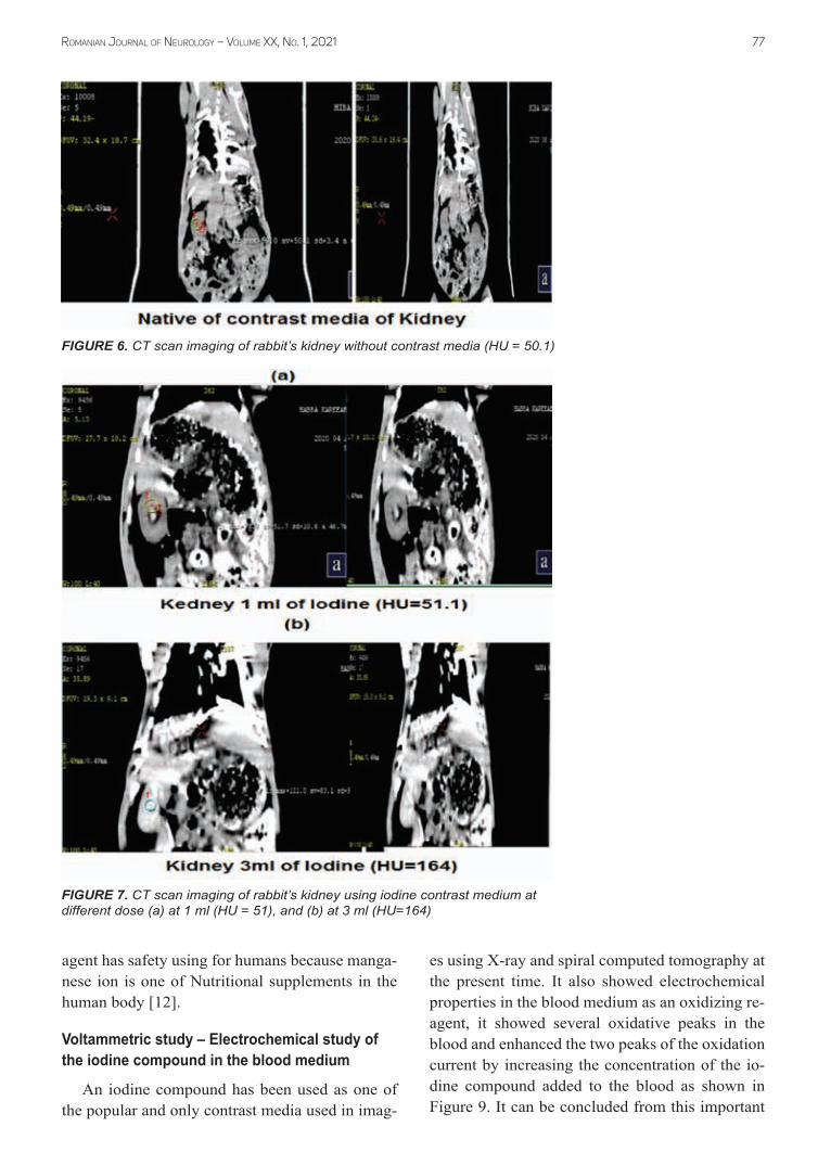
A. Native examinationthe native exam of ct scan for kidney organ
has 50.1 of HU value as shown in table 1 and Fig-ure 6. the resolution of ct scan imaging for native
exam is still low in the HU value because no con-trast used in both heart and kidney.
B. Iodine contrast medium examinationit was used iodine contrast medium at different
doses for the kidney organ of rabbit of 1, 2, and 3 ml. the resolution of ct scan imaging has HU val-ues at 52, 70, and 164 respectively as shown in Figure 7, the resolution of ct scan gradually en-hanced with increasing the iodine dose, but the
FIGURE 3. CT scan imaging of rabbit’s heart without contrast media (HU = 45)
FIGURE 4. CT scan imaging of rabbit’s heart using iodine contrast medium at different dose (a) at 1 ml (HU = 53), and (b) at 3 ml (HU=83)
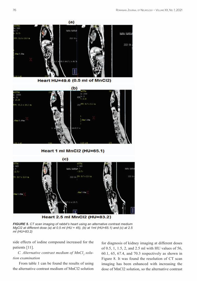
Romanian JouRnal of neuRology – Volume XX, No. 1, 202176
FIGURE 5. CT scan imaging of rabbit’s heart using an alternative contrast medium MgCl2 at different dose (a) at 0.5 ml (HU = 45), (b) at 1ml (HU=65.1) and (c) at 2.5 ml (HU=83.2)
side effects of iodine compound increased for the patients [11].
C. Alternative contrast medium of MnCl2 solu-tion examination
From table 1 can be found the results of using the alternative contrast medium of Mncl2 solution
for diagnosis of kidney imaging at different doses of 0.5, 1, 1.5, 2, and 2.5 ml with HU values of 56, 60.1, 65, 67.4, and 70.3 respectively as shown in Figure 8. it was found the resolution of ct scan imaging has been enhanced with increasing the dose of Mncl2 solution, so the alternative contrast
Romanian JouRnal of neuRology – Volume XX, No. 1, 2021 77
agent has safety using for humans because manga-nese ion is one of nutritional supplements in the human body [12].
Voltammetric study – Electrochemical study of the iodine compound in the blood medium
an iodine compound has been used as one of the popular and only contrast media used in imag-
es using X-ray and spiral computed tomography at the present time. it also showed electrochemical properties in the blood medium as an oxidizing re-agent, it showed several oxidative peaks in the blood and enhanced the two peaks of the oxidation current by increasing the concentration of the io-dine compound added to the blood as shown in Figure 9. it can be concluded from this important
FIGURE 6. CT scan imaging of rabbit’s kidney without contrast media (HU = 50.1)
FIGURE 7. CT scan imaging of rabbit’s kidney using iodine contrast medium at different dose (a) at 1 ml (HU = 51), and (b) at 3 ml (HU=164)

Romanian JouRnal of neuRology – Volume XX, No. 1, 202178
FIGURE 8. CT scan imaging of rabbit’s kidney using an alternative contrast medium MgCl2 at different dose (a) at 0.5 ml (HU = 56), and (b) at 2.5 ml (HU = 70.3)
analysis that all compounds containing iodine they are harmful oxidizing substances through their interaction with the blood composition, which causes undesirable symptoms when administered intravenously to all patients who undergo diagno-sis with both X-ray and tomography techniques, in addition to the dangerous symptoms they cause for those suffering from kidney or liver failure or heart disease iodine should not be given for contrast to avoid complications that may cause death in cer-tain cases or permanent diseases, and at the same time, the diagnosis required for this technique is not used [13,14].
Voltammetric effect of manganese chloride in the serum blood medium
Manganese chloride (Mncl2) the alternative contrast medium was used in ct scan, it was stud-
ied using an electrochemical method by cyclic voltammetry. Manganese chloride solution has good electrochemical properties, especially in the blood medium. it has been found that Mn(ii) in the blood which acted as an anti-oxidative agent as shown in Figure 10 illustrated the appearance of reduction current peak of Mg(ii) in potential re-gion at -0.55 V, so Mncl2 solution can be consid-ered as anti-oxidant agent because it received the free radical in the electrochemical reaction with blood composition. so, it can be used Mncl2 solu-tion as alternative contrast medium in safety be-havior in blood medium [15,16].
cONcLUsIONs
Manganese chloride (Mncl2) solution has good electrochemical properties and is safe to be used as

Romanian JouRnal of neuRology – Volume XX, No. 1, 2021 79
an alternative contrast medium without side effects for ct scan examination. the Mncl2 solution con-tains only the peak of the reducing current in the blood medium, while the iodine complex contains two oxidation current peaks, so manganese chlo-ride is an antioxidant compound and iodine is an oxidizing compound. On the other hand, the Mncl2 solution has been used as an alternative contrast medium due to the improved imaging for diagnos-ing the abdominal organs as founding in the rabbit
heart of the value of HU = 83.2 when using alter-native contrast medium and the same value when using iodine contrast medium. the HU value is 70 for kidney organs when using the alternative con-trast medium and iodine compound. the resolu-tion of ct scan imaging has the same in iodine and Mncl2 but the side effect is high in iodine com-pound and safe in Mncl2, so we recommend using Mncl2 solution as an alternative medium in the di-agnosis by ct scan.
FIGURE 9. Cyclic voltammogram of Iodine solution at different concentra-tions in blood medium
FIGURE 10. Cyclic voltammogram of MnCl2 solution in blood medium
1. Radhi MM, Al-Shimmari HAT, Al-Mulla EAJ et al. New voltammetric study of MgCl2 as alternative contrast media in MRI molecular imaging. Nano Biomed Eng. 2017;9(2):152-161.
2. Mustafa DA, Al-Shimmari HAT, Radhi MM. Use of MgCl2 Nanoparticles as Alternative Contrast Media in Magnatic Resonance Imaging Molecular Imaging and Analyzed by Voltammetric Technique. Nano Biomed Eng. 2020;12(2):148-152.
3. Radhi MM. Use of MgSO4 as alternative contrast medium in computed tomography scanning and analyzed by voltammetric technique. Ro J Neurol. 2020;19(3):200-205.
4. Du Mustafa DA, Mohammed BH, Radhi MM. Oxidative and anti-oxidative effect of anesthesia compounds (ketamine and xylazine) on the rabbit blood sample using electrochemical method. Ro J Neurol. 2020;19(2):76-83.
rEfErENcEs

Romanian JouRnal of neuRology – Volume XX, No. 1, 202180
5. Pan D, Schmieder AH, Wickline SA, Lanza GM. Manganese-based MRI contrast agents: past, present and future. Tetrahedron. 2011 Nov 4;67(44):8431-8444.
6. Brunnquell CL, Hernandez R, Graves SA, Smit-Oistad I, Nickles RJ, Cai W, Meyerand ME, Suzuki M. Uptake and retention of manganese contrast agents for PET and MRI in the rodent brain. Contrast Media Mol Imaging. 2016 Sep;11(5):371-380.
7. Ye Z, Jeong EK, Wu X, Tan M, Yin S, Lu ZR. Polydisulfide manganese(II) complexes as non-gadolinium biodegradable macromolecular MRI contrast agents. J Magn Reson Imaging. 2012 Mar;35(3):737-44.
8. Chabanova E, Logager VB, Moller JM, Thomsen HS. Manganese Based MR Contrast Agents: Formulation and Clinical Applications. The Open Drug Safety Journal. 2011;2:29-38.
9. Herberholz J, Mims CJ, Zhang X, Hu X, Edwards DH. Anatomy of a live invertebrate revealed by manganese-enhanced Magnetic Resonance Imaging. J Exp Biol. 2004 Dec;207(Pt 26):4543-50.
10. Radhi MM, Abdullah HN, abir MS, Al-Mulla EAJ. Electrochemical Effect of Ascorbic Acid on Redox Current Peaks of CoCl2 in Blood Medium. Nano Biomed. Eng. 2017;9(2):103-106.
11. Abdullah AL, Radhi MM, Khelkal IN, Naji EN. Azithromycin nanoparticles: Cyclic voltammetric study in human blood serum samples at electrochemical analysis. Ro J Neurol. 2020;19(1):5-11.
12. Radhi MM, Alosfur FKM, Ridha NJ. Voltammetric Characterization Of Grafted Polymer Modified With ZnO Nanoparticles On Glassy Carbon Electrode. Russian Journal of Electrochemistry. 2018;54(1):7–32.
13. Karastogianni S, Girousi S. Electrochemical Behavior and Voltammetric Determination of a Manganese(II) Complex at a Carbon Paste Electrode. Anal Chem Insights. 2016 Jan 18;11:1-11.
14. Islam GJ, Naseem Akhtar HM, Mamun MA, Ehsan MQ. Investigations on the redox behaviour of manganese in manganese(II)–saccharin and manganese(II)–saccharin–1,10-phenanthroline complexes. Journal of Saudi Chemical Society. 2009;13(2):177-183.
15. Heidari S, Singh JP, Feizi H, Bagheri R, Chae KH, Song Z, Khatamian M, Najafpour MM. Electrochemical water oxidation by simple manganese salts. Sci Rep. 2019 May 23;9(1):7749.
16. Kaabi N, Chouchene B, Mabrouk W, Matoussi F, Selmane E, Hmida BH. Electrochemical properties of a modified electrode with δ-MnO2-ased new nanocomposites, Solid State Ionics. 2018;325(1):74-79.