a study of hypokalemic paralysis- etiology, clinical
TRANSCRIPT
A STUDY OF HYPOKALEMIC PARALYSIS- ETIOLOGY, CLINICAL PROFILE AND OUTCOME
Dissertation submitted to
THE TAMILNADU DR.M.G.R. MEDICAL UNIVERSITY
in partial fulfillment of the requirements for the award of the degree of
DM (NEPHROLOGY) – BRANCH – III
THE TAMILNADU DR.M.G.R. MEDICAL UNIVERSITY CHENNAI
AUGUST 2011
DECLARATION
I solemnly declare that this dissertation titled “A STUDY OF
HYPOKALEMIC PARALYSIS- ETIOLOGY, CLINICAL PROFILE
AND OUTCOME” is done by me in the Department of Nephrology, Madras
Medical College & Rajiv Gandhi Government General Hospital, Chennai under
the guidance and supervision of Prof.N.Gopalakrishnan, MD., DM., FRCP.,
Professor & Head of the Department, Department of Nephrology, Madras
Medical College & Rajiv Gandhi Government General Hospital, Chennai. This
dissertation is submitted to the Tamil Nadu Dr.MGR Medical University,
Chennai in partial fulfillment of the university requirements for the award of
the degree of DM.Nephrology.
Place : Chennai Date :
Dr.G.CHANDRAMOHAN Postgraduate student, Dept of nephrology, Madras medical college, Chennai.
CERTIFICATE
This is to certify that the Dissertation entitled, “A STUDY OF
HYPOKALEMIC PARALYSIS- ETIOLOGY, CLINICAL PROFILE
AND OUTCOME” is the bonafide record work done by
Dr.G.Chandramohan, under our guidance and supervision in the
Department of Nephrology, Government General Hospital, Madras
Medical College, Chennai, submitted as partial fulfillment for the
requirements of D.M. Degree examination Branch III NEPHROLOGY,
AUGUST 2011, under The Dr.M.G.R. Medical University, Chennai.
Dr.V.KANAGASABAI, M.D., THE DEAN, MADRAS MEDICAL COLLEGE, CHENNAI,
Dr.N.GOPALAKRISHNAN, M.D., D.M.,FRCP., PROFESSOR AND HEAD, DEPT OF NEPHROLOGY, MADRAS MEDICAL COLLEGE, CHENNAI.
ACKNOWLEDGEMENT
I sincerely thank the Dean, Dr.V.KANAGASABAI M.D., for having permitted
me to carry out this dissertation work at Government General Hospital, Madras
Medical College, Chennai.
I have great pleasure in expressing my gratitude and respect to PROF.
Dr.M.JAYAKUMAR, M.D., D.M., former Professor and Head, Department of
Nephrology, Madras Medical College, Chennai, for his valuable suggestions, kind
guidance, constant supervision and moral support without which this study would not
have been possible.
I have great pleasure in expressing my gratitude and respect to PROF.
Dr.N.GOPALAKRISHNAN, M.D., D.M., FRCP., Professor and Head, Department
of Nephrology, Madras Medical College, Chennai, who allowed me to continue my
dissertation, for his valuable suggestions, kind guidance, constant supervision and
moral support without which this study would not have been possible.
I am thankful to DR.T.BALASUBRAMANIAN, M.D., D.M., Associate
Professor, Department of Nephrology, Madras Medical College, Chennai, for his
valuable suggestions and guidance in doing the study.
I am thankful to Dr.R.VENKATRAMAN, M.D., D.M., and
Dr.S.JAYALAKSHMI, M.D., D.M., DR.N.MALATHY, M.D., D.M., Assistant
Professors, Department of Nephrology, Madras Medical College, Chennai, for their
valuable suggestions and guidance.
Last but not the least, my sincere thanks to the patients who co-
operated for this study, without whom the study could not have been completed and to
all my colleagues who shared their knowledge.
1
INTRODUCTION
Acute flaccid paralysis is a potentially reversible medical emergency and
has a wide differential diagnosis that includes neurologic, metabolic and
infectious etiologies. Acute hypokalemic paralysis (HP) constitutes a group of
heterogenous disorders that present with acute muscular weakness and can at
times be potentially life threatening1. Complications secondary to hypokalemia
such as a cardiac arrhythmia or respiratory failure lead to morbidity and
mortality. Although there are many potential causes of hypokalemia, there are
far fewer entities in the differential diagnosis of hypokalemic paralysis.
Hypokalemia and paralysis can be divided into 2 types, hypokalemic periodic
paralysis (HPP) where there is short-term shift of potassium into cells and non-
HPP resulting from a large deficit of potassium due to various etiologies. The
differential diagnosis in a patient with HP can be challenging due to
heterogeneity of its etiologies, but it is important to make the diagnosis promptly
because different therapies are required for each type and identifying causes that
are reversible are important2. Presence of a positive family history and recurrent
episodes in a patient can be helpful in making a diagnosis of HPP , but HPP
and non-HPP are almost indistinguishable and there is diagnostic difficulty2.
Familial periodic paralysis has been reported as the most common cause
of hypokalemic paralysis in Caucasians. Thyrotoxic hypokalemic paralysis
(TPP) is common in the Asian (oriental) population. The etiology of
2
hypokalemic paralysis is likely to depend on ethnicity, vigor of the investigation,
and the setting of the medical practice3.
In this study an attempt has been made to analyze the various etiologies
of HP that appears to be common in our region. We have also analyzed the
metabolic profile that will aid in diagnosis and outcome of patients with
hypokalemic paralysis.
3
AIM
To analyze the clinical presentation, etiology, and outcome of patients
presenting with hypokalemic paralysis.
4
Review of Literature
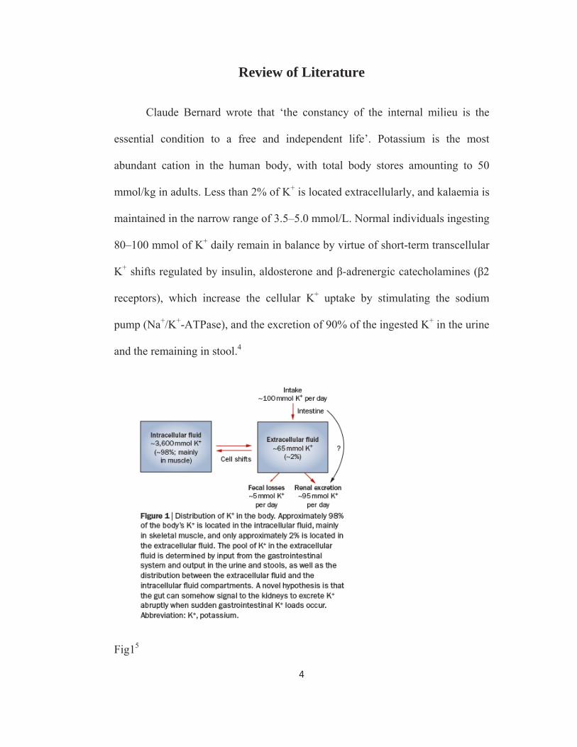
Claude Bernard wrote that ‘the constancy of the internal milieu is the
essential condition to a free and independent life’. Potassium is the most
abundant cation in the human body, with total body stores amounting to 50
mmol/kg in adults. Less than 2% of K+ is located extracellularly, and kalaemia is
maintained in the narrow range of 3.5–5.0 mmol/L. Normal individuals ingesting
80–100 mmol of K+ daily remain in balance by virtue of short-term transcellular
K+ shifts regulated by insulin, aldosterone and β-adrenergic catecholamines (β2
receptors), which increase the cellular K+ uptake by stimulating the sodium
pump (Na+/K+-ATPase), and the excretion of 90% of the ingested K+ in the urine
and the remaining in stool.4
Fig15
5
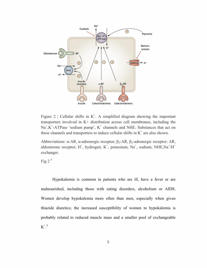
Figure 2 | Cellular shifts in K+. A simplified diagram showing the important transporters involved in K+ distribution across cell membranes, including the Na+,K+-ATPase ‘sodium pump’, K+ channels and NHE. Substances that act on these channels and transporters to induce cellular shifts in K+ are also shown.
Abbreviations: α-AR, α-adrenergic receptor; β2-AR, β2-adrenergic receptor; AR, aldosterone receptor; H+, hydrogen; K+, potassium; Na+, sodium; NHE,Na+/H+ exchanger.
Fig 2 5
Hypokalemia is common in patients who are ill, have a fever or are
malnourished, including those with eating disorders, alcoholism or AIDS.
Women develop hypokalemia more often than men, especially when given
thiazide diuretics; the increased susceptibility of women to hypokalemia is
probably related to reduced muscle mass and a smaller pool of exchangeable
K+.5
6
The most prominent clinical features of hypokalemia or potassium
depletion are neuromuscular, although other systems, such as cardiovascular and
gastrointestinal, may also be affected. Some patients complain of muscular
weakness, especially of the lower extremities, while marked and generalized
weakness of skeletal muscles is common with more severe potassium depletion.
Very severe hypokalaemia may lead to virtually total paralysis including
respiratory, bulbar and cranial musculature. Deaths from respiratory failure and
arrhythmia have been reported.
Differential diagnosis of hypokalaemic paralysis (modified from ref 6)
Transcellular distribution of K (no depletion)
• Familial periodic paralysis
• Thyrotoxic periodic paralysis
• Barium poisoning
Actual K depletion
Renal loss
• RTAs
• Sjögren’s syndrome
• Medullary sponge kidney
• Chronic toluene exposure
• Fanconi’s syndrome
7
• Tubulointerstitial disorder, Gitelman syndrome, Barrter syndrome
• Primary hyperaldosteronism
• Drug-induced: Gentamicin, carbenicillin, amphotericin B, degraded
tetracycline, vitamin B12, alcohol, carbenoxolone, overdoses of
chloroquine, the antipsychotic agents risperidone or quetiapine9
• Others - water intoxication, nephrotic syndrome,diuretic phase of acute
tubular necrosis, treatment phase of diabetic ketoacidosis, chlorothiazide-
associated hypokalaemia, hypokalaemia following ureterosigmoidostomy
, Licorice6 consumption, Leptospirosis18, Cleistanthus collinus
poisoning.19
Extra-renal loss
• Celiac disease
• Tropical sprue
• Acute gastroenteritis
• Short bowel syndrome
Hypokalemic periodic paralysis
Ion channels constitute a class of proteins that is ultimately responsible
for generating and orchestrating the electrical signals passing through the
thinking brain, the beating heart, and the contracting muscle. Defective ion-
channel proteins are responsible for cystic fibrosis, the long-QT syndrome,
8
heritable hypertension (Liddle’s syndrome), familial persistent hyperinsulinemic
hypoglycemia of infancy, hereditary nephrolithiasis (Dent’s disease), and a
variety of hereditary myopathies, including generalized myotonia (Becker’s
disease), myotonia congenita (Thomsen’s disease), periodic paralyses, malignant
hyperthermia, and central core storage disease7 .
The periodic paralyses, paradigm for muscle channelopathies
The periodic paralyses are rare and dominantly inherited disorders
characterized by neuromuscular symptoms (paralysis and myotonia) associated
with marked and transitory variations of K+ levels in the plasma 8 . Symptoms
usually appear during the first or second decade of life and can be life-
threatening if heart or respiratory muscles are involved.
Familial Hypokalemic periodic paralysis
Nomenclature
Hypokalemic periodic paralysis was also known as Cavare-Romberg
syndrome, Cavare-Westphal syndrome, Cavare-Romberg-Westphal syndrome,
Westphal's disease, Westphal's neurosis . Karl Friedrich Otto Westphal (1833-
1890) first described extensively and convincingly the main characteristics of the
disease, which had previously been described as "periodic palsy" by Musgrave
9
in 1727, Cavare in 1853, and Romberg in 1857. Hartwig reported a case of palsy
with muscle inexcitability provoked by rest after exercise in 1875. It was not
until 1887 that a dominant pedigree was described by Cousot10.
Ion channels involved 11
Sodium channel
Hypokalemic PP (HypoPP2)
Calcium channel
Hypokalemic PP (HypoPP1)
Potassium channel
Andersen-Tawil syndrome
(Hyperkalemic PP or hypokalemic PP)
Prevalence 12
The prevalence of HOKPP is unknown but thought to be approximately
1:100,000. Inheritance is autosomal dominant with reduced penetrance in
women. Symptoms begin early in life and rarely after the age of 25 years.
Typically affects Caucasians with male preponderance. Male to female ratio is
3:1.2 Age of onset was earlier and the duration of episodes was longer in
HypoPP1 (10 years; 20 h) versus HypoPP2 (16 years;1 h).
10
Genetics and Inheritance 10
Calcium Channel –
• Voltage-Dependent, L Type,
• Skeletal Muscle Dihydropyridine-Sensitive, Alpha-1 Subunit;
• CACNA1S Gene map locus 1q32
• Seven Mutations described to Date: ARG1239HIS, ARG528HIS ,
ARG528GLY , ARG1239GLY , ARG1086CYS & ARG1086HIS
Sodium Channel –
• Voltage-Gated, Type IV,
• Alpha Subunit;
• SCN4A; Gene map locus 17q23.1-q25.3
• Six mutations described to date: ARG669HIS , ARG672HIS,
ARG672GLY , ARG672SER , DIII-S4 ARG1132Q and alpha subunit,
between 4th/5th transmembrane segments, domain III SCN4A,
Pro1158Ser.
Potassium channel-
• Inward-rectifying potassium channel Kir2.1
• KCNJ2 gene; Gene map locus 17q24.3;
• More than 20 mutations described
11
Pathophysiology
The pathophysiology behind the disorder is poorly understood. This is a
type of skeletal muscle channelopathy. Several mutations have been reported in
skeletal muscle dihydropyridine receptor calcium channel(CACNL1A3),
tetrodoxin sensitive voltage sensitive sodium channel(SCN4A) and potassium
channel(Kir2.1). However sporadic cases without documented evidence of
mutation have been reported8.
Cav1.1 is the main subunit of the voltage-gated pentameric L type Ca2+
channel complex (also called the dihydropyridine receptor) located in the
transverse (t) tubular system. Via Cav1.1, a t-tubular action potential activates
the Ca2+ release channel (also called the ryanodine receptor) and the
Ca2+released from the sarcoplasmic reticulum activates the contractile
machinery. Hypokalemic PP types 1 and 2 are clinically similar and in both
responsible channels, the mutations are located exclusively in the voltage
sensing S4 segments: those of SCN4A are situated in domain 2 and those of
Cav1.1 in domains 2 or 4. Functionally, the inactivated state is stabilized in the
Na+ channel mutants, while the channel availability is reduced for the
Ca2+channel mutants. It is still unclear how the loss-of-function mutations of
these two cation channels can produce the long-lasting and pronounced
membrane depolarization that inactivates the sodium channels and thereby leads
to the fiber inexcitability8 .
12
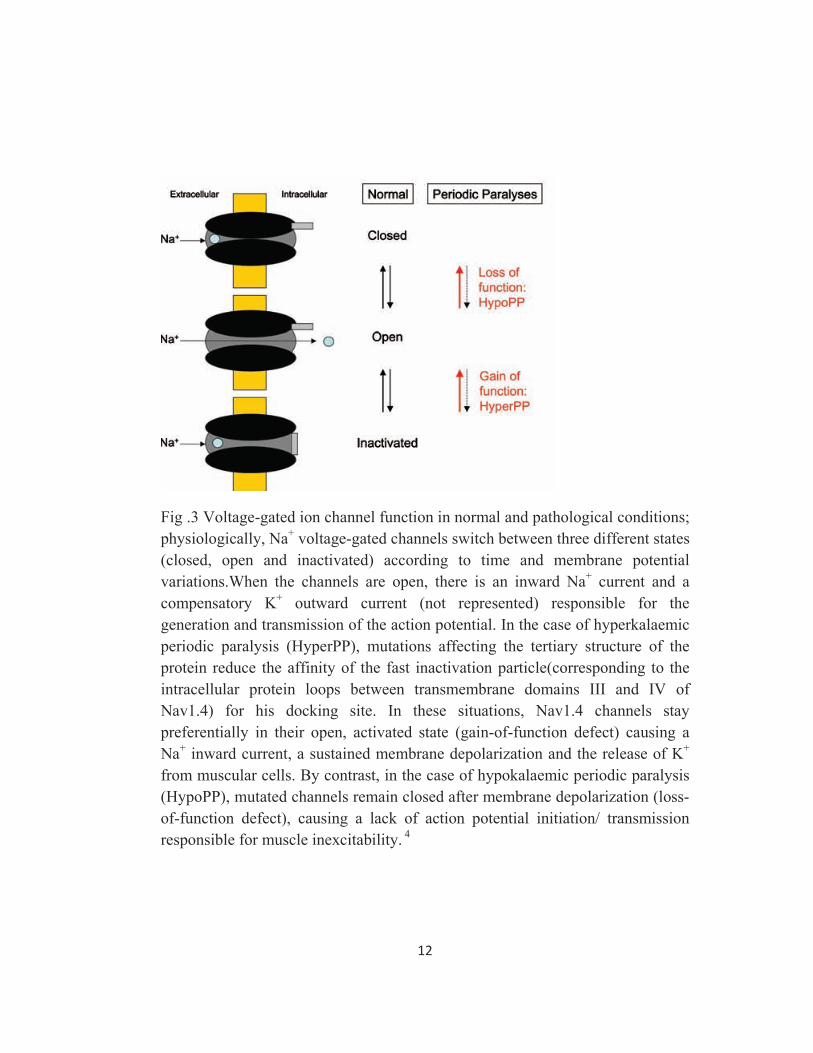
Fig .3 Voltage-gated ion channel function in normal and pathological conditions; physiologically, Na+ voltage-gated channels switch between three different states (closed, open and inactivated) according to time and membrane potential variations.When the channels are open, there is an inward Na+ current and a compensatory K+ outward current (not represented) responsible for the generation and transmission of the action potential. In the case of hyperkalaemic periodic paralysis (HyperPP), mutations affecting the tertiary structure of the protein reduce the affinity of the fast inactivation particle(corresponding to the intracellular protein loops between transmembrane domains III and IV of Nav1.4) for his docking site. In these situations, Nav1.4 channels stay preferentially in their open, activated state (gain-of-function defect) causing a Na+ inward current, a sustained membrane depolarization and the release of K+ from muscular cells. By contrast, in the case of hypokalaemic periodic paralysis (HypoPP), mutated channels remain closed after membrane depolarization (loss-of-function defect), causing a lack of action potential initiation/ transmission responsible for muscle inexcitability. 4
13
Kir2.1 channels stabilize the resting membrane potential in skeletal and
cardiac muscle. Reduced Kir2.1 channel function in skeletal muscle may cause
sustained membrane depolarization, leading to failure of action potential
propagation and flaccid paralysis12.
Clinical features and Diagnosis
The attack frequency varies from daily to yearly and attacks frequently
lasts for 3 to 4 hours to a day or more and are frequently precipitated by sleep or
rest after exercise, alcohol or high carbohydrate meal and almost never occur
during exercise. The limb muscles are more affected than the trunk muscles and
proximal muscles are more affected than the distal ones.11 Diagnosis is made by
demonstrating the presence of low potassium during a paralytic attack and by
excluding other secondary causes of hypokalemia. ECG will show features of
hypokalemia. A clinical diagnosis of sporadic periodic paralysis can be made if
hyperthyroidism and family history of HPP are both absent13.
Thyrotoxic periodic paralysis
Epidemiology
Incidence of Thyrotoxic periodic paralysis is highest among Asian
population. Approximately 2% of patients in China and Japan reportedlyhave
TPP. Despite a higher incidence of thyrotoxicosis in women TPP occurs
predominantly in men; the male-female ratio is approximately 20:1. Presence of
Certain HLA antigen subtypes such as HLA-DRw8 , A2, Bw22,Aw19 and B17
14
is suspected to make some of the Asian population susceptible to TPP. TPP
occurs most commonly in summer and autumn. Increased consumption of sweet
drinks, outdoor activities and exercise and increased potassium loss in sweat are
possible explanations for the seasonal pattern14.
Pathophysiology15
• Hyperthyroidism results in hyperadrenergic state. β2 adrenergic
stimulation increases the activity of Na+,k+-ATPase pump.
• Thyroid hormone per se increases the activity of Na+,k+-ATPase pump.
• Hyperinsulinemia observed in patients with acute attack of TTP indirectly
also stimulates Na+,k+-ATPase pump.
Clinical & Laboratory findings
Occurs in persons aged 20-40 years in contrast to familial HPP which
occurs in persons aged less than 20 years15. Nearly half of the patients with TPP
have no obvious symptoms related to hyperthyroidism during an attack17.
Patients usually have abnormal thyroid functions, hypokalemia,with a urinary
potassium-creatinine ratio less than 2meq/mmol. Urinary phosphate excretion is
reduced remarkably as a result of increased shift of phosphate into the cells .
Hypercalciuria and hypophosphaturia need to be emphasized in diagnosing
TPP14.
15
TPP attacks occur only when thyrotoxicosis is present. Attacks can be
induced by insulin and carbohydrate administration in hyperthyroid patients
with a history of TPP, but not in TPP patients who have become euthyroid.
Paralytic attacks can recur with relapse into a thyrotoxic state, and can be
induced by exogenous thyroid hormone.17
Barium poisoning
The first cases were referred to as Pa Ping disease, due to an outbreak of
paralysis in the Pa Ping area of the Szechwan province of China caused by
ingestion of table salt contaminated by a periodic barium salt. Most of the
instances of acute toxicity have occurred due to ingestion of barium carbonate
(rodenticide), food contaminated by barium carbonate (used in error instead of
potato meal), or carelessness in handling rat poison whereby it is mixed with
flour and eaten. The fatal dose of barium carbonate is about 0.8 g. However,
barium doses as low as 0.2–0.5 mg/kg body weight, resulting from barium
carbonate or chloride ingestion, have been found to produce toxicity in adults. A
shift of potassium from extracellular to intracellular fluid is the basis of acute
hypokalaemia in cases of barium carbonate toxicity. The exact mechanism of
hypokalaemia is not known, however, it may be due to the activation of sodium–
potassium-stimulated ATPase at the cell surface causing potassium entry into the
cell at the cost of extracellular fluid. Barium is reported to block the potassium
channels and thereby reduce the potassium efflux from muscles. It also
16
competitively reduces the permeability of the cell membrane to potassium which
may lead to membrane depolarization. The treatment of barium-induced
hypokalaemic paralysis has been intravenous potassium administration.The
potassium reverses the hypokalaemia as well as displacing barium from
potassium channels, allowing it to be excreted in urine. 6
Renal Tubular Acidosis
Introduction
Renal tubular acidosis (RTA) comprises of a group of disorders
characterized by a low capacity for net acid excretion and persistent
hyperchloremic metabolic acidosis.
Physiology
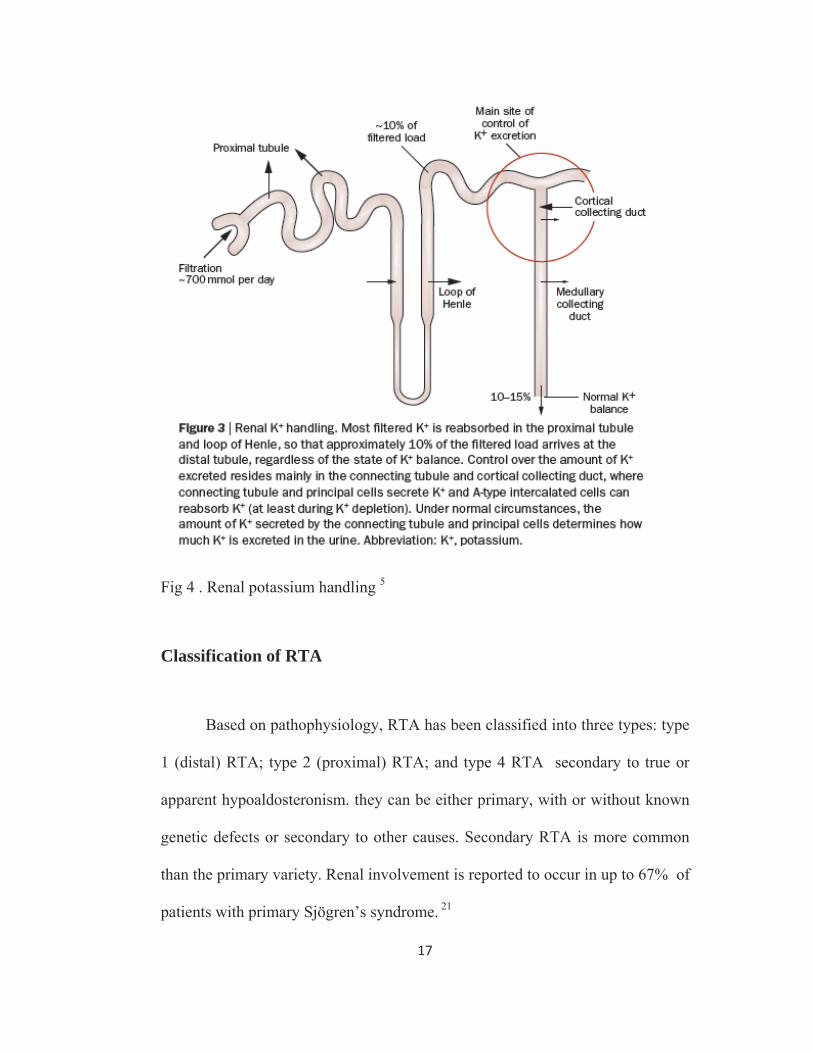
The proximal renal tubule is the site of the bulk of solute and water
reabsorption in the nephron. Approximately 60% of the filtered sodium (Na +) is
reabsorbed in the proximal segments, along with water, potassium (K+),
bicarbonate (HCO3−), phosphate, amino acids and low molecular weight
proteins. In contrast, the distal tubule has a specialized role in the final
modification of urine concentration and pH. Specialized transporters are
involved in the regulation of Na+ and K+ reabsorption and H+ secretion.20
17
Fig 4 . Renal potassium handling 5
Classification of RTA
Based on pathophysiology, RTA has been classified into three types: type
1 (distal) RTA; type 2 (proximal) RTA; and type 4 RTA secondary to true or
apparent hypoaldosteronism. they can be either primary, with or without known
genetic defects or secondary to other causes. Secondary RTA is more common
than the primary variety. Renal involvement is reported to occur in up to 67% of
patients with primary Sjögren’s syndrome. 21
18
Pathophysiological basis
The primary defect in proximal RTA is reduced renal threshold for HCO3-
, resulting in bicarbonaturia. Renal NaHCO3 losses lead to intravascular volume
depletion, which in turn activates the renin-angiotensin-aldosterone system.
Distal Na+ delivery is increased as a result of the impaired proximal reabsorption
of NaHCO3. Because of the associated hyperaldosteronism and increased distal
nephron Na+ reabsorption, there is increased K+ secretion. The net result is renal
potassium wasting and the development of hypokalemia. In the steady state,
when virtually all the filtered HCO3− is reabsorbed in the proximal and distal
nephron, renal potassium wasting is less and the degree of hypokalemia tends to
be mild. Proximal RTA may represent isolated or generalized proximal tubular
dysfunction, the latter (Fanconi syndrome) characterized by tubular proteinuria
and aminoaciduria and variable degrees of bicarbonaturia, phosphaturia, Na +
and K + wasting and glucosuria22. K + wasting is enhanced due to increased distal
tubular delivery of Na+ and hyperaldosteronism secondary to volume
contraction. Proximal RTA without Fanconi syndrome may be inherited as
autosomal dominant and recessive form.
The diagnosis of proximal RTA calls for study of other proximal tubular
functions. Proximal RTA should be suspected in a patient with a normal anion
gap acidosis and hypokalemia who has an intact ability to acidify the urine to
below 5.5 while in a steady state.23 Proximal tubular dysfunction, such as
19
euglycemic glycosuria, hypophosphatemia, hypouricemia, and mild proteinuria,
helps support this diagnosis. The UAG is greater than zero, indicating the lack
of increase in net acid excretion. Disorders that are associated with proximal
RTA and Fanconi syndrome should be specifically screened for, including
cystinosis, Lowe's syndrome, galactosemia and Wilson's disease24.
Causes of Type 2 RTA22
Not associated with Fanconi syndrome
Sporadic
Familial
Disorders of Carbonic anhydrase
Drugs: Acetazolamide, sulfanilamide, topiramate
Carbonic anhydrase II deficiency
Associated with Fanconi syndrome
Selective(no systemic disease present)
Sporadic
Familial
AR proximal RTA with ocular abnormalities
AR proximal RTA with osteopetrosis and cerebral calcification
Generalized(systemic disorder present)
Genetic disorders
Cystinosis
Wilson’s disease
20
Hereditary fructose intolerance
Lowe syndrome
Metachromatic leukodystrophy
Dysproteinemic states
Myeloma kidney
Light chain deposition disease
Primary and Secondary hyperparathyroidism
Drugs and toxins
Outdated tetracycline
Iphosphamide
Gentamicin
Lead, Cadmium, Mercury
Tubulointerstitial disease
Post-tansplant rejection
Balkan nephropathy
Medullary cystic disease
Others
Bone fibroma
Osteopetrosis
Paroxysmal Nocturnal Hemoglobinuria
21
Hypokalemic Distal RTA
Metabolic acidosis secondary to decreased secretion of H+ ions in the
absence of marked decrease in the glomerular filtration rate is characteristic of
distal RTA. Patients with distal RTA are unable to excrete ammonium (NH4+)
ions in amounts adequate to keep pace with a normal rate of acid production. In
hypokalemic distal RTA, urine pH cannot reach maximal acidity (i.e., remains
>5.5) despite systemic acidemia indicating low H+ concentration in the collecting
duct. Distal RTA can be caused by either impaired H+ secretion (secretory
defect) or an abnormally permeable distal tubule, resulting in increased backleak
of normally secreted H+ (gradient defect); it may be genetic or acquired22. In the
secretory defect, the rate of secretion of H+ is low for the degree of acidosis.
Ideally, with a rate defect the ability to maximally acidify the urine should be
retained. However, with a severe rate defect, the time spent in tubule lumen may
be insufficient for acidification, and there is failure to maximally decrease the
urine pH. The defect in secretory distal RTA may be secondary to defective
function of H+ ATPase, H + /K + ATPase, or the Cl - / HCO3− exchanger.
In distal RTA, the titrable acidity and NH4+ secretion is low resulting in
systemic acidosis. The development of systemic acidosis tends to diminish net
proximal fluid reabsorption with an increase in distal delivery, resulting in
volume contraction and activation of the renin-aldosterone system. Increased
distal Na+ delivery coupled to increased circulating levels of aldosterone then
leads to increased renal K+ secretion22. Incomplete distal RTA is a variant or
22
milder form of classic distal RTA, in which there is defective tubular H+
secretion but plasma HCO3− levels are normal. Daily net acid excretion is
maintained by enhanced ammoniagenesis. Hypercalciuria and hypocitraturia are
present, and there is a risk for nephrolithiasis and nephrocalcinosis.25
Distal RTA may be a primary disorder, either idiopathic or inherited, but
it most commonly occurs in association with a systemic disease, of which one of
the most common is Sjögren's syndrome. Hypergammaglobulinemic states as
well as drugs and toxins may also cause this disorder. A common cause of
acquired distal RTA is glue sniffing. Inhalation of toluene from the fumes of
model glue, spray paint, and paint thinners can give rise to hypokalemic normal
gap acidosis through multiple mechanisms. First, toluene inhibits collecting duct
proton secretion. Second, metabolism of toluene produces the organic acids
hippuric and benzoic acid. These are buffered by sodium bicarbonate, resulting
in metabolic acidosis and the production of sodium hippurate and sodium
benzoate. If plasma volume is normal, these salts are rapidly excreted in the
urine, and a non–anion gap metabolic acidosis develops. If plasma volume is
decreased, urinary excretion is limited, they accumulate, and an anion gap
metabolic acidosis develops.22
Causes of Type 1 RTA 22 Primary
Idiopathic
Familial
23
Secondary
Autoimmune disorders
Hypergammaglobulinemia
Sjogren’s syndrome
Primary biliary cirrhosis
SLE
Genetic diseases
AD type 1 RTA
AR
Drugs and toxins
Amphoterecin B
Toluene
Disorders with nephocalcinosis
Hyperparathyroidism
Vit D intoxication
Idiopathic hypercalciuria
Tubulointerstitial diseases
Obstructive uropathy
Renal transplantation
In patients with minimal disturbances in blood pH and plasma HCO3−
concentration, a test of urinary acidification is required. Traditionally, such a test
involved oral NH4Cl administration to induce metabolic acidosis with
assessment of the renal response by serial measurement of urine pH. Many
patients poorly tolerate NH4Cl ingestion because of gastric irritation, nausea, and
vomiting. An alternative way to test the capacity for distal acidification is to
administer furosemide and the mineralocorticoid fludrocortisone
24
simultaneously.26 The combination of both increased distal Na+ delivery and
mineralocorticoid effect will stimulate distal H+ secretion by both an increase in
the luminal electronegativity and a direct stimulatory on H+ secretion. Normal
subjects will lower urine pH to values below 5.5 with either maneuver.
Urine pH & Ammonium chloride loading test 27
Urine pH is useful for assessing the overall integrity of distal urinary
acidification. In the presence of systemic acidosis, present spontaneously or
induced by ammonium chloride load, the urine pH is normally <5.5. Presence of
urine pH >5.5 during metabolic acidosis suggests defective distal secretion of
H+. It should however be appreciated that the urine pH measures the
concentration of free H+ in the urine. This constitutes <1% of the total amount of
H+ secreted in the distal nephron during systemic acidosis, since most protons
are excreted as NH4+ or titrable acidity. Urine pH values < 5.5 are seen in
subjects with proximal RTA during systemic acidosis and low filtered load
(plasma HCO3- <15 mEq/L), or in patients with selective aldosterone deficiency.
If systemic acidosis is absent, an oral ammonium chloride challenge (0.1
g/Kg) might be given, followed by the measurement of urine pH every hr for the
next 28 hr. If the plasma total CO2 content falls by 3-5 mEq/L, the urine pH
should fall to <5.5. Another protocol involves giving the same dose of
ammonium chloride daily for 3-5 days, followed by measurement of urine pH
and urinary NH4+ excretion; the latter should increase 35 times the baseline by
25
the third day of induced acidosis. In patients with liver disease, calcium chloride
may be used as an acidifying agent at a dose of 2 mEq/Kg. The pH is measured
electrometrically on fresh voided, early morning urine specimen. The use of
dipstick is not recommended. Urine kept standing is likely to get contaminated
or infected with urea-splitting organisms, resulting in high urine pH. The urine
pH must be evaluated in conjunction with the urinary NH4+ content to assess the
acidification process. Low urine pH does not always imply an intact urinary
acidification mechanism, if excretion of NH4+ is low, as might occur in proximal
RTA.
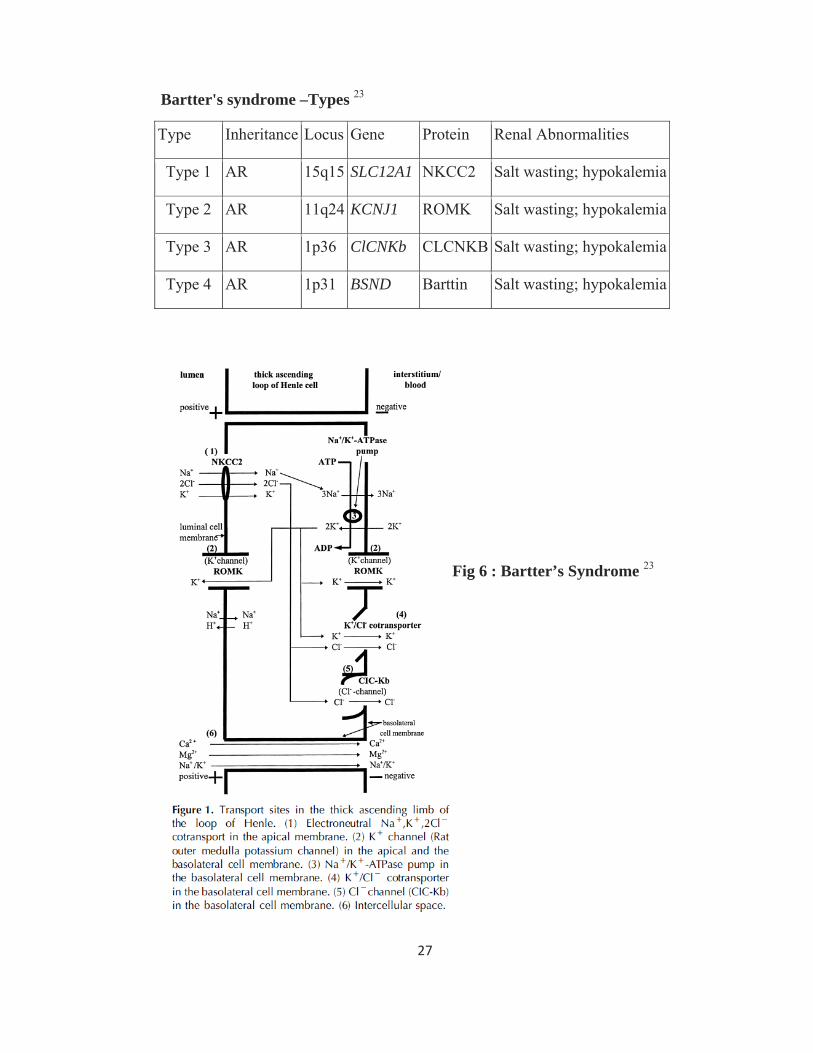
Bartter Syndrome
Introduction
Bartter syndrome is a heterogeneous group of disorders characterized by
primary renal tubular disorders associated with hypokalemic metabolic alkalosis
and hyperreninemia with normotension. Bartter syndrome is rare and is
manifested in childhood or in the perinatal period with severe hypokalemia,
metabolic alkalosis, and low-normal blood pressure, all of which are caused by
tubular wasting of Na+ and Cl−. In contrast, Gitelman's syndrome is mostly a
disorder of adults, and hypomagnesemia is a defining feature22.
Bartter's syndrome may result from mutations affecting any of the four
ion transport proteins in the TAL. The proteins affected include the apical loop-
26
diuretic sensitive sodium-potassium-chloride co-transporter NKCC2 (type 1), the
apical potassium channel ROMK (type 2), and the basolateral chloride channel
ClC-Kb (type 3). Bartter type 4 results from mutations in barttin, an essential
subunit of ClC-Ka and ClC-Kb that enables transport of the chloride channels to
the cell surface. The TAL transporters function in an integrated manner to
maintain both the electrical potential difference and sodium gradient between the
lumen and the cell. Loss of the lumen-positive electrical transport potential that
normally drives the paracellular reabsorption of sodium, calcium, and
magnesium causes NaCl wasting, hypercalciuria, and mild hypomagnesemia.
The clinical syndrome mimics a state induced by chronic ingestion of a loop
diuretic. Bartter syndrome from mutations in NKCC2, ROMK, or barttin has a
more severe phenotype than that caused by mutations of ClC-Kb. The more
severe phenotype typically presents in the perinatal period and is called antenatal
Bartter syndrome. The milder variety is called classic Bartter syndrome( Type
3)22.
In some cases prenatal diagnosis is possible in the presence of important
signs and symptoms such as polyhydramnios, polyuria and dehydration. In other
patients diagnosis is usually made during childhood due to the appearance of
tetanic crises or growth failure. Other patients are completely asymptomatic and
the diagnosis is made in adolescent or adult life following blood tests prescribed
for intercurrent diseases.
27
Bartter's syndrome –Types 23
Type Inheritance Locus Gene Protein Renal Abnormalities
Type 1 AR 15q15 SLC12A1 NKCC2 Salt wasting; hypokalemia
Type 2 AR 11q24 KCNJ1 ROMK Salt wasting; hypokalemia
Type 3 AR 1p36 ClCNKb CLCNKB Salt wasting; hypokalemia
Type 4 AR 1p31 BSND Barttin Salt wasting; hypokalemia
Fig 6 : Bartter’s Syndrome 23
28
Neonatal Bartter’s syndrome
Features of neonatal Bartter’s syndrome include marked polyhydramnios,
premature delivery, weight loss, lethargy, failure to thrive and polyuria28. An
important biochemical abnormality of the amniotic fluid is consistently elevated
chloride levels. In the first week of life, laboratory investigation shows a
metabolic alkalosis associated with hypokalaemia. The urine has low specific
gravity with very high sodium, chloride and calcium levels, while potassium is
normal. However, after 1–3 weeks, the level of potassium in the urine rises to
considerably above normal, with relatively less sodium than in the first week of
life. Prostaglandin levels are high, both in blood and in urine29.
Molecular basis
A genetic heterogeneity was clearly demonstrated by Simon et al in those
with the antenatal (hypercalciuric) variant of Bartter syndrome, also termed
hyper prostaglandin E syndrome . The hypothesis that a loss of function of the
absorptive form of the bumetanide-sensitive Na-K-2Cl reabsorption of this
segment could produce the features of these patients was proved by Simon et al
who demonstrated that the human NKCC2 gene maps on chromosome 15q15-
21, is encoded by 26 exons, like TSC, and is made up of 1,099 amino acids. The
same group subsequently showed that mutations in another gene encoding the
inwardly-rectifying renal potassium channel (ROMK) may also cause the
29
hypercalciuric variant of Bartter syndrome. This last gene was mapped on
chromosome 11q21-2530,31.
Defects in either Na+-K+-2CI- cotransport or K+ channels will result in
malreabsorption of Na+, K+, Cl-, and Ca2+ in the thick ascending limb of loop of
Henle primarily, with subsequent reabsorption of H2O in the descending loop of
Henle. The result of such a defect will be the delivery of large volumes of urine
with a high content of Na+, K+, Cl-, and Ca2+ to the distal tubule. In the distal
tubule, part of the delivered Na+ will be reabsorbed in exchange for intracellular
K+. By this action, partial but incomplete concentration of the intraluminal fluid
will be accomplished, while more potassium wasting becomes evident.
However, this impaired sodium absorption in the thick ascending limb of loop of
Henle will result in increased levels of prostaglandin E2. This interrelation has
been documented in normal individuals using loop diuretics32.
Increased prostaglandin E2 levels will exacerbate the primary defect of chloride
transport in the thick ascending loop of Henle which will28:
(i) stimulate the renin-angiotensin- aldosterone axis causing hypokalemia
due to increased aldosterone activity;
(ii) impede ROMK channel activity and hence decrease NaCl transport;
and
(iii) impede H2O reabsorption in the collecting ducts due to a secondary
effect on vasopressin activity, resulting in hyposthenuria.
30
Classic Bartter Syndrome
(Bartter syndrome type 3)
Classical Bartter syndrome is characterized by early childhood onset. The
patients fail to thrive but have no tetany. Symptoms may include polyuria,
polydipsia, vomiting, constipation, salt craving, and a tendency to dehydration.
Failure to thrive and growth retardation follows if treatment is not initiated.
However the normal adult height usually achieved by untreated individuals is
due to a delayed adolescent growth spurt. They have hypokalaemic metabolic
alkalosis and their urinary Ca2+ is either normal or slightly elevated, with the
urine concentration being almost normal. There are reports of Bartter syndrome
presenting with hypokalemic periodic paralysis.
Molecular Basis
The biochemical abnormalities of classical Bartter syndrome are all
suggestive of a defect related to Cl- transport in the medullary thick ascending
loop of Henle. However, the precise pathway involved is not yet clear. The
familial cases of classical Bartter syndrome are inherited as an autosomal
recessive entity. A group of patients with this phenotype all had either a large
deletion or nonsense, missense, or splice mutations of the gene (CIC-Kb,
chromosome 1p36) encoding the renal chloride channels of the basolateral cell
membrane.
31
However, in some patients with classical Bartter syndrome, no
abnormality in this gene could be identified. It has therefore been suggested that
NaCl transport in the ascending loop of Henle (and the relevant gene/s) may also
be involved28.
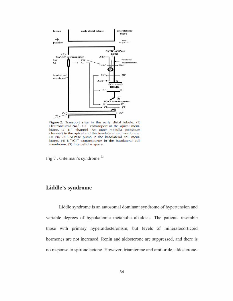
Gitelman’s syndrome
Gitelman's syndrome is due to mutations in the thiazide-sensitive Na-Cl
co-transporter (NCCT) in the DCT. Defects in NCCT in Gitelman's syndrome
impair sodium and chloride reabsorption in the DCT and, thus, resemble the
effects of thiazide diuretics.
Onset is late, usually after the age of 20 years. Patients present with
fatigue, muscle weakness and recurrent episodes of tetany33 Biochemically, there
is metabolic alkalosis, (serum bicarbonate >29 meq/1) profound hypokalaemia,
(serum potassium <3 meq/l; normal >3.5 meq/1) hypomagnesaemia (serum
magnesium <0.5 meq/l; normal 0.8–1.0 meq/1) and hypocalciuria, (urinary
calcium <2 mg/kg per day).35 Urinary concentrating ability in this disease is
mildly impaired.
Pathophysiology
The basic pathology in this disease is an impaired Na-Cl cotransporter in
the distal nephron. Distal tubule and collecting duct together reabsorb about 12%
of the filtered Na+. The early distal tubule, also called the cortical diluting
32
segment, is the site of absorption of NaCl by Na+-Cl- cotransport (NCCT) and is
the site of action of thiazide diuretics. A similar biochemical abnormality can be
seen in long-term use of thiazide diuretics in an otherwise normal individual.
NaCl wasting in this part of the distal nephron will lead to mild hypovolemia,
and stimulation of the renin- angiotensin axis. Simon et al. showed that there is a
complete linkage of Gitelman’s syndrome to the locus encoding the renal
thiazide sensitive Na+- Cl- cotransporter on chromosome 16q13, with an
autosomal recessive pattern and a 99% penetrance. Mutant alleles in this disease
have been reported by Simon and others. 28
The late distal tubule and collecting duct have two kinds of cells, each
with special feature and function. Principal cells reabsorb Na+ and H2O and
secrete K+ . Aldosterone acts on principal cells to increase Na+ reabsorption and
increase K+ secretion. Intercalated cells secrete H+ ions in exchange for
reabsorption of K+ ions. Aldosterone increases H+ ion secretion by intercalated
cells. Impairment of Na+-Cl- cotransport in the early part of the distal tubule
results in excessive amounts of Na+ ion in the late distal tubule. Maximal
reabsorption of Na+ and H2O and maximal secretion of K+ ion by the principal
cells takes place in this segment.At the same time, H+ is excreted by intercalated
cells and this, together with impaired Cl- reabsorption in the early distal tubule,
results in metabolic alkalosis.
33
The high intake of Ca2+ in distal tubule and hence hypocalciuria is
probably due to
(a) decreased apical Na+ uptake driving basolateral Na+/ Ca2+ exchange
with subsequent increase of Ca2+ uptake at the apical membrane level and
(b) decreased intracellular Cl- content increasing the polarity of the apical
cell membrane, which stimulates Ca2+uptake.34
Hypomagnesaemia in Gitleman syndrome is perhaps due to magnesium
wasting in distal convoluted tubules of the nephron due to inhibition of Mg2+
uptake in the presence of hypokalaemia.35 It has also been suggested that the
metabolic alkalosis may be an important cause of hypomagnesaemia by
increasing the resistance of distal tubular cells to Mg2+ uptake.40 With low Mg2+
levels in the blood, magnesium wasting has been observed in patients with
Gitelman’s syndrome, indicating a too-low renal Mg2+ threshold.
34
Fig 7 . Gitelman’s syndrome 23
Liddle’s syndrome
Liddle syndrome is an autosomal dominant syndrome of hypertension and
variable degrees of hypokalemic metabolic alkalosis. The patients resemble
those with primary hyperaldosteronism, but levels of mineralocorticoid
hormones are not increased. Renin and aldosterone are suppressed, and there is
no response to spironolactone. However, triamterene and amiloride, aldosterone-
35
independent inhibitors of distal Na+ transport, correct hypertension, renal K+
loss, and hypokalemia.52
Pathophysiology
Liddle syndrome is related to mutations of the β or γ subunit of the
collecting duct sodium channel ENaC. In Liddle syndrome, the mutated ENaC
protein cannot be recognized by NEDD4, a ubiquitin ligase protein; hence, the
channels remain in the cell membrane for prolonged periods. This action results in
enhanced sodium reabsorption, hypertension, and hypokalemic alkalosis.53
Primary Hyperaldosteronism
Primary hyperaldosteronism accounted for about 42% of patients with
periodic paralysis in a case series from south India36. It is suspected in a patient
with hypokalemic periodic paralysis if there is a combination of metabolic
alkalosis and hypertension. It is then confirmed by imaging studies and
biochemical investigations. Periodic paralysis as a presentation of primary
hyperaldosteronism is commonly reported among the oriental races. In a series
of 50 patients with primary hyperaldosteronism from Taiwan, 42% presented
with periodic paralysis, although all 50 had hypokalaemia37.
36
Approach to the patient with hypokalemic palaysis
Failure to distinguish HPP from non-HPP may lead to overly aggressive
treatment of an apparent potassium deficit, with rebound hyperkalemia on
recovery. In retrospective studies by Manoukain and Lin and colleagues,
rebound hyperkalemia occurred in 30% to 42% of patients with HPP, especially
if more than 90 mmol of potassium chloride was given within 24 hours14. Hence
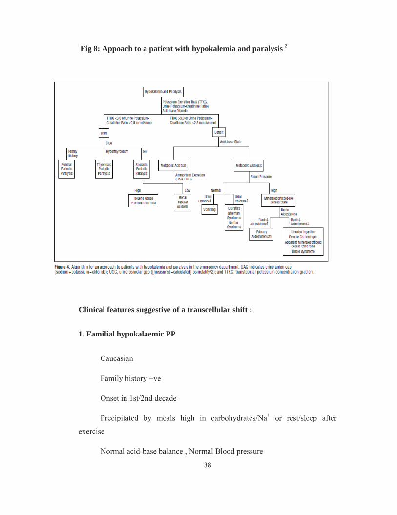
diagnosis of the etiology is important.
Diagnosis for the cause of hypokalemia is based mainly on determining if there
is a net loss of K+ or only a transcellular shift.
Diagnostic tests
Serum K+
Blood urea nitrogen,
Serum creatinine
Acid-base status
Urinary Potassium
Transtubular potassium concentration gradient (TTKG) and
Urinary Potassium creatinine ratio (K+/C)
Thyroid function tests – if features suggestive of a transcellular shift
37
Three urinary indices of renal response to hypokalemia can be easily
measured: spot urine potassium concentration, TTKG, and Urine potassium-
creatinine ratio.2
The spot urine potassium concentration is lower in the HPP than the non-
HPP (<15-20 mmol/L); however, there can overlapping values because polyuria
is common in patients with hypokalemia . The polyuria may be due to thirst or
defective renal concentration in patients with chronic hypokalemia; this leads to
a low value for the urine potassium concentration even if significant renal
potassium wasting is present.
The TTKG is a semiquantitative index for events in the lumen of the
cortical collecting ducts, because it adjusts for the plasma potassium
concentration and for water reabsorption in the medullary collecting ducts.11,18
Although calculating the TTKG is a reliable way to distinguish between HPP
and non-HPP, a TTKG is invalid if theUosm is lower than the Posm in a patient.
The potassium- creatinine ratio is independent of the Uosm. Urine
potassium-creatinine ratio less than 2.5 mmol/mmol and TTKGs less than 3.0 in
hyperosmolar urine are reliable cutoff values to differentiate between patients
with HPP and non-HPP.2
38
Fig 8: Appoach to a patient with hypokalemia and paralysis 2
Clinical features suggestive of a transcellular shift :
1. Familial hypokalaemic PP
Caucasian
Family history +ve
Onset in 1st/2nd decade
Precipitated by meals high in carbohydrates/Na+ or rest/sleep after
exercise
Normal acid-base balance , Normal Blood pressure
39
Low K+ excretion
2. Thyrotoxic PP
Asian
Family history –ve
Onset in 2nd/4th decade
Sign/symptoms of hyperthyroidism
Predominantly males
Normal acid-base balance, Normal Blood pressure
Low K+ excretion
Hypokalemia secondary to K+ loss
Hypokalemia secondary to K+ loss can be differentiated based on clinical
features like Blood pressure and also history suggestive of the aetiologies
mentioned below and also by looking at the acid base status and urinary K+ loss
in the patient.
Determination of the urinary K+ concentration alone might be misleading,
because K+ depletion can cause polyuria and a relatively low K+
concentration(<20mmol/l) could still represent substantial urinary K+ loss. The
TTKG provides a better way to evaluate the excretory process. 13
The transtubular K+ concentration ([K+]) gradient (TTKG) is calculated using the
following formula:
40
TTKG = [urine K+/plasma K+] / [urine osmolality/plasma osmolality]
To differentiate individual causes of renal K+ wasting the following
investigations may be done
Urinary pH
Ammonium loading test
Serum and urinary electrolytes – Na , K, Cl, Ca, Phosphate,Mg
Alkaline phosphatase
Creatine kinase
24 h urinary calcium, inorganic phosphate, and creatinine
Urinary aminoacids
Seum aldosterone and renin levels
Management
Treatment of Periodic paralyses
Body potassium stores are normal in patients with FPP and TPP. Hence
the aim of potassium supplementation is to normalize the plasma potassium
instead of repairing the potassium deficit. Initial treatment of acute episodes of
FPP is oral potassium supplementation (0.2-0.4mmol/kg) every 15 to 30
41
minutes depending on potassium levels, ECG changes and muscle strength.
Intravenous therapy is necessary when the patients are unable to swallow or
vomiting. Prophylaxis against recurrent episodes has been successful with wide
variety of drugs like acetazolamide (125-1000mg/day), spiranolactone (25-100
mg/day) and triamterine (25-100 mg/day).6
In a study conducted by Lin et al, they found that the average recovery
time is two times shorter in patients with TPP treated with intravenous potassium
chloride supplementation at a rate of 10meq/h than in controls. However
rebound hyperkalemia occurred in 70% of patients treated with potassium
chloride. Patients receiving 50 mmols or less rarely developed hyperkalemia. It
appears that low doses may be efficacious while substantially reducing the risk
of hyperkalemia8. Oral β blockers (propranolol 3-4mg/kg) appear to be effective
with out causing rebound hyperkalemia.
Propranolol is effective in preventing recurrent attacks by suppressing the
activity of Na+,k+-ATPase pump15. Regular potassium chloride supplementation
is futile because potassium levels are normal during the inter attack period.
Acetazolamide can precipitate recurrent attacks of TPP and should not be used8.
Management of Hypokalemia secondary to potassium loss
Any depletion of potassium – renal or non renal has to be corrected by
replacing the lost potassium and also the underlying cause corrected to prevent
futher loss.
42
Treatment of Proximal RTA
Children should treated to prevent growth retardation. Alkali must be
given in large amounts daily, 5 to 15 mmol/kg/day, because bicarbonate is
rapidly excreted in urine. A thiazide diuretic can be used in conjunction with low
salt to reduce the amount of bicarbonate required. Potassium requirements
increase during alkali therapy due to increased renal loss of potassium from
bicarbonaturia38.
Adults with proximal RTA are frequently not treated as aggressively as children
are because of the lack of systemic metabolic abnormalities or bone disease.22
Treatment of Distal RTA
Distal RTA is treated with potassium replacement and alkali solution. The
dose is 0.5 to 2 mmol/kg/day with an aim to keep the bicarbonate level above
18mEq/L. Alkali supplementation is either as sodium bicarbonate or Shohl’s
solution38 . It is a mixture of 98 gm of hydrated sodium citrate and 140 gm of
citric acid dissolved in 1000ml of distilled water. Potassium citrate can be used
instead of sodium citrate if hypokalemia correction is required . In patients with
osteomalacia, calcium and vitamin D form integral part of therapy in addition to
correction of acidosis .
Treatment of Bartter syndrome
The therapeutic management of Bartter syndrome is composed of two
major aspects:
43
(i) replacement therapy and (ii) use of drugs.
Dietary intake of sodium and potassium should be liberal. Potassium
supplements are usually required.. Nonsteroidal anti-inflammatory drugs
(NSAIDs) are useful, particularly in patients with antenatal Bartter’s syndrome,
since they reduce prostaglandin production. Indomethacin (1 to 3 mg/kg per 24
hours) 39 or ibuprofen is commonly used for both all types and initial response in
good in both types although better in antenatal Bartter.
If not treated, patients may succumb to episodes of dehydration, electrolyte
disturbance, or intercurrent infection. With appropriate therapy, most children
improve clinically and show catch-up growth; pubertal and mental developments
are usually normal. Lifelong therapy is needed. Chronic tubulointerstitial
nephropathy due to persistent hypokalemia, hypercalciuria, and nephrocalcinosis
may lead to progressive decline in renal function22.
Treatment of Gitelman’s syndrome
Replacement therapy is the main treatment for Gitelman’s syndrome,
which means magnesium supplementation throughout life. Administration of
magnesium in the form of MgCl2 partially corrects hypomagnesaemia and hence
prevents the appearance of tetany as well as compensating for ongoing chloride
losses by the kidney.40 Acid-base status, urinary Ca excretion and renin-
angiotensin axis are all corrected. Also correction of hypokalemia may
44
occasionally require the addition of potassium salts and/or anti-aldosterone drugs
such as spironolactone or amiloride.41
Treatment of Liddle’s syndrome
Therapy consists of sodium restriction and K+ supplements. Triamterene
directly inhibits apical Na+ channels, resulting in increased urinary Na+ and
decreased K+ excretion and resolution of hypertension. Amiloride also normalizes
the blood pressure and K+ levels. However, most patients continue to have growth
retardation. Because the pathogenetic disorder is not correctable with age, lifelong
therapy is required.5
45
MATERIALS AND METHODS
Study population
Patients admitted in medical and general wards of Rajiv Gandhi
Government General Hospital, Chennai-3, from January 2009- March 2011 with
acute onset of flaccid weakness and documented serum potassium of < 3.5
mEq/l during the episode were included in the study. Patients with renal failure
(serum creatinine >1.5 mg/dl) were excluded.
Evaluation
Clinical data collected included Age, Sex, ethnic origin, history regarding
the evolution of the symptoms, precipitating factors like, high carbohydrate
intake in the preceding 24 Hrs, alcohol consumption and treatment taken. Any
history of similar disease in the family was enquired about, and reports of
weakness, thyroid disease, diarrhea, vomiting, hypertension, bone pain,
fractures, dry mouth, dry eyes, and kidney disease were recorded. Intake of
drugs like diuretics, β2 agonists, decongestants, insulin, laxatives and
antipsychotics were noted. Excessive caffeine and herbal drugs usage were
excluded.
The examination included anthropometry, pulse, blood pressure, anemia,
thyroid status. Complete neurological examination was performed. Schirmer’s
test was performed in selected patients.
46
Laboratory investigations
All patients underwent:
Routine urine analysis
Haemogram,
Blood urea, serum creatinine, serum electrolytes
Serum calcium, inorganic phosphorus
Serum magnesium
Serum albumin,globulin, alkaline phosphatase, creatine kinase
Spot urine potassium, urine chloride
Spot urine potassium, creatinine ratio
Arterial blood gas analysis
Electrocardiogram
Thyroid Function Tests
Ultrasonogram Abdomen.
Early morning urinary pH
Patients with urinary K+ loss and hyperchloraemic metabolic acidosis with
normal anion gap underwent:
Lip biopsy
SSA,SSB antibody
ANA
Urinary aminoacids.
Urine Bence Jones Protein, serum electrophoresis.
47
Patients with urinary K+ loss and metabolic alkalosis underwent:
24 hrs urinary calcium
Patients with Urinary K+ loss with normal acid base status underwent an
ammonium chloride loading test (0.1 g/kg). TTKG was done when there was
doubt in diagnosis of hypokalemia due to transcellular shifts or due to renal loss.
Serum aldosterone, plasma renin levels, and CT scan of the abdomen was
done in patients presenting with hypertension, hypokalemia and alkalosis to rule
out primary hyperaldosteronism.
Statistical analysis
All data are expressed as mean ± SD. Differences in group means were
compared using one-way analysis of variance (ANOVA). Differences in
categorical variables were compared using Fisher’s exact test. The difference
was considered significant if p-value was > 0.05. Data was analyzed using SPSS
(V: 17) software.
48
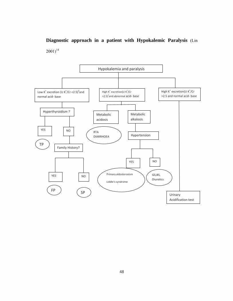
Diagnostic approach in a patient with Hypokalemic Paralysis (Lin
2001)14
Hypokalemia and paralysis
Low K+ excretion (U K+/Cr <2.5)2and normal acid‐ base
High K+ excretion(U K+/Cr
>2.5)2and abnormal acid‐ base
Hyperthyroidism ?
YES NO
Metabolic alkalosis
Metabolic acidosis
NO
High K+ excretion(U K+/Cr >2.5 and normal acid‐ base
YES
Family History?
Hypertension
YES NO
Urinary Acidification test
TP
FP SP
RTA DIARRHOEA
Primary aldosteronism
Liddle’s syndrome
GS,BS, Diuretics
49
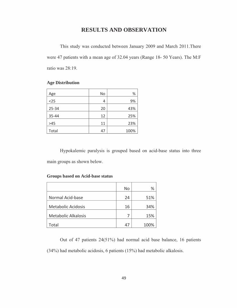
RESULTS AND OBSERVATION
This study was conducted between January 2009 and March 2011.There
were 47 patients with a mean age of 32.04 years (Range 18- 50 Years). The M:F
ratio was 28:19.
Age Distribution
Age No %
<25 4 9%
25‐34 20 43%
35‐44 12 25%
>45 11 23%
Total 47 100%
Hypokalemic paralysis is grouped based on acid-base status into three
main groups as shown below.
Groups based on Acid-base status
No %
Normal Acid‐base 24 51%
Metabolic Acidosis 16 34%
Metabolic Alkalosis 7 15%
Total 47 100%
Out of 47 patients 24(51%) had normal acid base balance, 16 patients
(34%) had metabolic acidosis, 6 patients (15%) had metabolic alkalosis.
50
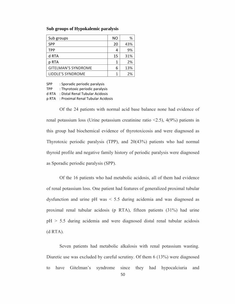
Sub groups of Hypokalemic paralysis
Sub groups NO %SPP 20 43%TPP 4 9%d RTA 15 31%p RTA 1 2%GITELMAN’S SYNDROME 6 13%LIDDLE’S SYNDROME 1 2% SPP : Sporadic periodic paralysis TPP : Thyrotoxic periodic paralysis d RTA : Distal Renal Tubular Acidosis p RTA : Proximal Renal Tubular Acidosis Of the 24 patients with normal acid base balance none had evidence of
renal potassium loss (Urine potassium creatinine ratio <2.5), 4(9%) patients in
this group had biochemical evidence of thyrotoxicosis and were diagnosed as
Thyrotoxic periodic paralysis (TPP), and 20(43%) patients who had normal
thyroid profile and negative family history of periodic paralysis were diagnosed
as Sporadic periodic paralysis (SPP).
Of the 16 patients who had metabolic acidosis, all of them had evidence
of renal potassium loss. One patient had features of generalized proximal tubular
dysfunction and urine pH was < 5.5 during acidemia and was diagnosed as
proximal renal tubular acidosis (p RTA), fifteen patients (31%) had urine
pH > 5.5 during acidemia and were diagnosed distal renal tubular acidosis
(d RTA).
Seven patients had metabolic alkalosis with renal potassium wasting.
Diuretic use was excluded by careful scrutiny. Of them 6 (13%) were diagnosed
to have Gitelman’s syndrome since they had hypocalciuria and
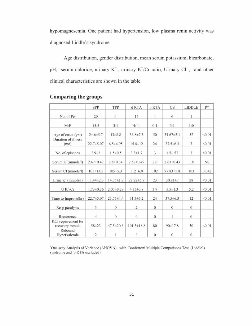
51
hypomagnesemia. One patient had hypertension, low plasma renin activity was
diagnosed Liddle’s syndrome.
Age distribution, gender distribution, mean serum potassium, bicarbonate,
pH, serum chloride, urinary K+ , urinary K+/Cr ratio, Urinary Cl- , and other
clinical characteristics are shown in the table.
Comparing the groups
SPP TPP d RTA p RTA GS LIDDLE P*
No. of Pts 20 4 15 1 6 1
M:F 15:5 3:1 4:11 0:1 5:1 1:0
Age of onset (yrs) 24.6±5.7 43±8.8 36.8±7.3 50 34.67±3.1 32
<0.01 Duration of illness
(mo) 22.7±5.07 6.5±4.95 15.4±12 24 37.5±6.3 3
<0.01
No. of episodes 2.9±2 1.5±0.5 3.3±1.7 3 1.5±.57 3 <0.01
Serum K+(mmols/l) 2.47±0.47 2.8±0.34 2.52±0.49 2.6 2.63±0.43 1.8 NS
Serum Cl-(mmols/l) 105±13.5 105±5.3 112±6.9 102 97.83±5.8 103 0.042
Urine K+ (mmols/l) 11.44±2.3 14.75±1.9 28.22±6.7 23 30.91±7 28 <0.01
U K+/Cr 1.73±0.36 2.07±0.29 4.35±0.8 3.9 5.5±1.3 5.2 <0.01
Time to Improve(hr) 22.7±5.07 23.75±4.4 31.3±6.2 24 37.5±6.3 12 <0.01
Resp paralysis 3 0 2 0 0 0
Recurrence 4 0 0 0 1 0 KCl requirement for
recovery mmols 58±23 47.5±20.6 101.3±18.8 80 90±17.8 50
<0.01 Rebound
Hyperkalemia 2 1 0 0 0 0
*One-way Analysis of Variance (ANOVA) with Bonferroni Multiple Comparisons Test. (Liddle’s syndrome and p RTA excluded)
52
There is a statistically significant difference in the mean age of onset, the
lowest being SPP group followed by Gitelman’s syndrome group, d RTA, and
then by TPP. Serum chloride is highest in d RTA and lowest in Gitelman’s
syndrome group. Highest level of renal K+ loss is seen in Gitelman’s syndrome
followed by d RTA whereas it is normal in SPP and TPP.
HPP (SPP and TPP) required lower potassium chloride supplementation.
Rebound Hyperkalemia (serum K+ >5 mEq/L) after recovery occurred in 2
patients in SPP group and 1 patient in TPP. None of the secondary HP patients
had rebound hyperkalemia.
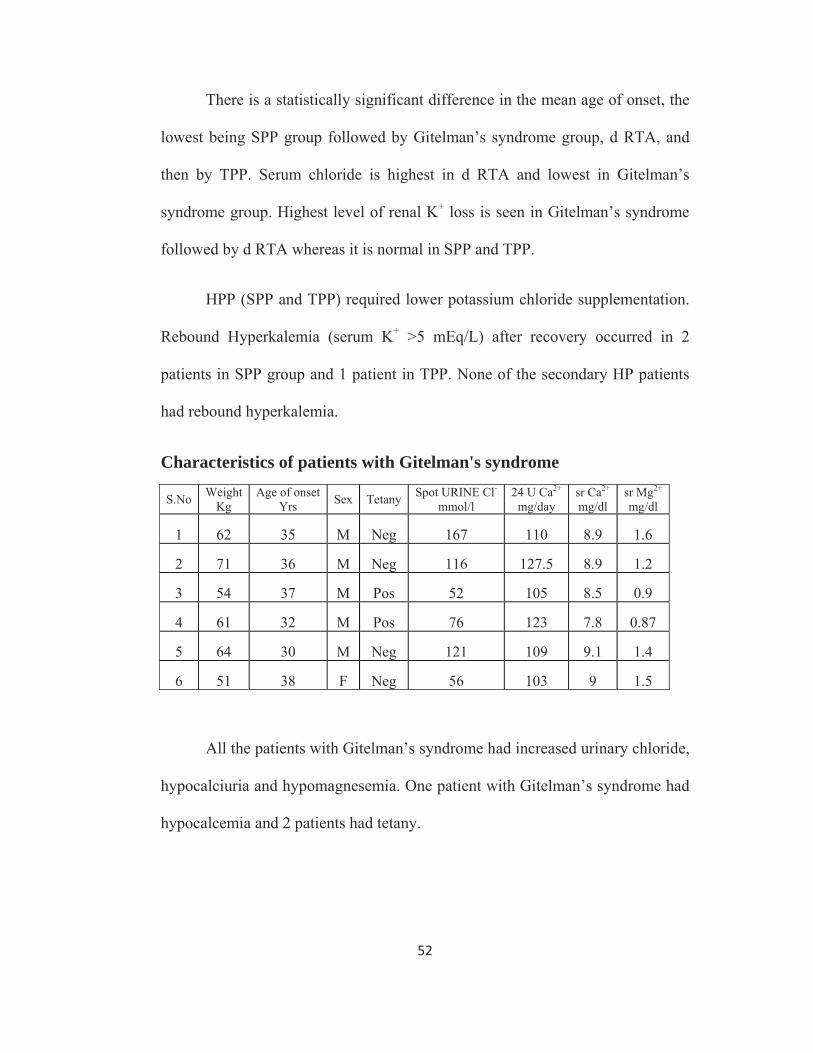
Characteristics of patients with Gitelman's syndrome
S.No Weight Kg
Age of onset Yrs Sex Tetany Spot URINE Cl-
mmol/l 24 U Ca2+
mg/day sr Ca2+
mg/dl sr Mg2+
mg/dl
1 62 35 M Neg 167 110 8.9 1.6
2 71 36 M Neg 116 127.5 8.9 1.2
3 54 37 M Pos 52 105 8.5 0.9
4 61 32 M Pos 76 123 7.8 0.87
5 64 30 M Neg 121 109 9.1 1.4
6 51 38 F Neg 56 103 9 1.5
All the patients with Gitelman’s syndrome had increased urinary chloride,
hypocalciuria and hypomagnesemia. One patient with Gitelman’s syndrome had
hypocalcemia and 2 patients had tetany.
53
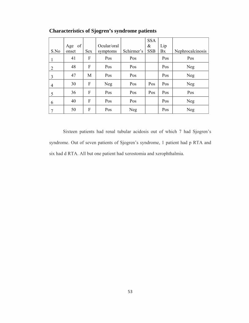
Characteristics of Sjogren’s syndrome patients
S.No Age of onset Sex
Ocular/oral symptoms Schirmer’s
SSA & SSB
Lip Bx
Nephrocalcinosis
1 41 F Pos Pos Pos Pos
2 48 F Pos Pos Pos Neg
3 47 M Pos Pos Pos Neg
4 30 F Neg Pos Pos Pos Neg
5 36 F Pos Pos Pos Pos Pos
6 40 F Pos Pos Pos Neg
7 50 F Pos Neg Pos Neg
Sixteen patients had renal tubular acidosis out of which 7 had Sjogren’s
syndrome. Out of seven patients of Sjogren’s syndrome, 1 patient had p RTA and
six had d RTA. All but one patient had xerostomia and xerophthalmia.
54
DISCUSSION
In this study in a South Indian population there were 47 patients who
fulfilled the inclusion criteria of whom 24(51%) had HPP with potassium shifts,
out of which 20(43%) had sporadic periodic paralysis (SPP) and 4 (9%) had non
familial Thyrotoxic periodic paralysis. Twenty three patients (49%) had non-
HPP (due to potassium deficit), out of which 16 (33%) had renal tubular acidosis
(15 d RTA and 1 p RTA), 6 (13%) had Gitelman’s syndrome and one patient had
Liddle’s syndrome.
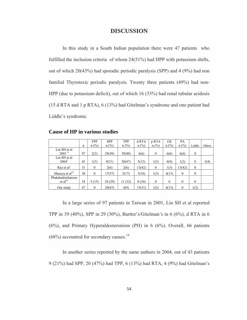
Cause of HP in various studies
n FPP
n (%) SPP
n (%) TPP
n (%) d RTA n (%)
p RTA n (%)
GS n (%)
PA n (%) Liddle Others
Lin SH et al 200114 97 2(2) 29(30) 39(40) 6(6) 0 6(6) 6(6) 0
Lin SH et al 20042 43 1(2) 9(21) 20(47) 5(12) 1(2) 4(9) 1(2) 0 2(4)
Rao et al1 31 0 2(6) 2(6) 13(42) 0 1(3) 13(42) 0
Maurya et al43 30 0 17(57) 5(17) 3(10) 1(3) 4(13) 0 0 Phakdeekitcharoen
et al42 34 5 (15) 10 (29) 11 (32) 8 (24) 0 0 0 0
Our study 47 0 20(43) 4(9) 15(31) 1(2) 6(13) 0 1(2)
In a large series of 97 patients in Taiwan in 2001, Lin SH et al reported
TPP in 39 (40%), SPP in 29 (30%), Bartter’s/Gitelman’s in 6 (6%), d RTA in 6
(6%), and Primary Hyperaldosteronism (PH) in 6 (6%). Overall, 66 patients
(68%) accounted for secondary causes.14
In another series reported by the same authors in 2004, out of 43 patients
9 (21%) had SPP, 20 (47%) had TPP, 6 (13%) had RTA, 4 (9%) had Gitelman’s
55
syndrome and 1 (2%) had Primary Hyperaldosteronism. Secondary causes
accounted for 33 cases (76%).2
Rao et al published a series of 31 cases from South India, 2 each had SPP
and TPP, 13 (42%) had d RTA, 1 (3%) had Gitelman’s syndrome and 13 (42%)
had Primary Hyperaldosteronism.1
In a series by Phakdeekitcharoen et al from Thailand of the 34 patients,
11 (32%), 8 (24%) and 15 (44%) had TPP, d RTA, SPP respectively.42
In another study from North India, Maurya et al reported HP in 30
patients, 17 (56.7%) had primary idiopathic hypokalemic paralysis, 5 (16.7%)
had TPP, 4 (13.3%) had RTA and 4 (13.3%) had Gitelman’s syndrome.43
In most Asian studies TPP is observed as the most common cause of HP,
18,44 whereas in Caucasians, FPP is more common.45 In our study and the study
by Maurya et al, SPP is the commonest group and no FPP is reported. 43 In the
study by Rao et al, HP due to Primary Hyperaldosteronism (PH) and d RTA are
the most common causes.1 None of our patients had Primary Hyperaldosteronism
(PH). The difference in etiology of HP in our study and Rao et al’s study may
be due to difference in study setting. Our study was conducted in a tertiary
nephrology practice, where as Rao et al’s study was undertaken in a tertiary
endocrinology practice.
56
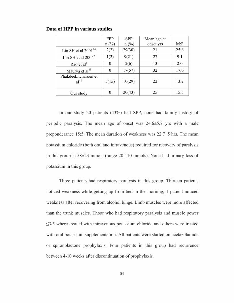
Data of HPP in various studies
FPP n (%)
SPP n (%)
Mean age at onset yrs M:F
Lin SH et al 200114 2(2) 29(30) 21 25:6
Lin SH et al 20042 1(2) 9(21) 27 9:1
Rao et al1 0 2(6) 13 2:0
Maurya et al43 0 17(57) 32 17:0 Phakdeekitcharoen et
al42 5(15) 10(29) 22 13:2
Our study 0 20(43) 25 15:5
In our study 20 patients (43%) had SPP, none had family history of
periodic paralysis. The mean age of onset was 24.6±5.7 yrs with a male
preponderance 15:5. The mean duration of weakness was 22.7±5 hrs. The mean
potassium chloride (both oral and intravenous) required for recovery of paralysis
in this group is 58±23 mmols (range 20-110 mmols). None had urinary loss of
potassium in this group.
Three patients had respiratory paralysis in this group. Thirteen patients
noticed weakness while getting up from bed in the morning, 1 patient noticed
weakness after recovering from alcohol binge. Limb muscles were more affected
than the trunk muscles. Those who had respiratory paralysis and muscle power
≤3/5 where treated with intravenous potassium chloride and others were treated
with oral potassium supplementation. All patients were started on acetazolamide
or spiranolactone prophylaxis. Four patients in this group had recurrence
between 4-10 weeks after discontinuation of prophylaxis.
57
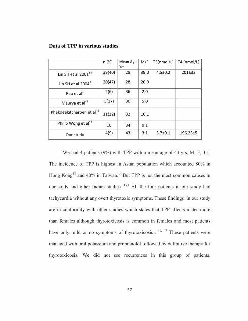
Data of TPP in various studies
n (%) Mean AgeYrs
M/F T3(nmol/L) T4 (nmol/L)
Lin SH et al 200114 39(40) 28 39:0 4.5±0.2 201±33
Lin SH et al 20042 20(47) 28 20:0
Rao et al1 2(6) 36 2:0
Maurya et al43 5(17) 36 5:0
Phakdeekitcharoen et al42 11(32) 32 10:1
Philip Wong et al46 10 34 9:1
Our study 4(9) 43 3:1 5.7±0.1 196.25±5
We had 4 patients (9%) with TPP with a mean age of 43 yrs, M: F, 3:1.
The incidence of TPP is highest in Asian population which accounted 80% in
Hong Kong18 and 40% in Taiwan.14 But TPP is not the most common causes in
our study and other Indian studies. 43,1 All the four patients in our study had
tachycardia without any overt thyrotoxic symptoms. These findings in our study
are in conformity with other studies which states that TPP affects males more
than females although thyrotoxicosis is common in females and most patients
have only mild or no symptoms of thyrotoxicosis . 46, 47 These patients were
managed with oral potassium and propranolol followed by definitive therapy for
thyrotoxicosis. We did not see recurrences in this group of patients.
58
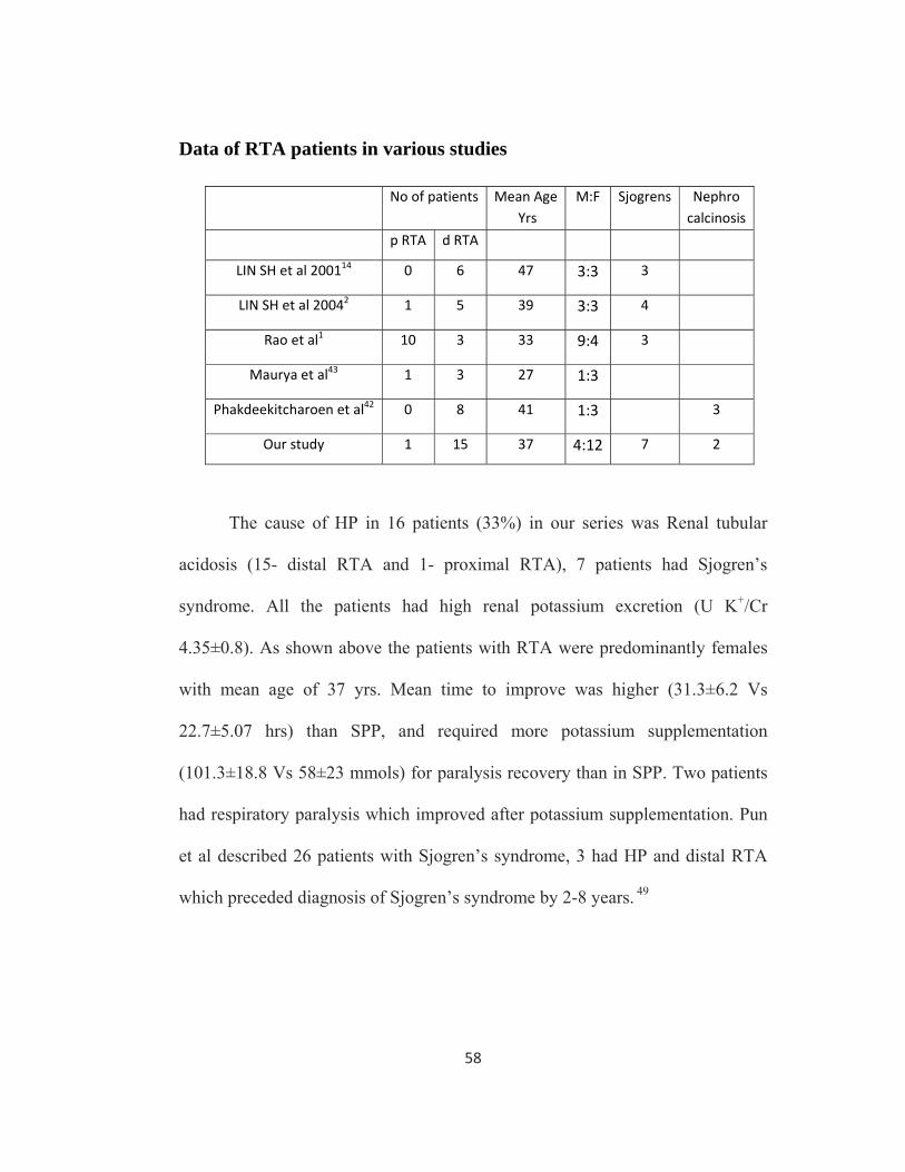
Data of RTA patients in various studies
The cause of HP in 16 patients (33%) in our series was Renal tubular
acidosis (15- distal RTA and 1- proximal RTA), 7 patients had Sjogren’s
syndrome. All the patients had high renal potassium excretion (U K+/Cr
4.35±0.8). As shown above the patients with RTA were predominantly females
with mean age of 37 yrs. Mean time to improve was higher (31.3±6.2 Vs
22.7±5.07 hrs) than SPP, and required more potassium supplementation
(101.3±18.8 Vs 58±23 mmols) for paralysis recovery than in SPP. Two patients
had respiratory paralysis which improved after potassium supplementation. Pun
et al described 26 patients with Sjogren’s syndrome, 3 had HP and distal RTA
which preceded diagnosis of Sjogren’s syndrome by 2-8 years. 49
No of patients Mean Age Yrs
M:F Sjogrens Nephrocalcinosis
p RTA d RTA
LIN SH et al 200114 0 6 47 3:3 3
LIN SH et al 20042 1 5 39 3:3 4
Rao et al1 10 3 33 9:4 3
Maurya et al43 1 3 27 1:3
Phakdeekitcharoen et al42 0 8 41 1:3 3
Our study 1 15 37 4:12 7 2
59
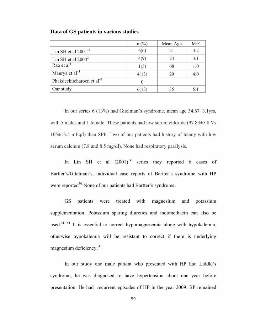
Data of GS patients in various studies
n (%) Mean Age M:F
Lin SH et al 200114 6(6) 21 4:2
Lin SH et al 20042 4(9) 24 3:1 Rao et al1 1(3) 68 1:0 Maurya et al43 4(13) 29 4:0 Phakdeekitcharoen et al42 0 Our study 6(13) 35 5:1
In our series 6 (13%) had Gitelman’s syndrome, mean age 34.67±3.1yrs,
with 5 males and 1 female. These patients had low serum chloride (97.83±5.8 Vs
105±13.5 mEq/l) than SPP. Two of our patients had history of tetany with low
serum calcium (7.8 and 8.5 mg/dl). None had respiratory paralysis.
In Lin SH et al (2001)14 series they reported 6 cases of
Bartter’s/Gitelman’s, individual case reports of Bartter’s syndrome with HP
were reported50. None of our patients had Bartter’s syndrome.
GS patients were treated with magnesium and potassium
supplementation. Potassium sparing diuretics and indomethacin can also be
used.41, 51 It is essential to correct hypomagnesemia along with hypokalemia,
otherwise hypokalemia will be resistant to correct if there is underlying
magnesium deficiency. 41
In our study one male patient who presented with HP had Liddle’s
syndrome, he was diagnosed to have hypertension about one year before
presentation. He had recurrent episodes of HP in the year 2009. BP remained
60
high despite being on 4 antihypertensives (telmesartan, thiazide, prazosin,
metaprolol). He had metabolic alkalosis with urinary potassium loss, low plasma
renin activity and low aldosterone. He had good response to triamterene and did
not have recurrence. There are case reports of muscle weakness52 and periodic
paralysis53 in Liddle’s syndrome.
In our study three patients had rebound hyperkalemia (>5 mEq/L), two
patients in SPP and one patient in TPP groups. None of the patients in Non- HPP
(large K+ deficit) group had rebound hyperkalemia. In Lin SH et al (2004)2 series
19 of 30 patients in the HPP group developed rebound hyperkalemia.
Since this study is from a single tertiary referral centre, only those
patients with severe or recurrent episodes may have been seen by us. This is a
limitation of our study.
61
CONCLUSIONS
1. The commonest causes for hypokalemic paralysis (HP) in our study were
sporadic periodic paralysis (SPP) and renal tubular acidosis (RTA).
2. Among the patients with hypokalemic paralysis, 57% of them were due to
secondary causes. Presence of acidosis or alkalosis in arterial blood gas
analysis suggests a renal cause for Hypokalemic paralysis.
3. Spot urine K+/Cr ratio helps to distinguish the diagnostic categories of
HPP (Hypokalemic paralysis due to K+ shifts) with non-HPP
(Hypokalemic paralysis due to K+ deficits)
4. There was a male predominance in Hypokalemic periodic paralysis
(HPP). Sporadic periodic paralysis (SPP) was more common than familial
periodic paralysis (FPP) in this study.
5. Male predominance was noted in Thyrotoxic periodic paralysis ( TPP).
Absence of history of thyroid disease or clinical thyrotoxicosis does not
exclude the diagnosis of TPP. So thyroid function tests should be done in
all patients with HP.
62
6. Though much less potassium is needed during therapy of HPP (SPP,
TPP), there is still a danger of rebound hyperkalemia.
7. Hypomagnesemia should be looked for and corrected along with the
correction of hypokalemia in patients with metabolic alkalosis.
Bibliography
1. Narsing Rao, Mathew John et al, Aetiological, Clinical and metabolic profile of
hypokalemic periodic paralysis in adults: A single centre experience. Natl Med J
India 2006; 19:246-9
2. Shih-Hua Lin, MD; Yuh-Feng Lin, MD et al, Laboratory Tests to Determine the
Cause of Hypokalemia and Paralysis: Arch Intern Med. 2004;164:1561-1566.
3. Stedwell RE, Allen KM, Binder LS. Hypokalemic paralyses: a review of the
etiologies, pathophysiology, presentation, and therapy. Am J Emerg Med
1992;10:143-8.
4. Jean-Philippe Lengel´e, Hendrica Belge and Olivier Devuyst . Periodic
paralyses: when channels go wrong. Nephrol Dial Transplant (2008); 23: 1098–
1101.
5. Robert J. Unwin, Friedrich C. Luft and David G. Shirley. Pathophysiology and
management of hypokalemia: a clinical perspective Nat. Rev. Nephrol(2011) ; 7,
75–84
6. Sushil K Ahlawat, Anita Sachdev Hypokalaemic paralysis. Postgrad Med J
1999;75:193–197
7. Michael J. Ackerman et al . Ion channels — basic science and clinical disease.
NEJM 1997 Volume 336 ; Number 22:1575-1586
8. Jurkat-Rott K, Lehman-Horn F. Paroxysmal muscle weakness—the familial
periodic paralyses. J Neurol 2006; 253: 1391–1398
9. Salem CB et al, Drug-Induced Hypokalemia. Current drug safety,2009;4:55-61
10. Damien Sternberg et al, Hypokalemic Periodic Paralysis. Gene Reviews- NCBI
BookShelf, University of Washington, Seatle, web page, Assessed on
17/10/2010. http://www.ncbi.nlm.nih.gov/books/NBK1338/
11. Naganand Sripathi. Periodic Paralyses . eMedicine Neurology web page
downloaded on 30/9/2009. http://emedicine.medscape.com/article/1171678-
overview.
12. Shannon L. Venance et al. The primary periodic paralyses: diagnosis,
pathogenesis and treatment . Brain (2006); 129, 8–17
13. Lin SH, Lin YF, Halperin ML.Hypokalemia and paralysis. QJM 2001; 94:133-9.
14. Lin SH. Thyrotoxic periodic paralysis. Mayo Clin Proc 2005; 80:99–105.
15. Annie W. C. Kung Thyrotoxic Periodic Paralysis:A Diagnostic Challenge J Clin
Endocrinol Metab, July 2006, 91(7):2490–2495.
16. Goh SH. Thyrotoxic periodic paralysis: reports of 7 cases presenting with
weakness in an Asian emergency department. Emerg Med J 2002;19:78-79.
17. G.T.C. Ko etal Thyrotoxic periodic paralysis in a Chinese population QJ Med
1996; 89:463-468.
18. Seguro AC, Lomar AV, Rocha AS. Acute renal failure of leptospirosis:
nonoliguric and hypokalemic forms. Nephron. 1990;55(2):146-51.
19. Benjamin SPE , Fernando M E et al. Cleistanthus Collinus Poisoning. J Assoc
Physicians India 2006;54:742-4.
20. Bagga A, Sinha A. Evaluation of renal tubular acidosis. Indian J Pediatr
2007;74:679-686.
21. Eriksson P, Denneberg T, Larsson L, Lindstrom F. Biochemical markers of
renal disease in primary Sjögren’s syndrome. Scand J Urol Nephrol
1995;29:383–92.
22. Bliff F, Palmer et al, Metabolic Acidosis. In Floege J Feehally J (eds)
Comprehensive Clinical Nephrology, Fourth edition, Page 157-8.
23. Rodriguez S. Renal tubular acidosis, the clinical entity. J Am Soc Nephrol
2002;13: 2160-2170.
24. Dell KM, Avner ED. Renal tubular acidosis. In Behrman RE, Kliegman RM,
Jenson HB, ed. Nelson Textbook of Pediatrics. Philadelphia; WB
Saunders,2003; 1758-1762.
25. Jovelic A, Stafanovic D. Distal renal tubular acidosis as a cause of osteomalacia
in a patient with primary Sjogren’s syndrome.Vojnosaint Pregl 2005;
62(10):769-73.
26. Walsh S, Shirley D, Wrong O, Unwin R: Urinary acidification assessed by
furosemide and fludrocortisone treatment: An alternative to ammonium
chloride. Kidney Int 2007; 71:1310-1316.
27. Backman, U.et al Incidence and Clinical Importance of Renal Tubular Defects
in Recurrent Renal Stone Formers Nephron 1980;25:96-101.
28. Amirlak I and Dawson K.P. Bartter syndrome: an overview. Q J Med 2000;
93:207-215.
29. Proesmans WC. Bartter syndrome and its neonatal variant. Eur J Pediatr1997;
156: 669–79.
30. Simon DB, Karet FE, Hamdan JM, Di Pietro A, Sanjad SA, Lifton RP. Bartter's
syndrome hypokalaemic alkalosis with hypercalciuria, is caused by mutations in
the Na-K-2Cl cotransporter NKCC2. Nature Gen 1996; 183-8.
31. Simon DB, Karet FE, Rodriguez-Soriano J, et al. Genetic heterogeneity of
Bartter's syndrome revealed by mutations in the K+ channel, ROMK. Nature
Genet 1996; 12: 152-6.
32. Katayama S, Attallah AA, Stahl RA, Bloch DL, Lee JB. Mechanism of
furosemide-induced natriuresis by direct stimulation of renal prostaglandin E2.
Am J Physiol1984; 247:F555–61.
33. Rodrı´guez-Soriano J, Vallo A. Familial hypokalaemia- hypomagnesemia
(Gitelman’s syndrome). Pediatr Nephrol 1990; 4:C22.
34. Colussi G, Macaluso M, Brunati C, Minetti L. Calcium metabolism and
calciotropic hormone levels in Giteman's syndrome. Miner Electrolyte
Metab1994; 20:294–301.
35. Quamme GA. Renal magnesium handling: new perspectives in understanding
old problems. Kidney Int1997; 52:1180–95.
36. Arya SN. Periodic paralysis. JIACM 2002; 3:374-82.
37. Ma JT, Wang C, Lam KS, Yeung RT, Chan FL, Boey J, et al. Fifty cases of
primary hyperaldosteronism in Hong Kong Chinese with a high frequency of
periodic paralysis: Evaluation of techniques for tumour localisation. QJM
1986;61: 1021–37.
38. David J. Salant, Parul S. Patel . Polycystic Kidney Disease and Other Inherited
Tubular Disorders in Harrison's Internal Medicine 17 th ed, Fauci , Kasper,
Braunwald (eds). McGraw-Hill,2008.
39. Dillon MJ, Shah V, Mitchell MD: Bartter syndrome: 10 cases in childhood.
Results of long term indomethacin therapy. Q J Med 1979; 48:429-446
40. Juan Rodrı´guez-Soriano. Bartter and related syndromes; the puzzle is almost
solved. Pediatr Nephrol 1998; 12:315–27.
41. Colussi G, Rombola G, De Ferrari ME, Macaluso M, Minetti L. Nephrol 1996;
10:551–4. correction of hypokalemia with antialdosterone therapy in Gitleman’s
syndrome. Am J Nephrol 1994; 14:127–35.
42. Bunyong Phakdeekitcharoen et al; Hypokalaemia and paralysis in the Thai
population; Nephrol Dial Transplant (2004) 19: 2013–2018.
43. Pradeep Kumar Maurya et al, Spectrum of hypokalaemic periodic paralysis in a
tertiary care centre in India; Postgrad Med J 2010 ; 86: 692-695.
44. Sinharay R. Hypokalaemic thyrotoxic periodic paralysis in an Asian man in
theUnited Kingdom. Emerg Med J 2004;21:120-1.
45. Ober KP. Thyrotoxic periodic paralysis in the United States. Report of 7 cases
and review of the literature. Medicine (Baltimore) 1992;71:109-20.
46. Phillip Wong; Hypokalemic thyrotoxic periodic paralysis: A case series: Journal
of the Canadian Association of Emergency Physicians; Sep 2003;353-5.
47. Karen M O'Neil;Thyrotoxic hypokalemic paralysis: A case study;Critical Care
Nurse; Dec 1999; 31-4.
48. Raskin RJ, Tesar JT, Lawless OJ. Hypokalemic periodic paralysis in Sjogren’s
syndrome. Arch Intern Med 1981;141:1671–3.
49. Pun KK, Wong CK et al;Hypokalemic periodic paralysis due to the Sjogren
syndrome in Chinese patients. Ann Intern Med 1989;110:405–6.
50. Shiah CJ, Tsai DM, Liao ST, et al. Acute muscular paralysis in an adult with
subclinical Bartter’s syndrome associated with gentamicin administration. Am J
Kidney Dis 1994;24:932–5.
51. Tang NL, Hui J, To KF, Ng HK, Hjelm NM, Fok TF. Severe hypokalemic
myopathy in Gitelman’s syndrome. Muscle Nerve 1999;22:545–7.
52. Kang JH, Lee CH, Han SM et al. A case of Liddle’s syndrome associated with
muscle weakness. Korean J Nephrology 1998;17:124-127.
53. Boorugu H, Mathews KP, Liddle’s Syndrome Presenting with Periodic
Paralysis:JAPI • JUNE 2009 • VOL. 57; 481-2

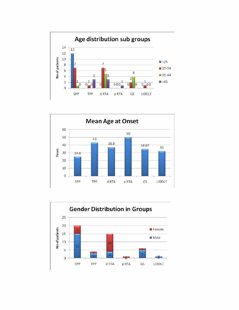
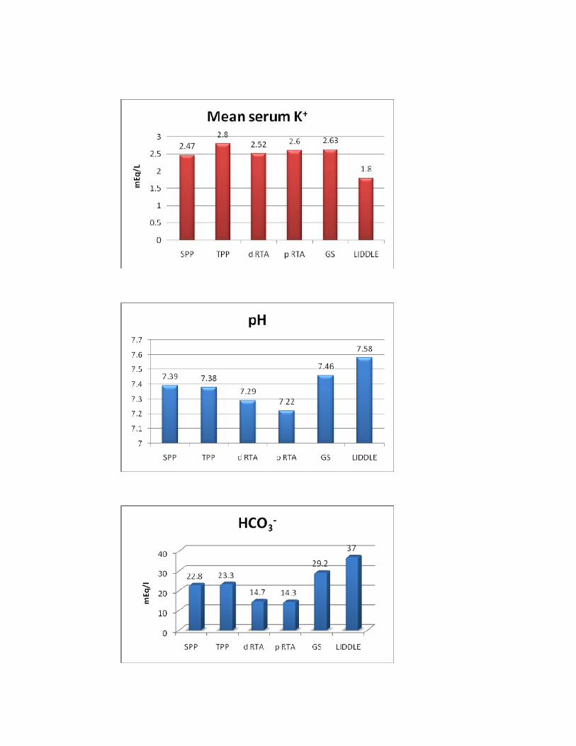
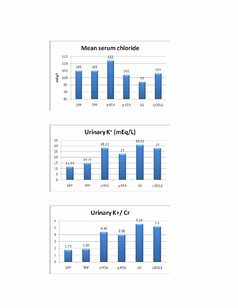
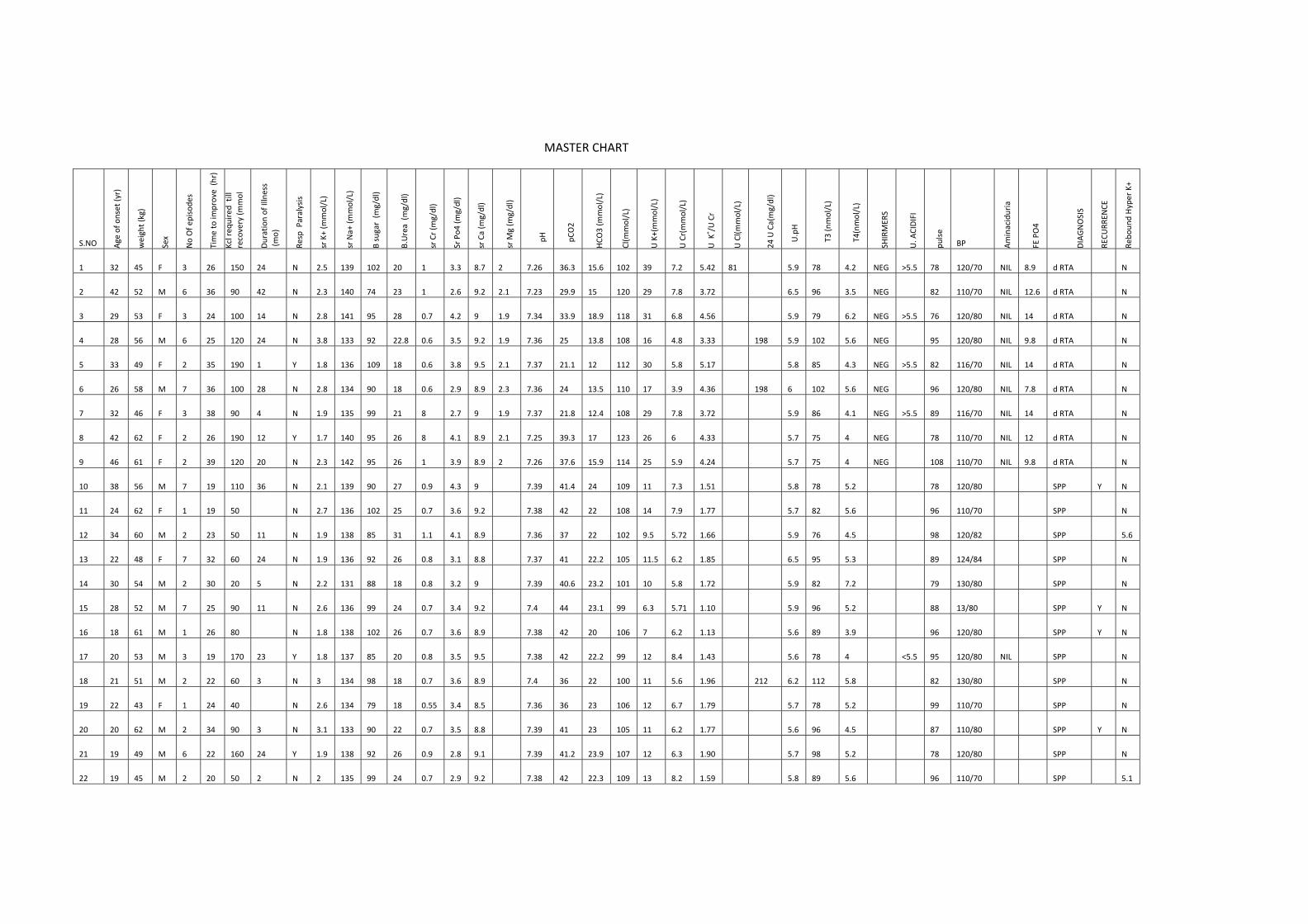
MASTER CHART
S.NO Age of o
nset (yr)
weight (kg)
Sex
No Of e
pisode
s
Time to im
prove (h
r)
Kcl req
uired till
recovery (m
mol
Duration of Illness
(mo)
Resp Paralysis
sr K+ (m
mol/L)
sr Na+ (m
mol/L)
B sugar (m
g/dl)
B.Urea (m
g/dl)
sr Cr (m
g/dl)
Sr Po4
(mg/dl)
sr Ca (m
g/dl)
sr M
g (m
g/dl)
pH
pCO2
HCO
3 (m
mol/L)
Cl(m
mol/L)
U K+(mmol/L)
U Cr(mmol/L)
U K
+ /U Cr
U Cl(m
mol/L)
24 U Ca(mg/dl)
U.pH
T3 (n
mol/L)
T4(nmol/L)
SHIRMER
S
U. A
CIDIFI
pulse
BP Aminaciduria
FE PO4
DIAGNOSIS
RECU
RREN
CE
Rebo
und Hyper K+
1 32 45 F 3 26 150 24 N 2.5 139 102 20 1 3.3 8.7 2 7.26 36.3 15.6 102 39 7.2 5.42 81 5.9 78 4.2 NEG >5.5 78 120/70 NIL 8.9 d RTA N
2 42 52 M 6 36 90 42 N 2.3 140 74 23 1 2.6 9.2 2.1 7.23 29.9 15 120 29 7.8 3.72 6.5 96 3.5 NEG 82 110/70 NIL 12.6 d RTA N
3 29 53 F 3 24 100 14 N 2.8 141 95 28 0.7 4.2 9 1.9 7.34 33.9 18.9 118 31 6.8 4.56 5.9 79 6.2 NEG >5.5 76 120/80 NIL 14 d RTA N
4 28 56 M 6 25 120 24 N 3.8 133 92 22.8 0.6 3.5 9.2 1.9 7.36 25 13.8 108 16 4.8 3.33 198 5.9 102 5.6 NEG 95 120/80 NIL 9.8 d RTA N
5 33 49 F 2 35 190 1 Y 1.8 136 109 18 0.6 3.8 9.5 2.1 7.37 21.1 12 112 30 5.8 5.17 5.8 85 4.3 NEG >5.5 82 116/70 NIL 14 d RTA N
6 26 58 M 7 36 100 28 N 2.8 134 90 18 0.6 2.9 8.9 2.3 7.36 24 13.5 110 17 3.9 4.36 198 6 102 5.6 NEG 96 120/80 NIL 7.8 d RTA N
7 32 46 F 3 38 90 4 N 1.9 135 99 21 8 2.7 9 1.9 7.37 21.8 12.4 108 29 7.8 3.72 5.9 86 4.1 NEG >5.5 89 116/70 NIL 14 d RTA N
8 42 62 F 2 26 190 12 Y 1.7 140 95 26 8 4.1 8.9 2.1 7.25 39.3 17 123 26 6 4.33 5.7 75 4 NEG 78 110/70 NIL 12 d RTA N
9 46 61 F 2 39 120 20 N 2.3 142 95 26 1 3.9 8.9 2 7.26 37.6 15.9 114 25 5.9 4.24 5.7 75 4 NEG 108 110/70 NIL 9.8 d RTA N
10 38 56 M 7 19 110 36 N 2.1 139 90 27 0.9 4.3 9 7.39 41.4 24 109 11 7.3 1.51 5.8 78 5.2 78 120/80 SPP Y N
11 24 62 F 1 19 50 N 2.7 136 102 25 0.7 3.6 9.2 7.38 42 22 108 14 7.9 1.77 5.7 82 5.6 96 110/70 SPP N
12 34 60 M 2 23 50 11 N 1.9 138 85 31 1.1 4.1 8.9 7.36 37 22 102 9.5 5.72 1.66 5.9 76 4.5 98 120/82 SPP 5.6
13 22 48 F 7 32 60 24 N 1.9 136 92 26 0.8 3.1 8.8 7.37 41 22.2 105 11.5 6.2 1.85 6.5 95 5.3 89 124/84 SPP N
14 30 54 M 2 30 20 5 N 2.2 131 88 18 0.8 3.2 9 7.39 40.6 23.2 101 10 5.8 1.72 5.9 82 7.2 79 130/80 SPP N
15 28 52 M 7 25 90 11 N 2.6 136 99 24 0.7 3.4 9.2 7.4 44 23.1 99 6.3 5.71 1.10 5.9 96 5.2 88 13/80 SPP Y N
16 18 61 M 1 26 80 N 1.8 138 102 26 0.7 3.6 8.9 7.38 42 20 106 7 6.2 1.13 5.6 89 3.9 96 120/80 SPP Y N
17 20 53 M 3 19 170 23 Y 1.8 137 85 20 0.8 3.5 9.5 7.38 42 22.2 99 12 8.4 1.43 5.6 78 4 <5.5 95 120/80 NIL SPP N
18 21 51 M 2 22 60 3 N 3 134 98 18 0.7 3.6 8.9 7.4 36 22 100 11 5.6 1.96 212 6.2 112 5.8 82 130/80 SPP N
19 22 43 F 1 24 40 N 2.6 134 79 18 0.55 3.4 8.5 7.36 36 23 106 12 6.7 1.79 5.7 78 5.2 99 110/70 SPP N
20 20 62 M 2 34 90 3 N 3.1 133 90 22 0.7 3.5 8.8 7.39 41 23 105 11 6.2 1.77 5.6 96 4.5 87 110/80 SPP Y N
21 19 49 M 6 22 160 24 Y 1.9 138 92 26 0.9 2.8 9.1 7.39 41.2 23.9 107 12 6.3 1.90 5.7 98 5.2 78 120/80 SPP N
22 19 45 M 2 20 50 2 N 2 135 99 24 0.7 2.9 9.2 7.38 42 22.3 109 13 8.2 1.59 5.8 89 5.6 96 110/70 SPP 5.1

23 23 52 M 3 28 160 7 Y 2 136 85 25 8 4 8.5 7.36 36.9 22 101 9.5 5.78 1.64 5.9 79 7 79 120/80 SPP N
24 32 48 F 2 18 60 2 N 2.9 134 89 20 0.6 2.8 8.8 7.35 35.8 22.9 108 12 8.2 1.46 5.7 88 5.6 102 110/70 SPP N
25 30 54 M 2 20 70 3 N 3.2 133 88 24 0.7 3.6 8.5 7.39 41 23 105 11 8.2 1.34 5.6 96 4.5 85 110/80 SPP N
26 25 52 M 2 21 30 3 N 1.9 140 98 23 0.9 2.8 9 7.38 42.6 22.6 109 12 6.5 1.85 6.1 95 3.8 102 120/80 SPP N
27 26 49 F 2 18 30 5 N 3 141 96 18 0.6 3.5 9.3 7.38 41.4 24.4 108 16 6.6 2.42 214 5.6 82 4 86 120/80 SPP N
28 19 56 M 2 15 40 1 N 2.9 135 102 22 0.9 3.6 8.5 7.41 43 24.1 102 14 6.2 2.26 245 5.7 99 5.9 75 130/80 SPP N
29 22 50 M 2 19 60 5 N 2.9 140 102 21 0.8 3.4 8.6 2.1 7.40 37.1 24.1 111 14 5.8 2.41 5.6 87 5.6 75 130/80 SPP N
30 35
62
M 1 32 120 N 2.5 135 96 25 0.8 4.1 8.9 1.6 7.46 37 26.5 105 25.6 4.8 5.33 167 110 5.6 79 3.9 84 130/80 GS N
31 36
71
M 2 38 100 14 N 3.1 136 95 26 0.8 2.9 8.9 1.2 7.5 44.5 34 89 31 5.8 5.34 116 127.5 5.9 89 5.4 78 120/80 GS Y N
32 37
54
M 2 47 80 14 N 2.8 135 109 19 0.7 3.8 8.5 0.9 7.45 41.5 27.8 103 28 3.9 7.18 52 105 5.8 102 5.6 82 130/80 GS N
33 32
61
M 1 30 90 N 3 126 92 24 1 3.7 7.8 0.87 7.42 46.1 29 98 36 7.8 4.62 76 123 6 96 7.5 89 130/80 GS N
34 30
64
M 1 36 80 N 1.9 136 99 25 0.9 3.3 9.1 1.4 7.48 42.3 28.5 98 42 6 7.00 121 109 5.9 85 6 98 120/80 GS N
35 38
51
F 2 42 70 16 N 2.5 135 88 26 0.9 2.8 9 1.5 7.47 42.1 29.8 94 22.9 5.9 3.88 56 103 5.9 103 4.2 82 130/80 GS N
36 41 50 F 3 23 90 14 N 2.1 140 113 23 0.8 4 8.8 2 7.24 26.6 11.3 123 40.3 7.3 5.52 6.6 84 5.4 POS >5.5 96 120/80 NIL 12 d RTA/SJ N
37 48 52 F 1 32 90 N 3 136 73 24 0.7 4.2 8.5 1.9 7.33 27 14 112 32 7.9 4.05 5.6 78 5.4 POS >5.5 95 120/70 NIL 14 d RTA/SJ N
38 47 49 M 2 30 100 1 N 2 140 109 18 0.6 3.8 8.6 1.8 7.33 27.9 15 115 26 5.72 4.55 5.9 82 6.1 POS >5.5 82 110/70 NIL 9.1 d RTA/SJ N
39 30 45 F 4 41 110 6 N 2.5 138 99 20 0.7 3.2 8.9 2 7.24 36.7 15.6 110 32 6.2 5.16 5.6 89 4.5 POS 99 120/70 NIL 7.8 d RTA/SJ N
40 36 46 F 2 24 80 2 N 2.9 139 80 22 0.7 3 8.8 2.1 7.26 39.2 16.8 102 29 6.3 4.60 5.6 98 5.6 POS 87 110/70 NIL 11 d RTA/Sj N
41 40 60 F 3 35 90 24 N 2 139 95 24 0.9 4.1 8.5 2.3 7.23 30.1 15.1 103 22 8.2 2.68 5.9 82 5.6 POS >5.5 79 110/70 NIL 15.2 d RTA/SJ N
42 50 58 F 3 24 80 4 N 2.6 138 102 24 0.7 3.2 8.6 2 7.22 35.6 14.3 107 23 5.78 3.98 5.7 75 3.2 POS <5.5 89 110/70 NIL 26 p RTA/SJ N
43 49 54 M 2 28 70 10 N 2.3 140 102 26 0.7 3.4 8.6 7.36 39.3 23.6 110 16 8.8 1.82 5.8 202 11.8 102 120/80 TPP N
44 30 52 M 2 21 80 3 N 2.9 138 102 24 0.7 3 9.2 7.38 41.8 21.8 108 15 6.2 2.42 5.7 198 8.9 112 120/80 TPP N
45 45 61 F 1 27 40 N 2.9 138 90 24 1 2.9 9 7.38 42 24.1 104 12 6.5 1.85 5.9 226.5 16 110 130/80 TPP 5.2
46 48 53 M 1 19 40 N 3.1 132 79 25 0.9 2.8 9.5 7.38 41.2 23.6 98 16 7.1 2.25 5.6 195 12.1 108 120/80 TPP N
47 32 54 M 3 12 50 3 N 1.8 144 78 26 1 3 9 2.3 7.58 35 37 103 28 5.3 5.28 201 6.5 78 4 78 160/100 LIDDLE N
PROFORMA
1. Name: NC No.: DOA:
2. Age: Sex:
3. Occupation:
4. Address:
5. Presenting History:
a. Vomiting:
b. Diarrhoea:
c. Drugs:
d. Laxative abuse:
e. Other predisposing Factors:
f. Fever:
g. Polyuria:
h. Dry eyes:
i. Dry mouth:
6. Family History:
7. Examination:
Rt Lt
Power UL
LL
DTR UL
LL
Neck muscles
Resp Paralysis
8. Vital signs:
Pulse: BP: RR:
a. Systems:
9. Previous episodes:
10. Treatment:
11. Investigations :
Sl No. Test
1 ECG
2 TC
3 DC
4 ESR
5 Hb
6 UREA
7 CREATININE
8 B.SUGAR
9 Sr Na
10 Sr K
11 Sr Po4
12 Sr calcium
13 Sr uric acid
14 Sr osmolality
15 ABG
16 URINALYSIS
Protein
Sugar
Deposits
17 URINE CULTURE
18 URINE pH
19 URINE K
20 URINE Cl
21 URINE PO4
22 URINE UA
23 Urine amino acids
24 Urine osmolality
25 TTKG
26 24 urine Ca
27 Ca/Cr Ratio
28 Sr Mg
29 NCS
30 Thyroid profile
31 U Acidification test
ABG
Urine pH
32 KUB
33 USG
34 CT KUB
35
36
37
38
39
40
Diagnosis
Follow up