a sequence-specific dna binding small molecule triggers the ......(ab2907), polyclonal rabbit...
TRANSCRIPT

REPORT
A sequence-specific DNA bindingsmall molecule triggers the release ofimmunogenic signals and phagocytosisin a model of B-cell lymphoma
JeenJoo S. Kang and Peter B. Dervan*
Division of Chemistry and Chemical Engineering, California Institute of Technology, Pasadena, CA 91125, USA
Quarterly Reviews of Biophysics (2015), 48(4), pages 453–464 doi:10.1017/S0033583515000104
Abstract. Means to cause an immunogenic cell death could lead to significant insight into how cancer escapes immune control. In this study,we screened a library of five pyrrole–imidazole polyamides coding for different DNA sequences in a model of B-cell lymphoma for the upre-gulation of surface calreticulin, a pro-phagocytosis signal implicated in immunogenic cell death. We found that hairpin polyamide 1 triggersthe release of the damage-associated molecular patterns calreticulin, ATP and HMGB1 in a slow necrotic-type cell death. Consistent with thissignaling, we observed an increase in the rate of phagocytosis by macrophages after the cancer cells were exposed to polyamide 1. The DNAsequence preference of polyamide 1 is 5′-WGGGTW-3′ (where W = A/T), indicated by the pairing rules and confirmed by the Bind-n-Seqmethod. The close correspondence of this sequence with the telomere-repeat sequence suggests a potential mechanism of action throughligand binding at the telomere. This study reveals a chemical means to trigger an inflammatory necrotic cell death in cancer cells.
Key words: Py-Im Polyamide, damage-associated molecular patterns, necrotic cell death, immunogenic cell death, DNA-binding smallmolecule, telomere sequence.
IntroductionAvoidance of immune destruction has been called the seventhhallmark of cancer (Dunn et al. 2004a; Hanahan &Weinberg,2011). According to the immuno-editing paradigm, the im-mune system recognizes and destroys those proto-oncogeniclesions capable of triggering an immune response, while thosethat escape immune control grow to become clinically detect-able disease (Dunn et al. 2004b; Koebel et al. 2007; Schreiberet al. 2011; O’Sullivan et al. 2012). Studies suggest that thera-pies that enlist the immune system maintain more durabledisease control in the clinical setting (Eggermont et al.2014). Chemical methods to cause immunogenicity in cancercells would be an important tool toward understanding
immunomodulation in the treatment of cancer. A prerequi-site for the activation of an anti-cancer immune responseis the recognition of the damaged cells as a threat. Damagedcells release immunostimulatory molecules, called damage-associated molecular patterns (DAMPs), to recruit and acti-vate professional phagocytes such as macrophages and den-dritic cells (Matzinger, 2002; Obeid et al. 2007; Jaiswal et al.2010). These antigen-presenting cells engulf and process thecancer cells to further prime the immune system for targetedelimination of cancer (Tseng et al. 2013).
Although most chemotherapeutic regimens cause a non-immunogenic or even tolerogenic cell death, recent reportssuggest anthracyclins or γ-radiation are particularly effectivebecause they result in the release of DAMPs (Obeid et al.2007; Zitvogel et al. 2008). The extracellular exposure ofthe intracellularly abundant molecules calreticulin (CRT),
* Author for correspondence: P. B. Dervan, Division of Chemistry and
Chemical Engineering, California Institute of Technology, 1200 E. California
Blvd., Mail Code 164-30, Pasadena, CA 91125, USA. Tel.: (626 ) 395-6002;
Fax: (626) 683-8753; E-mail: [email protected]
© Cambridge University Press 2015. This is an Open Access article, distributed under the terms of the Creative Commons Attribution licence (http://creativecommons.org/licenses/by/3.0/), which permits unrestricted re-use, distribution, and reproduction in any medium, provided the original work isproperly cited.https://www.cambridge.org/core/terms. https://doi.org/10.1017/S0033583515000104
Downloaded from https://www.cambridge.org/core. IP address: 54.39.106.173, on 03 Apr 2020 at 16:05:14, subject to the Cambridge Core terms of use, available at
HMGB1 and ATP have been suggested to form a spatiotem-poral code for immunogenicity (Zitvogel et al. 2010; Keppet al. 2011). The presentation of CRT, an abundantER-resident chaperone protein, to the cell surface was ident-ified as a necessary and sufficient pro-phagocytic signal forprofessional phagocytes (Obeid et al. 2007). The studyshowed that stimulation of CRT surface expression byanthracyclins or adsorbtion of the calreticulin protein onthe cell surface was sufficient to elicit an anti-cancer im-mune response in syngeneic mice (Obeid et al. 2007).Weissman and co-workers further demonstrated in theRaji cell line, a model of human B-cell non-Hodgkin’s lym-phoma, that CRT is the dominant pro-phagocytosis signalwhich is necessary for engulfment by human macrophages(Chao et al. 2010). Furthermore, ATP released from the cyto-sol into the local microenvironment serves as a lymphocyterecruiting and activating chemokine (Idzko et al. 2002;Aymeric et al. 2010). Lastly, the nucleus-resident proteinHMGB1 can be secreted into the surroundings as an inflam-matory adjuvant and was shown to be necessary for a durableanti-cancer response in mice (Rovere-Querini et al. 2004;Guerriero et al. 2011). Identification of additional smallmole-cules that trigger the release of these DAMPs from tumor cellswould be of utility to the field in addressing the heterogeneityof cancers. We became interested in expanding the examinedchemical space for compounds capable of causing an immu-nogenic cell death. Because the DNA damage pathway hasbeen implicated in immunogenic signaling (Gasser et al.2005) and anthracyclins are DNA-intercalating ligands, wesought to explore a class ofminor grooveDNA-binding oligo-mers hitherto not studied for this biological activity.
Hairpin pyrrole–imidazole (Py–Im) polyamides are a classof sequence-specific oligomers that bind in the minorgroove of DNA (Wade et al. 1992; Trauger et al. 1996;Kielkopf et al. 1998a, b; White et al. 1997, 1998).Sequence preference is achieved by the pair-wise, co-facialarrangement of aromatic amino acids that distinguish theedges of the four Watson–Crick base pairs as shown inFig. 1a (Dervan & Edelson, 2003). Pairing rules for pro-grammable specificity have been established: Im/Py specifiesa G•C base pair, Hp/Py codes for T•A base pairs and Py/Pybinds both T•A/A•T (White et al. 1998). Eight-ring hairpinpolyamides are linked in an antiparallel fashion by a centralaliphatic γ-aminobutyric acid unit (Mrksich et al. 1994).Polyamides of this hairpin architecture have affinities formatch sites similar in magnitude to natural DNA-bindingproteins, with Ka’s of 108–1010 M
−1 (Hsu et al. 2007).Eight-ring hairpins of this class are cell-permeable andmodulate transcription in both cells and mice (Nickolset al. 2007; Nickols & Dervan, 2007; Raskatov et al. 2012;Yang et al. 2013). In this study, we screened a small libraryof Py–Im polyamides coding for different six base pair DNAsequences in Raji cells for the upregulation of surface calre-ticulin. We found one hairpin polyamide which displayed
activity in this screen and characterize its potential for caus-ing an immunogenic cell death.
Materials and methodsChemicals and reagents
All chemicalswerepurchased fromSigma-Aldrichunless other-wise noted. Py–Impolyamides were synthesized bymicrowave-assisted, solid-phase synthesis on Kaiser oxime resin(Novabiochem) according to previously described protocols(Baird & Dervan, 1996; Puckett et al. 2012). Polyamide 5 wassynthesized on hydrazine resin (855037, Novabiochem) in thesame manner as the other polyamides and cleaved from resinin the same manner after 10 min oxidation of the hydrazineby Cu(II)SO4 in pyridine and DMF. Polyamides were purifiedby reverse-phase HPLC and lyophilized. EZ-Link NHS-PEG4-biotin (Pierce) was conjugated in 5% Hünig’s basein DMF. Purity and identity of compounds were verifiedby analytical HPLC and matrix-assisted laser desorption/ionization time-of-flight mass spectrometry (SI Table 2).
Brefeldin Awas purchased from BD Biosciences, 7-AAD fromeBiosciences and Z-VAD-fmk from Promega. Antibodies pur-chased from Abcam are: polyclonal rabbit anti-calreticulin(ab2907), polyclonal rabbit anti-ERp57 (ab10287), polyclonalgoat anti-rabbit Alexa Fluor 488 (ab150081). Antibodiesfrom Life Technologies are: anti-Annexin V Alexa Fluor 488conjugate (A13201) and mouse monoclonal anti-actin(AM4302). Antibodies from Cell Signaling Technologiesare: rabbit polyclonal anti-PARP (9542) and rabbit mono-clonal anti-phospho-H2AX (ser139, 9718).
Cell culture
Raji cells (ATCC) were maintained in RPMI media (LifeTechnologies) with 10% fetal bovine serum (FBS, OmegaScientific) and 5 mM glutamine (Life Technologies). K562cells (ATCC) were cultured in IMDM (ATCC) media and10% FBS. A549 and PC3 cells (ATCC) were maintained inKaign’s modified F-12 K media (Gibco) supplemented with10% FBS and 5 mM glutamine. Peripheral blood macrophages(Stemcell Technologies) were thawed and plated in DMEM(ATCC) and supplemented with 5 mM glutamine and 10%FBS for at least two days and no longer than 6 days before use.
Flow cytometry
Raji and K562 cells were plated at 1 × 10⁵ cells ml−1 in96-well plates at 100 μl and treated immediately after plat-ing. A549 and PC3 cells were plated in 24-well plates at6 × 104 cells ml−1 at 400 μl and were allowed to adhere over-night prior to treatment. Adherent cells were harvested withAccutase (Life Technologies).
For the CRT and ERp57 detection experiments, harvestedcells were washed with cold staining buffer: HBSS (LifeTechnologies) with 2·5 mg ml−1 BSA (Amresco), 10 mM
454
https://www.cambridge.org/core/terms. https://doi.org/10.1017/S0033583515000104Downloaded from https://www.cambridge.org/core. IP address: 54.39.106.173, on 03 Apr 2020 at 16:05:14, subject to the Cambridge Core terms of use, available at
HEPES (Life Technologies), 0·1 mM MgCl2 (Life Technologies)and DNAse (Roche). Cells were incubated with human FcRblock (eBiosciences) and primary antibody in cold stainingbuffer. This was followed by washing and incubation with dye-conjugated anti-rabbit secondary antibody. 7-AAD, forward-and side-scatter were used to gate for live cells. Secondaryantibody alone was used as a control (SI Fig. 2). The medianfluorescence intensity of live cells is normalized to non-treatment. Data were acquired on the FACS Calibur andanalyzed with Flowjo software. Measurements and standarddeviations are in triplicate and representative of at least two in-dependent experiments; asterisk indicates p < 0·05 by two-tailed Student’s t test compared with non-treatment.
For cell death analysis flow cytometry experiments, Raji cellswere harvested after treatment and washed with cold stain-ing buffer and incubated with FcR block and dye-conjugatedanti-Annexin V. Cells were washed with staining buffer andtaken for immediate analysis. 7-AAD was added to samplesmaintained at 4 °C and data was acquired soon thereafter onthe MACS VYB. Data was acquired in triplicate from twoindependent experiments and analyzed with Flowjo (SITables 5 and 6).
Bind-n-Seq of polyamide 1b
Highest affinity DNA binding sites of polyamide–biotinconjugate 1b were determined according to previously
reported methods (Meier et al. 2012). In brief, polyamide1b was equilibrated at 25 or 250 nM overnight in a DNA li-brary of all possible 21 mers. DNA bound to 1b was affinitypurified with streptavidin magnetic beads (M-280Dynabeads, Life Technologies) and eluted. Isolated DNAwas amplified by touchdown PCR and submitted forsequencing on an Illumina HiSeq 2000 Genome Analyzer.The MERMADE script was used to distribute data by bar-code and a fasta file of a random 25% of sequences was gen-erated for DREME motif analysis. Data are representative ofthree independent experiments (SI Fig. 4).
DNA thermal denaturation assay
DNA of the sequence 5′-CTTAGGGTTAGC-3′ and itscomplement were purchased from Integrated DNATechnologies and annealed. The oligonucleotides weremixed with hairpin polyamide 1 to a final concentrationof 2 and 3 μM, respectively, in 1 mL total volume. An aque-ous solution of 10 mM sodium cacodylate, 10 mM KCl, 10mM MgCl2 and 5 mM CaCl2 at pH 7·0 was used as analysisbuffer. The assay utilized a Varian Cary 100 spectrophot-ometer to heat samples to 90 °C, cool to a starting tempera-ture of 25 °C, and then heat at a rate of 0·5 °C min−1 to 90 °C. Denaturation profiles were recorded at λ = 260 nm andmelting temperatures were detected by the maximum ofthe first derivative. Data show the combined mean and
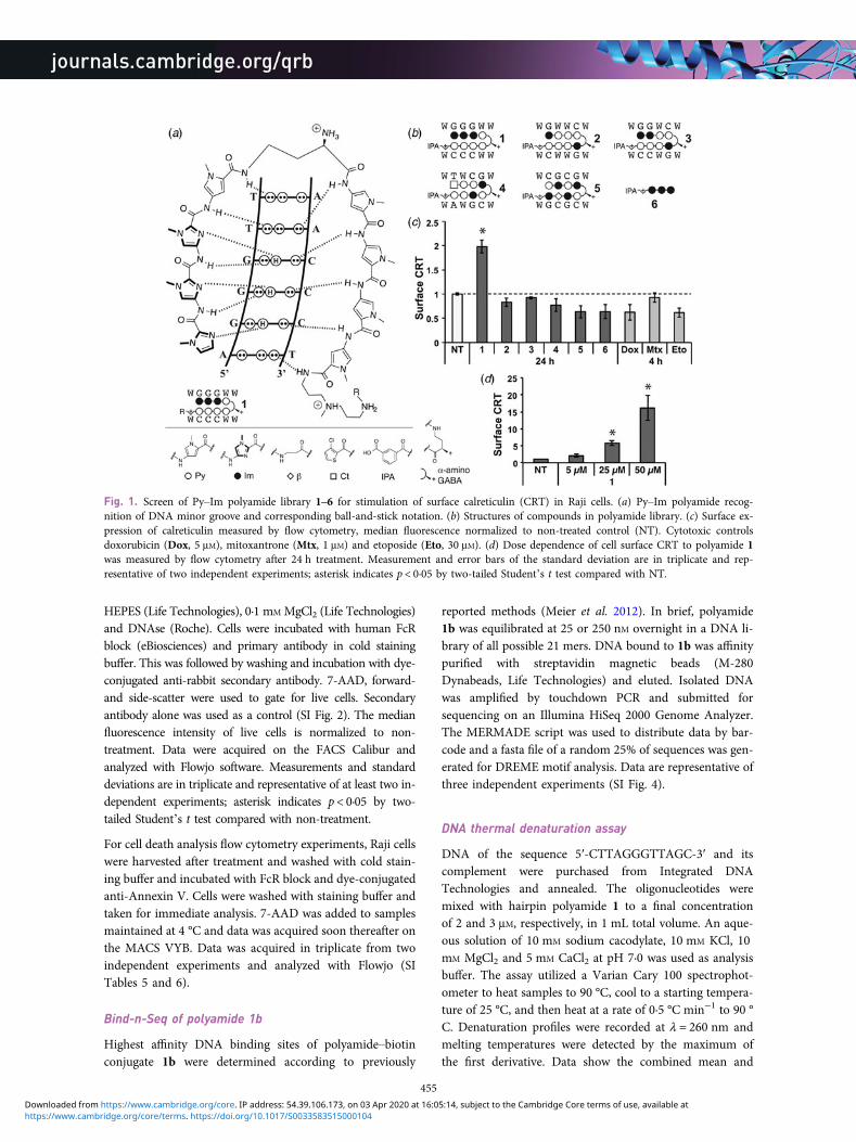
Fig. 1. Screen of Py–Im polyamide library 1–6 for stimulation of surface calreticulin (CRT) in Raji cells. (a) Py–Im polyamide recog-nition of DNA minor groove and corresponding ball-and-stick notation. (b) Structures of compounds in polyamide library. (c) Surface ex-pression of calreticulin measured by flow cytometry, median fluorescence normalized to non-treated control (NT). Cytotoxic controlsdoxorubicin (Dox, 5 μM), mitoxantrone (Mtx, 1 μM) and etoposide (Eto, 30 μM). (d) Dose dependence of cell surface CRT to polyamide 1was measured by flow cytometry after 24 h treatment. Measurement and error bars of the standard deviation are in triplicate and rep-resentative of two independent experiments; asterisk indicates p < 0·05 by two-tailed Student’s t test compared with NT.
455
https://www.cambridge.org/core/terms. https://doi.org/10.1017/S0033583515000104Downloaded from https://www.cambridge.org/core. IP address: 54.39.106.173, on 03 Apr 2020 at 16:05:14, subject to the Cambridge Core terms of use, available at
standard deviation of triplicate measurements from two in-dependent experiments.
Immunoblot assays
Raji cells were plated at 105 cells ml−1 in 10 cm diameterdishes and dosed with the indicated treatment. Cells werewashed with cold PBS and lysed for 10 min in lysis buffer(50 mM Tris–HCl pH 7·4, 1 mM EDTA, 150 mM NaCl, 1%Triton X-100) containing protease inhibitors (Complete,Roche), 1 mM PMSF and phosphatase inhibitors. Sampleswere clarified by centrifugation at 14 000 × g for 10 min,quantified with Bradford reagent (Bio-rad), denatured byboiling in Laemmli buffer (LI-COR) for 5 min and separatedby sodium dodecyl sulfate–polyacrylamide gel electrophor-esis using AnyKD gradient gels (Biorad). Protein was trans-ferred to a PVDF membrane (Millipore) and blocked withOdyssey blocking buffer (LI-COR). Both primary antibodiesand appropriate IR-dye conjugated secondary antibodies(LI-COR) were incubated in blocking buffer with 0·2%Tween. Anti-actin was used to control for equal loadingand experiments were done in at least two independent bio-logical replicates. Bands were visualized on a LI-COROdyssey infrared imager.
ATP bioluminescence assay
Raji cells were plated into 96-well cell culture plates at 200 μlper well and 105 cells ml−1, in quadruplicate per condition.After the indicated treatment, media and cells were trans-ferred to a 96-well PCR plate and centrifuged at 130 × gfor 5 min. The supernatant was collected for analysis ofATP content using an ATP bioluminescence kit (FLAA,Sigma). The assay was performed according to the manufac-turer’s protocol and luminescence was measured using aFlexstation 3. Measurements and standard deviation aretechnical quadruplicate and biological triplicate. Asteriskindicates p < 0·05 by two-tailed Student’s t test comparedwith non-treatment.
HMGB1 ELISA
Raji cells were plated into 96-well cell culture plates at 100 μlper well, 105 cells ml−1, in duplicate. Cells were treated withpolyamide 1 and 2 as indicated and the supernatant col-lected. HMGB1 was measured using the Shino-Test ELISAkit (IBL international) according to the manufacturer’sinstructions on the Flexstation 3. Measurement and stan-dard deviations were determined from technical duplicatefrom two independent replicates. Asterisk indicates p <0·05 by two-tailed Student’s t test compared withnon-treatment.
Caspase luciferase assay
Raji cells were plated into 96-well cell culture plates at 100 μlper well and 105 cells ml−1, in triplicate per condition.
Media was included as a blank control. After the indicatedtreatment, media and cells were transferred to an opaquewhite 96-well plate containing 100 μl in each well of thecaspase-dependent luciferase reagent, prepared as per themanufacturer’s instructions (Caspase-Glo 3/7, Promega).The mixture was left at room temperature for 1 h prior tomeasurement on the Flexstation 3 (Molecular Devices).Measurements and standard deviations were determinedin triplicate and done in biological duplicate.
Metabolic activity assay
Raji cells were plated into 96-well clear bottom cell cultureplates at 100 μl per well and 105 cells ml−1, in quadruplicateper indicated condition. Media was used as a backgroundcontrol. Metabolic activity was assessed using the WST-1 re-agent (Roche) as per the manufacturer’s instructions.Measurements and standard deviations were determinedin quadruplicate and done in biological duplicate.
Phagocytosis assay
Peripheral blood macrophages were plated at 2 × 10⁴ cellsper well in 24- or 48-well plates in DMEM. Immediatelyprior to use, macrophages were washed with HBSS andstained with carboxyfluorescein diacetate succinimidylester (CFSE, eBioscience) in HBSS for 5 min. Macrophageswere washed of excess dye and returned to DMEM for in-cubation. Target Raji cells were plated in 96-well plates at1 × 105 cells ml−1 in 200 μl of RPMI and treated as de-scribed. Target A549 cells were plated in 24-well plates at2 × 104 cells per well 15 h before beginning treatment.After treatment, cells were harvested with Accutase ifnecessary and washed with HBSS and incubated withpHrodo succinimidyl ester (Life Technologies) diluted to2 μM in HBSS for 5 min. Cells were then washed by centri-fugation at 150 × g, re-suspended in DMEM, counted on aBiorad TC10 cell counter, and 5 × 10⁴ cells per well wereadded to macrophages. After incubation for 2·5 h at 37 °C,media and non-adherent cells in each sample were aspiratedand saved. Adherent cells were trypsinized, aspirated andcombined with saved media mixture. Cells were washedonce with PBS and fixed in 1% formaldehyde and kept at4 °C until assessment on the MACS VYB flow cytometer.The percentage of phagocytosis was calculated as the per-centage of double positive cells among fluorescein+ macro-phages. Measurements and standard deviations are takenfrom three independent experiments and asterisks indicatep < 0·05 by two-tailed Student’s t test compared withnon-treatment.
Confocal microscopy
For images of phagocytosis, cells treated in the mannerdescribed above were put on 35 mM glass-bottom dishes(MatTek). Confocal images were acquired using a 40×
456
https://www.cambridge.org/core/terms. https://doi.org/10.1017/S0033583515000104Downloaded from https://www.cambridge.org/core. IP address: 54.39.106.173, on 03 Apr 2020 at 16:05:14, subject to the Cambridge Core terms of use, available at
oil immersion objective on a Zeiss LSM 5 Excitermicroscope.
ResultsPolyamide 1 upregulates calreticulin on the cellsurface
We tested five Py–Im polyamides (1–5, full structures in SIFig. 1) that bind five unique DNA sequences (5′-WGGGWW-3′, 5′-WGWWCW-3′, 5′-WGGWCW-3′, 5′-WTWCGW-3′ and 5′-WCGCGW-3′, respectively, where W =A/T) and have demonstrated biological activity (Fig. 1b)(Swalley et al. 1996; Nickols & Dervan, 2007; Nickolset al. 2007, 2013; Kang et al. 2014). Raji cells, which havepreviously been utilized in CRT, phagocytosis, and immu-notherapy animal models, were dosed at 5 μM for 24 hwith each of the polyamides 1–5. In addition, 5 μM doxoru-bicin (Dox) and mitoxantrone (Mtx, 1 μM) were included asrepresentative anthracyclins. The topoisomerase inhibitoretoposide (Eto, 30 μM) was also included for comparison.A 4 h exposure time point was used to measure CRT dueto the high toxicity of these chemotherapeutics and to bein the range of literature precedent (Obeid et al. 2007).We measured surface CRT by flow cytometry in a popu-lation gated for live cells and saw a statistically significant,twofold increase in cells treated with only polyamide1 (Fig. 1c). In contrast, we saw no activity with the otherpolyamides 2–5 despite their structural similarity. Notably,polyamide 2 has been most extensively studied among thisclass and has shown activity in prostate cancer xenograftmodels and DNA damage response (Yang et al. 2013;Martínez et al. 2015). Remarkably, polyamides 3 and 1share the same molar weight and composition of Im/Pypairs, and only differ by the exchange of one Im/Py ringpair. To assess the structure activity relationship of the imi-dazole trimer portion of the hairpin oligomer 1, we synthe-sized and examined ImImIm polyamide 6. This was testedbecause imidazoles are known to complex with calciumand CRT is involved in calcium homeostasis (Michalaket al. 2009). We did not, however, observe an increase ofCRT with polyamide 6, which suggests the imidazole trimeralone is not sufficient to trigger the surface expression ofCRT. We observed no increase in CRT with the two anthra-cyclins tested, Dox and Mtx, or with Eto. The diminishedresponse to anthracyclin treatment in Raji cells, as com-pared with that reported in the literature with murinecolon cancer cells, reflects a known range of CRT responseto anthracyclins and may be attributable to a difference incell lines (Tufi et al. 2008; Peters & Raghavan, 2011). Wenext increased the treatment concentration of 1–25 and50 μM and found that CRT exposure increased in a dose-dependent manner (Fig. 1d, SI Fig. 2a). The results indicatepolyamide 1 is unique in our small library of compounds inits modulation of surface expression of CRT.
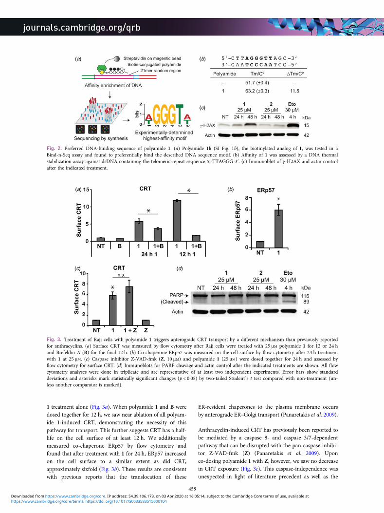
Polyamide 1 preferentially binds the sequence5′-WGGGTW-3′
Py–Im polyamides are a class of sequence-specificDNA-binding ligands and the DNA-binding preferencesof polyamide 1 may be related to a mechanism of action.By the pairing rules, polyamide 1 is a perfect match to5′-WGGGWW-3′, not unlike the TTAGGG-repeat sequencefound in human telomeres. Indeed, dye-conjugated tandemhairpin Py–Im polyamides that recognize 10 base pairs ofthis repeat sequence can be used to stain telomeres in per-meabilized cells (Maeshima et al. 2001; Kawamoto et al.2013). We sought to confirm the preferred binding sequencemotif of 1 using the Bind-n-Seq method. Bind-n-Seq cou-ples affinity enrichment with next-generation sequencingto query genome-sized sequence space for high-affinitybinding sequences (Meier et al. 2012). We modified polyam-ide 1 at the C-terminal position with a biotin-label to afford1b (full structure in SI Fig. 1b) for submission toBind-n-Seq. Polyamide 1b strongly preferred binding theDNA sequence motif of 5′-WGGGTW-3′ (Fig. 2a, SITable 4). We additionally confirmed the affinity of polyam-ide 1 for this sequence by measuring the thermal stabiliza-tion afforded to sequence-matched double-stranded DNA(Pilch et al. 1996). We tested a DNA fragment that includedthe telomeric DNA sequence 5′-TTAGGGTTAG-3′(Fig. 2b). Polyamide 1 stabilized the 12 base pair DNA frag-ment by 11·5 °C, suggesting a high-affinity hairpin polyam-ide. Lastly, we detected a marker of DNA stress,phosphorylation of serine 139 on the histone H2AX, aftertreatment with polyamide 1 but not 2 (Fig. 2c). We chosethe 25 μM dose for this experiment and others describedbelow because it resulted in a robust sixfold increase in sur-face CRT. We posit that the CRT effect may be due to theunique properties of the DNA target sequence of polyamide1 in cell biology. The DNA stress, thermal denaturation andBind-n-Seq results together suggest the telomere sequence isa plausible target for the mechanism of action of polyamide1-mediated CRT exposure.
Polyamide 1 triggers CRT in a different manner thando anthracyclins
To better understand the effects of polyamide 1, we com-pared its trigger of CRT with reports of CRT induction byanthracyclins (Panaretakis et al. 2009). Anthracyclin-induced CRT exposure has been described to occur withthe co-chaperone ERp57 by anterograde ER-Golgi transport(Panaretakis et al. 2009). To interrogate whether polyamide1 induces CRT export to the cell surface by ER–Golgi trans-port, in Fig. 3a, we applied the Golgi transport inhibitorBrefeldin A (B) to Raji cells (Lippincott-Schwartz et al.1989). Due to the toxicity of B, we could only dose B for12 h. With 24 h treatment with polyamide 1 and 12 h treat-ment with B, we saw statistically significant reduction ofCRT on the cell surface when compared with polyamide
457
https://www.cambridge.org/core/terms. https://doi.org/10.1017/S0033583515000104Downloaded from https://www.cambridge.org/core. IP address: 54.39.106.173, on 03 Apr 2020 at 16:05:14, subject to the Cambridge Core terms of use, available at
1 treatment alone (Fig. 3a). When polyamide 1 and B weredosed together for 12 h, we saw near ablation of all polyam-ide 1-induced CRT, demonstrating the necessity of thispathway for transport. This further suggests CRT has a half-life on the cell surface of at least 12 h. We additionallymeasured co-chaperone ERp57 by flow cytometry andfound that after treatment with 1 for 24 h, ERp57 increasedon the cell surface to a similar extent as did CRT,approximately sixfold (Fig. 3b). These results are consistentwith previous reports that the translocation of these
ER-resident chaperones to the plasma membrane occursby anterograde ER–Golgi transport (Panaretakis et al. 2009).
Anthracyclin-induced CRT has previously been reported tobe mediated by a caspase 8- and caspase 3/7-dependentpathway that can be disrupted with the pan-caspase inhibi-tor Z-VAD-fmk (Z) (Panaretakis et al. 2009). Uponco-dosing polyamide 1 with Z, however, we saw no decreasein CRT exposure (Fig. 3c). This caspase-independence wasunexpected in light of literature precedent as well as the
Fig. 2. Preferred DNA-binding sequence of polyamide 1. (a) Polyamide 1b (SI Fig. 1b), the biotinylated analog of 1, was tested in aBind-n-Seq assay and found to preferentially bind the described DNA sequence motif. (b) Affinity of 1 was assessed by a DNA thermalstabilization assay against dsDNA containing the telomeric-repeat sequence 5′-TTAGGG-3′. (c) Immunoblot of γ-H2AX and actin controlafter the indicated treatment.
Fig. 3. Treatment of Raji cells with polyamide 1 triggers anterograde CRT transport by a different mechanism than previously reportedfor anthracyclins. (a) Surface CRT was measured by flow cytometry after Raji cells were treated with 25 μM polyamide 1 for 12 or 24 hand Brefeldin A (B) for the final 12 h. (b) Co-chaperone ERp57 was measured on the cell surface by flow cytometry after 24 h treatmentwith 1 at 25 μM. (c) Caspase inhibitor Z-VAD-fmk (Z, 10 μM) and polyamide 1 (25 μM) were dosed together for 24 h and assessed byflow cytometry for surface CRT. (d) Immunoblots for PARP cleavage and actin control after the indicated treatments are shown. All flowcytometry analyses were done in triplicate and are representative of at least two independent experiments. Error bars show standarddeviations and asterisks mark statistically significant changes (p < 0·05) by two-tailed Student’s t test compared with non-treatment (un-less another comparator is marked).
458
https://www.cambridge.org/core/terms. https://doi.org/10.1017/S0033583515000104Downloaded from https://www.cambridge.org/core. IP address: 54.39.106.173, on 03 Apr 2020 at 16:05:14, subject to the Cambridge Core terms of use, available at
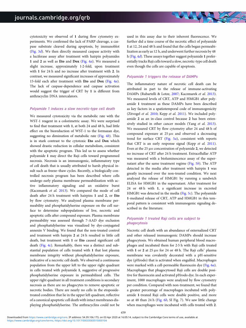
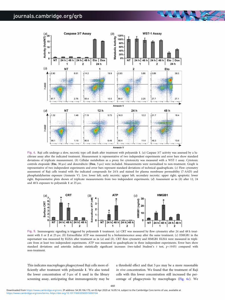
cytotoxicity we observed of 1 during flow cytometry ex-periments. We confirmed the lack of PARP cleavage, a cas-pase substrate cleaved during apoptosis, by immunoblot(Fig. 3d). We then directly measured caspase activity witha luciferase assay after treatment with hairpin polyamides1 and 2 as well as Eto and Dox (Fig. 4a). We measured aslight increase, approximately 1·2-fold, upon treatmentwith 1 for 24 h and no increase after treatment with 2. Incontrast, we measured significant increases of approximately15-fold each after treatment with Eto and Dox (Fig. 4a).The lack of caspase-dependence and caspase activationwould suggest the trigger of CRT by 1 is different fromanthracyclin DNA intercalators.
Polyamide 1 induces a slow necrotic-type cell death
We measured cytotoxicity via the metabolic rate with theWST-1 reagent in a colorimetric assay. We were surprisedto find that treatment with 1, at both 24 and 48 h, had littleeffect on the bioreduction of WST-1 to the formazan dye,suggesting no diminution of metabolic rate (Fig. 4b). Thisis in stark contrast to the cytotoxic Eto and Dox whichshowed drastic reduction in cellular metabolism, consistentwith the apoptotic program. This led us to assess whetherpolyamide 1 may direct the Raji cells toward programmednecrosis. Necrosis is an immunogenic, inflammatory typeof cell death that is usually attributed to harsh physical in-sult such as freeze–thaw cycles. Recently, a biologically con-trolled necrosis program has been described where cellsundergo early plasma membrane permeabilization with ac-tive inflammatory signaling and an oxidative burst(Kaczmarek et al. 2013). We compared the mode of celldeath after 24 h treatment with hairpins 1 and 2, or Etoby flow cytometry. We analyzed plasma membrane per-meability and phosphatidylserine exposure on the cell sur-face to determine subpopulations of live, necrotic andapoptotic cells after compound exposure. Plasma membranepermeability was assessed through 7-AAD dye exclusionand phosphatidylserine was visualized by dye-conjugatedannexin V binding. We found that the non-treated controland treatment with hairpin 2 at 24 h resulted in little celldeath, but treatment with 1 or Eto caused significant celldeath (Fig. 4c). Remarkably, there was a distinct and sub-stantial population of cells treated with 1 that lost plasmamembrane integrity without phosphatidylserine exposure,indicative of a necrotic cell death. We observed a continuouspopulation from the upper left to the upper right quadrantin cells treated with polyamide 1, suggestive of progressivephosphatidylserine exposure in permeabilized cells. Theupper right quadrant in all treatment conditions is secondarynecrosis as there are no phagocytes to remove apoptotic ornecrotic bodies. There are nearly no cells in the etoposide-treated condition that lie in the upper left quadrant, reflectiveof a canonical apoptotic cell deathwith intactmembranes dis-playing phosphatidylserine. The anthracyclins could not be
used in this assay due to their inherent fluorescence. Wefurther did a time course of the necrotic effect of polyamide1 at 12, 24 and 48 h and found that the cells began permeabi-lization as early as 12 h, and underwent further necrosis by 48h (Fig. 4d). These assays together suggest polyamide 1 prefer-entially tracks Raji cells toward a slow, necrotic-type cell deatheven though the cells are capable of apoptosis.
Polyamide 1 triggers the release of DAMPs
The inflammatory nature of necrotic cell death can beattributed in part to the release of immune-activatingDAMPs (Rubartelli & Lotze, 2007; Kaczmarek et al. 2013).We measured levels of CRT, ATP and HMGB1 after poly-amide 1 treatment as these DAMPs have been describedas key factors in a spatiotemporal code of immunogenicity(Zitvogel et al. 2010; Kepp et al. 2011). We included poly-amide 2 as an in-class control because 2 has been exten-sively studied in other cancer models (Yang et al. 2013).We measured CRT by flow cytometry after 24 and 48 h ofcompound exposure at 25 μM and observed a decreasingtrend for surface CRT (Fig. 5a), consistent with reportsthat CRT is an early response signal (Kepp et al. 2011).Even at the 25 μM concentration of polyamide 2, we detectedno increase of CRT after 24 h treatment. Extracellullar ATPwas measured with a bioluminescence assay of the super-natant after the same treatment regime (Fig. 5b). The ATPdetected in the media after treatment with hairpin 1 wasgreatly increased over the non-treated condition. We nextanalyzed the release of HMGB1 by running a sandwichELISA for HMGB1 in the supernatant. After treatment for24 or 48 h with 1, a significant increase in excretedHMGB1 was detected in the media (Fig. 5c). The polyamide1-mediated release of CRT, ATP and HMGB1 in this tem-poral pattern is consistent with immunogenic signaling de-scribed in the literature.
Polyamide 1 treated Raji cells are subject tophagocytosis
Necrotic cell death with an abundance of externalized CRTand other released immunogenic DAMPs should increasephagocytosis. We obtained human peripheral blood macro-phages and incubated them for 2·5 h with Raji cells treatedwith 1 or 2 at 25 μM for 24 or 48 h. The Raji cells’ plasmamembrane was covalently decorated with a pH-sensitivedye (pHrodo) that is activated when engulfed. Macrophageswere marked with a cell-permeable fluorescein dye (Fig. 6a).Macrophages that phagocytosed Raji cells are double posi-tive for fluorescein and activated pHrodo dye. In each exper-iment, 1000 macrophages were analyzed by flow cytometryper condition. Compared with non-treatment, we found thata greater percentage of macrophages incubated with poly-amide 1-treated Raji cells were double positive, and moreso at 48 than 24 h (Fig. 6b, SI Fig. 7). We saw little changewhen macrophages were incubated with cells treated with 2.
459
https://www.cambridge.org/core/terms. https://doi.org/10.1017/S0033583515000104Downloaded from https://www.cambridge.org/core. IP address: 54.39.106.173, on 03 Apr 2020 at 16:05:14, subject to the Cambridge Core terms of use, available at
This indicates macrophages phagocytosed Raji cells more ef-ficiently after treatment with polyamide 1. We also testedthe lower concentration of 5 μM of 1 used in the libraryscreening assay, anticipating that immunogenicity may be
a threshold effect and that 5 μM may be a more reasonablein vivo concentration. We found that the treatment of Rajicells with this lower concentration still increased the per-centage of phagocytosis by macrophages (Fig. 6c). We
Fig. 4. Raji cells undergo a slow, necrotic type cell death after treatment with polyamide 1. (a) Caspase 3/7 activity was assessed by a lu-ciferase assay after the indicated treatment. Measurement is representative of two independent experiments and error bars show standarddeviations of triplicate measurement. (b) Cellular metabolism as a proxy for cytotoxicity was measured with a WST-1 assay. Cytotoxiccontrols etoposide (Eto, 30 μM) and doxorubicin (Dox, 5 μM) were included. Measurements were normalized to non-treatment. Graph isrepresentative of two independent experiments and error bars represent standard deviations of technical quadruplicate. (c) Flow cytometryassessment of Raji cells treated with the indicated compounds for 24 h and stained for plasma membrane permeability (7-AAD) andphosphatidylserine exposure (Annexin V). Live: lower left; early necrotic: upper left; secondary necrotic: upper right; apoptotic: lowerright. Representative plots shown of triplicate measurements from two independent experiments. (d) Assessment as in (b) after 12, 24and 48 h exposure to polyamide 1 at 25 μM.
Fig. 5. Immunogenic signaling is triggered by polyamide 1 treatment. (a) CRT was measured by flow cytometry after 24 and 48 h treat-ment with 1 or 2 at 25 μM. (b) Extracellular ATP was measured by a bioluminescence assay after the same treatment. (c) HMGB1 in thesupernatant was measured by ELISA after treatment as in (a) and (b). CRT flow cytometry and HMGB1 ELISA were measured in tripli-cate from at least two independent experiments. ATP was measured in quadruplicate in three independent experiments. Error bars showstandard deviations and asterisks indicate statistically significant increases (two-tailed Student’s t test, p < 0·05) compared withnon-treatment.
460
https://www.cambridge.org/core/terms. https://doi.org/10.1017/S0033583515000104Downloaded from https://www.cambridge.org/core. IP address: 54.39.106.173, on 03 Apr 2020 at 16:05:14, subject to the Cambridge Core terms of use, available at
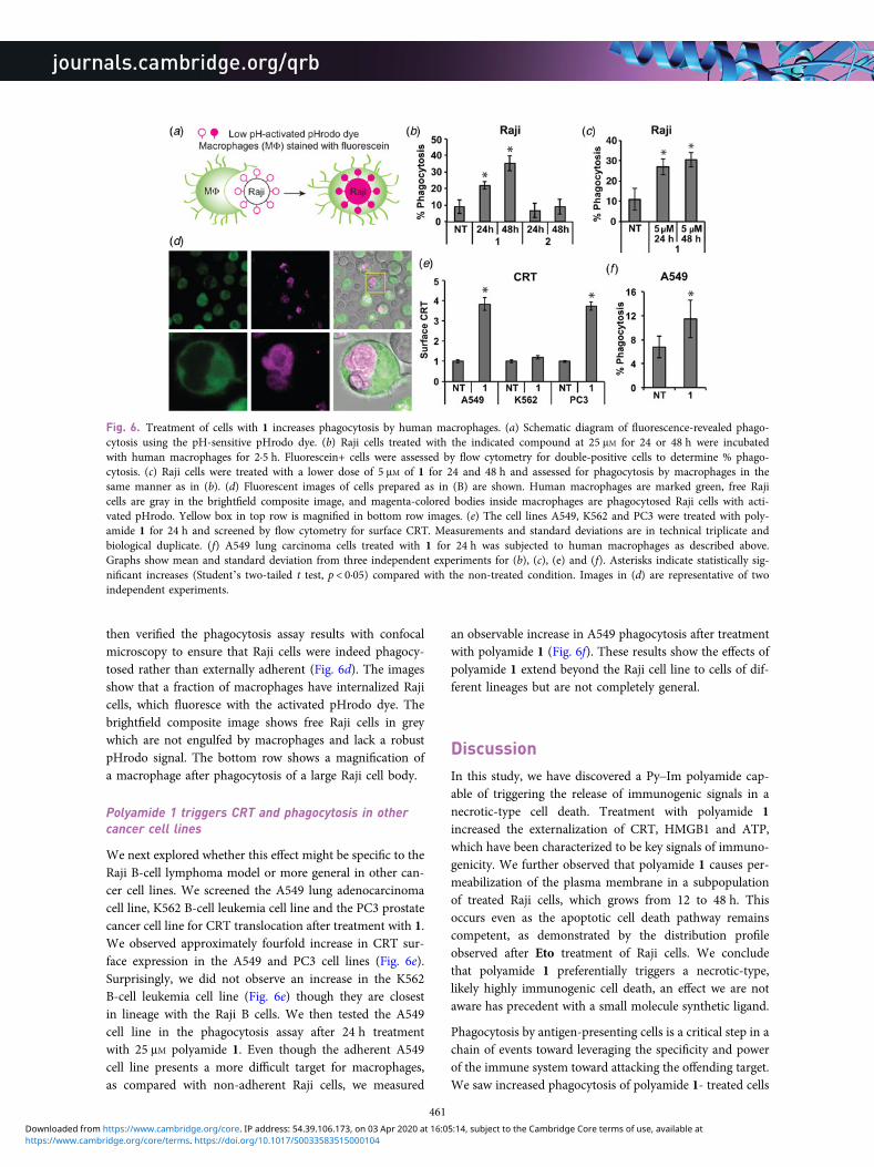
then verified the phagocytosis assay results with confocalmicroscopy to ensure that Raji cells were indeed phagocy-tosed rather than externally adherent (Fig. 6d). The imagesshow that a fraction of macrophages have internalized Rajicells, which fluoresce with the activated pHrodo dye. Thebrightfield composite image shows free Raji cells in greywhich are not engulfed by macrophages and lack a robustpHrodo signal. The bottom row shows a magnification ofa macrophage after phagocytosis of a large Raji cell body.
Polyamide 1 triggers CRT and phagocytosis in othercancer cell lines
We next explored whether this effect might be specific to theRaji B-cell lymphoma model or more general in other can-cer cell lines. We screened the A549 lung adenocarcinomacell line, K562 B-cell leukemia cell line and the PC3 prostatecancer cell line for CRT translocation after treatment with 1.We observed approximately fourfold increase in CRT sur-face expression in the A549 and PC3 cell lines (Fig. 6e).Surprisingly, we did not observe an increase in the K562B-cell leukemia cell line (Fig. 6e) though they are closestin lineage with the Raji B cells. We then tested the A549cell line in the phagocytosis assay after 24 h treatmentwith 25 μM polyamide 1. Even though the adherent A549cell line presents a more difficult target for macrophages,as compared with non-adherent Raji cells, we measured
an observable increase in A549 phagocytosis after treatmentwith polyamide 1 (Fig. 6f). These results show the effects ofpolyamide 1 extend beyond the Raji cell line to cells of dif-ferent lineages but are not completely general.
DiscussionIn this study, we have discovered a Py–Im polyamide cap-able of triggering the release of immunogenic signals in anecrotic-type cell death. Treatment with polyamide 1increased the externalization of CRT, HMGB1 and ATP,which have been characterized to be key signals of immuno-genicity. We further observed that polyamide 1 causes per-meabilization of the plasma membrane in a subpopulationof treated Raji cells, which grows from 12 to 48 h. Thisoccurs even as the apoptotic cell death pathway remainscompetent, as demonstrated by the distribution profileobserved after Eto treatment of Raji cells. We concludethat polyamide 1 preferentially triggers a necrotic-type,likely highly immunogenic cell death, an effect we are notaware has precedent with a small molecule synthetic ligand.
Phagocytosis by antigen-presenting cells is a critical step in achain of events toward leveraging the specificity and powerof the immune system toward attacking the offending target.We saw increased phagocytosis of polyamide 1- treated cells
Fig. 6. Treatment of cells with 1 increases phagocytosis by human macrophages. (a) Schematic diagram of fluorescence-revealed phago-cytosis using the pH-sensitive pHrodo dye. (b) Raji cells treated with the indicated compound at 25 μM for 24 or 48 h were incubatedwith human macrophages for 2·5 h. Fluorescein+ cells were assessed by flow cytometry for double-positive cells to determine % phago-cytosis. (c) Raji cells were treated with a lower dose of 5 μM of 1 for 24 and 48 h and assessed for phagocytosis by macrophages in thesame manner as in (b). (d) Fluorescent images of cells prepared as in (B) are shown. Human macrophages are marked green, free Rajicells are gray in the brightfield composite image, and magenta-colored bodies inside macrophages are phagocytosed Raji cells with acti-vated pHrodo. Yellow box in top row is magnified in bottom row images. (e) The cell lines A549, K562 and PC3 were treated with poly-amide 1 for 24 h and screened by flow cytometry for surface CRT. Measurements and standard deviations are in technical triplicate andbiological duplicate. (f) A549 lung carcinoma cells treated with 1 for 24 h was subjected to human macrophages as described above.Graphs show mean and standard deviation from three independent experiments for (b), (c), (e) and (f). Asterisks indicate statistically sig-nificant increases (Student’s two-tailed t test, p < 0·05) compared with the non-treated condition. Images in (d) are representative of twoindependent experiments.
461
https://www.cambridge.org/core/terms. https://doi.org/10.1017/S0033583515000104Downloaded from https://www.cambridge.org/core. IP address: 54.39.106.173, on 03 Apr 2020 at 16:05:14, subject to the Cambridge Core terms of use, available at
by macrophages, as would be expected upon the death ofRaji cells with externalized pro-phagocytic CRT. Theobserved phagocytosis and inflammatory DAMPs releasedinto the tumor microenvironment makes plausible thatthe immune system would become primed toward antigensof Raji cells. Reports have shown engulfment by macro-phages can activate an effective anti-cancer T cell response(Tseng et al. 2013). The application of polyamide 1 on can-cer in the context of a full immune system remains to beexplored.
That the DNA sequence preference of 1 is identical withthat of human telomeres was indicated by the pairingrules and confirmed by Bind-n-Seq. Whether the mechan-ism of action for polyamide 1 is through its sequence-specific DNA binding capacity remains an open question.Though polyamides have generally been characterized toact in a DNA-binding mode, we cannot exclude that poly-amide 1 may act as an aptamer binding to unknown targetproteins. The translocation of CRT after polyamide 1 treat-ment was observed in multiple, though not all, cell lines andsuggests there is a conserved pathway that leads to thisphenotype that is retained in many cancer cell lines. We ex-pect the elucidation of the mechanism of action will revealimportant signaling networks involved in immunosurveil-lance of cancer cells.
ConclusionThe effects of polyamide 1 are compelling because immuno-genic cancer cell death is cleared in the natural setting andthus difficult to observe. Further study may reveal tumorsuppressive signaling pathways that can be exploited to ex-trinsically control cancer even as cell-intrinsic mechanismsfail. The failure of anthracyclins to elicit CRT in Raji cellswhen it has previously been shown to be effective in CT26murine colon cancer cells (Obeid et al. 2007) underscoresthe heterogeneity of cancers. As cancers have heterogeneousmechanisms for evading elimination by the immune system,a corresponding diversity of immunogenicity agents will beimportant for both shedding light on new biology and de-veloping immuno-oncologic therapies.
Supplementary materialThe supplementary material for this article can be found athttp://dx.doi.org/10.1017/S0033583515000104
AcknowledgmentsWe thank Rochelle Diamond and Diana Perez of theCaltech Flow Cytometry Cell Sorting Facility for help withflow cytometry. Sequencing was conducted with the helpof Dr. Igor Antoshechkin at the Millard and Muriel
Jacobs Genetics and Genomics Laboratory at theCalifornia Institute of Technology; mass spectrometryanalyses were performed in the Mass SpectrometryLaboratory of the Division of Chemistry and ChemicalEngineering at the California Institute of Technology; gelswere scanned in the Center for the Chemistry of CellularSignaling at the California Institute of Technology.
Financial supportThis work was supported by the National Institutes ofHealth Grant GM51747 and Tobacco-Related DiseaseResearch Program (award number 20DT-0037 to J.S.K., dis-sertation research award).
Conflicts of InterestNone.
ReferencesAYMERIC, L., APETOH, L., GHIRINGHELLI, F., TESNIERE, A., MARTINS, I.,KROEMER, G., SMYTH, M. J. & ZITVOGEL, L. (2010). Tumor celldeath and ATP release prime dendritic cells and efficient antican-cer immunity. Cancer Research 70, 855–858.
BAIRD, E. E. & DERVAN, P. B. (1996). Solid phase synthesis of polya-mides containing imidazole and pyrrole amino acids. Journal ofthe American Chemical Society 118, 6141–6146.
CHAO, M. P., JAISWAL, S., WEISSMAN-TSUKAMOTO, R., ALIZADEH, A. A.,GENTLES, A. J., VOLKMER, J., WEISKOPF, K., WILLINGHAM, S. B.,RAVEH, T., PARK, C. Y., MAJETI, R. & WEISSMAN, I. L. (2010).Calreticulin is the dominant pro-phagocytic signal on multiplehuman cancers and is counterbalanced by CD47. ScienceTranslational Medicine 2, 63ra94.
DERVAN, P. B. & EDELSON, B. S. (2003). Recognition of the DNAminor groove by pyrrole-imidazole polyamides. CurrentOpinion in Structural Biology 13, 284–299.
DUNN, G. P., OLD, L. J. & SCHREIBER, R. D. (2004a). The three Es ofcancer immunoediting. Annual Review of Immunology 22, 329–360.
DUNN, G. P., OLD, L. J. & SCHREIBER, R. D. (2004b). The immunobiol-ogy of cancer immunosurveillance and immunoediting.Immunity 21, 137–148.
EGGERMONT, A., ROBERT, C., SORIA, J. C. & ZITVOGEL, L. (2014). Har-nessing the immune system to provide long-term survival inpatients with melanoma and other solid tumors.Oncoimmunology 3, e27560.
GASSER, S., ORSULIC, S., BROWN, E. J. & RAULET, D. H. (2005). TheDNA damage pathway regulates innate immune system ligandsof the NKG2D receptor. Nature 436, 1186–1190.
GUERRIERO, J. L., DITSWORTH, D., CATANZARO, J. M., SABINO, G., FURIE,M. B., KEW, R. R., CRAWFORD, H. C. & ZONG, W-X. (2011). DNAalkylating therapy induces tumor regression through anHMGB1-mediated activation of innate immunity. Journal ofImmunology 186, 3517–3526.
462
https://www.cambridge.org/core/terms. https://doi.org/10.1017/S0033583515000104Downloaded from https://www.cambridge.org/core. IP address: 54.39.106.173, on 03 Apr 2020 at 16:05:14, subject to the Cambridge Core terms of use, available at
HANAHAN, D. & WEINBERG, R. A. (2011). Hallmarks of cancer: thenext generation. Cell 144, 646–674.
HSU, C. F., PHILLIPS, J. W., TRAUGER, J. W., FARKAS, M. E., BELITSKY, J.M., HECKEL, A., OLENYUK, B. Z., PUCKETT, J. W., WANG, C. C. &DERVAN, P. B. (2007). Completion of a programmable DNA-binding small molecule library. Tetrahedron 63, 6146–6151.
IDZKO, M., DICHMANN, S., FERRARI, D., DI VIRGILIO, F., LA SALA, A.,GIAMPIERO, G., PANTHER, E. & NORGAUER, J. (2002). Nucleotides in-duce chemotaxis and actin polymerization in immature but notmature human dendritic cells via activation of pertussis toxin-sensitive P2y receptors. Blood 100, 925–932.
JAISWAL, S., CHAO, M. P., MAJETI, R. & WEISSMAN, I. L. (2010).Macrophages as mediators of tumor immunosurveillance.Trends in Immunology 31, 212–219.
KACZMAREK, A., VANDENABEELE, P. & KRYSKO, D. V. (2013).Necroptosis: the release of damage-associated molecular patternsand its physiological relevance. Immunity 38, 209–223.
KANG, J. S., MEIER, J. L. & DERVAN, P. B. (2014). Design of sequence-specific DNA binding molecules for DNA methyltransferase inhi-bition. Journal of the American Chemical Society 136, 3687–3694.
KAWAMOTO, Y., BANDO, T., KAMADA, F., LI, Y., HASHIYA, K., MAESHIMA,K. & SUGIYAMA, H. (2013). Development of a new method for syn-thesis of tandem hairpin pyrrole-imidazole polyamide probes tar-geting human telomeres. Journal of the American ChemicalSociety 135, 16468–16477.
KEPP, O., GALLUZZI, L., MARTINS, I., SCHLEMMER, F., ADJEMIAN, S.,MICHAUD, M., SUKKURWALA, A. Q., MENGER, L., ZITVOGEL, L. &KROEMER, G. (2011). Molecular determinants of immunogeniccell death elicited by anticancer chemotherapy. CancerMetastasis Reviews 30, 61–69.
KIELKOPF, C. L., BAIRD, E. E., DERVAN, P. B. & REES, D. C. (1998a).Structural basis for G.C recognition in the DNA minor groove.Nature Structural Biology 5, 104–109.
KIELKOPF, C. L., WHITE, S., SZEWCZYK, J. W., TURNER, J. M., BAIRD, E.E., DERVAN, P. B. & REES, D. C. (1998b). A structural basis for rec-ognition of A•T and T•A base pairs in the minor groove ofB-DNA. Science 282, 111–115.
KOEBEL, C. M., VERMI, W., SWANN, J. B., ZERAFA, N., RODIG, S. J., OLD,L. J., SMYTH, M. J. & SCHREIBER, R. D. (2007). Adaptive immunitymaintains occult cancer in an equilibrium state. Nature 450,903–907.
LIPPINCOTT-SCHWARTZ, J., YUAN, L. C., BONIFACINO, J. S. & KLAUSNER,R. D. (1989). Rapid redistribution of Golgi proteins into the ERin cells treated with Brefeldin A: evidence for membrane cyclingfrom Golgi to ER. Cell 56, 801–813.
MAESHIMA, K., JANSSEN, S. & LAEMMLI, U. K. (2001). Specific targetingof insect and vertebrate telomeres with pyrrole and imidazolepolyamides. The EMBO Journal 20, 3218–3228.
MARTÍNEZ, T. F., PHILLIPS, J. W., KARANJA, K. K., POLACZEK, P., WANG,C.-M., LI, B. C., CAMPBELL, J. L. & DERVAN, P. B. (2015).Replication stress by Py-Im polyamides induces a non-canonicalATR-dependent checkpoint response. Nucleic Acids Research 42,11546–11559.
MATZINGER, P. (2002). The danger model: a renewed sense of self.Science 296, 301–305.
MEIER, J. L., YU, A. S., KORF, I., SEGAL, D. J. & DERVAN, P. B. (2012).Guiding the design of synthetic DNA-binding molecules withmassively parallel sequencing. Journal of the AmericanChemical Society 134, 17814–17822.
MICHALAK, M., GROENENDYK, J., SZABO, E., GOLD, L. I. & OPAS, M.(2009). Calreticulin, a multi-process calcium-buffering chaperoneof the endoplasmic reticulum. The Biochemical Journal 417, 651–666.
MRKSICH, M., PARKS, M. E. & DERVAN, P. B. (1994). Hairpin peptidemotif. A new class of oligopeptides for sequence-specific recog-nition in the minor groove of double-helical DNA. Journal ofthe American Chemical Society 116, 7983–7988.
NICKOLS, N. G. & DERVAN, P. B. (2007). Suppression of androgenreceptor-mediated gene expression by a sequence-specificDNA-binding polyamide. Proceedings of the National Academyof Sciences of the United States of America 104, 10418–10423.
NICKOLS, N. G., JACOBS, C. S., FARKAS, M. E. & DERVAN, P. B. (2007).Modulating hypoxia-inducible transcription by disrupting theHIF-1-DNA interface. ACS Chemical Biology 2, 561–571.
NICKOLS, N. G., SZABLOWSKI, J. O., HARGROVE, A. E., LI, B. C.,RASKATOV, J. A. & DERVAN, P. B. (2013). Activity of a Py-Im poly-amide targeted to the estrogen response element. MolecularCancer Therapeutics 12, 675–684.
OBEID, M., TESNIERE, A., GHIRINGHELLI, F., FIMIA, G. M., APETOH, L.,PERFETTINI, J. L., CASTEDO, M., MIGNOT, G., PANARETAKIS, T.,CASARES, N., MÉTIVIER, D., LAROCHETTE, N., VAN ENDERT, P.,CICCOSANTI, F., PIACENTINI, M., ZITVOGEL, L. & KROEMER, G.(2007). Calreticulin exposure dictates the immunogenicity of can-cer cell death. Nature Medicine 13, 54–61.
O’SULLIVAN, T., SADDAWI-KONEFKA, R., VERMI, W., KOEBEL, C. M.,ARTHUR, C., WHITE, J. M., UPPALURI, R., ANDREWS, D. M., NGIOW,S. F., TENG, M.W., SMYTH, M. J., SCHREIBER, R. D. & BUI, J. D.(2012). Cancer immunoediting by the innate immunesystem in the absence of adaptive immunity. The Journal ofExperimental Medicine 209, 1869–1882.
PANARETAKIS, T., KEPP, O., BROCKMEIER, U., TESNIERE, A., BJORKLUND,A. C., CHAPMAN, D. C., DURCHSCHLAG, M., JOZA, N., PIERRON, G.,VAN ENDERT, P., YUAN, J., ZITVOGEL, L., MADEO, F., WILLIAMS, D.B. & KROEMER, G. (2009). Mechanisms of pre-apoptotic calreticu-lin exposure in immunogenic cell death. The EMBO Journal 28,578–590.
PETERS, L. R. & RAGHAVAN, M. (2011). Endoplasmic reticulum cal-cium depletion impacts chaperone secretion, innate immunity,and phagocytic uptake of cells. Journal of Immunology 187,919–931.
PILCH, D. S., POKLAR, N., GELFAND, C. A., LAW, S. M., BRESLAUER, K. J.,BAIRD, E. E. & DERVAN, P. B. (1996). Binding of a hairpin polyam-ide in the minor groove of DNA: sequence-specific enthalpic dis-crimination. Proceedings of the National Academy of Sciences ofthe United States of America 93, 8306–8311.
PUCKETT, J. W., GREEN, J. T. & DERVAN, P. B. (2012). Microwave as-sisted synthesis of Py-Im polyamides. Organic Letters 14, 2774–2777.
RASKATOV, J. A., MEIER, J. L., PUCKETT, J. W., YANG, F., RAMAKRISHNAN,P. & DERVAN, P. B. (2012). Modulation of NF-κB-dependent genetranscription using programmable DNA minor groove binders.Proceedings of the National Academy of Sciences of the UnitedStates of America 109, 1023–1028.
ROVERE-QUERINI, P., CAPOBIANCO, A., SCAFFIDI, P., VALENTINIS, B.,CATALANOTTI, F., GIAZZON, M., DUMITRIU, I. E., MÜLLER, S.,IANNACONE, M., TRAVERSARI, C., BIANCHI, M. E. & MANFREDI, A.A. (2004). HMGB1 is an endogenous immune adjuvant releasedby necrotic cells. EMBO Reports 5, 825–830.
463
https://www.cambridge.org/core/terms. https://doi.org/10.1017/S0033583515000104Downloaded from https://www.cambridge.org/core. IP address: 54.39.106.173, on 03 Apr 2020 at 16:05:14, subject to the Cambridge Core terms of use, available at
RUBARTELLI, A. & LOTZE, M. T. (2007). Inside, outside, upside down:damage-associated molecular-pattern molecules (DAMPs) andredox. Trends in Immunology 28, 429–436.
SCHREIBER, R. D., OLD, L. J. & SMYTH, M. J. (2011). Cancer immunoe-diting: integrating immunity’s roles in cancer suppression andpromotion. Science Signalling 331, 1565.
SWALLEY, S. E., BAIRD, E. E. & DERVAN, P. B. (1996). Recognition of a5′-(A,T)GGG(A,T)2–3′ Sequence in the minor groove of DNA byan eight-ring hairpin polyamide. Journal of the AmericanChemical Society 118, 8198–8206.
TRAUGER, J. W., BAIRD, E. E. & DERVAN, P. B. (1996). Recognition ofDNA by designed ligands at subnanomolar concentrations.Nature 382, 559–561.
TSENG, D., VOLKMER, J. P., WILLINGHAM, S. B., CONTRERAS-TRUJILLO, H.,FATHMAN, J. W., FERNHOFF, N. B., SEITA, J., INLAY, M. A., WEISKOPF,K., MIYANISHI, M. & WEISSMAN, I. L. (2013). Anti-CD47 antibody-mediated phagocytosis of cancer by macrophages primes an ef-fective antitumor T-cell response. Proceedings of the NationalAcademy of Sciences of the United States of America 110,11103–11108.
TUFI, R., PANARETAKIS, T., BIANCHI, K., CRIOLLO, A., FAZI, B., DI SANO,F., TESNIERE, A., KEPP, O., PATERLINI-BRECHOT, P., ZITVOGEL, L.,PIACENTINI, M., SZABADKAI, G. & KROEMER, G. (2008). Reduction
of endoplasmic reticulum Ca2+ levels favors plasma membranesurface exposure of calreticulin. Cell Death and Differentiation15, 274–282.
WADE, W. S., MRKSICH, M. & DERVAN, P. B. (1992). Design of pep-tides that bind in the minor groove of DNA at 5′-(A,T)G(A,T)C(A,T)-3′ sequences by a dimeric side-by-side motif. Journal ofthe American Chemical Society 114, 8783–8794.
WHITE, S., BAIRD, E. E. & DERVAN, P. B. (1997). On the pairing rulesfor recognition in the minor groove of DNA by pyrrole-imidazolepolyamides. Chemistry & Biology 4, 569–578.
WHITE, S., SZEWCZYK, J. W., TURNER, J. M., BAIRD, E. E. & DERVAN, P.B. (1998). Recognition of the four Watson-Crick base pairs inthe DNA minor groove by synthetic ligands. Nature 391,468–471.
YANG, F., NICKOLS, N. G., LI, B. C., MARINOV, G. K., SAID, J. W. &DERVAN, P. B. (2013). Antitumor activity of a pyrrole-imidazolepolyamide. Proceedings of the National Academy of Sciences ofthe United States of America 110, 1863–1868.
ZITVOGEL, L., APETOH, L., GHIRINGHELLI, F. & KROEMER, G. (2008).Immunological aspects of cancer chemotherapy. NatureReviews. Immunology 8, 59–73.
ZITVOGEL, L., KEPP, O. & KROEMER, G. (2010). Decoding cell deathsignals in inflammation and immunity. Cell 140, 798–804.
464
https://www.cambridge.org/core/terms. https://doi.org/10.1017/S0033583515000104Downloaded from https://www.cambridge.org/core. IP address: 54.39.106.173, on 03 Apr 2020 at 16:05:14, subject to the Cambridge Core terms of use, available at