a review on various targeted anticancer therapies
TRANSCRIPT

REVIEW
A review on various targeted anticancer therapies
Junjie Li & Feng Chen & Marlein Miranda Cona &
Yuanbo Feng & Uwe Himmelreich & Raymond Oyen &
Alfons Verbruggen & Yicheng Ni
Received: 6 September 2011 /Accepted: 30 January 2012 /Published online: 15 February 2012# Springer-Verlag 2012
Abstract Translational oncology aims to translate laborato-ry research into new anticancer therapies. Contrary to con-ventional surgery, chemotherapy, and radiotherapy, targetedanticancer therapy (TAT) refers to systemic administrationof drugs with particular mechanisms that specifically act onwell-defined targets or biologic pathways that, when acti-vated or inactivated, may cause regression or destruction ofthe malignant process, meanwhile with minimized adverseeffects on healthy tissues. In this article, we intend to firstgive a brief review on various known TAT approaches thatare deemed promising for clinical applications in the currenttrend of personalized medicine, and then we will introduceour newly developed approach namely small molecular
sequential dual targeting theragnostic strategy as a general-ized class of TAT for the management of most solid malig-nancies, which, after optimization, is expected to helpimprove overall cancer treatability and curability.
Keywords Targeted anticancer therapy . Inhibitors .
Monoclonal antibody . Radiotherapy . Small molecules
Introduction
Along with human longevity is the fact that cancer becomesa commonest disease and a major cause of human sufferingand death. A recent report from the International Agency onCancer indicates that in 2008 alone, 12.4 million cases ofcancer were diagnosed worldwide with 7.6 million cancerdeaths; by 2030, there will be 27 million incident cases ofcancer, and deaths from cancer are projected to continue torise to over 11 million in the world [1]. The rapid increase incancer cases and social burdens represents a real crisis forpublic health and health systems worldwide [1, 2].
Meanwhile, rapid progress in our knowledge on cancer atsub-cellular and molecular levels has been made during thelast 50 years. Now it is widely accepted that disruption ofthe normal regulation of cell-cycle progression and divisionleads to cancer [3, 4]. Multifarious factors, such as onco-genes, viruses, cytokines, hormones, bacteria, and carcino-gens have been identified to impose crucial effects on theinitiation and promotion of cancer. Sub-cellular mechanismsthat drive hyper-proliferation, invasion, angiogenesis, andmetastasis of cancer have been explored (Fig. 1a). More-over, the structure of entire human genome and at least someof those genes that mediate tumorigenesis become quiteapparent now [4]. In spite of tremendous increase in ourunderstanding about cancer, the war against cancer over
J. Li : F. Chen :M. M. Cona :Y. Feng :U. Himmelreich :R. Oyen :Y. NiSection of Radiology, Department of Diagnostic Sciences,Faculty of Medicine, University of Leuven,Herestraat 49,BE-3000 Leuven, Belgium
J. Li : F. Chen :M. M. Cona :Y. Feng :U. Himmelreich :A. Verbruggen :Y. NiMolecular Small Animal Imaging Center, Faculty of Medicine,University of Leuven,Herestraat 49,BE-3000 Leuven, Belgium
A. VerbruggenLaboratory of Radiopharmacy,Faculty of Pharmaceutical Sciences, University of Leuven,Herestraat 49,BE-3000 Leuven, Belgium
Y. Ni (*)Department of Radiology, Faculty of Medicine, K.U. Leuven,Herestraat 49,BE-3000 Leuven, Belgiume-mail: [email protected]
Targ Oncol (2012) 7:69–85DOI 10.1007/s11523-012-0212-2

decades has experienced an awkward imbalance betweenthe input and output with limited improvement of overallcancer mortality.
Translational cancer research aims to translate scientificdiscoveries into new methods of cancer treatment and is,therefore, overwhelmingly important for cancer control.Relative to conventional surgery, chemotherapy, and radio-therapy, targeted anticancer therapies (TATs) utilize cutting-edge translational research findings either from the uniquecharacteristics of molecules, antibodies, proteins, and pep-tides or from structures, metabolisms, and other phenotypicproperties of cancer to destroy cancer cells more preciselyand therefore may significantly improve cancer treatability.Thus, TATs bear the most expectations by the researchersand clinicians to be an integral part of state-of-the-art cancertherapies.
TATs refer to drugs with particular mechanisms thatspecifically act on a well-defined target or biologic pathwaythat, when activated or inactivated, causes regression ordestruction of the malignant process [5]. Anticancer anti-bodies, especially conjugated with cytotoxic drugs, radio-isotopes or poisons, are widely considered as typical TATagents that seek out and kill malignant cells bearing thetarget antigens [6]. Besides, small molecular inhibitors ofprotein kinases have also emerged as viable drugs for TATs.Moreover, multifarious agents such as pro-apoptotic agents,PARP-inhibitors, vascular disrupting agents (VDAs), angio-genesis inhibitors, radiolabeled peptides, radiolabeled meta-iodo-benzyl-guanidine (MIBG), immunoconjugates, as wellas antisense strategies, immunologic therapies, etc., canselectively target cancer cells, stroma, or parenchyma, hencefalling into the category of TAT in its broad sense (Fig. 1b–d).
Ideally, cancer targets are expected to be macromoleculesthat are crucial to malignant phenotypes but not significantlyexpressed in normal organs and tissues [6]. Biologic rele-vance of such targets can be measured reproducibly [7].Besides, when interrupted or inhibited, significant clinicaloutcomes can present in targeted patients, but no or lessresponse occurs in patients whose tumors do not expresssuch a target. By interfering with the ability of cancer cellsto grow, divide, repair, and intercellular communicate, TATsthat focus on specific molecular and cellular changes maybring relatively high therapeutic index and are currently in avery active research area [8]. In addition, TATs that canachieve an optimally effective treatment at a dose belowthe maximal tolerated dose may improve treatability withfewer side effects.
As illustrated in Fig. 2, TATs comprise a variety of directand indirect approaches. Direct approaches hit targets oftumor cells to alter their molecular pathways by either mAbsor small molecular inhibitors. Indirect approaches target tumorstroma to disrupt tumoral vascularture, inhibit angiogenesis,interrupt tumor fibroblasts macrophages, and contaminate
tumoral micro-environment by monocolonal antibodies(mAbs) or peptide ligands or radiolabeled chemicals specificto tumor interstitia [9]. Among many possible TATs, smallmolecular inhibitors, mAbs and antivascular agents are cur-rently the top three that have received the most attentions(Fig. 1b–d). The emerging therapies based on the mech-anisms involving critical molecular pathways or variousmechanisms of malignancies have given rise to consid-erable interests [8].
In this review article, we intend to first give a briefoverview on various TAT approaches that are deemedpromising for clinical applications in the trend of per-sonalized medicine, and then introduce our newly de-veloped approach SMSDTTS as a promising generalizedclass of TAT for the management of most solid malig-nancies [10].
Current targeted anticancer therapies
Targeting mutant kinases by small molecule kinaseinhibitors
Structure-based design of anticancer drugs has emerged as akey tool for addressing the challenges of specificity, e.g., aselectivity profile has been identified from the adenosinetriphosphate (ATP) binding site of highly conserved nature[11]. Protein tyrosine kinases exist in different molecularand cellular contexts and have different mechanisms ofactivation. They share a conserved structural similarity inthe region of the ATP binding site where most inhibitorsinteract [12]. In polypeptides, the transfer of phosphate fromATP to tyrosine residues is catalyzed by protein tyrosinekinases [13]. In a variety of cancers, diversified proteinkinases are activated and mutated. A series of therapeuticinhibitors have been explored utilizing the fundamental roleand structure of protein kinases in progression of malignantcells [14]. Small molecule kinase inhibitors (SMKIs) are aclass of chemicals that have been successfully developed bythe pharmaceutical industry for the treatment of a number of
Fig. 1 Schema illustrates the mechanisms of common TATs (e.g.,monoclonal antibodies (mAbs), small-molecule kinase inhibitors(SMKIs), and vascular targeting therapies). Deregulated activation incell signaling lead to abnormal activities in proliferation, differentiationof cells and induce cancer, sub-cellular mechanisms that drive hyper-proliferation, invasion, angiogenesis, and metastasis of cancer havebeen explored (a). SMKIs can pass into the cytoplasm and therebyact on any molecules inside the cell (b). Owing to the large molecularsize, mAbs cannot pass through the cellular membrane, they can onlyact on molecules that are expressed on the cell surface or secreted (c).Vascular disrupting agents (VDAs) cause tumor-associated endothelialcells to change from a flat to a round shape, which leads to blocking ofthe blood vessels, hence depriving the tumor of the oxygen andnutrients it needs to survive (d)
b
70 Targ Oncol (2012) 7:69–85

malignancies. By selectively attaching to the ATP-bindingsite or adjacent small pocket within the kinase domain,SMKIs generally inhibit enzymatic domains on mutated,
overexpressed, or critical proteins inside cancer cells[13–16]. Prominent examples are the tyrosine kinase inhib-itors Imatinib and Gefitinib.
Targ Oncol (2012) 7:69–85 71

Imatinib is currently used in chronic myelogenous leuke-mia (CML), gastrointestinal stromal tumors (GISTs), and anumber of other malignancies [14, 17]. CML is driven bythe mutant kinase fusion protein breakpoint cluster region/the Abelson tyrosine (Bcr-Abl), which displays constitutiveactivation of the Abl kinase in the pathogenesis of thedisease process [17, 18], whereas GIST is caused by acti-vating point mutations in the c-Kit or the platelet-derivedgrowth factor receptor (PDGFR)-α kinases [13, 19]. Imati-nib binds to the site of kinases, blocks their activity effec-tively, and therefore produces dramatic prevention effectthat correlates precisely with the presence of these mutationsin the tumor [19, 20]. By inhibiting Bcr-Abl kinase activity,Imatinib thus blocks the proliferation signal within leukemicprogenitor cells and induces apoptotic cell death in cellsexpressing this activated kinase, and leads to rapid andselective death of CML cells [17]. Imatinib has shownclinical efficacy against at least three different cancers aswell as a favorable safety profile [11, 17, 18]. Clinicaleffectiveness of Imatinib has been demonstrated in large-scale of clinical trials, in 454 patients with chronic CMLwho were either refractory or intolerant to IFN-α, a completehematological remission, a major cytogenetic response, and a
complete cytogenetic remission were achieved in 95%, 60%,and 41% of the patients, respectively, when treated withImatinib [18, 21].
Activation of epidermal growth factor receptor (EGFR) isa key factor in cell proliferation and has been shown to playan important role in growth of many solid tumors, i.e.,EGFR effects on cell motility, adhesion, invasion, survival,and angiogenesis [22]. Approximately 70–80% of metaplas-tic breast carcinomas overexpress the EGFR [23]. Gefitinibeffectively binds to the ATP-binding site of EGFR tyrosinekinase, thus the function of the EGFR tyrosine kinase inactivating the Ras signal transduction cascade is hinderedand thereby malignant cells are inhibited [24]. In clinical andpreclinical evaluations, significant variability in patients'response to Gefitinib has been identified due to the presenceor absence of mutations in the ATP binding site of the EGFR[25–27]. Non-small cell lung cancer (NSCLC) with somaticmutations in the kinase domain of EGFR is highly responsiveto Gefitinib [25–28]. According to the results of a phase IIItrial in 230 NSCLC patients who were selected on the basis ofEGFR mutations, significantly higher response rates and lon-ger progression-free survival have been achieved in patientswho received Gefitinib comparing with patients who received
Fig. 2 A scheme listing currently known targeted anticancer therapies(TATs): both small molecular kinase inhibitors (SMKIs) and monoclo-nal antibodies (mAbs) can directly target cancer cells or indirectlytarget cancer stroma by inhibiting various cancer molecular pathways.Vascular targeting therapies target cancer stroma by disrupting estab-lished tumor vessels or inhibiting neovascularization. Most TATs havebeen proven to be effective but not thoroughly. Remnant tumor cells
always exist and tumor relapse sooner or later. Accordingly, we pro-posed a new generalized strategy, namely small molecular sequentialdual targeting theragnostic strategy (SMSDTTS), which sequentiallycombines a small molecular VDA and a stroma-targeted radiotherapy,providing a non-overlapping complementary mechanism to most solidtumors. Synergetic anticancer efficacy has been achieved in recentresearch in rodent tumors
72 Targ Oncol (2012) 7:69–85

standard chemotherapy (73.7% vs. 30.7% and 10.8 vs.5.4 months, respectively) [28].Meanwhile, a tolerable toxicityprofile including less hematologic toxicity and neurotoxicitywas observed comparing with chemotherapy.
Patients with sensitive EGFR mutations are also veryresponsive to Erlotinib treatment. Besides, a handful of thebest-characterized kinases, i.e., PDGFR, c-KIT, mTOR,BCR-ABL, VEGF, etc., have been successfully targeted.Crizotinib was recently approved by the US Food and DrugAdministration (FDA) for treatment of locally advanced ormetastatic NSCLC that is anaplastic lymphoma kinase(ALK) positive [29, 30]; Vemurafenib was newly approvedas an inhibitor of BRAF kinase for the treatment of patientswith unresectable metastatic melanoma with the BRAFV600E mutation [31, 32]. Larger numbers of SMKIs arecurrently under investigation in different stage of clinicaltrials, and so far, over ten of them have been approved bythe FDA for clinical use (Table 1).
Targeting specific antigen by monoclonal antibody
Due to the high binding specificity to targeted antigens onthe surface of cancer cells, mAbs have been extensivelyapplied as important therapeutic agents for the treatment of
increasing numbers of human malignancies [33]. SuchmAbs targeting cancer cells by disrupting and blocking thedownstream signaling (either anti-apoptotic or pro-mitotic)triggered by the overactive receptors. A wide range of tar-gets have been involved in mAb therapies including cell-surface proteins in both solid tumors and individual circu-lating malignant cells, antigens either on tumoral vascula-ture or associated with the stroma, and ligands that supporttumor growth, etc. [34]. Cytotoxicity effects with mAbs canbe achieved through various mechanisms, either byantibody-dependent, complement-mediated cytolysis, cell-mediated cytotoxicity, or by the focused delivery of radia-tion or cellular toxins [35–37]. Furthermore, mAbs may actas sole agents, or they can be conjugated to radioisotopes,small-molecular cytotoxic drugs, or protein toxins to im-prove the therapeutic efficacy [5]. Several mAbs have beendeveloped and approved by the FDA (Table 1). Particularexamples of such therapeutic mAbs are the anti-humanepidermal growth factor receptor 2 (HER2) antibody Tras-tuzumab for breast cancer [38] and the anti-CD20 antibodyRituximab used for a variety of B-cell malignancies [39, 40].
HER2, the membrane receptor, is one of the most prom-ising targets for immunotherapy [41]. HER2 overexpressesand/or amplifies in 20–30% of breast cancers and appears to
Table 1 FDA approved SMKIs and mAbs for use of cancer therapy
Name Targets Oncology uses
Small molecule inhibitors for cancer
Dasatinib BCR-ABL, SRC family, c-KIT, PDGFR Chronic myeloid leukemia (CML), acute lymphocytic leukemia
Erlotinib EGFR Non-small cell lung cancer(NSCLC), pancreatic cancer
Gefitinib EGFR NSCLC
Imatinib BCR-ABL, c-KIT, PDGFR Acute lymphocytic leukemia, CML, Gastrointestinal stromal tumor
Lapatinib HER2/neu, EGFR Breast cancer
Sorafeniba BRAF, VEGFR, EGFR, PDGFR Renal cell carcinoma(RCC), Hepatocellular carcinoma
Sunitiniba VEGFR, PDGFR, c-KIT, FLT3 RCC, gastrointestinal stromal tumor
Temsirolimusa mTOR, VEGF RCC
Pazopaniba VEGFR-1, VEGFR-2, VEGFR-3, PDGFR-a/β, and c-kit RCC
Nilotinib BCR-ABL CML
Crizotinib ALK, HGFR NSCLC
Vemurafenib BRAF Late-stage melanoma
Monoclonal antibodies for cancer
Alemtuzumab CD52 Chronic lymphocytic leukemia
Bevacizumaba VEGF Colorectal cancer, NSCLC, RCC
Cetuximab EGFR Colorectal cancer, head and neck cancer
Gemtuzumab Ozogamicin CD33 Relapsed acute myeloid leukemia
Ibritumomab Tiuxetan CD20 Non-Hodgkin's lymphoma (NHL) (with yttrium-90 or indium-111)
Panitumumab EGFR Colorectal cancer
Rituximab CD20 NHL
Tositumomab CD20 NHL (with Iodine-131)
Trastuzumab HER2/neu Breast cancer with HER2/neu overexpression
Ipilimumab CTLA-4 Late-stage melanoma
a Agents with antiangiogenic mechanism
Targ Oncol (2012) 7:69–85 73

be strongly associated with poor prognosis in breast carci-nomas [42–45]. Trastuzumab is an unconjugated monoclo-nal anti-HER2 antibody that can selectively bind to HER2protein and therefore inhibits proliferation of human tumorcells and suppresses angiogenesis, which in turn, prolongsthe survival of patients with breast cancer [46]. Trastuzumabcan be used alone, in combination with standard chemother-apy, or in adjuvant settings to reduce relapses and prolongdisease-free and overall survival period in high-risk patientsafter definitive local therapy for breast cancer [47]. Clinicalefficacy and safety of Trastuzumab have been investigatedin a few large phase III adjuvant trials (NSABP B-31,NCCTG N9831, HERA, and BCIRG 006) for 1 or 2 years.The addition of 1 year of Trastuzumab to adjuvant chemo-therapy significantly improved disease-free survival andoverall survival in these trials [46–51].
CD20, a transmembrane protein, is a signature B-cellantigen that plays an important role in the activation, func-tion, proliferation, and differentiation of B cells [39]. CD20is overexpressed on approximately 85% of B cell non-Hodgkin's lymphomas (NHL) and to a lesser degree on Bcell chronic lymphocytic leukemia (CLL) cells [39, 40].Rituximab is an unconjugated antitumor mAb that is directedagainst the CD20 antigen; when it binds to CD20 on surfaceof B cells, it triggers an immune response that results indestruction and apoptosis of the malignant cells [40]. Mecha-nism of action of Rituximab comprises direct growth inhibi-tion, induction of apoptosis, as well as increase sensitization ofcells to chemotherapy. A significant complement-dependentcytotoxicity (CDC) and antibody-dependent cellular cytotox-icity (ADCC) can also be achieved when binds to certainreceptors [52]. Rituximab is indicated for first-line treatmentof low-grade or follicular B cell, CD20-positive NHL or forother B cell malignancies such as intermediate grade or diffuselarge B cell lymphoma in combination with chemotherapeuticagents (cyclophosphamide, doxorubicin, vincristine, predni-sone, also called CHOP) [53]. Clinical efficacy of Rituximabhas been demonstrated in patients with various lymphoidmalignancies, including indolent and aggressive forms of Bcell NHL and B cell CLL [40, 53].
Targeting tumor vasculature
Solid tumors cannot grow beyond a certain size, generally1–2 mm3 without an angiogenic phenotype to generate newvessels [54]. Angiogenesis, the recruitment of new bloodvessels, is essential for metastatic growth and imperative inthe invading of malignant tumor cells into the neighboringhost tissues. Angiogenesis involves several processesincluding proliferation of endothelial cells, proteolytic deg-radation of the extracellular matrix and migration of endo-thelial cells, which lead to the formation of a functioningvessel with a lumen [55]. Malignant tumors overexpress
various proangiogenic factors through perturbing the localbalance of proangiogenic and antiangiogenic factors, so asto stimulate neoangiogenesis for metastatic potential anddevelopment [54, 56]. Antiangiogenesis inhibitors preventthe tumor-initiated angiogenic process by interrupting essen-tial aspects of angiogenesis, most notably signaling processamong the tumor and endothelial cells as well as endothelialcell function, through which new blood vessel formation iscompromised [56, 57].
Meanwhile, as the “lifeline” of solid tumors, tumor vas-culature, which delivers nutrition to and transports wastefrom the tumor, has become a major target for the develop-ment of new anticancer approaches. Endothelial cells liningthe blood vessels of malignant tumors proliferate rapidlywith increased permeability, abnormal morphology, andvariable density. Tumor vessels are irregularly shaped, dis-tended capillaries with leaky walls and sluggish flow, andoften demonstrate a lack of pericytes [58–60]. All thesecharacteristics lead to adequate phenotypic differences,which provide unique and selective targets for cancer ther-apies [58]. Given its characteristics of being relativelyimmature, proliferating, and more permeable and disorga-nized in comparison to normal vasculature, tumor vascula-ture has been exploited for developing vascular disruptingagents (VDAs) [61, 62]. VDAs induce direct damage to thepre-existing tumoral endothelium, cause collapse of thevasculature inside solid tumors, prohibit the tumor bloodflow and oxygen supply, and lead to rapid hemorrhagicnecrosis or tumor cell death [57, 61].
Based on whether to inhibit neovascularization or todamage the established tumor vasculature, vascular target-ing therapies are divided into two categories and expandedrapidly with a large number of investigational drugs under-going clinical evaluations. Both categories are under pre-clinical and clinical applications for the treatment of a widerange of malignant tumors.
Angiogenesis inhibitors
Angiogenesis and vascular remodeling are key processes fortumor growth and metastasis. Over a dozen of substances,e.g., Angiopoietin-1, basic fibroblast growth factor (bFGF),and vascular endothelial growth factor (VEGF), etc., havebeen identified that promote angiogenesis. They activateformation of new capillaries surrounding the tumor andcreate convenient routes for nutrients supply. Angiogenesisinhibitors bind to the substances or receptors on the surfaceof endothelial cells or in the downstream signaling path-ways, thereby blocking their angiogenesis activities [63,64]. Antiangiogenesis agents involved in tumor treatmentcan be classified into two major types: (1) mAbs directlyagainst specific proangiogenic growth factors and/or theirreceptors; and (2) SMKIs of multiple proangiogenic growth
74 Targ Oncol (2012) 7:69–85

factor receptors. In addition, mammalian target of rapamy-cin (mTOR) inhibitors and other approved antiangiogenicagents may also inhibit angiogenesis through direct or indi-rect mechanisms [65].
The most successful means of blocking angiogenesiscomes from the development of the monoclonal antibodyBevacizumab. Bevacizumab prevents the formation of newblood vessels by blocking the binding of VEGF to theirreceptors on vascular endothelium [66–68]. The FDA-approved indications of Bevacizumab include first- orsecond-line treatment of metastatic colorectal cancer whenused with standard chemotherapy treatment or in combina-tion with intravenous 5-fluorouracil-based chemotherapy;first-line treatment of advanced nonsquamous non-small celllung cancer (NSCLS) in combination with carboplatin/pacli-taxel chemotherapy; second-line treatment of glioblastoma;and treatment of metastatic renal cell carcinoma (RCC) incombination with interferon alfa [69].
Tyrosine kinase receptors play key roles in the generationof new blood vessels. SMKIs such as Sunitinib and Sorafe-nib that target VEGF receptors have shown clinical efficacyand benefit in patients with diverse cancer types includingrenal cell cancer. Sunitinib has been approved for treatmentof GISTs. Sorafenib that inhibits Raf serine kinase has beenapproved for treatment of hepatocellular carcinoma as well[70]. Besides these, mammalian target of Rapamycin(mTOR) inhibitors and other numerous potent antiangio-genic agents as well as more active treatment strategies arebeing investigated.
Vascular disrupting agents
VDAs comprise two main classes: ligand directed (biolog-ical) VDAs and small molecule VDAs [71]. Ligand-directedVDAs deliver toxins, procoagulant, and pro-apoptotic effec-tors to disease-associated vessels. The rationale behindligand-directed VDAs is that endothelial cells in tumorblood vessels express unique receptors on their surface;selectively identifying and targeting these receptors withsmall molecular drugs, monoclonal antibodies, peptides,growth factors, or gene therapy would cause collapse oftumor vasculature [72]. The localization property of thetherapeutic moiety to tumor vessels and its selective destruc-tion effect to tumor vasculature have been shown in preclin-ical studies [56, 57]. However, the clinical efficacy is limiteddue to the relatively high cost, lack of specificity, as well astoxicity concerns [57].
Small molecular VDAs comprise two classes: syntheticflavonoids and tubulin-binding agents, both selectively tar-get tumor blood vessels by exploiting differences betweennormal and tumoral endothelium through either induction oflocal cytokine production or depolymerization of tubulin[56, 57, 71]. Although through different approaches, the
intended results of VDA therapies are the same. Amongseveral VDAs that are actively pursued, small moleculesof 5,6-dimethylxanthenone-4-acetic acid (DMXAA) andCA4P are the furthest in clinical trials.
DMXAA is an active analog of flavone acetic acid with adistinct dual mechanism of action that comprises directeffects on cell apoptosis and indirect effects involving therelease of tumor necrosis factor-α (TNF) and nitric oxide[58, 71]. DMXAA reorganizes the cytoskeletal network ofendothelial cells and disrupts cell-to-cell junctions withinminutes of its administration, leaving the cells distorted andbasement membrane exposed. Then, platelets begin toaggregate and release serotonin or 5-hydroxytryptamine(5HT) in response to this damage [71]. 5HT is an antivas-cular agent that is metabolized into 5-hydroxyindoleaceticacid (5HIAA) in the liver. High concentration of 5HIAA inplasma has shown to reflect the intravascular effects ofDMXAA in previous studies [73]. Meanwhile, synthesisof TNFa in plasma and tumor tissue is triggered indirectlyfollowing DMXAA administration [73]. After 6 h, macro-phages release nitric oxide and other cytokines which, whensynergize with TNFa, can increase vascular permeabilityand lead to plasma leakage. These effects raise blood vis-cosity and restrict the diameter of capillaries, therebydecrease blood flow within the tumor. Approximately 1 hafter blood flow has ceased, apoptosis escalates rapidlyinside the tumor, and hemorrhagic necrosis develops afterfew hours of the complete cessation of blood flow. None-theless, possibly sustained by absorbing oxygen andnutrients from neighboring unaffected normal vessels, tu-mor cells at the peripheral rim survive and repopulate quick-ly after the treatment with DMXAA [58, 73].
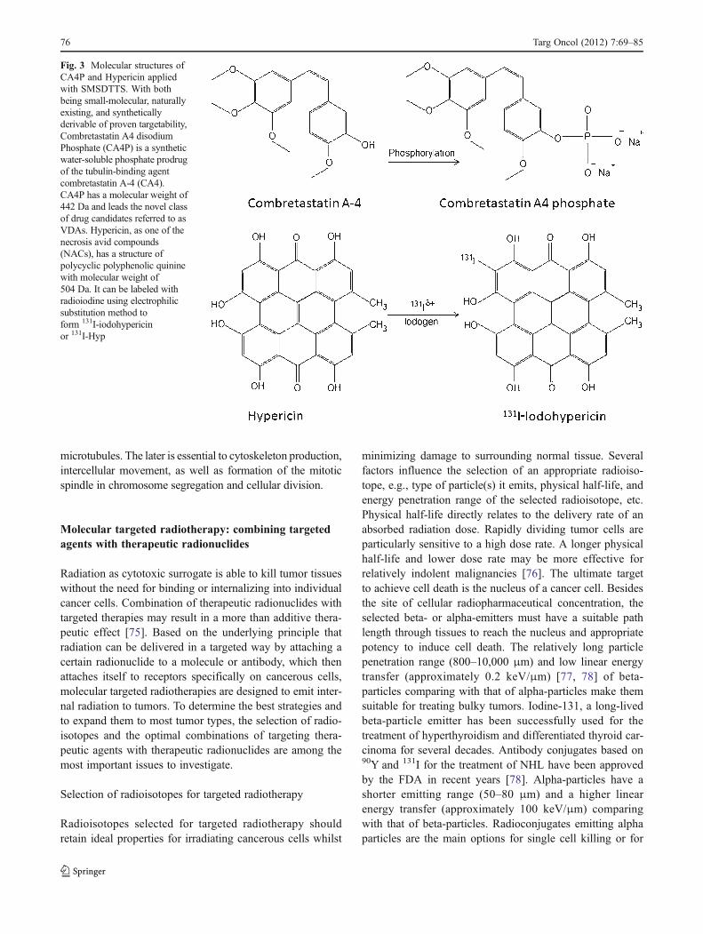
Combretastatin A4 Phosphate (CA4P) is a syntheticwater-soluble phosphate prodrug of the tubulin-bindingagent combretastatin A-4 (CA4) (Fig. 3). Following intra-cellular uptake, dephosphorylation of CA4P by endogenousphosphatases yields CA4, which binds to either the colchi-cines or vinblastine sites and causes depolymerization ofmicrotubules in endothelial cytoskeletons [61, 62]. Anti-cancer activities from this action lead to a change in shapeof vascular endothelial cells, i.e., they rapidly change fromflat- into balloon-like shape, which causes closure of capil-lary lumens and blockage of the tumor blood flow, resultingin necrosis of the tumor core within minutes to hours aftersystemic administration of CA4P [71]. As seen with otherVDAs, the tumor edge is less affected due to nutritioussupply from the surrounding normal tissues. This selectiveeffect is attributed to the fact that actin as another compo-nent of cytoskeleton is absent in tumoral endothelium butpresent in normal endothelium [74]. Thus, vascular shut-down due to endothelial disruption occurs only in tumorsbut not normal tissues. Depolymerization of tubulin alsoaffects cancer cells by preventing them from producing
Targ Oncol (2012) 7:69–85 75
microtubules. The later is essential to cytoskeleton production,intercellular movement, as well as formation of the mitoticspindle in chromosome segregation and cellular division.
Molecular targeted radiotherapy: combining targetedagents with therapeutic radionuclides
Radiation as cytotoxic surrogate is able to kill tumor tissueswithout the need for binding or internalizing into individualcancer cells. Combination of therapeutic radionuclides withtargeted therapies may result in a more than additive thera-peutic effect [75]. Based on the underlying principle thatradiation can be delivered in a targeted way by attaching acertain radionuclide to a molecule or antibody, which thenattaches itself to receptors specifically on cancerous cells,molecular targeted radiotherapies are designed to emit inter-nal radiation to tumors. To determine the best strategies andto expand them to most tumor types, the selection of radio-isotopes and the optimal combinations of targeting thera-peutic agents with therapeutic radionuclides are among themost important issues to investigate.
Selection of radioisotopes for targeted radiotherapy
Radioisotopes selected for targeted radiotherapy shouldretain ideal properties for irradiating cancerous cells whilst
minimizing damage to surrounding normal tissue. Severalfactors influence the selection of an appropriate radioiso-tope, e.g., type of particle(s) it emits, physical half-life, andenergy penetration range of the selected radioisotope, etc.Physical half-life directly relates to the delivery rate of anabsorbed radiation dose. Rapidly dividing tumor cells areparticularly sensitive to a high dose rate. A longer physicalhalf-life and lower dose rate may be more effective forrelatively indolent malignancies [76]. The ultimate targetto achieve cell death is the nucleus of a cancer cell. Besidesthe site of cellular radiopharmaceutical concentration, theselected beta- or alpha-emitters must have a suitable pathlength through tissues to reach the nucleus and appropriatepotency to induce cell death. The relatively long particlepenetration range (800–10,000 μm) and low linear energytransfer (approximately 0.2 keV/μm) [77, 78] of beta-particles comparing with that of alpha-particles make themsuitable for treating bulky tumors. Iodine-131, a long-livedbeta-particle emitter has been successfully used for thetreatment of hyperthyroidism and differentiated thyroid car-cinoma for several decades. Antibody conjugates based on90Y and 131I for the treatment of NHL have been approvedby the FDA in recent years [78]. Alpha-particles have ashorter emitting range (50–80 μm) and a higher linearenergy transfer (approximately 100 keV/μm) comparingwith that of beta-particles. Radioconjugates emitting alphaparticles are the main options for single cell killing or for
Fig. 3 Molecular structures ofCA4P and Hypericin appliedwith SMSDTTS. With bothbeing small-molecular, naturallyexisting, and syntheticallyderivable of proven targetability,Combretastatin A4 disodiumPhosphate (CA4P) is a syntheticwater-soluble phosphate prodrugof the tubulin-binding agentcombretastatin A-4 (CA4).CA4P has a molecular weight of442 Da and leads the novel classof drug candidates referred to asVDAs. Hypericin, as one of thenecrosis avid compounds(NACs), has a structure ofpolycyclic polyphenolic quininewith molecular weight of504 Da. It can be labeled withradioiodine using electrophilicsubstitution method toform 131I-iodohypericinor 131I-Hyp
76 Targ Oncol (2012) 7:69–85

treatment of minimal residual tumor cells. Researches on theefficacy of alpha-emitting radioconjugates seem encourag-ing, and clinical trials for leukemia, cystic glioma, andmelanoma are under way. The characteristics of clinicallyused common radioisotopes are summarized in Table 2.
Targeting tumor with radiolabeled peptides
Peptides have recently showed prominence in targeting ma-lignancies for several reasons. Key properties with peptidesinclude fast clearance, rapid tissue penetration, and lowantigenicity, as well as relatively easy and inexpensive pro-duction [79]. Radiolabeled peptides appear to be among themost promising vectors for TATs, as they offer an attractivevehicle for clinical use and commercialization [78]. Due tothe receptor-mediated internalization and intracellular reten-tion properties of radiopeptides, this approach can deliveradequate radiation doses to the tumor cells for achieving atleast volume reduction purpose [80]. Examples of currentradiopharmaceuticals include small peptides such as octreo-tide, neurotensin, α-melanocyte stimulating hormone, vaso-intestinal peptide, and others [81]. Somatostatin is a peptidehormone that naturally presents in neuroendocrine tumors,i.e., tumors in hypothalamus brain stem, gastrointestinaltract, and pancreas. The best clinically established radio-peptides for in vivo targeting to tumor are based on thesomatostatin receptors [78, 80–82]. A particularly largenumber of excellent radioligands have been developed fromsomatostatin agonists [82]. 111In-labeled somatostatin hasbeen widely used as a nuclear imaging agent. However,tumor size reduction was seldom achieved with such111In-labeled somatostatin analogs. Therapeutic radiopepti-des with beta-emitting isotopes like 90Yand 177Lu have beenmost extensively studied [78, 80]. 90Y-DOTA(0)-Tyr3-octreotide and 177Lu-DOTA(0)-Tyr3-octreotate have provedto be encouraging and promising in terms of neuroendocrine
tumor regression in various studies [78]. Anti-cancer effectsof 90Y-DOTA(0)-Tyr3-octreotide, as reported in literature,vary so much between various studies: complete plus partialregression of 50% or more was achieved in 22±11% of studiedpatients with neuroendocrine gastroenteropancreatic(GEP) tumors in multi-center phase I studies [83]. With177Lu-DOTA(0)-Tyr3-octreotate treatment in patients with neu-roendocrine GEP tumors, tumor regression of over 50% in28% and 25–50% in 19% was achieved with progressivedisease in 18% and stable disease in 35% of studied patients[83]. Thus, radiolabeled analogs of somatostatin represented awell-established paradigm of peptide radiopharmaceuticals fortargeting tumors [78, 80–84]. To further address the potentialof radiopeptide therapy and to establish the optimal treatmentscheme, both uniform pathology-oriented trials and random-ized clinical trials are required [80].
Radiolabeled MIBG
MIBG or Iobenguane, an iodinated arylalkylguanidine nor-epinephrine analog, resulted from the combination of thebenzyl group of bretylium and the guanidine group of gua-nethidine [80, 85]. Organ systems with rich adrenergic inner-vation and/or catecholamine excretion possess a high uptakeof MIBG. Thereby, MIBG radiolabeled with iodine-131 hasbeen used for imaging and therapy of neuroectodermallyderived tumors such as neuroblastomas, pheochromocytomas,paragangliomas, medullary thyroid carcinomas, carcinoidtumors, and Merkel cell tumors of the skin, etc. [80]. Thera-peutic doses of 131I-MIBG have been administered for exper-imental therapy of malignant pheochromocytoma and otherneuroendocrine tumors. The intense uptake and long retentionof 131I-MIBG indicate its therapeutic efficacy in metastatictumors. In a review of 116 patients, partial response in18–88% of patients was reported with varying dosesof 131I-MIBG [86]. A 30% overall objective response (com-plete and partial tumor response) was reported in a survey of131I-MIBG practice in over 99% of 537 treated patients withrefractory, stage III/IV disease, excluding childhood neuro-blastoma [76]. The result was associated with reduction inmeasurable tumor markers (complete and partial response) in38% of patients and subjective response in 52% [76]. Cur-rently, large dose of radio-iodinated MIBG has been used totreat relapsed or refractory metastatic neuroblastoma, moststudies reported a response rate of 30–40% with 131I-MIBGin these patients. More recent studies mainly focused on thecombination of 131I-MIBG with chemotherapy or myeloabla-tive regimens [87].
Monoclonal antibodies mediated targeting radiotherapy
Radioimmunotherapy (RIT) is currently a major researchtopic for personalized cancer treatment that combines the
Table 2 Characteristics of several radioisotopes more used for radio-therapeutic purposes
Isotope (symbol) T1/2 Emission Meantissue pathlength (μm)
Decayenergy
β/α (KeV)
Iodine-131 (131I) 8.04 days Beta/gamma 900 970
Yttrium-90 (90Y) 2.67 days Beta 3,900 2,280
Lutetium-177 (177Lu) 6.7 days Beta/gamma 700 497
Samarium-153 (153Sm) 1.95 days Beta/gamma 1,200 807
Rhenium-188 (188Re) 17 h Beta/gamma 3,500 2,120
Strontium-89 (89Sr) 50.5 days Beta 2,400 1,492
Actinium-225 (225AC) 10 days Alpha 65 5,935
Bismuth-213 (213Bi) 46 min Beta/alpha 80 1,422/5,982
T1/2 half-life, μm micrometer, mean tissue path length mean range insoft tissue, decay energyβ/α the energy released by a radioactive decaythrough beta or alpha emission
Targ Oncol (2012) 7:69–85 77

cancer killing of radiation therapy with the precise targetingcapacity of immunotherapy. Certain target structures ofmAbs have been identified for both hematological malig-nancies and solid tumors. Accordingly, radionuclides havebeen conjugated to such antibodies to increase specificity ofthe therapeutic intervention [88]. After intravenous injectionof a radiolabeled mAb, the binding ability of the antibody tothe tumor-associated antigen ensures that the tumor gets ahigh dose of radiation, which would be sufficient to kill thetargeted cancer cells and the nearby cells. RIT takes advan-tage of a growing number of mAbs to target tumor cellspreferentially while sparing normal and healthy tissues.Recent advances in chemistry have led to increasingly stableconjugation of radionuclide with mAbs [89]. Cancer thera-peutic index was potentially increased in comparison withother treatment modalities [77].
Currently the most promising area of RIT is in the treat-ment of NHL [89]. 90Y-Ibritumomab tiuxetan and 131I-Tosi-tumomab are the two radiolabeled mAbs that have beenapproved for treatment of follicular and recurred or resistedB cell NHL. High level expression of CD20 on normal andmalignant B cells has made it an attractive target for B cellNHL treatment. 90Y-Ibritumomab Tiuxetan and 131I-Tositu-momab bind to CD20 antigen on the surface of B cells,therefore deliver its radiation to target cancer cells.90Y-Ibritumomab Tiuxetan is indicated for the treatment ofadults with relapsed or refractory low-grade, follicular, ortransformed B cell lymphoma, but its safety has not beendetermined in children [90]. 131I-Tositumomab is indicatedfor the treatment of patients with CD20 positive, follicularNHL who are resistant to Rituximab and have relapsedfollowing chemotherapy [89]. Clinical results are veryencouraging with a high percentage of patients enteringlong-term remission with the above-mentioned RIT [89, 90].
Tumor necrosis therapy (TNT), an approach stems fromRIT, links a radioactive isotope to a targeted monoclonalantibody that is designed to bind to a universal intracellularantigen, i.e., DNA/histone H1 complex, which is exposedonly on dead and dying cells [91]. 131I-chTNT-1/B mAb is agenetically engineered, radiolabeled, chimeric mAb specificfor the necrotic core of malignant gliomas [91–93].131I-chTNT-1/B mAb, which delivers a cytotoxic dose ofradiation to the lesion core, has being investigated for thetreatment of newly diagnosed and recurrent high grade braintumors. It remains within the tumor necrosis and bombardsthe neighboring viable cells with radiation [93]. However,the current use of TNT is severely limited due to lowamount of tumor uptake, poor penetration into largerlesions, and heterogeneity of antibody uptake. Clinically,131I-chTNT-1/B mAb was delivered via convection-enhanced delivery in order to maximize coverage to thetumor and the invasive front of the glial tumor [90–93].Similar approach has been investigated both clinically and
experimentally in extra-cranial tumors [94], which alsoexposed the same drawbacks of insufficient tumoral tracerdistribution as well as unsatisfactory therapeutic outcomesafter systemic drug delivery. To compensate insufficient tar-getability, intratumoral injection was attempted in patientswith lung cancers [94].
Small-molecular sequential dual targeting theragnosticstrategy (SMSDTTS): a new TAT strategy?
Necrosis avid compounds (NACs) represent a new class oftargeting chemicals that show extraordinary affinity to non-viable tissues typically necrosis in the living body [95, 96].NACs were originally identified after disproving the tumorselectivity of porphyrin derivatives used for photodynamictherapy (PDT) or for tumor-seeking diagnostic imaging[95–97]. Both porphyrin and non-porphyrin species ofNACs have been reported [95, 96]. In addition to non-oncological applications such as visualization of myocardialinfarction [95, 96, 98], NACs can be exploited for diagnos-tic and even therapeutic utilities in experimental and clinicaloncology, e.g., to assess the presence and extent of sponta-neous tumor necrosis, to evaluate the necrotic tumor fractionafter necrosis-inducing therapies, and to deliver therapeuticradionuclide to tumor necrosis and kill adjacent living can-cer cells by crossfire radiation. The necrotic core of tumorsfunctions as an abundant, insoluble, non-diffusible anchorfor NACs. Similar to TNT, NACs can access and bind to thenecrotic areas of tumors but with a higher affinity, particu-larly at the interface between necrotic and viable tumortissues. Therefore, NACs have the potential to carry thera-peutic agents and to preferentially target virtually all solidtumors [99, 100]. By extending the capacity of NACs fromdiagnostic to therapeutic applications, the radiolabeledderivatives of NACs have shown the property of penetratingand localizing into the tumoral necrotic region and thereafterbombard the adjacent viable tumor cells with ionizingradiation [10].
Hypericin, as a nonporphyrin NAC, is a naturally derivedsubstance isolated from the plant genus Hypericum, and it isalso synthetically obtainable by binding two molecules ofemodin. Hypericin, with molecular weight of 504 Da, has astructure of polycyclic polyphenolic quinine (Fig. 3). Apeculiar affinity for necrotic or irreversibly damaged ische-mic tissues has been shown with Hypericin [99, 100].Radiolabeled derivatives of Hypericin mono-[123I]-iodohy-pericin ([123I]MIH) as diagnostic NACs have been studiedin animal models of hepatic and myocardial infarction [100,101] and found to concentrate in necrotic liver and myocar-dium of over 20-fold of that in normal surrounding tissues24 h after systemic injection [100, 101]. Similar affinity wasfound with native Hypericin in areas of necrosis in tumor
78 Targ Oncol (2012) 7:69–85

models after ablative therapies [102, 103]. RadiolabeledHypericin has achieved encouraging results in sensitivityand specificity of necrosis targeting. The virtual tumor-seeking property of hypericin enables it to home to thenecrotic core of solid tumor, which prompts our assumptionthat radioactive “payload” can be delivered to the center ofthe tumor mass if Hypericin is radiolabeled with therapeuticradionuclide [96, 100]. Experiments using iodine-131labeled hypericin to form 131I-iodohypericin (131I-Hyp) indifferent types of tumor models are under investigationswith preliminary promising outcomes [10].
As can be self-explained by its full terms, SMSDTTSstands for an anticancer strategy using two small moleculesto sequentially target tumors for achieving both diagnosticand therapeutic effects. Instead of directly attacking cancercells as mostly elaborated by others, SMSDTTS primarilytargets cancer stromas (soil) and indirectly but more thor-oughly destroys parenchymal cancer cells (seeds), which ison the basis of soil-to-seeds hypothesis [10]. As shown inFig. 4, both chemicals sequentially implemented inSMSDTTS are small-molecular, naturally extractable orsynthetically derivable, and clinically injectable. Theirrespective targeting mechanisms are: (1) CA4P selectivelyshuts down tumor vasculature and induces ischemic tumornecrosis; and (2) radiolabeled hypericin carries and deliversa therapeutic radionuclide iodine-131 to the prior existing orinduced necrotic region in the tumor and kills neighboringresidual tumor cells by crossfire radiation. Theoretically,these two components in SMSDTTS represent a perfectmatch of complementary tumoricidal effects. The VDACA4P kills the tumor from the inside out and leaves viabletumor cells at the periphery. Since those remaining tumorcells rely on the surrounding normal blood supply for rapidgrowth, they are sensitive to radiotherapies, which aredesigned to kill rapidly proliferative and well-oxygenatedtumor tissues [9, 104]. Such sequential and complementarytreatment may improve the likelihood of complete tumordestruction and, therefore, more satisfactory therapeutic
outcomes. With both being small molecules of proven hightargetabilities, CA4P and Hypericin possess favorable phar-macokinetics and safety profiles, i.e., high target/non-targetratio, short biological half-lives, and absence of toxic sideeffects commonly seen with conventional chemotherapies.Relative to currently available anticancer treatments or thenewly advocated personalized but sophisticated and costlyTAT approaches, this new strategy may prove to be a non-invasive, simple, workable, affordable, and depersonalizedanticancer treatment for all solid malignant tumors in vis-ceral organs, whether primary or metastatic in early or latestages [10].
Basic requirements for TATs
To obtain satisfactory therapeutic outcomes with TATs,several basic requirements have to be taken into account.
Dependence on tumor-types
A wide range of tumors have been covered by TATs, e.g.,breast cancer treated by trastuzumab and lapatinib, colorec-tal cancer treated by bevacizumab, lung and pancreaticcancers treated by Gefitinib and erlotinib, lymphoma treatedby rituximab and tositumomab, leukemia treated by dasati-nib, etc. (Table 1). Although TATs greatly benefit thepatients with their respective tumors, tumor cells resistantto TAT drugs are frequently induced in some tumors. TATthat based on the mechanism of SMKI requires the targetingtumor to have identified and overexpressed mutations inkinase domain. Therapies with mAbs rely on sufficientspecific tumor-associated antigens expressing on the surfaceof tumor cells. Due to a lack of particular and generalizedTATs that can target all tumor types, many cancers still havenot been covered by TATs.
The pathophysiology of solid tumors differs from that ofnormal tissues and has been explored and utilized for
Fig. 4 Schematic hypothesis and components for the novel targetedanticancer therapy of SMSDTTS: predicted sequential dual targetingtumoricidal events: imagine there is an inoperable liver tumor (T), wefirst treat the tumor using the available VDAs to cause massive tumornecrosis (N) that becomes the reliable target for the second attack
launched after 24 h. An IV injected radiolabeled NAC accumulates inthe intratumoral necrosis (N) particularly in close vicinity to theperipheral viable rim and constantly irradiates the remnant tumor cells,resulting in complete tumor necrosis (N) or cancer eradication
Targ Oncol (2012) 7:69–85 79

developing TATs. For example, based on the knowledge thatangiogenesis is a continuous process to keep the tumorgrowth and the fact that tumor vessels are often highlyabnormal and are prone to collapse comparing with vascu-latures of normal tissues [58], VDAs and angiogenesisinhibitors have been introduced to treat a wider variety oftumor types by targeting these unique features. Anothergeneral consensus about solid tumors is that they maycontain large necrosis; thereby, TNT has been developedto tentatively target intratumoral necrosis, but the resultswith TNT are not satisfactory due to the lack of sufficientnecrosis avidity and/or uncertain presence of spontaneoustumor necrosis. Along with our recent advances with theSMSDTTS that combines VDAs and radiolabeled necrosistargeting compounds, most solid tumors are expected to betargeted and treated [10].
Aspects on targeting agents
Specificity and affinity of TATs
Agents for TATs are designed to interact specifically withparticular receptors to avoid or reduce side effects. On theother hand, how tightly a TAT agent binds to its receptor isalso a substantial concern. Tumor specificity and affinity arethe most essential requirements for TATs. The exquisitetarget specificity as well as the high affinity to targetingantigens enables mAbs to selectively bind to antigen andtherefore reduce off-target effects. However, most antigensare not tumor specific because they are expressed not onlyby certain cancer cells but also by normal cells. Meanwhile,the expression of antigens in tumors recognized by mAbs isoften heterogeneous, and loss of expression may beobserved in anaplastic transformation and may result inimmune escape [105]. SMKIs show great promise as anew class of TAT because they target the ATP binding sitein protein kinases domain, whereas ATP site is present in allof the more than 500 protein kinases identified in the humangenome, which makes cross-reactivity inescapable. Molec-ular specificity and off-target interactions of SMKIs againstkinases must be assessed and identified before its clinicaluse. Yet for most SMKIs, specificity and affinity have beendetermined against only relatively small sets of kinases[106].
Cytotoxicity concerns
TATs should have the potential to induce selective tumorcytotoxicity while sparing normal tissues. Despite certainabilities to localize into the tumor and to bind to tumor cells,spectacular tumor regressions are not always seen withunconjugated mAbs and SMKIs due to the following rea-sons. First, tumors express the target but are not dependent
thoroughly on the target for proliferation and/or survival.Secondly, the development of resistance occurs regardlessof the size and type of the tumor. Moreover, generallyinsufficient cytotoxicity of mAbs and SMKIs to cancer cellsmay hinder the therapeutic efficacy. Furthermore, most can-cers are not sensitive to single-agent targeted therapies.Even when sensitive to single-agent therapies, cancersdevelop resistance. Thus, novel cytotoxic agents withunique mechanisms of actions are continuously being pur-sued [107] and TATs are required to combine with multipleagents or different mechanisms to achieve sufficient cyto-toxicity and to gain synergistic anticancer effects [108].Efforts to improve the cytotoxicity of mAbs have beenfocused on conjugates with cytotoxic agent, radioisotopes,and immunotoxins [109]. Our endeavors in developingSMSDTTS represent another example [10].
Highlighted by 90Y-Ibritumomab Tiuxetan and 131I-Tosi-tumomab, which have been clinically approved as radio-immunoconjugates used for the treatment of lymphomas,RIT could be heading for the mainstream in TAT develop-ment. Radioimmunoconjugates are produced either bycovalently binding the radioisotope directly to the mAb orby crosslinking them through a chemical linker or a chelator[110]. Besides the targeting specificity of the antibody to thecancer cells, the stability of the antibody-radionuclide con-jugate, the cytotoxic potentiality of the selected radionuclidewith regard to the targeted cells, are the key components forthe optimization of RIT.
Toxicity
Although TAT agents were deliberately chosen or designedto act on specific molecular targets, which may lead to fewerand less toxic side effects than conventional chemotherapy,toxic effects associated with TATs such as hypertension,fatigue, bone marrow toxicity, skin toxicity, gastrointestinalside-effects as well as immunosuppression, metabolic alter-ations, interstitial pneumonitis, and hypothyroidism do com-monly present [111]. Patients treated with TATs need to beclosely monitored for the development of drug-related tox-icities. Meanwhile, supportive measures to prevent interrup-tions of treatment, dose reductions, and eventualdevelopment of life-threatening complications should bevigorously taken to manage with drug-related toxicitieseven at mild and moderate levels [112].
Considerations for TATs
Cancer biology: inherent hurdles for cancer cure
Oncogenesis is a multi-step process with various genes andpathways, different mechanisms, multifarious carcinogens,
80 Targ Oncol (2012) 7:69–85

viruses, cytokines, hormones, bacteria, etc., as well as awhole bunch of possible gene mutations or disruptionsinvolved, which together allow the cells to undergo uncon-trolled division, thus forming a malignant mass [113].Besides, other nonexclusive detailed mechanisms that trig-ger resistance can be envisaged: target mutation, targetamplification, activation of a complementary pathway thatbypasses the target requirement, upregulation of mecha-nisms that lower the intracellular concentrations of the tar-get, etc [14]. Such an increasingly known complexity makescancer cure biologically almost impossible.
What to target and how to target in cancer
All targeted cancer therapies aim to maximize tumordestruction while minimize side-effects, which makes tumoraffinity or target binding an essential demand. High targettissue binding is the most important goal, whereas bloodpool residence and nonspecific binding of a TAT agent arealso important considerations. Equally important is the bio-distribution of metabolized components and their excretionroutes particularly for those radioactively labeled com-pounds. Although the discussed TATs by inhibiting a singlemolecular target, antigen, or neoangiogenesis may preventtumor cell proliferation or kill targeted malignant cellseffectively, tumor progression is unlikely to depend on asole signal transduction pathway. Furthermore, for anygenetically unstable diseases including cancer, resistance isan inevitable consequence of the treatment with a singlemolecular targeted agent or antibody [114]. Apparently,the newly introduced SMSDTTS which chooses noncancer-ous, less mutant, and more stable stromal targets may con-front less drug resistance and more therapeutic response[10].
Possible reasons for unsuccessful TATs
SMKIs are generally designed with intention to target onespecific kinase. However, as a result of the evolutionarilyconserved nature of the ATP binding pocket, a SMKI maypotently inhibit lots of other kinase members while targetingtheir specific kinase. Such off-target kinases may be a po-tential safety liability of SMKIs therapies and may hinderdrug development [115, 116]. For both mAb and its radio-labeled derivatives, only a handful of studies have shown asignificant number of complete remissions up to now. Sev-eral reasons account for the failure or unsatisfactory resultsfrom mAbs. Firstly, the specificity of antigen expression ontumor cells is poor, and tumor antigens often express tosome degree on normal cells. Secondly, intracellular com-partments, in particular the cytoplasm or nucleus, havegenerally been poorly accessible or inaccessible to mono-clonal antibodies [75, 117]. Moreover, the activity dose in
RITs is limited by myelotoxicity as a result of the continu-ous radiation exposure of the red bone marrow to the slow-clearing antibody. Thus, the success of mAb therapy andRIT for treatment of solid tumors has been limited so far[118]. As well, radiolabeled peptides and MIBG only showeffects in limited types of malignant tumors.
Limitations and obstacles of molecular targetedradiotherapies
Molecular targeted radiotherapy, being an evolving andpromising modality of cancer treatment, is required to beefficacious with minimal normal tissue toxicity [119]. Lim-itations concerning research on molecular targeted radio-therapies mainly include non-uniqueness of antibodies fortumor cell antigens (antibodies may bind to non-target anti-gens on normal cells), heterogeneous antigen expression ontumor cells, formidable myelotoxicity, slow blood clear-ance, and sub-optimal distribution of the relatively large(molecule weight 150 kDa) radiolabeled antibodies in thetumor [120]. Besides, other obstacles include inadequateunderstanding of the molecular mechanism and pharmacolo-gy of the agent, physical characteristics of selected cytotoxicradionuclides, intrinsic inferior cellular radiosensitivity, can-cer cell resistance factors, normal cell toxicity, and criteria ofclinical trial designs, etc. However, the major clinical limita-tion of targeted radiotherapy, particularly for treatment of solidtumors, lies in immunogenicity, cell specificity, and cell per-meability of the targeting molecular ligands.
Despite the collective and enriching knowledge on mo-lecular targeted radiotherapy thus far, many obstacles arestill in suspense and require further exploration. Basicrequirements for future molecular targeted radiotherapiesshould involve cytotoxic radioligands with high target spec-ificity, uniqueness for tumor cell or tumor stroma, rapidblood clearance, suitable physical characteristics for system-ic administration, appropriate potency for cancer cells, and awide coverage of tumors. In addition, the conjugation of thetargeting molecule to the radionuclide should be reliable.The final radioligand must be practical, affordable for clin-ical use, as well as stable in vivo and effective at targetingthe tumoral binding site [78]. Small molecular necrosis avidHypericin with a high target specificity as we proposed islikely to play a crucial role in such a context [10].
Potential challenges and new opportunities for TATs
Resulting from immature tumor blood vessels, discrepancybetween nutrition supply and tumor growth, immuneresponse and incomplete treatment, one unique characteris-tic of most solid tumors is that they encompass a proportionof dead tissue in addition to numerous proliferating viablecancer cells. The accumulation of dead cells results in the
Targ Oncol (2012) 7:69–85 81

formation of a necrotic core presented in virtually all solidtumors beyond a certain size. On the other hand, clinically,the most critical problem for all cancer therapies is incom-plete treatment, which sooner or later leads to tumor relapse.For instance, VDAs can cause tumor vessel shutdown andlead to tumor core necrosis. However, tumor relapses quicklydue to peripheral remaining viable cells. Radiofrequencyablation (RFA) may destroy the tumor, but the remainingviable cells frequently cause tumor recurrence. Thereby,sequential combination of targeted therapies by non-overlapping complementary mechanisms is imperative andshould be designed to achieve synergetic outcomes [58].SMSDTTS literally destroy the tumor “from the inside out”with minimal radiation exposure to healthy tissues due to thehigh target-to-nontarget ratio. Sequential use of CA4P andIodine-131 labeled Hypericin may provide an ingeniousapproach and simplify the cancer problems. Encouragingresults have been achieved with SMSDTTS in recent prelim-inary experiments in rodent tumor models [10]. Further opti-mizations are warranted before the implementation of this newstrategy in clinical oncology.
Acknowledgments This work was partially supported by the grantsawarded by FWO Vlaanderen ZWAP/05/018; Geconcerteerde Onder-zoeksactie of the Flemish Government, OT project (OT/06/70); the KULeuven Molecular Small Animal Imaging Center MoSAIC (KUL EF/05/08); the center of excellence In vivo Molecular Imaging Research(IMIR) of KU Leuven; the IWT SBO “Imagine” (SBO80017) and aEU project Asia-Link CfP 2006-EuropeAid/123738/C/ACT/Multi-Proposal No. 128-498/111. The corresponding author Yicheng Ni iscurrently a Bayer Lecture Chair holder.
Conflict of interest statement We, all the authors of the manuscripttitled “A review on various targeted anticancer therapies”, declare thatwe do not have any conflict of interest regarding the content of thepaper. We have no financial relationship with any organizations thatsponsored our research. We agree to allow the journal to review theirdata if requested.
References
1. Levin PBAB (2008) World Cancer Report 2008; ReportNo.9789283204237
2. Boyle P (2006) The globalisation of cancer. Lancet 368(9536):629–630
3. Meeran SM, Katiyar SK (2008) Cell cycle control as a basis forcancer chemoprevention through dietary agents. Front Biosci13:2191–2202
4. Aggarwal BB, Danda D, Gupta S, Gehlot P (2009) Models forprevention and treatment of cancer: problems vs promise. Bio-chem Pharmacol 78(9):1083–1094
5. Hamilton A, Gallipoli P, Nicholson E, Holyoake TL (2010)Targeted therapy in haematological malignancies. J Pathol 220(4):404–418
6. Ross JS, Schenkein DP, Pietrusko R, Rolfe M, Linette GP, Stec Jet al (2004) Targeted therapies for cancer. Am J Clin Pathol 122(4):598–609
7. Piccaluga PP, Martinelli G, Baccarani M (2006) Advances in thetreatment for haematological malignancies. Expert Opin Phar-macother 7(6):721–732
8. Green MR (2004) Targeting targeted therapy. N Engl J Med 350(21):2191–2193
9. Wachsberger P, Burd R, Dicker AP (2003) Tumor response toionizing radiation combined with antiangiogenesis or vasculartargeting agents: exploring mechanisms of interaction. ClinCancer Res 9:1957–1971
10. Li J, Sun Z, Zhang J, Shao H, Miranda Cona M, Wang H et al(2011) A dual-targeting anticancer approach: soil and seed prin-ciple. Radiology 260(3):799–807
11. Cherry M, Williams DH (2004) Recent kinase and kinase inhib-itor X-ray structures: mechanisms of inhibition and selectivityinsights. Curr Med Chem 11(6):663–673
12. McGregor MJ (2007) A pharmacophore map of small moleculeprotein kinase inhibitors. J Chem Inf Model 47(6):2374–2382
13. Krause DS, Van Etten RA (2005) Tyrosine kinases as targets forcancer therapy. N Engl J Med 353(2):172–187
14. Pearson MA, Fabbro D (2004) Targeting protein kinases in cancertherapy: a success? Expert Rev Anticancer Ther 4(6):1113–1124
15. Furge KA, MacKeigan JP, Teh BT (2010) Kinase targets in renal-cell carcinomas: reassessing the old and discovering the new.Lancet Oncol 11(6):571–578
16. Fasolo A, Sessa C (2011) Current and future directions in mam-malian target of rapamycin inhibitors development. Expert OpinInvestig Drugs 20(3):381–394
17. Gambacorti-Passerini C (2008) Part I: Milestones in personalisedmedicine—imatinib. Lancet Oncol 9(6):600
18. DeAngelo DJ, Ritz J (2004) Imatinib therapy for patients withchronic myelogenous leukemia: are patients living longer? ClinCancer Res 10(1 Pt 1):1–3
19. Sawyers C (2004) Targeted cancer therapy. Nature 432(7015):294–297
20. le Coutre P, Kreuzer KA, Pursche S, Bonin M, Leopold T,Baskaynak G et al (2004) Pharmacokinetics and cellular uptakeof imatinib and its main metabolite CGP74588. Cancer Chemo-ther Pharmacol 53(4):313–323
21. Kantarjian H, Sawyers C, Hochhaus A, Guilhot F, Schiffer C,Gambacorti-Passerini C et al (2002) Hematologic and cytogeneticresponses to imatinib mesylate in chronic myelogenous leukemia.N Engl J Med 346(9):645–652
22. Baselga J (2002) Why the epidermal growth factor receptor? Therationale for cancer therapy. Oncologist 7(Suppl 4):2–8
23. Reis-Filho JS, Milanezi F, Carvalho S, Simpson PT, Steele D,Savage K et al (2005) Metaplastic breast carcinomas exhibitEGFR, but not HER2, gene amplification and overexpression:immunohistochemical and chromogenic in situ hybridizationanalysis. Breast Cancer Res 7(6):R1028–R1035
24. Patra CR, Bhattacharya R, Mukhopadhyay D, Mukherjee P(2010) Fabrication of gold nanoparticles for targeted therapy inpancreatic cancer. Adv Drug Deliv Rev 62(3):346–361
25. Mantha AJ, Hanson JE, Goss G, Lagarde AE, Lorimer IA,Dimitroulakos J (2005) Targeting the mevalonate pathway inhib-its the function of the epidermal growth factor receptor. ClinCancer Res 11(6):2398–2407
26. Lynch TJ, Bell DW, Sordella R, Gurubhagavatula S, OkimotoRA, Brannigan BW et al (2004) Activating mutations in theepidermal growth factor receptor underlying responsiveness ofnon-small-cell lung cancer to gefitinib. N Engl J Med 350(21):2129–2139
27. Paez JG, Janne PA, Lee JC, Tracy S, Greulich H, Gabriel S et al(2004) EGFR mutations in lung cancer: correlation with clinicalresponse to gefitinib therapy. Science 304(5676):1497–1500
28. Maemondo M, Inoue A, Kobayashi K, Sugawara S, Oizumi S,Isobe H et al (2010) Gefitinib or chemotherapy for non-small-cell
82 Targ Oncol (2012) 7:69–85

lung cancer with mutated EGFR. N Engl J Med 362(25):2380–2388
29. US Food and Drug Administration (2011) FDA labeling infor-mation—Xalkori. FDA website: http://www.accessdata.fda.gov/drugsatfda_docs/label/2011/202570s000lbl.pdf
30. Shaw AT, Yasothan U, Crizotinib KP (2011) Nat Rev DrugDiscov 10(12):897–898
31. Chapman PB, Hauschild A, Robert C, Haanen JB, Ascierto P,Larkin J et al (2011) Improved survival with vemurafenib inmelanoma with BRAF V600E mutation. N Engl J Med 364(26):2507–2516
32. US Food and Drug Administration (2011) FDA labeling infor-mation—Zelboraf. FDA website: http://www.accessdata.fda.gov/drugsatfda_docs/label/2011/202429s000lbl.pdf
33. Von Mehren M, Adams GP, Weiner LM (2003) Monoclonalantibody therapy for cancer. Annu Rev Med 54:343–369
34. Adams GP, Weiner LM (2005) Monoclonal antibody therapy ofcancer. Nat Biotechnol 23(9):1147–1157
35. Zhang Q, Chen G, Liu X, Qian Q (2007) Monoclonal antibodiesas therapeutic agents in oncology and antibody gene therapy. CellRes 17(2):89–99
36. Ross JS, Gray K, Gray GS, Worland PJ, Rolfe M (2003) Anti-cancer antibodies. Am J Clin Pathol 119(4):472–485
37. Funaro A, Horenstein AL, Santoro P, Cinti C, Gregorini A,Malavasi F (2000) Monoclonal antibodies and therapy of humancancers. Biotechnol Adv 18(5):385–401
38. Gelmon K (2008) Part II: Milestones in personalised medicine—trastuzumab. Lancet Oncol 9(7):698
39. Cheson BD, Leonard JP (2008) Monoclonal antibody therapy forB-cell non-Hodgkin's lymphoma. N Engl J Med 359(6):613–626
40. Plosker GL, Figgitt DP (2003) Rituximab: a review of its use innon-Hodgkin's lymphoma and chronic lymphocytic leukaemia.Drugs 63(8):803–843
41. Slamon DJ, Clark GM, Wong SG, Levin WJ, Ullrich A, McGuireWL (1987) Human breast cancer: correlation of relapse andsurvival with amplification of the HER-2/neu oncogene. Science235(4785):177–182
42. Menard S, Pupa SM, Campiglio M, Tagliabue E (2003) Biologic andtherapeutic role of HER2 in cancer. Oncogene 22(42):6570–6578
43. Mandler R, Wu C, Sausville EA, Roettinger AJ, Newman DJ, HoDK et al (2000) Immunoconjugates of geldanamycin and anti-HER2 monoclonal antibodies: antiproliferative activity on humanbreast carcinoma cell lines. J Natl Cancer Inst 92(19):1573–1581
44. Shukla R, Thomas TP, Peters JL, Desai AM, Kukowska-Latallo J,Patri AK et al (2006) HER2 specific tumor targeting with den-drimer conjugated anti-HER2 mAb. Bioconjug Chem 17(5):1109–1115
45. Jahanzeb M (2008) Adjuvant trastuzumab therapy for HER2-positive breast cancer. Clin Breast Cancer 8(4):324–333
46. Slamon DJL-JB, Shak S, Fuchs H, Paton V, Bajamonde A,Fleming T et al (2001) Use of chemotherapy plus a monoclonalantibody against HER2 for metastatic breast cancer that over-expresses HER2. N Engl J Med 344(11):783–792
47. Tai W, Mahato R, Cheng K (2010) The role of HER2 in cancertherapy and targeted drug delivery. J Control Release 146(3):264–275
48. Smith I, Procter M, Gelber RD, Guillaume S, Feyereislova A,Dowsett M et al (2007) 2-year follow-up of trastuzumab afteradjuvant chemotherapy in HER2-positive breast cancer: a rando-mised controlled trial. Lancet 369(9555):29–36
49. Perez EA, Romond EH, Suman VJ, Jeong JH, Davidson NE,Geyer CE Jr et al (2007) Updated results of the combined analysisof NCCTG N9831 and NSABP B-31 adjuvant chemotherapywith/without trastuzumab in patients with HER2-positive breastcancer. 2007 ASCO Annual Meeting Proceedings Part I; 25(18S):512
50. Slamon DJ, Eiermann W, Robert N, Pienkowski T, Martin M,Pawlicki M et al (2006) 2nd interim analysis phase III random-ized trial comparing doxorubicin and cyclophosphamide fol-lowed by docetaxel with doxorubicin and cyclophosphamidefollowed by docetaxel and trastuzumab with docetaxel, carbopla-tin and trastuzumab in HER2neu positive early breast cancerpatients. Breast Cancer Res Treat 100(suppl 1):LBA 53
51. Verma S (2008) Trastuzumab in the adjuvant setting: concurrentor sequential? It takes two to tango! Curr Oncol 15(1):66–67
52. Amoroso A, Hafsi S, Militello L, Russo AE, Soua Z, MazzarinoMC et al (2011) Understanding rituximab function and resistance:implications for tailored therapy. Front Biosci 16:770–782
53. Hauptrock B, Hess G (2008) Rituximab in the treatment of non-Hodgkin's lymphoma. Biologics 2(4):619–633
54. Dudek A, Gupta K, Ramakrishnan S, Mukhopadhyay D (2010)Editorial Tumor angiogenesis. J Oncol doi:10.1155/2010/761671
55. Tortora G, Melisi D, Ciardiello F (2004) Angiogenesis: a targetfor cancer therapy. Curr Pharm Des 10(1):11–26
56. Siemann DW, Horsman MR (2009) Vascular targeted therapies inoncology. Cell Tissue Res 335(1):241–248
57. Siemann DW, Bibby MC, Dark GG, Dicker AP, Eskens FA,Horsman MR et al (2005) Differentiation and definition ofvascular-targeted therapies. Clin Cancer Res 11(2 Pt 1):416–420
58. Brown JM, Giaccia AJ (1998) The unique physiology of solidtumors: opportunities (and problems) for cancer therapy. CancerRes 58(7):1408–1416
59. Jain RK (2005) Normalization of tumor vasculature: an emergingconcept in antiangiogenic therapy. Science 307(5706):58–62
60. Hicklin DJ, Ellis LM (2005) Role of the vascular endothelialgrowth factor pathway in tumor growth and angiogenesis. J ClinOncol 23(5):1011–1027
61. O'Hanlon LH (2005) Taking down tumors: vascular disruptingagents entering clinical trials. J Natl Cancer Inst 97(17):1244–1245
62. Siemann DW (2011) The unique characteristics of tumor vascu-lature and preclinical evidence for its selective disruption bytumor-vascular disrupting agents. Cancer Treat Rev 37(1):63–74, Epub 2010 Jun 8
63. Griffioen AW, Molema G (2000) Angiogenesis: potentials forpharmacologic intervention in the treatment of cancer, cardiovas-cular diseases, and chronic inflammation. Pharmacol Rev 52(2):237–268
64. Kerbel RS (2008) Tumor angiogenesis. N Engl J Med 358(19):2039–2049
65. Angiogenesis Inhibitors for Cancer (2011) http://www.angio.org/understanding/inhib.php
66. McMahon G (2000) VEGF receptor signaling in tumor angio-genesis. Oncologist 5(Supplement 1):3–10
67. Eskens FA, Sleijfer S (2008) The use of bevacizumab in colorec-tal, lung, breast, renal and ovarian cancer: where does it fit? Eur JCancer 44(16):2350–2356
68. Uronis HE, Hurwitz HI (2007) Is bevacizumab effective and safein combination with chemotherapy in patients with colorectalcancer? Nat Clin Pract Oncol 4(4):214–215
69. FDA Approval for Bevacizumab. http://www.cancer.gov/cancertopics/druginfo/fda-bevacizumab
70. Gotink KJ, Verheul HM (2010) Anti-angiogenic tyrosine kinaseinhibitors: what is their mechanism of action? Angiogenesis 13(1):1–14
71. Hinnen P, Eskens FALM (2007) Vascular disrupting agents inclinical development. Br J Cancer 96:1159–1165. doi:10.1038/sj.bjc.6603694
72. Gridelli C, Rossi A, Maione P, Rossi E, Castaldo V, Sacco PC etal (2009) Vascular disrupting agents: a novel mechanism of actionin the battle against non-small cell lung cancer. Oncologist 14(6):612–620
Targ Oncol (2012) 7:69–85 83

73. Zhao L, Ching LM, Kestell P, Kelland LR, Baguley BC (2005)Mechanisms of tumor vascular shutdown induced by 5, 6-dimethylxanthenone-4-acetic acid (DMXAA): increased tumorvascular permeability. Int J Cancer 116(2):322–326
74. Wang H, Sun X, Chen F, De Keyzer F, Yu J, Landuyt W et al(2009) Treatment of rodent liver tumor with combretastatin a4phosphate: noninvasive therapeutic evaluation using multipara-metric magnetic resonance imaging in correlation with micro-angiography and histology. Investig Radiol 44(1):44–53
75. Miederer M, McDevitt MR, Sgouros G, Kramer K, Cheung NK,Scheinberg DA (2004) Pharmacokinetics, dosimetry, and toxicityof the targetable atomic generator, 225Ac-HuM195, in nonhumanprimates. J Nucl Med 45(1):129–137
76. Lewington VJ, Clarke SE, Hoefnagel CA, Behr TM, Brans B,deKlerk J, Vieira MR (2003) 131I mIBG therapy: results of aEuropean Survey. Presented at European Association of NuclearMedicine Medicine Meeting, August 2003, Amsterdam
77. Dancey G, Begent RH, Meyer T (2009) Imaging in targeteddelivery of therapy to cancer. Target Oncol 4:201–217.doi:10.1007/s11523-009-0119-8
78. Perkins A (2005) In vivo molecular targeted radiotherapy.Biomed Imaging Interv J 1(2):e9
79. Weiner RE, Thakur ML (2005) Radiolabeled peptides in oncology:role in diagnosis and treatment. BioDrugs 19(3):145–163
80. Oyen WJ, Bodei L, Giammarile F, Maecke HR, Tennvall J,Luster M et al (2007) Targeted therapy in nuclear medicine—current status and future prospects. Ann Oncol 18(11):1782–1792
81. Okarvi SM (2008) Peptide-based radiopharmaceuticals and cyto-toxic conjugates: potential tools against cancer. Cancer Treat Rev34(1):13–26
82. Ginj M, Zhang H, Waser B, Cescato R, Wild D, Wang X et al(2006) Radiolabeled somatostatin receptor antagonists are pref-erable to agonists for in vivo peptide receptor targeting of tumors.Proc Natl Acad Sci U S A 103(44):16436–16441
83. Van Essen M, Krenning EP, De JongM, Valkema R, KwekkeboomDJ (2007) Peptide receptor radionuclide therapy with radiolabelledsomatostatin analogues in patients with somatostatin receptor pos-itive tumours. Acta Oncol 46(6):723–734
84. Mariani G, Erba PA, Signore A (2006) Receptor-mediated tumortargeting with radiolabeled peptides: there is more to it thansomatostatin analogs. J Nucl Med 47(12):1904–1907
85. Giammarile F, Chiti A, Lassmann M, Brans B, Flux G (2008)EANM procedure guidelines for 131I-meta-iodobenzylguanidine(131I-mIBG) therapy. Eur J Nucl Med Mol Imaging 35(5):1039–1047
86. Shapiro B, Sisson JC, Wieland DM, Mangner TJ, Zempel SM,Mudgett E et al (1991) Radiopharmaceutical therapy of malignantpheochromocytoma with [131I]-metaiodobenzylguanidine: resultsfrom ten years of experience. J Nucl Biol Med 35(4):269–276
87. DuBois SG, Matthay KK (2008) Radiolabeled metaiodobenzyl-guanidine for the treatment of neuroblastoma. Nucl Med Biol 35(Suppl 1):S35–S48
88. Schrama D, Reisfeld RA, Becker JC (2006) Antibody targeteddrugs as cancer therapeutics. Nat Rev Drug Discov 5(2):147–159
89. Schaefer NG, Ma J, Huang P, Buchanan J, Wahl RL (2010) Radio-immunotherapy in non-Hodgkin lymphoma: opinions of U.S. med-ical oncologists and hematologists. J Nucl Med 51(6):987–994
90. Jacobs SA (2007) Yttrium ibritumomab tiuxetan in the treatmentof non-Hodgkin's lymphoma: current status and future prospects.Biologics 1(3):215–227
91. Chen FM, Taylor CR, Epstein AL (1989) Tumor necrosis treat-ment of ME-180 human cervical carcinoma model with 131I-labeled TNT-1 monoclonal antibody. Cancer Res 49(16):4578–4585
92. Shapiro WR, Carpenter SP, Roberts K, Shan JS (2006) (131)I-chTNT-1/B mAb: tumour necrosis therapy for malignant astro-cyticglioma. Expert Opin Biol Ther 6(5):539–545
93. Khawli LA, Mizokami MM, Sharifi J, Hu P, Epstein AL (2002)Pharmacokinetic characteristics and biodistribution of radioiodi-nated chimeric TNT-1, -2, and -3 monoclonal antibodies afterchemical modification with biotin. Cancer Biother Radiopharm17(4):359–370
94. Chen S, Yu L, Jiang C, Zhao Y, Sun D, Li S et al (2005) Pivotalstudy of iodine-131-labeled chimeric tumor necrosis treatmentradioimmunotherapy in patients with advanced lung cancer. JClin Oncol 23(7):1538–1547
95. Ni Y, Bormans G, Chen F, Verbruggen A, Marchal G (2005)Necrosis avid contrast agents: functional similarity versus struc-tural diversity. Investig Radiol 40(8):526–535
96. Ni Y (2008) Metalloporphyrins and functional analogues as MRIcontrast agents. Curr Med Imaging Rev 4:96–112
97. Ni Y, Marchal G, Yu J, Lukito G, Petre C, Wevers M et al (1995)Localization of metalloporphyrin-induced “specific” enhancementin experimental liver tumors: comparison of magnetic resonanceimaging,microangiographic, and histologic findings. AcadRadiol 2(8):687–699
98. Ni Y, Pislaru C, Bosmans H, Pislaru S, Miao Y, Bogaert J et al(2001) Intracoronary delivery of Gd-DTPA and Gadophrin-2 fordetermination of myocardial viability with MR imaging. EurRadiol 11(5):876–883
99. Van de Putte M, Ni Y, De Witte PA (2008) Exploration of themechanism underlying the tumor necrosis avidity of hypericin.Oncol Rep 19(4):921–926
100. Ni Y, Huyghe D, Verbeke K, de Witte PA, Nuyts J, Mortelmans Let al (2006) First preclinical evaluation of mono-[123I]iodohy-pericin as a necrosis-avid tracer agent. Eur J Nucl Med MolImaging 33(5):595–601
101. Fonge H, Jin L, Wang H, Ni Y, Bormans G, Verbruggen A (2007)Synthesis and preliminary evaluation of mono-[123I]iodohyper-icin monocarboxylic acid as a necrosis avid imaging agent. Bio-org Med Chem Lett 17(14):4001–4005
102. Van de Putte M, Wang H, Chen F, De Witte PA, Ni Y (2008)Hypericin as a marker for determination of tissue viability afterradiofrequency ablation in a murine liver tumor model. OncolRep 19(4):927–932
103. Van de Putte M, Wang H, Chen F, de Witte PA, Ni Y (2008)Hypericin as a marker for determination of tissue viability afterintratumoral ethanol injection in a murine liver tumor model.Acad Radiol 15(1):107–113
104. Brown JM (2007) Tumor hypoxia in cancer therapy. MethodsEnzymol 435:297–321
105. Scott AM, Renner C (2001) Tumour antigens recognized byantibodies. eLS. doi:10.1038/npg.els.0001433
106. FabianMA, BiggsWH, Treiber DK, Atteridge CE, Azimioara MD,Benedetti MG et al (2005) A small molecule-kinase interaction mapfor clinical kinase inhibitors. Nat Biotechnol 23(3):329–336
107. Chari RV (2008) Targeted cancer therapy: conferring specificityto cytotoxic drugs. Acc Chem Res 41(1):98–107
108. De Bono JS, Tolcher AW, Rowinsky EK (2003) The future ofcytotoxic therapy: selective cytotoxicity based on biology is thekey. Breast Cancer Res 5(3):154–159
109. Strome SE, Sausville EA, Mann D (2007) A mechanistic per-spective of monoclonal antibodies in cancer therapy beyondtarget-related effects. Oncologist 12(9):1084–1095
110. Linenberger ML, Maloney DG, Bernstein ID (2002) Antibody-directed therapies for hematological malignancies. Trends MolMed 8(2):69–76
111. Di Lorenzo G, Porta C, Bellmunt J, Sternberg C, Kirkali Z,Staehler M et al (2011) Toxicities of targeted therapy and theirmanagement in kidney cancer. Eur Urol 59(4):526–540
112. Seruga B, Gan HK, Knox JJ (2009) Managing toxicities andoptimal dosing of targeted drugs in advanced kidney cancer. CurrOncol 16(Suppl 1):S52–S59
84 Targ Oncol (2012) 7:69–85

113. Carcinogenesis. http://en.wikipedia.org/wiki/Carcinogenesis114. Goudar RK, Shi Q, Hjelmeland MD, Keir ST, McLendon
RE, Wikstrand CJ et al (2005) Combination therapy ofinhibitors of epidermal growth factor receptor/vascular endo-thelial growth factor receptor 2 (AEE788) and the mamma-lian target of rapamycin (RAD001) offers improvedglioblastoma tumor growth inhibition. Mol Cancer Ther 4(1):101–112
115. Manning G, Whyte DB, Martinez R, Hunter T, Sudarsanam S(2002) The protein kinase complement of the human genome.Science 298:1912–1934
116. Olaharski AJ, Gonzaludo N, Bitter H, Goldstein D, Kirchner S,Uppal H et al (2009) Identification of a kinase profile that predictschromosome damage induced by small molecule kinase
inhibitors. PLoS Comput Biol 5(7):e1000446. doi:10.1371/jour-nal.pcbi.1000446
117. Costantini DL, Hu M, Reilly RM (2008) Peptide motifs forinsertion of radiolabeled biomolecules into cells and routing tothe nucleus for cancer imaging or radiotherapeutic applications.Cancer Biother Radiopharm 23(1):3–24
118. Bidros DS, Liu JK, Vogelbaum MA, Debinski W, Tatter SB(2009) Future of convection-enhanced delivery in the treatmentof brain tumors. Future Oncol 6(1):117–125
119. O’Donnell RT (2006) Nuclear localizing sequences: an innovativeway to improve targeted radiotherapy. J Nucl Med 47(5):738–739
120. Begg AC, Stewart FA, Vens C (2011) Strategies to improveradiotherapy with targeted drugs. Nat Rev Cancer 11:239–253.doi:10.1038/nrc3007
Targ Oncol (2012) 7:69–85 85