a rare giant gastrointestinal stromal tumor of the stomach traversing
TRANSCRIPT

Zhou et al. World Journal of Surgical Oncology 2012, 10:66WORLD JOURNAL OF SURGICAL ONCOLOGY
http://www.wjso.com/content/10/1/66
CASE REPORT Open Access
A rare giant gastrointestinal stromal tumor of thestomach traversing the upper abdomen: a casereport and literature reviewLei Zhou, Chang Liu*, Ji-Gang Bai, Ji-Chao Wei, Kai Qu, Feng Tian, Ming-Hui Tai, Rui-Tao Wang and Fan-Di Meng
Abstract
We present the case of a 66-year-old woman with a huge gastrointestinal stromal tumor of the stomach thattraversed her upper abdomen. The predominant abdominal sign was a huge, palpable mass, but there were noother distinctive findings in her physical examination or her routine blood workup, including biochemical markers. Itwas difficult to judge the origin of the mass upon imaging. Furthermore, radiological findings revealed that themass had a complex relationship with many major blood vessels. An exploratory laparotomy revealed a huge tumorprotruding from the anterior wall of the stomach fundus, on the lesser curvature of the stomach, measuringapproximately 21 × 34 × 11 cm in diameter and weighing 5.5 kg. A complete resection was performed and thetumor was characterized on immunohistochemistry as a gastrointestinal stromal tumor of the stomach. Preoperativediagnosis of gastrointestinal stromal tumors can be difficult, and we hope that the presentation of this rare caseand literature review will benefit other diagnosing clinicians having similar problems.
Keywords: Diagnosis, Gastrointestinal stromal tumor, GIST, Prognostic factor
BackgroundGastrointestinal stromal tumors (GISTs) are rare cancers,accounting for 0.1 % to 3.0 % of all gastrointestinal neo-plasms [1]. In adults, GISTs frequently occur in the stomach(approximately 60 %) and 30 % of them appear in the smallintestine. On very rare occasions, GISTs originate fromthe mesentery, omentum or retroperitoneum outsidethe gastrointestinal tract [2,3].GISTs range from incidental lesions a few millimeters
in diameter to large masses dozens of centimeters in sizeand they have a broad range of presentations. Some areidentified clinically because they cause symptoms; butmost GISTs are asymptomatic and discovered inciden-tally [4]. A large number of new studies on histologicaldiagnostic criteria, GIST molecular biology and patho-genesis, imaging strategies and surgical and adjuvanttreatment have been published [5,6] and consequentlymore aspects of GISTs have been revealed and understood.In spite of this, in some cases it is still difficult to diagnoseGISTs.
* Correspondence: [email protected] of Hepatobiliary Surgery, the First Affiliated Hospital, School ofMedicine, Xi’an Jiao tong University, Xi’an, 710061, China
© 2012 Zhou et al; licensee BioMed Central LtCommons Attribution License (http://creativecreproduction in any medium, provided the or
In this report, we describe a rare case of a huge GISTof the stomach that had traversed the upper abdomen.To the best of our knowledge, this is the largest tumorreported in the literature. The tumor was completelyresected and our department of pathology characterizedit through immunohistochemistry (using, for example,CD117, CD34, Dog-1) to evaluate the direction in whichit was tending to differentiate.
Case presentationOur hepatobiliary surgery outpatient departmentaccepted a 66-year-old woman, who presented with ayear-long history of aggravating abdominal discomfort thatwas mainly associated with distension and symptoms suchas epigastric fullness, eructation, decrease of food intakeand early satiety. She did not report any loss in bodyweight. A physical examination revealed a palpable massin the upper abdomen, extending from the right hypo-chondrial region to the left hypochondrial region, withordinary consistency and a smooth surface. Her liver andspleen were not palpable distance from the costal margin,and were not tender on palpation. All routine blood andbiochemical markers were normal. The levels of tumor
d. This is an Open Access article distributed under the terms of the Creativeommons.org/licenses/by/2.0), which permits unrestricted use, distribution, andiginal work is properly cited.

Figure 2 Enhanced abdominal computed tomography shows alarge low-density area over the entirety of the left and part ofthe right liver.
Zhou et al. World Journal of Surgical Oncology 2012, 10:66 Page 2 of 5http://www.wjso.com/content/10/1/66
markers assessed, including carbohydrate antigen 125 andcarcinoembryonic antigen, were within the normal ranges.An upper gastrointestinal barium series revealed the
antrum, fundus and body of her stomach, as well as thejejunum, were prominently compressed and infraplace-ment (Figure 1). However, the gastroscope examinationdid not show any abnormality. An abdominal ultrasoundrevealed a sharply defined mass with an intact capsuleand opaque dark areas of fluid detected in the middleregion, suggestive of a huge cystic solid myoma. We werenot able to identify any connection between the adjacentorgans and the mass because the tumor was too large toobtain a clear view.Enhanced abdominal computed tomography (CT)
scans demonstrated a deformed liver of abnormal sizeand structure, and a large low-density region featuring awell-circumscribed border that overlay the entire leftliver and partially overlay the right liver. The tumor con-tained inhomogeneous cystic components mixed withsolid elements (Figure 2). In the arterial phase, completefilling of the tumor with contrast material was neverobserved; patchy enhancement indicated possible tumornecrosis in the low-attenuation areas. The area of lowerdensity also did not enhance completely on delayedscans. There were no signs of lymphadenopathy or liveror pancreatic disease.CT angiography (CTA) showed that the tumor was sup-
plied mainly by branches from the left and right gastricarteries. The common hepatic artery, left hepatic artery,splenic artery, superior mesenteric artery and left renalartery next to the tumor were compressed. The portal vein,inferior vena cava, left hepatic vein and superior mesentericvein were also compressed (Figure 3).An exploratory laparotomy under general anesthesia
was performed and revealed a huge, thick-walled tumor
Figure 1 Upper gastrointestinal barium. (A) The fundus and body of theseen in the left-middle abdomen. (B) The antrum of the stomach and jejun
that almost filled the abdominal cavity. It appeared to pro-trude from the anterior wall of the fundus of her stomach,on the lesser curvature. The tumor was well-demarcatedfrom the surrounding organs (liver, spleen, transversecolon), which were displaced but not involved with thetumor. Furthermore, the tumor had no relationship withthe adjacent major vessels. In order to completely extirpatethe tumor, the proximal stomach and lower esophaguswere segmentally resected, and then an esophagogastrost-omy was performed. No evidence of liver metastasis,lymphadenopathy or peritoneal metastasis was found(Figure 4). The tumor measured approximately 21 × 34 ×11 cm in diameter and weighed 5.5 kg.
stomach are narrowly compressed and a high-density area can beum are also compressed and infraplacement.
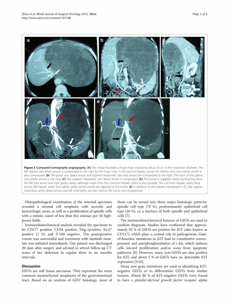
Figure 3 Computed tomography angiography. (A) The image illustrates a huge mass measuring about 30 cm in the maximum diameter. Theleft hepatic vein (black arrow) is compressed to the right by the huge mass. In the second hepatic portal, the inferior vena cava (white arrow) isalso compressed. (B) The portal vein (black arrow) and superior mesenteric vein (red arrow) are compressed to the right. The trace of the splenicvein (white arrow) is not clear. (C) The superior mesenteric vein (black arrow) is compressed. (D) The tumor is supplied mainly by branches fromthe left (red arrow) and right gastric artery, although origin from the common hepatic artery is also possible. The common hepatic artery (bluearrow), left hepatic artery and splenic artery (white arrow) are adjacent to the tumor. (E) In addition to the arteries mentioned in (C), the superiormesenteric artery (black arrow) and left renal artery are also next to the tumor and compressed.
Zhou et al. World Journal of Surgical Oncology 2012, 10:66 Page 3 of 5http://www.wjso.com/content/10/1/66
Histopathological examination of the resected specimenrevealed a stromal cell neoplasm with necrotic andhemorrhagic areas, as well as a proliferation of spindle cellswith a mitotic count of less than five mitoses per 50 high-power fields.Immunohistochemical analysis revealed the specimen to
be CD177 positive, CD34 positive, Dog-1positive, Ki-67positive (1 %) and S-100 negative. The postoperativecourse was uneventful and treatment with imatinib mesi-late was initiated immediately. Our patient was discharged20 days after surgery and advised to attend follow-up CTscans of her abdomen in regular three to six monthsintervals.
DiscussionGISTs are soft tissue sarcomas. They represent the mostcommon mesenchymal neoplasms of the gastrointestinaltract. Based on an analysis of GIST histology, most of
them can be sorted into three major histologic patterns:spindle cell type (70 %), predominantly epithelioid celltype (20 %), or a mixture of both spindle and epithelioidcells [7].The immunohistochemical features of GISTs are used to
confirm diagnosis. Studies have confirmed that approxi-mately 95 % of GISTs are positive for KIT (also known asCD117), which plays a central role in pathogenesis. Gain-of-function mutations in KIT lead to constitutive overex-pression and autophosphorylation of c-kit, which inducescells toward proliferation and/or away from apoptoticpathways [8]. However, many non-GISTs are also positivefor KIT, and about 5 % of GISTs have no detectable KITexpression [9,10].Many new gene mutations are used in identifying KIT-
negative GISTs or to differentiate GISTs from similartumors. About 80 % of KIT-negative GISTs were foundto have a platelet-derived growth factor receptor alpha

Figure 4 The tumor protrudes from the anterior wall of thefundus, on the lesser curvature of the stomach, measuring 21 ×34 × 11 cm in diameter and weighing 5.5 kg.
Zhou et al. World Journal of Surgical Oncology 2012, 10:66 Page 4 of 5http://www.wjso.com/content/10/1/66
gene (PDGFRA) mutation, which results in an epithelioidmorphology. This discovery had been used in discrim-inating between KIT-negative GISTs and other gastro-intestinal mesenchymal lesions [11,12]. BRAF mutationshave been revealed in a small number of high-risk GISTswhich develop from intestine lacking KIT or PDGFRAmutations [13]. A promising calcium-dependent andreceptor- activated chloride channel protein, known asDOG1, has emerged as a sensitive and specific markerused in the setting of KIT-negative GISTs [14]. Proteinkinase C theta, a downstream effector in the KIT signalingpathway, is used to discriminate between GISTs and leio-myosarcoma or other tumors which have similar histo-pathology to GISTs [15]. Recently, a novel biomarkercalled carbonic anhydrase II, which can promote tumorgrowth by contributing to intracellular alkalization andextracelluar acidification, has been demonstrated to bequite selective to GISTs among mesenchymal tumors [16].The clinical presentation of GISTs is erratic. About
70 % of patients are symptomatic and GISTs are asso-ciated with a broad range of presentations, includingearly satiety, bloating and some form of gastrointestinalbleeding, either acute or chronic [17]. However, approxi-mately 30 % are asymptomatic or the GIST is detectedincidentally at autopsy. There is no physical finding thatspecifically suggests the presence of a GIST. Althoughthere are several diagnostic modalities available, such asbarium examination of the gastrointestinal tract, CT orabdominal ultrasound, none of them can confirm thediagnosis. On some occasions, these examinations proveto be deceptive with regards to tumor identification. As
in our case, CT images and an upper gastrointestinalbarium series suggested that the tumor possibly origi-nated from the liver or gastrointestinal tract. In addition,the abdominal ultrasound indicated the possibility thatthe tumor came from the retroperitoneal organs. We couldnot determine the diagnosis because signs were evident forboth possible diagnoses. Under these circumstances, wedecided to perform an exploratory laparotomy. In order toexplicitly determine the relationship between importantvessels, neighboring organs and the mass, CTA examinationwas performed before the operation.Because GISTs are usually radioresistant and insensitive
to chemotherapeutic agents, surgery remains the maintherapy for patients with primary GISTs who have no evi-dence of metastasis. The tumor in our report was so hugethat it almost filled the abdominal cavity, approximately21 × 34 × 11 cm. Although no such advanced case hadbeen previously reported in the literature, we were able tocompletely remove the tumor with the proximal stomachand lower esophagus.There are also many GISTs which have metastases or
are unresectable using current technology. Imatinibmesilate, which is an inhibitor of a family of structurallyrelated tyrosine kinase signaling enzymes, is currently themost effective treatment for GISTs [18]. However, somepatients are insensitive to imatinib mesilate, as almost im-mediately after initiation or disease stabilization they thenexperience disease progression while on medication. Inthis situation, a multi-kinase inhibitor which inhibits, forexample, KIT, PDGFR and vascular endothelial growthfactor receptors 1 to 3, such as sunitinib, is indicated forthose who fail imatinib treatment [19]. There are a fewpatients with GISTs who fail both imatinib and sunitinibtreatment. In these cases, research has shown that nilotinib,which specifically inhibits KIT, PDGFRA and BCR-ABL,resolves the problem [20].There is general agreement that tumor size and mitotic
count are the most important prognostic factors inGISTs. These two features were the foundation for aconsensus approach to risk stratification of GISTs pub-lished in 2002 [21]. Subsequently, the criteria for the riskstratification of GISTs have constantly been expandeddue to the availability of long-term clinical follow-up.Based on the long-term follow-up of more than 1,600patients, Miettinen and Lasota [2] suggested guidelinesfor the risk stratification of primary GISTs based on mi-totic index, size and site. Moreover, other pathologicfeatures, including cellularity, mucosal ulceration, andthe presence or absence of KIT or PDGFRA mutations,have been clinically evaluated [22].
ConclusionWe have described an uncommon huge gastric GIST.According to fundamental surgical principles in the

Zhou et al. World Journal of Surgical Oncology 2012, 10:66 Page 5 of 5http://www.wjso.com/content/10/1/66
management of gastric GISTs, we completely resected thetumor with the proximal stomach and lower esophagus.The patient has been followed-up after the operation. Fur-thermore, we will periodically examine the patient and fol-low support guidelines for medical therapy. We hope thepresentation of this rare case could benefit others whenthey encounter a similar diagnostic problems.
ConsentWritten informed consent was obtained from the patientfor publication of this case report and any accompanyingimages.
Competing interestsThe authors declare that they have no competing interests.
Authors’ contributionsCL designed the study and LZ drafted the manuscript. CL is the guarantor.All authors contributed to the intellectual context and read and approvedthe final version.
Received: 19 December 2011 Accepted: 27 April 2012Published: 27 April 2012
References1. Liegl-Atzwanger B, Fletcher JA, Fletcher CD: Gastrointestinal stromal
tumors. Virchows Arch 2010, 456:111–127.2. Miettinen M, Lasota J: Gastrointestinal stromal tumors: pathology and
prognosis at different sites. Semin Diagn Pathol 2006, 23:70–83.3. Fülöp E, Marcu S, Milutin D, Borda A: Gastrointestinal stromal tumors:
review on morphology, diagnosis and management. Rom J MorpholEmbryol 2009, 50:319–326.
4. van der Zwan SM, DeMatteo RP: Gastrointestinal stromal tumor: 5 yearslater. Cancer 2005, 104:1781–1788.
5. Casali PG, Jost L, Reichardt P, Schlemmer M, Blay JY: Gastrointestinalstromal tumors: ESMO clinical recommendations for diagnosis, treatmentand follow-up. Ann Oncol 2008, 19(2):35–38.
6. Kochlar R, Manoharan P, Leahy M, Taylor MB: Imaging in gastrointestinalstromal tumours: current status and future directions. Clin Radiol 2010,65:584–592.
7. Miettinen M, Lasota J: Gastrointestinal stromal tumors: review onmorphology, molecular pathology, prognosis, and differential diagnosis.Arch Pathol Lab Med 2006, 130:1466–1478.
8. Hirota S, Isozaki K, Moriyama Y, Hashimoto K, Nishida T, Ishiguro S, KawanoK, Hanada M, Kurata A, Takeda M, Tunio GM, Matsuzawa Y, Kanakura Y,Shinomura Y, Kitamura Y: Gain-of-function mutations of c-kit in humangastrointestinal stromal tumors. Science 1998, 279:577–580.
9. Medinger M, Kleinschmidt M, Mross K, Wehmeyer B, Unger C, Schaefer HE,Weber R, Azemar M: c-kit (CD117) expression in human tumors and itsprognostic value: an immunohistochemical analysis. Pathol Oncol Res2010, 16:295–301.
10. Medeiros F, Corless CL, Duensing A, Hornick JL, Oliveira AM, Heinrich MC,Fletcher JA, Fletcher CDM: KIT-negative gastrointestinal stromal tumors:proof of concept and therapeutic implications. Am J Surg Pathol 2004,28:889–894.
11. Heinrich MC, Corless CL, Duensing A, McGreevey L, Chen CJ, Joseph N,Singer S, Griffith DJ, Haley A, Town A, Demetri GD, Fletcher CDM, FletcherJA: PDGFRA activating mutations in gastrointestinal stromal tumors.Science 2003, 299:708–710.
12. Miselli F, Millefanti C, Conca E, Negri T, Piacenza C, Pierotti MA, Tamborini E,Pilotti S: PDGFRA immunostaining can help in the diagnosis ofgastrointestinal stromal tumors. Am J Surg Pathol 2008, 32:738–743.
13. Hostein I, Faur N, Primois C, Boury F, Denard J, Emile JF, Bringuier PP,Scoazec JY, Coindre JM: BRAF mutation status in gastrointestinal stromaltumors. Am J Clin Pathol 2010, 133:141–148.
14. Abdel-Hadi M, Bessa SS, Hamam SM: Evaluation of the novel monoclonalantibody against DOG1 as a diagnostic marker for gastrointestinalstromal tumors. J Egypt Natl Canc Inst 2009, 21:237–247.
15. Ou Wb, Zhu Mj, Demetri GD, Fletcher CDM, Fletcher JA: Protein kinase C-[theta] regulates KIT expression and proliferation in gastrointestinalstromal tumors. Oncogene 2008, 27:5624–5634.
16. Parkkila S, Lasota J, Fletcher JA, Ou WB, Kivelä A, Nuorva K, Parkkila AK,Ollikainen J, Sly WS, Waheed A, Pastorekova S, Pastorek J, Isola J, MiettinenM: Carbonic anhydrase II. A novel biomarker for gastrointestinal stromaltumors. Mod Pathol 2010, 23:743–750.
17. Sepe PS, Brugge WR: A guide for the diagnosis and management ofgastrointestinal stromal cell tumors. Nat Rev Gastroenterol Hepatol 2009,6:363–371.
18. Cohen MH, Farrell A, Justice R, Pazdur R: Approval summary: imatinibmesylate in the treatment of metastatic and/or unresectable malignantgastrointestinal stromal tumors. Oncologist 2009, 14:174–180.
19. Demetri GD, Heinrich MC, Fletcher JA, Fletcher CDM, Van den Abbeele AD,Corless CL, Antonescu CR, George S, Morgan JA, Chen MH, Bello CL, HuangX, Cohen DP, Baum CM, Maki RG: Molecular target modulation, imaging,and clinical evaluation of gastrointestinal stromal tumor patients treatedwith sunitinib malate after imatinib failure. Clin Cancer Res 2009, 15:5902–5909.
20. Montemurro M, Schoffski P, Reichardt P, Gelderblom H, Schütte J, HartmannJT, Moos RV, Seddon B, Joensuu H, Wendtner CM, Weber E, Grünwald V,Roth A, Leyvraz S: Nilotinib in the treatment of advanced gastrointestinalstromal tumours resistant to both imatinib and sunitinib. Eur J Cancer2009, 45:1959–1968.
21. Fletcher CDM, Berman JJ, Corless C, Gorstein F, Lasota J, Longley BJ,Miettinen M, O’ Leary TJ, Remotti H, Rubin BP, Shmookler B, Sobin LH, WeissSW: Diagnosis of gastrointestinal stromal tumors: a consensus approach.Hum Pathol 2002, 33:459–465.
22. Demetri GD, von Mehren M, Antonescu CR, DeMatteo RP, Ganjoo KN, MakiRG, Pisters PWT, Raut CP, Riedel RF, Schuetze S, Sundar HM, Trent JC, WayneJD: NCCN task force report: update on the management of patients withgastrointestinal stromal tumors. J Natl Compr Canc Netw 2010, 8(Suppl 2):S1–S41.
doi:10.1186/1477-7819-10-66Cite this article as: Zhou et al.: A rare giant gastrointestinal stromaltumor of the stomach traversing the upper abdomen: a case report andliterature review. World Journal of Surgical Oncology 2012 10:66.
Submit your next manuscript to BioMed Centraland take full advantage of:
• Convenient online submission
• Thorough peer review
• No space constraints or color figure charges
• Immediate publication on acceptance
• Inclusion in PubMed, CAS, Scopus and Google Scholar
• Research which is freely available for redistribution
Submit your manuscript at www.biomedcentral.com/submit