a rare case of traumatic ulcerative granuloma with stromal ... · traumatic ulcerative granuloma...
TRANSCRIPT
A Rare Case of Traumatic Ulcerative Granuloma with Stromal Eosinophiliaof the Tongue (Tugse) in a Child: Diagnostic Problems and DifferentialDiagnosisBarca Ida, Cordaro Raffaella, Colangeli Walter, Novembre Daniela, Cristofaro Maria GDepartment of Experimental and Clinical Medicine, Unit of Oral and Maxillofacial Surgery, Magna Graecia University, VialeEuropa, Catanzaro, Italy.
AbstractTraumatic Ulcerative granuloma with stromal eosinophilia (TUGSE) is an infrequent condition marked by the presence of a solitaryulcer in the oral mucosa. It is generally considered a reactive, benign and self-limiting ulcer with raised and indurated margins,mainly affecting the tongue, cheeks, or, less frequently, lips. His typical histology shows a diffuse polymorphic cell inflammatoryinfiltrate composed predominantly of eosinophils involving the superficial mucosa and extending deep into the submucosa withdegeneration of the underlying muscle layer. Aetiology is unknown and pathogenesis is not clear although trauma could have animportant role in the origin and evolution of this lesion. Nevertheless, a correct diagnosis is difficult because the ulcer mimics awide range of pathologies including microbial infections or cancer.This work describes a case of traumatic granuloma of the tongue in a child female focusing attention on the differential diagnosis ofthis unclear lesion
Key Words: Eosinophilic granuloma, Reactive benign lesion of the tongue, Traumatic ulcerative granuloma
IntroductionTUGSE is a reactive benign lesion of oral mucosa with anobscure clinical behavior generally affecting the tongue. Itsulcerative aspect can mimics malignancies or infectiousprocesses but it tends to resolve spontaneously and, in amajority of the cases, trauma is considered to have a centralrole in the pathogenesis [1]. This lesion affects usuallyfemales with a remarkable predilection of the older populationespecially the black ethnic groups. A bioptic exam of thelesion is crucial to define a TUGSE diagnosis.Histopathological findings are typical and consist ofeosinophil-rich mixed infiltrate with small lymphocytes T andB. In many cases rapid healing is generally achievedspontaneously or after a biopsy; and do not requires anytreatment [2]. For clinicians knowledge of this condition isimportant to guide appropriate patient care and counseling.
We report an uncommon case of TUGSE in a young patientseen at our Department of Oral and Maxillofacial Surgery,University “Magna Graecia” of Catanzaro, Italy.
Case ReportAn 11-year-old female presented herself at the Department ofOral and Maxillo Facial Surgery University Magna Graecia ofCatanzaro with non-healing isolated painful ulcer of thetongue for 7 days.
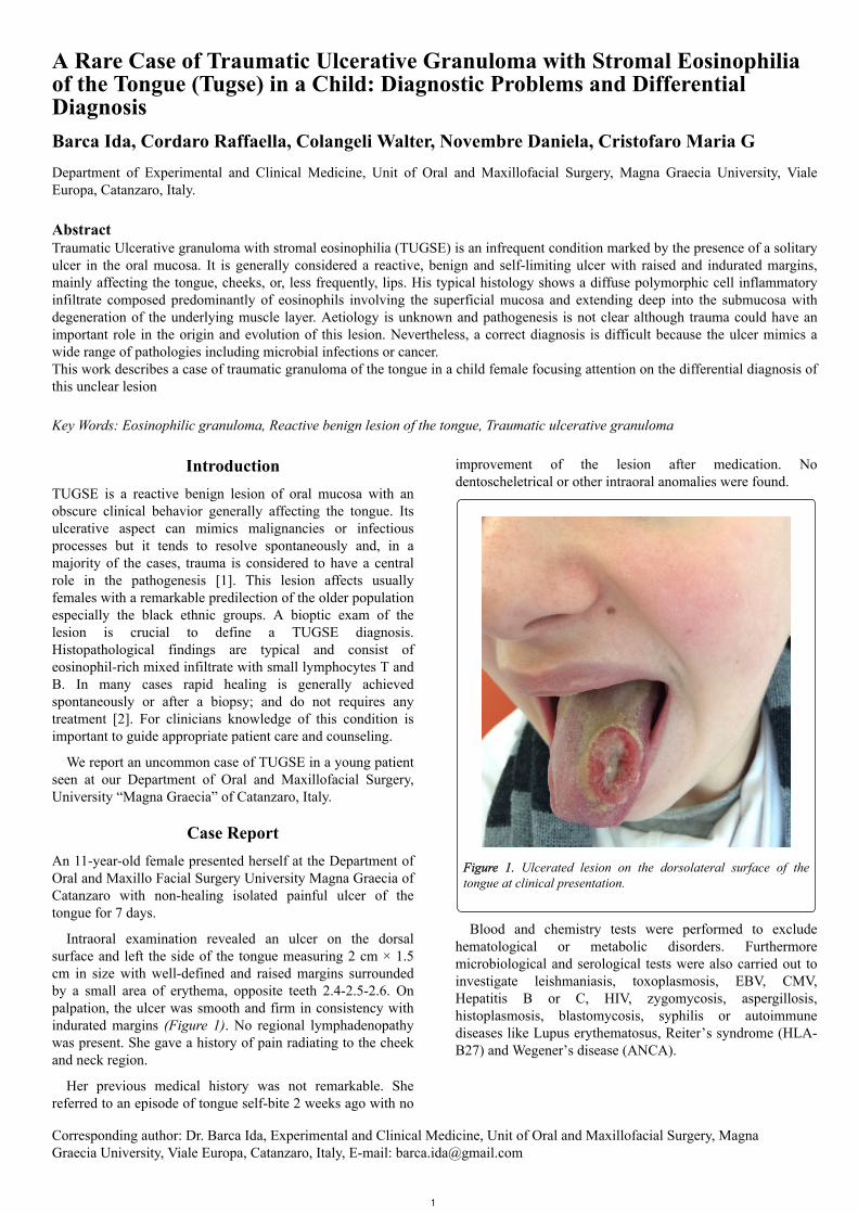
Intraoral examination revealed an ulcer on the dorsalsurface and left the side of the tongue measuring 2 cm × 1.5cm in size with well-defined and raised margins surroundedby a small area of erythema, opposite teeth 2.4-2.5-2.6. Onpalpation, the ulcer was smooth and firm in consistency withindurated margins (Figure 1). No regional lymphadenopathywas present. She gave a history of pain radiating to the cheekand neck region.
Her previous medical history was not remarkable. Shereferred to an episode of tongue self-bite 2 weeks ago with no
improvement of the lesion after medication. Nodentoscheletrical or other intraoral anomalies were found.
Figure 1. Ulcerated lesion on the dorsolateral surface of thetongue at clinical presentation.
Blood and chemistry tests were performed to excludehematological or metabolic disorders. Furthermoremicrobiological and serological tests were also carried out toinvestigate leishmaniasis, toxoplasmosis, EBV, CMV,Hepatitis B or C, HIV, zygomycosis, aspergillosis,histoplasmosis, blastomycosis, syphilis or autoimmunediseases like Lupus erythematosus, Reiter’s syndrome (HLA-B27) and Wegener’s disease (ANCA).
Corresponding author: Dr. Barca Ida, Experimental and Clinical Medicine, Unit of Oral and Maxillofacial Surgery, MagnaGraecia University, Viale Europa, Catanzaro, Italy, E-mail: [email protected]
1

Figure 2. Ulcerated area with mixed inflammatory cells (10X, Hand E).
Figure 3. Inflammatory infiltrate of granuloma composed bysmall lymphocytes B and T and granulocytes (40X, H and E).
Figure 4. Immunohistochemical analysis showing CD3+ TLymphocytes aggregates.
Figure 5. Immunohistochemical analysis showing composed bysmall lymphocytes B and T and CD20+ B Lymphocytes.
Figure 6. Complete resolution two months post biopsy.
Tuberculosis skin test was added too. Negative results ofthese laboratories exams excluded a systemic condition andthen biopsy was recommended to define the diagnosis.Ultrasonographic exam of Lymph Nodes in Head and Neckshowed reactive lymph nodes in the left submandibularregion. One week later an incisional biopsy was performedunder local anesthesia for histopathological analysis.
Microscopic examination showed superficial hyperplasticepithelium with hyperkeratosis and central area of ulceration.This ulcerated area was infiltrated with mixed inflammatorycells chiefly composed of eosinophils, CD 20+ and CD3+ Band T lymphocytes, CD68+ histiocytes, CD31+ cells andmacrophages extending deep into the muscle layer. Theinfiltrated tissue was well vascularized. No presence ofatypical cells (Figures 2-5).
On the basis of these results, a diagnosis of TUGSE wasmade. The patient underwent antibiotic therapy with oralAmoxicillin 1gr for 10 days and was advised to usecorticosteroid and chlorhexidine 0.2% mouthwashes. Then
OHDM- Vol. 18- No.3-June, 2019
2

she has been monitored closely with routine check-ups. Nofurther treatment was required. Complete healing was noticedafter two months with structural and functional resolution(Figure 6).
DiscussionTUGSE is an infrequent lesion of the oral mucosa withunclear aetiology and pathogenesis. It has been known bydifferent names such as Riga-Fede disease, eosinophilicgranuloma, and traumatic granuloma. This lesion wasoriginally described clinically in 1881 by Riga [3] andhistologically in 1890 by Fede [4] and identified as a distinctentity in 1970 by Shapiro and Juhlin but only in the 1983,Elzy coined the term TUGSE [5].
Nowadays etiology of TUGSE remains still obscure. Theclinical presentation mimics a large range of differentpathologies like oral cancer, infectious diseases, metabolic orautoimmune disorders, and aphthous-like lesions. Someauthors identified trauma as the main factor in thedevelopment of lesion in less than 50% of cases [6]; amongthem, an accidental bite or repeated injury leads to theintroduction of viral or toxic agents in the tissues causinginflammatory response [7,8] and local immune reaction [9].However, an absolute linkage between injuries and thedevelopment of these lesions wasn’ t found in all studies[10,11].
Clinically ulcer appears solitary with elevated and induratedmargins associated with pain in many cases. It can persist forseveral weeks or months without treatment and generallytends to resolve spontaneously [12].
It affects a wide age range of patients from childhood to oldage with a peak incidence between the 6th and 7th decades oflife and a slight female predominance. The ventral or lateralsurface of the tongue is generally involved perhaps because ismore vulnerable to injuries. Other oral areas like lip, palateand vestibular mucosa may also be involved. In our case,TUGSE presented in a female child atypically on the leftdorsal surface of the tongue measuring about 2 cm × 1.5 cm,tender on palpation with indurated margins surrounded byhyperemic area. Dentoscheletrical system has presented noanomalies. A traumatic episode of an accidental bite referredby the patient a week ago has not justified completely theentity of lesion. No lymphadenopathy was founded. Negativeresults of laboratory exams excluded a systemic condition,and then biopsy was recommended to define the diagnosis.
Indeed benign nature of lesion has been confirmed byhistopathological findings. Microscopical examination of ourlesion revealed a polymorphic inflammatory infiltrate mainlycomposed of eosinophils and histiocytes CD68+,accompanied by a population of CD3+ lymphocytes T,CD20+ lymphocytes B and macrophages [10] with abundantcytoplasm, irregular nuclear contours, small nucleoli and finechromatin [11]; immunohistochemical technique resultsconsisted of CD31+ in vascular component and CD30 andS100 rarely expressed. This inflammatory infiltrate extendsfrom the superficial mucosa to the submucosa involvingmuscle fibers and sometimes salivary glands. The role ofeosinophils is not completely clear because they are notpresent in all traumatic oral ulcers; they may be involved in a
tissue reaction to some unknown antigen introduced throughthe traumatic lesion. Degeneration of oral mucosa may beattributed to the proliferation of cytotoxic T cells or toxicproduct released by degranulating eosinophils. It supports therole of cytotoxic T cells in the pathogenesis of TUGSE.
The differential diagnosis is complex because clinicalpresentation mimics malignancies such as squamous cellcarcinoma, lymphoma, salivary gland tumors, infectivediseases such as leishmaniasis, toxoplasmosis, hepatitis orautoimmune diseases. The histologic differential diagnosismay include many lesions characterized by infiltration ofeosinophils within the connective tissue such as Langerhanscell disease, Angiolymphoid Hyperplasia with Eosinophilia(ALHE), Kimura disease, certain types of lymphomas,allergic reactions and parasitic diseases [12,13]. In our caserarely expressed CD30 antigen excluded alymphoproliferative disorder [14].
In our patient we observed a marked improvement of thelesion after incisional biopsy with no need for more radicalsurgery; a complete resolution at 2 and 6 months follow-upvisits was observed, indicating a full recovery [15].
ConclusionTUGSE is a benign lesion of the oral mucosa of unclearpathogenesis. This case report analyzed clinical andhistopathological characteristics of TUGSE highlighting thecomplexity of diagnosis, due to a large number of pathologieswith overlapping clinical and histopathological features.According to literature, some cases show a correlationbetween TUGSE and recurrent traumatic injury to the tongue.The present case characterized by the young age of thepatient, history of an accidental bite and spontaneous self-healing of the lesion related to the typical clinical picture andhistopathological findings led up to the diagnosis of TUGSE.This lesion could be considered a reactive process secondaryto trauma excluding other similar suspected ulcerative lesions.
Awareness of this entity is important to emphasize thecorrect diagnosis of ulcerated lesions and deliver appropriateand effective treatment.
AcknowledgmentThe authors would like to thank the medical staff of theAnatomopathology Department of Magna Graecia Universityfor providing us with detailed histological images and reportsused in this study.
References1. Soyele O, Adesina O, Ladeji A, Kuye K, Owotade F.
Traumatic ulcerative granuloma with stromal eosinophilia: Reviewof the literature and case report. African Journal of Medical andHealth Sciences. 2017; 16: 115.
2. Bacem AE Ottoman. Granular cell tumor of the tongue: a casereport with emphasis on the diagnostic and therapeutic proceedings.Oncology and Cancer Case Reports. 2015, 1: 1.
3. Riga A. Di una malattia della prima infanzia, Probabilmentenon-trattata, di movimenti patologici. Napoli. 1881.
4. Fede F. Della produzione sottolinguale o malattia di Riga. AttoCongresso italiano di pediatria 1890. Napoli. 1891: 251.
OHDM- Vol. 18- No.3-June, 2019
3

5. Elzay RP. Traumatic ulcerative granuloma with stromaleosinophilia (Riga-Fede ’ s disease and traumatic eosinophilicgranuloma). Oral Surgery Oral Medicine Oral Pathology. 1983; 55:497-506.
6. Segura S, Romero D, Colomo L. Eosinophilic ulcer of the oralmucosa: Another rhistological simulator of CD30+lymphoproliferative disorders. British Journal of Dermatology. 2006;155: 460-63.
7. Gao S, Wang Y, Liu N, Li S, Du J. Eosinophilic ulcer of theoral mucosa: A clinicopathological analysis. Chinese Journal ofDental Research. 2000; 3: 47-50.
8. Hirshberg A, Amariglio N, Akrish S, et al. Traumaticulcerative granuloma with stromal eosynophilia: Reactive lesion ofthe oral mucosa. American Journal of Clinical Pathology. 2006; 126:522-529.
9. El-Mofty SK, Swanson PE, Wick MR, Miller AS.Eosinophilic ulcer of the oral mucosa: Report of 38 new cases withimmunohistochemical observations. Oral Surgery Oral MedicineOral Pathology. 1993; 75: 716-722.
10. Elovic AE, Gallgher GT, Kabani S, Galli SJ, Weller PF, et al.Lack of TGF-a and TGF-b synthesis by human eosinophils in
chronic oral ulcers. Oral Surgery Oral Medicine Oral Pathology,Oral Radiology, and Endodontology. 1996; 81: 672-681.
11. Vélez A, Alamillos FJ, Dean A, Rodas J, Acosta A.Eosinophilic ulcer of the oral mucosa: Report of a recurrent case onthe tongue. Clinical and Experimental Dermatology.1997; 22:154-156.
12. Gonçales ES, Rubira-Bullen IF, Rubira CM, Miyazawa M,Chinellato LE, et al. Eosinophilic ulcer of the oral mucosa versussquamous cell carcinoma. Quintessence International. 2007; 38:677-680.
13. Cepeda LT, Pieretti M, Chapman SF, Horenstein MG. CD30-positive atypical lymphoid cells in common on neoplastic cutaneousinfiltrate rich in neutrophils and eosinophils. American Journal ofSurgical Pathology. 2003; 27: 912-918.
14. Ada S, Seckin D, Tarhan E, Buyuklu F, Cakmak O, et al.Eosinophilic ulcer of the tongue. Australasian Journal ofDermatology. 2007; 48: 248-250.
15. Sharma B, Koshy G, Kapoor S. Traumatic ulcerativegranuloma with stromal eosinophilia: A case report and review ofpathogenesis. Journal of Clinical and Diagnostic Research. 2016;10: ZD07-Zd09.
OHDM- Vol. 18- No.3-June, 2019
4