a rare case of identification and preservation … · a rare case of identification and...
TRANSCRIPT
A rare case of human remains preservation Rev Arg de Anat Clin; 2013, 5 (3): 240-249__________________________________________________________________________________________
Todos los derechos reservados. Reg. Nº: 5104953 www.anatclinar.com.ar240
Case Report
A RARE CASE OF IDENTIFICATION AND PRESERVATION OF HUMAN REMAINS
Horacio E. Solla1*, Mehmet Y. Iscan2, Barbara McCabe2
1 Judicial Morgue of Montevideo City, Montevideo, Uruguay2 Florida Atlantic University, Florida, United States of America
RESUMEN
Los estudios de casos son ideales para probar la validez de técnicas antropológicas y los estudios antropológico-forenses de casos suelen funcionar como campo de pruebas en varios tópicos de la Antropología Física. El creciente rol que ha jugado los antropólogos en las ciencias forenses ha ayudado a las disciplinas médico-legales en varios aspectos. Por ejemplo, la identificación restos óseos es ahora más segura que nunca. El propósito de éste artículo es mostrar un raro caso de preservación de tejidos blandos de los restos de una niña color que se han conservado transformándose en un molde compacto y describir las técnicas antropológicas usadas para identificar los restos. Los estudios antropológicos forenses indicaban que se trataba de los restos de una niña de color entre 10 y 12 años de edad al momento de la muerte y de 150 cms de estatura. Basados en esos datos preliminares y en la sospecha de que los restos podrían ser los de Jane Doe, víctima de una violación y posterior asesinato. Una superposición digital fue realizada usando una fotografía de la supuesta víctima y el cráneo desconocido. Los estu-dios indicaron que el cráneo hallado se correspondía de manera consistente con el individuo de la fotografía. Estos resultados fueron luego ratificados por un análisis de ADN. Por lo tanto, la Antropología Forense y sus técnicas pueden ser utilizadas en investigaciones médico-legales concretas de manera muy satisfactoria.
Palabras clave: Preservación de tejidos blandos, Comparaciones cráneo-fotográficas, Identificación humana.
ABSTRACT
Case studies are ideal to test the validity of anthropological techniques and forensic anthropology cases function as a testing ground for this aspect of physical anthropology. The increasing role that
anthropologists have played in forensic sciences has aided the medico legal disciplines in a number of ways. For example, identification of skeletal remains is now more accurate than ever before. The purpose of this paper is to show a peculiar case of rare preservation of soft tissue of a black child that formed an unique soft tissue casting and to describe anthropological techniques used to identify the remains. The anthropological analysis indicated that she was black racial affinity, between 10-12-year old and was about 150 cm of stature. Based on preliminary evidence that the victim might be Jane Doe, a victim of possible rape and murder, a digital superimposition was made using one victim's photograph and the unknown skull. This examination revealed that the skull corresponded consistently with the individual in the photograph. Results were supported by a DNA analysis. Therefore, forensic anthropology techniques can be successfully used in medico-legal investigations.
Keywords: Soft tissue preservation; skull-photo comparison; Human identification.
INTRODUCTION
Biological anthropologists have developed numerous demographic techniques to under-stand the biology of ancient people. Many of these techniques derived from skeletal remains of known identity but case studies are ideal to test the validity of these techniques and forensic anthropology may function as a testing ground for this aspect of biological anthropology. _________________________________________________
* Correspondence to: Horacio E. Solla. Benito Chain 1925, Montevideo. Uruguay. [email protected]
Received: 26 August, 2013. Revised: 12 October, 2013. Accepted: 31 October, 2013.

A rare case of human remains preservation Rev Arg de Anat Clin; 2013, 5 (3): 240-249__________________________________________________________________________________________
Todos los derechos reservados. Reg. Nº: 5104953 www.anatclinar.com.ar241
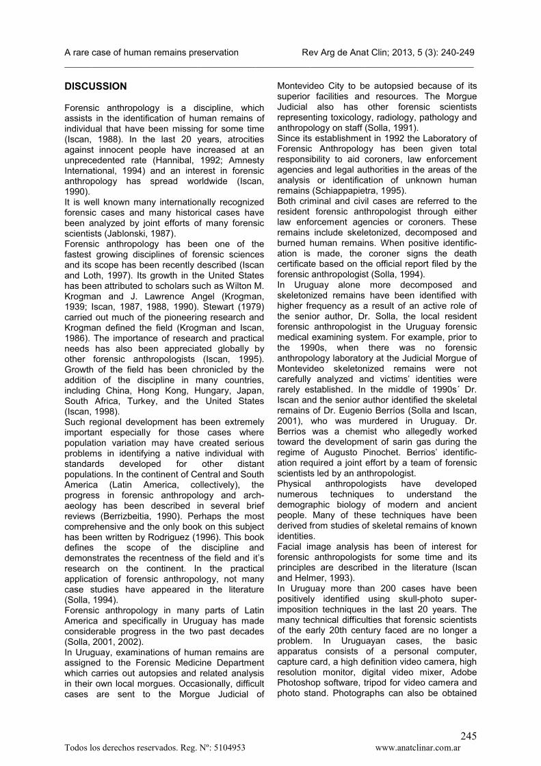
Latin America has shown a trend in the development of forensic anthropology research and practice (Iscan and Solla, 2000; Rodríguez, 1996, 2004). Along with these changes, many internationally known cases have been studied in Latin America (Basauri, 1967; Soto, 1989; Solla et al, 2001, 2004, 2005, 2010)); some of the mostimportant cases include the identification of Joseph Mengele (Eckerct et al, 1985; Curran, 1986; Helmer, 1986, 1987; Jeffreys et al, 1992)and 16th century explorer Francisco Pizarro (Maples, et al, 1989). In the last few years, forensic anthropology has been an active part of the coroner system in Uruguay. The number of cases has increased considerably since the inclusion of a resident forensic anthropologist to the Uruguayan coroner system. This eventually led to a higher rate of positive identifications of human skeletal remains (Solla, 2001, 2002).In February 2010, the remains of a child were discovered inside an abandoned water well in a rural location about 30 miles from Montevideo City, Uruguay. There was differential decomp-osition and a rare and unique adipocere formation which had been preserved like a cast on several parts of child’s body, especially on the lumbar area and the upper legs (Fig.1). Decomposition of the remaining portions of the body was more advanced, showing the following skeletal structures: a skull without the mandible, two femurs (right and left), two tibias (right and left), two fibulas (right and left), two pubis (right
and left), two iliums (right and left), two ischiums (right and left), a right scapula, a sacrum, a right ulna, two radius (right and left), two humerus (right and left), a left clavicle, seven left ribs, eight right ribs, six dorsal vertebrae, two lumbar vertebrae, four cervical vertebrae, two patellas (right and left), a right femoral head and two humeral heads (right and left). No injuries were found on the remains. The skull was completely skeletonized and in a very good state of preservation and without fractures or observable trauma. The mandible was never found and the anterior teeth on the maxilla were lost post-mortem. The victim remains were totally naked except for a stocking. After a preliminary examination the remains were carried to the Judicial Morgue of Montevideo City (the largest morgue in Uruguay) for an in-depth analysis by the resident forensic anthropologist.
CASE REPORT
a) Anthropological Analysis of the Recovery Remains The recovered skeletal remains were sent to the Forensic Anthropology Laboratory at the Judicial Morgue of Montevideo City to be analyzed (Karagioziz et al, 2005).
Figure1. Adipocere cast ventral and dorsal view
A rare case of human remains preservation Rev Arg de Anat Clin; 2013, 5 (3): 240-249__________________________________________________________________________________________
Todos los derechos reservados. Reg. Nº: 5104953 www.anatclinar.com.ar242
First of all, the bones were analyzed to determine the minimum number of individuals. The examination indicated that the remains belonged to a single individual (Pickering and Bachman, 1997; Burns, 2000).Upon arrival the skull was cleaned of debris and all remaining soft tissues were removed. The preservation of the skull was excellent and the preliminary analysis revealed that remains were of a pre-adolescent individual. Skeletal characteristics of the corpse were analyzed to establish sex, race, stature and age at time of death. The determination of sex of the immature remains was achieved using Hunt and Gleiser (1955) formulae. The results of this study indicated that the bones belonged to a female. Appraisal of the victim’s racial affinity was based on a number of Negroid characteristics such as; a guttered nasal margin, a broad nasal aperture and absence of nasal spine (Stewart, 1979); (Krogman and Iscan, 1986). To confirm this, cranial dimensions were put into discriminative function formulae derived from an American black sample (Giles and Elliot, 1962). The result obtained was above the section point (92.2) between American Whites and Blacks females indicating a black racial affinity. Stature estimation is another way to determine whether the victim's body size was within the range of reported missing persons and also to rule it out if there was a large discrepancy. There are very few standards to estimate height from the skeleton. The most reliable was obtained from the long bones of the lower and, to a lesser degree, the upper extremities. The given standard error of estimation can cover a safety range around the mean. Estimation of stature was made using lengths of the femur, tibia, fibula, humerus, radius and ulna and applying Trotter's regression equations for black females (Trotter,
1970). The average stature was found to be 150 cm with about 4.0 cm estimated standard error. The range was from 146 to 154 cm. In general the femur, tibia and fibula give better estimates than the bones of the upper extremity (table 1). Numerous techniques, with varying degrees of accuracy for the estimation of age at death from a sub-adult individual are available to anthropologists. For the present case age at death was estimated from tooth development (Ubelaker, 1989), union of epiphyses (Bass, 1971), long bone length (Primeau et al, 2012)and illium growth study (Rissech and Malgosa, 2005). Although the mandible was never found, the development of the maxilla superior teeth indicated an age at death between 10 and 12 years old. The epiphyseal union of the long bones indicated an age at death between 10 and 14 years old. Long bones length indicated an age at death between 9 and 13 years old and the growth of the illium indicated an age between 9 and 12 years old.Because of the advanced state of decomposition the cause of death was not determined. However, the manner of death was assumed to be homicide due to the circumstances and evidence found at the crime scene. These suspicions were later confirmed.The skeletal remains were examined to determine if there was any evidence of pathology and fractures, healed or otherwise. None were observed. The absence of any fresh trauma suggested that death might have occurred as a result of asphyxiation or other factors.The final results of the anthropological analysis performed on the skeletal remains indicated that the victim was a black female of about 150 cm of stature, between an age of 10 and 12 years at the time of death.
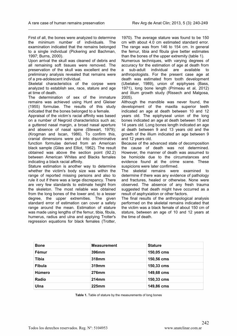
Bone Measurement Stature
Fémur 396mm 150,05 cms
Tibia 318mm 150,56 cms
Fibula 319mm 150,33 cms
Húmero 276mm 149,68 cms
Radio 214mm 150,33 cms
Ulna 225mm 149,86 cms
Table 1. Table of stature by the measurements of long bones

A rare case of human remains preservation Rev Arg de Anat Clin; 2013, 5 (3): 240-249__________________________________________________________________________________________
Todos los derechos reservados. Reg. Nº: 5104953 www.anatclinar.com.ar243
b) Time From DeathEstimation of time since death is one of the most difficult aspects of forensic assessment (Morse, 1983a). It is made even more complex when the decomposition process is advanced or remains are mostly skeletonized. The stage of decom-position and presence of soft tissue was also taken into account. Based on the level of decomposition and the rare cast of adipocere the time of death was estimated to be two or more years. Decomposition odor was minimal. The rate of deterioration of personal clothing, only a little stocking, was also assessed to be minimal (Morse, 1983b). The temperature and season preceding death added further input to the estimation of time since death. The remains were found in summer with a temperature range from 25 to 35ºC. The author’s experience with exhumations in general had indicated that the
total decomposition process may take as long as 2-3 years in Uruguay. It is a tradition in Uruguay to exhume the remains of deceased relatives after 2 years and place them in an urn. It has been observed that some remains still contain adipocere after two years probably because of the high humidity in the region. The process should proceed at a faster rate in cases that have not been buried in a coffin as was the situation for the present remains. Based on the evaluation of these facts, estimation was that death may have occurred at least two years before the corpse was discovered, the minimal time necessary to transform a body of soft tissue into adipocere. It should be mentioned that adipocere rarely is total and generally is of partial development in several parts of the body. The formation of adipocere in all soft tissue of a human body is very rare.
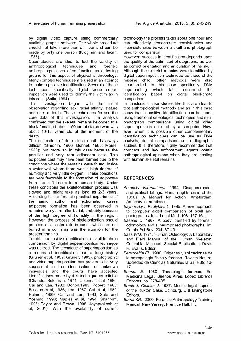
Figure 2. Skull-photo images compared by digital superimposition

A rare case of human remains preservation Rev Arg de Anat Clin; 2013, 5 (3): 240-249__________________________________________________________________________________________
Todos los derechos reservados. Reg. Nº: 5104953 www.anatclinar.com.ar244
c) Identification of the Human RemainsBased on these findings, the local database was consulted to determine whether any missing children might be compared with the anthropological data of the skeleton recovered from the well. A match was made with a child who had disappeared in May 2006 near the location where the remains were found. The demographic characteristics of age, race, sex, and stature seemed to correspond to those of the missing girl listed as Jane Doe. A careful assessment of specific individual osteological characteristics is necessary to make a positive identification (Krogman and Iscan, 1986). These characteristics ensure that the victim matches only one individual. A number of procedures were put together to make the positive identification. These included cranial characteristics and digital skull-photo superimposition techniques (Iscan and Helmer, 1993). The technique of skull-photo superimposition has been used to assist in the identification of numerous victims and is accepted in courts in a number of countries (Brash and Glaister, 1937; Basauri, 1967; Eckert and Texeira,1985; Curran, 1986; Helmer, 1986,1987; Soto, 1989; Kritscher and Szilvássy, 1991; Ivanov and Abramov, 1991; Ubelaker, 1996; Solla and Iscan, 2001; Solla et al, 2005, 2010). The use of a computer in this technique has added a number of advantages to the process (Pesce Delfino et al, 1983, Pesce Delfino et al, 1986; Nickerson et al, 1991; Bajinoczky and Kiralyfalvi, 1995; Smeets and De Valck, 1996; Ubelaker and O´Donnell, 1997; Humpire and Soto, 2013).Examination of the skeletal remains had shown that these were the remains of a 10-12 year old black female who was approximately 150 cm in stature. Surmising that the victim might be Jane Doe it was decided that a positive identification could be made by comparing the skull with a good quality photograph submitted by relatives using digital superimposition techniques. The use of computer has a number of advantages as well, one of which is that the whole process can beaccomplished faster and by only one person. The apparatus utilized was standard equipment and consisted of a computer with a good graphic card and Pinnacle Studio capture card, one high definition digital video camera, digital video mixer unit, a high definition image monitors, a tripod for the video camera, the skull, a positioning stand, printer unit and a photo stand to hold the photo for comparison. The images obtained were later processed with the Adobe Photoshop software. The photograph was then placed under the video camera and illuminated by white fluorescent
lamps while the image was adjusted on the monitor. The image was then digitized and stored within the digital video mixer unit. Key anatomical landmarks were traced on the photograph submitted for comparison. The image of the photo was then momentarily removed from the monitor by the digital mixer controls and it was placed under the video camera by the skull with tissue thicknesses marked and illuminated again by white fluorescent lamps. The skull was manipulated by a servomotor until the position approximated that of the individual in the photograph. After the skull had been correctly oriented, using the video camera zoom, the size of the skull image was adjusted so that it was as close as possible to that of the individual in the victim's photograph. After comparing anatomical landmarks on the skull with their counterparts marked on the photograph by the Above Photoshop software, the image of the skull was digitized and stored within the digital video mixer unit into the computer. Both images (photo and skull) were then superimposed on the monitor for a detailed comparison. The digital video mixer unit allows some combinations of photo-skull comparison, including removing the soft tissue to view the underlying skeletal structures such as auditory canal, eye sockets, root of the nose, teeth, skull contours and so forth. All the images obtained were later compared in detail by the Adobe Photoshop software using anatomical landmarks and the eight examining lines described by Cai and Lan, (1982, 1983, 1993) The analysis showed that conformity was found between skull image and all recognizable proportions of head, face, eyes, nose and mouth on the photograph submitted for comparison. Also, the outline of the soft tissue on the skull was congruent with the facial contours lying in the photograph. The final comparisons revealed an excellent match of photograph and underlying skeletal structures (Fig. 2). A complete report was released indicating that the remains belonged to the missing child, Jane Doe, based on the skeletal characteristics and digital superimposition comparisons. Dental charts or radiographs were not available for comparison. The only other avenue for identification was DNA comparison. For this analysis blood samples from close relatives were obtained. Four months after identification by skull - photo comparison techniques were made, the DNA results ratified the primary identification of the remains as being those of Jane Doe, who was 11 years old at the time of her dis-appearance. Later, the police confirmed what she was victim of rape and murder.
A rare case of human remains preservation Rev Arg de Anat Clin; 2013, 5 (3): 240-249__________________________________________________________________________________________
Todos los derechos reservados. Reg. Nº: 5104953 www.anatclinar.com.ar245
DISCUSSION
Forensic anthropology is a discipline, which assists in the identification of human remains of individual that have been missing for some time (Iscan, 1988). In the last 20 years, atrocities against innocent people have increased at an unprecedented rate (Hannibal, 1992; Amnesty International, 1994) and an interest in forensic anthropology has spread worldwide (Iscan, 1990). It is well known many internationally recognized forensic cases and many historical cases have been analyzed by joint efforts of many forensic scientists (Jablonski, 1987).Forensic anthropology has been one of the fastest growing disciplines of forensic sciences and its scope has been recently described (Iscan and Loth, 1997). Its growth in the United States has been attributed to scholars such as Wilton M. Krogman and J. Lawrence Angel (Krogman, 1939; Iscan, 1987, 1988, 1990). Stewart (1979) carried out much of the pioneering research and Krogman defined the field (Krogman and Iscan,1986). The importance of research and practical needs has also been appreciated globally by other forensic anthropologists (Iscan, 1995). Growth of the field has been chronicled by the addition of the discipline in many countries, including China, Hong Kong, Hungary, Japan, South Africa, Turkey, and the United States (Iscan, 1998).Such regional development has been extremely important especially for those cases where population variation may have created serious problems in identifying a native individual with standards developed for other distant populations. In the continent of Central and South America (Latin America, collectively), the progress in forensic anthropology and arch-aeology has been described in several brief reviews (Berrizbeitia, 1990). Perhaps the most comprehensive and the only book on this subject has been written by Rodriguez (1996). This book defines the scope of the discipline and demonstrates the recentness of the field and it’s research on the continent. In the practical application of forensic anthropology, not many case studies have appeared in the literature (Solla, 1994). Forensic anthropology in many parts of Latin America and specifically in Uruguay has made considerable progress in the two past decades (Solla, 2001, 2002).In Uruguay, examinations of human remains are assigned to the Forensic Medicine Department which carries out autopsies and related analysis in their own local morgues. Occasionally, difficult cases are sent to the Morgue Judicial of
Montevideo City to be autopsied because of its superior facilities and resources. The Morgue Judicial also has other forensic scientists representing toxicology, radiology, pathology and anthropology on staff (Solla, 1991).Since its establishment in 1992 the Laboratory of Forensic Anthropology has been given total responsibility to aid coroners, law enforcement agencies and legal authorities in the areas of the analysis or identification of unknown human remains (Schiappapietra, 1995). Both criminal and civil cases are referred to the resident forensic anthropologist through either law enforcement agencies or coroners. These remains include skeletonized, decomposed and burned human remains. When positive identific-ation is made, the coroner signs the death certificate based on the official report filed by the forensic anthropologist (Solla, 1994).In Uruguay alone more decomposed and skeletonized remains have been identified withhigher frequency as a result of an active role of the senior author, Dr. Solla, the local resident forensic anthropologist in the Uruguay forensic medical examining system. For example, prior to the 1990s, when there was no forensic anthropology laboratory at the Judicial Morgue of Montevideo skeletonized remains were not carefully analyzed and victims’ identities were rarely established. In the middle of 1990s´ Dr. Iscan and the senior author identified the skeletal remains of Dr. Eugenio Berríos (Solla and Iscan, 2001), who was murdered in Uruguay. Dr. Berrios was a chemist who allegedly worked toward the development of sarin gas during the regime of Augusto Pinochet. Berrios’ identific-ation required a joint effort by a team of forensic scientists led by an anthropologist. Physical anthropologists have developed numerous techniques to understand the demographic biology of modern and ancient people. Many of these techniques have been derived from studies of skeletal remains of known identities. Facial image analysis has been of interest for forensic anthropologists for some time and its principles are described in the literature (Iscan and Helmer, 1993).In Uruguay more than 200 cases have been positively identified using skull-photo super-imposition techniques in the last 20 years. The many technical difficulties that forensic scientists of the early 20th century faced are no longer a problem. In Uruguayan cases, the basic apparatus consists of a personal computer, capture card, a high definition video camera, high resolution monitor, digital video mixer, Adobe Photoshop software, tripod for video camera and photo stand. Photographs can also be obtained
A rare case of human remains preservation Rev Arg de Anat Clin; 2013, 5 (3): 240-249__________________________________________________________________________________________
Todos los derechos reservados. Reg. Nº: 5104953 www.anatclinar.com.ar246
by digital video capture using commercially available graphic software. The whole procedure should not take more than an hour and can be made by only one person (Krogman and Iscan, 1986). Case studies are ideal to test the validity of anthropological techniques and forensic anthropology cases often function as a testing ground for this aspect of physical anthropology. Many complex techniques are used in an attempt to make a positive identification. Several of these techniques, specifically digital video super-imposition were used to identify the victim as in this case (Solla, 1994).This investigation began with the initial observation regarding sex, racial affinity, stature and age at death. These techniques formed the core data of this investigation. The analysis confirmed that the skeletal remains belonged to a black female of about 150 cm of stature who was about 10-12 years old at the moment of the death.The estimation of time since death is always difficult (Simonín, 1966; Bonnet, 1980; Morse, 1983); but more so in this case because the peculiar and very rare adipocere cast. This adipocere cast may have been formed due to the conditions where the remains were found, inside a water well where there was a high degree of humidity and very little oxygen. These conditions are very favorable to the formation of adipocere from the soft tissue in a human body. Under these conditions the skeletonization process was slowed and might take as long as 2-3 years. According to the forensic practical experience of the senior author and exhumation cases adipocere formation has been observed in remains two years after burial, probably because of the high degree of humidity in the region. However, the process of skeletonization should proceed at a faster rate in cases which are not buried in a coffin as was the situation for the present remains.To obtain a positive identification, a skull to photo comparison by digital superimposition technique was utilized. The technique of superimposition as a means of identification has a long history (Grüner et al, 1959; Grüner, 1993). photographic and video superimposition has proven to be very successful in the identification of unknown individuals and the courts have accepted identifications made by this technique as reliable (Chandra Sekharan, 1971; Colonna et al, 1980; Cai and Lan, 1982; Dorion,1983; Robert, 1983; Bassian et al, 1986; Iten, 1987; Cai et al, 1989; Helmer, 1989; Cai and Lan, 1993; Seta and Yoshino, 1993; Maples et al, 1994; Shahrom, 1996; Taylor and Brown, 1998; Jayaprakash et al, 2001). With the availability of current
technology the process takes about one hour and can effectively demonstrate consistencies and inconsistencies between a skull and photograph used for comparison.However, success in identification depends upon the quality of the submitted photographs, as well as correct orientation and articulation of the skull. Although the skeletal remains were identified by digital superimposition technique as those of the missing child, other methods were also incorporated. In this case specifically, DNA fingerprinting which later confirmed the identification based on digital skull-photo comparison. In conclusion, case studies like this are ideal to test anthropological methods and as in this case show that a positive identification can be made using traditional osteological techniques and skull photograph comparisons using digital video superimposition assisted by a computer. How-ever, when it is possible other complementary identification techniques can be use as DNA analysis, dental comparisons and radiographic studies. It is, therefore, highly recommended that coroners and law enforcement agents obtain anthropological opinions when they are dealing with human skeletal remains.
REFERENCES
Amnesty International. 1994. Disappearances and political killings: Human rights crisis of the 1990s. A Manual for Action. Amsterdam. Amnesty International.
Bajinoczky I, Kiralyfalvi L. 1995. A new approach to computer aided comparison of skull and photographs. Int J Legal Med. 108: 157-161.
Basauri C. 1967. A body identified by forensic odontology and superimposed photographs. Int. Crimin Pol Rev; 204: 37-43.
Bass WM. 1971. Human Osteology: A Laboratory and Field Manual of the Human Skeleton. Columbia, Missouri. Special Publications David R. Evans, Editor.
Berrizbeitia EL. 1990. Origenes y aplicaciones de la antropología fisica y forense. Revista Natura, Sociedad de Ciencias Naturales la Salle 89: 13-17.
Bonnet E. 1980. Tanatología forense. En: Medicina Legal. Buenos Aires. López Libreros Editores, pp. 278-405.
Brash J, Glaister J. 1937. Medico-legal aspects of the Ruxton Case. Edinburg, E & Livingstone Editors.
Burns KR. 2000. Forensic Anthropology Training Manual. New Yersey, Prentice Hall, Inc.
A rare case of human remains preservation Rev Arg de Anat Clin; 2013, 5 (3): 240-249__________________________________________________________________________________________
Todos los derechos reservados. Reg. Nº: 5104953 www.anatclinar.com.ar247
Cai D, Lan Y. 1982. Research on standards for skull to photo superimposition. Criminal Technol (Suppl), Beijing, pp 34- 40.
Cai D, Lan Y. 1989. A study on the standard for forensic anthropologic identification of skull-image superimposition. J Forensic Sci 34:1343-56.
Cai D, Lan Y. 1993. Standards for skull to photo superimposition. In: M Y Iscan, R P Helmer (Eds), Forensic Analysis of the Skull: Craniofacial Analysis, Reconstruction and Identification. New York, Wiley pp. 171-181.
Chandra Sekharan P. 1971. A revised super-imposition technique for identification of the individual from the skull and photograph. J Criminol Pol Sci 62: 107-113.
Colonna M, Pesce Delfino V, Introina F Jr. 1980. Identificazione mediante sovraposizione cranio-photo del viso a meso di circuito televisivo: applicazione sperimentale di una nova metodica. Boll Soc Ital Biol Sper 56: 2271-2277.
Curran, W. J. 1986. The forensic investigation of the death of Josef Mengele. N Engl J Med 315:1071-1073.
Dorion RBJ. 1983. Photographic super-impositions. J Forensic Sci 28: 724-34.
Eckert WG, Teixeira WR. 1985. The identification of Josef Mengele. A triumph of international cooperation. Am. J. Forensic Med Pathol 6:188-191.
Giles E, Elliot O. 1962. Sex Determination by Discriminants Function. Analysis of Crania. Am J Physical Anthropol 21: 53-68.
Grüner O, Reinhard R. 1959. Ein photographis-ches Verfahren zur schädelidentifizierung.Dtsch Z. Gerichtl Med. 48: 247-256.
Grüner O. 1993. Identification of skulls: a historical review and practical applications. In: M Y Iscan, R P Helmer (Eds) Forensic Analysis of the Skull: Craniofacial Analysis, Reconstruc-tion and Identification. New York, Wiley pp 29-45.
Hannibal K. 1992. Taking up the challenge: Promotion of human rights. A guide for the Scientific Community. Science and Human Rights Program. American Association for the Advancement of Science Publication 92: 328.
Helmer R. 1986. Identifizierung der Leichenuber-reste des Josef Mengele. Arch. Kriminol 177: 130-44.
Helmer R. 1987. Identification of the Cadaver Remains of Josef Mengele. J Forensic Sci 32: 1622-1644.
Helmer R, Schimmler JB, Rieger J. 1989. On the conclusiveness of skull identification via video superimposition technique. Can Soc Forensic Sci J. 22: 177-194.
Humpire DJ, Soto B. 2013. Análisis del Cráneo, Aproximación Facial e Identificación
por Superposición de Imágenes en la Criminalística. Lima. Grupo Editorial Cromeo, 178 pp.
Hunt Junior EE, Gleiser I. 1955. The estimation of age and sex of preadolescent children from bones and teeth. Am J Phys Anthropol. 13: 479-487.
Iscan MY. 1987. John Lawrence Angel, 1915-1986, a tribute and remembrance. J Forensic Sci 32: 1484-1485.
Iscan MY. 1988. Rise of Forensic Anthropology.Yrbk Phys Anthropol 31: 203-230.
Iscan MY, Krogman WM. 1988. (1903-1987): the end of an era, J Forensic Sci 33: 1473–1476.
Iscan MY. 1990. Forensic Anthropology in International Forum. Adli Tip Derg, 6: 103-105.
Iscan MY. 1990. The Wisdom of Wilton Marion Krogman. Adli tip Derg, 6: 107- 117.
Iscan MY, Helmer R. 1993. Forensic Analysis of the Skull: Craniofacial Analysis, Reconstruction and Identification. New York. Wiley Inc.
Iscan MY. 1995. Forensic anthropology around the world. Forensic Sci Int 74: 1-3.
Iscan MY, Loth SR. 1997. The scope of forensic anthropology. W.G. Eckert (Ed.), Introduction To Forensic Sciences, Boca Raton, FL, CRC Press pp. 343-369.
Iscan MY. 1998. Progress in forensic anthrop-ology: the 20th century. Forensic Sci Int 98: 1-8.
Iscan MY, Solla HE. 2000. Forensic anthropology in Latin America. Forensic Sci Int 109: 15-30.
Iten PX. 1987. Identification of skulls by video superimposition. J Forensic Sci 32: 173-188
Ivanov PL, Abramov SS. 1991. Authetication of the skeletal remains of the Last Russian Tsar and Royal Family: Cooperation between forensic craniofacial specialists and DNA experts. Moscow: Bureau of the Chief Forensic Medical Examiner.
Jablonski NG. 1987. The role of anthropology in forensic investigations in Hong Kong, or how did a nice girl like you get mixed up in a messy business like this? Hong Kong Anthrop Bull pp 12-13.
Jayaprakash PT, Srinivasan G, Amravaneswaran MG. 2001. Cranio-facial morphanalysis: a new method for enhancing reliability while identifying skulls by photosuperimposition. Forensic Sci Int 117: 121-43.
Jeffreys AJ, Allen MJ, Hagelberg E, Sonnberg A.1992. Identification of the skeletal remains of Josef Mengele by DNA analysis. Forensic Sci Int 56: 65-76.
Karagioziz MF, Scaglio R. 2005. An Introduction to the collection, preservation, analysis and presentation of evidence. Springfield IL. Charles C Thomas Publisher.
Kritscher VH, Szilvássy J. 1991. Zur Identifizierrung des Mozartschädels. Wien.

A rare case of human remains preservation Rev Arg de Anat Clin; 2013, 5 (3): 240-249__________________________________________________________________________________________
Todos los derechos reservados. Reg. Nº: 5104953 www.anatclinar.com.ar248
Annalen des Naturhistorischen Museums in Wien. Serie A.
Krogman WM. 1939. Guide to the Identification of Human Skeletal Material. F.B.I. Law Enforce-ment. Bulletin 8: 3-31.
Krogman WM, Iscan MY. 1986. The HumanSkeleton in Forensic Medicine. 2nd Ed. Springfield, IL, Charles C Thomas, Publisher.
Maples WR. Browing M. 1989. The death and mortal remains of Francisco Pizarro. J Forensic Sci 34: 1021-1036.
Maples WR, Browning M. 1994. The Reliability of Skull Photograph Superimposition on Individual Identification. J Forensic Sci 39: 446-455.
Morse D. 1983a. The time of death. Handbook of Forensic Archeology and Anthropology. Morse D, Duncan J, Stoutamire J. (Eds) Tallahassee Bill’s Book Store, pp124-144.
Morse D. 1983b. Studies on the deterioration of associated death scene materials. D. Morse, J. Duncan, J. Stoutamire (Eds.) Handbook of Forensic Archaeology and Anthropology, Tallahassee, Bill's Book Store pp. 1-15.
Nickerson BA, Fitzhorn PA, Koch SK, Charney M. 1991. A methodology for near-optimal computational superimposition of two-dimensional digital facial photographs and three-dimensional cranial surface meshes. J Forensic Sci 36: 480-500.
Pesce Delfino V, Colonna M, Vacca E, Potente F, Introina F Jr. 1986. Computer-aided skull/face superimposition. Am J Forensic Medicine and Pathology 7: 201-12.
Pesce Delfino V, Vacca E, Potente F, Lettinii T, Colonna M. 1993. Shape analytic morphometry in computer-aided skull identification via video superimposition. M Y Iscan. R P Helmer (Eds) Forensic Analysis of the Skull: Craniofacial Analysis, Reconstruction and Identification. New York, Wiley, pp 131-159.
Pickering RB, Bachman DC. 1997. The Use of the Forensic Anthropology. Boca Raton, FL. CRC Press.
Primeau C, Friis L, Sejrsen B, Lynnerup N. 2012. A method for estimating age of danish medieval sub-adults based on long bone length.Anthropol Anz 69: 317-33.
Rissech C, Malgosa A. 2005. Ilium growth study: applicability in sex and age diagnosis. Forensic Sci Int 147: 165-74.
Robert BJ. 1983. Photographic Superimposition. Am. J Forensic Sci 28: 724-734.
Rodríguez JV. 1996. Panorama de la Antropología Biológica en Colombia y su relación con el ámbito latinoamericano y mundial. Bogotá, Maguare 11: 75-102.
Rodríguez JV. 2004. La Antropología en la Identificación Humana. Bogotá. Universidad Nacional de Colombia.
Schiappapietra L. 1995. Conceptos sobre la organización y funcionamiento de la justicia: aspectos generales sobre la justicia penal y civil. Montevideo. Medicina Legal, Oficina del Libro, pp. 27–39.
Seta S, Yoshino MY. 1993. A combined apparatus for photographic and video superimposition. M Y Iscan, R P Helmer (Eds). Forensic Analysis of the Skull: Craniofacial Analysis, Reconstruction and Identification. New York, Wiley, pp 161-169.
Simonín C. 1966. Fecha de la muerte. Medicina Legal Judicial. 2da Edición. Barcelona. Jims. pp. 735-745.
Smeets B, De Valck E. 1996. L´utilisation de l´ordinateur en odontologie: superposition video et reproduction faciale par le biais d´une interface informatique. Rev Belge Med Dent 51:272-283.
Solla HE. 1991. La Antropología Forense, Revista MIDU: 34–35.
Solla HE. 1994. Antropología Forense: estudio de casos. Montevideo, (EPPAL) Ediciones Populares para América Latina.
Solla HE. 2001. Los peritajes antropológico-forenses en Uruguay “1950-1999”. Montevideo. Suprema Corte de Justicia.
Solla HE, Iscan MY. 2001. Skeletal Remains of Dr. Eugenio Antonio Berríos Sagredo. Forensic Sci Int 116: 201-211.
Solla HE. 2002. Study and identification of human skeletal remains in Uruguay (1950-2001). The Forensic Examiner. 14: 20-25.
Solla HE, Iscan MY, Mc Cabe B. 2004.Identification of the Skeletal Remains of a Child. Forensic Examiner Int. 13: 32-36.
Solla HE, Iscan MY, McCabe B. 2005. A victim of a dictatorial regime: Identification of Mr. Roberto Gomensoro Josman. Forensic Sci Int 151: 213-20.
Solla HE, Iscan MY, McCabe B. 2010. Skeletal remains of Ubagesner Chaves Sosa and Dr. Fernando Miranda Pérez: victims of a dictatorial regime in Uruguay. The Forensic Examiner, 19: 28-39.
Soto H. 1989. Identificación de las víctimas de un sicópata sexual en la República del Ecuador. Estudios de Antropología Física. México. UNAM. pp, 727-737.
Stewart TD. 1979. Essentials of Forensic Anthropology: Especially as Developed in the United States. Charles C. Springfield, IL, Thomas Publisher.
Taylor JA, Brown KA. 1998. Superimposition techniques. J G Clement, R L Ranson (Eds). Craniofacial Identification in Forensic Medicine, Arnold London, pp 151-164.
Trotter M. 1970. Estimation of stature from intact limb bones. Personal Identification in Mass

A rare case of human remains preservation Rev Arg de Anat Clin; 2013, 5 (3): 240-249__________________________________________________________________________________________
Todos los derechos reservados. Reg. Nº: 5104953 www.anatclinar.com.ar249
Disasters. T.D Stewart, Washington D.C., (Ed)National Museum of Natural History, pp 71-83.
Ubelaker DH. 1989. Human Skeletal Remains: Excavation, Analysis and Interpretation. Washington DC. Smithsonian Institute.
Ubelaker DH. 1992. Computer assisted photographic superimposition. J Forensic Sci. 37: 750-762.
Ubelaker DH. 1996. The Remains of Dr. Carl Austin Weiss: Anthropological Analysis. J Forensic Sci 41: 60-79.
Ubelaker DH, O'Donnell GE. 1997. Computer Assisted Facial Reproductions, Journal of Forensic Scie 2: 155-162.