a rare case of disseminated cysticercosis - neurology asia · 127 a rare case of disseminated...
TRANSCRIPT
127
A rare case of disseminated cysticercosis1Th Shanti Devi, 1Th Bhimo Singh, 1Th Suraj Singh, 1N Biplab Singh, 2W Jatishwor Singh, 3Y Chingsuingamba
Departments of 1Medicine, 2Radiodiagnosis, and 3Ophthalmology, Regional Institute of Medical Sciences (RIMS), Imphal, Manipur, India
Abstract
This is the report of a case of disseminated cysticercosis, with simultaneous involvement of the brain, spinal cord, eyes, muscles and subcutaneous tissues. Such an extensive involvement of cysticercosis is extremely rare and has not been reported previously. A 57 year old Christian male presented with recurrent seizures, progressive cognitive deterioration, abnormal gait, headache, impaired vision and multiple subcutaneous nodules all over the body. Cysts in the subretinal space and lateral rectus muscle of the right eye were seen on funduscopy and ultrasound examination of the eyeball. CT brain showed multiple punctuate calcifications with a starry sky appearance. MRI showed multiple cysts in different stages in the brain, spinal cord, eyes, neck muscles and tongue. Soft tissue calcifications were shown by plain radiographs of the limbs. A larval cyst was seen on microscopic examination of an excised nodule. Serological test for cysticercal antibodies was positive.
Neurology Asia 2007; 12 : 127 – 130
Address for correspondence to: Dr. Th. Suraj Singh, Uripok Yambem Leikai, Imphal – 795001, Manipur, India
INTROUDCTION
Human cysticercosis is an important cause of epilepsy and neurological morbidity in many developing countries. Cysts occur especially in striated muscles, subcutaneous tissues, the nervous system and the eye. Cysticercosis becomes symptomatic almost exclusively in the nervous system or the eye. Central nervous system involvement with T. solium cysts, neurocysticercosis, is a pleomorphic disease whose clinical manifestations vary with the number, size, location and stage of cysticerci as well as the intensity of the host’s immune response.1 Common manifestations include epilepsy, focal neurological signs, intracranial hypertension, cognitive decline, cerebellar ataxia, symptoms of hydrocephalus and psychiatric disorders. Neurocysticercosis is the most common parasitic infection of the brain and a leading cause of epilepsy in the developing world. Late-onset seizures in otherwise healthy individuals in endemic areas are highly suggestive of neurocysticercosis. The major forms of neurocysticercosis are parenchymal, ventricular, subarachnoid, spinal and orbital. Ventricular and basal cisternal locations are considered to be malignant forms as the mortality rate is high (50%) when hydrocephalus is present.1
A set of diagnostic criteria based on neuroimaging studies, serological tests, clinical presentation and exposure history has been proposed by Del Brutto et al.2 CT and MRI remain
the most effective means of diagnosis. Sensitivity of serological tests tends to be high for patients with multiple cysts (94%) but substantially lower for patients with a single cyst or calcified cysts (28%).3
Cysticidal drugs, albendazole and praziquantel, destroy most parasites. The control of seizures with epileptic drugs is also better after treatment with cysticidal drugs than when the disease is left untreated. Del Brutto et al found 83% of those who received cysticidal treatment became seizure free, compared to only 26% of those patients who did not receive treatment.4 Most treated patients with neurocysticercosis also experience noticeable recovery of cognitive functioning.5 However, surgery may be necessary in the management of hydrocephalus and intraventricular cysts. There is no role for cysticidal drugs in inactive neurocysticercosis, i.e. calcified cysts, since the parasites are dead. Simultaneous and extensive involvement of the brain, spinal cord, eyes, muscles and subcutaneous tissues is extremely rare and has not been reported previously in review of literature. This is the report of a case of disseminated cysticercosis from Manipur, North-East India.
CASE REPORT
A 57- year old Christian male, resident of a remote tribal village in Manipur (a state in North- East India), farmer by profession, presented with a history of recurrent seizures and gradual cognitive
Neurology Asia December 2007
128
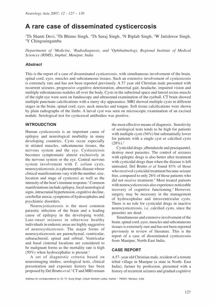
decline for the past 6 months, unsteady gait, headache and diminution of vision for 2 months. There was no history of recurrent fever, chronic cough, chronic diarrhea,weight loss, decreased appetite, joint pain and past history suggesstive of diabetes and tuberculosis. On examination, he was afebrile, with normal blood pressure. He has multiple asymptomatic pea-sized subcutaneous nodules all over the body, especially over the trunk and extremities (Figure 1). On cognitive assessment, he had mild to moderate deficits
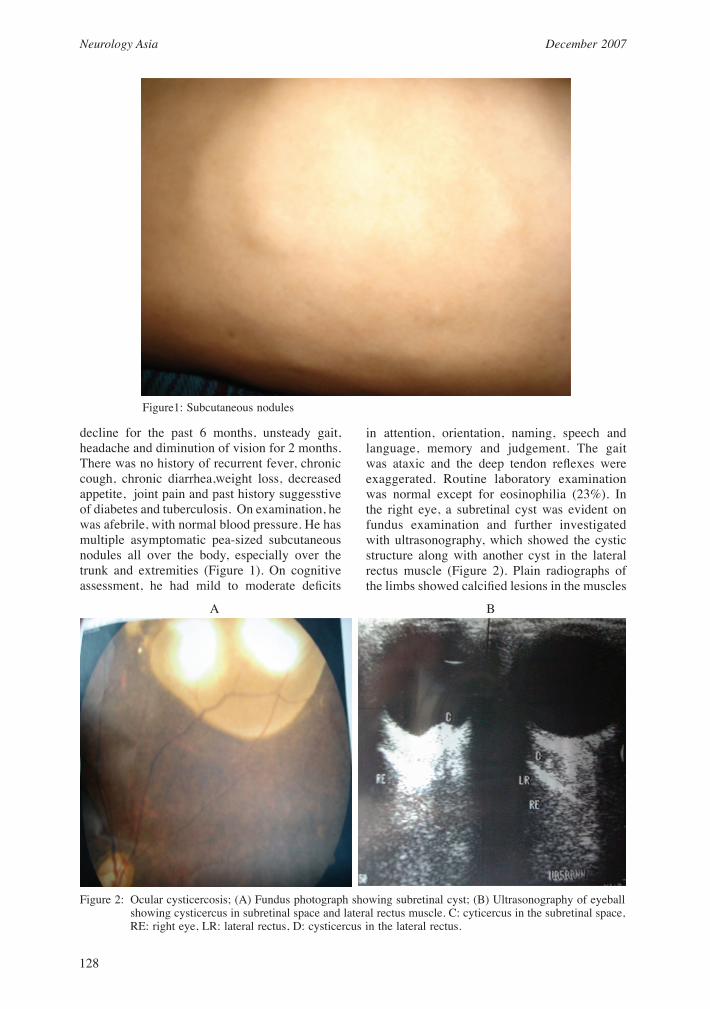
in attention, orientation, naming, speech and language, memory and judgement. The gait was ataxic and the deep tendon reflexes were exaggerated. Routine laboratory examination was normal except for eosinophilia (23%). In the right eye, a subretinal cyst was evident on fundus examination and further investigated with ultrasonography, which showed the cystic structure along with another cyst in the lateral rectus muscle (Figure 2). Plain radiographs of the limbs showed calcified lesions in the muscles
Figure1: Subcutaneous nodules
Figure 2: Ocular cysticercosis; (A) Fundus photograph showing subretinal cyst; (B) Ultrasonography of eyeball showing cysticercus in subretinal space and lateral rectus muscle. C: cyticercus in the subretinal space, RE: right eye, LR: lateral rectus, D: cysticercus in the lateral rectus.
A B
129
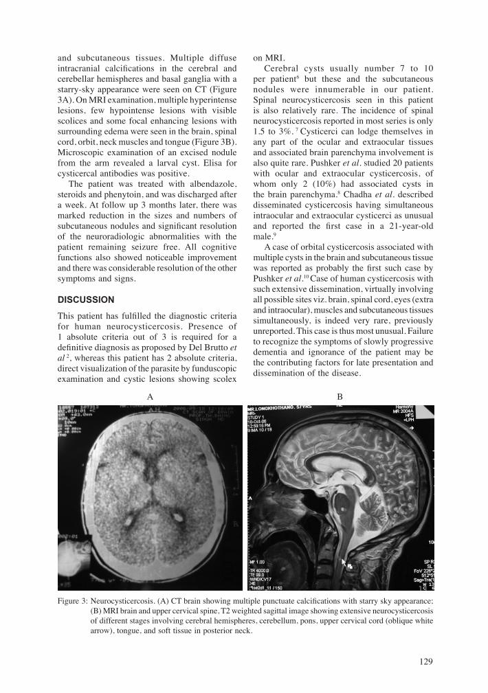
and subcutaneous tissues. Multiple diffuse intracranial calcifications in the cerebral and cerebellar hemispheres and basal ganglia with a starry-sky appearance were seen on CT (Figure 3A). On MRI examination, multiple hyperintense lesions, few hypointense lesions with visible scolices and some focal enhancing lesions with surrounding edema were seen in the brain, spinal cord, orbit, neck muscles and tongue (Figure 3B). Microscopic examination of an excised nodule from the arm revealed a larval cyst. Elisa for cysticercal antibodies was positive. The patient was treated with albendazole, steroids and phenytoin, and was discharged after a week. At follow up 3 months later, there was marked reduction in the sizes and numbers of subcutaneous nodules and significant resolution of the neuroradiologic abnormalities with the patient remaining seizure free. All cognitive functions also showed noticeable improvement and there was considerable resolution of the other symptoms and signs.
DISCUSSION
This patient has fulfilled the diagnostic criteria for human neurocysticercosis. Presence of 1 absolute criteria out of 3 is required for a definitive diagnosis as proposed by Del Brutto et al 2, whereas this patient has 2 absolute criteria, direct visualization of the parasite by funduscopic examination and cystic lesions showing scolex
on MRI. Cerebral cysts usually number 7 to 10 per patient6 but these and the subcutaneous nodules were innumerable in our patient. Spinal neurocysticercosis seen in this patient is also relatively rare. The incidence of spinal neurocysticercosis reported in most series is only 1.5 to 3%. 7 Cysticerci can lodge themselves in any part of the ocular and extraocular tissues and associated brain parenchyma involvement is also quite rare. Pushker et al. studied 20 patients with ocular and extraocular cysticercosis, of whom only 2 (10%) had associated cysts in the brain parenchyma.8 Chadha et al. described disseminated cysticercosis having simultaneous intraocular and extraocular cysticerci as unusual and reported the first case in a 21-year-old male.9 A case of orbital cysticercosis associated with multiple cysts in the brain and subcutaneous tissue was reported as probably the first such case by Pushker et al.10 Case of human cysticercosis with such extensive dissemination, virtually involving all possible sites viz. brain, spinal cord, eyes (extra and intraocular), muscles and subcutaneous tissues simultaneously, is indeed very rare, previously unreported. This case is thus most unusual. Failure to recognize the symptoms of slowly progressive dementia and ignorance of the patient may be the contributing factors for late presentation and dissemination of the disease.
Figure 3: Neurocysticercosis. (A) CT brain showing multiple punctuate calcifications with starry sky appearance; (B) MRI brain and upper cervical spine, T2 weighted sagittal image showing extensive neurocysticercosis of different stages involving cerebral hemispheres, cerebellum, pons, upper cervical cord (oblique white arrow), tongue, and soft tissue in posterior neck.
A B

Neurology Asia December 2007
130
ACKNOWLEDGEMENT
We are indebted to the Director, RIMS, for his kind permission to publish this case report.
REFERENCES
1. Takayanagui OM, Odashima NS. Clinical aspects of neurocysticercosis. Parasito Int 2006; 55 Suppl: S111 - 5. Epub 2005 Dec 5.
2. Del Brutto, O. H., V. Rajshekhar, A. C. White, Jr., V. C. Tsang, T. E. Nash, O. M. Takayanagui, P. M. Schantz, C. A. Evans, A. Flisser, D. Correa, D. Botero, J. C. Allan, E. Sarti, A. E. Gonzalez, R. H. Gilman, and H. H. Garcia. 2001. Proposed diagnostic criteria for neurocysticercosis. Neurology 57:177-183
3. Wilson M, Bryan RT, Fried JA, et al clinical evaluation of the cysticerciosis enzyme, linked immunoelectrotransfer blot in patients with neurocysticercosis. J Infect Dis. 1991; 164; 1007-1009
4. Del Brutto OH, Santibanez R, Noboa CA, et al. Epilepsy due to neurocysticercosis: analysis of 203 patients. Neurology 1992; 42: 389-92.
5. Ramirez-Bermudez J, Higuera J, Sosa AL, et al. Is dementia reversible in patients with neurocysticercosis? J Neurol Neurosurg Psychiatry 2005; 76:1164-6.
6. King CH. Cestodes (Tapeworms). In: Mandell GL, Bennett JE, Dolin R, eds: Mandell, Douglas and Bennett’s Principles and Practice of Infectious Diseases, 5th ed (Vol. 2). Churchill Livingstone, New York, 2000: 2956-64.
7. Alsina GA, Johnson JP, Mc Bride DQ, et al. Spinal neurocysticercosis. Neurosurg Focus 2002: 12(6): e8.
8. Pushker N, Bajaj MS, Chandra M, et al. Ocular and orbital cysticercosis. Acta Opthalmol Scand 2001; 79(4): 408-13.
9. Chadha V, Pandey PK, Chauhan D, et al. Simultaneous intraocular and bilateral extraocular muscle involvement in a case of disseminated cysticercosis. Int Opthalmol 2005; 26(1-2): 35-7. Epub 2006 Jun 15.
10. Pushker N, Bajaj MS, Balasubramanya R. Disseminated cysticercosis involving orbit, brain and subcutaneous tissue. J Infect 2005; 51(5): e 245-8.