a protocol for preparation and transfection of rat
TRANSCRIPT

University of North Dakota University of North Dakota
UND Scholarly Commons UND Scholarly Commons
Biomedical Sciences Faculty Publications Department of Biomedical Sciences
10-18-2017
A Protocol for Preparation and Transfection of Rat Entorhinal A Protocol for Preparation and Transfection of Rat Entorhinal
Cortex Organotypic Cultures for Electrophysiological Whole-Cell Cortex Organotypic Cultures for Electrophysiological Whole-Cell
Recordings Recordings
Nicholas I. Cilz
James E. Porter University of North Dakota, [email protected]
Saobo Lei University of North Dakota, [email protected]
Follow this and additional works at: https://commons.und.edu/bms-fac
Recommended Citation Recommended Citation Cilz, Nicholas I.; Porter, James E.; and Lei, Saobo, "A Protocol for Preparation and Transfection of Rat Entorhinal Cortex Organotypic Cultures for Electrophysiological Whole-Cell Recordings" (2017). Biomedical Sciences Faculty Publications. 18. https://commons.und.edu/bms-fac/18
This Article is brought to you for free and open access by the Department of Biomedical Sciences at UND Scholarly Commons. It has been accepted for inclusion in Biomedical Sciences Faculty Publications by an authorized administrator of UND Scholarly Commons. For more information, please contact [email protected].

Protocol Article
A protocol for preparation and transfectionof rat entorhinal cortex organotypic cultures forelectrophysiological whole-cell recordings
Nicholas I. Cilz, James E. Porter, Saobo Lei*Department of Biomedical Sciences, School of Medicine and Health Sciences, University of North Dakota,Grand Forks, ND 58203, USA
G R A P H I C A L A B S T R A C T
A B S T R A C T
Understanding how neuromodulators influence synaptic transmission and intrinsic excitability within theentorhinal cortex (EC) is critical to furthering our understanding of the molecular and cellular aspects of thisregion. Organotypic cultures can provide a cost-effective means to employ selective molecular biologicalstrategies in elucidating cellular mechanisms of neuromodulation in the EC. We therefore adapted our acute slicemodel for organotypic culture applications and optimized a protocol for the preparation and biolistic transfectionof cultured horizontal EC slices. Here, we present our detailed protocol for culturing EC slices. Using an n-methyl-D-glucamine (NMDG)-containing cutting solution, we obtain healthy EC slice cultures for electrophysiologicalrecordings. We also present our protocol for the preparation of “bullets” carrying one or more constructs anddemonstrate successful transfection of EC slices. We build upon previous methods and highlight specific aspectsin our method that greatly improved the quality of our results. We validate our methods usingimmunohistochemical, imaging, and electrophysiological techniques. The novelty of this method is that itprovides a description of culturing and transfection of EC neurons for specifically addressing their functionality.This method will enable researchers interested in entorhinal function to quickly adopt a similar slice culturetransfection system for their own investigations.
* Corresponding author.E-mail address: [email protected] (S. Lei).
https://doi.org/10.1016/j.mex.2017.10.0032215-0161/© 2017 The Authors. Published by Elsevier B.V. This is an open access article under the CC BY license (http://creativecommons.org/licenses/by/4.0/).
MethodsX 4 (2017) 360–371
Contents lists available at ScienceDirect
MethodsX
journal homepage: www.elsevier.com/locate/mex

© 2017 The Authors. Published by Elsevier B.V. This is an open access article under the CC BY license (http://creativecommons.org/licenses/by/4.0/).
A R T I C L E I N F OProtocol name: Biolistic transfection of entorhinal sectionsKeywords: Organotypic, Entorhinal, Biolistic, Slice, TransfectionArticle history: Received 24 April 2017; Accepted 11 October 2017; Available online 18 October 2017
Method details
Animal Welfare Declaration
Animal procedures described here conformed to guidelines approved by the University of NorthDakota Animal Care and Use Committee.
Materials
General resources� Chemical fume hood� Isoflurane� Bell jar� Ice bucket� 50 mL and 15 mL conical tubes� Microcentrifuge tubes� Biological safety cabinet (BSC)� Vibratome (VT1000, Leica)� Cyanoacrylate glue� Millicell culture inserts (#picmorg50, Millicell)� 40 � 11 mm petri dishes or 6-well plates� 100 � 20 mm petri dish� Ice packs� 1 mL transfer pipette� Sterile transfer pipette tips� Sterile package of 18 in. � 26 in. surgical draping (4410-imc, IMCO products)� 1 250 mL beaker� 1 30 mL beaker� 1 20 mL beaker� 37 �C incubator� Biorad Gene-Gun Low-Pressure System (#1652451, Biorad)� Polyvinylpyrrolidone (PVP, #PVP360-100G, Sigma)� Spermidine (#S2501-1G, Sigma)� 1.6 mm gold microcarriers (#1652264, Biorad)� Tefzel Tubing (#1652441, Biorad)� Molecular biology grade ethanol� Mini centrifuge (e.g. Daigger Sprout Mini-Centrifuge)� Nitrogen tank� Helium tank� Serological pipettes� Pipette aid� Vacuum and syringe filters (0.22 mm)
N.I. Cilz et al. / MethodsX 4 (2017) 360–371 361

� Autoclavable instrument bags
Supplies for dissection instrument packages (Fig. 1A)
� Large Surgical scissors (#RS-6818, Roboz)� Fine Surgical Scissors (#RS-5914, Roboz)� 2 Fine forceps (#RS-5041, Roboz)� 2 Spatulas (#13523, Ted Pella)� 2 pieces of filter paper (#09-803-5A, Fisherbrand)� 1 brush (11860, Ted Pella)� 1 mL syringe with hand-bent 0.5 in. needle (we use this tool with the brush further dissect slices)� Scalpel blade (RS-9861-21, Roboz)
Fig. 1. Equipment and setup for culturing and transfecting EC slices. A. Top, Surgical package #1 used for removal of the brain.Bottom, Package #2 used for further reducing the brain prior to and after slicing. B. Autoclaved tool package with contentsnecessary for one transfection condition. C. Basic layout of equipment in BSC prior to UV sterilization before the dissection andslicing procedure. The 4–40 mm petri dishes are absent from picture. D. Example of improvised transfection platform toimprove the consistency of transfections.
362 N.I. Cilz et al. / MethodsX 4 (2017) 360–371

Supplies for transfection instrument packages (Fig. 1B)
� Biorad Helios Gene-Gun barrel liner with rubber gasket� Modified diffusing screen (#9317T109, McMaster-Carr) [1]� Biorad diffuser screen washer (#1652475, Biorad)� Fine forceps
Recipes
1. Cutting Solution (in mM): 130 n-methyl-D-glucamine-Cl (NMDG), 24 NaHCO3, 3.5 KCl,1.25 NaH2PO4, 0.5 CaCl2, 5.0 MgCl2, and 10 glucose. Adjust pH to 7.3–7.4 and filter sterilize
2. Rinsing Solution: MEM (Life technologies cat #: 12360)3. Slice medium (SM): 50% HMEM without glutamine (Lonza cat#: 12-137F) + 25% heat-inactivated
horse serum (Hyclone cat #: SH30074.03HI) + 25% HBSS (Life technologies cat #: 24020) + 2 mMglutamine (ThermoFisher cat#: 25030081), 5.95 mg/mL glucose, 100 ug/mL penicillin andstreptomycin (100� Pen/strep stock; Cellgro cat#: 30-002-CI)a Adjust pH to 7.3 and osmolarity to �310 mOsm and filter sterilize
Plasmid constructs
Plasmids carrying reporter genes for either green fluorescent protein (GFP, pGFP-V-RS) or redfluorescent protein (RFP, pRFP-C-RS) were purchased from OriGene (Rockville, MD). Plasmids wereamplified in E. coli and isolated using a mega prep kit purchased from Qiagen (cat#: 12181). DNAworking solutions used for cartridge preparation were diluted to 1 mg/mL in TE.
Procedure overview
EC slice cultureSetup for culturing procedure (estimated time�1 h)1. Prepare and autoclave instrument packages and foil-covered 20, 30, and 250 mL beakersa. Package #1: large surgical scissors, fine surgical scissors, fine forceps, spatula (Fig. 1A)b. Package #2: spatula, scalpel blade, syringe with modified needle, brush, filter paper, fine forceps
(Fig. 1A)2. Aliquot necessary solutions in BSC and pre-equilibrate mediaa. Aliquot �200 mL of filter sterilized cutting solution into a small sterile bottle for freezing at
�80 �Ci. Critical step: Ensure that cutting solution is a very icy slurry prior to dissection to avoid poor slice
healthb. Add �1.1 mL of SM to anticipated number of petri dishes/6-well plates neededi. We typically culture 3–4 EC slices per insert and thus require between 4 and 6 inserts for one
animalii. Pre-equilibrate dishes by placing them into a 37 �C humidified 5% CO2 incubator at least 30 min
prior to dissection procedurec. Aliquot �2 mL and �10 mL of SM and MEM, respectively, and place on ice for use during
dissection procedure3. Prepare and UV sterilize BSC for slicing (Fig. 1C)a. Spray down BSC with 70% ethanol (EtOH) and lay out: vibratome equipment, both dissection
packages, sterile beakers, 4–40 mm petri dishes, 1 mL transfer pipette, 1 mL transfer pipette tips,anticipated number of cell culture inserts, and cyanoacrylate glue
b. UV sterilize cabinet and materials for at least 15 min4. Place foil covered sterile beakers on ice with SM and MEM5. Use sterile scissors from dissection package to cut and modify tip of 1 mL transfer tip
N.I. Cilz et al. / MethodsX 4 (2017) 360–371 363

a. These tips are used for transferring slice from cutting chamber, MEM washes, and placement oninsert (see Graphical abstract)
6. Invert lids of 100 mm petri dish to use as brain-prepping stage (used prior to gluing brain tovibratome platform) and place filter paper atop
7. Fill vibratome trough with ice, turn on and prepare vibratome8. Once cutting solution is sufficiently slushy and chilled, aliquot �150 mL into 250 mL beaker,
�25 mL into 30 mL beaker, and �15 mL into 20 mL beaker in BSCa. Recover with foil and return to ice bucketDissection and slice collection procedure (estimated time �30 min)1. In a fume hood, deeply anesthetize animal in bell jar with isoflurane, spray animal with 70%
EtOH, decapitate using large scissors from dissection package #1, and transfer head to 30 mL beaker oficy slurry solution
a. Replace foil, change gloves, and move to BSC with remaining tools in dissection package #12. On surgical draping, dissect brain into 20 mL beaker of icy solution and allow to cool for at least
1 mina. Meanwhile, use EtOH to disinfect dissection region of BSC3. Once the brain is chilled, pour off excess cutting solution and guide brain onto a piece of filter
paper placed on prepping stage4. With scalpel blade, gently invert brain so that it sits on its dorsal region and remove cerebellum
and anterior portion of brain not containing hippocampus5. Using the spatula and scalpel, carefully transfer the brain to the second piece of filter paper to
remove excess cutting solution from the bottom (dorsal) part of the brain6. Apply glue to vibratome platform, gently guide brain onto glue, transfer platform to cutting
reservoir, and add �150 mL ice cold cutting solution without disturbing brain with large ice chunks7. Collect 300 mm horizontal sections in reservoir8. While slicing, remove an ice pack from the freezer and place in BSC; place the four petri dishes
atop the pack9. Using a paintbrush and modified syringe/needle, carefully isolate the EC by making two cuts (see
Graphical abstract)a. The first cut removes a portion of the perirhinal cortex and provides a visual cue for medial vs.
lateral orientation at later stages in cultureb. The second cut near the hippocampal sulcus removes the hippocampus proper and leaves the EC
intact10. Fill three of the four dishes with chilled MEM and rinse slices by serial transfer, transferring as
many slices as possible at once (see Graphical abstract)11. Add cold SM to the fourth dish and transfer slices from MEM to SM12. Remove pre-equilibrated dishes/plates from incubator and place in BSC13. Open a new culture insert and transfer one slice at a timea. Allow the slice to fall to the bottom of the modified tip prior to placement on insert (see Graphical
abstract)b. Once it is at the bottom, gently touch the solution bead containing the slice to the insertc. Repeat for desired number of slices per insert14. Switch to a regular 1 mL transfer tip and carefully remove excess SM around EC slicesa. Critical Step: Take care not to leave too much media on the slices15. Using sterile forceps, transfer culture insert to pre-warmed media dish/plate16. Repeat steps 13–15 until all slices have been successfully transferred17. Clean and UV sterilize BSCMaintenance of EC slice cultures1. The media should be changed on day-in-vitro (DIV) 1 and then every two days, thereafter2. On DIV 1, examine slices under an inverted light microscope and use a marker to denote the
medial aspect of the slice on the bottom of the plate
Biolistic transfection of cultured EC slicesPreparation of gene gun cartridges (estimated time �40 min)
364 N.I. Cilz et al. / MethodsX 4 (2017) 360–371

1. Insert a length of Tefzel Tubing into the tubing prep station and leave about a two-inch overhangat the end of the tubing rod
2. Turn on the N2 and open the tubing prep station regulator to just over 0.4 L per minute for about30 min
3. Measure out 12.5–15 mg of 1.6 mm gold particles in a 1.5 mL microcentrifuge tube4. Measure out 20 mg PVP into a 1.5 mL microcentrifuge tube and dissolve with 1 mL 100% EtOH5. Aliquot 3.1 mL of 100% EtOH into a 15 mL tube and prepare a 50 mg/mL PVP dilution using the
20 mg/mL stock (7.75 mL of stock solution to 3.1 mL)6. Prepare DNA working solution; we typically prepare 1 mg/mL dilutions of cDNA to be transfecteda. If more than one construct is to be transfected, we combine all constructs in a separate tube and
mix by aspiratingb. Ratios will need to be determined for each condition and by individual investigators, but our
results have indicated a high success rate of near equal co-transfection rates by simply using a 1:1 ratioof two constructs with the same promoter (see below)
7. To the gold, add 100 mL 0.5 M spermidine (filter sterilized); tap to mix and sonicate briefly8. Add 50 mg of DNA to gold mixture; tap well to mix and sonicate very briefly9. With the cap open, vortex on a low speed and add 100 mL of CaCl2 dropwise to the gold mixture10. Sonicate briefly and tap well to mix11. Allow 10 min for the mixture to precipitate while tapping every couple of minutes to re-agitate
the mixture12. Spin mixture for 30 s using a mini-centrifuge13. Remove as much of the supernatant as possible without disturbing the gold pellet14. Resuspend the mixture with 1 mL of 100% EtOH; tap mixture well and briefly sonicate to aid in
resuspension15. Repeat steps 12–14 for a total of three EtOH washes of gold pellets16. Remove supernatant from final wash and resuspend pellet in 500 mL of the 50 mg/mL PVP
solution17. Tap this mixture very well to resuspend the pellet and transfer to a new 15 mL conical tube18. Repeat this process (steps 16–17) in 500 mL iterations to thoroughly resuspend gold pelleta. After transferring �1.5 mL, brief sonication may be employed to facilitate dissolving of remaining
gold in PVP solution19. Once 3 mL of the gold solution has been resuspended, turn off N2 and remove tubing from the
prep station rod20. Attach a piece of 18 in. length, 0.104 in. internal diameter silicone tubing to a 10 cc syringe and
attach the other end of the silicone tubing to the Tefzel Tubing21. Vigorously shake the suspension to resuspend and quickly draw up the solution into the Tefzel
Tubing, leaving about 2 in. at the end of the tubing22. Carefully, but quickly, place Tefzel Tubing into the prep station rod and mark where the solution
stands in the tubing using a marker23. With the syringe attached, wait five minutes to allow the gold to settle24. After five minutes, draw off EtOH and rotate the tubing 180� and wait about 2 s before turning
on constant rotation25. Remove the syringe and continue to rotate for 30 s, after which turn on the N2 and continue
rotating for another 5 min26. Turn off N2, remove tubing, and use the Biorad tubing cutter to cut 0.5 in. cartridges from
between the marked regions of the tubing27. Store cartridges with a desiccant pellet in a 20 mL scintillation vial wrapped in parafilm at 4 �CTransfection procedure (estimated time �20 min)1. In a BSC, place the EtOH disinfected Biorad Helios Gene-Gun, autoclaved gene-gun transfection
package (Fig. 1B), vials of gene gun cartridges, a sterile 40 mm petri dish, warmed SM, a 2 mLserological pipette, and a pipette-aid
a. We also include a shooting platform in the BSC to assist in consistent shooting distances (seebelow under Additional information)
N.I. Cilz et al. / MethodsX 4 (2017) 360–371 365

b. A simple shooting platform can be constructed using a microcentrifuge tube rack that holds a rodupright (e.g. seriological pipette) marked at 1.25 in. distance from slice surface (Fig. 1D)
2. Open gene gun kits and load appropriate number of cartridges into cartridge holder using forceps3. Connect gene gun to helium supply, as per manufacturer instructions, and adjust helium supply
to 150 psi4. Prime the gun by discharging twice with the cartridge holder positioned on an empty barrel5. Remove EC slice dishes/plates from incubator and transfect one insert at a timea. Transfer insert to a new empty sterile 40 mm petri dish and change the media on the dish/plateb. Place dish containing only the insert on shooting platform, remove cover, position gun directly
above insert at a distance of 1.25 in. away from slice surface and discharge cartridgei. Critical Step: Slice health is important for obtaining quality electrophysiological recordings from
slice cultures and we observed that shooting distance can drastically influence the quality oftransfected, patchable cells
ii. Thus, using a shooting platform with a guide to improve consistency is strongly recommended(Fig. 1D)
c. Return insert to original dish containing fresh mediad. Repeat steps 5a–5c until all slices are transfectedi. Note: if transfecting with multiple constructs, be sure to use a different gene-gun instrument
package with a clean mesh diffuser screen for each construct6. Turn off helium source, return slices to incubator, clean up BSC, and return sealed cartridge
constructs to 4 �C.
Method validation
We first ensured that cultured slices (Fig. 2A) maintained a laminar distribution and that we hadhealthy neurons in our preparation. Slices were fixed in 4% paraformaldehyde for 24 h, after whichslices were individually transferred to frosted microscope slides (VWR #12-550-15). Immuno-staining was performed using methods adapted from previous work [2]. Immunofluorescentstaining for the neuronal marker NeuN (1:200, Milipore, #ABN78) demonstrated that EC layers werewell-preserved in slice cultures and that neurons were detected after being in culture for severaldays (Fig. 2B). We next assessed slice health using electrophysiological methods described inprevious work [2]. Under high-magnification, differential interference contrast microscopy (DIC)images revealed numerous healthy principal neurons for patching (Fig. 2C). Direct positive currentinjections to cultured layer II principal neurons (DIV 4) displayed action potential firing that wasgraded proportionally to current steps and negative current injections displayed early sag responses,typical of superficial layer II EC neurons. Input-output curves for action potential firing in responseto current injections showed no significant difference between DIV 4 cultured slices and postnatalday 16 (p16) acute slices, suggesting no substantial changes in EC principal neuron excitability werepresent early on in culture.
Having validated that cultured EC slices were healthy and contained patchable neurons, we nextwanted to validate our biolistic transfection procedures. Similar to other methods [3], we initiallytransfected slices within a few hours following the slicing procedure. Transfecting at two hours on DIV0 resulted in several slices being lost from the insert following the pressure burst, likely owing toinsufficient time for adherence to the membrane. Furthermore, we observed consistently highertransfection rates when slices were permitted to acclimate to culture conditions for a few days. Theadditional time in culture likely allowed the slice to reach a new physiological steady-state andpermitted intrinsic processes to clear away unhealthy cells and other debris, which may haveotherwise obstructed the penetrating transfection particles from reaching healthy neurons. For thesereasons, we adopted a transfection scheme where slices were transfected on DIV 2 and experimentswere conducted 36–72 h later (Fig. 3A). It is important to point out that because the culturingprocedure results in massive denervation of the EC and because denervation can result in theoccurrence of homeostatic changes in the target cell [4], careful attention should be given to timepoints of culturing, transfecting, and recording to ensure consistency across experiments. This methodprovided high transfection rates per slice in EC regions of interest (Fig. 3B). Recordings from layer III EC
366 N.I. Cilz et al. / MethodsX 4 (2017) 360–371

neurons transfected with GFP remained healthy and continued to be electrically active (Fig. 3C and D).Because some experimental conditions may require co-transfection of multiple plasmids to permiteither expression of reporter molecules or because conditions may require introduction of multipleprotein components, we assessed transfection ratios of 1:1 plasmid DNA constructs (i.e. 25 mg of eachplasmid added during cartridge preparation) using GFP and RFP reporters to evaluate levels of co-expression. Under these conditions, we consistently observed a prominent level of co-expressionacross many experiments spanning different slice culture batches, although minor variations inintensity were occasionally seen in a few cells (Fig. 3E, see white arrows). These results suggest that a1:1 ratio may be sufficient when introducing multiple gene products. However, for experiments whereone of the constructs is simply a reporter (e.g. GFP), we adjust our ratios to 0.75:0.25 for gene of
Fig. 2. Healthy EC slices are prepared that exhibit typical cortical laminar distribution and the electrophysiological properties ofprincipal neurons are generally well-preserved. A. DIC photograph of atypical EC slice section. B. NeuN immunofluorescentstaining of organotypic cultures demonstrated that an abundant number of neurons were present and arranged in typicallaminar fashion seen in acute slices. C. Higher magnification of a typical EC slice, imaged near layer II/layer III border, exhibited arespectable number of healthy and patchable principal neurons, indicated by asterisks (*). D. Example of voltage responses to arange of current injections obtained from a layer II principal neuron, confirming electrophysiological viability of neurons andtypical signatures of this layer (i.e. preserved sag response). E. Input-output curve for number of action potentials generated peramount of direct-current injected, where responses from DIV 4 (from p12) slice cultures were compared with acute slicesprepared from animals of the same litter (p16). No significant changes were observed (acute n = 3; DIV 4 n = 5).
N.I. Cilz et al. / MethodsX 4 (2017) 360–371 367
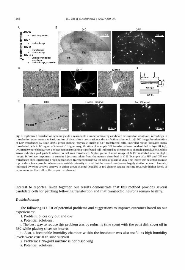
interest to reporter. Taken together, our results demonstrate that this method provides severalcandidate cells for patching following transfection and that transfected neurons remain healthy.
Troubleshooting
The following is a list of potential problems and suggestions to improve outcomes based on ourexperiences:
1. Problem: Slices dry out and diea. Potential Solutions:i. The best way to reduce this problem was by reducing time spent with the petri dish cover off in
BSC while placing slices on insertsii. Also, a breathable humidity chamber within the incubator was also useful as high humidity
levels were crucial to slice survival2. Problem: DNA-gold mixture is not dissolvinga. Potential Solutions:
Fig. 3. Optimized transfection scheme yields a reasonable number of healthy candidate neurons for whole-cell recordings intransfection experiments. A. Basic outline of slice culture preparation and transfection scheme. B. Left, DIC image for orientationof GFP-transfected EC slice. Right, green channel greyscale image of GFP transfected cells. Encircled region indicates manytransfected cells in EC region of interest. C. Higher magnification of example GFP transfected neuron identified in layer III. Left,DIC image where black arrow denotes region containing transfected cell, indicated by the presence of a gold particle. Note, whitearrow indicates gold particle where no cell was transfected. Center, green channel image of GFP-transfected neuron. Right,merge. D. Voltage responses to current injections taken from the neuron described in C. E. Example of a RFP and GFP co-transfected slice illustrating a high degree of co-transfection using a 1:1 ratio of plasmid DNA. This image was selected becauseit provides a few examples where some variable intensity existed, but the overall levels were largely similar between channels,indicated by white arrows. Arrows in either green channel (middle) or red channel (right) indicate relatively higher levels ofexpression for that cell in the respective channel.
368 N.I. Cilz et al. / MethodsX 4 (2017) 360–371

i. The mixture is tough to dissolve but this process is aided by constant brief sonicationii. If it is impossible to dissolve, double-check the DNA stock solution concentration to ensure its
accuracy; if more than 50 mg is added, it is nearly impossible to dissolve the clump, in which case it isbest to start over
3. Problem: Transfected slices are uneven, in that some slices are transfected while others are blanka. Potential Solutions:i. This problem is likely due to poor quality bullets and is likely the result of poor spreading of gold
mixture during the coating stage; there should not be a visible gold line in the cartridgeii. Better bullets can be generated by ensuring a well dissolved mixture and spinning the tubing for
a longer duration after drawing off the EtOH, but before turning on the N2
4. Problem: Both transfected and non-transfected cells from a shot culture dish are visible but verydifficult to patch and seem to be encased in some difficult-to-penetrate matrix
a. Potential Solutions:i. Use a control non-transfected (i.e. non-shot) plate to ensure cell viability; if cells are visible and
patchable, the problem is almost certainly due to pressure intensity or distance of shot from the top ofthe slice
ii. We strongly recommend using at least 1.25 in. shooting distance from the top of the slice to thebarrel liner
Additional information
Background and discussion
Organotypic cultures are a well-established in vitro model to investigate the cytoarchitecture andneurophysiology of brain tissue [5,6]. Whereas such cultures comprised of hippocampal regions havebeen utilized by many labs, relatively fewer reports involve organotypic cultures consisting ofentorhinal tissue. Horizontal organotypic slices consisting of both entorhinal and hippocampalregions of P0–P5 animals have been employed to study axonal regeneration and synaptic scaling at thedentate gyrus following lesions to the perforant pathway in vitro [4,7]. Our method is unique in that itspecifically details the culturing and transfection protocols used for the electrophysiologicalinvestigations of EC neurons and will be helpful for mechanism studies involving EC neuromodulation.
There are several steps we employ that distinguish our method from other methods of organotypicslice culture. First, we have chosen to use a vibratome as opposed to either a tissue chopper or slicerthat others have described [6,8,9]. Although use of a vibratome requires additional time for collectingtissue, we find that it produces healthy slices that are well-suited for electrophysiological recordingsas previously demonstrated by others, e.g. in spinal cord organotypic cultures [10]. Second, we chooseto utilize a horizontal preparation because this approach provides slices that are similar in orientationto our acute model. Third, unlike most organotypic slice culture methods, we use a cutting solutionwhere Na+ has been largely replaced with NMDG, in order to minimize excitotoxicity that may occurduring the traumatic slicing process. Alternative methods to minimize excitotoxicity involvesupplementing the cutting solution with antioxidants, such as pyruvate and ascorbic acid [8,11].NMDG-containing cutting solutions have become widely adopted in electrophysiological laboratoriesworking with acute slices because these steps are neuroprotective, especially toward the morevulnerable interneuron populations [12]. Overall, our EC slice model attempts to yield a similar qualityof initial tissue preparation for longer-term culture that resembles our acute slice model and we havehad success culturing slices using animal ages ranging from p6 to P18.
Our optimized transfection conditions are informed by both the instruction manual provided withthe Biorad Helios Gene-Gun, as well as other detailed published protocols describing biolisticcartridge preparation and transfection [1,9,13]. The reader is referred to those references for additionaldetail, however, we obtained excellent results in our method by employing a few modifications. First,it is essential that the gold mixture be homogenously distributed about the cartridge to increasetransfection efficiency for all available slices on a plate. We found that including a more thoroughsonication periods of the DNA gold mixture was necessary to obtain a better suspension for coating theTefzel Tubing. Additionally, a longer period of tubing rotation following EtOH removal, but prior to
N.I. Cilz et al. / MethodsX 4 (2017) 360–371 369

turning on N2, achieved an improved mixture distribution. Secondly, neurons are very susceptible tothe pressure burst during the transfection procedure and the quality of cells available forelectrophysiological recordings are greatly influenced by the shooting distance used during thetransfection. Initially, we held the gun and barrel liner at a distance similar to what other methodsreported. On recording days, we observed an unknown matrix that seemed to encase the neurons andimpede our ability to form tight gigaohm seals with transfected neurons. However, when the shootingdistance was increased, this matrix was no longer a problem and whole-cell success rates weresubstantially improved. We therefore addressed this problem by optimizing a shooting a distance forour 11 mm petri dishes, which we empirically found to be at 1.25 in. or greater. To improve consistencyacross experiments, we improvised a shooting platform that was clearly demarcated with the idealshooting distance for an easy reference (see Fig. 1D). Thirdly, although not rigorously tested, we alsofound that the media composition seemed to influence the quality of transfected cell-health forrecordings. Initially, we were able to obtain healthy slice cultures using a more simplistic media recipedescribed by other labs [3]. However, transfection efficiency seemed to be less than ideal whenfollowing the biolistic transfection process. We observed a consistent improvement in efficiency aftermodifying the SM with the addition of glucose and glutamine, similar to the Stoppini method [6] (seeunder Recipes above). Finally, we report that transfecting with GFP and RFP constructs in a 1:1 ratioprovided a high degree of co-transfection, in agreement with a recent report involving biolisticdouble-transfections [8]. Ideally, co-transfection experiments should use plasmids containing thesame promoter, as was the case in our experiment. Results are expected to vary if different and/or lessefficient promotor-carrying plasmids are to be used, in which case ratios will need to be determined byindividual investigators.
We expect this method to be useful for other investigators interested in addressing molecular andcellular mechanisms of entorhinal neurophysiology. The details described herein will assist others inquickly adopting a similar EC slice culture method for their lab. Additionally, this information may beuseful to those troubleshooting biolistic transfection methods in other brain regions.
Acknowledgements
We wish to thank Dr. Kumi Nagamoto-Combs and Dr. Colin Combs for their time and thoughtfulcomments in addressing technical issues encountered when culturing entorhinal slices. We wouldalso like to thank Dr. Othman Ghribi, Dr. John Watt, and Jared Schommer for their comments andassistance in the development of our culturing technique. This work was supported with fundingprovided by the National Institute of Mental Health (RO1MH082881) and a graduate student researchfellowship through the National Science Foundation and North Dakota Experimental Program toStimulate Competitive Research (#IIA-1355466).
References
[1] G. Woods, K. Zito, Preparation of gene gun bullets and biolistic transfection of neurons in slice culture, J. Vis. Exp. 12 (2008)e675, doi:http://dx.doi.org/10.3791/675.
[2] N.I. Cilz, S. Lei, Histamine facilitates GABAergic transmission in the rat entorhinal cortex: roles of H1 and H2 receptors, Na+-permeable cation channels, and inward rectifier K+ channels, Hippocampus 27 (2017) 613–631.
[3] R.C. Foehring, D. Guan, T. Toleman, A.R. Cantrell, Whole cell recording from an organotypic slice preparation of neocortex, J.Vis. Exp. 52 (2011)e2600, doi:http://dx.doi.org/10.3791/2600.
[4] A. Vlachos, D. Becker, P. Jedlicka, R. Winkels, J. Roeper, T. Deller, Entorhinal denervation induces homeostatic synapticscaling of excitatory postsynapses of dentate granule cells in mouse organotypic slice cultures, PLoS One 7 (2012)e32883,doi:http://dx.doi.org/10.1371/journal.pone.0032883.
[5] B.H. Gähwiler, Organotypic monolayer cultures of nervous tissue, J. Neurosci. Methods 4 (1981) 329–342.[6] L. Stoppini, P.-A. Buchs, D. Muller, A simple method for organotypic cultures of nervous tissue, J. Neurosci. Methods 37
(1991) 173–182.[7] D. Del Turco, T. Deller, Organotypic entorhino-hippocampal slice cultures—a tool to sudy the molecular and cellular
regulation of axonal regeneration and collateral sprouting in vitro, Neuroprot. Methods Protoc. (2007) 55–66.[8] L. Lossi, C. Cocito, S. Alasia, A. Merighi, Ex vivo imaging of active caspase 3 by a FRET-based molecular probe demonstrates
the cellular dynamics and localization of the protease in cerebellar granule cells and its regulation by the apoptosis-inhibiting protein survivin, Mol. Neurodegener. 11 (2016) 34.
[9] A.K. McAllister, Biolistic transfection of neurons, Sci. Signal. (2000) pl1, doi:http://dx.doi.org/10.1126/stke.2000.51.pl1.
370 N.I. Cilz et al. / MethodsX 4 (2017) 360–371

[10] F. Ferrini, C. Salio, L. Lossi, G. Gambino, A. Merighi, Modulation of inhibitory neurotransmission by the vanilloid receptortype 1 (TRPV1) in organotypically cultured mouse substantia gelatinosa neurons, Pain 150 (2010) 128–140, doi:http://dx.doi.org/10.1016/j.pain.2010.04.016.
[11] S. Alasia, A. Merighi, L. Lossi, Transfection techniques and combined immunocytochemistry in cell cultures andorganotypic slices, Immunocytochem. Relat. Tech. (2015) 329–355.
[12] Y. Tanaka, Y. Tanaka, T. Furuta, Y. Yanagawa, T. Kaneko, The effects of cutting solutions on the viability of GABAergicinterneurons in cerebral cortical slices of adult mice, J. Neurosci. Methods 171 (2008) 118–125.
[13] J.A. O’Brien, M. Holt, G. Whiteside, S.C.R. Lummis, M.H. Hastings, Modifications to the hand-held Gene Gun: improvementsfor in vitro Biolistic transfection of organotypic neuronal tissue, J. Neurosci. Methods 112 (2001) 57–64.
N.I. Cilz et al. / MethodsX 4 (2017) 360–371 371