a prospective randomized trial of intravitreal bevacizumab versus ranibizumab for the management...
TRANSCRIPT
A Prospective Randomized Trial of IntravitrealBevacizumab Versus Ranibizumab for the Management
of Diabetic Macular Edema
ANTONIO BRUNNO NEPOMUCENO, ERIKA TAKAKI, FELIPE PIACENTINI PAES DE ALMEIDA,RENATO PERONI, JOSE AUGUSTO CARDILLO, RUBENS CAMARGO SIQUEIRA, INGRID URSULA SCOTT,
ANDRE MESSIAS, AND RODRIGO JORGE
� PURPOSE: To compare visual acuity and spectral-domain optical coherence tomography (SDOCT)outcomes associated with intravitreal (IV) bevacizumabvs IV ranibizumab for the management of diabeticmacular edema (DME).� DESIGN: Prospective randomized trial.� METHODS: Forty-eight patients (63 eyes) with center-involved DME were randomly assigned to receive1.5 mg (0.06 cc) IV bevacizumab or 0.5 mg (0.05 cc)IV ranibizumab at baseline andmonthly if central subfieldthickness was greater than 275 mm.� RESULTS: Forty-five patients (60 eyes) completed48weeks of follow-up. At baseline, mean ± standard errorbest-corrected visual acuity (BCVA) (logMAR)was 0.60(20/80) ± 0.05 in the IV bevacizumab group and 0.63(20/85) ± 0.05 in the IV ranibizumab group. A significantimprovement in mean BCVA was observed in bothgroups at all study visits (P < .05); this improvementwas significantly greater in the IV ranibizumab groupcompared with the IV bevacizumab group at weeks 8(P [ .032) and 32 (P [ .042). A significant reductionin mean central subfield thickness was observed in bothgroups at all study visits compared with baseline (P <.05), with no significant difference in the magnitude ofmacular thickness reduction between groups. The meannumber of injections was significantly higher (P [ .005)in the IV bevacizumab group (9.84) than in the IV ranibi-zumab group (7.67).� CONCLUSIONS: IV bevacizumab and IV ranibizumabare associated with similar effects on central subfieldthickness in patients with DME through 1 year offollow-up. IV ranibizumab is associated with greaterimprovement in BCVA at some study visits, andthe mean number of injections is higher in the IV
Accepted for publication Apr 18, 2013.From the School of Medicine of Ribeirao Preto, University of Sao
Paulo, Department of Ophthalmology, Otorhinolaryngology and Headand Neck Surgery, Ribeirao Preto, Brazil (A.B.N., E.T., F.P.P.A.,R.C.S., A.M., R.J.); Hospital de Olhos de Araraquara, Araraquara,Brazil (R.P., J.A.C.); and Departments of Ophthalmology and PublicHealth Sciences, Penn State College of Medicine, Hershey,Pennsylvania (I.U.S.).
Inquiries to Prof Dr Rodrigo Jorge, Hospital das Clınicas da Faculdadede Medicina de Ribeirao Preto, Avenida Bandeirantes 3900, RibeiraoPreto-SP, 14049-900, Brazil; e-mail: [email protected]
502 � 2013 BY ELSEVIER INC.
bevacizumab group. (Am J Ophthalmol 2013;156:502–510. � 2013 by Elsevier Inc. All rights reserved.)
MACULAR EDEMA IS THE LEADING CAUSE OF
decreased visual acuity in patients with diabeticretinopathy.1,2 Laser photocoagulation has been
the standard-of-care treatment for diabetic macular edema(DME) for decades, based on the Early Treatment DiabeticRetinopathy Study (ETDRS) and other more recent clinicaltrials.3–6 However, because visual acuity improvementpost laser is observed infrequently, and because of thefrequent recurrence or persistence of DME after lasertreatment, there is a need for better treatments for themanagement of DME (especially for diffuse DMEinvolving the foveal center, since focal DME not involvingthe foveal center may have a good prognosis after focallaser treatment).3,4,6–8 Care must be taken, however,because the terms ‘‘diffuse’’ and ‘‘focal’’ DME have not beendefined consistently in the literature; these terms havereferred to a variety of diverse parameters (clinical andangiographic) itemized differently by various authors.7–9 Inaddition, the definition of center- and non-center-involvedDMEmay vary; the Diabetic Retinopathy Clinical ResearchNetwork (DRCRnet) has defined non-center-involvedDME as ‘‘a baseline central subfield thickness<250 micronsand a baseline photograph assessment of retinal thickness atthe center of the macula graded as none or questionable.’’7
Moreover, the parameters of a ‘‘normal’’ central subfieldthreshold may vary depending on the optical coherencetomography (OCT) machine employed.10
Among pharmacologic treatments currently availablefor DME, antiangiogenic agents such as bevacizumab andranibizumab have been reported to be associated withvisual acuity improvement and favorable remodeling ofthe macular architecture in patients with DME.11–16
Ranibizumab has been evaluated in phase III prospectiverandomized clinical trials and reported to be associatedwith better visual acuity outcomes compared to focal/gridlaser in patients with DME.12,13 To our knowledge andbased on a Medline search, there is no published studycomparing intravitreal (IV) bevacizumab and IVranibizumab for the treatment for DME. We conducteda randomized, prospective study to compare the visual
0002-9394/$36.00http://dx.doi.org/10.1016/j.ajo.2013.04.026
ALL RIGHTS RESERVED.
acuity and spectral-domain optical coherence tomography(SDOCT) outcomes associated with IV bevacizumab vs IVranibizumab for the management of DME.
METHODS
THE CURRENT STUDY IS A PROSPECTIVE RANDOMIZED CLIN-
ical trial registered at ClinicalTrials.gov (NCT01487629).The study protocol adhered to the tenets of the Declarationof Helsinki and was approved by the local InstitutionalReview Board, Comite de Etica em Pesquisa do Hospitaldas Clınicas da Faculdade de Medicina de Ribeirao Preto,and all participants gave written informed consent beforeentering into the study. All patients evaluated in theRetina Section of the Department of Ophthalmology,School of Medicine of Ribeirao Preto of the University ofSao Paulo with center-involved DME in at least 1 eyebetween July 1, 2010 and August 31, 2011 were invitedto participate in the study.
� PATIENT ELIGIBILITY AND BASELINE EVALUATION:
Inclusion criteria. Inclusion criteria were as follows: (1)center-involved DME, defined as a central subfieldthickness >300 mm on SDOCT, despite at least 1 sessionof macular laser photocoagulation performed at least3 months previously; (2) best-corrected ETDRS visualacuity (BCVA) measurement between 0.3 logMAR(Snellen equivalent: 20/40) and 1.6 logMAR (Snellenequivalent: 20/800); (3) signed informed consent.
Exclusion criteria. Exclusion criteria were: (1) vitreo-macular traction on SDOCT; (2) proliferative diabeticretinopathy needing panretinal photocoagulation (PRP)or anticipated to need PRP in the next 12 months; (3)macular capillary dropout on fluorescein angiography; (4)history of glaucoma or ocular hypertension (defined as anintraocular pressure higher than 22 mm Hg); (5) an ocularcondition (other than diabetes) that, in the opinion of theinvestigator, might affect macular edema or alter visualacuity during the course of the study (eg, retinal vein occlu-sion, uveitis or other ocular inflammatory disease, neovas-cular glaucoma, etc); (6) systemic corticosteroid therapy;(7) any condition that, in the opinion of the investigator,might preclude follow-up throughout the study period.
Each patient received a detailed ophthalmologic exami-nation including measurement of BCVA according to thestandardized ETDRS refraction protocol using a retroillu-minated Lighthouse for the Blind distance visual acuitytest chart (using modified ETDRS charts 1, 2, and R; Preci-sion Vision, IL), as well as applanation tonometry, undi-lated and dilated slit-lamp biomicroscopic examination,indirect fundus examination, and fluorescein angiographyusing high-resolution angiography (HRA; HeidelbergEngineering, Heidelberg, Germany).
VOL. 156, NO. 3 BEVACIZUMAB VERSUS RANIBIZUMAB
Fourier-domain OCT evaluation (Spectralis EyetrackerTomographer, HRA-OCT; Heidelberg Engineering) wasperformed in all patients, and retinal thickness measure-ments were acquired using a standard 203 15-degree rasterscan protocol consisting of 19 horizontal sections (eachcomputed out of 25 frames) with a distance of 240 mmbetween each horizontal scan, covering a square of 20 315 degrees on the retina and centered on the foveal region.Follow-up mode was used to reduce test-retest variability.In order to optimize the accuracy of OCT data, automaticdelineation of the inner and outer boundaries of the neuro-sensory retina generated byOCT built-in software was veri-fied for each of the scans. Central subfield thickness valueswere calculated automatically as the average thickness ofa central macular region 1000 mm in diameter centeredon the patient’s foveola by built-in Heidelberg softwareusing retinal map analysis.
� INTRAVITREAL INJECTION: If both eyes were eligible fortreatment and the patient agreed to treat both eyes withanti-VEGF therapy, 1 eye received the randomized treat-ment according to a computer-generated sequence andthe contralateral eye received the other anti-VEGF agenton the next day; thus, if an eye was randomized to the rani-bizumab group, the contralateral eye was allocated to thebevacizumab group. All injections were performed usingtopical proparacaine drops under sterile conditions (eyelidspeculum and povidone-iodine). Before the injection wasperformed, the eyelids were scrubbed with 10% povidone-iodine, and 5% povidone-iodine drops were applied tothe conjunctiva. The time between application of 5%povidone-iodine solution to the conjunctiva and adminis-tration of the intravitreal injection was 2 minutes.Povidone-iodine was applied to the conjunctiva directlyover the intended injection site.17–20 Care was taken inall cases to insure that the needle did not touch the lidsor lashes. Bevacizumab (1.5 mg/0.06 cc; F. Hoffmann- LaRoche Ltd., Basel, Switzerland) or ranibizumab (0.5 mg/0.05 cc; Novartis Pharma Stein AG, Stein, Switzerland)was injected into the vitreous cavity using a 29-gauge 0.5-inch needle inserted through the inferotemporal pars plana3.0-3.5 mm posterior to the limbus.21 After the injection,central retinal artery perfusion was confirmed with indirectophthalmoscopy. Patients were instructed to instill 1 dropof 0.3% ciprofloxacin into the injected eye 4 times dailyfor 1 week after the procedure.
� RETREATMENTPROTOCOL: Retreatment with the origi-nally assigned treatment was performed monthly if centralsubfield thickness was greater than 275 mm.
� RESCUE THERAPY: If, after 3 consecutive injections,there was not a reduction in central subfield thickness ofat least 10% or an increase in BCVA of at least 5 letterscompared with baseline, the patient could, at the discretionof the treating ophthalmologist, receive focal/grid laser
503FOR DIABETIC MACULAR EDEMA

photocoagulation or continue to receive the same intravi-treal medication for an additional 3 consecutive visits.
� FOLLOW-UP EXAMINATIONS AND OUTCOMEMEASURES: Patientswere scheduled for follow-up examina-tions at monthly intervals. At these visits, patients’ BCVAwas determined after ETDRS refraction, and they under-went complete ophthalmic examination using the sameprocedures as at baseline, with the exception of fluoresceinangiography, which was performed only at the final follow-up visit. Examiners (E.T., F.P.P.A., R.P.) were maskedregarding which treatment drug was used for each patient.Throughout the study, a single masked, certified examinerperformed BCVA measurements prior to any other studyprocedure. Patients, OCT technicians, and fundus photog-raphers were also masked to treatment group. Outcomemeasures include changes in ETDRS BCVA, changes incentral subfield thickness, and occurrence of complications.
� STATISTICAL ANALYSIS: BCVA and central subfieldthickness measured at each follow-up visit were comparedwith baseline BCVA and central subfield thickness valuesfor within- and between-group comparisons, which wereperformed using multiple analysis of variance (MANOVA)for repeatedmeasurements. Proportions of eyes with centralsubfield thickness <_275 mmwere compared using the likeli-hood ratio x2 test. In addition, a multivariate analysiscomparing BCVA and central subfield thickness outcomesin the IV bevacizumab group and IV ranibizumab group wasperformed, taking into account number of injections, base-line BCVA, and central subfield thickness as effects.
A statistically significant effect was defined if P< .05, anda trend towards significance was reported if P < .1. Statis-tical analyses were performed using JMP 10.0.0 (2010;SAS Institute Inc, Cary, North Carolina, USA) software.
� SAMPLE SIZE: Sample size and powering were based ona previous clinical trial on bevacizumab use for diabeticmacular edema,14 where a mean change observed in centralsubfield thickness from baseline was �130 mm with a stan-dard deviation of 122mm. Therefore, to have 80% power todetect a difference of 50mmbetween central subfield thick-ness change found in both groups, the sample size requiredin each group was 25 eyes. Thirty eyes per treatment groupwere required if one assumed a 10% dropout rate. With thissample size, there is a 20% chance for a failure to detecta true mean difference of at least 50 mm between the treat-ment groups (type I error), or for an incorrect conclusionthat a difference of at least 50 mm exists between the treat-ment groups (type II error).
RESULTS
A TOTAL OF 48 PATIENTS WITH CENTER-INVOLVED DME IN
at least 1 eye were identified during the study period.
504 AMERICAN JOURNAL OF
Forty-five patients (60 eyes; IV ranibizumab: 28 eyes, IVbevacizumab: 32 eyes) were included in the outcomes anal-yses; all patients were included in the safety analyses. The 3patients excluded from the outcomes analyses consisted of1 patient in the IV ranibizumab group who developedStaphylococcus aureus endophthalmitis after the first injec-tion (this patient chose to exit the study and he did notcomplete any further study visits); 1 patient in the IV beva-cizumab group who developed advanced posterior subcap-sular cataract, which precluded adequate SDOCT images,after the ninth follow-up visit; and 1 patient from the IVbevacizumab group who missed 3 consecutive follow-upvisits.Another patient in the IV ranibizumab group developed
Streptococcus mitis endophthalmitis after the 44-week studyvisit, but he completed all study visits and his data wereincluded in the analysis. One patient in the IV bevacizu-mab group developed transient inferior vitreous hemor-rhage attributable to acute posterior vitreous detachmentat week 36 and was also maintained in the analysis.Fifteen patients with bilateral DME received IV ranibi-
zumab in 1 eye and IV bevacizumab in the other eye, and30 patients received unilateral treatment. Forty percentof eyes (24/60) had proliferative diabetic retinopathytreated with PRP at least 6 months before the initial eval-uation. Mean duration of DME estimated by the patients’reported duration of decreased vision was 37.3 monthsand 38.1 months in the IV bevacizumab and IV ranibizu-mab groups, respectively. The time interval between thelast anti-VEGF or steroid treatment and study enrollmentwas at least 6 months. In the bevacizumab group, thenumber of eyes that had received IV triamcinolone, beva-cizumab, or ranibizumab prior to entering the current studywas 1, 3, and 2 eyes, respectively; in the ranibizumab group,the number of eyes that had received IV triamcinolone,bevacizumab, or ranibizumab prior to entering the currentstudy was 2, 3, and 2 eyes, respectively. Baseline character-istics are summarized in Table 1.
� OUTCOME MEASURES: Best-corrected visual acuity. Atbaseline, mean BCVA (logMAR) 6 standard error (SE)was 0.60 (Snellen equivalent: 20/80) 6 0.05 and 0.63(Snellen equivalent: 20/85)6 0.06 in the IV bevacizumaband IV ranibizumab groups, respectively (P ¼ .680). Intra-group significant improvement in mean BCVA comparedwith baseline was observed at all study follow-up visits (P< .05). Maximum mean BCVA improvement occurred atweeks 44 and 48 (�0.23 6 0.02 logMAR: w2.5 ETDRSlines) in the IV bevacizumab group and at week 48(�0.29 6 0.04 logMAR: w3 ETDRS lines) in the IVranibizumab group. There was a significantly greatermean improvement in BCVA in the IV ranibizumabgroup compared with the IV bevacizumab group at weeks8 (P ¼ .0318) and 32 (P ¼ .0415), with a trend towardssignificance at weeks 28, 36, and 40 (P < .10) (Table 2,and Figure 1, Top).
SEPTEMBER 2013OPHTHALMOLOGY
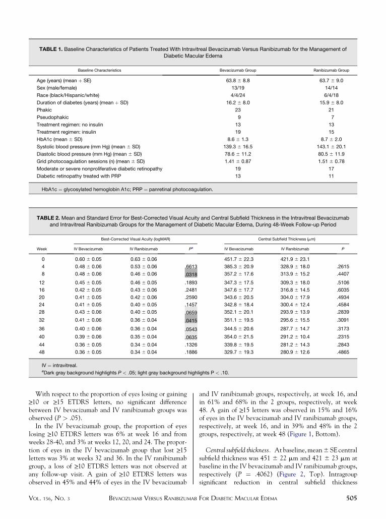
TABLE 1. Baseline Characteristics of Patients Treated With Intravitreal Bevacizumab Versus Ranibizumab for the Management ofDiabetic Macular Edema
Baseline Characteristics Bevacizumab Group Ranibizumab Group
Age (years) (mean þ SE) 63.8 6 8.8 63.7 6 9.0
Sex (male/female) 13/19 14/14
Race (black/Hispanic/white) 4/4/24 6/4/18
Duration of diabetes (years) (mean þ SD) 16.2 6 8.0 15.9 6 8.0
Phakic 23 21
Pseudophakic 9 7
Treatment regimen: no insulin 13 13
Treatment regimen: insulin 19 15
HbA1c (mean 6 SD) 8.6 6 1.3 8.7 6 2.0
Systolic blood pressure (mm Hg) (mean 6 SD) 139.3 6 16.5 143.1 6 20.1
Diastolic blood pressure (mm Hg) (mean 6 SD) 78.6 6 11.2 80.5 6 11.9
Grid photocoagulation sessions (n) (mean 6 SD) 1.41 6 0.87 1.51 6 0.78
Moderate or severe nonproliferative diabetic retinopathy 19 17
Diabetic retinopathy treated with PRP 13 11
HbA1c ¼ glycosylated hemoglobin A1c; PRP ¼ panretinal photocoagulation.
TABLE 2. Mean and Standard Error for Best-Corrected Visual Acuity and Central Subfield Thickness in the Intravitreal Bevacizumaband Intravitreal Ranibizumab Groups for the Management of Diabetic Macular Edema, During 48-Week Follow-up Period
Week
Best-Corrected Visual Acuity (logMAR) Central Subfield Thickness (mm)
IV Bevacizumab IV Ranibizumab Pa IV Bevacizumab IV Ranibizumab P
0 0.60 6 0.05 0.63 6 0.06 451.7 6 22.3 421.9 6 23.1
4 0.48 6 0.06 0.53 6 0.06 .6613 385.3 6 20.9 328.9 6 18.0 .2615
8 0.48 6 0.06 0.46 6 0.06 .0318 357.2 6 17.6 313.9 6 15.2 .4407
12 0.45 6 0.05 0.46 6 0.05 .1893 347.3 6 17.5 309.3 6 18.0 .5106
16 0.42 6 0.05 0.43 6 0.06 .2481 347.6 6 17.7 316.8 6 14.5 .6035
20 0.41 6 0.05 0.42 6 0.06 .2590 343.6 6 20.5 304.0 6 17.9 .4934
24 0.41 6 0.05 0.40 6 0.05 .1457 342.8 6 18.4 300.4 6 12.4 .4584
28 0.43 6 0.06 0.40 6 0.05 .0659 352.1 6 20.1 293.9 6 13.9 .2839
32 0.41 6 0.06 0.36 6 0.04 .0415 351.1 6 19.5 295.6 6 15.5 .3091
36 0.40 6 0.06 0.36 6 0.04 .0543 344.5 6 20.6 287.7 6 14.7 .3173
40 0.39 6 0.06 0.35 6 0.04 .0635 354.0 6 21.5 291.2 6 10.4 .2315
44 0.36 6 0.05 0.34 6 0.04 .1326 339.8 6 19.5 281.2 6 14.3 .2843
48 0.36 6 0.05 0.34 6 0.04 .1886 329.7 6 19.3 280.9 6 12.6 .4865
IV ¼ intravitreal.aDark gray background highlights P < .05; light gray background highlights P < .10.
With respect to the proportion of eyes losing or gaining>_10 or >_15 ETDRS letters, no significant differencebetween IV bevacizumab and IV ranibizumab groups wasobserved (P > .05).
In the IV bevacizumab group, the proportion of eyeslosing >_10 ETDRS letters was 6% at week 16 and fromweeks 28-40, and 3% at weeks 12, 20, and 24. The propor-tion of eyes in the IV bevacizumab group that lost >_15letters was 3% at weeks 32 and 36. In the IV ranibizumabgroup, a loss of >_10 ETDRS letters was not observed atany follow-up visit. A gain of >_10 ETDRS letters wasobserved in 45% and 44% of eyes in the IV bevacizumab
VOL. 156, NO. 3 BEVACIZUMAB VERSUS RANIBIZUMAB
and IV ranibizumab groups, respectively, at week 16, andin 61% and 68% in the 2 groups, respectively, at week48. A gain of >_15 letters was observed in 15% and 16%of eyes in the IV bevacizumab and IV ranibizumab groups,respectively, at week 16, and in 39% and 48% in the 2groups, respectively, at week 48 (Figure 1, Bottom).
Central subfield thickness. Atbaseline,mean6SEcentralsubfield thickness was 451 6 22 mm and 421 6 23 mm atbaseline in the IV bevacizumab and IV ranibizumab groups,respectively (P ¼ .4062) (Figure 2, Top). Intragroupsignificant reduction in central subfield thickness
505FOR DIABETIC MACULAR EDEMA

FIGURE 1. Best-corrected visual acuity in intravitreal (IV)bevacizumab vs ranibizumab for the management of diabeticmacular edema. (Top) Best-corrected visual acuity (BCVA)plotted against follow-up visit for IV bevacizumab vs ranibizu-mab for the management of diabetic macular edema. Pointsrepresent the mean BCVA change in logMAR and error barsthe 95% confidence limits at each study follow-up visitcompared with baseline. The magnitude of BCVA improvementwas significantly higher in the IV ranibizumab group comparedwith the IV bevacizumab group at weeks 8 (P [ .03) and 32(P [ .04), with a trend towards significance at weeks 28(P[ .06), 36 (P[ .05), and 40 (P[ .06). (Bottom) Propor-tion of eyes treated with IV bevacizumab (black circles) andranibizumab (open circles) gaining ‡10 or ‡15 ETDRS letters:No statistically significant difference was observed betweengroups for gain of ‡10 or ‡15 letters at any study visit.
FIGURE 2. Intravitreal (IV) bevacizumab vs ranibizumab forthemanagement of diabetic macular edema. (Top) Circles repre-sent the mean and error bars the 95% confidence limits forcentral subfield thickness at each study visit. The mean centralsubfield thickness in the IV ranibizumab group was lower thanthe central subfield thickness in the IV bevacizumab group atall follow-up visits, although the difference between the 2groups was not statistically significant. (Bottom) Mean changein central subfield thickness (mm) ± 95% confidence limits ateach study follow-up visit compared with baseline. There wasno significant difference between the IV bevacizumab groupand IV ranibizumab group in the magnitude of central subfieldthickness change at any of the study follow-up visits.
compared with baseline was observed at all study follow-upvisits (P < .05). Maximum mean central subfield thicknessreduction occurred at week 44 (�136 6 23 mm) in the IVranibizumab group and at week 48 (�126 6 25 mm) inthe IV bevacizumab group (Table 2, and Figure 2,Bottom). There was no difference in mean central subfieldthickness reduction between the IV bevacizumab and IVranibizumab groups at any of the study follow-up visits.However, there was a significantly higher proportion ofeyes with a central subfield thickness <_275 mm in the IVranibizumab group compared with the IV bevacizumabgroup at weeks 4 (P ¼ .0029; likelihood ratio), 28 (P ¼.0077), 36 (P ¼ .0028), and 44 (P ¼ .0292) (Figure 3).
Number of intravitreal injections. The mean (6 standarderror of the mean; SEM) number of injections in the IV
506 AMERICAN JOURNAL OF
bevacizumab groupwas 9.846 0.55, whichwas significantly(P ¼ .005; Wilcoxon) higher than the mean (6 SEM)number of injections in the IV ranibizumab group (7.67 60.60 injections). In the IV bevacizumab group, 16 eyesreceived 12 injections, while only 4 eyes from the IV rani-bizumab group were treated with 12 injections (Figure 4).
Rescue therapy. Two eyes from 2 different patientsreceived rescue laser therapy: 1 from the IV ranibizumabgroup at week 32 and the other from the IV bevacizumabgroup at week 36. An additional 8 patients (8 eyes) fromthe IV bevacizumab group and 3 patients (3 eyes) fromthe IV ranibizumab group met the criteria for rescuetherapy during the study period and these patients electedto be treated with 3 additional consecutive injections oftheir originally assigned treatment. The number of eyesthat met the criteria for rescue therapy during the studyperiod was significantly higher in the IV bevacizumabgroup (n ¼ 9) compared with the IV ranibizumab group(n ¼ 4) (P ¼ .042; paired t test).
SEPTEMBER 2013OPHTHALMOLOGY

FIGURE 3. Proportion of eyes with diabetic macular edema treated with intravitreal (IV) bevacizumab (black bars) and ranibizumab(white bars) with central subfield thickness £275 mm. The proportion was significantly higher in the IV bevacizumab group at weeks4 (P[ .029), 28 (P[ .007), 36 (P[ .0028), and 44 (P[ .029) (likelihood ratio) when compared with the IV ranibizumab group.
FIGURE 4. Mean diamond plots summarizing the distributionof number of intravitreal (IV) injections of bevacizumab andranibizumab for management of diabetic macular edema after48 weeks. The center horizontal line represents the mean, andthe superior and inferior lines represent the 95% and 5% confi-dence limits, respectively. The mean number of injections(± standard error) was 9.84 ± 0.55 and 7.67 ± 0.60 in the IVbevacizumab group and IV ranibizumab group, respectively(P [ .005; Wilcoxon).
Multivariate analysis. A multivariate analysis comparingBCVA and central subfield thickness outcomes betweenthe IV bevacizumab and IV ranibizumab groups, takinginto account number of injections, baseline BCVA, andcentral subfield thickness, demonstrated a statisticallysignificant influence of baseline BCVA on follow-upBCVA (P < .001) but no other significant differencesbetween groups (P ¼ .051) across follow-up time (P ¼.490) regarding these 2 outcomes.
VOL. 156, NO. 3 BEVACIZUMAB VERSUS RANIBIZUMAB
Adverse events. There was no significant change in meanintraocular pressure compared with baseline at any of thestudy follow-up visits in either group (P < .05). In the IVbevacizumab group, 1 patient experienced clinicallysignificant cataract progression that prevented a clearview of the fundus after his ninth visit and anotherpatient developed transient vitreous hemorrhage after anacute posterior vitreous detachment.There were 2 patients who developed endophthalmitis in
the IV ranibizumab group (both patients were treatedunilaterally) and1patient, also in the IV ranibizumabgroup,who experienced increased blood pressure, controlled withoral antihypertensive agents. Additionally, 1 patient devel-oped transient worsening of renal function. This patient,who had the right eye treated with ranibizumab and theleft eye treated with bevacizumab, had a serum creatininelevel of 2.0 mg/dL at baseline and, during the study, hiscreatinine level increased to 2.9 mg/dL; at the last studyvisit, his creatinine level had returned to 2.0 mg/dL. Nopatient experiencedmyocardial infarction, stroke, or gastro-intestinal bleeding throughout the study period.
DISCUSSION
IN THE PRESENT STUDY, BOTH GROUPS ACHIEVED SIGNIFI-
cant improvement in BCVA compared with baseline atall study visits (P < .05). At week 48, there was a meanBCVA improvement of 0.23 logMAR (w11 letters)and 0.27 logMAR (w13 letters) in the IV bevacizumaband IV ranibizumab groups, respectively. Similarly,DRCR.net12 reported a mean BCVA improvement of 8.2letters in patients with DME treated with IV ranibizumabplus prompt laser and 8.4 letters in patients treated withIV ranibizumab plus deferred laser after 1 year of follow-up. More recently, the RISE and RIDE13 studies alsoshowed significant improvements in BCVA associatedwith IV ranibizumab treatment for DME. In the RISE
507FOR DIABETIC MACULAR EDEMA
study, the IV ranibizumab 0.5 mg group demonstrateda mean improvement of 12 letters in BCVA at 1 year,and in the RIDE study, the IV ranibizumab 0.5 mg groupdemonstrated a mean improvement of 11 letters inBCVA at 1 year. Similarly, the BOLT14 study reporteda significant mean improvement in BCVA after anti-VEGF treatment for DME; eyes treated with IV bevacizu-mab gained a mean of 5.6 letters at 1 year of follow-up.
Although both groups achieved a significant improve-ment in mean BCVA, IV ranibizumab eyes demonstratedsignificantly greater BCVA gains when compared with IVbevacizumab eyes at weeks 8 and 32 and a trend towardsignificance at weeks 28, 36, and 40. This differencebetween the groups at these time points during follow-up may be attributable to lower central subfield thicknessvalues in the IV ranibizumab group compared with the IVbevacizumab group at these periods (Figure 2, Top) and,consequently, a significantly higher proportion of patientswith a central subfield thickness <_275 mm in the IV rani-bizumab group (Figure 3). Correspondingly, the propor-tion of IV bevacizumab eyes that met the criterion forrescue therapy was significantly higher in the IV bevacizu-mab group compared with the IV ranibizumab group.Despite significant differences between groups in BCVAat weeks 8 and 32, it is important to note that becausethe sample size calculation for this study was based onthe difference between treatment groups with respect tocentral subfield thickness, conclusions regarding BCVAare limited: the lack of a significant difference betweentreatment groups with respect to BCVA at some studyvisits does not necessarily indicate that both anti-VEGFtreatments have an equivalent effect on BCVA. In otherwords, a significant difference between groups may havebeen detected at other study visits if the study had beenconducted with a sample size based on differences inBCVA rather than on differences in central subfieldthickness.
Significant improvements in central subfield thicknesscompared with baseline were observed in both the IV beva-cizumab and IV ranibizumab groups. At week 48, bothgroups demonstrated a mean central subfield thicknessreduction compared with baseline of 120 mm. Similarly,the DRCR.net12 reported a mean improvement in centralsubfield thickness of 131 mm and 137 mm in patientswith DME treated with IV ranibizumab plus prompt ordeferred laser, respectively, after 1-year follow-up. Morerecently, the RISE and RIDE13 studies reported a meancentral subfield thickness reduction at 1 year of 250 mmin patients with DME treated with IV ranibizumab. Thegreater absolute value of central subfield thickness reduc-tion observed in the RISE and RIDE studies may be relatedto higher baseline central foveal thickness values and/ormore constant VEGF blockage with monthly treatmentcompared to the DRCR.net study,12 in which the meannumber of injections was 8 per year, and the present study,in which the mean number of injections was 7.67 per year.
508 AMERICAN JOURNAL OF
It is also important to note that the multivariate analysis inthe current study did not demonstrate any influence ofbaseline central subfield thickness on the number of injec-tions in either study group. Treatment with IV bevacizu-mab has also been reported to be associated withfavorable anatomic effects in patients with DME; theBOLT14 study reported a mean central subfield thicknessreduction at 1 year of 130 mm, which is very similar tothe 120 mm reduction observed in the IV bevacizumabgroup of the present study.Despite no significant difference in the magnitude of
absolute central subfield thickness reduction between theIV bevacizumab and IV ranibizumab groups, there wasa higher proportion of eyes with a central subfield thickness<_275 mm in the IV ranibizumab group compared with theIV bevacizumab group at all study follow-up visits; at weeks4, 28, 36, and 44, this difference was statistically significant.Since reinjections were guided by this anatomic parameter(central subfield thickness), IV bevacizumab eyes weretreated with a significantly higher mean number of intravi-treal injections (9.89) compared with IV ranibizumab eyes(7.67), yet achieved similar central subfield thicknessand BCVA outcomes compared with IV ranibizumab eyesat week 48. It is also important to point out a possible cross-over effect of bevacizumab in the contralateral eyes of the15 patients treated bilaterally, which may have positivelyinfluenced central subfield reduction in ranibizumab-treated contralateral eyes. However, there also may havebeen a crossover effect of ranibizumab. This potential cross-over effect represents a limitation for studies that permitbilateral anti-VEGF treatment.The reinjection criterion (a central subfield thickness
>275 mm) was based on data from patients with chronicDME that responded with favorable macular remodelingand were considered to demonstrate ‘‘no fluid’’ on OCTafter intravitreal anti-VEGF treatment (L. Barroso et al,unpublished data, November 2012). It has been reportedthat for patients with chronic DME, a lower central subfieldthickness threshold value should be established in compar-ison to normal population values,22,23 probably because ofsome degree of central retinal atrophy related to previouslaser or mild to moderate ischemia.24 Consistent with thelatter report, in the present study no patients with ‘‘no fluid’’on OCT at week 48 had a central subfield thickness>_275 mm. In addition, in the present study, among the 42eyes that had any degree of concave foveal contour atweek 48 despite some fluid on OCT, only 5 (12%) hada central subfield thickness >275 mm (L. Barroso et al,unpublished data, November 2012).No difference in intraocular pressure between the 2
groups was observed throughout the study, and no signifi-cant change in intraocular pressure was observed at anystudy visit compared with baseline in either group. Theresults of the current study are consistent with data fromother studies that reported no apparent associationbetween intravitreal anti-VEGF injection and increase in
SEPTEMBER 2013OPHTHALMOLOGY

intraocular pressure,25,26 and are in contrast to some studiesthat have suggested such an association.27,28 There were 2cases of endophthalmitis in the IV ranibizumab groupamong a total of 553 injections administered in thestudy. The DRCR.net25 reported 3 cases of endophthalmi-tis out of a total of 3973 injections (0.08%) in ranibizumabarms. The RISE and RIDE studies,13 taken together,reported a total of 4 endophthalmitis cases among a totalof 10 584 injections administered. In the current study,all injections were performed in an ambulatory operatingroom, following recommended aseptic practices.17–20 Therelatively high endophthalmitis rate in our study may berelated to patient-related characteristics, such as poorsocioeconomic status and hygiene habits.17 Finally, admin-istering anti-VEGF to both eyes may increase the risk ofsystemic complications; in fact, 1 of these patients hadtransient increase in creatinine levels during the study.
VOL. 156, NO. 3 BEVACIZUMAB VERSUS RANIBIZUMAB
In sum, in the current study, IV bevacizumab and IVranibizumab were associated with improvement in meanBCVA and mean central subfield thickness in patientswith center-involved DME at 48 weeks of follow-upwhen compared with baseline. Eyes in the IV bevacizumabgroup received a significantly higher number of injectionsthan eyes in the IV ranibizumab group. During the study,eyes in the IV ranibizumab group experienced a fasterrecovery of BCVA compared with eyes in the IV bevacizu-mab group, which may be explained by the higher propor-tion of eyes in the IV ranibizumab group with a centralsubfield thickness <275 mm at intermediate-term studyfollow-up visits. To our knowledge and based on a Medlinesearch, this is the first report comparing IV bevacizumaband IV ranibizumab for the treatment of DME. The currentstudy is limited by a small sample size; larger prospectivestudies are warranted to confirm our preliminary findings.
ALL AUTHORSHAVE COMPLETED AND SUBMITTED THE ICMJE FORM FOR DISCLOSUREOF POTENTIAL CONFLICTS OF INTEREST.Rodrigo Jorge received travel support from Novartis to attend the 2012 American Society of Retina Specialists (ASRS) meeting. This study was supportedby Fundacao de Amparo a Pesquisa do Estado de Sao Paulo (FAPESP), grant number 2010/013368; and Fundacao Apoio ao Ensino, Pesquisa e Assistencia(FAEPA) do Hospital das Clınicas da Faculdade de Medicina de Ribeirao Preto da Universidade de Sao Paulo. Contributions of authors: conception anddesign of the study (I.U.S., A.M., R.C.S., R.J.); analysis and interpretation (A.B.N., E.T., F.P.P.A., R.P., R.C.S., J.A.C., A.M., I.U.S., R.J.); writing thearticle (A.B.N., E.T., F.P.P.A., R.P., J.A.C., A.M., I.U.S., R.J.); critical revision (A.B.N., J.A.C., R.C.S., I.U.S., A.M., R.J.); final approval of the article(A.B.N., E.T., F.P.P.A., R.P., R.C.S., J.A.C., A.M., I.U.S., R.J.); data collection (A.B.N., E.T., F.P.P.A., R.P., R.C.S.); provision of materials (A.B.N.,E.T., F.P.P.A., R.P., R.C.S., J.A.C., R.J.); statistical analysis (A.M., R.J.); obtaining funding (A.B.N., E.T., A.M., R.J.); literature search (A.B.N., E.T.,R.C.S., I.U.S., R.J.); and administrative, technical, or logistic support (A.B.N., J.A.C., A.M., R.J.). This trial was registered at clinicaltrials.gov undernumber: NCT01487629.
The authors would like to thank Prof Dr Klaus Dietz, professor emeritus of Medical Biometry (University of Tubingen–Germany), for review of andconstructive comments on the statistical analysis.
REFERENCES
1. Klein R, Klein BE, Moss SE. Visual impairment in diabetes.Ophthalmology 1984;91(1):1–9.
2. Klein R, Klein BE, Moss SE, Davis MD, DeMets DL. TheWisconsin epidemiologic study of diabetic retinopathy.IV. Diabetic macular edema. Ophthalmology 1984;91(12):1464–1474.
3. Early Treatment Diabetic Retinopathy Study ResearchGroup. Photocoagulation for diabetic macular edema. EarlyTreatment Diabetic Retinopathy Study report number 1.Arch Ophthalmol 1985;103(12):1796–1806.
4. Elman MJ, Raden RZ, Sloan MD, et al. Randomized trialcomparing intravitreal triamcinolone acetonide and focal/grid photocoagulation for diabetic macular edema. Ophthal-mology 2008;115(9):1447–1459.
5. Googe J, Brucker AJ, Bressler NM, et al. Randomized trialevaluating short-term effects of intravitreal ranibizumab ortriamcinolone acetonide on macular edema after focal/gridlaser for diabetic macular edema in eyes also receiving panre-tinal photocoagulation. Retina 2011;31(6):1009–1027.
6. Scott IU, Edwards AR, Beck RW, et al. A phase II random-ized clinical trial of intravitreal bevacizumab for diabeticmacular edema. Ophthalmology 2007;114(10):1860–1867.
7. Scott IU, Danis RP, Bressler SB, Bressler NM, Browning DJ,Qin H. Diabetic Retinopathy Clinical Research Network.Effect of focal/grid photocoagulation on visual acuity and
retinal thickening in eyes with non-center-involved diabeticmacular edema. Retina 2009;29(5):613–617.
8. Kinyoun J, Barton F, Fisher M, Hubbard L, Aiello L,Ferris F III. Detection of diabetic macular edema. Ophthal-moscopy versus photography–Early Treatment Diabetic Reti-nopathy Study Report Number 5. The ETDRS ResearchGroup. Ophthalmology 1989;96(6):746–750.
9. Browning DJ, Altaweel MM, Bressler NM, et al. Diabeticmacular edema: what is focal and what is diffuse? Am JOphthalmol 2008;146:649–655.
10. Huang J, Liu X, Wu Z, Xiao H, Dustin L, Sadda S. Macularthickness measurements in normal eyes with time-domainand Fourier-domain optical coherence tomography. Retina2009;29(7):980–987.
11. Audren F, Lecleire-Collet A, Erginay A, et al. Intravitrealtriamcinolone acetonide for diffuse diabetic macular edema:phase 2 trial comparing 4 mg vs 2 mg. Am J Ophthalmol2006;142(5):794–799.
12. Elman MJ, Aiello LP, Beck RW, et al. Randomized trial eval-uating ranibizumab plus prompt or deferred laser or triamcin-olone plus prompt laser for diabetic macular edema.Ophthalmology 2010;117(6):1064–1077.
13. Nguyen QD, Brown DM, Marcus DM, et al. Ranibizumab fordiabetic macular edema: results from 2 phase III randomizedtrials: RISE and RIDE. Ophthalmology 2012;119(4):789–801.
14. Michaelides M, Kaines A, Hamilton RD, et al. A prospectiverandomized trial of intravitreal bevacizumab or laser therapy
509FOR DIABETIC MACULAR EDEMA
in the management of diabetic macular edema (BOLT study)12-month data: report 2. Ophthalmology 2010;117(6):1078–1086.
15. Paccola L, Costa RA, Folgosa MS, Barbosa JC, Scott IU,Jorge R. Intravitreal triamcinolone versus bevacizumab fortreatment of refractory diabetic macular oedema (IBEMEstudy). Br J Ophthalmol 2008;92(1):76–80.
16. Haritoglou C, Kook D, Neubauer A, et al. Intravitreal beva-cizumab (Avastin) therapy for persistent diffuse diabeticmacular edema. Retina 2006;26(9):999–1005.
17. Das T, Hussain A, Naduvilath T, Sharma S, Jalali S,Majji AB. Case control analyses of acute endophthalmitisafter cataract surgery in South India associated with tech-nique, patient care, and socioeconomic status. J Ophthalmol2012;2012:298459.
18. Gomez-Ulla F, Basauri E, Arias L, Martınez-Sanz F. ComplejoHospitalario Universitario de Santiago de Compostela, Insti-tuto Tecnologico de Oftalmologıa, Espana. Management ofintravitreal injections. Arch Soc Esp Oftalmol 2009;84(8):377–388.
19. Mason J, White M, Feist R, et al. Incidence of acute onsetendophthalmitis following intravitreal bevacizumab (Avas-tin) injection. Retina 2008;28(4):564–567.
20. Aiello LP, Brucker AJ, Chang S, et al. Evolving guidelines forintravitreal injections. Retina 2004;24(5 Suppl):S3–19.
21. Ribeiro JA, Messias A, Scott IU, et al. Alternative techniquefor reducing compound waste during intravitreal injections.Arq Bras Oftalmol 2009;72(5):641–644.
510 AMERICAN JOURNAL OF
22. Grover S, Murthy RK, Brar VS, Chalam KV. Normative datafor macular thickness by high-definition spectral-domainoptical coherence tomography (Spectralis). Am J Ophthalmol
2009;148(2):266–271.23. Menke MN, Dabov S, Knecht P, Sturm V. Reproducibility of
retinal thickness measurements in healthy subjects usingspectralis optical coherence tomography. Am J Ophthalmol2009;147(3):467–472.
24. Lovestam-Adrian M, Agardh E. Photocoagulation of diabeticmacular oedema–complications and visual outcome. Acta
Ophthalmol Scand 2000;78(6):667–671.25. Elman MJ, Bressler NM, Qin H, et al. Diabetic Retinopathy
Clinical Research Network. Expanded 2-year follow-up ofranibizumab plus prompt or deferred laser or triamcinoloneplus prompt laser for diabetic macular edema. Ophthalmology2011;118(4):609–614.
26. Filho JA, Messias A, Jorge R, et al. Panretinal photocoagula-tion (PRP) versus PRP plus intravitreal ranibizumab for high-risk proliferative diabetic retinopathy. Acta Ophthalmol 2011;89(7):e567–e572.
27. Tseng JJ, Vance SK, Della Torre KE, et al. Sustainedincreased intraocular pressure related to intravitreal antivas-cular endothelial growth factor therapy for neovascular age-related macular degeneration. J Glaucoma 2012;21(4):241–247.
28. Kahook MY, Liu L, Ruzycki P, et al. High-molecular-weightaggregates in repackaged bevacizumab. Retina 2010;30(6):887–892.
SEPTEMBER 2013OPHTHALMOLOGY

Biosketch
Antonio Brunno Nepomuceno received his medical degree from the Federal University of Ceara, Fortaleza, Brazil. In 2011,
he completed his ophthalmology residency and started working as a retinal fellowship trainning at the School of Medicine
of Ribeirao Preto, University of Sao Paulo, Ribeirao Preto, Brazil. His primary research interests pertain to diabetic
retinopathy, macular edema and anti-VEGF agents.
VOL. 156, NO. 3 510.e1BEVACIZUMAB VERSUS RANIBIZUMAB FOR DIABETIC MACULAR EDEMA

Biosketch
Rodrigo Jorge is an Associate Professor and coordinates the Postgraduate program in the Ophthalmology Department and
in the Retina and Vitreous Division in Clinics Hospital, School of Medicine of Ribeirao Preto, University of Sao Paulo. He
has taught and published in the field of vitreoretinal diseases, with over 80 publications. Since 2006, his research has focused
on anti-VEGF therapy studies and his collaborative study group has published the most cited prospective studies of age-
related macular degeneration and diabetic retinopathy conceived and conducted in Brazil.
510.e2 SEPTEMBER 2013AMERICAN JOURNAL OF OPHTHALMOLOGY