a physiological role for amyloid beta protein: enhancement ... · 1 a physiological role for...
TRANSCRIPT
1
A Physiological Role for Amyloid Beta Protein:
Enhancement of Learning and Memory
John E. Morley1,2
Susan A. Farr1,2
William A. Banks1,2
Steven N. Johnson1,2
Kelvin A. Yamada3
Lin Xu3
1Division of Geriatric Medicine, Saint Louis University School of Medicine
2GRECC, VA Medical Center, St. Louis, Missouri
3Department of Neurology, Washington University School of Medicine,
St. Louis, Missouri
Contact:
John E. Morley, M.B., B.Ch.
Geriatric Medicine
Saint Louis University School of Medicine
1402 S. Grand Blvd., M238
St. Louis, MO 63104
Email: [email protected]

2
Abstract
Amyloid beta protein (Aβ) is well recognized as having a significant role in the
pathogenesis of Alzheimer’s disease (AD). The reason for the presence of Aβ and its
physiological role in non-disease states is not clear. In these studies, low doses of Aβ
enhanced memory retention in two memory tasks and enhanced acetylcholine production
in the hippocampus in vivo. We then tested whether endogenous Aβ has a role in learning
and memory in young, cognitively intact mice by blocking endogenous Aβ in healthy 2-
month-old CD-1 mice. Blocking Aβ with antibody to Aβ or DFFVG (which blocks Aβ
binding) or decreasing Aβ expression with an antisense directed at the Aβ precursor APP
all resulted in impaired learning in T-maze foot-shock avoidance. Finally, Aβ1-42
facilitated induction and maintenance of long term potentiation in hippocampal slices,
whereas antibodies to Aβ inhibited hippocampal LTP. These results indicate that in
normal healthy young animals the presence of Aβ is important for learning and memory.
Introduction
Alzheimer’s disease is widely believed to be mediated by an excess of amyloid-
beta peptide (Aβ). Aβ has been shown to impair learning and memory in vivo
(1,2,3,4,5,6,7). Transgenic mice that overproduce amyloid precursor protein have
decreased memory (8,9). Despite a large literature on the pathology of Aß, its
physiological role remains unclear. Recent studies have suggested that Aβ, which is
secreted by neurons during excitatory neuronal activity (10), down regulates excitatory
synaptic transmission (11). The negative feedback loop thus formed would provide a
homeostatic mechanism by which Aβ could maintain appropriate levels of neuronal
activity. This suggests that whereas excess Aβ suppresses memory, appropriate levels of

3
Aß support mnemonic processes. This would be consistent with the general principle that
excess of mnemonics results in cognitive impairments (12).
Long term potentiation (LTP) is believed to be the synaptic correlate of memory
formation. Most studies have shown that amyloid-beta peptide inhibits LTP (13,14),
though a few have shown that amyloid-beta peptide facilitates LTP (15,16,17). Further
support for the concept that amyloid-beta may stimulate LTP comes from amyloid
precursor protein (APP) null mice, which have reduced synapses, impaired LTP, and
perform poorly on spatial memory tasks (18,19). Furthermore, presenilin-1-deficient
mice have a reduced level of Aß and impaired LTP (20). Whereas antibodies against Aβ
improve memory in the SAMP8 mouse, a strain which overexpressses Aß (21), APP
antibodies impair performance of passive avoidance tasks in rats (22) and chicks (23) that
are not overexpressers. In the studies reported here, we demonstrated in young mice that
inhibition of Aß with antibodies, inhibition of amyloid-beta expression with antisense, or
blocking Aß with a putative inhibitor all impaired learning. In addition, we found that
low doses of Aß enhanced memory in young mice and increased hippocampal
acetylcholine secretion. Finally, we demonstrated that Aβ-1-42 facilitated induction and
maintenance of long term potentiation and that antibodies to Aß inhibits it.
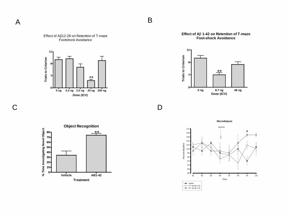
Aβ 12-28 and 1-42 Improved Retention of T-maze Foot-shock Avoidance
The 12-28 region of the Aβ peptide has been shown to be the region important for
learning and memory (3). Administering high doses of Aß 12-28 produces impaired
learning and memory (3). Here, we determined whether low doses of Aß12-28 can
improve retention when administered by intracerebroventricular (ICV) injection

4
immediately after a training session in the active avoidance T-maze. The one-way
ANOVA with trials to criterion as the independent variable showed a significant
treatment effect (F(4,42)=14.90, P<0.0001 (Figure 1a). Dunnett’s post hoc analysis
indicated that the mice that received 20 ng of Aß12-28 took significantly fewer trials to
reach criterion than the mice that received saline, thus demonstrating enhanced memory.
Aβ 1-42 is considered by many to be the peptide that produces learning and
memory impairments in AD. Here, we injected Aβ 1-42 ICV immediately after training
in T-maze foot-shock avoidance. The one-way ANOVA with trials to criterion as the
independent variable showed a significant treatment effect (F(2,27)=12.82, P<0.0001
(Figure 1b). Dunnett’s post hoc test indicated that the mice which received 8.7 ng of Aβ
1-42 took significantly fewer trials to reach criterion on the retention than the mice which
received saline.
Aβ 1-42 Improves Retention in Object Recognition
Object recognition is a non-shock episodic memory task that relies on the
inclination of mice to spend more time with novel than familiar objects (24). Here, mice
were administered Aβ 1-42 or saline immediately after training. Twenty-four hours later,
mice were tested for recognition of the original object by determining the amount of time
spent with the new versus the original object. A T-test showed that the mice which
receive Aβ 1-42 spent a significantly greater amount of time with the novel object,
T(14)=4.303, P<0.0007 (Figure 1c), indicating that they had improved recognition of the
original object.

5
Low Dose of Aβ 1-42 Increases Acetylcholine in the Hippocampus
A one-way ANOVA assessing percent baseline for the 61-90 minutes post
administration period showed a significant treatment effect (F(2,16) = 4.129, P<0.05.
Dunnett’s post hoc analysis indicated that the mice which received 43 ng had
significantly higher levels of acetylcholine than the mice which received saline (Figure
1d).
Decreasing Beta-Amyloid Prior to Training Impairs Learning of T-Maze Foot Shock
Avoidance
Mice were prepared for ICV administration of an antibody to Aß1-42 (8 ng) or
rabbit anti-mouse IgG (8 ng)as described below. Seventy-two hours later, mice were
trained in T-maze foot-shock avoidance. A T-test indicated that the mice which received
antibody to Aß prior to training took significantly longer to make criterion than the mice
which received rabbit anti-mouse IgG, T(16) = 8.102, P<0.0001 (Figure 2a), indicating
impaired learning.
In order to further verify that blocking Aβ will impair learning in CD-1 mice, we
administered the Aß blocking peptide DFFVG (2 ug) or vehicle ICV 72 hrs prior to
training. A T-test indicated that mice which received DFFVG took significantly longer to
reach criterion than the mice which received vehicle, T(12) 9.238, P<0.0001 (Figure 2B),
indicating impaired learning.
Decreasing Aβ with Antisense (AO) Impairs Learning of T-maze Foot Shock
Avoidance
We have previously shown that in SAMP8 mice, a strain with elevated Aβ, that
the administration of an antisense directed against the Aß precursor APP (AO) decreases
6
brain levels of APP and Aß and results in improved learning and memory (25). Here, we
administered AO or a random antisense (RA) ICV to young CD-1 mice 3x (2 weeks
between each administration) and trained the mice in the T-maze 2 weeks after the last
injection. A T-test indicated that mice which received AO took significantly longer to
reach first avoidance than the mice which receive RA, T(10) = 4.037, P<0.0024 (Figure
2c), demonstrating that AO produced an impairment in learning.
Aβ1-42 facilitates LTP induction and maintenance
In order to determine whether the presence of Aβ could facilitate LTP under some
circumstances, we used induction stimuli that were below threshold for inducing LTP
(Two TBS instead of three), and found that Aβ1-42 converted a subthreshold induction
stimulus to one that facilitated LTP induction and maintenance. Successful LTP induction
and maintenance (defined as at least 20% potentiation of the fEPSP 60 minutes after
induction) was observed in only 1 of 9 control slices 60 minutes after two x TBS
(diamonds, filled circle is the mean). In contrast, successful LTP induction and
maintenance was observed in 7 of 9 slices incubated with 10ng/ml Aβ1-42 (Figure 3a
triangles, filled circle is the mean, the dotted line indicates 20% fEPSP potentiation,
p=0.015 Fisher’s exact test). This result suggests that Aβ1-42 can facilitate LTP.
The effect of Aβ antibody on hippocampal LTP in CD-1 mice
The Aβ antibodies 4G8 and 6E10 (Sigma-Aldrich) have not been reported to
affect LTP when applied alone, but rather they counteract the Aβ-mediated LTP
inhibition in vitro and in vivo when applied with Aβ or in transgenic animals expressing
human forms of Aβ associated with Alzheimer’s disease and impaired LTP (26). A
possible explanation is that these antibodies have much higher affinities for the human or

7
oligometric forms of Aβ, and thereby have greater effects on Aβ-mediated LTP
inhibition. If this is true, then higher concentrations of these antibodies may inhibit LTP
by themselves in wild type hippocampal slices. There are different commercially
available antibodies that recognize different segments of Aβ. If we can reduce Aβ by
application of these antibodies, then we would predict that we would inhibit induction
and maintenance of LTP. Different Aβ antibodies producing the same result woudl be
strong evidence that this approach supports a role for Aβ in normal cognition.
LTP was induced by 3 applications of theta burst stimulation (figure 3b, TBS,
arrow). Non-specific IgG antibody at a similar concentration was used as control. Slices
incubated in ACSF (squares, n=9 slices) or IgG (circles, n=5 slices) exhibited sustained
LTP, but slices incubated with 1:100 dilution of the DAKO anti-Aβ antibody (triangles,
n=8 slices) had prolonged synaptic depression after TBS, and did not exhibit post-TBS
potentiation or LTP 55-60 minutes after induction (p=0.001, ANOVA Figure 3b).
The Aß blocking peptide DFFVG inhibits hippocampal LTP
The blocking peptide DFFVG that impaired T-maze foot shock learning (Figure
2b) was applied to hippocampal slices to determine if it would inhibit TBS-induced LTP.
DFFVG (1 μM) did not affect the baseline slope of the fEPSP. However, in the presence
of DFFVG TBS failed to induce post TBS potentiation or LTP (Figure 3c).
Discussion
We have provided here multiple lines of evidence that Aβ’s physiological role is
to enhance learning and memory retention. This suggests that it is an excess of Aβ that
inhibits learning and memory (27). This is not surprising in view of the fact that

8
numerous memory enhancers have been shown to follow the law of hormesis, with low
doses enhancing and high doses inhibiting memory retention (12).
The strengths of the study are that we have shown that Aβ enhances memory in
two totally different forms of memory testing, via the aversive T maze and the non-
aversive object recognition test. Recently, there has been increasing interest in the use of
object recognition as a memory test. Studies show that this task involves the hippocampal
formation (entorhinal cortex, dentate gyrus, CA1-4 and subiculum), amygdala and
parahippocampal cortices all of which comprise the declarative memory system (24).
The aversive T-maze has been shown to be a hippocampal dependent task (28). We also
demonstrated that not only increasing Aβ at low levels enhanced memory, but that
antibodies to Aβ and an antisense to amyloid precursor protein inhibited learning in
young mice. DFFVG has previously been shown to block the memory inhibiting effects
of Aβ in large doses by binding to its receptor site (29). Here, we found that it inhibited
learning in young mice. Previously, we have found that an antibody directed against Aβ
increases acetylcholine production in the hippocampus of the SAMP8, an animal model
of Alzheimer’s disease with learning and memory deficits caused by an overexpression of
APP (30). This is consistent with high doses of Aß suppressing acetylcholine production.
Here, we showed that low doses of Aβ enhanced acetylcholine production in the
hippocampus in vivo. This is consistent with our hypothesis that physiologic or near
physiologic levels of Aß support acetylcholine production. Finally, we have shown that
induction of Schaffer collateral pathway LTP can be facilitated by a low concentration of
Aβ and blocked by Aβ antibodies. Schaffer collateral LTP is thought to underlie
hippocampal-dependent spatial learning and memory (31).

9
Overall, we believe these studies strongly suggest that the physiological role of
Aβ is to enhance learning and memory. Only when there is excess production does Aβ
result in memory deficits. These findings are important in understanding the optimal
design of drugs to treat Alzheimer’s disease.
Methods Summary
All studies were conducted in CD-1 mice. Memory testing in CD-1 male mice, 8
weeks old, was done using the aversive T-maze and object recognition tasks. Aβ was
administered by intracerebroventricular (ICV) injection, as were the antagonists – Aβ
antibodies, DFFVG and antisense to the Aβ portion of APP. Acetylcholine levels were
measured by microdialysis in response to Aβ1-42. The effects of Aβ1-42, Aβ antibodies
and DFFVG were studied in long term potentiation.
Financial Support:
Financial Support for this research was provided by: NINDA Type R01 Grant “Polypeptide Modification for Enhanced Brain Delivery” 2006-2009 and NIH/NIA, R01, “Oxidative Dysfunction of LRP at the Blood-Brain Barrier in Alzheimer’s Disease” 2008-2013.

10
Figure Legends
Figure 1a. Low doses of Aß12-28 administered to CD-1 mice ICV immediately after
training improves retention in T-maze foot-shock avoidance. The ** indicates P<0.01.
Figure 1b. Low doses of Aβ 1-42 administered to CD-1 mice ICV immediately after
training improves retention in T-maze foot-shock avoidance. The ** indicates P<0.01.
Figure 1c. Low dose of the Aβ 1-42 administered to CD-1 immediately after training
improves retention of object recognition 24 hours later. The ** indicates P<0.01.
Figure 1d. Administration of Aβ 1-42 increased acetylcholine levels in the
hippocampus of CD-1 mice during the 61-90 minute time period of collection. The *
indicates P<0.05.
Figure 2a. Antibody to Aβ (DAKO) administered 72 hours prior to training to CD-1
mice impaired acquisition of T-maze foot-shock avoidance. The ** indicates P<0.01.
Figure 2b. DFFVG administered 72 hours prior to training impaired T-maze
acquisition in CD-1 mice. The ** indicates P<0.01.
Figure 2c. Antisense directed at the Aß region of the APP peptide impairs acquisition
of T-maze acquisition in CD-1 mice. The ** indicates P<0.01.

11
Figure 3a. Aβ 1-42 is important for hippocampal LTP. A, Two theta-burst stimuli
(TBS) applied to the Schaffer collateral pathway does not induce long-term synaptic
potentiation (LTP) at CA1 synapses. Each symbol represents one hippocampal slice, and
the mean is depicted by a filled circle. The dotted line indicates 20% increase in the slope
of the fEPSP, which is the threshold used to define successful induction/maintenance of
LTP. Only 1/9 slices under control conditions (open diamonds); in contrast 7/9 slices in
the presence of Aβ (10 ng/ml) exhibit LTP (p=0.015 Fisher’s exact test).
Figure 3b. B, Three TBS applied to the Schaffer collateral pathway after obtaining
baseline responses (arrow) induces LTP in control buffer (filled squares) and in the
presence of a nonspecific IgG (open circles), whereas in the presence of an Aβ antibody
(DAKO, open triangles) there is post TBS synaptic depression, and no LTP.
Figure 3c. C, As in B, three TBS (arrow) induces LTP in control buffer (filled
squares), but application of the putative Aß inhibitor peptide DFFVG, blocks LTP
without affecting the slope of the baseline fEPSP (open triangles). DFFVG blocked
TBS-LTP in hippocampal slices of CD-1 mice compared to vehicle. The TBS-LTP
consisted of a train of 5 pulses of 100Hz applied at 200ms intervals 10 times.

12
References
1. Flood JF, Roberts E, Sherman MA, Kaplan BE, Morley JE. Topography of a
binding site for small amnestic peptides deduced from structure-activity studies: relation
to amnestic effect of amyloid beta protein. Proc Natl Acad Sci USA. 1994;91:380-384.
2. Flood JF, Morley JE, Roberts E. Amnestic effects of mice of four synthetic
peptides homologous to amyloid beta protein from patients with Alzheimer disease. Proc
Natl Acad Sci USA. 1991;88:3363-3366.
3. Flood JF, Morley JE, Roberts E. An amyloid beta-protein fragment, A beta [12-
28], equipotently impairs post-training memory processing when injected into different
limbic system structures. Brain Res. 1994;663:271-176.
4. Terranova JP, Kan JP, Storme JJ, Perreaut P, Le Fur G, Soubrie P. Administration
of amyloid beta-peptides in the rat medical septum causes memory deficits: reversal by
SR 57746A, a non-peptide neurotrophic compound. Neurosci Letters. 1996;213:79-82.
5. Cleary J, Hittner JM, Semotuk M, Mantyh P, O’Hare E. Beta-amyloid [1-40]
effects on behavior and memory. Brain Res. 1995;682:69-74.
6. Mazzola C, Micale V, Drago F. Amnesia induced by beta-amyloid fragments is
counteracted by cannabinoid CB1 receptor blockade. Aur J Pharm. 2003;477:219-225.
7. McDonald MP, Dahl EE, Overmier JB, Mantyh P, Clearly J. Effects of an
exogenous beta-amyloid peptide on retention for spatial learning. Beh Neur Biol.
1994;62:60-67.
8. Hardy J, Selkoe DJ. The amyloid hypothesis of Alzheimer’s disease: progress and
problems on the road to therapeutics. Science. 2002;297:353-356.
13
9. Robinson SR, Bishop GM. Aβ as a bioflocculant: implications for the amyloid
hypothesis of Alzheiemr’s disease. Neurobiol Aging. 2002;23:1051-1072.
10. Cirritto JR, Yamada KA, Finn MB, Sloviter RS, Bales KR, May PC, Schoepp
DD, Paul SM, Mennerick S, Holtzman DM. Synaptic activity regulates interstitial fluid
amyloid-beta levels in vivo.
11. Kamenetz F, Tomita T, Hsieh H, Seabrook G, Borchelt D, Iwatsubo T, Sisodia S,
Malinow R. APP processing and synaptic function. Neuron 2003; 37:925-937.
12. Flood JF, Farr SA, Uezu K, Morley JE. Age-related changes in septal
serotonergic, GABAergic and glutamatergic facilitation of retention in SAMP8 mice.
Mech Ageing Dev. 1998;105:173-188.
13. Itoh A, Akaike T, Sokabe M, et al. Impairments of long-term potentiation in
hippocampal slices of β-amyloid-infused rats. Eur J Pharmacol. 1999;382:167-175.
14. Raymond CR, Ireland DR, Abraham WC. NMDA receptor regulation by
amyloid-B does not account for its inhibition of LTP in rat hippocampus. Brain Res.
2003;968:263-272.
15. Wu H, Anwyl R, Rowan MJ. B amyloid [1-40] increases long-term potentiation
in rat hippocampus in vitro. Eur J Pharmacol. 1995;284:R1-3.
16. Kim JH, Anwyl R, Suh YH, et al. Use-dependent effects of amyloidogenic
fragments of β-amyloid precursor protein on synaptic plasticity in rat hippocampus in
vivo. J Neurosci. 2001;21:1327-1333.
17. Trubetskaya VV, Stepanichev MY, Onufriev MV et al. Administration of
aggregated β-amyloid peptode [25-35] induces changes in long-term potentiation in the
hippocampus in vivo. Neurosci Behav Physiol. 2003;33:95-98.

14
18. Dawson GR, Seabrook GR, Zheng H, et al. Age-related cognitive deficits,
impaired long-term potentiation and reduction in synaptic marker density in mice lacking
the β-amyloid precursor protein. Neurosci. 1999;90:1-13.
19. Seabrook GR, Smith DW, Bowery BJ, et al. Mechanisms contributing to the
deficits in hippocampal synaptic plasticity in mice lacking amyloid precursor protein.
Neuropharmacology. 1999;38:349-359.
20. Morton RA< Kuenzi FM, Fitzjohn SM, et al. Impairment in hippocampal long-
term potentiation in mice under-expressing the Alzheimer’s disease related gene
presenilin-1. Neurosci Lett. 2002;319:37-40.
21. Morley JE, Farr SA, Flood JF. Antibody to amyloid beta protein alleviates
impaired acquisition, retention, and memory processing in SAMP8 mice. Neurobiol
Learning & Memory. 2002;78:125-138.
22. Huber G, Martin JR, Loffler J, et al. Involvement of amyloid precursor protein in
memory formation in the rat: an indirect antibody approach. Brain Res. 1993;603:348-
352.
23. Mileusnic R, Lancashire CL, Johnston AN, et al. APP is required during an early
phase of memory formation. Eur J Neurosci. 2000;12:4487-4495.
24. Dere E, Huston JP, De Souza Silva MA. The pharmacology, neuroanatomy and
neurogenetics of one-trial object recognition in rodents. Neurosci. Biobehav. Rev.
2007:673-704.
25. Kumar VB, Farr SA, Flood JF, Kamlesh V, Franko M, Banks WA, Morley JE.
Site-directed anitisense oligonucleotide decreases the expression of amyloid precursor

15
protrein and reverses deficits in learning and memory in aged SAMP8 mice. Peptides
200;21:1769-1775.
26. Klybin I, Walsh DM, Lemere CA, Cullen WK, Shankar GM, Spooner ET, Jiang
L, Anwyl R, Selkoe DJ, Rowan MJ. Amyloid beta protein immunotherapy neutralizes
Abeta oligomers that disrupt synaptic plasticity in vivo. Nat. Med. 2005;11:556-561.
27. Christensen R, Marcussen AB, Wörtwein G, Knudsen GM, Aznar S. Abeta(1-42_
injection causes memory impairment, lowered cortical and serum BDNF levels, and
decreased hippocampal 5-HT(2A) levels. Exp. Neurol. 2008;210:164-171.
28. Farr SA, Banks WA, La Scola ME, Morley JE. Permanent and temporary
inactivation of the hippocampus impairs T-maze footshock avoidance acquisition and
retention. Brain Res. 2000; 872:242-249.
29. Flood JF, Roberts E, Sherman MA, Kaplan BE, Morley JE. Topography of a
binding site for small amnestic peptides deduced from structure-activity studies: relation
to amnestic effect of amyloid beta protein. Proc. Natl. Acad. Sci. 1994; 91:380-384.
30. Farr SA, Banks WA, Uezu K, Sano A, Gaskin FS, Morley JE. Antibody to beta-
amyloid protein increases acetylcholine in the hippocampus of 12 month SAMP8 male
mice. Life Sci. 2003; 73:555-562.
31. Malenka RC, Bear MF. LTP and LTD: an embarrassment of riches. Neuron 2004;
44:5-21.

16
Methods
Mice
CD-1 male mice from an in house colony, 8 weeks of age, served as test subjects.
This colony has been maintained as an outbred strain obtained from Charles Rivers
Breeding Laboratories of Wilmington, MA. The mice are tested regularly to ensure that
they are virus and pathogen free. All subjects were experimentally naïve. Mice were on
a 12 h light:12 h dark cycle with lights on at 0600. Water and food (PMI Nutrition
LabDiet 5001) were available ad libitum. All experiments were conducted after
institutional approval of the animal use subcommittee, which subscribes to the NIH
Guide for Care and Use of Laboratory Animals.
Drugs
Amyloid beta protein 1-42 (Aβ) was purchased from American Peptide Co.
(Sunnyvale, CA). Aβ 12-28 was purchased from Phoenix Pharmaceuticals, Inc.
(Belmont CA). Antisense oligonucleotide (AO) and random antisense were purchased
from Midland Certified Reagent Co. (Midland, TX). Antibody to Aβ was purchased from
DAKO Corporation (Carpinteria, CA). DFFVG was obtained from Sigma-Genosys (The
Woodlands, TX). All drugs were dissolved in saline.
Surgery and Drug Administration
Forty-eight hours prior to training, mice were anesthetized with tribromoethylene,
placed in a stereotaxic instrument, the scalp was deflected and a hole drilled through the
skull over the injection site. The injection coordinates for the ICV injections is 0.5 mm
posterior to the bregma and 1.0 mm to the right and left of the sagittal suture. The

17
injection depth was 2.0 mm. In the acquisition studies mice were injected immediately
after the hole was drilled and trained 72 hours later. In the retention studies, mice were
trained 48 hr after surgery. Immediately after training, mice were again placed in the
stereotaxic apparatus under light isoflurane anesthesia. Within three minutes after
training, a 0.5 ml solution of saline or drug solution was injected into each injection site
over 60 s through a 30 gauge needle, which was attached to a 10 μl syringe. After ICV
injection, the scalp was closed and the mice were returned to their cages.
T-Maze training and testing procedures
The T-maze is a working memory learning task and reference memory task. The
T-maze consisted of a black plastic alley with a start box at one end and two goal boxes
at the other. The start box was separated from the alley by a plastic guillotine door, which
prevented movement down the alley until training began. An electrifiable stainless steel
rod floor ran throughout the maze to deliver scrambled foot-shock.
Mice were not permitted to explore the maze prior to training. A block of training
trials began when a mouse was placed into the start box. The guillotine door was raised
and a buzzer sounded simultaneously; 5 sec later foot-shock was applied. The goal box
entered on the first trial was designated “incorrect” and the foot-shock was continued
until the mouse entered the other goal box, which in all subsequent trials was designated
as “correct” for the particular mouse. At the end of each trial, the mouse was returned to
its home care until the next trial.
Mice in the pretraining injection groups were trained until they made 1 avoidance.
The parameters for post-training were set so that the control groups would have poor
retention (mean trials to criterion between 9 and 10) so that drug-induced improvement of

18
retention could be detected. Training used an intertrial interval of 35 sec, the buzzer was
of the door-bell type, sounded at 55 dB and shock was set at 0.35 mA (Coulbourn
Instruments scrambled grid floor shocker model E13-08). Retention was tested one week
later by continuing training until mice reach a criterion (5 avoidences in 6 consecutive
trials). The results were reported as the number of trials to criterion for the retention test.
Object Recognition
Object recognition is an episodic memory task that involves the hippocampus. In
this task, an animal was exposed to two similar objects which it was allowed to explore
for 5 minutes. Twenty-four hours later, the mouse was exposed to one of the same objects
and a new novel object. The mouse was injected ICV after the training day where it is
exposed to the two similar objects. The underlying concept of the task is the animal
spends more time exploring the new novel object versus the old object. Thus, the greater
the retention/memory at 24 hour, the less time spent with the old object. However, if the
animal spends an equal or greater amount of time exploring the old object, then the
weaker the memory of the original object. Mice were habituated to an empty apparatus
for 5 minutes a day for 3 days prior to entry of the objects. On the first day, two similar
objects were placed in the maze. Mice are placed in the maze and allowed to explore the
objects for 5 minutes. During the one day retention test, one of the same objects was
placed in the maze as well as a new object in a new location. The percent time spent
exploring the new versus the old object was recorded.
Cannula Implantation and Microdialysis
Mice were anesthetized with tribromoethylene and a guide cannula was
sterotaxically implanted into the right hippocampus (2.6 mm dorsal and 3.5 mm to the

19
right of the bregma and 2.6 mm below the skull surface). A second cannula was
implanted into the ventricle (0.5 mm anterior and 1.0 mm to the left of the bregma and
1.0 mm below the surface of the skull). The guide cannulas were fixed to the skull and
sealed until probe insertion. The mice were allowed to recover from surgery for 2 days
and were again lightly anesthetized with isoflurane for insertion of the CMA/7
Microdialysis Probe 2 mm (CMA/Microdialysis, 73 Princeton St, North Chelmsford,
MA) through the guide cannula. Each probe was tested for percent recovery prior to use
by placing it in artificial cerebral spinal fluid with a known amount of acetylcholine.
Mice were individually placed into a lidless round cage and connected to a wire extended
from a swivel (Instech, Swivel Model 375/22QE). Artificial cerebropinal fluid
containing 10 μM physostigmine to block acetylcholinesterase activity was perfused at a
flow rate of 1.0 μl/min with a Sage Syringe Pump (Model 341A). After a 1 hr
equilibration period, dialysates were collected in micro test tubes. Sampling was done
every 30 min for two hours to establish baseline. At the end of 2 hrs, an injection cannula
was placed inside the guide cannula and Aβ 1-42 or saline was infused into the ventricle.
Samples were collected every 30 min for 2 hrs post injection. The brains were then
removed to ascertain the location of the dialysis probe and injection cannula.
Acetylcholine Measurements
Acetylcholine in perfusate samples was measured by HPLC-electrochemical
detection (HPLC-ED) coupled to a post column solid phase reactor containing
immobilized enzymes (choline oxidase and acetylcholinesterase) which were loaded into
ESA Model 5040 analytical cell with platinum target at a potential of +0.3 V. Samples
(30 μl) were injected onto the ACH-3 column (ESA) using a mobile phase that consists
20
of 100 mM Na2HPO4, 0.5 mM TMAC1, 0.005% reagent MB, 2.0 mM OSA at pH 8.0
adjusted with H3PO4. The flow rate was 0.35 ml/min at a temperature of 35°C. After
separation on the analytical column, acetylcholine was detected with the use of a post-
column solid phase reactor. Acetylcholine was converted into hydrogen peroxide which
was quantified at the amperometric platinum working electrode.
TBS-LTP (Theta burst stimuli-LTP)
Hippocampal slices (400 mm thick) were obtained from CD-1 mice. Extracellular
recordings were made from the stratum radiatum of the CA1 region of hippocampal
slices using a glass electrode filled with 2M NaCL (~5 MΩ DC resistances). Bipolar
constant-current pulses were applied to the Schaffer collateral pathway to elicit excitatory
postsynaptic potentials (EPSPs). A theta burst stimulus consisted of a train of 5 pulses at
100 H2 applied at 200 mg intervals EPSP ten times. LTP induction was achieved by
applying 3 theta burst stimuli (a total of 200 pulses). The stimulus intensity utilized was
that which would evoke a 50% maximal amplitude EPSP.
0 ng 0.2 ng 2.0 ng 20 ng 200 ng5
7
9
11
Effect of Aβ12-28 on Retention of T-mazeFootshock Avoidance
**
Dose (ICV)
Tria
ls to
Cri
teri
on
A
0 ng 8.7 ng 46 ng5
7
9
11
Effect of Aβ 1-42 on Retention of T-mazeFoot-shock Avoidance
**
Dose (ICV)
Tria
ls to
Cri
teri
on
B
Object Recognition
Vehicle AB1-420
1020304050607080 **
Treatment
% T
ime
Inve
stig
atin
g N
ovel
Obj
ect
C
Microdialysis
Time
B1 B2 B3 B4 30 60 90 120
Perc
ent B
asel
ine
88
90
92
94
96
98
100
102
104
106
108
110
Saline8.7 ng Aβ 1-4243 ng Aβ 1-42
Injection
*
D

Anti-IgG Anti-A-beta6
8
10
12
Effect of Antibody to Aβ Administered 72 hrs Priorto Training on Acquisition of T-maze Foot-shock Avoidance
**
Dose (ICV)
Tria
ls to
Cri
teri
on
A
Vehicle DFFVG 2ug6
8
10
12
Effect of DFFVG 72 hours Administered 72 hrs Prior to Trainingon Acquisition of T-maze Foot-shock Avoidance
**Dose 2ug/2ul ICVMW 480.5
Treatment (ICV)
Tria
ls to
Cri
teri
on
B
RA AO5
10
15
Effect of Antisense to Aβ Administered 3x Prior to Trainingon Acquisition of T-maze Footshock Avoidance
**
Treatment (ICV)
Tria
ls to
Crit
erio
nC

0
50
100
150
200
250
Abeta 60 min post TBS
No LTPfEP
SP
slo
pe,
% o
f bas
elin
e
LTP
CTRL
A
0 20 40 60 800
50
100
150
CTRL DAKO IgG
EP
SP
slo
pe,
% o
f bas
elin
e
Time, min
B
C
0 20 40 60
50
100
150
200
DFFVG E
PS
P s
lope
, %
of b
asel
ine
Time, min
CTRL