a pde model for imatinib-treated chronic myelogenous leukemiadlevy/papers/cml-pde.pdf · a pde...
TRANSCRIPT

Bulletin of Mathematical Biology (2008) 70: 1994–2016DOI 10.1007/s11538-008-9336-z
O R I G I NA L A RT I C L E
A PDE Model for Imatinib-Treated Chronic MyelogenousLeukemia
Peter S. Kima, Peter P. Leeb, Doron Levyc,∗
aLaboratoire des Signaux et Systèmes, École Supérieure d’Électricité, 91192 Gif-sur-Yvette,France
bDivision of Hematology, Department of Medicine, Stanford University, Stanford, CA 94305,USA
cDepartment of Mathematics and Center for Scientific Computation and MathematicalModeling (CSCAMM), University of Maryland, College Park, MD 20742, USA
Received: 4 February 2008 / Accepted: 29 April 2008 / Published online: 29 July 2008© Society for Mathematical Biology 2008
Abstract We derive a model for describing the dynamics of imatinib-treated chronicmyelogenous leukemia (CML). This model is a continuous extension of the agent-basedCML model of Roeder et al. (Nat. Med. 12(10), 1181–1184, 2006) and of its recent formu-lation as a system of difference equations (Kim et al. in Bull. Math. Biol. 70(3), 728–744,2008). The new model is formulated as a system of partial differential equations that de-scribe various stages of differentiation and maturation of normal hematopoietic cells andof leukemic cells.
An imatinib treatment is also incorporated into the model. The simulations of the newPDE model are shown to qualitatively agree with the results that were obtained withthe discrete-time (difference equation and agent-based) models. At the same time, fora quantitative agreement, it is necessary to adjust the values of certain parameters, suchas the rates of imatinib-induced inhibition and degradation.
Keywords Chronic myelogenous leukemia · Gleevec · Imatinib · Mathematical models ·Agent-based models · Difference equations · Partial differential equations
1. Introduction
Chronic Myelogenous Leukemia (CML) is a cancer that results in the overproduction ofwhite blood cells. It represents nearly 20% of all leukemias and affects approximately 1in 100,000 people. More than 90% of all CML cases are associated with the Philadelphia(Ph) chromosome, a genetic abnormality caused by a reciprocal translocation betweenchromosomes 9 and 22 (Thijsen et al., 1999). The recently developed, molecular targeted
∗Corresponding author.E-mail address: [email protected] (Doron Levy).

A PDE Model for Imatinib-Treated Chronic Myelogenous Leukemia 1995
drug imatinib has proven to be a highly effective treatment against CML (Druker and Ly-don, 2000). While imatinib does not cure CML, it provides for most patients an effectivemean of controlling CML expansion without resorting to more aggressive treatments suchas chemotherapy or stem-cell transplantation (Campbell et al., 2001).
In recent years, there has been an ongoing activity in deriving mathematical modelsof CML. These works were motivated by the desire to explore the mechanisms that con-trol the disease with the hope that this will lead, e.g., to new therapeutical strategies. Webriefly mention a few and refer to the references therein for a complete picture. The firstmathematical CML model is due to Fokas et al. (1991). A model that accounts for theimmune response in CML is due to Neiman (2002). This work attempted to explain thetransition of leukemia from the stable chronic phase to the erratic accelerated and acutephases. A more recent work is of Moore and Li (2004), whose aim was to identify theparameters that control cancer remission. Their main conclusion was that lower growthrates lead to a greater chance of cancer elimination. Komarova et al. used methods ofstochastic networks to study drug resistance with a particular view toward imatinib (Ko-marova and Wodarz, 2005). Together with deConde, we have published in DeConde et al.(2005) a model for the interaction between the immune system and cancer cells after astem cell (or a bone-marrow) transplant. The main result of that work was that a slightlyelevated autologous (pretransplant) immune response greatly favors remission. Hence,mini-transplants may increase the chances of a full remission when compared with fullallogeneic transplants. These ideas were further developed in Kim et al. (2007).
Recently, two models of stem cell differentiation and imatinib treatment have beenproposed by Michor et al. (2005) and by Roeder et al. (2006). Michor et al. developeda differential equations model, in which leukemia cells progressively differentiate fromstem cells to terminally differentiated cells (Michor et al., 2005). By comparing theirmodel to patient data measured up to 450 days after the start of treatment, they concludedthat leukemic stem cells are largely immune to imatinib. On the other hand, Roeder et al.proposed an agent-based model in which stem cells circulate between proliferating andquiescent states. The data set considered by Roeder et al. shows a sustained leukemiaremission for at least 4 years. Consequently, the conclusion of that work is that imatinibcan affect proliferating stem cells.
The different point of views were further stressed in a recent review paper by Abbottand Michor (2006). There, the conclusion that imatinib does not affect stem cells is re-asserted. In a related work, Dingli and Michor (2006) derive a modified model of celldifferentiation that is structurally similar to the original model in Michor et al. (2005).The analysis of this modified model lead them to conclude that an effective therapy toprevent a future relapse must target cancer stem cells. Hence, it still remains an importantquestion to what degree imatinib affects leukemia stem cells.
Offering yet another perspective, Roeder and Glauche study the same issue in a re-view paper where they clearly point out that recently collected patient data indicates thatimatinib-induced remission is sustained for at least 4 years in nearly all cases (Roeder andGlauche, 2008). These observations in conjunction with their mathematical models leadthem to conclude that imatinib must have a nontrivial effect on leukemic stem cells.
Both groups seem to agree that the current data does not provide sufficient grounds toeliminate either hypothesis and that it is very important to determine the impact, if any, ofimatinib on stem cells.
1996 Kim et al.
In this paper, we revisit the agent-based model of Roeder et al. (2006). This is astochastic agent-based model for studying the effect of imatinib on CML. This modelaccounts for the progression of normal and leukemia cells through three stages of themyeloid lineage: stem cells, precursor cells, and mature cells. Their agent-based formu-lation captures the inherent diversity of individual members of a large population andaccounts for the probabilistic behavior of individual cells. However, to capture this levelof complexity, the algorithm is computationally demanding.
To accelerate the computational time of the Roeder model, we have recently derivedan analogous system of difference equations that captures the dynamics of the originalmodel (Kim et al., 2008). Our approach consisted of grouping cells with similar statevariables into clusters and treating each cluster as a collective agent. This reformulationof the model, reduces the number of agents in the model and can be implemented deter-ministically, which eliminates the need to generate huge numbers of random variables,and hence it is substantially more efficient than the original ABM.
The goal of the present work is to derive a system of partial differential equations(PDEs) that describes the Roeder model. Instead of keeping the original discretization oftime, we assume that time is continuous, an assumption that leads us to a system of PDEsthat is analogous to the original ABM. The system of PDEs presented in this paper is thecontinuous version of our system of difference equations (Kim et al., 2008).
A PDE model has several advantages over the stochastic ABM: The PDE model canbe solved much faster than the stochastic ABM. Complexity issues limited the originalABM from Roeder et al. (2006) to population sizes that were about 10% of the real sizes.Such limitations do not exist with a PDE model as the number of variables is fixed andis independent of the number of cells. In addition, the PDE model has the advantage ofproviding a direct access to macroscopic quantities. Various quantities can now depend ondensities and constants can be treated from a macroscopic point of view instead of keep-ing track over individual parameters that correspond to individual cells. In addition, sincethe biological processes occur in continuous time—such a representation is desirable alsofrom a modeling perspective. Furthermore, more accurate estimates of the problem’s pa-rameters can potentially be obtained in a continuous model that captures the dynamics intime with better resolution.
The system of PDEs developed in this paper is an alternative to the system presented inRoeder (2003). In this work, Roeder formulates a system in which the population variablesare functions of time, t , and a state variable, a. Despite the simplicity of this system,numerical difficulties arise from an accumulation point in one of the populations at a = 1.At this point, the population density blows up in finite time. Various strategies can beconsidered for treating this problem, in particular, distributing the point mass at a = 1over a small interval around a = 1. This is in fact the approach adopted in Roeder (2003).
The adjustment makes the system numerically solvable, but does not eliminate thenumerical difficulties that arise from the huge difference in population densities insideand outside the interval around the point mass. Indeed, the difference between ordersof magnitude can be arbitrarily large depending on how small one makes the interval.Alternatively, in our PDE system, we introduce a new population variable correspondingto the point mass at a = 1. In this fashion, we eliminate the difficulties that result fromhaving an accumulation point. Furthermore, we also formulate our system in terms of twostate variables, a and c, which coincide with those used in the original ABM. Hence, wepreserve the complexity of the original ABM in Roeder et al. (2006).

A PDE Model for Imatinib-Treated Chronic Myelogenous Leukemia 1997
The system of equations obtained in this paper is a hyperbolic system of maturity-structured PDEs. Several other maturity-structured models have been formulated, in par-ticular in the context of cell differentiation. For example, Colijn and Mackey (2005)and Pujo-Menjouet and Mackey (2004) provide two age-structured models for normalhematopoiesis and for periodic CML, respectively. In these models, cells spend constantamounts of time in various stages of development before progressing to successive stages.The models are formulated as systems of delay-differential equations (DDEs), where thedelay values correspond to the time duration of each cell of the cell development. As analternative approach, Adimy and Pujo-Menjouet (2003) proposes a model of cell division,in which the duration of each round of proliferation depends on the maturity of the cell.This model is formulated as a system of hyperbolic PDEs with age and maturity variablesthat both increase over time. The ABM in Roeder et al. (2006) provides, however, an al-ternative stem cell paradigm that allows stem cells to increase or decrease in affinity overtime. Hence, affinity is not a measure of maturity, at least in the conventional sense, butrather of a more generalized state variable. Since stem cells can switch between increasingand decreasing their “maturity” arbitrarily many times, cells do not necessarily mature infinite time. Furthermore, the “left-moving” and “right-moving” populations continuallyinteract and exchange members so that information does not only travel in one direction.These features, inherent in the original formulation of the ABM, reappear in the analogousPDE system presented in this paper.
The paper is organized as follows. We briefly review the Roeder agent-based model inSection 2. A system of PDEs for the emerging cancer dynamics is then derived in Sec-tion 3. In this context, we first derive the PDEs that govern the dynamics of the stemcell populations, and pay special attention to the appropriate boundary conditions. Wethen derive PDEs for normal (nonleukemic) differentiated cells, for leukemia cells andimatinib-affected leukemia cells. In Section 4, we present the results of our numericalsimulations. We compare the solutions of the PDE model to the difference equation andthe agent-based models by computing steady states of nonleukemia cells, by simulatingthe CML genesis, and by studying the progression in time of an imatinib treatment. Con-cluding remarks are provided in Section 5.
2. A brief overview of the Roeder CML model
In this section, we briefly overview the Roeder CML model (Roeder et al., 2006). A statediagram for this model is shown in Fig. 1. In this model, hematopoietic stem cells (HSCs)are assumed to exist in two growth compartments: quiescent (denoted by A) and prolif-erating (denoted by Ω). At the beginning of every time step (representing one hour), astem cell may transfer from A to Ω with probability ω or from Ω to A with probabilityα. Each stem cell has a time-dependent affinity, denoted by a(t), and the affinity rangesbetween amin and amax (which are estimated to be 0.002 and 1.0, respectively (Roederet al., 2006)).
A cell with a high affinity has a high chance of remaining in the A environment ortransferring to it. Likewise, a cell with a low affinity is more likely to remain in the Ω
environment or transfer to it, where it starts proliferating. The transition probabilities ω

1998 Kim et al.
and α are given by
ω(Ω(t), a(t)
) = amin
a(t)fω
(Ω(t)
),
α(A(t), a(t)
) = a(t)
amaxfα
(A(t)
).
(1)
Here, A(t) and Ω(t) denote the total number of cells in each compartment. The functionsfα and fω are sigmoidal functions, whose definition can be found in Appendix B.
Proliferating cells in the Ω compartment progress through various stages of the cellcycle: G1, S, G2, and M. The G1 phase is the longest period of growth during whichthe cell generates new organelles. The S phase is the period when DNA synthesis andreplication occurs. The G2 phase is the short period of growth when the cell prepares formitosis, and the M phase, or mitosis, is when the cell replicates its DNA and divides intotwo daughter cells. Only Ω cells in the G1 phase of the cell cycle can transfer to A. TheΩ cells spend about two-thirds of their time in the G1 phase.
For each cell that remains in the A compartment, its affinity increases by a factor of r
(estimated as 1.1). Similarly, cells that remain in Ω , decrease their affinity by a factor ofd (estimated as 1.05). The affinity of a cell stops increasing once it reaches the maximalvalue, amax. Stem cells whose affinity reaches the minimum affinity amin, differentiate intoa proliferating precursor and then into a non-proliferating mature cell (see Fig. 1).
Each cell in Ω has an internal time counter, c(t), that indicates its position in the cellcycle (measured in hours). Each time step is equivalent to one hour. Consequently, at eachtime step, c(t) increases by 1. After c(t) reaches its maximal value of 48, it recycles backto 0 at the next time step, resulting in a 49-hour cell cycle. Cells entering Ω start with acounter that is set at c(t) = 32 corresponding to the beginning of the S phase. For the first17 hours, the cell progresses through the S, G2, and M phases and divides into two cellsonce c(t) = 48. Then for the next 32 hours, (c(t) = 0, . . . ,31), the cell remains in the G1
phase. If at the end of this period the cell has not transferred to A, it reenters the S, G2,and M phases and the cycle repeats.
The work Roeder et al. (2006) includes an algorithm for simulating the effect ofimatinib-treatment on leukemia stem cells. For the sake of brevity, we do not describethe full mechanism here. The complete ABM is summarized in Appendix A.
3. A PDE CML model
3.1. The stem cells
Following Kim et al. (2008), we note that the log of the affinities change linearly in time.We thus index the stem cells with respect to the log of their affinities, by letting x =− loga where a is the affinity. Then x ∈ [xmin, xmax], where xmin = − logamax and xmax =− logamin. In our case, we have x ∈ [0,6.2146].
Let A(x, t) denote the population density of Alpha cells with x = loga at time t . Astime progresses, the x-components of these cells decrease at a constant rate until theyreach xmin. At this point, cells start accumulating at the boundary point x = xmin.
We thus let A∗(t) denote the population of Alpha cells at the accumulation spot, i.e.,the population of cells with minimum log affinity x = xmin (consult Fig. 2).

A PDE Model for Imatinib-Treated Chronic Myelogenous Leukemia 1999
Fig. 1 A state diagram for the Roeder CML model (Roeder et al., 2006). (1) At every time step, stemcells may transfer between the A (nonproliferating) and Ω (proliferating) compartments. While in A, theaffinity of each cell increases by a factor of r up to the maximal value, amax. While in Ω , a cell’s affinitydecreases by a factor of d until it reaches the minimum affinity, amin. (2) Ω cells progress through theG1, S, G2, and M phases of the cell cycle. The counter c(t) increases cyclically from 0 to 48. When acell first enters Ω , c(t) = 32 to mark the beginning of S phase. Only cells in the G1 phase can transferback to A. (3) When the affinity of a cell drops below amin, it differentiates into a precursor cell. Precursorcells proliferate for 20 days, dividing once per day. (4) At the end of 20 days, precursors differentiate intomature cells and live for 8 additional days without dividing.
For cells in the Omega compartment, let Ω(x, c, t) denote the population densityof these cells with log affinity x and counter c at time t . As time progresses, the x-components of these cells increase at a constant rate, until they reach xmax. At the sametime, the c-components (that record the position of the cells in their cell cycle) also in-crease at a constant rate.
There is a continuous supply of cells that are added to the Omega compartment eitherby transferring from the Alpha compartment or by dividing every 49 hours. As stated inRoeder et al. (2006), cells that transfer from the Alpha compartment, begin with their timecounters set to 32. Hence, these cells enter the Omega compartment at the line c = 32.In addition, cells that reach a time counter of 49, double and reset their time countersto 0. The cells that transfer into Omega from the A∗ state are entering at a point source Pand travel along the appropriate characteristic curve with respect to x and c. Let Ω∗(x, t)
denote the population of cells that transferred from A∗ into the point source P at time t
(see Fig. 2).We let A and Ω denote the total population of cells in the Alpha and Omega compart-
ments, respectively. Then
A(t) =∫ xmax
xmin
A(x, t) dx + A∗(t), (2)
Ω(t) =∫ xmax
xmin
∫ 49
0Ω(x, c, t) dc dx +
∫ xmax
xmin
Ω∗(x, t) dx. (3)
For the rest of this chapter, we will use the words “Alpha” and “Omega” to refer to thecollection of cells in the entire Alpha and Omega compartments, respectively, and we will

2000 Kim et al.
Fig. 2 State space for the PDE CML model. The variable A(x, t) represents cells in the Alpha compart-ment that have log affinity x at time t . The variable A∗(t) represents cells in the Alpha compartment thathave attained the minimum log affinity xmin. The variable Ω(x, c, t) represents the cells in the Omegacompartment that have log affinity x and time counter c at time t . The population density Ω(x, c, t) isalways 0 in the striped regions. The shaded region of Ω space between c = 32 and c = 49 corresponds tothe S, G2, and M phases of the cell cycle. The unshaded region between c = 0 and c = 32 correspondsto the G1 phase of the cell cycle. The variable Ω∗(x, t) corresponds to the population of Omega cellssupplied by A∗ . These cells travel along the characteristic curve originating at point source P. The pointsy1, y2, y3, y4, and y5 correspond to the x-values at which Ω∗ cells attain time counters of 49, 32, 49, 32,and 49, after entering the point source P.
use the variable names A, A∗, Ω , and Ω∗ to refer to the corresponding subpopulations ofthe compartments.
We are now ready to formulate the PDEs for each of the populations. For x ∈[xmin, xmax), A satisfies
∂A
∂t− ρr
∂A
∂x= −ω
(Ω,e−x
)A + α
(A,e−x
)∫ 32
0Ω(x, c, t) dc
+{
0, x ∈ Xa,
α(A, e−x)Ω∗, x ∈ Xb,(4)
where Xa = (xmin, y1] ∪ (y2, y3] ∪ (y4, y5], and Xb = (y1, y2] ∪ (y3, y4] ∪ (y5, xmax]. Theconstants y1, y2, y3, y4, and y5 correspond to the values of x at which Ω∗ cells attaintime counters of 49, 32, 49, 32, and 49, after entering at the point source P. Assuming thatxmin = 0, the values of yi are given by
y1 = 17ρd, y2 = 49ρd, y3 = 66ρd, y4 = 98ρd, y5 = 115ρd,
where the advection rate ρd is given by logd , and d is the differentiation factor estimatedas 1.05 in Roeder et al. (2006) (see Table B.1 in Appendix B).

A PDE Model for Imatinib-Treated Chronic Myelogenous Leukemia 2001
The expression on the LHS of (4) accounts for the linear advection of the A popula-tion in the negative x-direction. The advection rate ρr is given by log r , where r is theregeneration factor estimated as 1.1 in Roeder et al. (2006).
The first term on the RHS of (4) accounts for the cells that transfer out of A intoΩ . The transition rate ω is given by (1), where Ω is defined in (3). The expression e−x
recovers the affinity of a cell from its x-coordinate. In the original model, ω is defined asthe probability that an individual cell transfers to Omega in a single time step of 1 hour,but if we assume that time is measured in units of hours, this probability ω also gives thetransfer rate out of Alpha into Omega.
The second term on the RHS of (4) is the rate in which cells transfer into A from Ω .The transition rate α is given by (1), and A is given by (2). Only Ω cells in the G1 phase(i.e., with time counters c between 0 and 32) can transfer into A, which explains theboundaries in the integral.
The last term on the RHS of (4) is the rate that cells transfer from Ω∗ into A. Also inthis case, the only cells that can transfer are those with time counters between 0 and 32. Asshown in Fig. 2, cells in Ω∗ only originate at the point source P and travel at a constantrate along the characteristic curve. Hence, the value of an Ω∗ cell’s time counter is afunction of its x-coordinate. The Ω∗ cells that have time counters between 0 and 32 havex-coordinates in the set Xb. Similarly, Ω∗ cells that do not have time counters between 0and 32 have x-coordinates in the complementary set Xa .
For A∗, the following ODE holds:
dA∗
dt= ρrA(xmin, t) − ω
(Ω,e−xmin
)A∗. (5)
The first term on the RHS of (5) is the rate in which cells flow from A into A∗. These A
cells flow from the endpoint x = xmin into A∗.The second term on the RHS of (5) is the rate in which cells flow out of A∗ into Ω∗.
Cells coming from A∗ enter Ω∗ at the point source P.The PDE for Ω is
∂Ω
∂t+ ρd
∂Ω
∂x+ ∂Ω
∂c=
{−α(A, e−x)Ω, for c ∈ (0,32],0, for c ∈ (32,49]. (6)
The second term on the LHS of (6) accounts for the advection of the Ω population inthe positive x-direction at rate ρd = logd . The third term on the LHS of (6) accounts forthe constant rate of increase of the time counter c (the rate is 1). The expressions on theRHS of (6) are the rates that cells transfer out of Ω , depending the values of their timecounters. Only cells with time counters between 0 and 32 can leave Ω .
Finally, the PDE for Ω∗ is
∂Ω∗
∂t+ ρd
∂Ω∗
∂x=
{0, x ∈ Xa,
−α(A, e−x)Ω∗, x ∈ Xb.(7)
The second term on the LHS of (7) accounts for the constant advection rate of Ω∗ cellsin the positive x-direction. The RHS of (7) has the rates that cells flow out of Ω∗ into A.The RHS is the negative of the last term on the RHS of (4).

2002 Kim et al.
3.1.1. Boundary conditions and source terms for stem cellsAny cell in the Omega compartment that has attained the maximal log affinity xmax isdestined to differentiate into a precursor cell. Hence, only cells with a smaller log affinitycan exist in the Alpha compartment, which means that the boundary condition for A atthe endpoint x = xmax is
A(xmax, t) = 0. (8)
Once Ω cells reach the boundary c = 49 they divide, and hence we have
Ω(x,0, t) = 2Ω(x,49, t). (9)
At c = 32, we have:
Ω(x,32+, t
) = Ω(x,32−, t) + ω(Ω,e−x
)A, (10)
where 32+ and 32− denote the upper and lower limits as c approaches 32, respectively.The first term on the RHS of (10) is the population of cells already in Ω . The second termon the RHS of (10) corresponds to the rate that cells transfer from A into Ω at c = 32. Thisterm is the negative of the first term on the right-hand side of (4). Since the orthogonalrate of advection away from the boundary c = 32 is 1, the scaling factor for the secondterm is also 1.
The boundary condition for Ω∗ at the point source P is
Ω∗(xmin, t) = ω(Ω,e−xmin)
ρd
A∗. (11)
This is the rate that cells transfer from A∗ into Ω∗, scaled by the advection rate away fromP. When the time counters c of the Ω∗ cells reach 49, i.e., at y1, y3, and y5, cells divide,and hence we have
Ω(y+
i , t) = 2Ω(y−
i , t), i = 1,3,5. (12)
Note that Ω∗ is continuous at y2 and y4, and hence Ω(y+i , t) = Ω(y−
i , t), at i = 2,4.
3.2. The differentiated cells
Once the affinity of a stem cell drops to the minimal value (loga(t) ≤ −127ρ) or under it,the cell differentiates into a proliferating precursor and later into a nonproliferating maturecell. It then remains in the proliferating precursor state for λp = 20 days (480 hours) anddivides every τc = 24 hours. At the end of 480 hours, the precursor cell differentiates intoa mature cell and lives for λm = 8 additional days (192 hours) without dividing. Thesedurations are given in Roeder et al. (2006) and are summarized in Table B.1. The time-line diagram is shown in Fig. 3.
The PDE for the precursor cells can thus be written as a linear advection equation thatrepresents a simple age-based formulation
∂P
∂t+ ∂P
∂s= 0, s ∈ [0,480). (13)

A PDE Model for Imatinib-Treated Chronic Myelogenous Leukemia 2003
Fig. 3 A time-line diagram for differentiated cells. Precursor cells, P , live for 20 days (480 hours) anddivide once per day. Mature cells, M , live for eight days and do not divide.
Here, P (s, t) denotes the population density of precursor cells of age s at time t . Due tocell division, we consider 0,24,48, . . . ,456, to be “boundary” points. The correspondingboundary conditions are
{P (0, t) = ρd(
∫ 320 Ω(xmax, c, t) dc + Ω∗(xmax, t)),
P (v+, t) = 2P (v−, t), v = 24,48,72, . . . ,456.(14)
The expression on the RHS of the first line in (14) is the rate at which Omega cells withminimum affinity flow into precursor state. The second line accounts for cell division ofprecursor cells every 24 hours.
Similarly, mature cells can be represented by an advection equation
∂M
∂t+ ∂M
∂s= 0, s ∈ [0,192), (15)
where M(s, t) is the population density of mature cells of age s at time t . Precursor cellsdevelop into mature cells after completing a final round of division, and hence (15) isaugmented by
M(0, t) = 2P (480, t). (16)
3.3. Leukemia cells and imatinib treatment
We label leukemia cells as Ph+ and nonleukemia cells as Ph−. These labels indicatewhether or not a cell possesses the Philadelphia chromosome. We formulate a separateset of PDEs for each subpopulation: Ph− cells, Ph+ cells, and imatinib-affected Ph+ cells.These PDEs are similar to (4)–(7), (13), and (15) with few modifications that dependon the specific subpopulation. In all cases, the boundary conditions remain the same as(8)–(12), (14), and (16).
From now on, we define A and Ω to denote the total Alpha and Omega populationsfor all three populations of cells.
We denote the Ph− populations by A−, A∗,−, Ω−, Ω∗,−, P −, and M−. The equationsfor these populations are the original equations (4)–(7), (13), and (15).

2004 Kim et al.
We denote the unaffected Ph+ populations by A+, A∗,+, Ω+, Ω∗,+, P +, and M+.These cells are governed by the transition functions, fα/ω , that are defined in terms of
the parameters that correspond to unaffected Ph+ cells in Table B.1. Taking into accountthese parameters, the populations A+, A∗,+, P +, and M+ retain the same form as (4), (5),(13), and (15).
On the other hand, proliferating stem cells, Ω+ and Ω∗,+, become imatinib affected atrate rinh and undergo apoptosis at rate −rdeg. Let α+(·, ·) denote the transition probabilitygiven by (1) that corresponds to unaffected Ph+ cells. Then Eqs. (6) and (7) are replacedby
∂Ω+
∂t+ ρd
∂Ω+
∂x+ ∂Ω+
∂c
= −(rinh + rdeg)Ω+ +
{−α+(A, e−x)Ω+, c ∈ (0,32],0, c ∈ (32,49], (17)
∂Ω∗,+
∂t+ ρd
∂Ω∗,+
∂x
= −(rinh + rdeg)Ω∗,+ +
{0, x ∈ Xa,
−α+(A, e−x)Ω∗,+, x ∈ Xb.(18)
Finally, we denote the imatinib-affected Ph+ populations by Ai , A∗,i , Ωi , Ω∗,i , P i , andMi . These cells are governed by the transition functions, fα/ω corresponding to imatinib-affected Ph+ cells in Table B.1. Otherwise as before, the populations Ai , A∗,i , P i , and Mi
retain the same form as (4), (5), (13), and (15).In addition, as discussed above, proliferating Ph+ stem cells become imatinib affected
at rate rinh, and imatinib-affected proliferating Ph+ cells, Ωi and Ω∗,i , also undergo apop-tosis at rate rdeg. Furthermore, the transition function fα is the same for both unaffectedand imatinib-affected Ph+ cells, meaning that the transition probability α+(·, ·) also ap-plies to imatinib-affected Ph+ cells. Hence, the PDEs for Ωi and Ω∗,i are
∂Ωi
∂t+ ρd
∂Ωi
∂x+ ∂Ωi
∂c
= rinhΩ+ − rdegΩ
i +{
−α+(A, e−x)Ωi, c ∈ (0,32],0, c ∈ (32,49], (19)
∂Ω∗,i
∂t+ ρd
∂Ω∗,i
∂x
= rinhΩ∗,+ − rdegΩ
∗,i +{
0, x ∈ Xa,
−α+(A, e−x)Ω∗,i , x ∈ Xb.(20)
We point out that in this model imatinib directly acts on stem cells by causing themto undergo apoptosis and by turning them into imatinib-affected cells, whose transitionfunctions fα and fω are different from those of unaffected cells. In contrast, the model ofMichor et al. (2005) assumes that imatinib does not affect stem cells. Instead, in Michoret al. (2005), the effects of imatinib only become apparent at later stages of differentiation.

A PDE Model for Imatinib-Treated Chronic Myelogenous Leukemia 2005
The terms in (17)–(20) corresponding to coefficients rdeg correspond to the rate ofapoptosis of Ph+ stem cells during treatment. The terms in (17)–(20) corresponding tocoefficients rinh correspond to the rate that imatinib causes Ph+ cells to become imatinib-affected.
4. Numerical simulations
4.1. A numerical method
We start by describing the discretization of the equations for the nonleukemia (Ph−) cells.This system is given by (4)–(7), (13), and (15).
For the stem cells, Eqs. (4)–(7), we divide the domain [xmin, xmax] × [0,49] × R+0
into an equally spaced grid. Then the grid points are given by xj = jx, ck = kc, andtn = nt , where j = 0, . . . , J , k = 0, . . . ,K , and n = 0, . . . ,N . x, c, and t denotethe spacings between grid points in the x, c, and t directions, respectively. We let
x = xmax − xmin
Jand c = 49
K,
and set λx = t/x as the fixed mesh ratio.We let Tu(f ) denote the composite trapezoidal rule evaluated on the function f with
respect to the coordinate u. All integrals that appear in the equations are replaced by acomposite trapezoidal rule. Specifically, we let Tc(Ω) denote the composite trapezoidalrule approximation for the integral
∫ 320 Ω(x, c, t) dc.
Let An, Ωn, Aj,n, A∗n, Ωj,k,n, and Ω∗
j,n be our numerical approximations for A(tn),
Ω(tn), A(xj , tn), A∗(tn), Ω(xj , ck, tn), and Ω∗(xj , tn), respectively. Then
An = Tx(A−,n) + A∗n,
Ωn = Tx ◦ Tc(Ω−,−,n) + Tx
(Ω∗
−,n
).
From (4), we obtain the numerical scheme
Aj,n+1 = Aj,n + λxρr(Aj+1,n − Aj,n)
− t(ω
(Ωn, e
−xj)Aj,n + α
(An, e
−xj)
Tc(Ωj,−,n))
+{
0, xj ∈ Xa,
(t)α(An, e−xj )Ω∗
j,n, xj ∈ Xb.
From (8), we obtain the following boundary condition at x = xJ :
AJ,n+1 = 0.
Next, we approximate (5) by
A∗n+1 = A∗
n + t(ρrA0,n − ω
(Ωn, e
−x0))
A∗n.
Note that x0 = 0 for the parameters in Table B.1.

2006 Kim et al.
The equation for Ω cells, (6), is discretized as
Ωj,k,n+1 = Ωj,k,n − λxρd(Ωj,k,n − Ωj−1,k,n) − λc(Ωj,k,n − Ωj,k−1,n)
+{
−(t)α(An, e−xj )Ωj,k,n, for c ∈ (0,32],
0, for c ∈ (32,49].
On the left edge x = x0, we set
Ω0,k,n = 0, ∀k,n.
From (9), for c = 0, we have
Ωj,0,n+1 = 2Ωj,K,n.
Let k be the index between 0 and K such that ck is as close to 32 as possible. Then forc = 32, we have
Ωj,k+,n+1 = Ωj,k−,n+1 + ω(Ω, e−xj
)Aj,n+1. (21)
Note that (21) implies a jump discontinuity, which occurs at the transition between indices(j, k−) and (j, k+). Similar jump discontinuities occur with Ω∗ and P .
The PDE for Ω∗, (7), is discretized as
Ω∗j,n+1 = Ω∗
j,n − λxρd
(Ω∗
j,n − Ω∗j−1,n
) +{
0, xj ∈ Xa,
−(t)α(A, e−xj )Ω∗j,n, xj ∈ Xb.
The boundary condition (11) becomes
Ω∗0,n+1 = ω(Ωn, e
−x0)
ρd
A∗n.
For i = 1, 3, and 5, let ji be the index between 0 and J such that xji is as close to yi aspossible. Then from (12), for x = xj1 , xj3 , and xj5 , we have
Ω∗j+,n+1 = 2Ω∗
j−,n+1.
For the precursor and mature cells, we use the same time discretization as above anddivide the age domains [0,480] and [0,192] into equally spaced meshes. For simplicity,we choose step sizes of the form s = 1/w, where w is an integer. This way, we can usethe same step size for both precursor and mature cells. Hence, the grid points are given bysi = is for i = 1, . . . , Im, . . . , Ip , where Ims = 192 and Ips = 480.
The explicit upwind scheme applied to Eqs. (13), and (15) (for the precursor and themature cells) reads
Pj,n+1 = Pj,n − λs(Pj,n − Pj−1,n),
Mj,n+1 = Mj,n − λs(Mj,n − Mj−1,n).

A PDE Model for Imatinib-Treated Chronic Myelogenous Leukemia 2007
Fig. 4 Steady state profiles for the PDE model and the original agent-based model in the case where thereare no leukemia stem cell populations. Cells from the PDE model are plotted with respect to their logaffinity values, x. The number is Ph− cells with maximum affinity is given by A∗. Cells from the RoederABM model are grouped into wells based on their log affinity values. The kth well corresponds to cellswith affinities between e−(k+1/2)ρd and e−(k−1/2)ρd for k = 0, . . . ,127. This grouping coincides withwhat was used in Kim et al. (2008) and allows the ABM data to scale the same way as the solutions of thedifference equation model.
Here, λs = t/s, and P and M are the numerical approximations for P and M . Theboundary conditions are given by
⎧⎪⎨
⎪⎩
P0,n = ρd(Tc(ΩJ,−,n) + Ω∗J,n),
Pvw+,n = 2Pvw−,n, for v = 24,48,72, . . . ,456,
M0,n = 2P480,n,
where 1/w = s as before.The discretization of the equations for leukemia cells are derived in the same manner
as the scheme for nonleukemia cells. All equations retain the same form, and are omittedfor the sake of brevity.
4.2. Nonleukemia cells
Using the scheme from in Section 4.1, we set t = 0.1, x = 2ρdt = 0.0098, andc = 0.2 (which corresponds to J = 636 and K = 245) and consider the scenario inwhich there are no leukemia cells. With the parameters in Table B.1 for nonleukemiccells, we obtain the steady state profile shown in Fig. 4 for the Alpha and the Omega stemcells. Omega cells from the PDE model are plotted with respect to their log affinity values,x. Since Ω is a function of both x and c, this variable is integrated with respect to c toobtain the total population of Ω cells with a given log affinity x. Hence, the total Omegapopulation is plotted as
∫ 49
0Ω(x, c, t0) dc + Ω∗(x, t0),
where t0 is fixed.Figure 4 demonstrates that our new PDE model captures the behavior of the original
agent-based model. In the same way, the PDE model agrees closely with the differenceequation model of Kim et al. (2008). Figure 5 compares the steady state profiles for non-leukemic cells for the PDE and the difference equation models.
The steady state solution of the PDE model demonstrates that most cells in the Omegacompartment originate at the point source P shown in Fig. 2. Specifically, the Ω∗ sub-

2008 Kim et al.
Fig. 5 Steady state profiles for the PDE model and the difference equation model from Kim et al. (2008)in the case where there are no leukemia stem cells.
Fig. 6 Steady state profiles for the Omega subpopulations, Ω and Ω∗ , of the PDE model.
population accounts for 95% of the total Omega population at steady state. Figure 6 showsthe steady state profile of the total Omega population and the Ω∗ and Ω subpopulations.
4.3. CML genesis
One of the numerical studies that were conducted in Roeder et al. (2006) was of CMLgenesis from one leukemia stem cell. The transition from one leukemia cell to a BCR-ABL ratio of over 99%, was captured by simulating the dynamics for up to 15 years.
Such a long time simulation with a PDE model is challenging. We conduct a ratherstraightforward study by using a coarser mesh that is given by t = 0.5, x = 0.0488,c = 1, and s = 0.5.
Figure 7 shows the results of the numerical simulation of the PDE model. For compar-ison, they are shown along with simulations of the difference equation model from Kimet al. (2008) and the ABM from Roeder et al. (2006).
From Figure 7, we see that the PDE model captures the same qualitative behavioras the ABM. At the same time, it estimates higher steady state concentrations of Ph−and Ph+ cells. On the other hand, the difference equation model from Kim et al. (2008)achieves essentially the same steady state concentrations as the ABM. Hence, it appearsthat there is a difference between the (continuous time) PDE model and the (discrete time)difference equation and agent-based models.
The steady state solutions of the PDE and of the discrete models have the same qual-itative and even structural behavior as shown in Fig. 5. However, from a quantitativeperspective, the PDE model estimates higher Alpha and Omega stem cell populations,which in turn result in higher precursor and mature cell populations. Indeed, at the steadystate for Ph− cells, the total numbers of Alpha (dormant) and Omega (proliferating) stem

A PDE Model for Imatinib-Treated Chronic Myelogenous Leukemia 2009
Fig. 7 Simulations of the PDE model and of the difference equation model from Kim et al. (2008). Theplot shows the numbers of mature Ph− and mature Ph+ cells for both models. The figure also shows twoexamples of simulations of the ABM from Roeder et al. (2006). The two examples of ABM simulationsare the extreme cases out of 100 runs, and most ABM simulations fall between the two examples, closerto the solution given by the difference equation model.
Fig. 8 Comparison between population distributions of stem cells with respect to log(affinity) for thePDE and difference equation models. (a) Ph− cells. (b) Ph+ cells after CML genesis.
cells in the PDE model are 9.32×10⁴ and 2.00×10⁴, whereas for the difference equationmodel the total numbers of Alpha and Omega stem cells are 9.29 × 10⁴ and 1.94 × 10⁴.
The difference in the way the time variable is handled results in slight differences inaccounting for stem cell transitions. These effects accumulate over time, resulting in dif-ferent quantitative behaviors between the two models. Figure 8 shows an example of how

2010 Kim et al.
Fig. 9 Time evolution of BCR-ABL ratios under imatinib treatment. The BCR-ABL ratio is defined by(# of mature Ph+ cells)/((# of mature Ph+ cells) + 2(# of mature Ph− cells)) as in Roeder et al. (2006).(a) Plots of the PDE, difference equation, and average of 20 ABM simulations using parameters fromRoeder et al. (2006) listed in Appendix B with rinh = 0.05 and rdeg = 0.033. As we can see, the differ-ence equation and ABM simulations almost coincide, except that the ABM exhibits some stochasticity,whereas the difference equation model is fully deterministic. (b) Numerical solutions of the PDE modelwith (rinh, rdeg) values of (0.1,0.037), (0.1,0.04), and (0.1,0.43) for solutions 1, 2, and 3, respectively.
the population distributions of Alpha and Omega cells differ between the PDE and differ-ence equation models. As we notice from the figure, the PDE model does not necessarilyaverage the behavior of the difference equation model. As we see from Figs. 8(a) and8(b), there are slight differences at every discrete interval along the x-axis of the popula-tion distribution. Furthermore, these differences accumulate as x increases, especially forthe Omega cells.
4.4. An imatinib treatment
Whereas the differences discussed in Section 4.3 seem relatively minor, the differencesbetween the continuous time and discrete time models become more pronounced in sim-ulations of imatinib treatment. For these simulations. we use the same mesh used in Sec-tion 4.3, except that we set t = 0.45 rather than 0.5.
Figure 9(a) compares numerical solutions of the PDE model and the difference equa-tion model using the parameters listed in Appendix B. Figure 9(a) shows how greatly thedynamics vary between the PDE and difference equation models. In particular, the solu-tion to the PDE is much flatter than that of the difference equation model and does notexhibit the bi-phasic decline that was also observed in Roeder et al. (2006). On the otherhand, the solutions of the difference equation and agent-based models follow each othervery closely (which is not surprising since they are really two equivalent formulations ofthe same problem).
It is interesting to note that we can obtain the behavior of the difference equation modelfrom the PDE model by increasing the values of rinh and rdeg from 0.050 and 0.033 to 0.1and 0.04, respectively. Figure 9(b) shows three examples of solutions of the PDE modelwith (rinh, rdeg) values of (0.1,0.037), (0.1,0.04), and (0.1,0.43). Like the difference
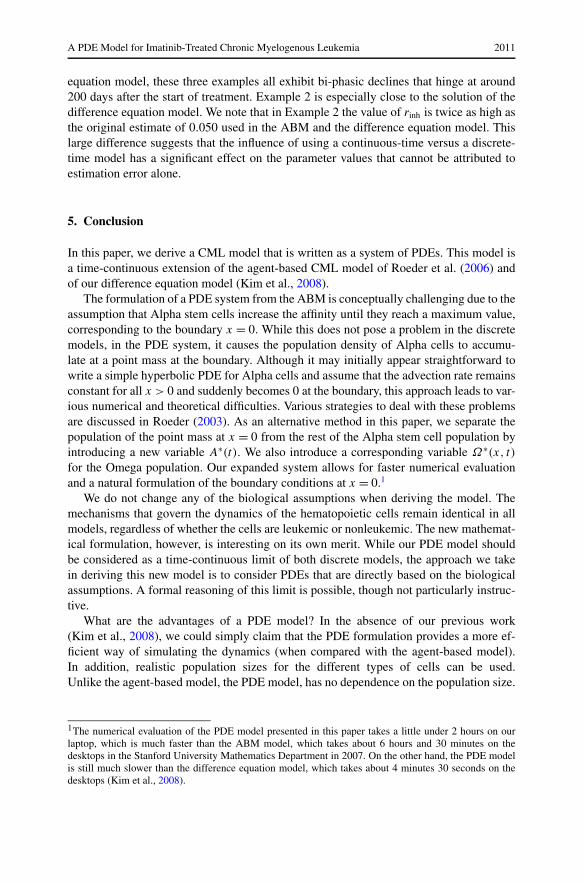
A PDE Model for Imatinib-Treated Chronic Myelogenous Leukemia 2011
equation model, these three examples all exhibit bi-phasic declines that hinge at around200 days after the start of treatment. Example 2 is especially close to the solution of thedifference equation model. We note that in Example 2 the value of rinh is twice as high asthe original estimate of 0.050 used in the ABM and the difference equation model. Thislarge difference suggests that the influence of using a continuous-time versus a discrete-time model has a significant effect on the parameter values that cannot be attributed toestimation error alone.
5. Conclusion
In this paper, we derive a CML model that is written as a system of PDEs. This model isa time-continuous extension of the agent-based CML model of Roeder et al. (2006) andof our difference equation model (Kim et al., 2008).
The formulation of a PDE system from the ABM is conceptually challenging due to theassumption that Alpha stem cells increase the affinity until they reach a maximum value,corresponding to the boundary x = 0. While this does not pose a problem in the discretemodels, in the PDE system, it causes the population density of Alpha cells to accumu-late at a point mass at the boundary. Although it may initially appear straightforward towrite a simple hyperbolic PDE for Alpha cells and assume that the advection rate remainsconstant for all x > 0 and suddenly becomes 0 at the boundary, this approach leads to var-ious numerical and theoretical difficulties. Various strategies to deal with these problemsare discussed in Roeder (2003). As an alternative method in this paper, we separate thepopulation of the point mass at x = 0 from the rest of the Alpha stem cell population byintroducing a new variable A∗(t). We also introduce a corresponding variable Ω∗(x, t)
for the Omega population. Our expanded system allows for faster numerical evaluationand a natural formulation of the boundary conditions at x = 0.1
We do not change any of the biological assumptions when deriving the model. Themechanisms that govern the dynamics of the hematopoietic cells remain identical in allmodels, regardless of whether the cells are leukemic or nonleukemic. The new mathemat-ical formulation, however, is interesting on its own merit. While our PDE model shouldbe considered as a time-continuous limit of both discrete models, the approach we takein deriving this new model is to consider PDEs that are directly based on the biologicalassumptions. A formal reasoning of this limit is possible, though not particularly instruc-tive.
What are the advantages of a PDE model? In the absence of our previous work(Kim et al., 2008), we could simply claim that the PDE formulation provides a more ef-ficient way of simulating the dynamics (when compared with the agent-based model).In addition, realistic population sizes for the different types of cells can be used.Unlike the agent-based model, the PDE model, has no dependence on the population size.
1The numerical evaluation of the PDE model presented in this paper takes a little under 2 hours on ourlaptop, which is much faster than the ABM model, which takes about 6 hours and 30 minutes on thedesktops in the Stanford University Mathematics Department in 2007. On the other hand, the PDE modelis still much slower than the difference equation model, which takes about 4 minutes 30 seconds on thedesktops (Kim et al., 2008).

2012 Kim et al.
In the agent-based case (Roeder et al., 2006), this was a rather strict restriction that limitedthe sizes of the populations simulated to 10% of their real sizes. These advantages werealready noted with our previous work (Kim et al., 2008), and indeed, when compared withthat work, our new model, does not improve the efficiency of the simulations.
Nevertheless, the PDE approach has several advantages over the discrete models:
(i) A PDE model provides an alternative representation of the dynamics of the progres-sion of the disease that is continuous in time.
(ii) The PDE approach allows a direct study (and a better fit) of the model parametersthat are sensitive to the time being a continuous or a discrete variable.
(iii) The PDE approach allows direct adjustments of global parameters that may dependon macroscopic quantities (such as densities of cells). These are usually less accessi-ble in the discrete models. It also provides direct access to the macroscopic quantitiesthemselves, as the model is formulated in these terms.
(iv) From an implementation point of view, the present model amounts to several(complex) equations. This should be compared with tens of thousands (of rathersimple) difference equations in the model of Kim et al. (2008), or with the hundredthousand or so iterations of simple rules at each time step in the ABM of Roederet al. (2006).
Our numerical simulations demonstrate that the PDE model captures the same qual-itative (and structural) behavior of the stem cells in the ABM. Indeed, at equilibrium,most Alpha cells accumulate in the A∗ state, representing Alpha cells that have at-tained maximum affinity. Also, the Omega cells show a step-like behavior with respectto the x-coordinate as shown in Fig. 5. These results coincide with the behavior ofthe ABM.
The PDE model starts to diverge from the discrete-time (difference equation and agent-based) models when we compare simulations of CML genesis and imatinib treatment. InCML genesis, the PDE model shows the same qualitative behavior as the discrete-timemodels, but the initial rise of Ph+ cells occurs earlier, and the Ph+ cells end at a higherequilibrium concentration than in the discrete-time models.
In simulations of imatinib treatment, the PDE and the discrete-time models divergemore greatly. The results of all models can match if the rates of imatinib-induced inhibi-tion (rinh) and degradation (rdeg) are increased. These differences between the ABM andthe PDE models demonstrate that some critical aspects of the model are highly sensitiveto the discretization of time. This observation is important, because it suggests that it isnot straightforward to assume that a discrete-time ABM, albeit with small time steps,accurately approximate the continuous-time behavior.
While it could be argued that a continuous-time description of disease dynamics maybe more realistic than discrete-time models, there still are multiple directions in whichthe present model can be improved, even at the existing resolution (i.e., without addingadditional types of cells, signaling, etc.). One example is the deterministic “clock” thatgoverns the progression of cells in the Omega state, the deterministic nature of the affinityvariable, or the deterministic life-cycle of precursor cells. These could be ideal places toadd some uncertainty the model, an issue we plan to address in the future.

A PDE Model for Imatinib-Treated Chronic Myelogenous Leukemia 2013
Appendix A: The ABM algorithm
We summarize the algorithm of the ABM from Roeder et al. (2006).At every time step, the ABM is defined as the following set of actions:
A. Preliminary calculations
1. Calculate the total populations of A and Ω cells.2. During imatinib treatment:
• Remove the proliferative Ph+ cells (Ω+ and Ω+/i ) that undergo apoptosis.• Determine which unaffected proliferative Ph+, Ω+, become imatinib-affected.
B. Proliferation, death, change of state, clocks
At this stage, all cells fall into one of three categories: A stem cells, Ω stem cells, differ-entiated cells.
1. For each A stem cell:
• Determine whether the cell transfers to Ω . If a cell transfers, skip the remainingactions for A cells. Note that the transition function depends on whether the cell isPh−, Ph+, or imatinib-affected. Calculate transition probabilities based on the totalpopulation of Ω calculated in Step A1.
• Increase the cell’s affinity by a factor of r .
2. For each Ω stem cell:
• Determine whether the cell transfers to A. If a cell transfers, skip the remainingactions for Ω cells. Calculate transition probabilities based on the total populationof A calculated in Step A1.
• If the cell’s affinity is less than or equal to amin, the cell becomes a differentiated cellof age 0. If the cell differentiates, skip the remaining actions for Ω cells.
• If a cell’s affinity is greater than amin, decrease the cell’s affinity by a factor of d .• Increase the counter c by 1.• If the counter c is greater than or equal to 49, set c to 0 and create a new cell with
identical attributes and state values as the current cell.
3. For each differentiated cell:
• Increase the cell’s age by one.• If the cell’s age is a multiple of 24 between 24 and 480, inclusively, create a new
differentiated cell with the same age as the current cell.• If a cells age reaches 672, that cell dies.

2014 Kim et al.
Table B.1 Parameters from Roeder et al. (2006)
Parameter Description Ph− Ph+/imatinib-affected
amin Min value of affinity a 0.002 0.002amax Max value of affinity a 1.0 1.0
d Differentiation coefficient 1.05 1.05r Regeneration coefficient 1.1 1.1
τc Cell cycle duration 48 hours 48 hoursτS Duration of S phase 8 hours 8 hoursτG2/M Duration of G2 and M phases 8 hours 8 hours
λp Lifespan of proliferating precursor cells 20 days 20 daysλm Lifespan of mature cells 8 days 8 days
τc Cell cycle of proliferating precursors 24 hours 24 hours
fα(0) Transition characteristic for fα 0.5 1.0
fα(NA/2) Transition characteristic for fα 0.45 0.9
fα(NA) Transition characteristic for fα 0.05 0.058fα(∞) Transition characteristic for fα 0.0 0.0
NA Scaling factor 105 105
fω(0) Transition characteristic for fω 0.5 1.0/0.0500
fω(NA/2) Transition characteristic for fω 0.3 0.99/0.0499
fω(NA) Transition characteristic for fω 0.1 0.98/0.0498fω(∞) Transition characteristic for fω 0.0 0.96/0.0496
NA Scaling factor 105 105
Note that differentiated cells of age less than 480 are considered to be proliferating pre-cursors, whereas differentiated cells of age greater than or equal to 480 are considered tobe non-proliferating mature cells.
Appendix B: Parameter estimates
The sigmoidal transition functions are given in Roeder et al. (2006) by
fα/ω(A/Ω) = 1
ν1 + ν2 exp(ν3
A/Ω
NA/Ω
) + ν4, (B.1)
where A and Ω denote the total populations in the Alpha and Omega compartments,respectively (see (2) and (3)). Furthermore,
ν1 = (h1h3 − h2
2
)/(h1 + h3 − 2h2),
ν2 = h1 − ν1,
ν3 = ln(h3 − ν1/ν2),
ν4 = fα/ω(∞),

A PDE Model for Imatinib-Treated Chronic Myelogenous Leukemia 2015
Table B.2 Imatinib-related parameters from Roeder et al. (2006). The inhibition intensity, rinh, refers tothe probability that a proliferative Ph+ cell (i.e., an Ω cell) becomes imatinib-affected in a given timeinterval. The degradation intensity, rdeg, refers to the probability that an imatinib-affected, proliferativePh+ cell dies in a given interval
Parameter Description Estimate
rinh Inhibition intensity 0.050rdeg Degradation intensity 0.033
and
h1 = (fα/ω(0) − fα/ω(∞)
)−1,
h2 = (fα/ω(NA/Ω/2) − fα/ω(∞)
)−1,
h3 = (fα/ω(NA/Ω) − fα/ω(∞)
)−1.
The values of the various parameters are listed in Table B.1.
Acknowledgements
This work was supported by a Research Scholar Award from the American Cancer Societyto PPL. The work of DL was supported in part by the NSF under Career Grant DMS-0133511. The work of PSK was supported in part by the Chateaubriand fellowship.
References
Abbott, L.H., Michor, F., 2006. Mathematical models of targeted cancer therapy. Br. J. Cancer. 95(9),1136–1141.
Adimy, M., Pujo-Menjouet, L., 2003. A mathematical model describing cellular division with a proliferat-ing phase duration depending on the maturity of cells. Electron. J. Differ. Equ. 2003(107), 1–14.
Campbell, J.D., Cook, G., Holyoake, T.L., 2001. Evolution of bone marrow transplantation—the originalimmunotherapy. Trends Immunol. 22(2), 88–92.
Colijn, C., Mackey, M.C., 2005. A mathematical model of hematopoiesis–I. Periodic chronic myelogenousleukemia. J. Theor. Biol. 237(2), 117–132.
DeConde, R., Kim, P.S., Levy, D., Lee, P.P., 2005. Post-transplantation dynamics of the immune responseto chronic myelogenous leukemia. J. Theor. Biol. 236(1), 39–59.
Dingli, D., Michor, F., 2006. Successful therapy must eradicate cancer stem cells. Stem Cells 24(12),2603–2610.
Druker, B.J., Lydon, N.B., 2000. Lessons learned from the development of an ABL tyrosine kinase in-hibitor for chronic myelogenous leukemia. J. Clin. Invest. 105(1), 3–7.
Fokas, A.S., Keller, J.B., Clarkson, B.D., 1991. Mathematical model of granulocytopoiesis and chronicmyelogenous leukemia. Cancer Res. 51(8), 2084–2091.
Kim, P.S., Lee, P.P., Levy, D., 2007. Mini-Transplants for Chronic Myelogenous Leukemia: A ModelingPerspective. In: Queinnec et al. (Eds.), Biology and Control Theory: Current Challenges, LectureNotes in Control and Information Sciences, vol. 357, pp. 3–20.
Kim, P.S., Lee, P.P., Levy, D., 2008. Modeling imatinib-treated chronic myelogenous leukemia: reducingthe complexity of agent-based models. Bull. Math. Biol. 70(3), 728–744.
Komarova, N.L., Wodarz, D., 2005. Drug resistance in cancer: Principles of emergence and prevention.Proc. Natl. Acad. Sci. USA 102(27), 9714–9719.

2016 Kim et al.
Michor, F., Hughes, T.P., Iwasa, Y., Branford, S., Shah, N.P., Sawyers, C.L., Nowak, M.A., 2005. Dynamicsof chronic myeloid leukaemia. Nature 435, 1267–1270.
Moore, H., Li, N.K., 2004. A mathematical model for chronic myelogenous leukemia (CML) and T cellinteraction. J. Theor. Biol. 225(4), 513–523.
Neiman, B., 2002. A mathematical model of chronic myelogenous leukemia. Master’s thesis, UniversityCollege, Oxford University, UK.
Pujo-Menjouet, L., Mackey, M.C., 2004. Contribution to the study of periodic chronic myelogenousleukemia. C. R. Biol. 327, 235–244.
Roeder, I., 2003. Dynamical modeling of hematopoietic stem cell organization—Design and validation ofthe new concept of within-tissue plasticity. PhD thesis, University of Leipzig, Germany.
Roeder, I., Glauche, I., 2008. Pathogenesis, treatment effects, and resistance dynamics in chronic myeloidleukemia—insights from mathematical model analyses. J. Mol. Med. 85(1), 17–27.
Roeder, I., Horn, M., Glauche, I., Hochhaus, A., Mueller, M.C., Loeffler, M., 2006. Dynamic modelingof imatinib-treated chronic myeloid leukemia: functional insights and clinical implications. Nat. Med.12(10), 1181–1184.
Thijsen, S.F.T., Schuurhuis, G.J., van Oostveen, J.W., Ossenkoppele, G.J., 1999. Chronic mlyeloidleukemia from basics to bedside. Leukemia 13(11), 1646–1674.