a patient with obese hypoventilation syndrome with ventilator-associated pneumonia: brain mapping...
TRANSCRIPT

lable at ScienceDirect
J Exp Clin Med 2014;6(4):147e148
Contents lists avai
Journal of Experimental and Clinical Medicine
journal homepage: http : / /www.jecm-onl ine.com
LETTER TO THE EDITOR
A Patient with Obese Hypoventilation Syndrome with Ventilator-associated Pneumonia: Brain Mapping and Polysomnography Outcomes
Obese hypoventilation syndrome is also historically described asthe Pickwickian syndrome. Patients may clinically present them-selves with symptoms such as excessive daytime sleepiness,fatigue, or morning headaches, which are similar to the symptomsof obstructive sleep apnea-hypopnea syndrome.1 Ventilator-associated pneumonia is the most common nosocomial infectionin patients receiving mechanical ventilation.2
A 65-year-old male patient was admitted because of breathless-ness and decreased responsiveness in the previous 3 hours. The pa-tient also complained of insomnia, confusion, and restlessness. Pasthistory showed lower lobe consolidation of the lung and multi-nodular goiter. The patient was later diagnosed as having hypoven-tilation syndrome and carbon dioxide (CO2) narcosis. Two yearspreviously, the patient underwent an operation. He was insertedwith a metal tracheostomy tube. He had a known case of diabetesmellitus since the previous 6 months and received 500 mg of met-formin daily. He had a morbid body mass index of 42.8 kg/m2. Theresults of atrial blood gases were as follows: pH, 7.37; arterial par-tial pressure of oxygen (PaO2), 130 mm Hg; arterial partial pressureof carbon dioxide (PaCO2), 47.7 mm Hg; and oxygen saturation(SaO2), 96.5%. The patient had normal findings in the completeblood counts, renal and liver function, serum glucose, electrolytes,ammonia, erythrocyte sedimentation rate, and thyroid functiontests. He was treated intravenously every 6 hours with 250 mg ofcefotaxime. From his personal history, he was a nonsmoker andnonalcoholic. From his past medication history, he was taking dailybactrim (100 mg tablet), diltiazem (30 mg), and pantoprazole(40 mg). On the 4th day of admission, the patient was referred tothe metabolic ward of the SRM Medical College Hospital andResearch Centre (Tamil Nadu, India) for an overnight polysomnog-raphy (PSG) study that was performed simultaneously with brainmapping. The sleep study was performed on two consecutivenights. The total PSG recording time was 6 hours 20 minutes, totalsleep time was 217 minutes, sleep latency was 30.5 minutes, wakeafter sleep onset was 42.7minutes, sleep efficiency was 59.9%, stage1 (N1) sleep was 104.7 minutes, stage 2 (N2) sleep was 3.1 minutes,stage 3 (N3) sleep was 1.6 minutes, rapid eye movement (REM)sleep was 5.9 minutes, and wake was 101.8 minutes (Figure 1).The apnea/hypopnea duration index was 23.2. Hypopnea episodesand desaturation (1e3%) episodes were frequent (Figure 2). Sinustachycardia was detected on his overnight electrocardiogram.From the brain mapping results, 10e11 Hz low amplitude a-
Conflicts of interest: The authors have no conflicts of interest to declare.
http://dx.doi.org/10.1016/j.jecm.2014.06.0041878-3317/Copyright © 2014, Taipei Medical University. Published by Elsevier Taiwan LL
frequency waves were in the occipital and posterior parietal re-gions in response to eye opening. Predominant delta waves ranged4e6 Hz amplitude. Photostimulation and hypoventilation re-sponses were unremarkable. No spikes or sharp waves wereobserved.
The patient received an overnight PSG, conducted by the guide-lines published by the American Academy of Sleep Medicine(Darien, Illinois, USA). The PSG findings consisted of continuous re-cordings of central and occipital electroencephalograms (EEGs),bilateral electro-oculograms, submental and bilateral tibial electro-myograms, and an electrocardiogram. The nasal and oral airflowswere measured using thermocouple sensors and pressure trans-ducer airflow monitoring devices. Body positioning was verifiedby infrared video recording.3
It is unclear why only 10%e15% of patients with obstructivesleep apnea-hypopnea develop hypoventilation, although obstruc-tive sleep apnea-hypopnea syndrome has been postulated as a
Figure 1 Graphic representation of the duration of sleep in each stage.
C. All rights reserved.
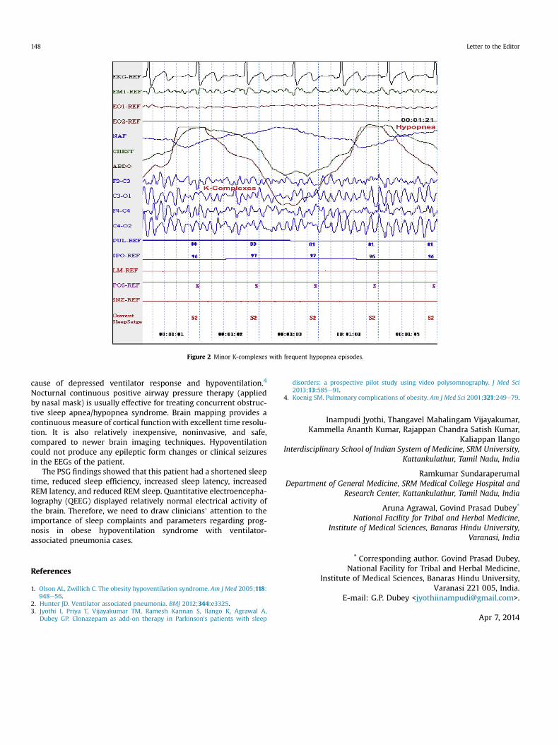
Figure 2 Minor K-complexes with frequent hypopnea episodes.
Letter to the Editor148
cause of depressed ventilator response and hypoventilation.4
Nocturnal continuous positive airway pressure therapy (appliedby nasal mask) is usually effective for treating concurrent obstruc-tive sleep apnea/hypopnea syndrome. Brain mapping provides acontinuous measure of cortical functionwith excellent time resolu-tion. It is also relatively inexpensive, noninvasive, and safe,compared to newer brain imaging techniques. Hypoventilationcould not produce any epileptic form changes or clinical seizuresin the EEGs of the patient.
The PSG findings showed that this patient had a shortened sleeptime, reduced sleep efficiency, increased sleep latency, increasedREM latency, and reduced REM sleep. Quantitative electroencepha-lography (QEEG) displayed relatively normal electrical activity ofthe brain. Therefore, we need to draw clinicians’ attention to theimportance of sleep complaints and parameters regarding prog-nosis in obese hypoventilation syndrome with ventilator-associated pneumonia cases.
References
1. Olson AL, Zwillich C. The obesity hypoventilation syndrome. Am J Med 2005;118:948e56.
2. Hunter JD. Ventilator associated pneumonia. BMJ 2012;344:e3325.3. Jyothi I, Priya T, Vijayakumar TM, Ramesh Kannan S, Ilango K, Agrawal A,
Dubey GP. Clonazepam as add-on therapy in Parkinson's patients with sleep
disorders: a prospective pilot study using video polysomnography. J Med Sci2013;13:585e91.
4. Koenig SM. Pulmonary complications of obesity. Am J Med Sci 2001;321:249e79.
Inampudi Jyothi, Thangavel Mahalingam Vijayakumar,Kammella Ananth Kumar, Rajappan Chandra Satish Kumar,
Kaliappan IlangoInterdisciplinary School of Indian System of Medicine, SRM University,
Kattankulathur, Tamil Nadu, India
Ramkumar SundaraperumalDepartment of General Medicine, SRM Medical College Hospital and
Research Center, Kattankulathur, Tamil Nadu, India
Aruna Agrawal, Govind Prasad Dubey*
National Facility for Tribal and Herbal Medicine,Institute of Medical Sciences, Banaras Hindu University,
Varanasi, India
* Corresponding author. Govind Prasad Dubey,National Facility for Tribal and Herbal Medicine,
Institute of Medical Sciences, Banaras Hindu University,Varanasi 221 005, India.
E-mail: G.P. Dubey <[email protected]>.
Apr 7, 2014